Alessandro Pini - d2cax41o7ahm5l.cloudfront.net · The new AMP M33 M33 is a novel non natural...
21
A novel synthetic antimicrobial peptide for the cure of Gram-negative infections. Mechanism of action, efficacy in vivo, toxicity, biodistribution and resistance selection Alessandro Pini [email protected] Department of Medical Biotechnology University of Siena Italy SetLance srl Toscana Life Sciences Italy
Transcript of Alessandro Pini - d2cax41o7ahm5l.cloudfront.net · The new AMP M33 M33 is a novel non natural...

A novel synthetic antimicrobial peptide for the cure of Gram-negative infections. Mechanism
of action, efficacy in vivo, toxicity, biodistribution and resistance selection
Alessandro Pini [email protected]
Department of Medical Biotechnology University of Siena
Italy
SetLance srl Toscana Life Sciences
Italy
The new AMP M33
M33 is a novel non natural antimicrobial peptide discovered at the University of
Siena. It is under development for the set up of a new antibacterial drug.
PIPELINE OF DRUG DEVELOPMENT FROM LAB TO MARKET
Research Development Commercialization
Target identification
and acquisition
Lead generation
Lead optimization
Preclinical and
pharmacology
Process devel.
manufacturing Clinical trials and
Launch and
marketing
MARKET
TIME (Y)
1 2 3 4 5 6 7 8 9 10
M33
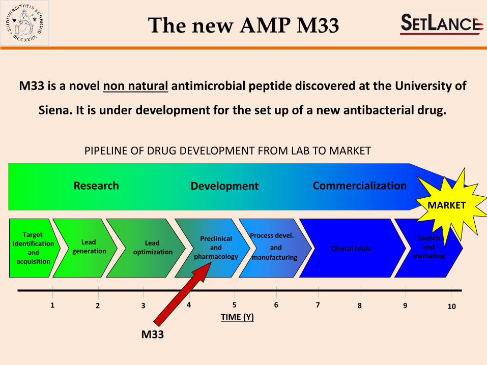
Tetrabranched (MAP) peptides acquire high resistance to protease activity making these molecules good candidates for in vivo use.
Tetrabranched peptide
Monomeric peptide O
N H
N H
O
NH
NH
O
NH
NH
O H
monomeric peptide incubated in serum at 0 h
monomeric peptide incubated in serum at 2 h
MAP peptide incubated in serum at 0 h
MAP peptide incubated in serum at 24 h
monomeric peptide incubated in serum at 0 h
monomeric peptide incubated in serum at 2 h
MAP peptide incubated in serum at 0 h
MAP peptide incubated in serum at 24 h
HPLC profiles of monomeric and tetrabranched peptides incubated in serum
Technology
Bracci et al., 2002, Biochemistry-US
Bracci et al., 2003, J Biol Chem
Lozzi et al., 2003, Chem Biol
Pini et al., 2005, Antimicrob Agents Chemother
Pini et al., 2006, Biochem J
Falciani et al., 2007, Mol Cancer Therapeut
Falciani et al., 2007, Chem Biol Drug Des
Pini et al., 2007, J Pept Sci
Pini et al., 2008, Cur Prot Pept Sci
Falciani et al., 2009, Exp Opin Biol Ther
Pini et al., 2010, FASEB J
Falciani et al., 2010, Curr Cancer Drug Targets
Falciani et al., 2010, ChemMedChem
Falciani et al., 2011, ChemMedChem
Pini et al., 2012, AminoAcids
Falciani et al., 2012, PLOS One
Falciani et al., 2013, J Med Chem
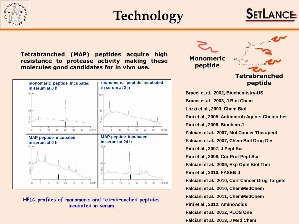
Identification and optimization of new antimicrobial peptides
QKKIRVRLSA (M6) Pini et al., 2005; Pini et al., 2007
Antimicrobial activity
-20
0
20
40
60
80
100
3 6,25 12,5 25 50
peptide [µg/ml]
%C
FU/m
l E.c
oli
M6 lot1 M6 lot2 M6 lot3 PyroM6 M33
HPLC profile
KKIRVRLSA (M33) Pini et al., 2010; Pini et al., 2012
M6 different peptide species
M33
HPLC profile
-20
0
20
40
60
80
100
3 6.25 12.5 25 50
%C
FU/m
l E.c
oli
peptide [µg/ml]
M33 lot 1 M33 lot 2 M33 lot 3
Antimicrobial activity
phage library
selection

Antimicrobial activity Pini et al., 2010, FASEB J; Pini et al., 2012, AminoAcids
aTested strains included either reference strains (indicated) or clinical isolates (mostly showing an MDR phenotype); relevant resistance traits and resistance mechanisms are indicated: FQr, resistant to fluoroquinolones; AGr, resistant to aminoglycosides (gentamicin, amikacin, and/or tobramycin); ESCr, resistant to expanded-spectrum cephalosporins; NEMr, resistance to carbapenems (imipenem and/or meropenem), ETPr resistance to ertapenem; ESBL, extended spectrum β-lactamase; MBL, metallo-β-lactamase; OXA, oxacillinase; MR methicillin-resistant; PENr resistance to penicillin; VANi, vancomycin-intermediate; bClinical isolates from Cystic Fibrosis patients cMucoid phenotype
MICs (M) of M33 in comparison with polymyxin B against bacterial strains representative of several pathogenic species, including MDR strains of clinical origin
Species and strains Resistancea MIC (M)
M33 Polymyxin B
Pseudomonas aeruginosa ATCC 27853 Reference strain, wild type 1.5 1.5
P. aeruginosa PAO-1 Reference strain, wild type 1.5 1.5
P. aeruginosa VR-143/97 FQr AGr ESCr NEMr (MBL/VIM-1) 1.5 1.5
P. aeruginosa SC-MDr03-06b FQr AGr ESCr NEMr 3 1.5
P. aeruginosa SC-VMr04-05b FQr AGr ESCr NEMr 3 1.5
P. aeruginosa SC-DMr05-04b FQr AGr ESCr NEMr 1.5 1.5
P. aeruginosa SC-BGr12-02b FQr AGr ESCr NEMr 1.5 1.5
P. aeruginosa EF-OBG6-1b FQr AGr ESCr NEMr (MBL/IMP-13) 1.5 0.7
P. aeruginosa SC-MDm03-02b,c FQr AGr ESCr NEMr 3 1.5
P. aeruginosa SC-GMm03-05b,c FQr AGr ESCr NEMr 1.5 1.5
P. aeruginosa SC-CNm03-07b,c FQr AGr ESCr NEMr 0.3 0.7
Klebsiella pneumoniae ATCC 13833 Reference strain, wild type 1.5 0.7
K. pneumoniae 7086042 FQr AGr ESCr NEMr (MBL/VIM-1) 3 1.5
K. pneumoniae C8-27 FQr AGr ESCr ETPr (ESBL/CTX-M-15) 1.5 0.7
K. pneumoniae FIPP-1 FQr AGr ESCr NEMr (MBL/KPC-3) 3 1.5
Escherichia coli ATCC 25922 Reference strain, wild type 1.5 0.7
E. coli W03BG0025 FQr AGr ESCr (ESBL/CTX-M-15) 0.7 0.7
Enterobacter aerogenes W03BG0067 AGr ESCr (ESBL/SHV-5) 1.5 0.7
Enterobacter cloacae W03AN0041 ESCr (ESBL/SHV-12) 1.5 0.7
Acinetobacter baumannii RUH 134 Reference strain, European clone II 1.5 1.5
A. baumannii RUH 875 Reference strain, European clone I 3 1.5
A. baumannii MR157 FQr AGr ESCr NEMr (OXA/OXA-58) 3 1.5
Staphylococcus aureus ATCC 29213 Reference strain, PENr 6 96
S. aureus 3851 MR VANi 6 96
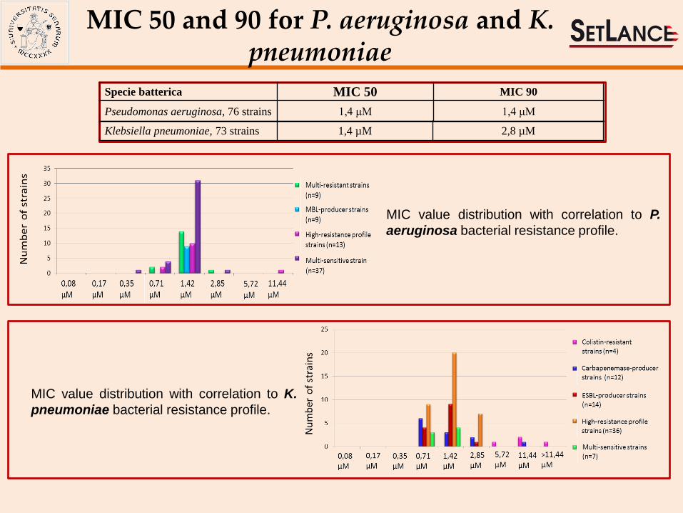
MIC value distribution with correlation to P.
aeruginosa bacterial resistance profile.
Klebsiella pneumoniae, 73 strains 1,4 µM 2,8 µM
Specie batterica MIC 50 MIC 90
Pseudomonas aeruginosa, 76 strains 1,4 μM 1,4 μM
MIC value distribution with correlation to K.
pneumoniae bacterial resistance profile.
MIC 50 and 90 for P. aeruginosa and K. pneumoniae

TWO STEPS MECHANISM
1- LPS and LTA recognition and binding
2 – Bacteria membrane is crossed and impaired
M33
LPS binding Pini et al., 2007
KD = 3.17e–9
Confocal laser microscopy Pini et al., 2007
CLSM experiments showed that rhodamine-labelled M33 is able to enter the cells within 5 minutes from incubation
Mechanism of action
LTA binding Falciani et al., 2012
Scanning Electron Microscopy
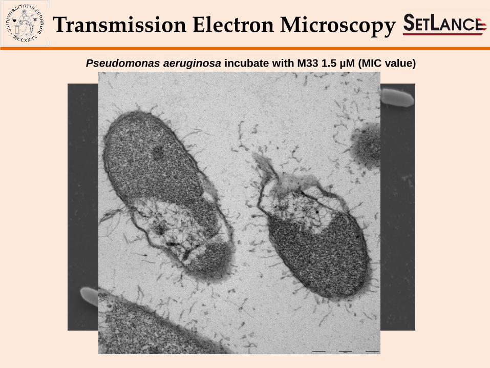
Pseudomonas aeruginosa incubate with M33 1.5 µM (MIC value)
Transmission Electron Microscopy
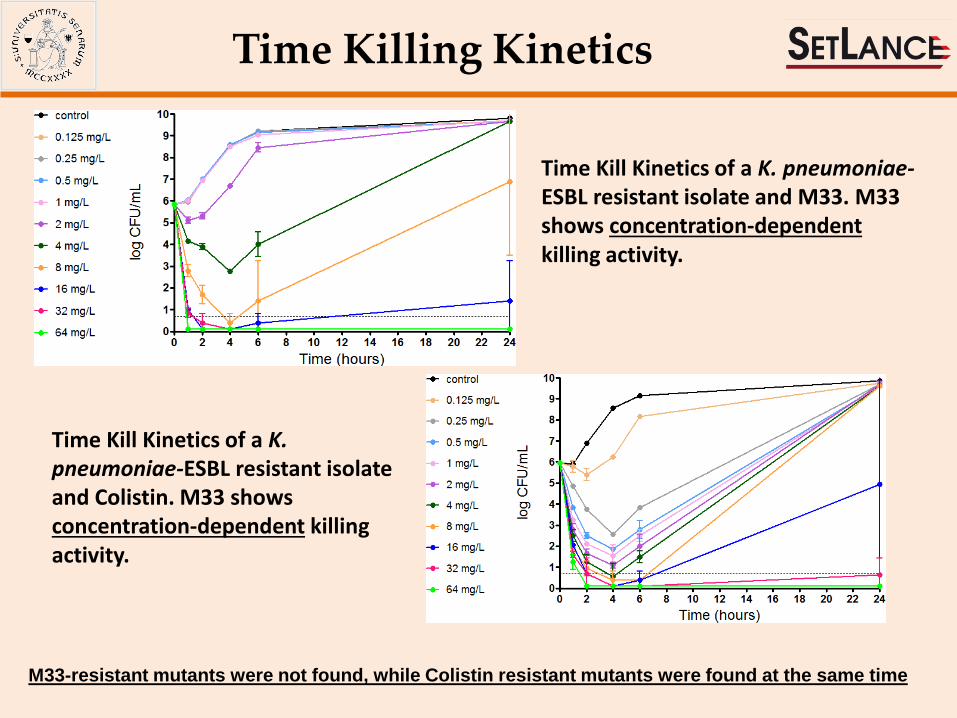
Time Killing Kinetics
Time Kill Kinetics of a K. pneumoniae-ESBL resistant isolate and M33. M33 shows concentration-dependent killing activity.
Time Kill Kinetics of a K. pneumoniae-ESBL resistant isolate and Colistin. M33 shows concentration-dependent killing activity.
M33-resistant mutants were not found, while Colistin resistant mutants were found at the same time
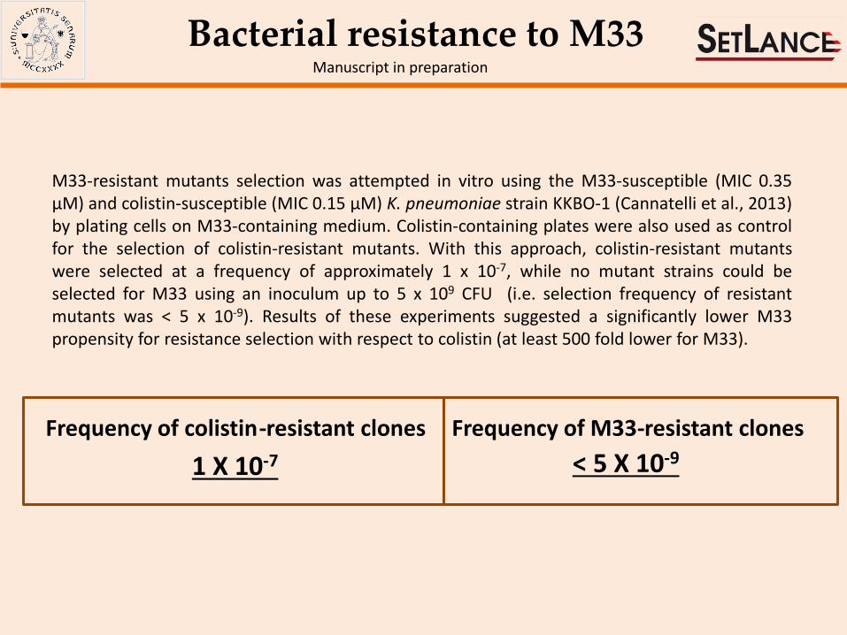
Bacterial resistance to M33 Manuscript in preparation
M33-resistant mutants selection was attempted in vitro using the M33-susceptible (MIC 0.35 µM) and colistin-susceptible (MIC 0.15 µM) K. pneumoniae strain KKBO-1 (Cannatelli et al., 2013) by plating cells on M33-containing medium. Colistin-containing plates were also used as control for the selection of colistin-resistant mutants. With this approach, colistin-resistant mutants were selected at a frequency of approximately 1 x 10⁻⁷, while no mutant strains could be selected for M33 using an inoculum up to 5 x 10⁹ CFU (i.e. selection frequency of resistant mutants was < 5 x 10⁻⁹). Results of these experiments suggested a significantly lower M33 propensity for resistance selection with respect to colistin (at least 500 fold lower for M33).
Frequency of colistin - resistant clones Frequency of M33 - resistant clones
< 5 X 10⁻⁹ 1 X 10⁻⁷

Neutralization of LPS
Inhibition of TNF-a release by LPS neutralization. Raw 264.7 (mouse leukaemic monocyte macrophage cells) were incubated with LPS from P. aeruginosa and Klebsiella pneumonie and M33. Triangles indicates incubation with LPS and different concentrations of M33. Squares indicates incubation with M33 only.
Pini et al., 2010, FASEB J

IL1-β
iNOS
MIP1
MIP2
Control LPS
Klebsiella
LPS
Klebsiella
with M33
TNF-α 100% 69% 100% 64%
100% 59% 100% 27%
100% 17% 100% 16%
100% 71% 100% 37%
100% 100% 100% 58%
LPS
Pseudomonas
LPS
Pseudomonas
with M33
LPS Klebsiella Control
NFk
B
Control LPS Pseudomonas LPS Pseudomonas with M33
Anti-inflammatory activity Manuscript in preparation
Cells incubated with LPS or with LPS and M33 Control = cells not incubated. LPS Pseudomonas = cells stimulated with LPS and producing NFkB (green signal). LPS Pseudomonas with M33 = cells incubated with LPS and M33 where the green signal is disappeared
• TNF-α is the most important cytokine involved in systemic inflammation and is implicated in acute phase reaction • IL1-beta is an important mediator of the inflammatory response, and is involved in a variety of cellular activities •iNOS is a proximate cause of septic shock • MIP1 and MIP2 are among the major factors produced by macrophages after they are stimulated with bacterial endotoxins •NF-κB is involved in cellular responses to several stimuli including bacterial or viral antigens. •Cox-2 is an enzyme that acts to speed up the production prostaglandins that play a key role in in promoting inflammation.
Gene expression (P. aeruginosa or K. pneumoniae) LPS Klebsiella
with M33
NFkB Protein production
IL1-b
iNOS
MIP1
MIP2
TNF-a
Control LPS E. coli LPS E. coli
with M33
Gene expression (E. coli)
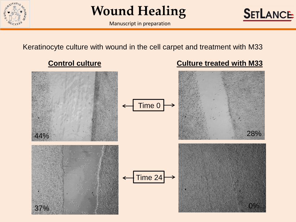
Wound Healing Manuscript in preparation
Keratinocyte culture with wound in the cell carpet and treatment with M33
Control culture Culture treated with M33
Time 0
Time 24
44%
37%
28%
0%

In vivo activity – The sepsis animal model
20%
M33 tested against P. aeruginosa PAO1 (1.5 X 10e3/mouse) inoculated IP after cyclophosphamide (160 mg/Kg -4 and -1 days)
0 24 48 72 96 120 144 168 192 216 240 264 2880
10
20
30
40
50
60
70
80
90
100
CTR
M33 5 mg/Kg IV, 24h and 72h after infection
M33PEG4 5 mg/Kg IV, 24h and 72h after infection
hours
% s
urv
ival
M33PEG4 5mg/Kg
M33 5mg/Kg
OH
ONH
NH
O
NH
NH
O
NH
NH
ONH
NH
O
NH
NH
O
NH
NH
PEG4
Manuscript in preparation

In vivo activity – The lung infection model
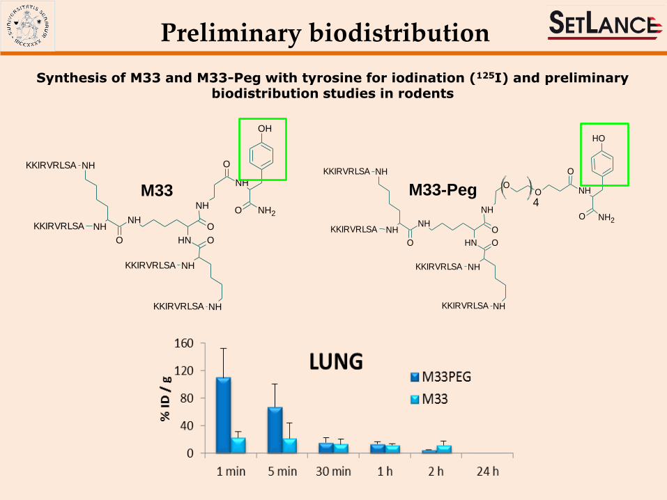
Number of CFU present in lungs of animals infected IT with P. aeruginosa and then treated IT with M33
Manuscript in preparation
M33PEG4 5mg/Kg
M33 5mg/Kg
0 5 0 1 0 0 1 5 0
1
1 0
1 0 0
T im e (m in )
% I
D /
gM 33
M 3 3 -P E G
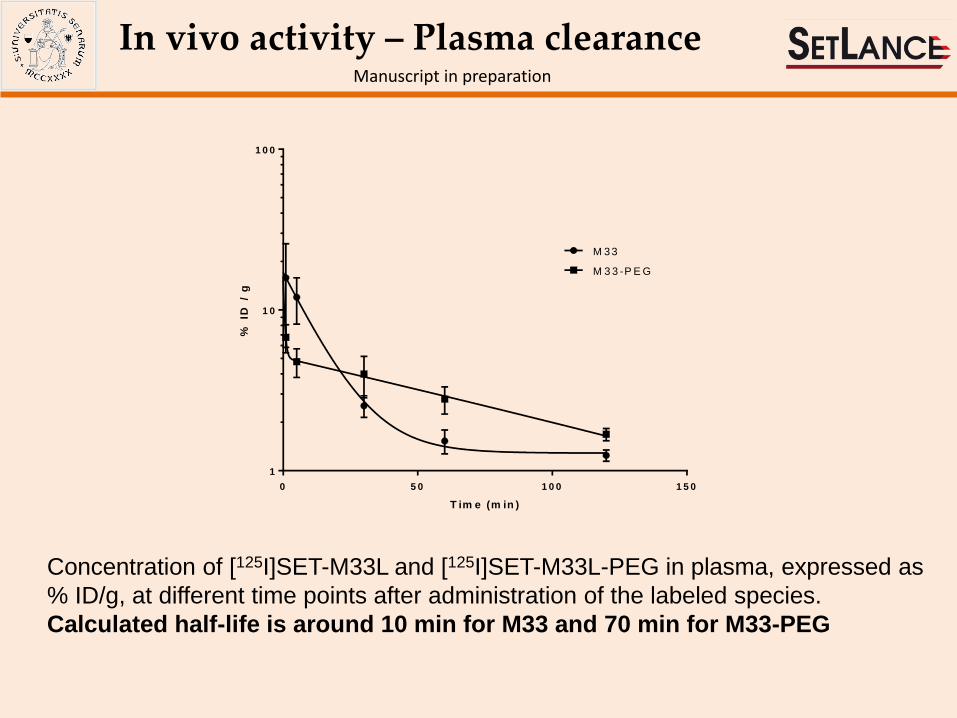
In vivo activity – Plasma clearance Manuscript in preparation
Concentration of [125I]SET-M33L and [125I]SET-M33L-PEG in plasma, expressed as
% ID/g, at different time points after administration of the labeled species.
Calculated half-life is around 10 min for M33 and 70 min for M33-PEG
Controls M33 treated
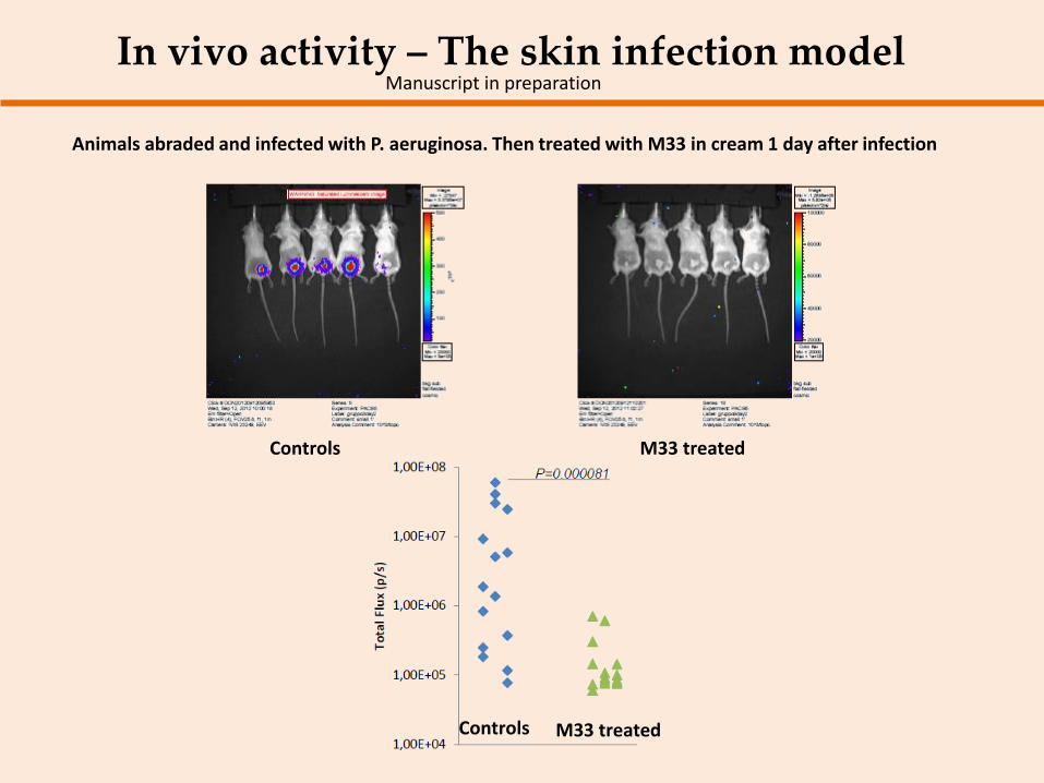
In vivo activity – The skin infection model Manuscript in preparation
Animals abraded and infected with P. aeruginosa. Then treated with M33 in cream 1 day after infection
Controls M33 treated
t 10 min t 24h t 48 h t 72 h M33
t 96 h
mouse without signs
mouse with mild signs
mouse with manifest signs
LEGEND mouse dead for toxicity
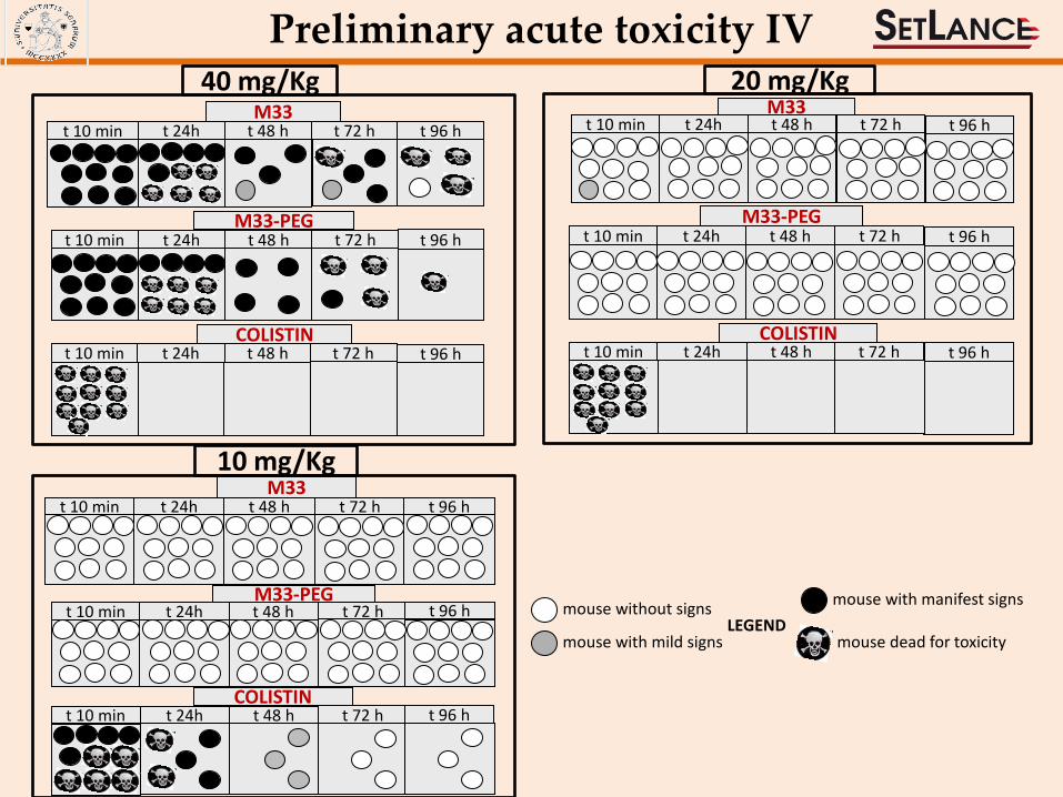
Preliminary acute toxicity IV
10 mg/Kg
t 10 min t 24h t 48 h t 72 h COLISTIN
t 96 h
40 mg/Kg
t 10 min t 24h t 48 h M33-PEG
t 72 h t 96 h
t 10 min t 24h t 48 h COLISTIN
t 72 h t 96 h
t 10 min t 24h t 48 h t 72 h t 96 h M33
t 10 min t 24h t 48 h M33
t 72 h t 96 h
20 mg/Kg
t 10 min t 24h t 48 h COLISTIN
t 72 h t 96 h
t 10 min t 24h t 48 h M33-PEG
t 72 h t 96 h
t 10 min t 24h t 48 h t 72 h M33-PEG
t 96 h
NH
O
NH
NH
O
NH
NH
O
NH
NHKKIRVRLSA
KKIRVRLSA
KKIRVRLSA
KKIRVRLSA
O
NH2O
NH
OH
Synthesis of M33 and M33-Peg with tyrosine for iodination (125I) and preliminary biodistribution studies in rodents
Preliminary biodistribution
4NH
O
NH
NH
O
NH
NH
O
NH
NH
O
KKIRVRLSA
KKIRVRLSA
KKIRVRLSA
KKIRVRLSA
O
O
O
NH
OH
NH2
M33 M33-Peg
In progress
Preclinical Development •Bioanalytical method set up •GLP production of M33 •Pharmacokinetics and biodistribution •Safety pharmacology in rodents and non rodents Research and back up molecules •Animal model of K. pneumoniae infection and M33 treatment •Conjugation with nanoparticles and formulation for delivery in lungs •Broadening spectrum of activity using M33 with D-aminoacids •Preliminary efficacy and toxicity with M33-D

Luisa Bracci
Jlenia Brunetti
Stefano Bindi
Luisa Lozzi
Silvia Scali
Giulia Roscia
Elisa Ibba
Leila Quercini
Lorenzo Depau
Giulia Riolo
Elisabetta Mandarini
Microbiology Section
Gian Maria Rossolini
Simona Pollini
Antonio Cannatelli
MIUR
Biochemistry Section
Medical Biotechnology Department
University of Siena
Chiara Falciani Federica Ceccherini
Martina Onori
Maria Luisa Mangoni
Vincenzo Luca
Department of Biochemical Sciences
A. Rossi Fanelli
University of Rome 1
Department of Experimental Medicine
University of Perugia Anna Vecchiarelli
Donatella Pietrella
FP7
Erasmus MC
Rotterdam, The
Netherlands
San Sebastian, Spain
Unai Cossio
Vanessa Gomez-Vallejo
Maria Puigivila
Jordi Llop Roig
John Hays
Irma Bakker