Ahmed Raad Emnati
43
Ministry of high Education & scientific research University of Baghdad College of Dentistry A Project Submitted to the College of Dentistry, University of Baghdad, Department of Orthodontic dentistry in partial fulfillment for the requirement to award the degree B.D.S. By Ahmed Raad Emnati 5th Grade Supervised by Dr. Jinan Eliewy Saloom B.D.S., M.Sc. Orthodontic Dent. 2017-1438
Transcript of Ahmed Raad Emnati

Ministry of high Education& scientific researchUniversity of BaghdadCollege of Dentistry
A ProjectSubmitted to the College of Dentistry, University of Baghdad,
Department of Orthodontic dentistry in partial fulfillment for therequirement to award the degree B.D.S.
ByAhmed Raad Emnati
5th Grade
Supervised byDr. Jinan Eliewy SaloomB.D.S., M.Sc. Orthodontic Dent.
2017-1438

الرحمن الرحیم
ھ لا أنھ شھد ا إلا إلوأولووالملائكة ھو
بالقسط قائماالعلم ھ لا العزیز ھو إلا إل
الحكیم
18

Declaration
This is to certify that the organization and preparation of thisthesis have been made by graduate student Ahmed Raad Emnatiunder my supervision in the College of Dentistry, University ofBaghdad in partial fulfillment of the requirement for the 5th Grade.
Signature:
Dr. Jinan Eliewy Saloom
The supervisor

Dedication
This thesis is dedicated to my parents.For their endless love, support and
encouragement.
Acknowledgment
I
Acknowledgment
Thanks Allah (ǀ ɯɯƵǡү ƴɯɯү ) for everything, for providing me with
power and patience to perform this study.
I would like to express grateful thanks to dean of college of
dentistry, University of Baghdad Prof. Dr. Hussain Al-Huwaizi.
Grateful thanks are expressed to Prof. Dr. Dhiaa Jaafar Al-
Dabagh, Head of the Department of Orthodontic Dentistry, for his
scientific support and advice.
To my supervisor Dr. Jinan Eliewy Saloom words fail my gratitude
for you, I would like to express gratitude and gratitude to scientific care
and to the spirit of high morality that encourage and advise me always to
right way throughout this research, Ask Allah to reward her the best
reward.
Great thanks to all members of Orthodontic dentistry department for
high ethics and for standing help.
Thank everyone who helped me in the completion of the search for
scientific truth.
Finally I would like to express grateful thanks to my lovely family,
my wonderful parents, my brothers and my sister for everything.

List of Contents
II
Subjects PagesNo.
Introduction 1
1.1 Definition 3
1.2 Etiology of class III malocclusion 4
1.2.1 Another classification for the etiology of class III malocclusion 5
1. General factors 5
2. Local factor 7
1.3 Classification of class III malocclusion
1.3.1 Classification of skeletal class III malocclusion according tothe location of the problem
8
1.3.2 Classification of class III malocclusion according tomorphology
10
1.3.3 Classification of skeletal class III according to Head formtypes
10
1.4 Clinical Features of skeletal Class III Malocclusion 11
1.5 Treatment of class III malocclusion 14
1.5.1 Treatment aims 14
1.5.2 Treatment planning 14
1.5.3 Treatment options 14
1. No treatment 14
2.Extractions only 15
3.Removable appliance 15

List of Contents
III
4.Single arch fixed appliance 15
5.Full arch fixed appliance 16
6.Functional appliance 17
7.Orthognathic surgery 21
1.6 Post-treatment stability 23
References 24

List of figure
IV
FigureNo.
Title PageNo.
Figure 1 Class III malocclusion according to the molars, canines andincisors relationship
3
Figure 2 Profile of a patient with maxillary retrognathism 11
Figure 3 Profile of a patient with mandibular prognathism 12
Figure 4 Intraoral feature of mandibular prognathism 13
Figure 5 Combined maxillary retrognathism and mandibularprognathism
13
Figure 6 Class III with single arch 16
Figure 7 Treatment of class III with upper and lower fixedorthodontic appliance
17
Figure 8 Frankel III 18
Figure 9 Chin cup 19
Figure10
Protraction face mask 21
Figure11
Sever class III malocclusion with orthodontic surgery 22

Introduction
- 1 - | P a g e
Introduction
Class III malocclusion is a subject of interest and concern to theorthodontist in both research and clinical practice in addition to that, class IIImalocclusion has long been viewed as one of the most severe facialdeformities. The appearance of protruding mandible with reverse overlap ofthe anterior teeth is easy to identify (Graber, 1988).
Hunter (1788) addressing mandibular prognathism stated that “It is notun common to find the lower jaw projecting too far forward, so that its foreteeth pass before those of the upper jaw, when the mouth is shut; which isattended with inconvenience and disfigure the face”.
In our face-conscious society it seem that any departure from the usualare the “normal” attracts attention; especially for children this often meansderision and ridicule because the child want to be like this playmates andbecause facial disharmony is predisposing to psychological aberrations(Graber, 1988).
Studies conducted to identify the etiological features of class IIImalocclusion show that the deformity is not constricted to the jaws butinvolves the entire the craniofacial complex (Chang et al., 1992; Mackay etal., 1992; Battagel, 1993).
The prevalence of class III malocclusion varies among different racesand population. The highest prevalence is among Asians of the Far East andthe lowest is in Caucasians (Bukhary, 2005). In Iraq it ranges from 2.4% to6.3% (Agha et al., 2002; Khamarco et al., 2002).
In orthodontic diagnosis and treatment planning, great importance hasbeen attached to evaluate the sagittal apical base relationship. Both angularand liner measurement have been incorporated into various cephalometricanalysis to help the clinician diagnosis anteroposterior dysplasia and establishthe most appropriate treatment plan. Any cephalometric analysis based oneither angular or liner measurements has obvious shortcomings, which havebeen discussed in detail by Moyers et al (1979).
Freeman (1988) stated that, even before Angle introduced hisClassification of malocclusion to profession in the early 1900s, theanteroposterior relationship of mandible to maxilla was the most importantdiagnostic criterion. It is important to identify whether the etiology of class IIImalocclusion is dental, functional or skeletal. If the problem is skeletal, it

Introduction
- 2 - | P a g e
must be determined whether the cause is overdeveloped mandible,underdeveloped maxilla or combination of both (Baik et al., 2004).
So, when treating class III patients orthodontically whether they aregrowing children or mature adults, anteroposterior and vertical position offacial components as well as dental relationship must be considered so that theexcess or deficiency may be treated where it actually exists (Guyer et al,.1986).

Definition
- 3 - | P a g e
1.1 Definition
According to Angle’s classification, the lower arch is at least one-halfcusp width too far forward in relation to the upper arch, judged by the firstpermanent molar relationship.
According to the canine classification, in class III the upper permanentcanine will occlude backwards to the embrasure between the lower canine andfirst premolar.
According to the British standard definition, class III incisorrelationship includes that malocclusion where the lower incisor edge occludesanterior to the cingulum plateau of upper incisors. Class III malocclusionaffects around 3 percent of Caucasians (Jones and Oliver, 2000).
Fig. 1: Class III malocclusion according to the molars, canines andincisors relationship

Etiology of class III malocclusion
- 4 - | P a g e
1.2 Etiology of class III malocclusion
1. Skeletal factors
Osseous class III is the problem of skeletal morphology andosseous growth that leads to the skeletal imbalance. It also called True ClassIII malocclusion (Graber, 1972), True prognathism (Jacobosn et al., 1974),Skeletal Class III malocclusion (Foster, 1982) and True Mesi-occlusion
(Nakasima et al., 1986).
Jacson et al. (1974) stated that class III skeletal imbalance is usuallyattributed to one or more of the following components variable:
1. The mandible may be too large relative to maxilla (lengthened ramus,lengthened mandibular body or large total length).
2. The maxilla may be too small relative to the mandible.3. The maxilla may be retro-positioned relative to the mandible.4. The mandible may be positioned too far forward relative to the maxilla.5. A forward rotation of the mandible relative to cranium will cause the
chin point to move into a horizontally more protrusive position; aprognathic mandible may thereby result together with a reduction inlower anterior facial height.
2. Soft tissues factorWhere the anterior intermaxillary height is large the lips are frequently
incompetent. Such cases often have a skeletal anterior open bite, and duringswallowing there will be an adaptive variation of swallowing behavior withthe tongue coming forwards into the gap between the incisors. where theintermaxillary height is reduced sometimes the upper lip may also be shorterand hypotonic (Jones and Oliver, 2000).
3. Dental factors
Class III malocclusion are often associated with a narrow upper archand a broad lower arch, With the result that crowding is seen more commonly,and two greater degree, in the upper arch than in the lower. Frequently, thelower arch is well aligned for even spaced (Jones and Oliver, 2000).

Etiology of class III malocclusion
- 5 - | P a g e
1.2.1 Another classification for the etiology of class III
malocclusion
The permanent interaction between genetic and environmental factors guidesand controls the process of growth and development and determines themorphologic and physiologic traits of the individual (Van Der Linden, 1969).The poly genic or multifactorial theory is responsible in the development ofclass III malocclusion (Litton et al., 1970). So, they are complex inter actionsof genetic and environmental factors, which may act synergistically or inisolation or may cancel each other out (Battagel, 1993).
Graber (1972) classified the etiologic factors into general and localfactors; this system was used to describe the etiology of class IIImalocclusion;
1. General factorsA. Genetic: familial studies of mandibular prognothism are
suggestive of heredity in the etiology of this condition (Casto,1928; Downs). The best known example of the transmission ofdistinctive abnormal condition is certainly the mandibularprognathism of the Hapsburg family (Rubbrecht, 1939).Heredity determines both tooth size and the pattern of jaw growth(Gresham, 1975). Some genetic syndromes are associated withmandibular prognothism such as basal cell nevus, osteogenesisimperfect, Marfan syndrome and Klinefelter syndrome (Cohen,1980). The correlation between parents and offspring with classIII malocclusion were strong especially for skeletal measurement,indicating that skeletal pattern was more directly related togenetic factor, also both parents and offspring had concaveprofile (Nakasima e al., 1982). Ethnic is also a part of geneticsand different morphology of class III malocclusion can be seen indifferent ethnic groups, Masaki (1980) reported that maxillaryskeletal retrusion occurred more often in Asians face, whereasmandibular prognathism often observed as a component of classIII malocclusion in individuals of European American ancestry.Singh (1999) reported that some Asian ethnic groupsdemonstrate an increase prevalence of class III malocclusion; It islikely that the skeletal component and soft tissue matrices are

Etiology of class III malocclusion
- 6 - | P a g e
genetically determined. Presumably, the co-morphologies ofcraino-maxillary and mandibular complex are likely dependentupon candidates genes that undergone gene-environmentinteraction to yield class III malocclusion.
B. Congenital anatomical defectCleft palate cause extreme vertical and anterposterior growthdeficiency of the maxilla leading to maxillary retrognathism(Graber, 1972). About 72.7% of cleft cases are associated withclass III malocclusion while 18% associated with class I and8.8% with class II (Baek et al., 2002).Chemicals and drugs are capable of producing embryologicaldefect if given at critical time; agents like Valium, Aspirin,cigarette and alcohols affected the dento-facial development andmay result in cleft palate which in turn leads to deficient maxilla(Proffit et al., 2007).
C. Predisposing metabolic climate and diseasePatient with acromegaly, which is cause by anterior pituitarytumor where there is excessive secretion of growth hormone, hasexcessive growth of the mandible which will create class IIImalocclusion in adult life (Strang & Thompson, 1958; Pascoeet al., 1960; Graber, 1972; Proffit et al., 2007).
D. Abnormal pressure habit and functional aberrationI. Tongue habit: tongue habit may result from size, posture or function.
Large tongue will cause the mandible to be protruded at all time(Graber, 1972; Proffit 2000) and may lead to abnormal growthpattern of the mandible and flaring of the incisors (McDonald &Avery, 2000). True macroglosia may be either congenital or acquired;congenital cause likes muscular Hypertrophy, Glandular hyperplasia,hemangioma and Lymphangioma. Macroglosia also occur inconditions like Down’s syndrome, while acquired causes may includeacromegaly, tertiary syphilis, cyst or tumor involving the tongue andneurologic injury (Wolford & Cottrell, 1996) and also thyroiddeficiency (Graber, 1972; Proffit, 2000).Regarding tongue posture, flat and anteriorly positioned tongue isresponsible for the anterior position of the mandible with resultantincrease in its length and flaring of the lower labial segment(McCallin et al., 1985; Rakosi & Schilli, 1981). There is acontroversy whether tongue posture is a compensatory, adaptivephenomenon or a primary etiologic factor (Graber et al., 1985).

Etiology of class III malocclusion
- 7 - | P a g e
Tongue thrust was found in 17% of patient with class III malocclusion(Subtelny & Subtelny, 1972).
II. Lip pressure: some muscle functions may be changed to adapt foralready existent mal-relationship between the maxilla and themandible. In class III malocclusion, the lower lip is impotent andhypofunctional while the upper lip is quite active in addition to lowtongue posture. All these may be an adaptive activity; however, theymay accentuate the deformation (Thompson, 1949; Graber, 1972).Soft tissue matrices, especially labial pressure from circomoralmusculature, may influence the final outcome of the craniofacialgrowth of a child skeletally predispose to class III condition (Singh,1999).
E. Habitual occlusionThe habit of protruding the mandible may accelerate mandibulargrowth leading to class III malocclusion (Rakosi and Schilli,1981).
2. Local factorA. Premature tooth loss and irregular eruption path:
Premature bilateral losses of maxillary six years molars (1st molar) mayresult in a foreshortening of the maxillary arch, which is turn, producemandibular prognathism (Gold, 1949).
When over eruption of molars occur, several unfavorable change takeplace. If the patient’s growth is low, the mandible may rotate backward.Consequently an anterior open bite may be created. However, if the patienthas enough growth potential, vertical growth of condyle is stimulated, then themandible rotates forward creating skeletal class III malocclusion (Sato, 1994).
B. Teeth wear and bruxismThe flat plane of occlusion of deciduous teeth makes it simple to
reposition his mandible anteriorly; this may result from relative softness of thedeciduous enamel which permits rapid wear and in a rather short period oftime, this forward position becomes the new position of mandible (Gold,1949).
C. Teeth numberPatients with more sever hypodontia in maxilla demonstrated
tendencies to a class III skeletal relationship (Chung et al., 2000).

Classification of class III malocclusion
- 8 - | P a g e
1.3 Classification of class III malocclusion
1.3.1 Classification of skeletal class III malocclusion
according to the location of the problem
One of the old classifications of class III malocclusion was describe bySanborn (1955), who divided class III cases according to anteroposteriorposition of the maxilla and mandible as determined by SNA and SNB intofour subgroups:
1. Group A: Those presenting a maxilla within the normal range ofprognathism and mandible beyond normal range of prognathism.
2. Group B: Those presenting a maxilla below normal range ofprognathism and a mandible within the normal range of prognathism.
3. Group C: Those presenting a maxilla and a mandible within the normalrange of prognathism.
4. Group D: Those presenting a maxilla below normal range ofprognathism and a mandible beyond the normal range of prognathism.
Pascoe et al. (1960), using vertical height and antero-posteriordimension, classified their class III malocclusion patients into:
1. Type A: In which both the maxilla and mandible were within the rangeof prognathism.
2. Type B: The maxilla of normal length, where mandibular length iswithin the range of prognathism.
3. Type C: An underdeveloped, shortened maxilla (retrognathic), wherethe mandibular is within normal.
4. Type D: Maxilla within normal range, occlusion and alveolar process innormal relationship and the mandible is prognathic because of basalprognathism.
5. Type E: The maxilla within normal range, where the mandible isprognathic with lengthening of lower third of face and open biteanteriorly.
Tweed (1966) classified skeletal class III into 2 categories:
1. Pseudo class III malocclusion: with small maxilla and conventionalshape mandible.
2. True class III malocclusion: with overdeveloped mandible.Ellis and McNamara (1984) divided the measurements of craniofacial
structure of adult class III surgical patient into 4 horizontal components;

Classification of class III malocclusion
- 9 - | P a g e
maxillary skeletal position, maxillary dental position, mandibular dentalposition, and mandibular skeletal position and one of vertical component.When each of these five component is divided into 3 classes: protruded,normal and retruded, permits 243 possible subgroup; actually 69 subgroupwhere found.
Rakosi in 1982 mentioned that six types of class III relationships maybe distinguished.
1. Normal extent of maxillary and mandibular bases: The upper incisorshow lingual, the lower incisors labial inclination. The cause of theanomaly can usually be localized in the dento-alvcolar region. This typeis often difficult to distinguish from trans-located closure with markedmandibular prognathism.
2. Large mandibular base and ascending ramus: The gonial angle islarge, the articular angle is small. The upper incisors show labial, thelower incisors lingual inclination. Edge to edge or open bite is usuallyseen frontally and cross bite laterally prognathism with fault in themandible).
3. Underdeveloped maxilla: This present with crowding in the upperfront region, with the mandibular base prominent. Two variations of thetype may be distinguished:
a) Vertical growth tendency. The ascending ramus and posteriorcranial base are short, the gonial angle large and the upper gonialangle (GO1) greater than 65 degree.
b) Horizontal growth tendency: The ascending ramus and posteriorcranial base are large, the gonial angle small and the upper gonialangle (GO1) 40-55 degree.
The crowding in the maxilla complicates treatment with these twotypes, so that fixed appliance is usually required (mandibular prognathismwith fault in the maxilla).
4. Maxilla underdeveloped, mandible normal: This type occur withmal-development of the maxilla, e.g. in subject with cleft palate andcertain syndromes where mid-face underdeveloped is characteristic(mandibular prognathism with the fault in the maxilla).
5. Maxilla normal overdeveloped mandible: This group includes“genuine” mandibular prognathism, with poor prognosis for effectivetreatment (prognathism with the fault in the mandible).
6. Pseudo translocation closure: A fully developed skeletal prognathismmay be partly compensated by lingual inclination of lower incisor and

Classification of class III malocclusion
- 10 - | P a g e
labial inclination of the upper incisors. On clinical examination, theanomaly gives the impression of being Trans-located closure, butcephalometric radiography and “mental repositioning “ of incisorsangulation will reveal a genuine mandibular prognathism.
Classification of class III malocclusion according to1.3.2
morphology
Jacobson et al. (1974) suggested two basic morphologic types of ClassIII malocclusion:
1. Divergent Class III pattern: In which palatal, occlusal and mandibularplanes tend to diverge, an obtuse gonial angle and anterior open bite inextreme cases.
2. Convergent Class III pattern: Palatal, occlusal and mandibular planestend toward parallelism, an acute gonial angle and deep overbite.
III according to HeadClassification of skeletal class1.3.3
form types
Marton et al, (1992) reported that three groups exist within Class IIIrelating to differences configuration related to head form types:
1. The brachycephalic Class III: It has a relatively wider and flatter face,a vertically shorter and less protrusive nasal region and more forwardrotated mandible when compared with brachycephalic Class I type.
2. The dolichocephalic Class III: It is a less common variant of the longand narrow head form, has a more elongated and protrusive nasomaxillaand a more downward and backward mandibular alignment.
3. Dinaric or Mesocephalic Class III: Vertical nasal length tends to beshortened, producing a more forward mandibular alignment associatedwith average head form.

Clinical Features of skeletal Class III Malocclusion
- 11 - | P a g e
1.4 Clinical Features of skeletal Class III Malocclusion
Carlotti and George (1981) describe the clinical features of maxillaryretrognathism and mandibular prognathism as follows:
Maxillary retrognathism
A. Facial features:1- Tendency of upper lip to be thin and retruded relative to lower lip.2- Normal to deficient upper anterior teeth to upper lip relation.3- Nasolabial line-subnasale: subnasale-tip of nose, usually not 1:1 ratio.4- Obtuse nasolabial angle.5- Less incisor appearance during smiling and the teeth seem to be under
the upper lip.6- Nearly normal chin projection.7- Normal to decrease lower facial height.8- Concave profile.
Fig. 2: Profile of a patient with maxillary retrognathism
B. Intraoral features:1. Class III malocclusion (molars, canines and incisors).2. Tendency toward crowding and mission or impacted teeth.3. Transverse deficiencies noticeable in maxillary arch.4. More nearly normal inclination of mandibular anterior teeth.

Clinical Features of skeletal Class III Malocclusion
- 12 - | P a g e
Mandibular prognathism
A. Facial features:1- Normal upper lip form.2- Normal relation of upper anterior teeth to upper lip.3- Normal 1:1 ratio between nasolabial line-subnasale: subnasale-tip of
nose.4- Normal to acute nasolabial angle.5- Good incisor / lip relationship.6- The lower lip is protruded.7- Prominent chin.8- Normal to increase lower facial height.9- Concave profile.
Fig. 3: Profile of a patient with mandibular prognathism
B. Intraoral features:1- Class III malocclusion (molars, canines and incisors).2- Normal maxillary arch.3- Broad mandibular arch form.4- Tendency toward lingo-version of lower anterior teeth.

Clinical Features of skeletal Class III Malocclusion
- 13 - | P a g e
Fig. 4: Intraoral feature of mandibular prognathism
Combined maxillary retrognathism and mandibular prognathism willhave the prominent features of both.
Fig. 5: Combined maxillary retrognathism and mandibular prognathism

Treatment of class III malocclusion
- 1 - | P a g e
1.5 Treatment of class III malocclusionThis earlier intervention in class III patients obviously results in a longer
period between the start of the initial phase of treatment and the end of the
comprehensive treatment phase after the permanent dentition has erupted. The
early treatment of Class III malocclusion may be characterized by more than
one period of intervention during the mixed dentition (Graber and Vanarsdall,
1994).
1.5.1 Treatment aimsThe principal treatment aims can be summarized as follows (Wada et al,
1981):
1. To improve the aesthetics of the teeth and the function of the teeth and
jaw whilst maintaining or improving the facial profile.
2. To relieve crowding and produce alignment within the arches.
3. To correct the incisor relationship to obtain a more normal and inter-
incisal angle.
4. To eliminate antero-posterior and unilateral lateral cross bites together
with associated displacements.
1.5.2 Treatment planningWhen treatment planning a Class III case it is important to establish the
true occlusal position after all displacements have been eliminated. It is often of
value to have two sets of records, one with the occlusion in the displaced
position and the other set at the retruded condylar position with displacements
eliminated. The patient will often present complaining of upper arch (canine)
crowding associated with a narrow and/or short dental arch. In such cases the
crowding should not be relieved without some consideration begin given to the
likely effect of future growth on the dental arch relationship. It is wise to
Treatment of class III malocclusion
- 2 - | P a g e
develop a longer-term provisional treatment plan before arranging extraction of
any permanent teeth (Ellis et al, 1984).
1.5.3 Treatment options1. No treatment
Where crowding of the dental arches is minimal, there are no
displacements apparent, and the Class III appearance of the incisors and/or the
jaws is acceptable, the ‘no-treatment’ option is a reasonable approach to
management. It also has the advantages of keeping the Class III growth
tendency under review and minimizing any intervention until growth has largely
finished and the jaw profile has been finally established (Shareef et al, 2009).
2. Extractions only
In many cases where the lower arch is well aligned, the upper arch is
crowded, there is no displacement and the appearance of the Class III incisor
and jaw discrepancy is acceptable to the patient; under arch extractions only
may appear a simple and attractive treatment. Usually upper first premolar loss
is considered to facilitate the alignment of buccally excluded upper permanent
canines, always provided that they are favorably (mesially) inclined (Downs et
al, 1928).
Great care should be taken with this approach, since upper incisors can
drop back into any residual extraction space, to worsen the incisor pattern.
However, on occasions it is appropriate, although an upper removable space
maintainer may, in addition to its usual role, act to support the position of the
labial segment (Danaie et al, 2005).
3. Removable appliance
Treatment with an upper removable appliance works particularly well
where one or two incisors are ‘caught behind the bite’ and there is an associated
forward displacement of the lower jaw. Such an approach is most frequently

Treatment of class III malocclusion
- 3 - | P a g e
employed as an interceptive measure in the mixed dentition. An adequate
overbite is essential at the completion of tooth movement to maintain the
correction. Occasionally a removable appliance may be used in company with a
fixed appliance to clear the occlusion during the early stages of treatment or
alternatively to provide an intermittent anchor in the lower arch from which to
attach Class III intermaxillary elastics to an upper fixed appliance (Chnag et al,
2006).
4. Single arch fixed appliance
An upper single arch fixed appliance may be considered when the lower
is well aligned the jaw and incisor discrepancy is acceptable to the patient. In
addition there should be no displacement but there are substantial rotations in
the maxillary arch. Depending on the crowding, either first or second premolars
would often be extracted (Battagel et al, 1993).
Fig. 6: Class III with single arch

Treatment of class III malocclusion
- 4 - | P a g e
1. Full arch fixed applianceThis would be the usual orthodontic approach to a purely dento-alveolar
correction of this type of malocclusion. Before prescribing such an appliance a
careful assessment is required. The underlying skeletal discrepancy should be
relatively mild and susceptible to dento-alveolar camouflage; otherwise surgery
will be necessary to achieve a correction. Ideally the upper incisors should at
presentation be upright or retroelined and the lowers proclined, such that they
may be tipped to make the correction. It is an advantage if there is also an initial
anterior displacement on closure. The patient should be checked to see if they
can obtain an edge-to-edge incisor contact; this is often indicative of a good
prognosis for treatment providing that the incisal inclinations are favorable.
Before starting such treatment due consideration should be given to the pattern
of growth since if this is unfavorable it could rapidly outstrip the amount of
dento-alveolar movement available to disguise the underlying horizontal
skeletal discrepancy (Baik et al, 2004).
In patients with a tendency towards an increased lower facial height,
special care should be taken since most tooth movements in this type of case
will tend to open the bite on the molars and encourage a further increase in the
anterior intermaxillary height. This is especially true when upper arch
expansion device are employed. In patients with this type of tendency (towards
an anterior open bite), growth modification may be possible by means of a high
pull headgear to the upper first permanent molars. Such an approach is very
dependent on active growth and good patient cooperation (Baker et al, 1991).

Treatment of class III malocclusion
- 5 - | P a g e
Fig. 7: Treatment of class III with upper and lower fixed orthodontic
appliance
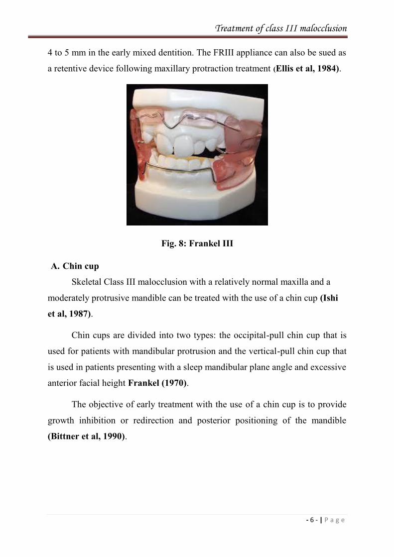
6. Functional applianceA. Frankel III
The Frankel III (FRIII) regulator is a functional appliance designed to
counteract the muscle forces acting on the maxillary complex. According to
Frankel (1970), the vestibular shields in the depths of the sulcus are placed
away from the alveolar buccal plates of the maxilla to stretch the periosteum
and allow for forward development of the maxilla. The shields are fitted closely
to the alveolar process of the mandible to hold or redirect growth posteriorly.
The effectiveness of each appliance is dependent on patient cooperation and
wearing them full time. In two separate studies the FRIII appliance appears to
effect occlusal changes (i.e. introducing dental compensations) by proclination
of upper incisors and retroclination of lower incisors (Lob and kerr, 1985;
Ulgen and Firath, 1994).
The mandible was repositioned downward and backward, decreasing the
prognathism of the mandible and increasing the lower facial height. Changes in
the position of the maxilla were minimal. The best response to FRIII treatment
was noted in patients with Class III malocclusions with an increased overbite of
Treatment of class III malocclusion
- 6 - | P a g e
4 to 5 mm in the early mixed dentition. The FRIII appliance can also be sued as
a retentive device following maxillary protraction treatment (Ellis et al, 1984).
Fig. 8: Frankel III
A. Chin cup
Skeletal Class III malocclusion with a relatively normal maxilla and a
moderately protrusive mandible can be treated with the use of a chin cup (Ishi
et al, 1987).
Chin cups are divided into two types: the occipital-pull chin cup that is
used for patients with mandibular protrusion and the vertical-pull chin cup that
is used in patients presenting with a sleep mandibular plane angle and excessive
anterior facial height Frankel (1970).
The objective of early treatment with the use of a chin cup is to provide
growth inhibition or redirection and posterior positioning of the mandible
(Bittner et al, 1990).

Treatment of class III malocclusion
- 7 - | P a g e
Effects on mandibular Growth
The orthopedic effects of a chin cup on the mandible include (D.
Nicodemo et al, 2008);
(1) Redirection of mandibular growth vertically.
(2) Backward repositioning (rotation) of the mandible.
Force Magnitude and Direction
Most of the reported studies recommended an orthopedic force of 300 to
500 g per side (Ishi et al, 1987; Uner et al, 1995; Deguchi and Kitsugi, 1996).
Patients are instructed to wear the appliance 14 hr/day. The orthopedic force is
usually directed either through the condyle or below the condyle.
Treatment Timing and Duration
Evidence exists that treatment to reduce mandibular protrusion is more
successful when it is started in the primary or early mixed dentition (Graber,
1977; Uner et al., 1995). The treatment time varies from 1 year to as long as 4
years depending on the severity of the original malocclusion.
Effects on the Temporomandibular Joint
There is some concern on the adverse effect of chin cup appliance on the
TMJ. In a study by Deguchi and Kitsugi (1996), several patients complained of
temporary soreness of the TMJ during the retention period. Of 40 patients, 2
continued to have TMJ pain and some degree of difficulty in opening the mouth
after the end of active treatment several studies indicated that the chin cup
affects the growth of not only the mandible, but also the cranial base structures
as well (Ritucci and Nanda, 1984).

Treatment of class III malocclusion
- 8 - | P a g e
Fig. 9: Chin cup
A. Protraction face mask
The face mask is most effective in the treatment of mild to moderate
skeletal Class III malocclusions with a retrusive maxilla and a hypodivergent
growth pattern (Bishara, 2001).
In 1944 Oppenheim, believed that one could not control the growth or
anterior displacement of the mandible and suggested moving the maxilla
forward in an attempt to counterbalance mandibular protrusion.
In the 1960 Delaire revived the interest in using a face mask for
maxillary protraction.
Petit 1983 later modified Delair’s basic concept by increasing the amount
of force generated by the appliance, thus decreasing the overall treatment time.
In 1987 McNamara introduced the use of a bonded expansion appliance
with acrylic occlusal coverage for maxillary protraction.
Turley 1988 improved patient cooperation in wearing the appliance by
fabricating customized face masks.

Treatment of class III malocclusion
- 9 - | P a g e
Parts of protraction face mask
The protraction face mask is made of two pads that contract the soft
tissue in the forehead and chin region. The pads are connected by a midline
framework and are adjustable through the loosening and tightening of a set
screw. Also it contains an adjustable anterior wire with hooks that connected to
the midline framework to accommodate a downward and forward pull on the
maxilla with elastics. To minimize the opening of the bite as the maxilla is
repositioned; the protraction elastics are attached near the maxillary canines
with a downward and forward pull of 30 degrees to the occlusal plane (Flanary
et al, 1990).
Force magnitude
Maxillary protraction generally requires 300 to 600 g of force per side,
depending on the age of the patient. Tension of the elastics can be estimated
using a tension stress gauge. Patients are instructed to wear the face mask for 12
hours a day (Gresham et al, 1957).
Treatment time and duration
Some studies suggest that face mask/ expansion therapy may be most
effective in the primary and early mixed dentitions (Jager et al, 2000).
Clinically, anterior cross bites can be corrected with 3 to 4 months of
maxillary expansion and protraction depending on the severity of the
malocclusion. Improvement in overbite and molar relationship can be expected
with an additional 4 to 6 months of maxillary protraction (Jacobson et al,
1974).
Treatment of class III malocclusion
- 10 - | P a g e
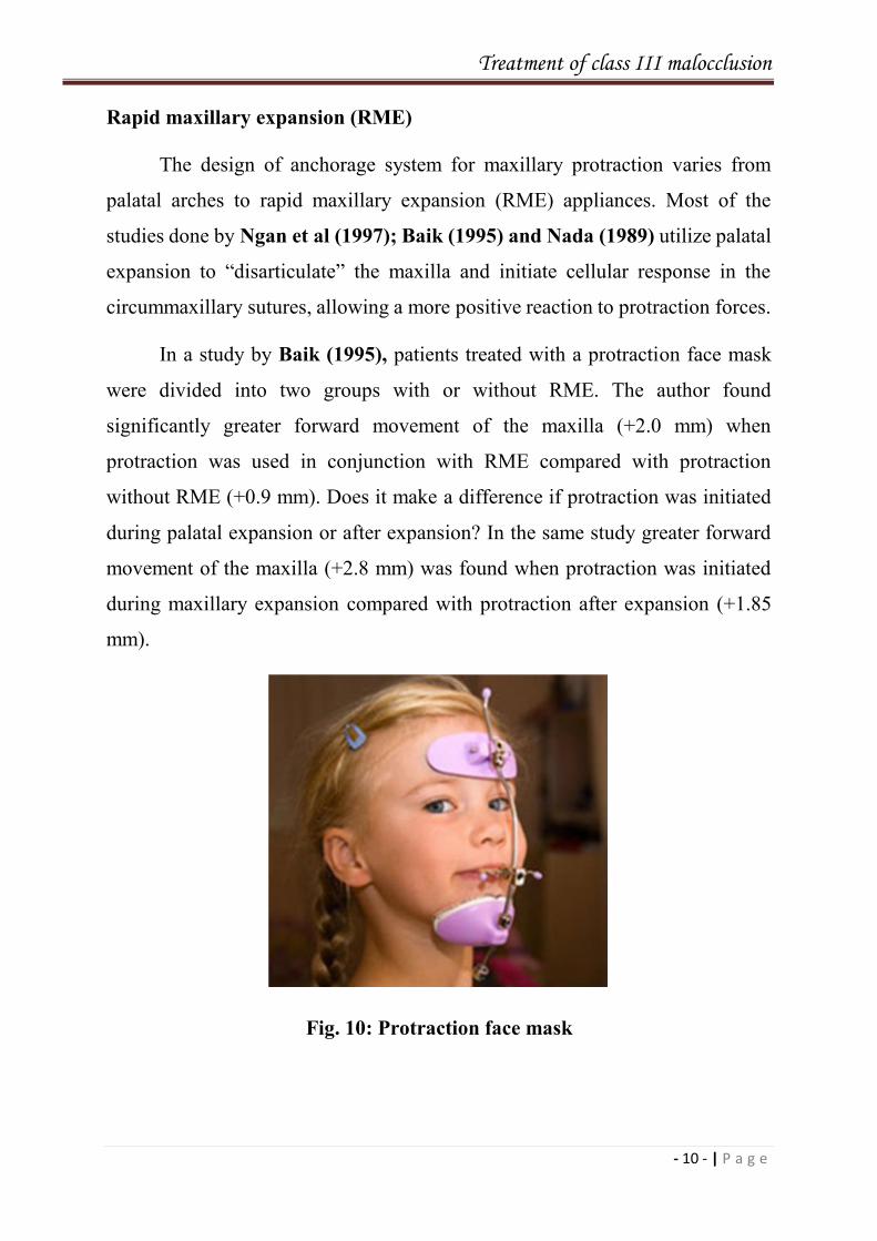
Rapid maxillary expansion (RME)
The design of anchorage system for maxillary protraction varies from
palatal arches to rapid maxillary expansion (RME) appliances. Most of the
studies done by Ngan et al (1997); Baik (1995) and Nada (1989) utilize palatal
expansion to “disarticulate” the maxilla and initiate cellular response in the
circummaxillary sutures, allowing a more positive reaction to protraction forces.
In a study by Baik (1995), patients treated with a protraction face mask
were divided into two groups with or without RME. The author found
significantly greater forward movement of the maxilla (+2.0 mm) when
protraction was used in conjunction with RME compared with protraction
without RME (+0.9 mm). Does it make a difference if protraction was initiated
during palatal expansion or after expansion? In the same study greater forward
movement of the maxilla (+2.8 mm) was found when protraction was initiated
during maxillary expansion compared with protraction after expansion (+1.85
mm).
Fig. 10: Protraction face mask

Treatment of class III malocclusion
- 11 - | P a g e
7. Orthognathic surgery
Orthognathic surgery, which is performed by the surgeon in conjunction
with the orthodontist, is used for the treatment of dentofacial deformities, and is
not only important for the correction of malocclusion but also for facial
esthetics. Patients with dentofacial deformities present problems of adjustment
and social adaptation, with negative consequences for their mental health. Thus,
the psychosocial aspects of surgery, such as changes in body image and
emotional and cognitive states, improvement of interpersonal relationships and
alterations in the reactions of society play an important role (Flanary et al
1990, Grossbart et al 1999, Ferreira et al 2004), Patients with dentofacial
deformities require a surgical orthodontic approach, and improvement of their
quality of life is one of the objectives of this type of intervention (Cunningham
et al 2002).
This has become increasingly popular in the treatment of patients with
moderate to severe Class III skeletal discrepancy. An initial orthodontic phase is
usually necessary in these patients to decompensate the arches by putting the
teeth in the ideal positions to facilitate the surgery. The maxilla may be
advanced or the mandible pushed back as the patient’s profile and occlusion
demands. Often a combination of upper and lower jaw surgery is necessary with
the addition of a reduction genioplasty of the chin. Vertical skeletal excess may
be dealt with by the addition of a Le Fort I posterior impaction osteotomy. This
is a commonly employed approach to the problem of skeletal anterior open bite.
Over the last decade surgical correction has become a common approach to
patients with significant Class III jaw and/or facial profile discrepancies.
Approximately 30-40 percent of patients presenting with a Class III might be
suitable to consider a surgical correction. If a young patient shows early signs of
developing such a problem and there is a chance of further unfavorable growth,
dento-alveolar camouflage generally should be avoided. Such untimely

Treatment of class III malocclusion
- 12 - | P a g e
interventions can create problems later if orthognathic correction is to be
considered. Surgery in these cases would usually be performed when all growth
has ceased since otherwise there is a danger of the skeletal discrepancy regrown
(Nicodemo et al 2008).
Fig. 11: Sever class III malocclusion with orthographic surgery

Treatment of class III malocclusion
- 13 - | P a g e
1.6 Post-treatment stabilityStability of overjet correction depends in the short term on an adequate
overbite and in the long term on facial growth. The greater part of orthodontic
treatment is undertaken in the growing patient. On average, the mandible grows
downwards and forwards slightly faster than the maxilla. In Class III patients
this is an adverse growth trend and may result both in a worsening (or relapse)
of the overjet and a reduction in overbite. An early sign of this happening is loss
of overbite on the upper lateral incisors with the result that they relapse into a
reverse overjet. In some patients the Class III skeletal pattern will become
markedly more sever after treatment and in these cases relapse is inevitable. In
other patients the facial proportions change little during the later stages of
growth and no adverse occlusal changes should result. In Class III, more than in
other types of malocclusion, long-term stability depends on a favorable growth
pattern, and this holds true whatever treatment approach is adopted (Jones and
Oliver, 2000).

References
- 24 - | P a g e
References
A
Agha NF, AL-Hamdany AK, Al-Khatib AR. Malocclusion assessment inorthodontically treated young Iraqis (6-18) years old. Al-Rafidain Dent J2002; 2: 80-86.
Ahiquist J, Eliasson S, Welander U. The effect of projection errors oncephalometric length measurements. Eur J Orthod 1986; 8: 141-8.
Allen AR, Connolly HI, Richardson A. Early treatment of Class III incisorrelationship using the chin cap appliance. Eur J Orthod 1993; 15:371-6.
B
Baek SH, Moon HS, Yang WS. Cleft type and Angle’s classification inKorean cleft patients. Eur J Orthod 2002; 24 (6): 647-53.
Baik CY, Ververidou M. A new approach of assessing sagittal discrepancies:The Beta angle. Am J Orthod Dentofacial Orthop 2004; 126:100-5.
Baik HS, Jee SGH, Lee KJ, Oh TK. Treatment effects of Frankel functionalregulator III in children with Class III malocclusions. Am J Orthod DentofacOrthop 2004; 125: 294-301.
Baker RW JR, Subtelny JD, Iran pour B. An American Board ofOrthodontics case report. Correction of a Class III mandibular prognathismand asymmetry through orthodontics and orthognathic surgery. Am J OrthodDentofac Orthop 1991; 99(3): 191-201.
Barrett MJ. A computer-based system of dental and craniofacialmeasurement and analysis. Aust. Dent J. 1968; 13: 207-12, [Cited by: Ali FA.Skeletodental characteristics of some Iraqi children at nine and ten years ofage: a cephalometric study. A master thesis, Department of Pedodontics,Orthodontics, and Preventive Dentistry, University of Baghdad, 1988].
Battagel JM. The etiological factors of Class III malocclusion. Eur J Orthod1993; 15(5): 347-70.
Baumrind S, Frantz R. The reliability of head film measurements I.Landmarks identification. Am J Orthod 1971; 60(2): 111-27.
Baumrind S, Miller DM. Computer-aided head film analysis. The Universityof California San Francisco method. Am J Orthod 1980; 78(1): 41-65.

References
- 25 - | P a g e
BeGole EA. Software development for the management of cephalometricradiographic data. Computer Programs Biomed 1981; 11(2): 175-82.
Biggerstaff RH, Allen RC, Tuncay OC, Berkowitz J. A verticalcephalometric analysis of the human craniofacial complex. Am J Orthod1977; 72(4): 397-405.
Bishara SE. Textbook of orthodontics. 1st ed. Philadelphia: W.B> SaundersCompany; 2001.
Bishara SE, Jorgensen GJ, Jakobsen JR. Changes in facial dimensionsassessed from lateral and frontal photographs. Part I – Methodology. Am JOrthod Dentofac Orthop 1995; 108(4): 389-03.
Bittner C, Pancherz H. Facial morphology and malocclusions. Am J OrthodDentofac Orthop 1990; 97(4): 308-15.
Bondevik O, Rosier M, Slagsvold O. The digital readout system CM-1: Aninstrument for rational measuring on radiographic head plates and dentalmodels. Eur J Orthod 1981; 3(1): 1-8.
Broadbent BS. A new X-ray technique and its application to orthodontia.Angle Orthod 1931; 1(2): 45-66.
Broch J, Slagsvold O, Rosier M. Error in landmark identification in lateralradiographic head plates. Eur J Orthod 1981; 3(1): 9-13.
Bukhary MT. Comparative cephalometric study of class III malocclusion inSaudi and Japanese adult females. J Oral Science 2005; 47(2): 83-90.
C
Carlotti AE, George R. Differential diagnosis and treatment planning of thesurgical orthodontic Class III malocclusion. Am J Orthod Dentofac Orthop1981; 79(4): 424-36.
Casto FM. Inherited and congenital causative factors in malocclusion. J AmDent Assoc 1928; 15: 1250-60.
Chang HP, Kinoshita Z, Kawamoto T. Craniofacial pattern of Class IIIdeciduous dentition. Angle Orthod 1992; 62(2): 139-44.
Cunningham SJ, Garratt AM, Hunt NP. Development of a condition-specific quality of life measure for patients with dentofacial deformity: II.Validity and responsiveness testing. Community Dent Oral Epidemiol 2002:30: 81–90.

References
- 26 - | P a g e
Chnag JZ, Chen Y, Chang FH, Yao JC, Liu P, Chang C, Lan W.Morphometric analysis of mandibular growth in skeletal class IIImalocclusion. J Formos Med Assoe 2006; 105(4): 318-28.
Choi YH, Sato K, Mitani H. Growth characteristics of prognathic face withrelapsed incisor reversed occlusion following chin cap therapy. J JapaneseOrthod Society 1999: 58: 1-14.
Chong YH, Ive JC, Artun J. Changes following the use of protractionheadgear for early correction Class III malocclusion. Angle Orthod 1996; 66:351-62.
Chung LKL, Hobson RS, Nunn JH, Gordon PH, Carter NE. An analysisof the skeletal relationships in a group of young people with hypodontia. J.Orthod 2000; 27(4): 315-8.
Cohen M, Bell WH, Proffit WR, White RP. Surgical correction ofdentofacial deformities. 1st ed. Philadelphia: W.B. Saunders Co.; 1980.
Cox NH, Vander Linden FP. Facial harmony. Am J Orthod 1971; 60: 175-83.
D
Danaie SM, Salehi P. Cephalometric evaluation of class-III Patients withchin cap and tongue guard. J Indian Soe Pedo Prev Dent 2005; 23(2):63-6.
Daskalogiannaks J. Glossary of orthodontic terms. 1st ed. Berlin:Quintessence Publishing Co.; 2000.
Davenport CB. Postnatal development of the human outer nose. Proc AmPhilos Soc 1939; 80(2): 175-356.
Deguchi T, Kitsugi A. Stability of changes associated with chin cup treatmentAngle Orthod 1996; 66: 139-46.
Deguchi T, Kuroda T, Minoshima Y, Graber TM. Craniofacial features ofpatients with Class III abnormalities: gowth-related chages and effects ofshort-term and long-term chin cup therapy. Am J Orthod Dentofac orthop2002; 121: 84-92.
Downs WB. Studies in the causes of dental anomalies. J Dent Res 1928; 8:367-79.
Downs WB. Variations in facisal relationship: their significance in treatmentand prognosis. Am J Orthod 1948; 34(10): 812-40.

References
- 27 - | P a g e
D. Nicodemo, M. D. Pereira, L. M. Ferreira: Effect of orthognathic surgeryfor class III correction on quality of life as measured by SF-36. Int. J. OralMaxillofac. Surg. 2008; 37: 131–134.
E
Ellis E, McNamara JA JR. Components of adult class III malocclusion. J.Oral and Max Surg 1984; 42: 295-305.
Enacar A, Demirhanolu M. Delaire-Verdon turii ortopedik ytiz masknn yapm ve uygulanmasnda Pratik bir yontem. Turk Ortodonti Dergisi, 1989; 2: 183-8.
Shareef PF Craniofacial features of skeletal class III malocclusion in asample of Kurdish adults in Hawler governorate. A master thesis. Departmentof Pedodontics, Orthodontics and Preventive Dentistry, University ofSulaimani, 2009.
F
Farkas LG. Anthropometry of the head and face in medicine. 1st ed. NewYork: Elsevier North Holland Inc.; 1981.
Flanary CM, Barnwel GM, Vansickels JE, Littlefield JH, Rugh AL.Impact of orthognathic surgery on normal and abnormal personalitydimensions: 2- year follow-up study of 61 patients. Am J Orthod DentofacialOrthop 1990: 98: 313–322.
Ferrario VF, Sforza C, Miani A, Tartaglia G. Craniofacial morphometry byphotographic evaluations. Am J Orfhod Dentofac Orthop 1993; 103(4): 327-37.
Ferreira JT, Telles CDS. Evaluation of the reliability of computerized profilecephalometric analysis. Braz Dent J 2002; 13(3): 201-4.
Foster TD. A textbook of orthodontics. 2nd ed. London: Blackwell; 1982.
Frank S. The occlusal plane: Reliability of its cephalometric location and itschanges with growth[thesis]. Oklahoma City: University of Oklahoma, 1983.
Freeman RS. Adjusting A-N-B angles to reflect the effect of maxillaryposition. Angle Orthod 1981; 51:162-71.
Ferreira LM, Rzeszetkowiski BSH. Plastic Surgery: an anthroposophicapproach. Rev Soc Bras Cir Pla´st 2004: 19: 39–40
G

References
- 28 - | P a g e
Grossbart TA, Sarwer DB. Cosmetic Surgery: surgical tools - psychologicalgoals. Semin Cutan Med Surg 1999: 18: 101–111.
Gelgor IE, Karaman Al. Non-surgical treatment of Class III malocclusion inadults: two case reports. J Orthod 2005; 32(2): 89-97.
Gold JK. A New approach to the treatment of mandibular prognathism. Am.J. Orthod 1949; 35(12): 893-912.
Graber TM. Orthodotics principles and practice. Philadelphia: W.B.Saunders Company; 1972.
Graber TM. Orthodonties principles and practice. Philadelphia: W.B.Saunders Company; 1988.
Graber TM, Rakosi T, Petrovic AG. Dentofacial orthopedics withfunctional appliances. St. Luis: Mosby Co.; 1985.
Graber TM, Vanarsdall RL. Orthodontics: Current principles andtechniques. 2nd ed. St. Louis: Mosby; 1994.
Gravely JF, Benzies PM. The clinical significance of tracing error incephalometry. Br J Orthod 1974; 1(3): 95-101.
Gresham H. A Manual of Orthodontics. New Zealand: N.M Peryer Ltd;1957.
Guyer EC, Ellis E, McNamara JA Jr, Behrents RG. Components of ClassIII malocclusion in juvenile and adolescents. Angle Orthod 1986; 56 (1): 7-31.
H
Haas A J. Palatal expansion: just the beginning of dentofacial orthopedics.Am J Orthod 1970; 57: 219-55.
Harris M, Reynolds IR. Fundamentals of orthognathic surgery. London:W.B. Saunders Co.; 1991.
Health MR. Measurement of cephalometric radiographs: Methods ofanalyzing data on a regional basis and improving reading efficiency. Am JOrthod 1980; 78(3): 303-9.
Hofrath Herbert. Die Bedeutung der Rontgenfern- und Abstandsaufnhme furdie Diagnostik der kieferanomalien. Fortschr Orthodont 1931; 1: 232-58[Citedby: Allen WI. Historical aspects of roentgenographic cephalometry. Am JOrthod 1963; 49(6): 451-9].
References
- 29 - | P a g e
Hohl TH, Wolford LM, Epker BN, Fonseca RJ. Craniofacial osteotomies:A photocephalometric technique for the prediction and evaluation of tissuechanges. Angle Orthod 1978; 48(2): 114-25.
Hong SK, Yi CK. A classification and characterization of skeletal Class IIImalocclusion on etio-pathogenic basis. Int J Oral Maxillofac Surg 2001; 30:264-71.
Houston WJB. The analysis of errors in orthodontic measurements. Am JOrthod 1983; 85(5): 382-90.
Hunter J. The natural history of the human teeth. Part II. A practical treatiseon the disease of the teeth intended as a supplement to the national history ofthose parts. London: J Johnson, 1778. [Cited by: Chang HP, Kinoshita Z,Kawamoto T. Craniofacial pattern of Class III deciduous dentition. AngleOrthod 1994; 62(2): 139044].
Hussels W, Nanda RS. Analysis of factors affecting angle ANB. Am JOrthod 1984;85: 411-23.
I
Irie M, Nakamura S. Orthopedic approach to severe skeletal Class IIImalocclusion. Am J Orthod 1975; 67: 377-92.
Ishii H, Morita S, Takeuchi Y, Nakamura S. Treatment effect of combinedMaxillary protraction and chin cap appliance in severe skeletal Class III cases.Am J Orthod Dentofac Orthop 1987; 92: 304-12.
J
Jacobson A. The “Wits” appraisal of jaw disharmony. Am J Orthod 1975; 67:125-38.
Jackson PH, Dickson GC, Birnie DJ. Digital image processing ofcephalometric radiographs: A preliminary report. Brit J Orthod 1985; 12(3):122-32.
Jacobson A, Evans WG, Preston CB, Sadowaski PL. Mandibularprognathism. Am J Orthod 1974; 66: 140-71.
Jager A, Braumann B, Kim C, Wahner S. Skeletal and dental effects ofmaxillary protraction in patients with Angle Class III malocclusion. A meta-analysis. J Orofac Orthop 2000; 62: 275-84.
K

References
- 30 - | P a g e
Kajiyama K, Murakami T, Suzuki A. Evaluation of the modified maxillaryprotractor applied to Class III malocclusion with retruded maxilla in earlydentition. Am J Orthod 2000; 118: 549-59.
Kama JD, Ozer T, Baran S. Orthodontic and orthopedic changes associatedwith treatment in subjects with Class III malocclusions. Eur J Orthod 2006;28(5): 496-502.
Khamarco TY, Al-Khatib AR, Agha NF. Occlusal criteria in two Iraqi ruralcommunitites. Al-Rafidain Dent J 2002; 2:360-8.
Kim YH, Vietas JJ. Anteroposterior dysplasia indicator: An adjunct tocephalometric differential diagnosis. Am J Orthod 1978; 73: 619-33.
Kim JY, Lee SJ, Kim TW, Nahm DS, Chang Y. Classification of theskeletal variation in normal occlusion. Angle Orthod 2005; 75(3): 303-11.
Kocadereli I. Early orthopedic treatment for Class III skeletal pattern. ASDCJ Dentistry Child 1998; 65: 177-181.
Konchak PA, Koehler JA. A pascal computer program for digitizing lateralcephalometric radiographs. Am J Orthod 1985; 87(3): 197-200.
M
McDonald RE, Avery DR. Dentistry for the child and adolescent. 7th ed., St.Louis, C.V. Mosby, 2000; P 141.
McNamara JA JR. A method of cephalometric evaluation. Am J Orthod.1984; 86(6): 449-69.
McWilliam JS. Evaluation and calibration of X-Y coordinatographs used incephalometric analysis. Scand J Dent Res 1980; 88(6): 496-504.
Meredith HV. Changes in the form of the head and face during childhood.Growth 1960; 24: 215-64.
Mermigos J, Full CA, Anderson G. Protraction of the maxillofacialcomplex. Am J Orthod 1990; 98: 47-55.
Michiels LYF, Tourne LPM. Nasion true vertical: A proposed method fortesting the clinical validity of cephalometric measurements applied to a newcephalometric reference line. Int J Adult Orthod Orthognath Surg 1990; 5(1):43-52.

References
- 31 - | P a g e
Midtgard J. Bjork G, Linder-Aronson S. Reproducibility of cephalometriclandmarks and errors of measurements of cephalometric cranial distances.Angle Orthod 1974; 44(1): 56-61.
Mackay F, John J A, Thompson R, Simpson W. Craniofacial from in ClassIII cases. Br J Orthod 1992; 19(1): 15-20.
Marton VD, Enlow DH, Hans MG, Broadbent BH, Oyen O. Class I andClass III malocclusion sub-groupings related to head form type. Angle Orthod1992; 62: 35-42.
Masaki F. Longitudinal study of morphological differences in the cranial baseand facial structure between Japanese and American White. J. Jpn. Orthod.Soe 1980; 39: 436-56. (In Japanese). [Cited by: Miyajima K, McNamara JAJR, Sana M, Murata S. An estimation of craniofacial growth in untreatedClass III female with anterior cross bite. Am. J. Orthod. Dentofac. Orthop.1997; 112(4): 425-434].
McCallin SG. Angle’s Class III malocclusion. Dent Pract 1956; 6(5): 151-64.
McCowen CS. Usefulness of an X-ray machine in orthodontia. Int JOrthodontia 1923; 9: 230-5, [Cited by: Allen WI. Historical aspects ofroentgenographic cephalometry. Am J Orthod. 1963; 49(6): 451-9].
Mitani H, Sakamoto T. Chin cap force to a growing mandible – long-termclinical reports. Am J Orthod 1984; 54: 93-122.
Mori Y, Miyajima T, Minami K, Sakuda M. An accurate three dimensionalcephalometric system: a solution for the correction of cephalic malpositioning.J Orthod 2001; 28(2): 143-9.
Moyers RE, Bookstein FL, Guire KE. The concept of pattern in craniofacialgrowth. Am J Orthod 1979; 76: 136-48.
Mucedero M, Coviello A, Baccetti T, Franchi L, Cozza P. Stability FactorsAfter Double-Jaw Surgery in Class III Malocclusion. Angle Orthod 2007;78(6): 1141-52.
N
Nakasima A, Lchinose M, Nakata S. Genetic and environmental factors inthe development of so-called pseudo- and true mesiocclusions. Am J OrthodDentofac Orthop 1986; 90: 106-16.
References
- 32 - | P a g e
Nakasima A, Ichinose M, Nakata S, Takahama Y. Heredity factors in thecraniofacial morphology of Angle’s Class II and Class III malocclusions. Am.J. Ortho Dentofac Orthop 1982; 82(2): 150-6.
Nagan P. Biomechanics of maxillary expansion and protraction in Class IIIpatients. Am J Orthod Dentofac Orthod 2002; 121: 582-3.
Nicodemo D, Rode SM. Psychological guidelines for the clinicalmanagement of patients with indication for ocular prosthesis. RPG Rev Po´sGrad 2002: 9: 224– 231.
P
Phillips C, Bennett ME, Broder HL. Dentofacial disharmony: psychologicalstatus of patients seeking a treatment consultation. Angle Orthod 1998: 68:547–556.
Pertschuk MJ, Sarwer DB, Wadden TA, Whitaker LA. Body imagedissatisfaction in male cosmetic surgery patients. Aesthet Plast Surg 1998: 22:20–24.
Prosterman B, Prosterman L, Fisher R, Gornitsky M. The use of implantsfor orthodontic correction of an open bite. Am J Orthod Dentofacial Orthop1995;107:245-50.
W
Wada K, Matsushita K, Shimazaki S, Miwa Y, Hasuike Y, Susami R. Anevaluation of a new case analysis of a lateral cephalometric roentgenogram. JKanazawa Med Univ 1981;6: 60-70.