Nanotechnology-Applied Curcumin for Different Diseases Therapy

Advances of Cancer Therapy by NanotechnologyXu Wang, Emory UniversityYiqing Wang, Emory UniversityGeorgia Chen, Emory UniversityDong M Shin, Emory University
Journal Title: Cancer Research and TreatmentVolume: Volume 41, Number 1Publisher: Korean Cancer Association | 2009-03, Pages 1-11Type of Work: Article | Final Publisher PDFPublisher DOI: 10.4143/crt.2009.41.1.1Permanent URL: http://pid.emory.edu/ark:/25593/fsww9
Final published version: http://e-crt.org/journal/view.php?number=224
Copyright information:© 2009 Korean Cancer Association. Open Access
Accessed January 25, 2022 3:04 PM EST

+ + + + + + + + + + + + + + + + + + + + + + + + + + + + + + + + + + + + + + + + + + + + + + + + + + + + + + + + + + + ++ + + + + + + + + + + + + + + + + + + + + + + + + + + + + + + + + + + + + + + + + + + + + + + + + + + + + + + + + + + ++ + + + + + + + + + + + + + + + + + + + + + + + + + + + + + + + + + + + + + + ++ + + + + + + + + + + + + + + + + + + ++ + + + + + + + + + + + + + + + + + + + + + + + + + + + + + + + + + + + + + + ++ + + + + + + + + + + + + + + + + + + ++ + + + + + + + + + + + + + + + + + + ++ + + + + + + + + + + + + + + + + + + + + + + + + + + + + + + + + + + + + + + ++ + + + + + + + + + + + + + + + + + + ++ + + + + + + + + + + + + + + + + + + ++ + + + + + + + + + + + + + + + + + + + + + + + + + + + + + + + + + + + + + + ++ + + + + + + + + + + + + + + + + + + ++ + + + + + + + + + + + + + + + + + + ++ + + + + + + + + + + + + + + + + + + ++ + + + + + + + + + + + + + + + + + + +
Advances of Cancer Therapy by Nanotechnology
Cancer Res Treat. 2009;41(1):1-11
Recent developments in nanotechnology offer researchers opportunities to significantlytransform cancer therapeutics. This technology has enabled the manipulation of thebiological and physicochemical properties of nanomaterials to facilitate more efficient drugtargeting and delivery. Clinical investigations suggest that therapeutic nanoparticles canenhance efficacy and reduced side effects compared with conventional cancer therapeuticdrugs. Encouraged by rapid and promising progress in cancer nanotechnology, researcherscontinue to develop novel and efficacious nanoparticles for drug delivery. The use oftherapeutic nanoparticles as unique drug delivery systems will be a significant addition tocurrent cancer therapeutics.
Key wordsNanoparticels, Cancer therapy, Drug delivery
Xu Wang, Ph.D.1
Yiqing Wang, Ph.D.2
Zhuo (Georgia) Chen, Ph.D.1
Dong M. Shin, M.D.1
1 Department of Hematology and MedicalOncology, Winship Cancer Institute and 2 Department of Biomedical Engineering,Emory University School of Medicine,Atlanta, GA, USA
Correspondence: Dong M. Shin, M.D.Department of Hematology and MedicalOncology, Winship Cancer Institute, EmoryUniversity School of Medicine, 1365 Clifton Rd.Building C. Room C3094. Atlanta, GA 30322, USA Tel: 1-404-778-2980Fax: 1-404-778-5520E-mail: [email protected] work was supported by National CancerInstitute (NCI) Specialized Program of ResearchExcellence (SPORE) grant (P50CA128613) toD.M. Shin and Centers of Cancer NanotechnologyExcellence (CCNE) grant (U54 CA119338) toD.M. Shin and Z.G. Chen.
VOLUM 41 NUMBER 1 MARCH 2009 1
I n t r o d u c t i o n
Conventional chemotherapeutic drugs are distributed non-specifically in the body where they affect both cancerous andhealthy cells, resulting in dose-related side effects and inadequatedrug concentrations reaching the tumor. Non-specific drug deliveryleads to significant complications that represent a serious obstacle toeffective anticancer therapy. In addition, the occurrence of resistancephenomena reduces the efficacy of cancer treatment. To overcomethe lack of specificity of conventional chemotherapeutic drugs,several ligand-targeted therapeutic strategies, including imm-unotoxins, radioimmunotherapeutics, and drug immunoconjugates, are being developed. Although these conjugated agents have shown
promising efficacy compared with conventional chemotherapydrugs, limitations in their delivery efficiency still remain.
Recent progress in cancer nanotechnology raises excitingopportunities for specific drug delivery. Nanoparticles, particularlyin the size range from 10 nm to 100 nm, are emerging as a class oftherapeutics for cancer treatment. Nanoparticles can be composed ofseveral functional molecules simultaneously, such as smallmolecule drugs, peptides, proteins, and nucleic acids. By using bothpassive and active targeting strategies, nanoparticles can increase theintracellular concentration of drugs in cancer cells while minimizingtoxicity in normal cells; thereby enhancing anticancer effects andreducing systemic toxicity simultaneously, when compared with thetherapeutic entities they contain. Furthermore, nanoparticles offer
DOI 10.4143/crt.2009.41.1.1
Review Article
Cancer Res Treat. 2009;41(1):1-11
2 CANCER RESEARCH AND TREATMENT
the potential to overcome drug resistance, since nanoparticles canbypass the P-glycoprotein efflux pump, one of the main drugresistance mechanisms, leading to greater intracellularaccumulation.
The purpose of this review article is to summarize the results ofthe use of therapeutic nanoparticles in the clinic and discuss theopportunities and challenges faced by therapeutic nanoparticles.Thus, the first part will emphasize the key properties of therapeuticnanoparticles and how these properties affect the efficiency andspecificity of nanoparticles as a drug delivery system. Next, we willsummarize current clinical uses of the first-generation therapeuticnanoparticles and the advances in new generation of therapeuticnanoparticles currently under preclinical and clinical investigation.Finally, we will discuss how nanoparticles will be developed toimprove their therapeutic efficacy and function for future cancertreatment.
N a n o p a r t i c l e s f o r Tu m o rTa r g e t i n g a n d D e l i v e r y
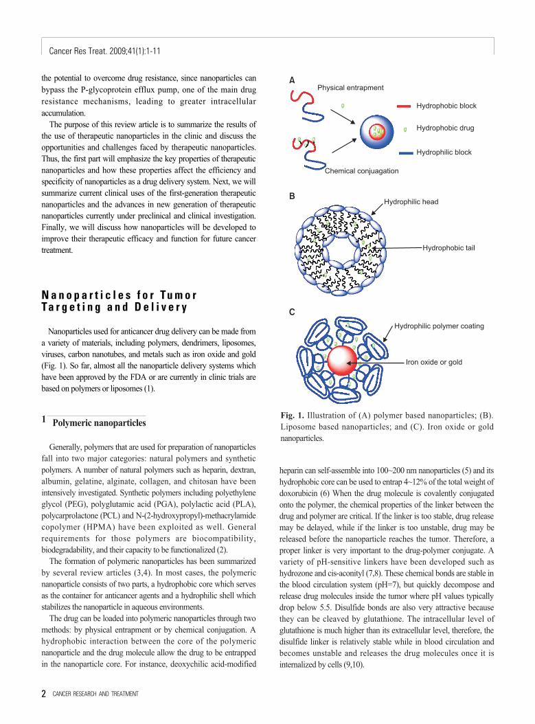
Nanoparticles used for anticancer drug delivery can be made froma variety of materials, including polymers, dendrimers, liposomes,viruses, carbon nanotubes, and metals such as iron oxide and gold(Fig. 1). So far, almost all the nanoparticle delivery systems whichhave been approved by the FDA or are currently in clinic trials arebased on polymers or liposomes (1).
1 Polymeric nanoparticles
Generally, polymers that are used for preparation of nanoparticlesfall into two major categories: natural polymers and syntheticpolymers. A number of natural polymers such as heparin, dextran,albumin, gelatine, alginate, collagen, and chitosan have beenintensively investigated. Synthetic polymers including polyethyleneglycol (PEG), polyglutamic acid (PGA), polylactic acid (PLA),polycarprolactone (PCL) and N-(2-hydroxypropyl)-methacrylamidecopolymer (HPMA) have been exploited as well. Generalrequirements for those polymers are biocompatibility,biodegradability, and their capacity to be functionalized (2).
The formation of polymeric nanoparticles has been summarizedby several review articles (3,4). In most cases, the polymericnanoparticle consists of two parts, a hydrophobic core which servesas the container for anticancer agents and a hydrophilic shell whichstabilizes the nanoparticle in aqueous environments.
The drug can be loaded into polymeric nanoparticles through twomethods: by physical entrapment or by chemical conjugation. Ahydrophobic interaction between the core of the polymericnanoparticle and the drug molecule allow the drug to be entrappedin the nanoparticle core. For instance, deoxychilic acid-modified
heparin can self-assemble into 100~200 nm nanoparticles (5) and itshydrophobic core can be used to entrap 4~12% of the total weight ofdoxorubicin (6) When the drug molecule is covalently conjugatedonto the polymer, the chemical properties of the linker between thedrug and polymer are critical. If the linker is too stable, drug releasemay be delayed, while if the linker is too unstable, drug may bereleased before the nanoparticle reaches the tumor. Therefore, aproper linker is very important to the drug-polymer conjugate. Avariety of pH-sensitive linkers have been developed such ashydrozone and cis-aconityl (7,8). These chemical bonds are stable inthe blood circulation system (pH=7), but quickly decompose andrelease drug molecules inside the tumor where pH values typicallydrop below 5.5. Disulfide bonds are also very attractive becausethey can be cleaved by glutathione. The intracellular level ofglutathione is much higher than its extracellular level, therefore, thedisulfide linker is relatively stable while in blood circulation andbecomes unstable and releases the drug molecules once it isinternalized by cells (9,10).
Fig. 1. Illustration of (A) polymer based nanoparticles; (B).Liposome based nanoparticles; and (C). Iron oxide or goldnanoparticles.

Xu Wang, et al _Nanotechnology for Cancer Therapy
VOLUM 41 NUMBER 1 MARCH 2009 3
It is important to note that dendrimers, synthetic supermacromolecules with highly branched repeated three dimensionalstructures, have emerged as important materials for biologicalapplication due to their unique features such as the precise control ofsize and shape, uncommon physical properties, controlleddegradation, and the ability to place numerous functional groups ontheir periphery and/or core (11,12).There are more than 50 differenttypes of dendrimers (13). Among them, polyamidoamine and poly(propylenemine) have been commercialized and used extensively asbiomaterials in gene and drug delivery (14,15) and for nanoparticleencapsulation (16,17) in imaging (18,19).
2 Liposomal nanoparticles
Liposomes are self-assembling spherical particles with amembrane composed of phospholipid bilayers. The size ofliposomes can range from 25 nm to 10μm depending on thepreparation method. They have been studied as candidates for drugdelivery for the last 50 years since being first discovered byBangham (20). The synthesis of liposomal nanoparticles has beenreviewed by Bellare, et al (21). Drug delivery systems based onunmodified liposomes are limited by their short blood circulationtime. This is mainly due to the fast clearance of liposomes bymacrophages of the reticuloendothelial system (RES) (22). Thesecond generation of polymer-coated liposomes can dramaticallyincrease blood circulation times from several minutes up to 3 days.
3 Gold and iron oxide nanoparticles
Recently, several novel nanotechnology concepts have beenapplied to the development of a new generation of anti-cancer drugdelivery systems. Gold nanoparticles can be synthesized through thereduction of HAlCl4 with a very narrow polydispersity. Several goldnanoparticle anticancer drug delivery systems have been reportedand showed good in vitro results (23).Although it seems that gold isinert under physiological environments, the long term toxicity ofgold nanoparticles remains an unanswered question. One attractiveproperty of gold nanoparticles is that gold concentrations arenaturally low in animal bodies, allowing the convenient use of thisnanoparticle model for in vivo pharmacokinetic and biodistributionstudies (24).
It is worth mentioning that a gold nanorod formulation is showingvery promising potential as a photothermal therapy agent. Goldnanorod can generate heat when it is radiated by a near infra-red(IR) laser (wavelengt > 650 nm). At this range, the laser is relativelysafe to the tissue and organs. Once the gold nanorod hasaccumulated inside the tumor through passive/active targeting, it canbe heated locally up to 43oC (25) by radiation with a near IR laser todestroy the tumor without causing damage to surrounding healthytissues (27).
Iron oxide nanoparticles have been clinically used as imaging
agents for MRI. Recently a number of research groups haveinvestigated them as drug carriers while retaining their imagingfunctions (26-28). One unique advantage of iron oxide nanoparticledelivery systems is that they can be delivered in a targeted manner toa desired region by applying an external magnetic field.
P r o p e r t i e s o f N a n o p a r t i c l e s
A suitable nanoparticle size is very important for efficient drugdelivery. Generally, 10~100 nm is considered the optimal size fornanoparticle drug carriers. If the particle size is less than 10 nm, thenanoparticles will be quickly eliminated by renal clearance(threshhold < 6 nm). At sizes greater than 100 nm, the chance of theparticle being captured by the RES will dramatically increase (29).
A proper surface coating is essential to the stability andcirculation time of nanoparticle delivery systems. For instance, asodium citrate-stabilized gold particle aggregates in PBS withinseveral minutes. But once coated with thiol-terminated polyethyleneglycol (PEG) polymer, this nanoparticle is stable not only in PBSbut also under low or high pH conditions (32). Generally, a neutral-charged nanoparticle can achieve a long circulation time and reducethe chance of nanoparticle capture by the immune system.
Ta r g e t e d D e l i v e r y o f T h e r a p e u t i cN a n o p a r t i c l e s
1 Passive targeting
Table 1 lists nanoparticles that have been used in the clinic andutilize passive targeting to achieve their selective delivery to tumors.Passive targeting takes advantage of the inherent size ofnanoparticles and the unique properties of tumor vasculature (30-33). As tumors grow and begin to outstrip the available supply ofoxygen and nutrients, they release cytokines and other signalingmolecules that recruit new blood vessels to the tumor in a processcalled angiogenesis. Unlike the tight blood vessels in normal tissues,angiogenic blood vessels in tumor tissues have gaps as large as 600to 800 nm between adjacent endothelial cells (34,35).This defectivevascular architecture coupled with poor lymphatic drainage inducesan enhanced permeability and retention effect (EPR) (36,37),Through these gaps, nanoparticles can selectively accumulate intothe tumor interstitium (38) (Fig. 2).
In general, the accumulation of nanoparticles in tumor tissues isdependent on several factors including the size, surfacecharacteristics, and circulation half-life of the nanoparticles and thedegree of angiogenesis of the tumor (39). It is speculated thatnanoparticles with a size between 10 and 100 nm will be optimal fortumor accumulation. For example, smaller polymeric micelles (20

nm) have been shown to accumulate more readily in tumors thanlarger liposomes (100 nm) (40,41). Proper surface characteristicsand longer circulation times of nanoparticles can also improvetumor uptake, as described earlier. The unmodified phospholipidsurface of liposomes can attract plasma proteins and thusrecognition by the mononuclear phagocytic system (MPS), resultingin their rapid clearance from the circulation. This property impedesthe distribution of liposome-associated drugs to solid tumors.Surface-modified (stealth) liposomes have solved the problem offast clearance from the circulation, yielding liposomes with asignificantly increased half-life in the blood (42,43). Dramaticallyreduced clearance rates have also been obtained with othernanoparticles such as Abraxane (44), Xyotax (45) and IT-101 (46).Tumor vascularization also affects nanoparticle accumulation;usually nanoparticles accumulate poorly in poorly vascularizedtumors, small pre-angiogenic tumors, or large necrotic tumors.
As drug delivery systems, nanoparticles have shown an ability toimprove pharmacokinetics, pharmacodynamics, efficacy, and toreduce the toxicity of associated drugs (40). For example, Abraxane
Cancer Res Treat. 2009;41(1):1-11
4 CANCER RESEARCH AND TREATMENT
Type of Nanoparticle Name and Refs Therapeutic agent StatusLiposomes DaunoXome (105) Dox Approved
Doxil /Caelyx (39, 51) Dox ApprovedMyocet (39, 106, 107) Dox Approved (Europe)
SPI-077 (108~110) Cisplatin Phase IIOncolipin (111) Interleukin 2 Phase IIOSI-7904L (112, 113) Thymidylate synthase inhibitor Phase IILEP ETU (114) Paclitaxel Phase I/IILE-SN38 (18, 19) SN-38 Phase I/IIOSI-211 (115) lurtotecan Phase IIAroplatin (116) Oxaliplatin Phase II
Polymeric micelles Genexol-PM (85, 117~119) Paclitaxel Approved (South Korea)NK911 (52, 120) Dox Phase ISP1049C (121) Dox Phase INC-6004 (122) Cisplatin Phase INK012 (122, 123) SN-38 Phase INK105 (50, 124) Paclitaxel Phase I
Polymer-drug conjugate-based nanoparticles CT-2103; XyotaxTM (45, 125) Paclitaxel Phase IIIPK1; FCE28068 (126, 127) Dox Phase IIPK2; FCE28069 (128) Dox Phase I/IIPNU166945 (97) Paclitaxel Phase IMAG-CPT (129, 130) Camptothecin Phase IAP5280 (131) Platinate Phase I/IIAP5346 (132) Platinum Phase IIAD-70, DOX-OXD (133) Dox Phase IDE-310 (134~136) Camptothecin Phase I/IIProthecan (137, 138) Camptothecin Phase IIEZN-2208 (139) SN-38 Phase IIT-101 (82) Camptothecin Phase IINKTR-102 (140) Irinotecan Phase II
Albumin-based nanoparticles Abraxane (47, 141, 142) or ABI-007 Paclitaxel Approved
Table 1. Examples of non-targeted nanoparticles in clinical development
Fig. 2 Schematic diagram of nanoparticle accumulation in tumortissue through EPR effect. Normal tissue vasculatures are lined bytight endothelial cells, thereby preventing nanoparticle drugs fromescaping, whereas tumor tissue vasculatures are leaky andhyperpermeable allowing preferential accumulation ofnanoparticles in the tumor interstitial space (passive targeting).

Xu Wang, et al _Nanotechnology for Cancer Therapy
(ABI-007), an albumin-bound nanoparticle of paclitaxel (Taxol)which has been approved for the treatment of metastatic breastcancer, showed significant greater efficacy than free paclitaxel in aphase III clinic trial (47).Despite the increased dose of paclitaxel inthe Abraxane group, the incidence of grade 4 neutropaenia wassignificantly lower than in patients treated with free paclitaxel.Pharmacokinetic studies also showed that paclitaxel clearance andthe volume of distribution were higher for Abraxane than forpaclitaxel: Clearance was 13 litres per hour per m2 for Abraxaneversus 14.76 litres per hour per m2 for paclitaxel (p=0.048), anddistribution was 663.8 litres per m2 for Abraxane versus 433.4 litresper m2 for paclitaxel (p=0.04) (44). Similar to Abraxane, NK105, amicellar nanoparticle formulation of paclitaxel also showedimproved pharmacokinetics, pharmacodynamics, efficacy, andreduced toxicity as compared with free paclitaxel in a preclinicalstudy and a phase I trial (48,49). The plasma area under the curve(AUC) value was approximately 90-fold greater for NK105 than forfree paclitaxel and the tumor AUC value was 25-fold higher forNK105 than for free paclitaxel in an animal model (48). In patients,the plasma AUC of NK105 at 180 mg/m2 was approximately 30-fold greater than that of the conventional formulation of paclitaxel(49).NK105 showed significantly more potent antitumor activity ina human colorectal cancer cell line HT-29 xenograft than freepaclitaxel, due to enhanced accumulation of the drug in the tumor(48). The phase I trial showed that NK105 was well tolerated andeffective in patients with pancreatic cancer (50).These differences inpharmacokinetic properties may contribute to the increased drugaccumulation inside the tumor observed with nanoparticlescompared with the corresponding free drugs. Other nanoparticlescurrently used in the clinic or undergoing clinic trials also showed animproved pharmacokinetic profile compared with the respective freedrugs, such as Doxil, a PEG-liposome loaded with doxorubicin(DOX) (51), SP1049C, a pluronic micelle loaded with DOX (40),NK911, a PEG-Asp micelle loaded with DOX (52), and Xyotax, apolyglutamic acid nanoparticle carrying paclitaxel (45).
2 Active targeting
The nanoparticles listed in Table 1 that have been used in theclinic so far mostly utilize the EPR effect of tumors and the tumormicroenvironment to promote their selective delivery to tumors.However, certain limitations of non-targeted nanoparticles as a drugdelivery system still remain. For example, in the case of the EPReffect, although poor lymphatic drainage helps the extravasateddrugs to be enriched in the tumor interstitium, it also induces drugoutflow from the cells as a result of higher osmotic pressure in theinterstitium, which eventually leads to drug redistribution in someportions of the cancer tissue (53). Most importantly, accumulationmerely within the tumor microenvironment by the EPR effect maynot always correlate with therapeutic efficacy since internalizationinto the tumor cells is required for most anticancer drugs to exert
their biological functions. To overcome these limitations, a rationalapproach is to incorporate a targeting moiety on the nanoparticlesurface. The targeting moiety is expected to bind a tumor-associatedantigen or receptor and facilitate the delivery of nanoparticles to theintracellular site of drug action, enabling a greater therapeutic effect(Fig. 3). Recent preclinical studies have shown that targetednanoparticles have better antitumor activity compared with non-targeted nanoparticles (54-57).Although targeted nanoparticles maynot always mediate an increase in tumor drug accumulation whencompared with non-targeted nanoparticles, targeted nanoparticlesshow greater intracellular drug delivery to cancer cells than non-targeted nanoparticles, resulting in dramatically increased antitumorefficacy (54-56).These findings suggest that the primary role of thetargeting ligands is to enhance cellular uptake into cancer cells andto minimize cellular uptake in normal cells.
Although current studies have shown that the use of targetednanoparticles as a drug delivery system is a promising strategy totreat human cancers, it is still in its early stage of development.Clinical data using targeted nanoparticles are limited since mosttargeted nanoparticles have not yet reached the clinic. Only a fewtargeted nanoparticles are currently under clinical investigation. Oneis MCC-465, which is an immunoliposome-encapsulateddoxorubicin (Dox). The liposome is tagged with PEG and theF(ab’)2 fragment of the human monoclonal antibody GAH, whichrecognizes a cell surface molecule on various types of cancer cells(58).Phase I studies have indicated that the PK parameters of MCC-465 differ from those of free Dox, but were very similar to those ofDoxil (non-targeted liposome-encapsulated Dox) in humans. Interms of skin toxicity, the patients who received MCC-465 did notexperience any severe skin toxicity such as palmar–plantarerythrodysesthesia (PPE) or mucositis, unlike the patients who
VOLUM 41 NUMBER 1 MARCH 2009 5
Fig. 3. Internalization of nanoparticles via receptor-mediatedendocytosis. Tumor-specific ligands/antibodies on the nanoparticlesbind to cell through an endosome-dependent mechanism. Drug-loaded nanoparticles bypass the drug efflux pump not beingrecognized when the drug enters cells, leading to high intracellularconcentration.

Cancer Res Treat. 2009;41(1):1-11
6 CANCER RESEARCH AND TREATMENT
received Doxil (58). Besides MCC-465, other examples of targetedtherapeutic nanoparticles include MBP-426 which contains thecytotoxic platinum-based drug oxaliplatin in a liposome (59), SGT-53, a liposome containing a plasmid coding for the tumor suppressorp53 (60), and CALAA-01, a polymer-siRNA conjugate (61,62)(Table 2) . These nanoparticles all target the transferrin receptorwhich is upregulated in many types of cancer (63).
3 Selection of target receptor and ligand
Selection of the target receptor or antigen on cancer cells iscrucial for the optimal design of targeted nanoparticles. In general,cell-surface antigens and receptors should have several propertiesthat render them particularly suitable as tumor-specific targets. First,they should be abundantly and uniquely expressed on tumor cells,but negligibly or less expressed on normal cells. Second, theyshould have a high density on tumor cells.
A targeting ligand should selectively and successfully transportnanoparticles into targeted cancer cells. It is believed thatinternalization of nanoparticles after binding to targeted tumor cellsis necessary for good therapeutic responses, so whether the targetednanoparticles can be internalized is an important issue in theselection of proper targeting ligand. Use of a ligand that can nottrigger the internalization process may result in drug release outsidethe cell and its redistribution to the surrounding normal tissues.
A variety of targeting ligands, including antibodies, antibodyfragments, peptides, growth factors, and aptamers (64), have beenused to facilitate the uptake of carriers into target cells (65-76).
R e d u c t i o n o f M u l t i d r u gR e s i s t a n c e
Drug resistance remains one of the major challenges in cancertherapy. A number of mechanisms for drug resistance have beendescribed. Drug resistance can be caused by physiological barriers(non-cellular based mechanisms), or alterations in the biology andbiochemistry of cancer cells (cellular mechanisms). Non-cellulardrug resistance can be caused by poorly vascularized tumor regionsand/or physiological barriers that greatly reduce drug access to thetumor tissues, thus protecting cancerous cells from drug-inducedcytotoxicity. Cellular drug resistance can be due to overexpresseddrug export pumps, such as P-glycoprotein (p-gp) and other drug-resistance proteins, increased DNA repair capacity, and reducedapoptosis regulation.
Among these mechanisms, the roles of the drug efflux pumps arethe most extensively investigated. P-glycoprotein (p-gp), a productof the MDR1 gene, is a 170-kD transmembrane glycoprotein thatfunctions as an efflux pump to remove drug from cells. Severalspecific p-gp inhibitors have been investigated to overcome drugresistance. Although in preclinical studies, some of these p-gpinhibitors have shown the restoration of cancer cell sensitivity toanticancer drugs, the clinical trials results have been disappointing(77,78).
Alternative strategies for overcoming drug resistance have beenstudied. Newly developed drug delivery systems, includingnanoparticle, allow selective drug accumulation in tumor tissues,tumor cells, or even compartments of tumor cells. Because with theaid of a targeting moiety nanoparticles enter cells throughendocytosis, it is expected that they can bypass the p-gp effluxpump, leading to their greater intracellular accumulation (Fig. 3).Many nanoparticles have been used to overcome or minimize drugresistance in preclinical studies and the results are very promising.For example, doxorubicin (DOX)-loaded poly (alkyl cyanoacrylate)nanoparticles (79), PACA nanoparticle (80,81), and IT-101 (a
Name and Refs Targeting agent Therapeutic agent StatusFCE28069 (128) Galactose DOX Phase I (stopped)MCC-465 (58) F(ab’)2 fragment of DOX Phase I
human antibody GAHMBP-426 (59) Transferrin Oxaliplatin Phase ISGT-53 (60) Transferrin receptor antibody fragment Plasmid DNA with p53 gene Phase ICALAA-01 (62) Transferrin Small interfering RNA Phase IDOX-PEG-FOL (143) Folate receptor Dox Pre-cliniccRGD-Functionalized Dox micelle (144) cRGD peptide Dox Pre-clinicDtxl-NP-Apt (56) RNA aptamer Dox Pre-clinic2C5 -Immunomicelles (145) mAntibody 2C5 Paclitaxel Pre-clinicASGPR-paclitaxel (146) Galactosel Paclitaxe Pre-clinicPt-NP-Apt (64) PSMA targeting aptamer Cisplatin Pre-clinic
Table 2. Examples of targeted nanoparticles in preclinical and clinical development

Xu Wang, et al _Nanotechnology for Cancer Therapy
VOLUM 41 NUMBER 1 MARCH 2009 7
nanoparticulate conjugate of 20 (S)-camptothecin) (82) have shownthe ability to overcome drug resistance in the tested models. Andmost importantly, the ability of targeted nanoparticles to overcomedrug resistance has been confirmed in humans. It has beendemonstrated in clinical studies that liposomal doxorubicin is able toovercome drug resistance in AIDS-related Kaposi’s sarcoma(83,84). Also, clinical trials showed positive results usingnanoparticles in patients who had previously failed chemotherapy(83,85,86). Ligand-targeted strategies, especially those usingreceptor-targeting ligands, have also been applied to overcome drugresistance since these ligands are internalized via receptor-mediatedendocytosis, bypassing the plasma membrane where p-gp primarilyacts. As an example, folate receptor-targeted pH-sensitive polymericmicelles containing DOX (87) and transferrin-conjugated paclitaxelnanoparticles exhibited greater cytotoxicity than the respective freedrugs in a drug-resistant model (88). As illustrated, usingnanoparticles as a drug delivery system may be able to overcomecertain kinds of cancer drug resistance.
P o t e n t i a l To x i c i t y o fN a n o p a r t i c l e s
An important consideration in nanoparticle development is thebiological behavior of carrier constituents and their potential toxicity,especially during chronic administration. Many candidate polymershave been defined with particular toxicities, such as hematotoxicity,complementactivation, carcinogenicity, teratogenicity, andimmunogenicity (89,90), indicating the importance of choosing safepolymers for the design of nanoparticles. In addition, the biologicalproperties of polymers are molecular weight-dependent and can bechanged once the respective conjugates are prepared. Therefore,careful characterization of the potential toxicity of both the polymerand the final nanoparticle is critically important. For non-biodegradable polymers, potential toxicity is concerning when thepolymer molecular weight is greater than the renal threshold.Increased understanding of the potentially deleterious properties ofpolymers leads to the design of new and safer polymericnanoparticles.
Currently, most nanoparticles use nontoxic and biodegradableingredients, so toxicities associated with the carrier molecules per setend to be mild. However, particular nanoparticles cause increasedaccumulation of drugs in MPS cells in the liver, spleen, and bonemarrow, with the possibility of increased toxicities to these organs.Among these organs, the liver has been identified in many studies asthe primary organ responsible for reticuloendothelial capture ofnanoparticles, often due to phagocytosis by Kupffer cells (91,92).Hepatic uptake has been shown to be a main mechanism of hepaticclearance from the blood circulation following the intravenousinjection of nanoparticles. In addition to hepatic accumulation, somenanoparticles have been reported to cause liver injury (decreased
function and hepatic morphology changes) (93,94). For example,intravenous administration of cationic PAMAM dendrimers causedliver injury when administered intravenously to mice (95).Hepatotoxicity has also been observed in mice treated orally withnano-zinc particles (96). Also there are safety concerns withparticular nanoparticles that are able to cross the blood brain barrier.Lessons have been learned from many of the early clinical studies.For example, due to neurotoxicity, a clinical trial testing an HPMA-conjugated paclitaxel was terminated (97). The failure of MAG-camptothecin due to cumulative bladder toxicity in phase I was alsoreported (98).
Attempts are being made to decrease the uptake of nanoparticlesby MPS cells and to increase their accumulation in the active site,through polymer or nanoparticle surface modifications, and/orincorporating targeting ligands (54,99,100). With more rationaldesign, many nanoparticles have shown an improved safety profileand enhanced antitumor efficacy compared with free drugs inpreclinical and clinical studies (100-104). For example, Doxil (PEG-liposome loaded with doxorubicin) showed a reduction incardiotoxicity over that of doxorubicin in a clinical study (103,104).Abraxane (albumin nanoparticle loaded with paclitaxel) showed agreater therapeutic outcome compared with free paclitaxel and,taking advantage of the water solubility of the nanoparticle,successfully eliminated the side effects associated with the toxicvehicle Cremophor EL (47).
I m p l i c a t i o n s a n d F u t u r eD i r e c t i o n s
Nanoparticles provide opportunities for designing and tuningproperties that are not possible with other types of therapeutic drugs,and have shown a bright future as a new generation of cancertherapeutics. Furthermore, the development of multifunctionalnanoparticles may eventually render nanoparticles able to detect andkill cancer cells simultaneously. Although there are certain criticalquestions and many challenges remaining for the clinicaldevelopment of nanoparticles, as more clinical data are available,further understanding in nanotechnology will certainly lead to themore rational design of optimized nanoparticles with improvedselectivity, efficacy, and safety.
A c k n o w l e d g e m e n t
We thank Dr. Anthea Hammond for her critical reading of themanuscript.

Cancer Res Treat. 2009;41(1):1-11
8 CANCER RESEARCH AND THREATMENT
01. Qiu LY, Bae YH. Polymer architecture and drug delivery. Pharm Res. 2006;23:1-30.02. Tong R, Cheng JJ. Anticancer polymeric nanomedicines. Polym Rev. 2007;47:345-81.03. Cho K, Wang X, Nie S, Chen ZG, Shin DM. Therapeutic nanoparticles for drug delivery
in cancer. Clin Cancer Res. 2008;14:1310-6.04. Zhang GZ, Niu AZ, Peng SF, Jiang M, Tu YF, Li M, et al. Formation of novel polymeric
nanoparticles. Acconuts Chem Res. 2001;34:249-56.05. Park K, Kim K, Kwon IC, Kim SK, Lee S, Lee DY, et al. Preparation and characterization
of self-assembled nanoparticles of heparin-deoxycholic acid conjugates. Langmuir.2004;20:11726-31.
06. Park K, Lee GY, Kim YS, Yu M, Park RW, Kim IS, et al. Heparin-deoxycholic acidchemical conjugate as an anticancer drug carrier and its antitumor activity. J ControlRelease. 2006;114:300-6.
0 7. Kratz F, Beyer U, Schutte MT. Drug-polymer conjugates containing acid-cleavablebonds. Crit Rev Ther Drug. 1999;16:245-88.
0 8. Ulbrich K, Subr V. Polymeric anticancer drugs with pH-controlled activation. Adv DrugDeliver Rev. 2004;561023-50.
09. Jones DP, Carlson JL, Samiec PS, Sternberg P, Mody VC, Reed RL, et al. Glutathionemeasurement in human plasma Evaluation of sample collection, storage andderivatization conditions for analysis of dansyl derivatives by HPLC. Clin Chim Acta.1998;275:175-84.
10. Koo AN, Lee HJ, Kim SE, Chang JH, Park C, Kim C, et al. Disulfide-cross-linked PEG-poly(amino acid)s copolymer micelles for glutathione-mediated intracellular drugdelivery. Chem Comm. 2008:6570-2.
11. Bosman AW, Janssen HM, Meijer EW. About dendrimers: Structure, physicalproperties, and applications. Chem Rev. 1999;99:1665-88.
12. Fischer M, Vogtle F. Dendrimers: From design to application - A progress report.Angew Chem Int Edit. 1999;38:885-905.
13. Klajnert B, Bryszewska M. Dendrimers: properties and applications. Acta BiochimicaPolonica. 2001;48:199-208.
14. Dykes GM. Dendrimers: a review of their appeal and applications. J Chem Tech Biot.2001;76:903-18.
15. Esfand R, Tomalia DA. Poly(amidoamine) (PAMAM) dendrimers: from biomimicry todrug delivery and biomedical applications. Drug Discov Today. 2001;6:427-36.
16. Balogh L, Tomalia DA. Poly(amidoamine) dendrimer-templated nanocomposites. 1.Synthesis of zerovalent copper nanoclusters. J Am Chem Soc. 1998;120:7355-6.
17. Crooks RM, Zhao MQ, Sun L, Chechik V, Yeung LK. Dendrimer-encapsulated metalnanoparticles: Synthesis, characterization, and applications to catalysis. AcconutsChem Res. 2001;34:181-90.
18. Balogh L, Bielinska A, Eichman JD, Valluzzi R, Lee I, Baker JR, et al. Dendrimernanocomposites in medicine. Chim Oggi. 2002;20:35-40.
19. Zheng J, Dickson RM. Individual water-soluble dendrimer-encapsulated silver nanodotfluorescence. J Am Chem Soc. 2002;124:13982-3.
20. Bangham AD, Horne RW. Negative Staining of Phospholipids + Their StructuralModification by-Surface Active Agents as Observed in Electron Microscope. J MolBiol. 1964;8:660-8.
21. Watwe RM, Bellare JR. Manufacture of Liposomes - a Review. Curr Sci. 1995;68: 715-24.
22. Gabizon AA. Stealth liposomes and tumor targeting: One step further in the quest forthe magic bullet. Clin Cancer Res. 2001;7:223-5.
23. Cheng Y, Samia AC, Meyers JD, Panagopoulos I, Fei BW, Burda C. Highly efficientdrug delivery with gold nanoparticle vectors for in vivo photodynamic therapy ofcancer. J Am Chem Soc. 2008;130:10643-7.
24. Qian X, Peng XH, Ansari DO, Yin-Goen Q, Chen GZ, Shin DM, et al. In vivo tumortargeting and spectroscopic detection with surface-enhanced Raman nanoparticletags. Nat Biotechnol. 2008;26:83-90.
25. Dewey WC. Arrhenius Relationships from the Molecule and Cell to the Clinic. Int JHyperther. 1994;10:457-83.
26. Sun C, Lee JSH, Zhang MQ. Magnetic nanoparticles in MR imaging and drug delivery.Adv Drug Deliver Rev. 2008;60:1252-65.
27. Jun YW, Lee JH, Cheon J. Chemical design of nanoparticle probes for high-performance magnetic resonance imaging. Angew Chem Int Edit. 2008;47:5122-35.
28. Xu CJ, Sun SH. Monodisperse magnetic nanoparticles for biomedical applications.Polym Int. 2007;56:821-6.
29. Davis ME, Chen ZG, Shin DM. Nanoparticle therapeutics: an emerging treatmentmodality for cancer. Nat Rev Drug Discov. 2008;7:771-82.
30. Gao X, Cui Y, Levenson RM, Chung LW, Nie S. In vivo cancer targeting and imagingwith semiconductor quantum dots. Nat Biotechnol. 2004;22:969-76.
31. Jain TK, Morales MA, Sahoo SK, Leslie-Pelecky DL, Labhasetwar V. Iron oxidenanoparticles for sustained delivery of anticancer agents. Mol Pharm. 2005;2:194-205.
32. Pelicano H, Martin DS, Xu RH, Huang P. Glycolysis inhibition for anticancer treatment.Oncogene. 2006;25:4633-46.
33. Deryugina EI, Quigley JP. Matrix metalloproteinases and tumor metastasis. CancerMetastasis Rev. 2006;25:9-34.
34. Edens HA, Levi BP, Jaye DL, Walsh S, Reaves TA, Turner JR, et al. Neutrophiltransepithelial migration: evidence for sequential, contact-dependent signaling eventsand enhanced paracellular permeability independent of transjunctional migration. JImmunol. 2002;169:476-86.
35. Wang X, Yang L, Chen ZG, Shin DM. Application of nanotechnology in cancer therapyand imaging. CA Cancer J Clin. 2008;58:97-110.
36. Jain RK. Transport of molecules across tumor vasculature. Cancer Metastasis Rev.1987;6:559-93.
37. Brannon-Peppas L, Blanchette JO. Nanoparticle and targeted systems for cancertherapy. Adv Drug Deliv Rev. 2004;56:1649-59.
38. Cuenca AG, Jiang H, Hochwald SN, Delano M, Cance WG, Grobmyer SR. Emergingimplications of nanotechnology on cancer diagnostics and therapeutics. Cancer.2006;107:459-66.
39. Allen TM, Cullis PR. Drug delivery systems: entering the mainstream. Science.2004;303:1818-22.
40. Sutton D, Nasongkla N, Blanco E, Gao J. Functionalized micellar systems for cancertargeted drug delivery. Pharm Res. 2007;24:1029-46.
41. Weissig V, Whiteman KR, Torchilin VP. Accumulation of protein-loaded long-circulating micelles and liposomes in subcutaneous Lewis lung carcinoma in mice.Pharm Res. 1998;15:1552-6.
42. Papahadjopoulos D, Gabizon A. Liposomes designed to avoid the reticuloendothelialsystem. Prog Clin Biol Res. 1990;343:85-93.
43. Wagner V, Dullaart A, Bock AK, Zweck A. The emerging nanomedicine landscape. NatBiotechnol. 2006;24:1211-7.
44. Sparreboom A, Scripture CD, Trieu V, Williams PJ, De T, Yang A, et al. Comparativepreclinical and clinical pharmacokinetics of a cremophor-free, nanoparticle albumin-bound paclitaxel (ABI-007) and paclitaxel formulated in Cremophor (Taxol). Clin CancerRes. 2005;11:4136-43.
45. Boddy AV, Plummer ER, Todd R, Sludden J, Griffin M, Robson L, et al. A phase I andpharmacokinetic study of paclitaxel poliglumex (XYOTAX), investigating both 3-weeklyand 2-weekly schedules. Clin Cancer Res. 2005;11:7834-40.
46. Schluep T, Cheng J, Khin KT, Davis ME. Pharmacokinetics and biodistribution of thecamptothecin-polymer conjugate IT-101 in rats and tumor-bearing mice. CancerChemother Pharmacol. 2006;57:654-62.
47. Gradishar WJ, Tjulandin S, Davidson N, Shaw H, Desai N, Bhar P, et al. Phase III trialof nanoparticle albumin-bound paclitaxel compared with polyethylated castor oil-based paclitaxel in women with breast cancer. J Clin Oncol. 2005;23:7794-803.
48. Hamaguchi T, Matsumura Y, Suzuki M, Shimizu K, Goda R, Nakamura I, et al. NK105,a paclitaxel-incorporating micellar nanoparticle formulation, can extend in vivoantitumour activity and reduce the neurotoxicity of paclitaxel. Br J Cancer.2005;92:1240-6.
R e f e r e n c e s

Xu Wang, et al _Nanotechnology for Cancer Therapy
VOLUM 41 NUMBER 1 MARCH 2009 9
49. Kato K, Hamaguchi T, Yasui H, Okusaka T, Ueno H, Ikeda M, et al. Phase I study ofNK105, a paclitaxel-incorporating micellar nanoparticle in patients with advancedcancer. Proc Am Soc Clin Oncol. 2006;24:83S (abstract 2018).
50. Hamaguchi T, Kato K, Yasui H, Morizane C, Ikeda M, Ueno H, et al. A phase I andpharmacokinetic study of NK105, a paclitaxel-incorporating micellar nanoparticleformulation. Br J Cancer. 2007;97:170-6.
51. Charrois GJ, Allen TM. Drug release rate influences the pharmacokinetics,biodistribution, therapeutic activity, and toxicity of pegylated liposomal doxorubicinformulations in murine breast cancer. Biochim Biophys Acta. 2004;1663:167-77.
52. Matsumura Y, Hamaguchi T, Ura T, Muro K, Yamada Y, Shimada Y, et al. Phase Iclinical trial and pharmacokinetic evaluation of NK911, a micelle-encapsulateddoxorubicin. Br J Cancer. 2004;91:1775-81.
53. Stohrer M, Boucher Y, Stangassinger M, Jain RK. Oncotic pressure in solid tumors iselevated. Cancer Res. 2000;60:4251-5.
54. Kirpotin DB, Drummond DC, Shao Y, Shalaby MR, Hong K, Nielsen UB, et al. Antibodytargeting of long-circulating lipidic nanoparticles does not increase tumor localizationbut does increase internalization in animal models. Cancer Res. 2006;66:6732-40.
55. Bartlett DW, Su H, Hildebrandt IJ, Weber WA, Davis ME. Impact of tumor-specifictargeting on the biodistribution and efficacy of siRNA nanoparticles measured bymultimodality in vivo imaging. Proc Natl Acad Sci U S A. 2007;104:15549-54.
56. Farokhzad OC, Cheng J, Teply BA, Sherifi I, Jon S, Kantoff PW, et al. Targetednanoparticle-aptamer bioconjugates for cancer chemotherapy in vivo. Proc Natl AcadSci U S A. 2006;103:6315-20.
57. Gu F, Zhang L, Teply BA, Mann N, Wang A, Radovic-Moreno AF, et al. Preciseengineering of targeted nanoparticles by using self-assembled biointegrated blockcopolymers. Proc Natl Acad Sci U S A. 2008;105:2586-91.
58. Matsumura Y, Gotoh M, Muro K, Yamada Y, Shirao K, Shimada Y, et al. Phase I andpharmacokinetic study of MCC-465, a doxorubicin (DXR) encapsulated in PEGimmunoliposome, in patients with metastatic stomach cancer. Ann Oncol.2004;15:517-25.
59. MedBiopharm Co. L. Safety study of MBP-426 (liposomal oxaliplatin suspension forinjection) to treat advanced or metastatic solid tumors. ClinivalTrials.gov web site2008 [online], http;//www.clinicaltrials.gove/ct/show/NCT00355888/
60. SynerGene Therapeutics I. safety study of infusion of SGT-53 tp treat solid tumors.ClinicalTrials.gov web site 2008 [online], http://www.clincaltrials.gov/ct2/show/NCT00470613/
61. Heidel JD, Yu Z, Liu JY, Rele SM, Liang Y, Zeidan RK, et al. Administration in non-human primates of escalating intravenous doses of targeted nanoparticles containingribonucleotide reductase subunit M2 siRNA. Proc Natl Acad Sci U S A. 2007;104:5715-21.
62 Calando-Pharmaceuticals. Safety study of CALAA-01 to treat solid tumor cancers.ClinicalTrials.gov web site 2008 [online], http://www.clincaltrials.gov/ct/show/NCT00689065
63. Gatter KC, Brown G, Trowbridge IS, Woolston RE, Mason DY. Transferrin receptors inhuman tissues: their distribution and possible clinical relevance. J Clin Pathol.1983;36:539-45.
64. Dhar S, Gu FX, Langer R, Farokhzad OC, Lippard SJ. Targeted delivery of cisplatin toprostate cancer cells by aptamer functionalized Pt(IV) prodrug-PLGA-PEGnanoparticles. Proc Natl Acad Sci U S A. 2008;105: 17356-61.
65. Mansour AM, Drevs J, Esser N, Hamada FM, Badary OA, Unger C, et al. A newapproach for the treatment of malignant melanoma: enhanced antitumor efficacy ofan albumin-binding doxorubicin prodrug that is cleaved by matrix metalloproteinase 2.Cancer Res. 2003;63:4062-6.
66. Kong G, Anyarambhatla G, Petros WP, Braun RD, Colvin OM, Needham D, et al.Efficacy of liposomes and hyperthermia in a human tumor xenograft model:importance of triggered drug release. Cancer Res. 2000;60:6950-7.
67. Guillemard V, Uri Saragovi H. Prodrug chemotherapeutics bypass p-glycoproteinresistance and kill tumors in vivo with high efficacy and target-dependent selectivity.Oncogene. 2004;23:3613-21.
68. Guillemard V, Saragovi HU. Novel approaches for targeted cancer therapy. CurrCancer Drug Targets. 2004;4:313-26.
69. Pechar M, Ulbrich K, Subr V, Seymour LW, Schacht EH. Poly(ethylene glycol)
multiblock copolymer as a carrier of anti-cancer drug doxorubicin. Bioconjug Chem.2000;11:131-9.
70. Lu ZR, Gao SQ, Kopeckova P, Kopecek J. Modification of cyclosporin A andconjugation of its derivative to HPMA copolymers. Bioconjug Chem. 2001;12:129-33.
71. Li C. Poly(L-glutamic acid)--anticancer drug conjugates. Adv Drug Deliv Rev. 2002;54:695-713.
72. Sudimack J, Lee RJ. Targeted drug delivery via the folate receptor. Adv Drug DelivRev. 2000;41:147-62.
73. Ni S, Stephenson SM, Lee RJ. Folate receptor targeted delivery of liposomaldaunorubicin into tumor cells. Anticancer Res. 2002;22:2131-5.
74. Backer MV, Gaynutdinov TI, Aloise R, Przekop K, Backer JM. Engineering S-proteinfragments of bovine ribonuclease A for targeted drug delivery. Protein Expr Purif.2002;26:455-61.
75. Backer MV, Aloise R, Przekop K, Stoletov K, Backer JM. Molecular vehicles fortargeted drug delivery. Bioconjug Chem. 2002;13:462-7.
76. Saba NF, Wang X. Müller, S, Tighiouart M, Cho K, Nie S, Chen Z(G), Shin DM.Examining expression of folate receptor in squamous cell carcinoma of the head andneck as a target for a novel nanotherapeutic drug. Head and Neck. 2008;in press.
77. Ferry DR, Traunecker H, Kerr DJ. Clinical trials of P-glycoprotein reversal in solidtumours. Eur J Cancer. 1996;32A:1070-81.
78. McDevitt CA, Callaghan R. How can we best use structural information on P-glycoprotein to design inhibitors? Pharmacol Ther. 2007;113:429-41.
79. Pepin X, Attali L, Domrault C, Gallet S, Metreau JM, Reault Y, et al. On the use of ion-pair chromatography to elucidate doxorubicin release mechanism frompolyalkylcyanoacrylate nanoparticles at the cellular level. J Chromatogr B Biomed SciAppl. 1997;702:181-91.
80. Vauthier C, Dubernet C, Chauvierre C, Brigger I, Couvreur P. Drug delivery to resistanttumors: the potential of poly(alkyl cyanoacrylate) nanoparticles. J Control Release.2003;93:151-60.
81. Peracchia MT, Fattal E, Desmaele D, Besnard M, Noel JP, Gomis JM, et al. StealthPEGylated polycyanoacrylate nanoparticles for intravenous administration and splenictargeting. J Control Release. 1999;60:121-8.
82. Schluep T, Hwang J, Cheng J, Heidel JD, Bartlett DW, Hollister B, et al. Preclinicalefficacy of the camptothecin-polymer conjugate IT-101 in multiple cancer models. ClinCancer Res. 2006;12:1606-14.
83. Northfelt DW, Dezube BJ, Thommes JA, Levine R, Von Roenn JH, Dosik GM, et al.Efficacy of pegylated-liposomal doxorubicin in the treatment of AIDS-related Kaposi'ssarcoma after failure of standard chemotherapy. J Clin Oncol. 1997;15:653-9.
84. Mamot C, Drummond DC, Hong K, Kirpotin DB, Park JW. Liposome-based approachesto overcome anticancer drug resistance. Drug Resist Updat. 2003;6:271-9.
85. Lee KS, Chung HC, Im SA, Park YH, Kim CS, Kim SB, et al. Multicenter phase II trial ofGenexol-PM, a Cremophor-free, polymeric micelle formulation of paclitaxel, inpatients with metastatic breast cancer. Breast Cancer Res Treat. 2008;108:241-50.
86. Nemunaitis J, Cunningham C, Senzer N, Gray M, Oldham F, Pippen J, et al. Phase Istudy of CT-2103, a polymer-conjugated paclitaxel, and carboplatin in patients withadvanced solid tumors. Cancer Invest. 2005;23:671-6.
087. Lee ES, Na K, Bae YH. Doxorubicin loaded pH-sensitive polymeric micelles forreversal of resistant MCF-7 tumor. J Control Release. 2005;103:405-18.
088. Sahoo SK, Labhasetwar V. Enhanced antiproliferative activity of transferrin-conjugated paclitaxel-loaded nanoparticles is mediated via sustained intracellulardrug retention. Mol Pharm. 2005;2:373-83.
089. Rihova B, Riha I. Immunological problems of polymer-bound drugs. Crit Rev TherDrug Carrier Syst. 1985;1:311-74.
090. Duncan R. The dawning era of polymer therapeutics. Nat Rev Drug Discov. 2003;2:347-60.
091. Cagle DW, Kennel SJ, Mirzadeh S, Alford JM, Wilson LJ. In vivo studies of fullerene-based materials using endohedral metallofullerene radiotracers. Proc Natl Acad SciU S A. 1999;96:5182-7.
092. Ogawara K, Furumoto K, Takakura Y, Hashida M, Higaki K, Kimura T. Surfacehydrophobicity of particles is not necessarily the most important determinant in their

Cancer Res Treat. 2009;41(1):1-11
10 CANCER RESEARCH AND THREATMENT
in vivo disposition after intravenous administration in rats. J Control Release.2001;77: 191-8.
093. Fernandez-Urrusuno R, Fattal E, Porquet D, Feger J, Couvreur P. Evaluation of livertoxicological effects induced by polyalkylcyanoacrylate nanoparticles. Toxicol ApplPharmacol. 1995;130:272-9.
094. Roberts JC, Bhalgat MK, Zera RT. Preliminary biological evaluation ofpolyamidoamine (PAMAM) Starburst dendrimers. J Biomed Mater Res. 1996;30:53-65.
095. Neerman MF, Zhang W, Parrish AR, Simanek EE. In vitro and in vivo evaluation of amelamine dendrimer as a vehicle for drug delivery. Int J Pharm. 2004;281: 29-32.
096. Wang B, Feng WY, Wang TC, Jia G, Wang M, Shi JW, et al. Acute toxicity of nano-and micro-scale zinc powder in healthy adult mice. Toxicol Lett. 2006;161:115-23.
097. Meerum Terwogt JM, ten Bokkel Huinink WW, Schellens JH, Schot M, Mandjes IA,Zurlo MG, et al. Phase I clinical and pharmacokinetic study of PNU166945, a novelwater-soluble polymer-conjugated prodrug of paclitaxel. Anticancer Drugs. 2001;12:315-23.
098. Wachters FM, Groen HJ, Maring JG, Gietema JA, Porro M, Dumez H, et al. A phase Istudy with MAG-camptothecin intravenously administered weekly for 3 weeks in a4-week cycle in adult patients with solid tumours. Br J Cancer. 2004;90:2261-7.
099. Clift MJ, Rothen-Rutishauser B, Brown DM, Duffin R, Donaldson K, Proudfoot L, etal. The impact of different nanoparticle surface chemistry and size on uptake andtoxicity in a murine macrophage cell line. Toxicol Appl Pharmacol. 2008;232:418-27.
100. Kukowska-Latallo JF, Candido KA, Cao Z, Nigavekar SS, Majoros IJ, Thomas TP, etal. Nanoparticle targeting of anticancer drug improves therapeutic response inanimal model of human epithelial cancer. Cancer Res. 2005;65:5317-24.
101. Benny O, Fainaru O, Adini A, Cassiola F, Bazinet L, Adini I, et al. An orally deliveredsmall-molecule formulation with antiangiogenic and anticancer activity. NatBiotechnol. 2008;26:799-807.
102. Langer CJ. CT-2103: emerging utility and therapy for solid tumours. Expert OpinInvestig Drugs. 2004;13:1501-8.
103. Rahman AM, Yusuf SW, Ewer MS. Anthracycline-induced cardiotoxicity and thecardiac-sparing effect of liposomal formulation. Int J Nanomedicine. 2007;2:567-83.
104. Batist G. Cardiac safety of liposomal anthracyclines. Cardiovasc Toxicol. 2007;7:72-4.105. Fassas A, Anagnostopoulos A. The use of liposomal daunorubicin (DaunoXome) in
acute myeloid leukemia. Leuk Lymphoma. 2005;46:795-802.106. Allison SD. Liposomal drug delivery. J Infus Nurs. 2007;30:89-95.107. Sapra P, Tyagi P, Allen TM. Ligand-targeted liposomes for cancer treatment. Curr
Drug Deliv. 2005;2:369-81.108. White SC, Lorigan P, Margison GP, Margison JM, Martin F, Thatcher N, et al. Phase
II study of SPI-77 (sterically stabilised liposomal cisplatin) in advanced non-small-celllung cancer. Br J Cancer. 2006;95:822-8.
109. Rosenthal DI, Yom SS, Liu L, Machtay M, Algazy K, Weber RS, et al. A phase I studyof SPI-077 (Stealth liposomal cisplatin) concurrent with radiation therapy for locallyadvanced head and neck cancer. Invest New Drugs. 2002;20:343-9.
110. Harrington KJ, Lewanski CR, Northcote AD, Whittaker J, Wellbank H, Vile RG, et al.Phase I-II study of pegylated liposomal cisplatin (SPI-077) in patients with inoperablehead and neck cancer. Ann Oncol. 2001;12:493-6.
111. Neville ME, Boni LT, Pflug LE, Popescu MC, Robb RJ. Biopharmaceutics of liposomalinterleukin 2, oncolipin. Cytokine. 2000;12:1691-701.
112. Ciuleanu T, Diculescu M, Hoepffner NM, Trojan J, Sailer V, Zalupski M, et al. Arandomised phase II study of OSI-7904L versus 5-fluorouracil (FU)/leucovorin (LV) asfirst-line treatment in patients with advanced biliary cancers. Invest New Drugs.2007;25:385-90.
113. Clamp AR, Schoffski P, Valle JW, Wilson RH, Marreaud S, Govaerts AS, et al. Aphase I and pharmacokinetic study of OSI-7904L, a liposomal thymidylate synthaseinhibitor in combination with oxaliplatin in patients with advanced colorectal cancer.Cancer Chemother Pharmacol. 2008;61:579-85.
114. Guo W, Johnson JL, Khan S, Ahmad A, Ahmad I. Paclitaxel quantification in mouseplasma and tissues containing liposome-entrapped paclitaxel by liquidchromatography-tandem mass spectrometry: application to a pharmacokinetics
study. Anal Biochem. 2005;336:213-20.115. Seiden MV, Muggia F, Astrow A, Matulonis U, Campos S, Roche M, et al. A phase II
study of liposomal lurtotecan (OSI-211) in patients with topotecan resistant ovariancancer. Gynecol Oncol. 2004;93:229-32.
116. Dragovich T, Mendelson D, Kurtin S, Richardson K, Von Hoff D, Hoos A. A Phase 2trial of the liposomal DACH platinum L-NDDP in patients with therapy-refractoryadvanced colorectal cancer. Cancer Chemother Pharmacol. 2006;58:759-64.
117. Kim DW, Kim SY, Kim HK, Kim SW, Shin SW, Kim JS, et al. Multicenter phase II trialof Genexol-PM, a novel Cremophor-free, polymeric micelle formulation of paclitaxel,with cisplatin in patients with advanced non-small-cell lung cancer. Ann Oncol.2007;18:2009-14.
118. Kim SI, Shin D, Choi TH, Lee JC, Cheon GJ, Kim KY, et al. Systemic and SpecificDelivery of Small Interfering RNAs to the Liver Mediated by Apolipoprotein A-I. MolTher. 2007;1145-52.
119. Kim TY, Kim DW, Chung JY, Shin SG, Kim SC, Heo DS, et al. Phase I andpharmacokinetic study of Genexol-PM, a cremophor-free, polymeric micelle-formulated paclitaxel, in patients with advanced malignancies. Clin Cancer Res.2004;10:3708-16.
120. Matsumura Y. [Micelle carrier system in clinical trial]. Nippon Rinsho. 2006;64:316-21.
121. Danson S, Ferry D, Alakhov V, Margison J, Kerr D, Jowle D, et al. Phase I doseescalation and pharmacokinetic study of pluronic polymer-bound doxorubicin(SP1049C) in patients with advanced cancer. Br J Cancer. 2004;90:2085-91.
122. Uchino H, Matsumura Y, Negishi T, Koizumi F, Hayashi T, Honda T, et al. Cisplatin-incorporating polymeric micelles (NC-6004) can reduce nephrotoxicity andneurotoxicity of cisplatin in rats. Br J Cancer. 2005;93:678-87.
123. Koizumi F, Kitagawa M, Negishi T, Onda T, Matsumoto S, Hamaguchi T, et al. NovelSN-38-incorporating polymeric micelles, NK012, eradicate vascular endothelialgrowth factor-secreting bulky tumors. Cancer Res. 2006;66:10048-56.
124. Negishi T, Koizumi F, Uchino H, Kuroda J, Kawaguchi T, Naito S, et al. NK105, apaclitaxel-incorporating micellar nanoparticle, is a more potent radiosensitisingagent compared to free paclitaxel. Br J Cancer. 2006;95:601-6.
125. Albain KS, Belani CP, Bonomi P, O'Byrne KJ, Schiller JH, Socinski M. PIONEER: aphase III randomized trial of paclitaxel poliglumex versus paclitaxel inchemotherapy-naive women with advanced-stage non-small-cell lung cancer andperformance status of 2. Clin Lung Cancer. 2006;7:417-9.
126. Bilim V. Technology evaluation: PK1, Pfizer/Cancer Research UK. Curr Opin Mol Ther.2003;5:326-30.
127. Thomson AH, Vasey PA, Murray LS, Cassidy J, Fraier D, Frigerio E, et al. Populationpharmacokinetics in phase I drug development: a phase I study of PK1 in patientswith solid tumours. Br J Cancer. 1999;81:99-107.
128. Seymour LW, Ferry DR, Anderson D, Hesslewood S, Julyan PJ, Poyner R, et al.Hepatic drug targeting: phase I evaluation of polymer-bound doxorubicin. J ClinOncol. 2002;20:1668-76.
129. Bissett D, Cassidy J, de Bono JS, Muirhead F, Main M, Robson L, et al. Phase I andpharmacokinetic (PK) study of MAG-CPT (PNU 166148): a polymeric derivative ofcamptothecin (CPT). Br J Cancer. 2004;91:50-5.
130. Sarapa N, Britto MR, Speed W, Jannuzzo M, Breda M, James CA, et al. Assessmentof normal and tumor tissue uptake of MAG-CPT, a polymer-bound prodrug ofcamptothecin, in patients undergoing elective surgery for colorectal carcinoma.Cancer Chemother Pharmacol. 2003;52:424-30.
131. Rademaker-Lakhai JM, Terret C, Howell SB, Baud CM, De Boer RF, Pluim D, et al. APhase I and pharmacological study of the platinum polymer AP5280 given as anintravenous infusion once every 3 weeks in patients with solid tumors. Clin CancerRes. 2004;10:3386-95.
132. Campone M, Rademaker-Lakhai JM, Bennouna J, Howell SB, Nowotnik DP, BeijnenJH, et al. Phase I and pharmacokinetic trial of AP5346, a DACH-platinum-polymerconjugate, administered weekly for three out of every 4 weeks to advanced solidtumor patients. Cancer Chemother Pharmacol. 2007;60:523-33.
133. Danhauser-Riedl S, Hausmann E, Schick HD, Bender R, Dietzfelbinger H, Rastetter J,

Xu Wang, et al _Nanotechnology for Cancer Therapy
VOLUM 41 NUMBER 1 MARCH 2009 11
et al. Phase I clinical and pharmacokinetic trial of dextran conjugated doxorubicin(AD-70, DOX-OXD). Invest New Drugs. 1993;11:187-95.
134. Shiose Y, Kuga H, Yamashita F, Hashida M. Quantitative acid hydrolysis of DE-310, amacromolecular carrier system for the camptothecin analog DX-8951f. J PharmBiomed Anal. 2007;43:1290-6.
135. Soepenberg O, de Jonge MJ, Sparreboom A, de Bruin P, Eskens FA, de Heus G, et al.Phase I and pharmacokinetic study of DE-310 in patients with advanced solidtumors. Clin Cancer Res. 2005;11:703-11.
136. Ochi Y, Shiose Y, Kuga H, Kumazawa E. A possible mechanism for the long-lastingantitumor effect of the macromolecular conjugate DE-310: mediation by cellularuptake and drug release of its active camptothecin analog DX-8951. CancerChemother Pharmacol. 2005;55:323-32.
137. Greenwald RB, Choe YH, McGuire J, Conover CD. Effective drug delivery byPEGylated drug conjugates. Adv Drug Deliv Rev. 2003;55:217-50.
138. Rowinsky EK, Rizzo J, Ochoa L, Takimoto CH, Forouzesh B, Schwartz G, et al. A phaseI and pharmacokinetic study of pegylated camptothecin as a 1-hour infusion every 3weeks in patients with advanced solid malignancies. J Clin Oncol. 2003;21: 148-57.
139. Sapra P, Zhao H, Mehlig M, Malaby J, Kraft P, Longley C, et al. Novel delivery ofSN38 markedly inhibits tumor growth in xenografts, including a camptothecin-11-refractory model. Clin Cancer Res. 2008;14:1888-96.
140. NektarTherapeutics. Study to Evaluate the Safety and Efficacy of NKTR-102 (PEG-Irinotecan) in Patients With Metastatic or Locally Advanced Breast Cancer. ClinicalTrials.gov web site 2008 [online], http://clinicaltrials.gov/ct2/show/NCT00802945
141. Gradishar WJ. Albumin-bound nanoparticle paclitaxel. Clin Adv Hematol Oncol.2005;3:348-9.
142. Gradishar WJ. Albumin-bound paclitaxel: a next-generation taxane. Expert OpinPharmacother. 2006;7:1041-53.
143. Yoo HS, Park TG. Folate-receptor-targeted delivery of doxorubicin nano-aggregatesstabilized by doxorubicin-PEG-folate conjugate. J Control Release. 2004;100:247-56.
144. Nasongkla N, Shuai X, Ai H, Weinberg BD, Pink J, Boothman DA, et al. cRGD-functionalized polymer micelles for targeted doxorubicin delivery. Angew Chem IntEd Engl. 2004;43:6323-7.
145. Torchilin VP, Lukyanov AN, Gao Z, Papahadjopoulos-Sternberg B. Immunomicelles:targeted pharmaceutical carriers for poorly soluble drugs. Proc Natl Acad Sci U S A.2003;100:6039-44.
146. Jeong YI, Seo SJ, Park IK, Lee HC, Kang IC, Akaike T, et al. Cellular recognition ofpaclitaxel-loaded polymeric nanoparticles composed of poly(gamma-benzyl L-glutamate) and poly(ethylene glycol) diblock copolymer endcapped with galactosemoiety. Int J Pharm. 2005;296:151-61.