Advances in and applications of proteasome inhibitors ... · PDF fileAvailable online at...
7
Available online at www.sciencedirect.com Advances in and applications of proteasome inhibitors Bradley S Moore 1,2 , Alessandra S Eusta ´ quio 1 and Ryan P McGlinchey 1 With the recent US Food and Drug Administration approval of bortezomib (Velcade 1 ) for the treatment of relapsed multiple myeloma, the proteasome has emerged as a new therapeutic target with diverse pathology. Drug discovery programs in academia and the pharmaceutical industry have developed a range of low nanomolar synthetic and natural inhibitors of the 20S proteasome core particle that have entered human clinical trials as significant anti-cancer and anti-inflammatory leads. Moreover, proteasome inhibitors continue to serve as valuable research tools in cellular biology through the elucidation of important biological processes associated with the ubiquitin– proteasome pathway of protein degradation. This review will highlight recent advances in the development and application of proteasome inhibitors. Addresses 1 Center for Marine Biotechnology and Biomedicine, Scripps Institution of Oceanography, University of California at San Diego, La Jolla, CA 92093, United States 2 Skaggs School of Pharmacy and Pharmaceutical Sciences, University of California at San Diego, La Jolla, CA 92093, United States Corresponding author: Moore, Bradley S ([email protected]) Current Opinion in Chemical Biology 2008, 12:434–440 This review comes from a themed issue on Next-generation therapeutics Edited by Floyd Romesberg and Anna Mapp Available online 24th July 2008 1367-5931/$ – see front matter # 2008 Elsevier Ltd. All rights reserved. DOI 10.1016/j.cbpa.2008.06.033 Introduction The ubiquitin–proteasome pathway in eukaryotes regulates many normal cellular processes including signal transduction, cell cycle control, transcriptional regulation, inflammation, and apoptosis through protein degradation and the maintenance of protein homeostasis [1,2,3 ]. This primary route of regulated proteolysis of bulk and mis- folded protein in mammalian cells is strictly controlled by the 26S proteasome complex, which recognizes polyubi- quitinated proteins marked for elimination by the E1, E2, and E3 ubiquitinating enzymes (Figure 1). Upon recog- nition, unfolding and transfer of the de-ubiquitinated target protein by the 19S regulatory cap into the interior of the cylindrical 20S proteasome core particle, protein degradation is facilitated by catalytic b-subunits having nucleophilic N-terminal threonine (Thr1) residues. Although eukaryotic 20S proteasomes harbor seven different b-subunits in their twofold symmetrical a 7 b 7 b 7 a 7 stacked complexes, only three b-subunits per b-ring [subunits b1 (caspase-like), b2 (trypsin-like), and b5 (chymotrypsin-like)] are proteolytically active (Figure 1). The disruption of this degradative process with small molecule inhibitors against one or more cat- alytic b-subunit has implications in a number of human diseases such as cancer, inflammation, and ischemic stroke and has exposed the proteasome as an important therapeutic target [4–7]. Chemical classes of proteasome inhibitors The nucleophilic character of the proteasome is governed by the active site Thr1 residue of each catalytic b-subunit in which the side chain hydroxyl group reacts with pep- tide bonds of substrates as well as with electrophilic functional groups of inhibitors. Selectivity is dictated by the composition of the substrate binding pockets (termed S1, S2, Sn and S1 0 , S2 0 ,Sn 0 depending on proxi- mity to the active site), which differs in the three catalytic b-subunits. A wide range of specific inhibitors has been developed as mechanism-based synthetic peptidyl elec- trophiles and natural products with IC 50 values in the low nanomolar range [8 ]. Tripeptide aldehydes such as the calpain inhibitor I (Ac- Leu-Leu-nLeu-al) and actinomycete natural product leu- peptin (Ac-Leu-Leu-Arg-al) were the first class of inhibi- tors to probe the biochemistry of the proteasome active sites [9] and reveal that the proteasome belongs to a novel class of N-terminal threonine proteases [10]. While the peptide aldehydes form reversible covalent hemiacetal intermediates with Thr1O g primarily of the b5-subunit, their moderate reactivity (low micromolar) and lack of in vivo specificity (also inhibit serine and cysteine proteases) led to the exploitation of other binding head groups with greater potency and selectivity. Diverse functional groups such as vinyl sulfones [11], boronates [12] and natural product-based a 0 ,b 0 -epoxyketones [13] were explored and provided a number of important leads. Peptide boronates, which are aldehyde surrogates, are much more reactive with subnanomolar potency and are selective towards the proteasome over common proteases [12]. Owing to their high selectivity, potency and low dissociation rates, the peptide boronates are ideal candi- dates for drug development, and many analogs have been prepared and evaluated. The dipeptide boronic acid bortezomib (Velcade 1 , PS-341) (Figure 2), a reversible inhibitor of the b5-subunit, is the first in class proteasome inhibitor approved by the US Food and Drug Adminis- tration for the treatment of relapsed multiple myeloma Current Opinion in Chemical Biology 2008, 12:434–440 www.sciencedirect.com
Transcript of Advances in and applications of proteasome inhibitors ... · PDF fileAvailable online at...

Available online at www.sciencedirect.com
Advances in and applications of proteasome inhibitorsBradley S Moore1,2, Alessandra S Eustaquio1 and Ryan P McGlinchey1
With the recent US Food and Drug Administration approval of
bortezomib (Velcade1) for the treatment of relapsed multiple
myeloma, the proteasome has emerged as a new therapeutic
target with diverse pathology. Drug discovery programs in
academia and the pharmaceutical industry have developed a
range of low nanomolar synthetic and natural inhibitors of the
20S proteasome core particle that have entered human clinical
trials as significant anti-cancer and anti-inflammatory leads.
Moreover, proteasome inhibitors continue to serve as valuable
research tools in cellular biology through the elucidation of
important biological processes associated with the ubiquitin–
proteasome pathway of protein degradation. This review will
highlight recent advances in the development and application
of proteasome inhibitors.
Addresses1 Center for Marine Biotechnology and Biomedicine, Scripps Institution
of Oceanography, University of California at San Diego, La Jolla, CA
92093, United States2 Skaggs School of Pharmacy and Pharmaceutical Sciences, University
of California at San Diego, La Jolla, CA 92093, United States
Corresponding author: Moore, Bradley S ([email protected])
Current Opinion in Chemical Biology 2008, 12:434–440
This review comes from a themed issue on
Next-generation therapeutics
Edited by Floyd Romesberg and Anna Mapp
Available online 24th July 2008
1367-5931/$ – see front matter
# 2008 Elsevier Ltd. All rights reserved.
DOI 10.1016/j.cbpa.2008.06.033
IntroductionThe ubiquitin–proteasome pathway in eukaryotes
regulates many normal cellular processes including signal
transduction, cell cycle control, transcriptional regulation,
inflammation, and apoptosis through protein degradation
and the maintenance of protein homeostasis [1,2,3�]. This
primary route of regulated proteolysis of bulk and mis-
folded protein in mammalian cells is strictly controlled by
the 26S proteasome complex, which recognizes polyubi-
quitinated proteins marked for elimination by the E1, E2,
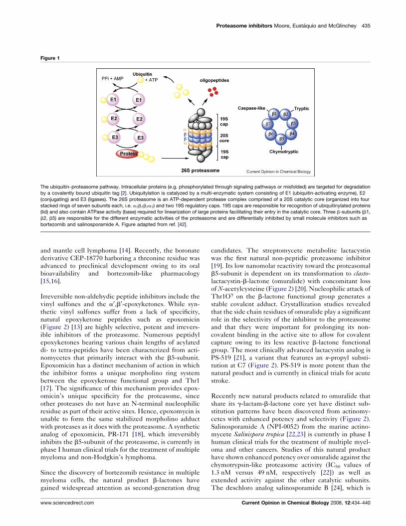
and E3 ubiquitinating enzymes (Figure 1). Upon recog-
nition, unfolding and transfer of the de-ubiquitinated
target protein by the 19S regulatory cap into the interior
of the cylindrical 20S proteasome core particle, protein
degradation is facilitated by catalytic b-subunits having
nucleophilic N-terminal threonine (Thr1) residues.
Although eukaryotic 20S proteasomes harbor seven
Current Opinion in Chemical Biology 2008, 12:434–440
different b-subunits in their twofold symmetrical
a7b7b7a7 stacked complexes, only three b-subunits per
b-ring [subunits b1 (caspase-like), b2 (trypsin-like), and
b5 (chymotrypsin-like)] are proteolytically active
(Figure 1). The disruption of this degradative process
with small molecule inhibitors against one or more cat-
alytic b-subunit has implications in a number of human
diseases such as cancer, inflammation, and ischemic
stroke and has exposed the proteasome as an important
therapeutic target [4–7].
Chemical classes of proteasome inhibitorsThe nucleophilic character of the proteasome is governed
by the active site Thr1 residue of each catalytic b-subunit
in which the side chain hydroxyl group reacts with pep-
tide bonds of substrates as well as with electrophilic
functional groups of inhibitors. Selectivity is dictated
by the composition of the substrate binding pockets
(termed S1, S2, Sn and S10, S20, Sn0 depending on proxi-
mity to the active site), which differs in the three catalytic
b-subunits. A wide range of specific inhibitors has been
developed as mechanism-based synthetic peptidyl elec-
trophiles and natural products with IC50 values in the low
nanomolar range [8��].
Tripeptide aldehydes such as the calpain inhibitor I (Ac-
Leu-Leu-nLeu-al) and actinomycete natural product leu-
peptin (Ac-Leu-Leu-Arg-al) were the first class of inhibi-
tors to probe the biochemistry of the proteasome active
sites [9] and reveal that the proteasome belongs to a novel
class of N-terminal threonine proteases [10]. While the
peptide aldehydes form reversible covalent hemiacetal
intermediates with Thr1Og primarily of the b5-subunit,
their moderate reactivity (low micromolar) and lack of invivo specificity (also inhibit serine and cysteine proteases)
led to the exploitation of other binding head groups with
greater potency and selectivity. Diverse functional groups
such as vinyl sulfones [11], boronates [12] and natural
product-based a0,b0-epoxyketones [13] were explored
and provided a number of important leads.
Peptide boronates, which are aldehyde surrogates, are
much more reactive with subnanomolar potency and are
selective towards the proteasome over common proteases
[12]. Owing to their high selectivity, potency and low
dissociation rates, the peptide boronates are ideal candi-
dates for drug development, and many analogs have been
prepared and evaluated. The dipeptide boronic acid
bortezomib (Velcade1, PS-341) (Figure 2), a reversible
inhibitor of the b5-subunit, is the first in class proteasome
inhibitor approved by the US Food and Drug Adminis-
tration for the treatment of relapsed multiple myeloma
www.sciencedirect.com
Proteasome inhibitors Moore, Eustaquio and McGlinchey 435
Figure 1
The ubiquitin–proteasome pathway. Intracellular proteins (e.g. phosphorylated through signaling pathways or misfolded) are targeted for degradation
by a covalently bound ubiquitin tag [2]. Ubiquitylation is catalyzed by a multi-enzymatic system consisting of E1 (ubiquitin-activating enzyme), E2
(conjugating) and E3 (ligases). The 26S proteasome is an ATP-dependent protease complex comprised of a 20S catalytic core (organized into four
stacked rings of seven subunits each, i.e. a7b7b7a7) and two 19S regulatory caps. 19S caps are responsible for recognition of ubiquitinylated proteins
(lid) and also contain ATPase activity (base) required for linearization of large proteins facilitating their entry in the catalytic core. Three b-subunits (b1,
b2, b5) are responsible for the different enzymatic activities of the proteasome and are differentially inhibited by small molecule inhibitors such as
bortezomib and salinosporamide A. Figure adapted from ref. [42].
and mantle cell lymphoma [14]. Recently, the boronate
derivative CEP-18770 harboring a threonine residue was
advanced to preclinical development owing to its oral
bioavailability and bortezomib-like pharmacology
[15,16].
Irreversible non-aldehydic peptide inhibitors include the
vinyl sulfones and the a0,b0-epoxyketones. While syn-
thetic vinyl sulfones suffer from a lack of specificity,
natural epoxyketone peptides such as epoxomicin
(Figure 2) [13] are highly selective, potent and irrevers-
ible inhibitors of the proteasome. Numerous peptidyl
epoxyketones bearing various chain lengths of acylated
di- to tetra-peptides have been characterized from acti-
nomycetes that primarily interact with the b5-subunit.
Epoxomicin has a distinct mechanism of action in which
the inhibitor forms a unique morpholino ring system
between the epoxyketone functional group and Thr1
[17]. The significance of this mechanism provides epox-
omicin’s unique specificity for the proteasome, since
other proteases do not have an N-terminal nucleophilic
residue as part of their active sites. Hence, epoxomycin is
unable to form the same stabilized morpholino adduct
with proteases as it does with the proteasome. A synthetic
analog of epoxomicin, PR-171 [18], which irreversibly
inhibits the b5-subunit of the proteasome, is currently in
phase I human clinical trials for the treatment of multiple
myeloma and non-Hodgkin’s lymphoma.
Since the discovery of bortezomib resistance in multiple
myeloma cells, the natural product b-lactones have
gained widespread attention as second-generation drug
www.sciencedirect.com
candidates. The streptomycete metabolite lactacystin
was the first natural non-peptidic proteasome inhibitor
[19]. Its low nanomolar reactivity toward the proteasomal
b5-subunit is dependent on its transformation to clasto-
lactacystin-b-lactone (omuralide) with concomitant loss
of N-acetylcysteine (Figure 2) [20]. Nucleophilic attack of
Thr1Og on the b-lactone functional group generates a
stable covalent adduct. Crystallization studies revealed
that the side chain residues of omuralide play a significant
role in the selectivity of the inhibitor to the proteasome
and that they were important for prolonging its non-
covalent binding in the active site to allow for covalent
capture owing to its less reactive b-lactone functional
group. The most clinically advanced lactacystin analog is
PS-519 [21], a variant that features an n-propyl substi-
tution at C7 (Figure 2). PS-519 is more potent than the
natural product and is currently in clinical trials for acute
stroke.
Recently new natural products related to omuralide that
share its g-lactam-b-lactone core yet have distinct sub-
stitution patterns have been discovered from actinomy-
cetes with enhanced potency and selectivity (Figure 2).
Salinosporamide A (NPI-0052) from the marine actino-
mycete Salinispora tropica [22,23] is currently in phase I
human clinical trials for the treatment of multiple myel-
oma and other cancers. Studies of this natural product
have shown enhanced potency over omuralide against the
chymotrypsin-like proteasome activity (IC50 values of
1.3 nM versus 49 nM, respectively [22]) as well as
extended activity against the other catalytic subunits.
The deschloro analog salinosporamide B [24], which is
Current Opinion in Chemical Biology 2008, 12:434–440
436 Next-generation therapeutics
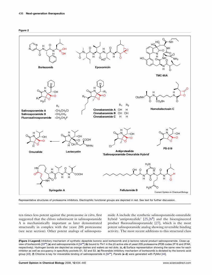
Figure 2
Representative structures of proteasome inhibitors. Electrophilic functional groups are depicted in red. See text for further discussion.
ten times less potent against the proteasome in vitro, first
suggested that the chloro substituent in salinosporamide
A is mechanistically important as later demonstrated
structurally in complex with the yeast 20S proteasome
(see next section). Other potent analogs of salinospora-
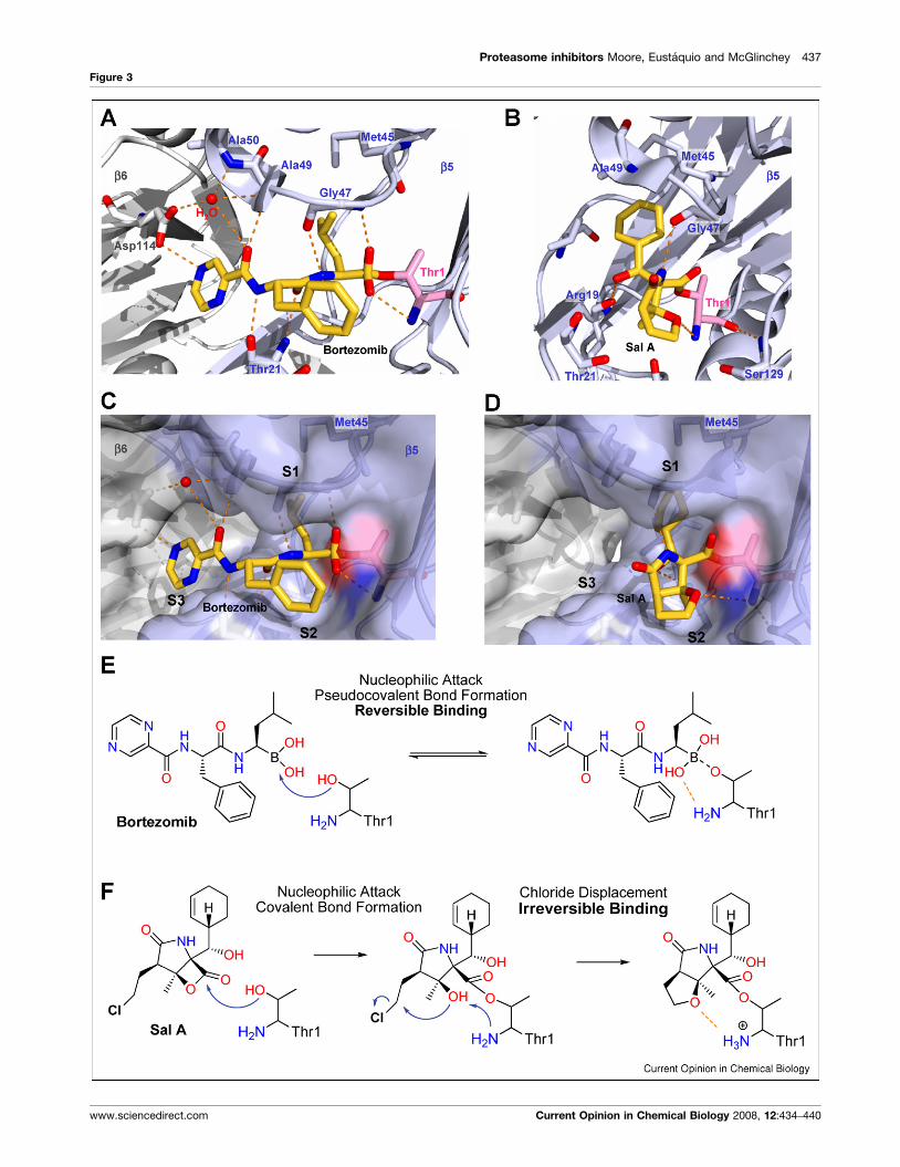
(Figure 3 Legend) Inhibitory mechanism of synthetic dipeptide boronic acid
view of bortezomib [33��] (a) and salinosporamide A [34��] (b) bound to Thr1 in
respectively). Hydrogen bonds are depicted as orange dashes and waters as
inhibitor as well as occupancy in specificity pockets S1, S2 and S3. (e) Reve
group [43]. (f) Chlorine is key for irreversible binding of salinosporamide A [3
Current Opinion in Chemical Biology 2008, 12:434–440
mide A include the synthetic salinosporamide-omuralide
hybrid ‘antiprotealide’ [25,26�] and the bioengineered
product fluorosalinosporamide [27], which is the most
potent salinoporamide analog showing reversible binding
activity. The most recent additions to this structural class
bortezomib and b-lactone natural product salinosporamide. Close-up
the b5 active site of yeast 20S proteasome (PDB codes 2F16 and 2FAK,
red dots. (c, d) Surface representation showing the same view for each
rsible inhibitory mechanism of bortezomib is dictated by the boronic acid
4��]. Panels (a–d) were generated with PyMol [44].
www.sciencedirect.com
Proteasome inhibitors Moore, Eustaquio and McGlinchey 437
Figure 3
www.sciencedirect.com Current Opinion in Chemical Biology 2008, 12:434–440
438 Next-generation therapeutics
are the cinnabaramides, which were isolated from a
terrestrial streptomycete [28]. These structural analogs,
which only differ from the salinosporamides in the C2
alkyl side chain, have comparable potency in vitro with
IC50 values in the low nanomolar range. It remains,
however, to be shown if the cinnabaramides have the
same anticancer properties as salinosporamide A.
Further proteasome inhibitors of the b-lactone family
include belactosines A and C (Figure 2) from Streptomycessp. UCK14 that selectively inhibit the b5-subunit of
the proteasome, with the modified homobelactosin
C derivative (Figure 2) having an IC50 in the low nano-
molar level [29]. Other natural proteasome inhibitors
include the TMC-95 family of cyclic peptides from
the fungus Apiospora montagnei [30], with TMC-95A
being the only natural product inhibitor to non-cova-
lently block all active sites of the proteasome selectively
and competitively in the low nanomolar range [31].
The majority of the most potent natural proteasome
inhibitors are derived from actinobacteria, which are
uncommon amongst prokaryotes to synthesize a 20S
proteasome complex. The simplified actinobacterial pro-
teasome is composed of identical a and b subunits with an
a7b7b7a7-stoichiometry and no regulatory caps reflective
of the absence of ubiquitin in bacteria. While the mech-
anism for self-resistance in these bacteria that produce
proteasome toxins has not yet been clarified, the recent
first biosynthetic gene cluster analysis of the natural
proteasome inhibitor salinosporamide A revealed an
associated b-subunit that may be involved with resistance
[32]. It will be intriguing to learn if other biosynthetic
gene clusters associated with actinomycete proteasome
inhibitors also harbor proteasome b-subunits, and if so,
whether this genetic signature may enable the discovery
of new inhibitor classes.
Molecular mechanism of actionHigh-resolution crystal structures of the 20S protea-
some (mainly from yeast) in complex with all of the
major inhibitors have been solved by Groll and co-
worker [8��]. These analyses illuminated their binding
mode and mechanism of action at the molecular level
and have been instrumental in the structure-based
design of new inhibitors. Most proteasome inhibitors
bind covalently to the catalytic Thr1 residue in the b5-
subunit with the exception of the cyclic peptide TMC-
95, which shows noncovalent binding in each catalytic
subunit. Recent crystal structures of the yeast 20S
proteasome with bound bortezomib [33��] and salinos-
poramide A [34��] have been reported and illustrate
some of the guiding principles in proteasome inhibition
(Figure 3).
As opposed to the reversible binding mode of bortezomib,
binding of salinosporamide A to the proteasome has been
Current Opinion in Chemical Biology 2008, 12:434–440
shown to be irreversible [35,36]. Moreover, bortezomib
and salinosporamide A differentially affect proteasome
activities, that is at low concentrations salinosporamide A
preferentially targets the chymotryptic (b5) and tryptic
(b2) while bortezomib affects chymotryptic and caspase-
like (b1) subunits [35].
The boronic acid moiety of bortezomib forms a (pseudo)-
covalent bond to the nucleophilic hydroxyl side chain of
Thr1. Further important interactions are summarized in
Figure 3a. The inhibitor occupies specificity pockets S1,
S2 and S3 (Figure 3c), which differ in charge and overall
architecture depending on the subunit in question. Selec-
tivity for the various proteasome active sites is controlled
by P1 (leucine boronic acid moiety) and P3 (pyrazine-2-
carboxyl group), while P2 (phenylalanine group) makes
no contacts with the protein so that S2 pockets in all active
sites can accept larger substituents. The leucine side
chain induces a fit to Met45 of b5 involved in key
proteasome–substrate interactions and the concerted
movements generated upon binding allow additional
hydrophobic contacts between P1 and S1. By contrast,
P1 does not interact with the larger S1 pocket in b2.
Furthermore, the S3 pocket of b2 fundamentally differs
from b5 explaining bortezomib’s lack of tryptic-like
inhibitory activity. In case of b1, Asp114 in S3
(Figure 3a) is replaced by a histidine preventing inter-
action with P3 and vindicating the lower affinity for the
caspase-like subunit [33��]. Figure 3e depicts bortezo-
mib’s binding mechanism.
As reported for omuralide, salinosporamide A is linked to
the Thr1-hydroxyl of proteasome active sites by an ester
bond with the carbonyl carbon of the b-lactone [34��](Figure 3b). However, while omuralide occupies only b5
subunits, salinosporamide A interacts with all catalytic
sites. The flexibility of Met45 (b5) affords accommo-
dation of larger P1 sites (isopropyl in omuralide, and
cyclohexenyl ring in salinosporamide A). Furthermore,
the bulkier P1 group in salinosporamide A allows for
additional hydrophobic interactions, helping explain at
least in part the enhanced potency of salinosporamide A
over omuralide [22,34��], and also the affinity to b2 which
presents a larger S1 pocket, consistent to salinosporamide
A’s inhibition of tryptic activity as opposed to bortezomib
[33��]. As shown in Figure 3d, the rather small b-lactone
inhibitor occupies only specificity pockets S1 and S2. Yet,
it represents an equipotent antitumor agent compared to
bortezomib [36].
As mentioned for bortezomib, the P2 group projects into
empty space. Therefore there is sufficient space to
accommodate larger side chains as exemplified by the
cinnabaramides [28]. Most important, P2 of b-lactone
inhibitors appears to be fundamental in determining if
binding is reversible or irreversible. Although omuralide
has been reported to bind to the proteasome irreversibly
www.sciencedirect.com

Proteasome inhibitors Moore, Eustaquio and McGlinchey 439
[20], based on a synthetic analog, binding of omuralide
and of the deschloro analog salinosporamide B should be
slowly reversible [34��]. After salinosporamide A becomes
covalently tethered to Thr1, the resulting C3 hydroxyl
displaces the C13 chlorine to yield an irreversibly bound
adduct, since the newly formed tetrahydrofuran ring (i)
blocks water attack on the ester bond preventing hydroly-
sis, (ii) engages C3O and circumvents reformation of the
b-lactone, and (iii) the resulting protonated state of
Thr1NH2 results in inactivation of its catalytic activity
(Figure 3b,f).
Therapeutic outlookProteasome inhibitors have been instrumental to our
fundamental understanding and appreciation of the
ubiquitin–proteasome system and are now rapidly
emerging as important new treatment options in cancer.
A new generation of proteasome inhibitors headed by
salinosporamide A and PR-171 are presently being
evaluated clinically and may offer alternative treatment
to patients intolerant or whose disease is refractory to
bortezomib. Comparative preclinical studies of these
irreversible inhibitors as single agents suggest reduced
toxicity and improved pathology [37,38], while combi-
nation therapy of salinosporamide A and bortezomib
affords synergistic anti-multiple myeloma activity at
reduced doses without the toxicity and resistance
attributed to bortezomib alone [39]. The landscape
of proteasome inhibitor-based therapeutics is quickly
evolving with promise in other diseases beyond clinical
oncology and represents an exciting example of transla-
tional medicine.
Primary resistance, as exemplified by bortezomid’s inef-
fectiveness against some solid tumors, as well as acquired
resistance may represent future hurdles for the wider
applicability of proteasome inhibitors [5]. Therefore,
further studies aimed to understand underlying mechan-
isms as well as the development of second-generation
drugs are imperative. In this context, new proteasome
inhibitors were reported during the production of this
article. The plant pathogen virulence factor syringolin A
from Pseudomonas syringae pv. syringae shows a novel
mechanism of covalent binding to the proteasome repre-
senting a new class of inhibitors containing a reactive a,b-
unsaturated carbonyl group that also includes glidobactin
A (Figure 2) [40��]. Moreover, the fungal peptide alde-
hyde fellutamide B, a known inducer of nerve growth
factor (NGF), was reported to inhibit the proteasome
[41��]. The authors also show that other proteasome
inhibitors induce production and secretion of NFG,
suggesting that targeting the proteasome may aid in
the treatment of neurodegenerative diseases. Together,
these recent additions provide further examples of pro-
teasome inhibition in nature as well as emphasize the vast
therapeutic potential of small molecule proteasome
inhibitors.
www.sciencedirect.com
AcknowledgementsThis work was supported by a grant from the National Institutes of Health(CA127622 to B.S.M.). A.S.E. is a Tularik postdoctoral fellow of the LifeSciences Research Foundation.
References and recommended readingPapers of particular interest, published within the period of review,have been highlighted as:
� of special interest�� of outstanding interest
1. Goldberg AL: Protein degradation and protection againstmisfolded or damaged proteins. Nature 2003, 426:895-899.
2. Adams J: The proteasome: a suitable antineoplastic target. NatRev Cancer 2004, 4:349-360.
3.�
Goldberg AL: Functions of the proteasome: from proteindegradation and immune surveillance to cancer therapy.Biochem Soc Trans 2007, 35:12-17.
This refreshing review article by Alfred Goldberg shares his personalinsight over the past 40 years on the discovery, function and clinicalpromise of the proteasome.
4. Orlowski RZ, Zeger EL: Targeting the proteasome as atherapeutic strategy against haematological malignancies.Expert Opin Invest Drugs 2006, 15:117-130.
5. Orlowski RZ, Kuhn DJ: Proteasome inhibitors in cancer therapy:lessons from the first decade. Clin Cancer Res 2008,14:1649-1657.
6. Zavrski I, Kleeberg L, Kaiser M, Fleissner C, Heider J, Sterz J,Jakob C, Sezer O: Proteasome as an emerging therapeutictarget in cancer. Curr Pharm Design 2007, 13:471-485.
7. Meiners S, Ludwig A, Stangl V, Stangl K: Proteasome inhibitors:poisons and remedies. Med Res Rev 2008, 28:309-327.
8.��
Borissenko L, Groll M: 20S proteasome and its inhibitors:crystallographic knowledge for drug development. Chem Rev2007, 107:687-717.
This review article provides a comprehensive account of the proteasomeand its inhibitors based on a structural perspective.
9. Vinitsky A, Michaud C, Powers JC, Orlowski M: Inhibition of thechymotrypsin-like activity of the pituitary multicatalyticproteinase complex. Biochemistry 1992, 31:9421-9428.
10. Lowe J, Stock D, Jap B, Zwickl P, Baumeister W, Huber R: Crystalstructure of the 20S proteasome from the archaeon T.acidophilium at 3.4 A resolution. Science 1995, 268:533-539.
11. Palmer JT, Rasnick D, Klaus JL, Bromme D: Vinyl sulfones asmechanism-based cysteine protease inhibitors. J Med Chem1995, 38:3193-3196.
12. Adams J, Behnke M, Chen S, Cruickshank AA, Dick LR, Grenier L,Klunder JM, Ma YT, Plamondon L, Stein RL: Potent and selectiveinhibitors of the proteasome: dipeptidyl boronic acids. BioorgMed Chem Lett 1998, 8:333-338.
13. Meng L, Mohan R, Kwok BH, Elofsson M, Sin N, Crews CM:Epoxomicin, a potent and selective proteasome inhibitor,exhibits in vivo anti-inflammatory activity. Proc Natl Acad Sci US A 1999, 96:10403-10408.
14. Williamson MJ, Blank JL, Bruzzese FJ, Cao Y, Daniels JS, Dick LR,Labutti J, Mazzola AM, Patil AD, Reimer CL et al.: Comparison ofbiochemical and biological effects of ML858 (salinosporamideA) and bortezomib. Mol Cancer Ther 2006, 5:3052-3061.
15. Dorsey BD, Iqbal M, Chatterjee S, Menta E, Bernardini R,Bernareggi A, Cassara PG, D’Arasmo G, Ferretti E, De Munari Set al.: Discovery of a potent, selective, and orally activeproteasome inhibitor for the treatment of cancer. J Med Chem2008, 51:1068-1072.
16. Piva R, Ruggeri B, Williams M, Costa G, Tamagno I, Ferrero D,Giai V, Coscia M, Peola S, Massaia M et al.: CEP-18770: a novel,orally active proteasome inhibitor with a tumor-selectivepharmacologic profile competitive with bortezomib. Blood2008, 111:2765-2775.
Current Opinion in Chemical Biology 2008, 12:434–440
440 Next-generation therapeutics
17. Groll M, Kim KB, Kairies N, Huber R, Crews CM: Crystal structureof epoxomycin:20S proteasome reveals a molecular basis forselectivity of a0,b0-epoxyketone proteasome inhibitors. J AmChem Soc 2000, 122:1237-1238.
18. Demo SD, Kirk CJ, Aujay MA, Buchholz TJ, Dajee M, Ho MN,Jiang J, Laidig GJ, Lewis ER, Parlati F et al.: Antitumor activity ofPR-171, a novel irreversible inhibitor of the proteasome.Cancer Res 2007, 67:6383-6391.
19. Omura S, Matsuzaki K, Fujimoto T, Kosuge K, Furuya T, Fujita S,Nakagawa A: Structure of lactacystin, a new microbialmetabolite which induces differentiation of neuroblastomacells. J Antibiot 1991, 44:117-118.
20. Fenteany G, Standaert RF, Lane WS, Choi S, Corey EJ,Schreiber SL: Inhibition of proteasome activities and subunit-specific amino-terminal threonine modification by lactacystin.Science 1995, 268:726-731.
21. Elliott PJ, Zollner TM, Boehncke WH: Proteasome inhibition:a new anti-inflammatory strategy. J Mol Med 2003, 81:235-245.
22. Feling RH, Buchanan GO, Mincer TJ, Kauffman CA, Jensen PR,Fenical W: Salinosporamide A, an antitumor proteasomeinhibitor from a novel microbial source, a marine bacteriumof the new genus Salinospora. Angew Chem Int Ed 2003,115:369-371.
23. Macherla VR, Mitchell SS, Manam RR, Reed KA, Chao TH,Nicholson B, Deyanat-Yazdi G, Mai B, Jensen PR, Fenical W et al.:Structure–activity relationship studies of salinosporamide A(NPI-0052), a novel marine derived proteasome inhibitor. J MedChem 2005, 48:3684-3687.
24. Williams PG, Buchanan GO, Feling RH, Kauffman CA, Jensen PR,Fenical W: New cytotoxic salinosporamides from the marineactinomycete Salinispora tropica. J Org Chem 2005,70:6196-6203.
25. Reddy LR, Fournier JF, Reddy BVS, Corey EJ: An efficient,stereocontrolled synthesis of a potent omuralide-salinosporinhybrid for selective proteasome inhibition. J Am Chem Soc2005, 127:8974-8976.
26.�
McGlinchey RP, Nett M, Eustaquio AS, Asolkar RN, Fenical W,Moore BS: Engineered biosynthesis of antiprotealide and otherunnatural salinosporamide proteasome inhibitors. J Am ChemSoc 2008, 130:7822-7823.
Together with reference [27], this paper describes the engineered bio-synthesis of diverse salinosporamide-based proteasome inhibitors.
27. Eustaquio AS, Moore BS: Mutasynthesis offluorosalinosporamide, a potent and reversible inhibitor of theproteasome. Angew Chem Int Ed 2008, 47:3936-3938.
28. Stadler M, Bitzer J, Mayer-Bartschmid A, Muller H, Benet-Buchholz J, Gantner F, Tichy H-V, Reinemer P, Baco KB:Cinnabaramides A-G: analogues of lactacystin andsalinosporamide from a terrestrial streptomycete. J Nat Prod2007, 70:246-252.
29. Groll M, Larionov OV, Huber R, de Meijere A: Inhibitor-bindingmode of homobelactosin C to proteasomes: New insights intoclass I MHC ligand generation. Proc Natl Acad Sci U S A 2006,103:4576-4579.
30. Koguchi Y, Kohno J, Nishio M, Takahashi K, Okuda T, Ohnuki T,Komatsubara S: TMC-95A, B, C, and D, novel proteasomeinhibitors produced by Apiospora montagnei Sacc. TC 1093.Taxonomy, production, isolation and biological activities. JAntibiot 2000, 53:105-109.
31. Groll M, Gotz M, Kaiser M, Weyher E, Moroder L: TMC-95-basedinhibitor design provides evidence for the catalytic versatilityof the proteasome. Chem Biol 2006, 13:607-614.
Current Opinion in Chemical Biology 2008, 12:434–440
32. Udwary DW, Zeigler L, Asolkar RN, Singan V, Lapidus A, Fenical W,Jensen PR, Moore BS: Genome sequencing reveals complexsecondary metabolome in the marine actinomycete Salinisporatropica. Proc Natl Acad Sci U S A 2007, 104:10376-10381.
33.��
Groll M, Berkers CR, Ploegh HL, Ovaa H: Crystal structure of theboronic acid-based proteasome inhibitor bortezomib incomplex with the yeast 20S proteasome. Structure 2006,14:451-456.
This report reveals specificity and binding mode of marketed bortezomib(Velcade1) at the molecular level. Knowledge gained herein could beused for the rational design of new agents.
34.��
Groll M, Huber R, Potts BCM: Crystal structures ofsalinosporamide A (NPI-0052) and B (NPI-0047) in complexwith the 20S proteasome reveal important consequences ofbeta-lactone ring opening and a mechanism for irreversiblebinding. J Am Chem Soc 2006, 128:5136-5141.
Likewise, the molecular basis of proteasome inhibition by salinospora-mide A, currently in human clinical trials, is discussed.
35. Chauhan D, Catley L, Li GL, Podar K, Hideshima T, Velankar M,Mitsiades C, Mitsiades N, Yasui H, Letai A et al.: A novel orallyactive proteasome inhibitor induces apoptosis in multiplemyeloma cells with mechanisms distinct from Bortezomib.Cancer Cell 2005, 8:407-419.
36. Chauhan D, Hideshima T, Anderson KC: A novel proteasomeinhibitor NPI-0052 as an anticancer therapy. Br J Cancer 2006,95:961-965.
37. Ahn KS, Sethi G, Chao TH, Neuteboom STC, Chaturvedi MM,Palladino MA, Younes A, Aggarwal BB: Salinosporamide A (NPI-0052) potentiates apoptosis, suppresses osteoclastogenesis,and inhibits invasion through down-modulation of NF-kappaB-regulated gene products. Blood 2007, 110:2286-2295.
38. Miller CP, Ban K, Dujka ME, McConkey DJ, Munsell M,Palladino MA, Chandra J: NPI-0052, a novel proteasomeinhibitor, induces caspase-8 and ROS-dependent apoptosisalone and in combination with HDAC inhibitors in leukemiccells. Blood 2007, 110:267-277.
39. Chauhan D, Singh A, Brahmandam M, Podar K, Hideshima T,Richardson P, Munshi N, Palladino MA, Anderson KC:Combination of proteasome inhibitors bortezomib and NPI-0052 trigger in vivo synergistic cytotoxicity in multiplemyeloma. Blood 2008, 111:1654-1664.
40.��
Groll M, Schellenberg B, Bachmann AS, Archer CR, Huber R,Powell TK, Lindow S, Kaiser M, Dudler R: A plant pathogenvirulence factor inhibits the eukaryotic proteasome by a novelmechanism. Nature 2008, 452:755-758.
A new mechanism of proteasome inhibition involving the virulence factorsyringolin A from the plant pathogen Pseudomonas syringie was recentlydescribed.
41.��
Hines J, Groll M, Fahnestock M, Crews CM: Proteasomeinhibition by fellutamide A induces nerve growth factorsynthesis. Chem Biol 2008, 15:501-512.
The discovery that fellutamide induces nerve growth factor secretion by amechanism involving proteasome inhibition suggests new therapeuticapplications by these inhibitors.
42. Landis-Piwowar KR, Milacic V, Chen D, Yang H, Zhao Y, Chan TH,Yan B, Dou QP: The proteasome as a potential target for novelanticancer drugs and chemosensitizers. Drug Resist Updat2006, 9:263-273.
43. Tsukamoto S, Yokosawa H: Natural products inhibiting theubiquitin–proteasome proteolytic pathway, a target for drugdevelopment. Curr Med Chem 2006, 13:745-754.
44. DeLano WL: The PyMOL Molecular Graphics System. http://www.pymol.org. 2002.
www.sciencedirect.com