Ubiquitin-Proteasome System Modulates Platelet Function
190
Cleveland State University Cleveland State University EngagedScholarship@CSU EngagedScholarship@CSU ETD Archive 2014 Ubiquitin-Proteasome System Modulates Platelet Function Ubiquitin-Proteasome System Modulates Platelet Function Nilaksh Gupta Cleveland State University Follow this and additional works at: https://engagedscholarship.csuohio.edu/etdarchive Part of the Biology Commons How does access to this work benefit you? Let us know! How does access to this work benefit you? Let us know! Recommended Citation Recommended Citation Gupta, Nilaksh, "Ubiquitin-Proteasome System Modulates Platelet Function" (2014). ETD Archive. 115. https://engagedscholarship.csuohio.edu/etdarchive/115 This Dissertation is brought to you for free and open access by EngagedScholarship@CSU. It has been accepted for inclusion in ETD Archive by an authorized administrator of EngagedScholarship@CSU. For more information, please contact [email protected].
Transcript of Ubiquitin-Proteasome System Modulates Platelet Function

Cleveland State University Cleveland State University
EngagedScholarship@CSU EngagedScholarship@CSU
ETD Archive
2014
Ubiquitin-Proteasome System Modulates Platelet Function Ubiquitin-Proteasome System Modulates Platelet Function
Nilaksh Gupta Cleveland State University
Follow this and additional works at: https://engagedscholarship.csuohio.edu/etdarchive
Part of the Biology Commons
How does access to this work benefit you? Let us know! How does access to this work benefit you? Let us know!
Recommended Citation Recommended Citation Gupta, Nilaksh, "Ubiquitin-Proteasome System Modulates Platelet Function" (2014). ETD Archive. 115. https://engagedscholarship.csuohio.edu/etdarchive/115
This Dissertation is brought to you for free and open access by EngagedScholarship@CSU. It has been accepted for inclusion in ETD Archive by an authorized administrator of EngagedScholarship@CSU. For more information, please contact [email protected].
UBIQUITIN-PROTEASOME SYSTEM MODULATES
PLATELET FUNCTION
NILAKSH GUPTA
Bachelor in Human Genetics (Honours)
Guru Nanak Dev University, Amritsar
April, 2000
Master in Human Genetics (Honours)
Guru Nanak Dev University, Amritsar
April, 2002
Submitted in partial fulfillment of requirements for the degree
DOCTOR OF PHILOSOPHY IN REGULATORY BIOLOGY
at the
CLEVELAND STATE UNIVERSITY
June, 2014
This dissertation has been approved for the
Department of Biological, Geological, and Environmental Sciences
And
CLEVELAND STATE UNIVERSITY
College of Graduate Studies by
_______________________________________________Date:____________
Dr. Thomas M. McIntyre Lerner Research Institute, Cleveland Clinic Major Advisor _______________________________________________Date:____________
Dr. Crystal M. Weyman, BGES, Cleveland State University Advisory Committee Member _______________________________________________Date:____________
Dr. Thomas Egelhoff Lerner Research Institute, Cleveland Clinic Advisory Committee Member _______________________________________________Date:____________
Dr. Jun Qin, Lerner Research Institute, Cleveland Clinic Advisory Committee Member _______________________________________________Date:____________
Dr. Barsanjit Mazumder, BGES, Cleveland State University Internal Examiner _______________________________________________Date:____________
Dr. Saurav Misra Lerner Research Institute, Cleveland Clinic, External Examiner

iii
DEDICATION
This thesis is dedicated to My father Subash Gupta,
My mother Rita Gupta, My mother-in-law Meenu Sharma,
My wife Arishya Sharma, and My son Aryan Gupta.
A very special thanks to all of you for your constant love,
support, encouragement and
belief in me.

iv
ACKNOWLEDGMENTS
First I would like to thank my mentor Dr. McIntyre. He has been a great mentor
in every aspect. I am truly grateful for his guidance in all aspects of my graduate
studies and his willingness to always help me and discuss things whenever it was
needed. He has encouraged me to always identify and pursue important questions
and has demonstrated to me how to tackle scientific questions in a systematic way. His
love for teaching and passion for science have been an inspiration to me
I would like to thank my committee members— Dr. Crystal Weyman, Dr.
Thomas Egelhoff and Dr. Jun Qin. Their feedback during my studies and for their time
in reviewing my progress during my Ph.D. years is greatly appreciated.
I would also like to thank my academic teachers at CSU— Dr. Mazumder, Dr.
Weyman, Dr. Komar, Dr. Shukla, Dr. Li and Dr. Börner and I want to extend my gratitude to
the administrative personnel at CSU, especially Monica and Carolee Pichler, for being so
helpful.
I would like to thank all the current and past members of McIntyre lab. They
were crucial for their technical assistance, guidance and support during my training.
Specifically, I would like to thank Latch and Rui for teaching me the techniques
needed for the completion of this project. I would like to thank Sowmya and Padmini
for assistance with experiments and moral support. I would also like to thank Dr. Wei Li
for the in vivo mouse experiments.

v
I would also like to thank my friends, Navneet, Manav, Arvind, Raminder,
Abhishek, Shreyas, Prateek, Rashim and Vikram. I would just like to thank you guys for
keeping my feet on the ground, being supportive, the nights out, holidays and general
good times.
Finally, I would like to thank my family. I would like to thank my mother and
father for giving me the best chance possible to reach my goals throughout my
childhood and into adulthood. I would like to thank my mother-in-law for her
blessings and constant support. Last, but by no means least, I would like to thank my
wife and son. To Arishya, for supporting me throughout this whole process and giving
me the drive to keep going when things got tough. To Aryan, for being a source of
inspiration and love and for being my bundle of joy. I love you guys...

vi
ABSTRACT OF THE DISSERTATION
Ubiquitin-proteasome system modulates
platelet function
By Nilaksh Gupta
Atherothrombotic diseases are responsible for more than 25% of all
deaths worldwide. Anti-platelet drugs are the mainstay treatment because of the
direct involvement of platelets in the initiation and propagation of
thrombosis. However, the currently available anti-platelet drugs, such as
antagonists of platelet receptors or of effector systems participating in platelet
activation, have their own limitations. A new mode of affecting platelet reactivity
may prove to offer unique advantages in a host of clinical settings.
Proteasome inhibitors are in clinical use to treat hematologic cancers, but
also reduce thrombosis. Whether the proteasome participates in platelet
activation or function is opaque since little is known of the proteasome in these
terminally differentiated cells. Therefore, I investigated the role of proteasome-
mediated proteolysis on platelet function (AIM 1). I find platelets displayed all
vii
three primary proteasome protease activities, which MG132 and bortezomib
(Velcade®) inhibited. Proteasome substrates are marked by ubiquitin, and
platelets contained a functional ubiquitination system that modified the proteome
by mono- and poly-ubiquitination. Proteasome inhibition suppressed platelet
aggregation by low thrombin concentrations and ristocetin-stimulated
agglutination through the GPIb-IX-V complex. Proteasome inhibitor MG132
reduced stimulated spreading and clot retraction. The effects of proteasome
inhibitors were not confined to a single receptor as MG132 and bortezomib
suppressed thrombin-, ADP-, and LPS-stimulated microparticle shedding.
Systemic MG132 strongly suppressed formation of occlusive, platelet-rich
thrombi in FeCl3-damaged carotid arteries. Transfusion of platelets treated ex
vivo with MG132 and washed prior to transfusion into thrombocytopenic mice
also reduced carotid artery thrombosis.
The inhibition of the proteasome quells the ultimate step of ubiquitin-
mediated protein degradation pathway. Proteasome-mediated degradation is the
final common step, however, multiple layers of regulated processes are involved
upstream of this degradative machine that determines whether to target a protein
for degradation or not. Platelets express a number of deubiquitinases that
reverse protein ubiquitination, but their potential function in platelets is unstudied.
So, I investigated the role of deubiquitinase enzymes in modulating platelet
reactivity (Aim 2). I show platelets express deubiquitinase activity and specific
inhibitor of the proteasome-associated deubiquitinases (b-AP15) as well as
general deubiquitinase inhibitors (PYR41 and PR619) increased mono- and poly-
viii
ubiquitination of platelet proteins. Deubiquitinase inhibition strongly
suppressed αIIbβ3 activation, degranulation, platelet aggregation and adhesion/
spreading in response to diverse platelet agonists. This inhibition also blocked
downstream signaling from platelet receptors by inhibiting agonist-induced Akt
phosphorylation and intracellular calcium release. Inhibition of platelet
deubiquitinase activity strongly suppressed formation of platelet-rich occlusive
thrombi in FeCl3-damaged murine carotid arteries and prevented in
vitro thrombus formation on collagen-coated surfaces at high shear rates.
Overall, this study uncovers the role of ubiquitin-proteasome system in
regulating platelet reactivity and thrombosis.
ix
Contents
ABSTRACT OF THE DISSERTATION .................................................................................................. vi
CHAPTER I ........................................................................................................................................ 1
Introduction ..................................................................................................................................... 1
1.1 Overview of platelet functions and roles ............................................................................... 1
1.1.1 Role of platelets in hemostasis and thrombosis ............................................................. 3
1.1.1.1 Blood coagulation and thrombin generation........................................................... 8
1.1.1.2 Spatial and temporal heterogeneity within growing thrombus in vivo- a revised
model of thrombus development ........................................................................................ 8
1.1.2 Platelet morphology and ultrastructure ....................................................................... 10
1.1.2.1 Plasma membrane (PM) and open canalicular system (OCS) ................................ 10
1.1.2.1.1 Platelet derived microparticles (PMPs) .......................................................... 12
1.1.2.2 Dense Tubular System (DTS) .................................................................................. 14
1.1.2.3 Platelet Cytoskeleton ............................................................................................. 14
1.1.2.4 Platelet Secretary Granules ................................................................................... 18
1.1.3 Platelet Receptors ......................................................................................................... 19
1.1.3.1 Adhesive receptors ................................................................................................ 19
1.1.3.1.1 Integrins .......................................................................................................... 19
1.1.3.1.2 Glycoprotein Ib-IX-V ........................................................................................ 22
1.1.3.1.3 Collagen Receptors ......................................................................................... 24
1.1.3.2 G- Protein Coupled Receptors ............................................................................... 26
1.1.3.2.1 Thrombin Receptors ....................................................................................... 26
1.1.3.2.1.1 PARs ......................................................................................................... 26
1.1.3.2.1.2 ADP Receptors ......................................................................................... 28
1.1.4 Current anti-platelet therapies and their limitations ................................................... 29
1.2 Overview of ubiquitin-proteasome system ......................................................................... 40

x
1.2.1 Ubiquitin Conjugation System ...................................................................................... 42
1.2.2 Ubiquitin Code .............................................................................................................. 46
1.2.3 The proteasome- a degradation nanomachine ............................................................ 50
1.2.3.1 20S catalytic core particle ...................................................................................... 50
1.2.4 Deubiquitination ........................................................................................................... 54
1.2.5 Targeting UPS ................................................................................................................ 58
CHAPTER II ..................................................................................................................................... 60
Proteasome proteolysis supports stimulated platelet function and thrombosis .......................... 60
2.1 Introduction ......................................................................................................................... 60
2.2 Materials and Methods ........................................................................................................ 63
2.2.1 Chemicals and reagents ................................................................................................ 63
2.2.2 Platelet preparation ...................................................................................................... 64
2.2.3 Proteasome function .................................................................................................... 64
2.2.4 In vivo thrombosis ......................................................................................................... 65
2.2.5 Platelet transfusion and murine carotid artery thrombosis assay ............................... 66
2.2.6 p53 ubiquitination......................................................................................................... 67
2.2.7 Western blotting and liquid chromatography-mass spectrometry .............................. 67
2.2.8 Total internal reflection microscopy (TIRF) microscopy ............................................... 69
2.2.9 Microparticle isolation and quantitation ...................................................................... 70
2.2.10 Peptidyl activity of tissue factor (TF) on microparticles ............................................. 70
2.2.11 Clot retraction ............................................................................................................. 71
2.2.12 Aggregation ................................................................................................................. 71
2.2.13 Flow cytometry ........................................................................................................... 72
2.2.14 Expression of data and statistics ................................................................................. 72
2.3 Results .................................................................................................................................. 72
2.3.1 The platelet proteasome aids occlusive thrombosis .................................................... 72
2.3.2 Platelets contain a stimulatable ubquitination system ................................................ 77
2.3.3 Filamin A is ubiquitinated, and truncated by the proteasome ..................................... 81
2.3.4 Proteasome inhibition reduces cytoskeleton-dependent functions ............................ 85
2.3.5 Proteasome inhibition selectively reduces aggregation stimulated by low
concentrations of thrombin ................................................................................................... 89
xi
2.4 Discussion............................................................................................................................. 93
2.5 Chapter summary ................................................................................................................ 97
Chapter III ....................................................................................................................................... 98
Unmasking platelet reactivity through deubiquitination .............................................................. 98
3.1 Introduction ......................................................................................................................... 98
3.2 Materials and Methods ...................................................................................................... 100
3.2.1 Chemicals and reagents .............................................................................................. 100
3.2.2 Platelet preparation .................................................................................................... 101
3.2.3 Western blotting ......................................................................................................... 101
3.2.4 Deubiquitination Assay and Ubiquitin chain disassembly .......................................... 102
3.2.5 In vivo thrombosis ....................................................................................................... 103
3.2.6 In vitro thrombus formation ....................................................................................... 104
3.2.7 Flow cytometry ........................................................................................................... 105
3.2.8 Total internal reflection fluorescence (TIRF) microscopy ........................................... 105
3.2.9 Aggregation ................................................................................................................. 106
3.2.10 Expression of data and statistics ............................................................................... 106
3.3 Results ................................................................................................................................ 106
3.3.1 Deubiquitination of the platelet proteome promotes thrombosis ............................ 106
3.3.2 Deubiquitinase inhibitors suppress platelet adhesion to collagen under shear flow 111
3.3.3 Stimulated homotypic platelet aggregation, adhesion, and spreading are suppressed
by deubiquitinase inhibitors ................................................................................................ 114
3.3.4 Agonist activation of glycoprotein αIIbβ3 is reduced by deubiquitinase inhibition ... 117
3.3.5 Inhibitors of platelet deubiquitinases suppressed ADP mediated αIIbβ3 activation and
degranulation ....................................................................................................................... 120
3.3.6 Proteasome associated deubiquitinases regulate platelet activation ........................ 123
3.3.7 Platelet deubiquitinases affect collagen mediated responses via the gpVI receptor 127
3.3.8 Inhibition of platelet deubiquitinases modulated platelet signaling responses
downstream of thrombin and collagen receptors ............................................................... 130
3.5 Discussion........................................................................................................................... 135
3.6 Chapter summary .............................................................................................................. 140
Chapter IV .................................................................................................................................... 142
Ubiquitin-specific protease 7 (USP7) activity promotes platelet activation ................................ 142
xii
4.1 Introduction and Rationale ................................................................................................ 142
4.2 Results ................................................................................................................................ 144
4.3 Chapter summary .............................................................................................................. 148
Chapter V ..................................................................................................................................... 150
Conclusion .................................................................................................................................... 150
5.1 Summary and Conclusion .................................................................................................. 150
Figure 5.1 Pharmacologic inhibition of different components of UPS modulates platelet reactivity ...................................................................................................................................... 152
Bibliography ................................................................................................................................. 153

xiii
List of Abbreviations
ADP Adenosine diphosphate Akt AK-8 thymoma (or transformed)
APC Activated protein C
BSS Bernard Soulier Syndrome
cAMP Cyclic adenosine monophosphate
cGMP Cyclic guanosine monophosphate
CVD Cardiovascular disease
DAG 1,2-diacyl-glycerol
DIC Differential interference contrast
DMSO Dimethylsulfoxide
DTS Dense tubular system DUB Deubiquitinase
FlnA Filamin A
gp VI Glycoprotein VI
GPCR G-protein coupled receptor
GPIb-IX-V Glycoprotein Ib-IX-V
MI Myocardial infarction
NO Nitric oxide
OCS Open canalicular system
PAR Protease activated receptor
PGI2 Prostaglandin I2
PI3K Phosphoinositide 3-kinase
PIP2 Phosphatidylinositol 1, 4-bisphosphate
PIP3 Phosphatidylinositol 3,4,5-trisphosphate
PLCβ Phospholipase C beta
PLCγ2 Phospholipase C gamma 2
PRP Platelet rich plasma
TFPI Tissue factor pathway inhibitor
TIRF Total internal reflection fluorescence
TxA2 Thromboxane A2 Ub Ubiquitin
UBD Ubiquitin binding domain
UFD Ubiquitin fold domain
UPS Ubiquitin-proteasome system
αIIbβ3 Integrin alpha IIb beta 3

xiv
List of Figures
Figure 1.1 Vascular endothelium keeps platelets quiescent and
coagulation under check 4
Figure 1.2 Mechanism of platelet recruitment following plaque
rupture 7
Figure 1.3 Spatial and temporal heterogeniety of thrombus
development 9
Figure 1.4 Platelet ultrastructure 13
Figure 1.5 Structure of Filamin A (FlnA) 16
Figure 1.6 Platelet receptors. 21
Figure 1.7 Ubiquitin- proteasome system 34
Figure 1.8 The Ub code 48
Figure 1.9 The 26S proteasome 52
Figure 1.10 General functions of DUBs 57
Figure 2.1 Platelets express a functional proteasome that
contributes to occlusive thrombosis 75
Figure 2.2 Platelets contain a functional ubiquitination system 79
Figure 2.3 MG132 protects cytoskeletal protein cleavage 83
Figure 2.4 MG132 suppresses stimulated spreading, microparticle
shedding, and clot retraction 87
Figure 2.5 MG132 reduces aggregation in response to low dose
thrombin, and prevents GP1bα downregulation 91
Figure 3.1 Platelets express functional deubiquitinases
that promote thrombosis 109
Figure 3.2 Deubiquitinase inhibitors suppressed in vitro thrombosis 112
xv
Figure 3.3 Deubiqutinase inhibition reduced platelet response
to agonists 115
Figure 3.4 Inhibitors of platelet deubiquitinases suppressed
protease activated receptor (PAR) mediated αIIbβ3 activation
and degranulation 118
Figure 3.5 Deubiquitinase inhibitors diminished ADP induced
platelet αIIbβ3 activation and degranulation 121
Figure 3.6 Pharmacologic inhibition of proteasome-associated
deubiquitinase enymes, USP14 and UCHL5, reduced platelet
responsiveness to agonists 125
Figure 3.7 Deubiquitinase inhibitors affect collagen
responses via the gpVI receptor 128
Figure 3.8 Deubiquitinase inhibitors impaired platelet
signaling following agonist stimulation 133
Figure 4.1 USP7/HAUSP inhibition blocked collagen-stimulated
aggregation and adhesion under flow 146
Figure 5.1 Pharmacologic inhibition of different components of UPS
modulates platelet reactivity 152
List of Tables
Table 1.1 Antiplatelet agents in the clinic or in clinical trials 32
1
CHAPTER I
Introduction
1.1 Overview of platelet functions and roles
Thrombotic cardiovascular diseases (CVD), predominantly manifested as
myocardial infarction and ischemic stroke, remain the single most common cause
of death and disability in the developed world. CVD is responsible for ~30% of all
deaths worldwide (Alwan 2011), with arterial thrombosis as the main underlying
cause. In the United States alone, more than 2000 patients die each day as a
direct result of myocardial infarction and ischemic stroke caused by arterial
thrombosis (Go, Mozaffarian et al. 2014).
The initial trigger for arterial thrombosis, formation of occlusive thrombi
within the lumen of arteries (Engelmann and Massberg 2013), is the disruption of
atherosclerotic plaque that exposes its thrombogenic molecules to the arterial
circulation. When released, these molecules cause concomitant platelet
activation and fibrin formation leading to platelet-rich occlusive thrombi (Ruggeri
2002; Mackman 2008). This occurs often with fatal consequences e.g. thrombotic
occlusion of coronary artery that results in acute myocardial infarction and

2
occlusive thrombi in cerebral artery that results in ischemic stroke (Michelson
2010).
Platelets are small, disc-shaped anuclear cells that originate from bone
marrow precursors, megakaryocytes, and circulate in blood as sentinels of
vascular integrity. Platelet aggregation is a hallmark of hemostasis and a
contributing factor in pathologic thrombosis (Italiano, Lecine et al. 1999; Hartwig
and Italiano 2003; Ruggeri and Mendolicchio 2007). In the event of injury to the
vessel wall, platelets adhere to the exposed subendothelial proteins and
polymers to undergo rapid aggregation to form the hemostatic plug, the first
response to stop bleeding. However, platelets cannot distinguish between
physiological wounds in the vessel lining and pathogenic lesions on diseased
atherosclerotic vessels. This contributes to atherothrombosis ( thrombosis at
sites of atherosclerotic plaque disruption), which stops adequate blood supply to
downstream tissues or organs (Fuster, Badimon et al. 1992; Ruggeri 2000).
Owing to the vital role of platelets in athrothrombosis, anti-platelet agents,
e.g. aspirin and clopidogrel, are widely prescribed for individuals at high risk of
arterial thrombosis (Michelson 2010). However, the downside is an increased risk
of bleeding, which limits use of such agents (Fisher and Loscalzo 2011).
Identifying new therapeutic approaches that prevent thrombosis without
undermining underlying hemostasis seems imperative, but is restricted by our
insufficient understanding of molecular events that enhance platelet adhesion,
and activation and thus prevents identification of new targets to control platelet
3
deposition. Therefore, uncovering of new pathways and advancements in the
understanding of molecular events that regulate platelet deposition and activation
in thrombus propagation are sought and are likely to provide new therapeutic
targets and insight for the improvement of existing anti-platelet agents.
1.1.1 Role of platelets in hemostasis and thrombosis
Blood platelets are all-important for maintaining hemostasis, the cessation
of bleeding. The critical role of platelets in hemostasis is highlighted by disease
states that affect platelet number and/or function. For example, disorders
resulting in abnormally low platelet count, thrombocytopenia, or genetic
conditions that impair platelet function, such as Bernard-Soulier syndrome (BSS,
quantitative or qualitative defects in the platelet GPIb-IX-V complex) results in
increased bleeding (Lanza 2006; Cox, Price et al. 2011). The mammalian
hemostatic system preserves the integrity of the high pressure circulatory system
by balancing processes that maintain blood fluidity under physiological conditions
with those that prevent hemorrhage after penetrating injuries to the vessel wall.
Maintenance of blood fluidity rests on an intact vessel wall lined by endothelial
cells (Fig.1.1) that maintains a quiescent inert surface through a series of
regulatory pathways that prevent platelet activation and keeps the coagulation
cascade in check (Pinsky, Broekman et al. 2002; Michelson 2010; Lippi,
Franchini et al. 2011).
4
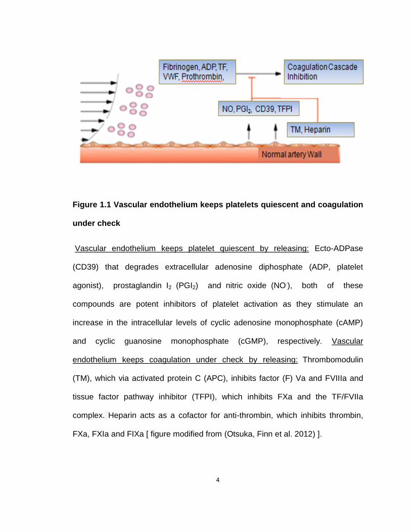
Figure 1.1 Vascular endothelium keeps platelets quiescent and coagulation
under check
Vascular endothelium keeps platelet quiescent by releasing: Ecto-ADPase
(CD39) that degrades extracellular adenosine diphosphate (ADP, platelet
agonist), prostaglandin I2 (PGI2) and nitric oxide (NO.), both of these
compounds are potent inhibitors of platelet activation as they stimulate an
increase in the intracellular levels of cyclic adenosine monophosphate (cAMP)
and cyclic guanosine monophosphate (cGMP), respectively. Vascular
endothelium keeps coagulation under check by releasing: Thrombomodulin
(TM), which via activated protein C (APC), inhibits factor (F) Va and FVIIIa and
tissue factor pathway inhibitor (TFPI), which inhibits FXa and the TF/FVIIa
complex. Heparin acts as a cofactor for anti-thrombin, which inhibits thrombin,
FXa, FXIa and FIXa [ figure modified from (Otsuka, Finn et al. 2012) ].
5
However, disruption or fissuring of an unstable atherosclerotic plaque
compromises endothelial integrity and triggers instant platelet recruitment and
activation to the exposed thrombogenic subendothelial matrix proteins (Fig.1.2)
(Schulz and Massberg 2012).
Recruitment of platelets at the site of the ruptured plaque is an immediate
response. This involves interactions between platelet-cell surface receptors and
exposed subendothelial matrix proteins including fibrillar collagen (type I and III)
and von Willebrand factor (VWF) (Savage, Almus-Jacobs et al. 1998; Massberg,
Gawaz et al. 2003; Denis and Wagner 2007). Platelet recruitment via individual
receptor-ligand interactions depends on the extent of vascular damage, as well
as prevailing rheological conditions. Importantly, shear rates up to 10,000 s-1
have been observed in the coronary artery occluded by 50% and even up to
50,000 s-1 in severe stenosis (Strony, Beaudoin et al. 1993; Mailhac, Badimon et
al. 1994) VS normal shear flow.
Under conditions of rapid blood flow, a feature of arterioles and stenotic
arteries, VWF recruits circulating platelets by reversible binding of its A1 domain
with multiple GPIb-IX-V receptors. Platelet GPIb-IX-V is essential for the initial
recruitment of platelets on the vascular lesion under high shear conditions
(Savage, Saldivar et al. 1996; Goto, Ikeda et al. 1998; Ruggeri 2001; Ruggeri
and Mendolicchio 2007). The VWF-GPIbα bond displays rapid on-off rates that
facilitates platelet translocation to the vessel wall, but it does not support stable
adhesion and requires the contribution of additional interactions between
6
platelets and other matrix macromolecules (Savage, Almus-Jacobs et al. 1998).
Stable platelet adhesion occurs through engagement of platelet glycoprotein
VI (gpVI) to collagen (Nieswandt, Brakebusch et al. 2001; Furie and Furie 2006)
that triggers downstream signaling leading to platelet activation.
This is critical for subsequent platelet activation as platelet stimulation
upon engagement with gpVI causes synthesis and release of secondary platelet
agonists, most notably thromboxane A2 (TxA2) and ADP, as well as locally
generated thrombin, further amplifying platelet activation in an autocrine or
paracrine fashion by stimulating their respective receptors on platelets (Share
1976; Offermanns 2006). Inside-out signaling from both gpVI and GPIb-IX-V
receptors induces a change in the activation status of β1 integrins {α2β1 [a
Collagen receptor], α5β1 [a fibronectin receptor] (Beumer, MJ et al. 1994), and
α6β1 [a laminin receptor] (Ruggeri 2009)} and β3 integrin, αIIbβ3 (binding VWF,
fibrinogen and fibronectin), from low affinity ligand binding states to high affinity
states (Shattil, Kim et al. 2010). Of all the integrins, αIIbβ3 is the dominant
integrin present on platelet surface, and high affinity adhesive interactions
between αIIbβ3 and adhesive proteins VWF, fibrinogen and fibronectin are
central to the irreversible platelet activation and thrombus propagation at the site
of ruptured plaque (Savage, Saldivar et al. 1996; Vinogradova, Velyvis et al.
2002).
7
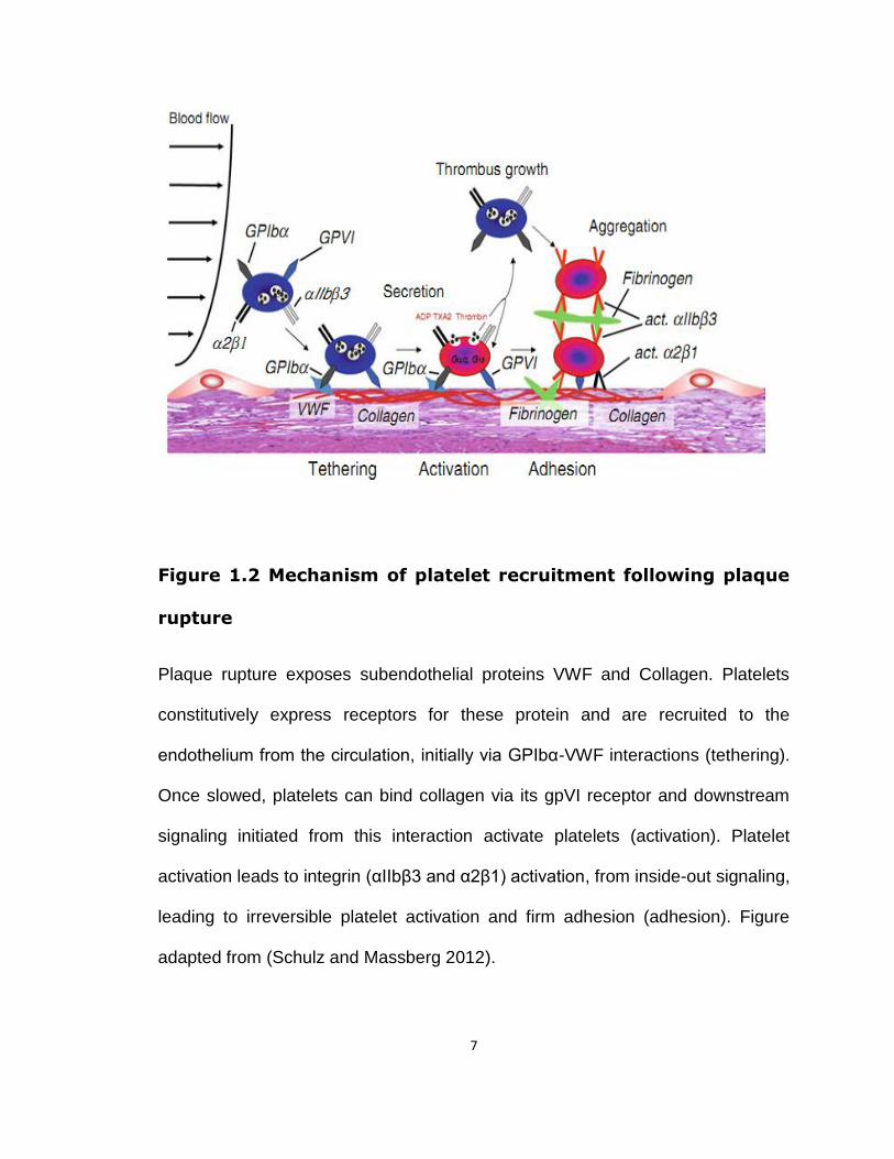
Figure 1.2 Mechanism of platelet recruitment following plaque
rupture
Plaque rupture exposes subendothelial proteins VWF and Collagen. Platelets
constitutively express receptors for these protein and are recruited to the
endothelium from the circulation, initially via GPIbα-VWF interactions (tethering).
Once slowed, platelets can bind collagen via its gpVI receptor and downstream
signaling initiated from this interaction activate platelets (activation). Platelet
activation leads to integrin (αIIbβ3 and α2β1) activation, from inside-out signaling,
leading to irreversible platelet activation and firm adhesion (adhesion). Figure
adapted from (Schulz and Massberg 2012).
8
1.1.1.1 Blood coagulation and thrombin generation
An important function of activated platelets is localization of subsequent
pro-coagulant events to the site of ruptured plaque. Platelet activation leads to
the rapid exposure of the membrane phospholipid phosphatidylserine (PS) from
inner membrane leaflet to platelet surface, which provides the anionic surface to
support the assembly of coagulation complexes on the platelet plasma
membrane. This is critical for localized thrombin generation and fibrin formation
(Jackson 2011). Thrombin is one of the most potent endogenous platelet
agonists, and plays a seminal role in thrombus formation under all rheological
conditions (Kahn, Zheng et al. 1998). Thrombin’s effect on human platelets is
mediated by surface G protein-coupled protease-activated receptor (PAR) 1 and
4 (Coughlin 1993; Coughlin 1999; Coughlin 2005). Thrombin-induced platelet
activation and fibrin generation is crucial for thrombus growth and stability, and
hence the drugs that inhibit thrombin generation or thrombin-induced platelet
activation, inhibit atherothrombosis (Michelson 2010).
1.1.1.2 Spatial and temporal heterogeneity within growing thrombus in
vivo- a revised model of thrombus development
Recent studies using high resolution intravital confocal microscopy,
genetically engineered mice, and flow chambers demonstrate that the above
description of occlusive thrombi formation may be more complicated than
previously anticipated. There studies also provide sufficient evidence of spatial
and temporal heterogeneity within the growing thrombi (Fig.1.3)

9
Figure 1.3 Spatial and temporal heterogeniety of thrombus development
Plaque rupture leads to the capture of platelets from circulation on to
thrombogenic endothelium matrix proteins and polymers. Subsequent platelet
activation via ADP and TxA2 leads to the formation of highly active platelet core.
Shear flow gradient generated at the site, due to narrowing of the lumen, causes
platelet tethering and formation of outer shell, composed mainly of discoid and
ready to be activated platelets. Adapted from (McFadyen and Jackson 2013).

10
(Nesbitt, Westein et al. 2009; Stalker, Traxler et al. 2013). Capturing thrombus
development in real-time reveals the heterogeneity in the growing thrombus,
which appears to have an inner core containing activated platelets and an outer
shell comprising of poorly activated discoid platelets. Elegant studies of blood
circulation in vivo and in flow chambers reveal that the growing platelet
aggregates were sensitive to prevailing rheological conditions i.e. to the flow
gradient established around the growing thrombus. This flow gradient results in
the recruitment of circulating discoid platelets to the growing thrombi through the
extension of their membrane, called tethers. These unactivated platelets in
thrombi are ready for activation dependent changes leading to thrombus
stabilization. (Nesbitt, Westein et al. 2009; Stalker, Traxler et al. 2013).
Importantly, accumulation of discoid platelets on developing thrombi by
shear gradients is not impeded by widely used anti-platelet agents aspirin and
clopidogrel or thrombin inhibitors (Nesbitt, Westein et al. 2009). Thus, improved
understanding of interaction of local conditions at sites of injury or plaque rupture
with the platelet signaling network offers a new context and rational into how
future anti-thrombotic drugs should be designed.
1.1.2 Platelet morphology and ultrastructure
1.1.2.1 Plasma membrane (PM) and open canalicular system (OCS)
Resting platelets are discoid, roughly 2-3 µm in diameter and their number
range from 150,000 to 450,000 cells/µL in human blood. About two-third of the
platelets exist in the circulation and the remaining cells are sequestered in the
11
spleen. Platelets have a life span of ~8-10 days (Hartwig and Italiano 2003; Thon
and Italiano 2012).
The platelet plasma membrane is a typical phospholipid bilayer, but also
contains an extensive series of complex indentations called the open canalicular
system (OCS) (Fig.1.4) (Behnke 1970; Frojmovic, Wong et al. 1992). The OCS is
a surface connected tubular system that connects the cytosol with the
surrounding medium and facilitates the quick release of secreted substances to
the extracellular environment (White and Clawson 1980; White and Krumwiede
1987). The OCS also constitutes an extensive membrane reservoir that upon
activation results in increased plasma membrane surface area that facilitates
fillopodia formation and platelet spreading (Thon and Italiano 2012). Embedded
in the platelet plasma membrane are numerous glycoprotein receptors (GP) and
integrins that are involved in the initial adhesion of platelets to the subendothelial
matrix and formation of a hemostatic plug (Phillips and Agin 1977; Kunicki 1989).
The negatively charged plasma membrane phospholipids (e.g.
phosphatidylserine and phosphatidylinositol) present in the inner leaflet of resting
platelets, are translocated and exposed on the platelet surface upon activation to
provide the surface for binding of proteins involved in coagulation (Heemskerk,
Bevers et al. 2002). The phosphatidylserine exposure on the surface of activated
platelets is crucial for normal hemostasis and this is evident from a rare bleeding
disorder, Scott syndrome, where the patients have reduced
12
phosphatidylserine exposure on surface and are unable to make microparticles
upon platelet activation (Toti, Satta et al. 1996; Heemskerk, Bevers et al. 2002).
1.1.2.1.1 Platelet derived microparticles (PMPs)
PMPs are small (0.1-1.0 µm in diameter) phospholipid vesicles, which
are shed from the cell membrane following platelet activation. PMPs are highly
pro-thrombotic because their surface contains exposed phosphatidylserine,
which provides the ideal catalytic surface that greatly expedites coagulation
(Sandberg, Bode et al. 1985; Sims, Faioni et al. 1988). Elevated numbers of
circulating PMPs have been detected during several diverse pathophysiological
processes such as acute myocardial infarction, peripheral artery disease,
cerebral malaria, rheumatoid arthritis, diabetes, multiple sclerosis, sepsis, and
ischemic stroke (Soriano, Jy et al. 2005; Faille, Combes et al. 2009; Boilard,
Nigrovic et al. 2010; Lannan, Phipps et al. 2014).
13
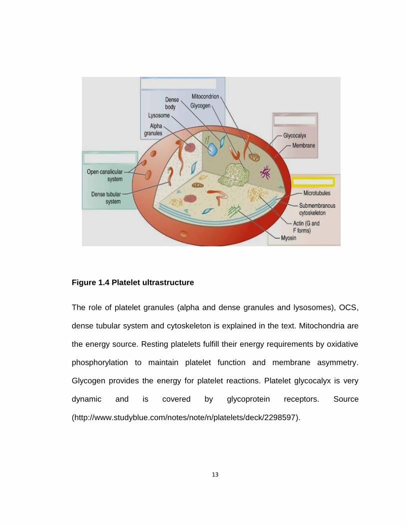
Figure 1.4 Platelet ultrastructure
The role of platelet granules (alpha and dense granules and lysosomes), OCS,
dense tubular system and cytoskeleton is explained in the text. Mitochondria are
the energy source. Resting platelets fulfill their energy requirements by oxidative
phosphorylation to maintain platelet function and membrane asymmetry.
Glycogen provides the energy for platelet reactions. Platelet glycocalyx is very
dynamic and is covered by glycoprotein receptors. Source
(http://www.studyblue.com/notes/note/n/platelets/deck/2298597).
14
1.1.2.2 Dense Tubular System (DTS)
Another membranous system present in platelets is termed as the DTS
that is a closed-channel network of residual smooth endoplasmic reticulum
(White 1972). The DTS sequesters ionized calcium and probably also a major
site of TxA2 and prostaglandin synthesis (Gerrard, White et al. 1976; Gerrard,
White et al. 1978). The release of intracellular calcium from DTS upon platelet
activation contributes to platelet degranulation, cytoskeletal reorganization and
redistribution of αIIbβ3.
1.1.2.3 Platelet Cytoskeleton
Platelets undergo rapid shape change, spreading, secretion and/or
aggregation upon activation. This transformation in stimulated platelets is largely
achieved by rapid cytoskeletal rearrangements within the platelet (Hartwig
2006). The platelet cytoskeleton consists of a marginal band consisting of a
microtubule coil, a cytoplasmic actin network, and cytoskeletal rim. Together
these structures support the platelet plasma membrane and confer shape to both
resting and activated platelets. This inherent capacity of platelets to rapidly
reorganize its cytoskeleton allows them to seal the leaks in the vasculature under
shear conditions (Hartwig 2006; Thon and Italiano 2012).
The marginal band lies beneath the plasma membrane and confers the
discoid shape to the resting platelet. Microtubule disassembly with drugs such as
nocodazole or colchicine or depolymerization induced by chilling at 4ºC, cause
15
platelets to lose their discoid shape and become round (White and Krivit 1967;
White 1968; Hartwig 2006).
The cytoskeletal rim is composed of actin, spectrin, talin, vinculin and actin
cross-linking proteins filamin and α-actinin (Fox 2001; Hartwig 2006). Filamins
are large cytoplasmic proteins that give mechanical stability to cells by cross-
linking actin into dynamic 3-dimensional structures (Pudas, Kiema et al. 2005).
There are three filamin isoforms: filamin A (FlnA), filamin B (FlnB) and filamin C
(FlnC), but platelets only express FlnA and FlnB. FlnA is an elongated 280-kDa
dimeric protein that self associates and is expressed ~10 fold in excess to FlnB
(Falet, Pollitt et al. 2010). Structurally, FlnA contains an actin-binding domain
(ABD) at the N-terminus, followed by 24 compact immunoglobulin-like repeats of
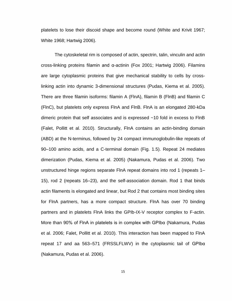
90–100 amino acids, and a C-terminal domain (Fig. 1.5). Repeat 24 mediates
dimerization (Pudas, Kiema et al. 2005) (Nakamura, Pudas et al. 2006). Two
unstructured hinge regions separate FlnA repeat domains into rod 1 (repeats 1–
15), rod 2 (repeats 16–23), and the self-association domain. Rod 1 that binds
actin filaments is elongated and linear, but Rod 2 that contains most binding sites
for FlnA partners, has a more compact structure. FlnA has over 70 binding
partners and in platelets FlnA links the GPIb-IX-V receptor complex to F-actin.
More than 90% of FlnA in platelets is in complex with GPIbα (Nakamura, Pudas
et al. 2006; Falet, Pollitt et al. 2010). This interaction has been mapped to FlnA
repeat 17 and aa 563–571 (FRSSLFLWV) in the cytoplasmic tail of GPIbα
(Nakamura, Pudas et al. 2006).
16
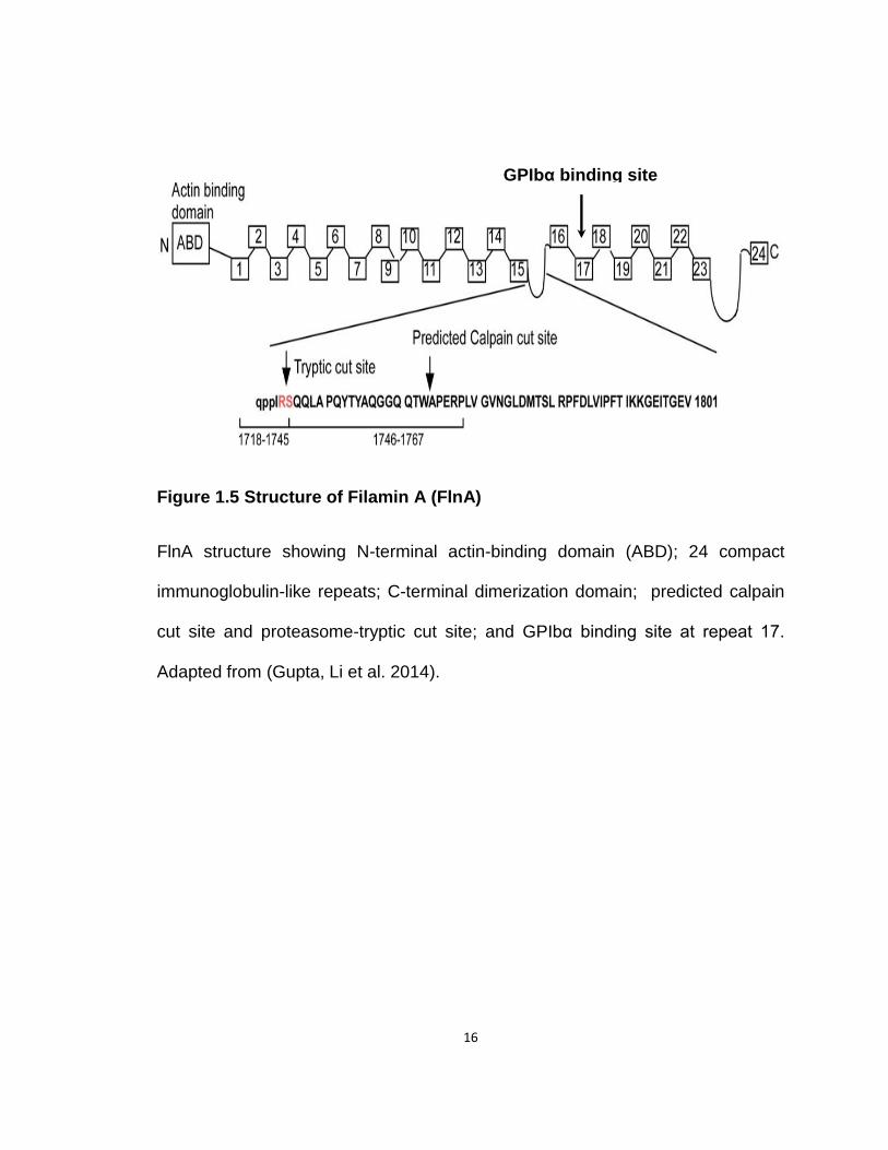
Figure 1.5 Structure of Filamin A (FlnA)
FlnA structure showing N-terminal actin-binding domain (ABD); 24 compact
immunoglobulin-like repeats; C-terminal dimerization domain; predicted calpain
cut site and proteasome-tryptic cut site; and GPIbα binding site at repeat 17.
Adapted from (Gupta, Li et al. 2014).
GPIbα binding site
17
The FlnA-GPIbα interaction in platelets is critical for anchoring the GPIb-IX-V
receptor complex to the membrane skeleton as well as for maintaining cell
adhesion under high shear (Cranmer, Ashworth et al. 2011). This FlnA-GPIbα
interaction is also vital for formation and release of discoid platelets from
megakaryocytes, the platelet precursor cells (Jurak Begonja, Hoffmeister et al.
2011).
The cytoplasmic actin network comprised of actin (which accounts for
~20% of total platelet protein mass) filaments and associated proteins (Oda,
Daley et al. 1992). In unstimulated platelets, 40% of the actin is filamentous F-
actin, and the rest is globular monomeric G-actin. Upon stimulation, the actin
filaments in platelets are severed and resulting smaller fragments are used as
the focal point for new, longer actin filaments. This leads to an increase in the F-
actin proportion to ~70% to 80% (Kovacsovics and Hartwig 1996). Myosin IIa, the
contractile protein, is rapidly phosphorylated by myosin light chain kinase (MLCK)
following platelet activation and becomes associated with F-actin (Fox and
Phillips 1982) and forms filaments that are anchored to the platelet plasma
membrane by attachment (via FlnA) to the GPIb-IX-V complex (Kovacsovics and
Hartwig 1996). Myosin IIa phosphorylation following platelet activation is
essential for clot retraction and granule centralization (Cohen, Gerrard et al.
1982; Stark, Golla et al. 1991).
18
1.1.2.4 Platelet Secretary Granules
Platelets contain secretary granules packed with preformed bioactive
molecules, released upon platelet activation, that are essential for normal platelet
function. Platelets have three distinct secretary granules; alpha granules, dense
granules and lysosomes, each with different morphologies, molecular content,
and kinetics of exocytosis (Flaumenhaft 2003; Coppinger, Cagney et al. 2004).
The α-granules are the largest and most abundant platelet granule,
outnumbering dense granules by ~10-fold (Koseoglu and Flaumenhaft 2013).
These granules contain pro-coagulant proteins (fibrinogen, VWF, platelet factor
4), coagulation factor V, glycoprotein CD62P (P-selectin), CD36, growth-
promoting factors and mitogens [platelet-derived growth factor (PDGF),
thrombospondin, vascular endothelial growth factor (VEGF), transforming growth
factor β (TGFβ) etc. (Coppinger, Cagney et al. 2004).
Dense granules are the smallest platelet granules and humans platelets
contain ~3-9 dense granules/platelet (White 1969). They house a variety of
hemostatically active molecules such as calcium, magnesium, serotonin and
ADP/ATP (ratio 3/2), that are released upon platelet activation to recruit
additional platelet at sites of vascular injury (Rendu and Brohard-Bohn 2001;
King and Reed 2002). Transport of serotonin in platelet dense granules is
essential for liver regeneration (Lesurtel, Graf et al. 2006).

19
P-selectin (from α-granules) expression on the surface of stimulated
platelets and ATP release from dense granules are frequently used markers to
assess the activation state of platelets in vitro.
Lysosomes are formed during megakaryocyte maturation earlier than α-
granules. Platelets have few primary and secondary lysosomes and their size lies
between dense and α-granules (Menard, Meyers et al. 1990). They contain
hydrolases (such as cathepsins D and E) elastase and membrane proteins
LAMP-1, -2 and -3 (Bentfeld-Barker and Bainton 1982; Israels, McMillan et al.
1996; McNicol and Israels 1999).
1.1.3 Platelet Receptors
Platelets express a great number of receptors (Fig. 1.6), respond to many
agonists, and trigger a surfeit of signaling pathways. Importantly, these receptors
are not merely redundant as genetic defects or pharmacological inhibition of any
of these molecules has a significant effect on platelet function e.g. patients
suffering from BSS (with defects in the GP1b-X-V complex) have bleeding
disorder or pharmacologic inhibition of αIIbβ3 inhibits platelet aggregation. In the
next section, I will focus on the receptors involved in my research work.
1.1.3.1 Adhesive receptors
1.1.3.1.1 Integrins
Integrins are a broadly distributed family of heterodimeric (α and β
subunits) cell surface adhesion receptors that occupy ~ 50% of surface area of

20
an activated platelet. A hallmark of integrins is their ability to cycle from a low
affinity ligand binding state (resting platelets) to a high affinity ligand binding state
(activated platelets), a property that is of particular importance to blood cells such
as platelets (Carman and Springer 2003).
Platelets express two major integrin β subunits, β1 and β3 and five α-
subunits. The β1-integrins present in platelets are α2β1, and α6β1(Hynes 2002;
Kasirer-Friede, Kahn et al. 2007). Of all the integrins, αIIbβ3 is the dominant
integrin on platelet surface and plays a prominent role in platelet aggregation by
binding plasma fibrinogen. Approximately 40,000-80,000 copies are present per
platelet (Kauskot and Hoylaerts 2012). Upon platelet activation through ADP,
thrombin, TxA2 or collagen, αIIbβ3 undergoes conformational change to a high
affinity state. This process is termed inside-out signaling (Shattil and Newman
2004). The signaling axis for αIIbβ3 activation involves Rap1, RIAM, talin and
kindlin-3 (Banno and Ginsberg 2008; Metcalf, Moore et al. 2010). Fibrinogen
binding to the activated αIIbβ3 relays outside-in signaling via the signaling
proteins FAK, Pyk2, SFKs (Src, Fyn and Yes), PI3Kβ, or SHIP1 to the actin-
myosin cytoskeleton and all of this is important for thrombus stability and clot
retraction (Shattil and Newman 2004; Suzuki-Inoue, Hughes et al. 2007;
Gratacap, Guillermet-Guibert et al. 2011).
21
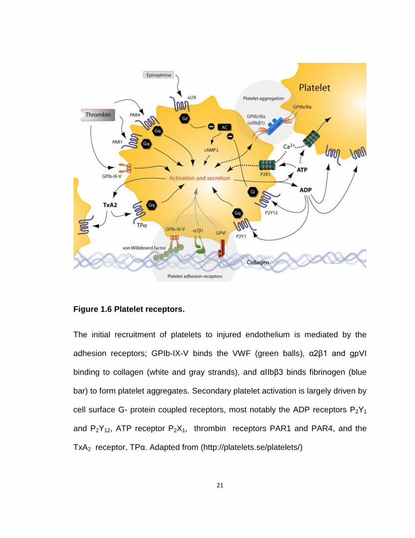
Figure 1.6 Platelet receptors.
The initial recruitment of platelets to injured endothelium is mediated by the
adhesion receptors; GPIb-IX-V binds the VWF (green balls), α2β1 and gpVI
binding to collagen (white and gray strands), and αIIbβ3 binds fibrinogen (blue
bar) to form platelet aggregates. Secondary platelet activation is largely driven by
cell surface G- protein coupled receptors, most notably the ADP receptors P2Y1
and P2Y12, ATP receptor P2X1, thrombin receptors PAR1 and PAR4, and the
TxA2 receptor, TPα. Adapted from (http://platelets.se/platelets/)
22
Since the αIIbβ3 receptor is absolutely essential for irreversible platelet
activation and thrombus propagation in vivo (Savage, Saldivar et al. 1996;
Vinogradova, Velyvis et al. 2002), pharmacologic inhibitors of this receptor had
been developed and are in clinical use. Three inhibitors, abciximab, eptifibatide
and tirofiban are FDA approved for percutaneous coronory intervention (PCI,
(abciximab and eptifibatide) and acute coronory syndrome (ACS, eptifibatide and
tirofiban). The major limitation of these drugs is increased incidence of bleeding,
which restricts their use to only high risk patients who have not been pretreated
with P2Y12 receptor antagonists (Michelson 2010; Muniz-Lozano, Rollini et al.
2013).
1.1.3.1.2 Glycoprotein Ib-IX-V
The GPIb-IX-V receptor complex is crucial for the initial adhesion of
circulating platelets to the injured vascular surface via its interaction with its
ligand VWF at high shear rates (Savage, Saldivar et al. 1996; Goto, Ikeda et al.
1998; Ruggeri 2001; Ruggeri and Mendolicchio 2007). This receptor complex is
constitutively expressed on platelet surface with the density of about 25000
copies per platelet (Modderman, Admiraal et al. 1992) and consists of 4 distinct
non-covalently attached subunits namely, glycoprotein Ibα (GPIbα), glycoprotein
Ibβ (GPIbβ), glycoprotein IX (GPIX), and glycoprotein V (GPV) in the ratio
2:2:2:1 (Ware 1998; Kauskot and Hoylaerts 2012). The N-terminal extracellular
region of GPIbα contains the binding sites for VWF, P-selectin, Mac-1 (integrin
α2βM), coagulation factors XI (Baglia, Badellino et al. 2002) and FXII (Bradford,
23
Pixley et al. 2000), high-molecular-weight kininogen (Lanza 2006) and thrombin
(De Marco, Mazzucato et al. 1994; Adam, Bouton et al. 2003). The intracellular
cytoplasmic domain of GPIbα is linked to FlnA and this interaction tethers the
GPIb-IX-V receptor complex to the platelet cytoskeleton, maintaining the
cytoskeletal architecture of resting platelets and those adhering in vessels at high
shear rates (Cranmer, Ashworth et al. 2011).
In the absence of any vascular trauma, plasma VWF does not normally
bind to platelets, since the GPIbα binding VWF-A1 domain is inaccessible
(Miyata, Goto et al. 1996). However, damage to vessel wall exposes
subendothelial collagen, enabling VWF to bind collagen through its A3 domain.
This and/or high shear stress induces conformational change in VWF, exposing
the otherwise cryptic binding site on VWF-A1 for the GPIb-IX-V complex
(Siedlecki, Lestini et al. 1996). The snake venom peptide botrocetin (Read, Smith
et al. 1989) or the antibiotic ristocetin (Scott, Montgomery et al. 1991) can also
be used to induce vWF-GPIbα interaction in vitro.
GPIbα, which is primarily regarded as the VWF receptor, also contains a
high affinity binding site for thrombin that contributes to platelet activation at low
thrombin concentrations and plays a significant role in the generation of platelet
microparticles and their procoagulant activity (Harmon and Jamieson 1986;
Dormann, Clemetson et al. 2000). The importance of thrombin binding to GPIbα
became evident after the observation that patients with a rare autosomal
recessive condition called BSS (quantitative or qualitative defects in the platelet
24
GPIb-IX-V complex) (Lanza 2006; Cox, Price et al. 2011), showed reduced
responsiveness to thrombin (Ganguly 1977; Jamieson and Okumura 1978).
GPIbα also increases the rate of protease-activated receptor 1 (PAR1, discussed
later) hydrolysis by 5 fold, indicating that GPIbα may act as a cofactor that
facilitates PAR 1 cleavage (De Candia, Hall et al. 2001). A specific mutation that
only blocked thrombin-GPIbα interaction reduced the ability of murine platelets to
form thrombi, suggesting this interaction is essential for thrombosis. Importantly,
blocking thrombin-GPIbα interaction in vivo had no effect on bleeding time,
making this interaction a suitable therapeutic target (Guerrero, Shafirstein et al.
2008).
1.1.3.1.3 Collagen Receptors
The subendothelial matrix protein collagen is a key initiator of platelet
responses by not only serving as a substrate for platelet adhesion, but also by
acting as a potent platelet agonist. Two major direct receptors have been
implicated in the platelet responses to collagen: α2β1 and glycoprotein VI (gpVI).
α2β1, a member of the integrin family, serves primarily to anchor platelets to
subendothelial collagen exposed after vascular injury. Basically, gpVI-collagen
interaction is weak, but provides a potent stimulus for intracellular signaling. As a
result of this signaling, α2β1 is induced to bind collagen with high affinity thereby
contributing to firm interaction between platelets and subendothelial collagen
(Jung and Moroi 1998; Polanowska-Grabowska, Simon et al. 1999; Nieswandt,
Brakebusch et al. 2001; Chen and Kahn 2003; Horii, Kahn et al. 2006).

25
GPVI is predominantly a signaling receptor that belongs to the
immunoglobulin (Ig) superfamily whose expression is restricted to platelets and
megakaryocytes. Approximately, 4000–6000 copies of gpVI are present/platelet
(Clemetson, Polgar et al. 1999; Jandrot-Perrus, Busfield et al. 2000). GPVI exists
as a single transmembrane receptor and its ability to generate signals rests on
the interaction between its transmembrane domain and immunoreceptor
tyrosine-based activation domain (ITAM)-containing Fc receptor γ chain (FcRγ)
(Kahn 2004). This gpVI- FcRγ chain interaction is critical for receptor stabilization
and downstream signaling as mice deficient in FcRγ are unresponsive to
collagen in part due to loss of signaling and in part because of their failure to
express the receptor on the surface of their platelets (Poole, Gibbins et al. 1997;
Tsuji, Ezumi et al. 1997; Kato, Kanaji et al. 2003). Collagen stimulation induces
cross-linking of the gpVI- FcRγ chain complex resulting in sequential activation of
Src and Syk family tyrosine kinases which further activates phosphatidylinositol
3-kinase β (PI3Kβ). This leads to phospholipase C γ2 (PLCγ2) activation, where
PIP2 hydrolysis increases intracellular calcium and induces diacylglycerol (DAG)
production. Both of these events are crucial for αIIbβ3 activation and subsequent
platelet aggregation (Watson, Auger et al. 2005).
Platelets from humans with gpVI deficiency are nonresponsive to collagen
(Sugiyama, Okuma et al. 1987; Kahn 2004) and gpVI polymorphisms are
associated with increased risk of myocardial infarction (Yee and Bray 2004).
Interestingly, inhibition or loss of gpVI prevented arterial thrombosis in animal
models, but only mildly perturbed the normal hemostasis in both mice and

26
humans (Kahn 2004), making gpVI a suitable therapeutic target. A soluble gpVI-
Fc fusion protein (Revacept) that blocks the gpVI-binding sites on exposed
collagen suppresses murine arterial thrombosis without a bleeding phenotype.
Revacept is currently being tested in phase II trials (Ungerer, Rosport et al.
2011).
1.1.3.2 G- Protein Coupled Receptors
1.1.3.2.1 Thrombin Receptors
Thrombin’s effect on human platelets is mainly mediated by surface
GPCRs called protease-activated receptor (PAR) 1 and 4 and GP1bα (described
above).
1.1.3.2.1.1 PARs
PAR1 was the first identified protease-activated receptor and out of four
known PARs, human platelets express PAR1 and PAR4, whereas, murine
platelets express only PAR3 and PAR4. (Coughlin 1993; Kahn, Zheng et al.
1998; Coughlin 1999; Coughlin 2005). PAR1 is a prototype of this family, and
accounts for the majority of cellular responses to thrombin. PAR1 is activated
through proteolytic cleavage of its extracellular N-terminal, carboxyl to the R41
residue, to yield the tethered ligand sequence S42FLLRN. This tethered ligand
can bind intra-molecularly to the cleaved PAR1 receptor to initiate signaling (Vu,
Hung et al. 1991; Macfarlane, Seatter et al. 2001). Proteolytic cleavage of the
extracellular N-terminus of PAR4 yields the tethered ligand sequence GYPGQV.
PAR1 or PAR4 activation can also be triggered by short synthetic peptides,
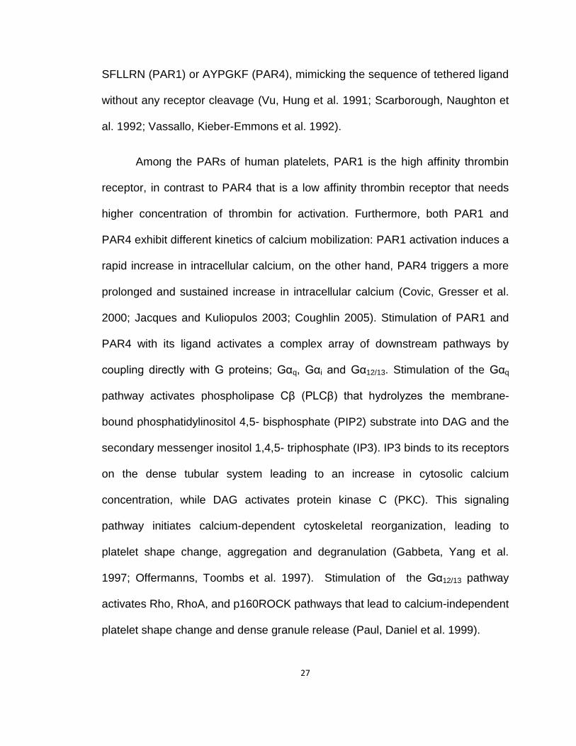
27
SFLLRN (PAR1) or AYPGKF (PAR4), mimicking the sequence of tethered ligand
without any receptor cleavage (Vu, Hung et al. 1991; Scarborough, Naughton et
al. 1992; Vassallo, Kieber-Emmons et al. 1992).
Among the PARs of human platelets, PAR1 is the high affinity thrombin
receptor, in contrast to PAR4 that is a low affinity thrombin receptor that needs
higher concentration of thrombin for activation. Furthermore, both PAR1 and
PAR4 exhibit different kinetics of calcium mobilization: PAR1 activation induces a
rapid increase in intracellular calcium, on the other hand, PAR4 triggers a more
prolonged and sustained increase in intracellular calcium (Covic, Gresser et al.
2000; Jacques and Kuliopulos 2003; Coughlin 2005). Stimulation of PAR1 and
PAR4 with its ligand activates a complex array of downstream pathways by
coupling directly with G proteins; Gαq, Gαi and Gα12/13. Stimulation of the Gαq
pathway activates phospholipase Cβ (PLCβ) that hydrolyzes the membrane-
bound phosphatidylinositol 4,5- bisphosphate (PIP2) substrate into DAG and the
secondary messenger inositol 1,4,5- triphosphate (IP3). IP3 binds to its receptors
on the dense tubular system leading to an increase in cytosolic calcium
concentration, while DAG activates protein kinase C (PKC). This signaling
pathway initiates calcium-dependent cytoskeletal reorganization, leading to
platelet shape change, aggregation and degranulation (Gabbeta, Yang et al.
1997; Offermanns, Toombs et al. 1997). Stimulation of the Gα12/13 pathway
activates Rho, RhoA, and p160ROCK pathways that lead to calcium-independent
platelet shape change and dense granule release (Paul, Daniel et al. 1999).

28
PAR1 plays a crucial role in platelet activation and thrombus stabilization
and in accordance with this PAR1 antagonist, Vorapaxar and Atopaxar, were
developed and clinical trials have been performed. Table 1.1 summarizes the
clinical trial status as well as limitations of PAR1 antagonists.
1.1.3.2.1.2 ADP Receptors
Adenosine 5’-diphosphate (ADP) is a physiologic, endogenous platelet
agonist critical for normal hemostasis and thrombosis. ADP is stored in platelet
dense granules and is secreted upon platelet activation. Shear stress can also
induce ADP release from erythrocytes. ADP amplifies platelet activation initiated
by other agonists, and is vital for maximal platelet aggregation under high shear
conditions. ADP acts in an autocrine/paracrine manner and binds two different G-
protein coupled purinergic receptors, P2Y1 and P2Y12 (Oury, Toth-Zsamboki et al.
2006). P2Y1, couples to Gαq and activates phospholipase Cβ (PLCβ) that triggers
mobilization of intra-platelet calcium stores. This calcium flux initiates cytoskeletal
reorganization leading to platelet shape change and aggregation (Ebbeling,
Robertson et al. 1992; Jardin, Lopez et al. 2008). P2Y12 is a Gαi-coupled receptor
that inhibits adenylate cyclase (AC) leading to reduction in the intracellular levels
of the signaling messenger cyclic adenosine monophosphate (cAMP), while Gβγ
dimers activate phosphoinositide 3-kinase (PI3Kβ/γ). This regulates platelet
aggregation via actin cytoskeleton-dependent integrin αIIbβ3 activation (Chen,
De et al. 2004). Continuous signaling through P2Y12 is critical for persistent αIIbβ3
activation and thrombus stability under shear flow conditions (Cosemans, Munnix
29
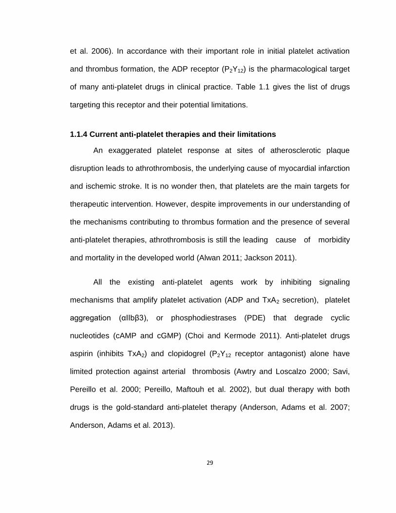
et al. 2006). In accordance with their important role in initial platelet activation
and thrombus formation, the ADP receptor (P2Y12) is the pharmacological target
of many anti-platelet drugs in clinical practice. Table 1.1 gives the list of drugs
targeting this receptor and their potential limitations.
1.1.4 Current anti-platelet therapies and their limitations
An exaggerated platelet response at sites of atherosclerotic plaque
disruption leads to athrothrombosis, the underlying cause of myocardial infarction
and ischemic stroke. It is no wonder then, that platelets are the main targets for
therapeutic intervention. However, despite improvements in our understanding of
the mechanisms contributing to thrombus formation and the presence of several
anti-platelet therapies, athrothrombosis is still the leading cause of morbidity
and mortality in the developed world (Alwan 2011; Jackson 2011).
All the existing anti-platelet agents work by inhibiting signaling
mechanisms that amplify platelet activation (ADP and TxA2 secretion), platelet
aggregation (αIIbβ3), or phosphodiestrases (PDE) that degrade cyclic
nucleotides (cAMP and cGMP) (Choi and Kermode 2011). Anti-platelet drugs
aspirin (inhibits TxA2) and clopidogrel (P2Y12 receptor antagonist) alone have
limited protection against arterial thrombosis (Awtry and Loscalzo 2000; Savi,
Pereillo et al. 2000; Pereillo, Maftouh et al. 2002), but dual therapy with both
drugs is the gold-standard anti-platelet therapy (Anderson, Adams et al. 2007;
Anderson, Adams et al. 2013).
30
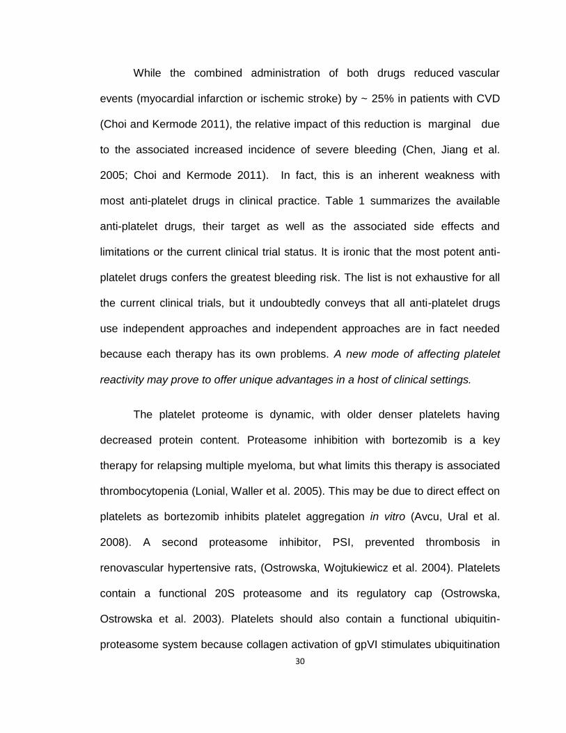
While the combined administration of both drugs reduced vascular
events (myocardial infarction or ischemic stroke) by ~ 25% in patients with CVD
(Choi and Kermode 2011), the relative impact of this reduction is marginal due
to the associated increased incidence of severe bleeding (Chen, Jiang et al.
2005; Choi and Kermode 2011). In fact, this is an inherent weakness with
most anti-platelet drugs in clinical practice. Table 1 summarizes the available
anti-platelet drugs, their target as well as the associated side effects and
limitations or the current clinical trial status. It is ironic that the most potent anti-
platelet drugs confers the greatest bleeding risk. The list is not exhaustive for all
the current clinical trials, but it undoubtedly conveys that all anti-platelet drugs
use independent approaches and independent approaches are in fact needed
because each therapy has its own problems. A new mode of affecting platelet
reactivity may prove to offer unique advantages in a host of clinical settings.
The platelet proteome is dynamic, with older denser platelets having
decreased protein content. Proteasome inhibition with bortezomib is a key
therapy for relapsing multiple myeloma, but what limits this therapy is associated
thrombocytopenia (Lonial, Waller et al. 2005). This may be due to direct effect on
platelets as bortezomib inhibits platelet aggregation in vitro (Avcu, Ural et al.
2008). A second proteasome inhibitor, PSI, prevented thrombosis in
renovascular hypertensive rats, (Ostrowska, Wojtukiewicz et al. 2004). Platelets
contain a functional 20S proteasome and its regulatory cap (Ostrowska,
Ostrowska et al. 2003). Platelets should also contain a functional ubiquitin-
proteasome system because collagen activation of gpVI stimulates ubiquitination
31
of platelet Syk kinase
(Dangelmaier, Quinter et al. 2005) through the E3 ligase Cbl-b (Daniel,
Dangelmaier et al. 2010).
Despite these fragmented observations, the proteasome of platelets is not
established as a component of cell signaling or function. Based on this, the
studies of this thesis examine the role of ubiquitin-proteasome system in
modulating platelet function.

32
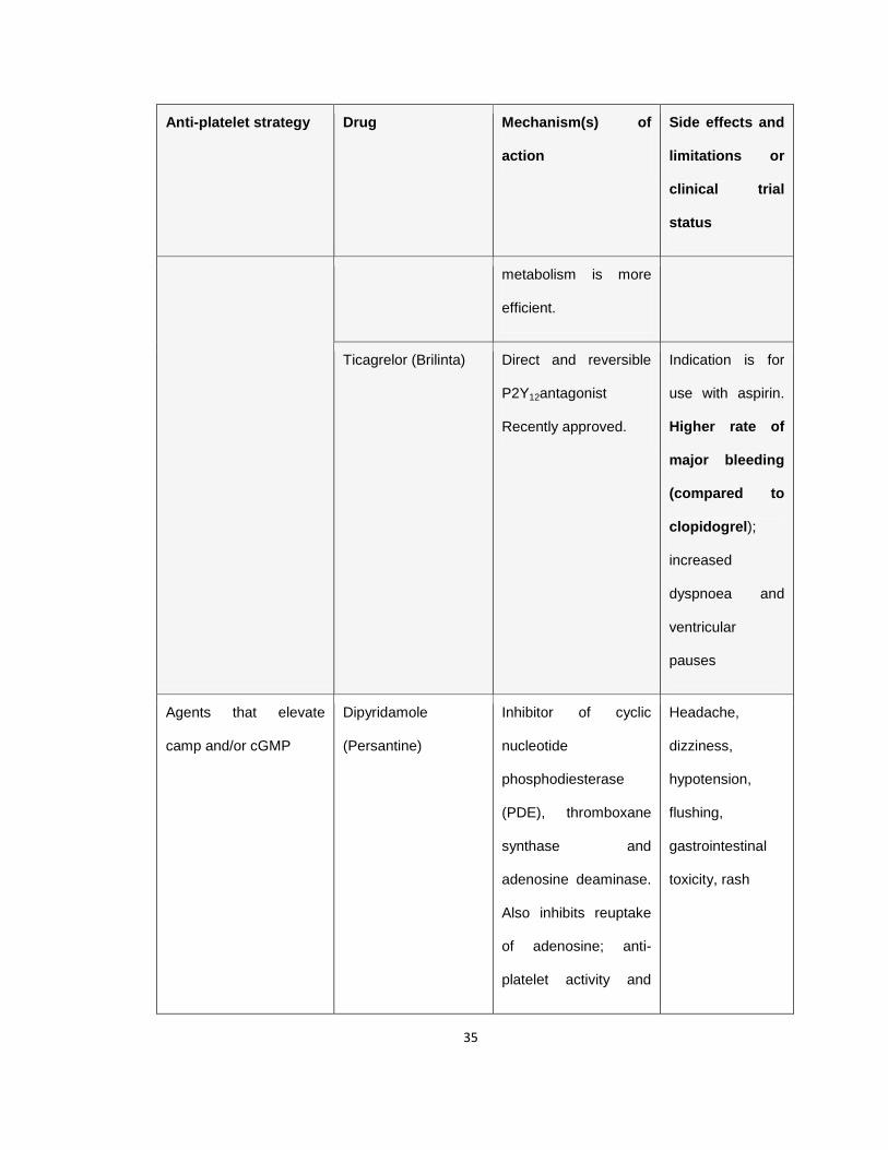
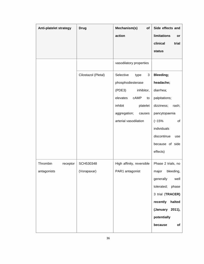
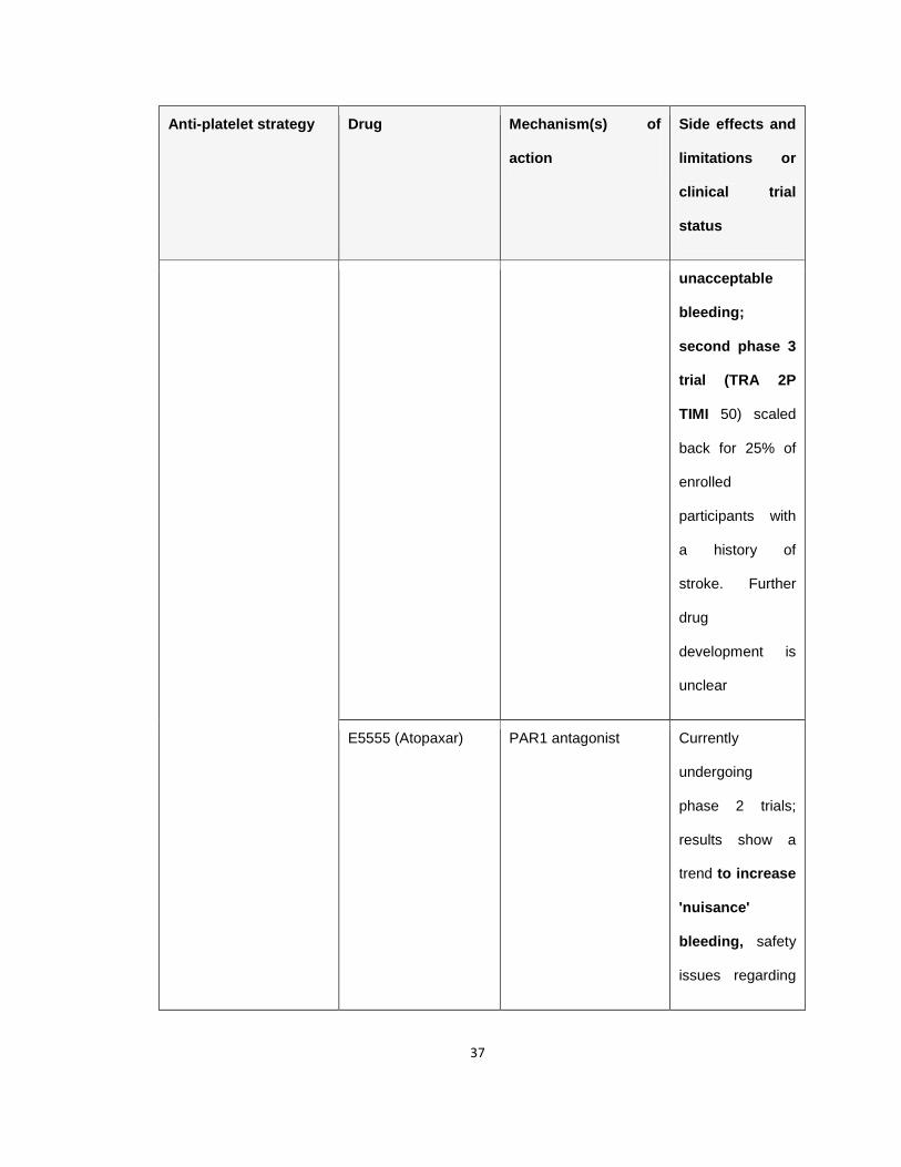
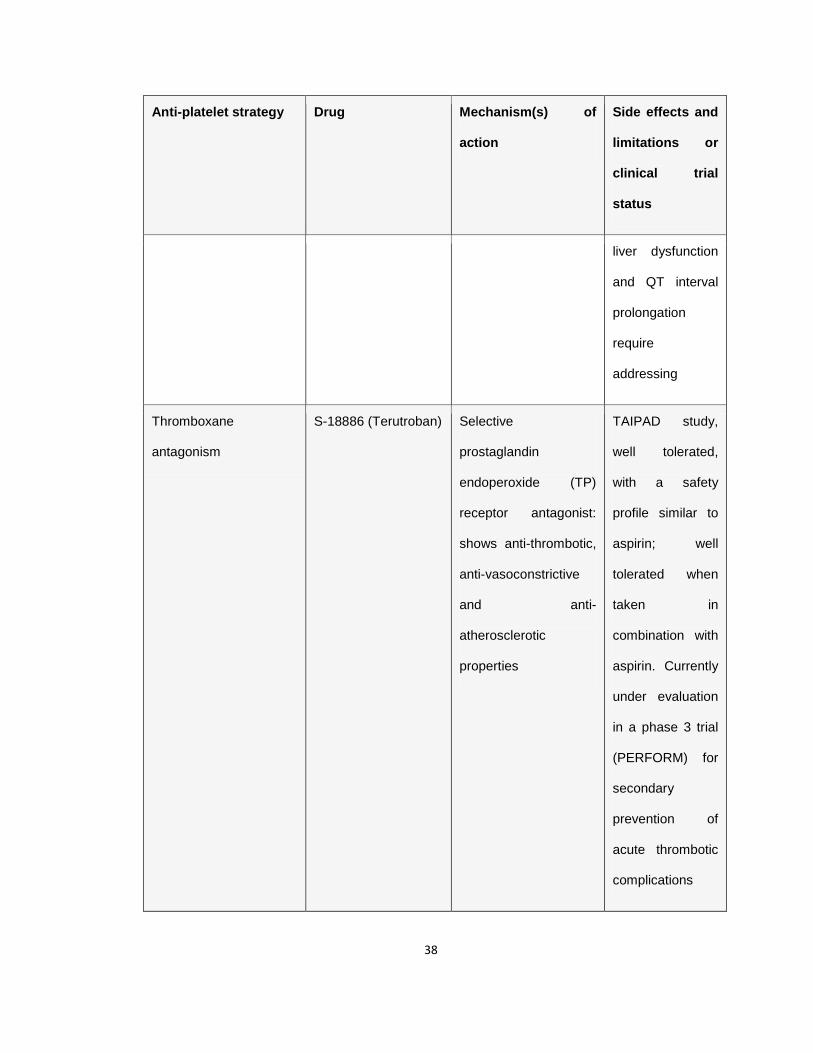
Table 1.1 Antiplatelet agents in the clinic or in clinical trials
Anti-platelet strategy Drug Mechanism(s) of
action
Side effects and
limitations or
clinical trial
status
Blockade of prostanoid
biosyntesis (TxA2)
Acetylsalicylic acid
(aspirin)
Irreversible acetylation
of cyclooxygenase 1
(COX-1), inhibiting
generation of TxA2
Bleeding,
gastrointestinal
toxicity Weak
anti-platelet
agent, ~25%
prevalence of
aspirin
'resistance'
GPIIb-IIIa inhibition Abciximab (ReoPro) Reversible inhibition of
integrinαIIbβ3 activation,
also blocks
integrin αvβ3
Bleeding,
thrombocytopenia
Restrictions
because of
intravenous
administration
Eptifibatide (Integrilin) Rapidly reversible,
Arg-Gly-Asp (RGD)
mimetic
Bleeding, small
increase in
profound
thrombocytopenia
Restrictions

33
Anti-platelet strategy Drug Mechanism(s) of
action
Side effects and
limitations or
clinical trial
status
because of
intravenous
administration
P2Y12antagonists Tirofiban (Aggrastat) RGD mimetic; rapidly
reversible, minimal
effects on αvβ3
Bleeding, severe
but reversible
thrombocytopenia
in small numbers
of recipients
Restrictions
because of
intravenous
administration
Ticlopidine (Ticlid) Active metabolite of
parent compound
irreversibly inhibits the
ADP receptor P2Y12
Bleeding,
gastrointestinal
toxicity, rash,
neutropenia,
thrombotic
thrombocytopenic
purpura (TTP)
(largely replaced
by clopidogrel

34
Anti-platelet strategy Drug Mechanism(s) of
action
Side effects and
limitations or
clinical trial
status
because of
increased
toxicity)
Clopidogrel (Plavix) Active metabolite of
parent compound
irreversibly inhibits
P2Y12
Rash,
neutropenia,
TTP, major
bleeding
corresponding
with >50%
inhibition of
P2Y12.
Interpatient
response
variability
Prasugrel (Effient) Active metabolite of
parent compound
irreversibly inhibits
P2Y12; improved
potency and
consistency over
clopidoogrel. Pro-drug
Bleeding (more
haemorrhagic
side-effects than
clopidogrel)
35
Anti-platelet strategy Drug Mechanism(s) of
action
Side effects and
limitations or
clinical trial
status
metabolism is more
efficient.
Ticagrelor (Brilinta) Direct and reversible
P2Y12antagonist
Recently approved.
Indication is for
use with aspirin.
Higher rate of
major bleeding
(compared to
clopidogrel);
increased
dyspnoea and
ventricular
pauses
Agents that elevate
camp and/or cGMP
Dipyridamole
(Persantine)
Inhibitor of cyclic
nucleotide
phosphodiesterase
(PDE), thromboxane
synthase and
adenosine deaminase.
Also inhibits reuptake
of adenosine; anti-
platelet activity and
Headache,
dizziness,
hypotension,
flushing,
gastrointestinal
toxicity, rash
36
Anti-platelet strategy Drug Mechanism(s) of
action
Side effects and
limitations or
clinical trial
status
vasodilatory properties
Cilostazol (Pletal) Selective type 3
phosphodiesterase
(PDE3) inhibitor,
elevates cAMP to
inhibit platelet
aggregation; causes
arterial vasodilation
Bleeding;
headache;
diarrhea;
palpitations;
dizziness; rash;
pancytopaenia
(~15% of
individuals
discontinue use
because of side
effects)
Thrombin receptor
antagonists
SCH530348
(Vorapaxar)
High affinity, reversible
PAR1 antagonist
Phase 2 trials, no
major bleeding,
generally well
tolerated; phase
3 trial (TRACER)
recently halted
(January 2011),
potentially
because of
37
Anti-platelet strategy Drug Mechanism(s) of
action
Side effects and
limitations or
clinical trial
status
unacceptable
bleeding;
second phase 3
trial (TRA 2P
TIMI 50) scaled
back for 25% of
enrolled
participants with
a history of
stroke. Further
drug
development is
unclear
E5555 (Atopaxar) PAR1 antagonist Currently
undergoing
phase 2 trials;
results show a
trend to increase
'nuisance'
bleeding, safety
issues regarding
38
Anti-platelet strategy Drug Mechanism(s) of
action
Side effects and
limitations or
clinical trial
status
liver dysfunction
and QT interval
prolongation
require
addressing
Thromboxane
antagonism
S-18886 (Terutroban) Selective
prostaglandin
endoperoxide (TP)
receptor antagonist:
shows anti-thrombotic,
anti-vasoconstrictive
and anti-
atherosclerotic
properties
TAIPAD study,
well tolerated,
with a safety
profile similar to
aspirin; well
tolerated when
taken in
combination with
aspirin. Currently
under evaluation
in a phase 3 trial
(PERFORM) for
secondary
prevention of
acute thrombotic
complications
39
Anti-platelet strategy Drug Mechanism(s) of
action
Side effects and
limitations or
clinical trial
status
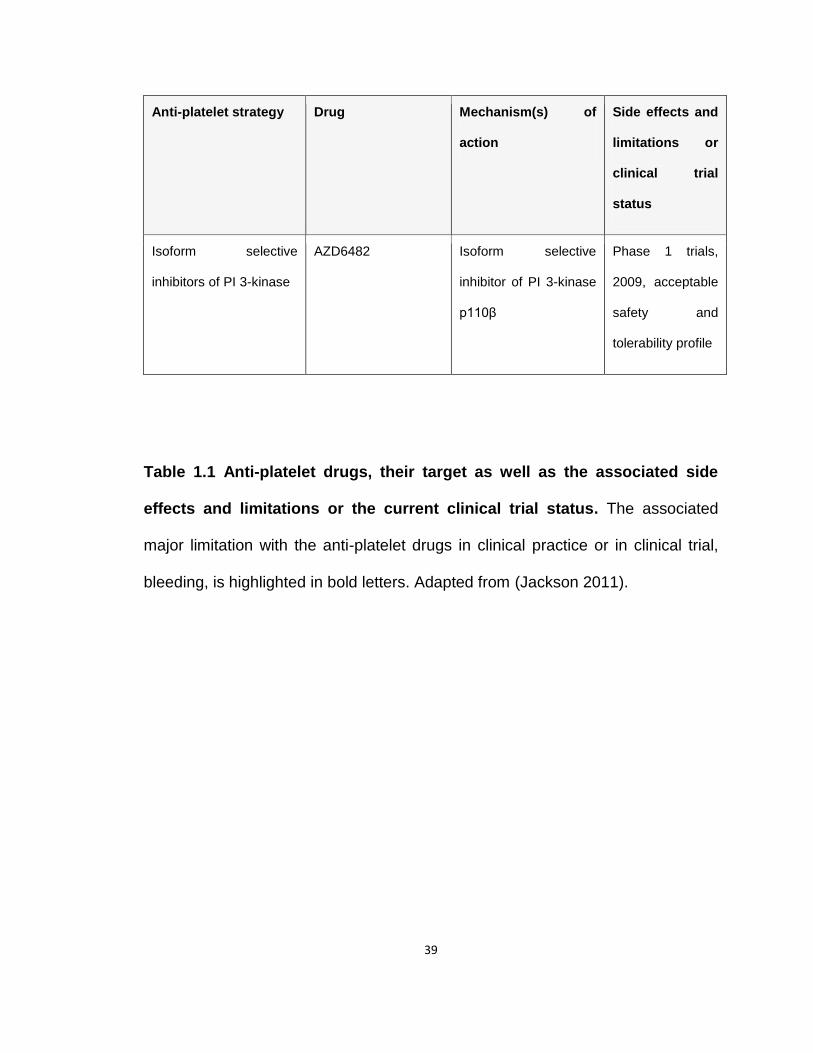
Isoform selective
inhibitors of PI 3-kinase
AZD6482 Isoform selective
inhibitor of PI 3-kinase
p110β
Phase 1 trials,
2009, acceptable
safety and
tolerability profile
Table 1.1 Anti-platelet drugs, their target as well as the associated side
effects and limitations or the current clinical trial status. The associated
major limitation with the anti-platelet drugs in clinical practice or in clinical trial,
bleeding, is highlighted in bold letters. Adapted from (Jackson 2011).

40
1.2 Overview of ubiquitin-proteasome system
Cellular proteins are subjected to a variety of post-translational
modifications that profoundly expand the functional repertoire and dynamics of
the eukaryotic proteome. Proteins can be modified by the covalent addition of
small molecules such as phosphate groups (phosphorylation), methyl groups
(methylation), sugar groups (glycosylation), acetyl groups (acetylation) or entire
proteins (Hochstrasser 2000; Pickart 2001; Xu and Peng 2006). The first such
protein-based modification to be described was ubiquitin (Ub). Ubiquitin is a
small 76- amino acid regulatory protein (~ 8.5 kDa) that is evolutionary conserved
throughout eukaryotes (only 3 amino acid difference from yeast to human), but is
absent from members of the other two super kingdoms, the eubacteria and the
archae bacteria (Hershko and Ciechanover 1998; Pickart and Eddins 2004).
The covalent decoration of cellular proteins with Ub, known as
ubiquitination, regulates a diverse array of biological processes, including
signaling, protein quality control, organelle biogenesis, cell cycle regulation, DNA
repair, transcription, inflammation, stress response, endocytosis and vesicular
trafficking (Weissman 2001; Greene, Whitworth et al. 2005; Hurley, Lee et al.
2006; Kerscher, Felberbaum et al. 2006; Ulrich and Walden 2010). Ubiquitination
of proteins is mediated through an enzymatic cascade that, in most cases,
results in the conjugation of either single Ub on one (mono-ubiquitination) or
multiple sites (multi-mono-ubiquitination), or multiple Ub monomers (poly-
ubiquitination) to the internal lysine (Lys) of a substrate (Ravid and Hochstrasser
41
2008). However, in rare instances Ub is conjugated to the N-terminus or the side
chain of the cysteine (Cys) moiety of the substrate (Deshaies and Joazeiro 2009;
Komander and Rape 2012). The generation of Ub linkages with distinct
topologies confers diversity and versatility in the ways ubiquitination modulates
various aspects of eukaryotic biology (Weissman 2001).
Ubiquitination is dynamic and reversible. Removal of Ub moieties, which
may modulate Ub signaling, is carried out by a specific class of proteases called
deubiquitinases (DUBs). DUBs hydrolyze the isopeptide linkages between Ub
and the substrate or between multiple Ub moieties. DUBs therefore play critical
roles in regulating the rate of protein turnover and in maintaining pools of free Ub
by recycling it from existing conjugates (Amerik and Hochstrasser 2004). In
mammalian cells, ~ 100 DUBs have been identified and classified in to five
subfamilies (Wilkinson 1997; Reyes-Turcu, Ventii et al. 2009).
Among all the processes regulated by Ub, the best characterized is the
role of poly-Ub chains in targeting the proteins for degradation by the 26S
proteasome (Elsasser and Finley 2005; Miller and Gordon 2005). The 26S
proteasome is a large multimeric, catalytic protease (2.5 MDa) that collaborates
with the Ub system and ensures the precise, rapid and irreversible degradation of
proteins tagged with poly-Ub chains. Like Ub, the proteasome is evolutionary
conserved in eukaryotes, however, simpler forms are found even in
archaebacteria and eubacteria. (Dahlmann, Kopp et al. 1989; Lupas, Zwickl et al.
1994; Coux, Tanaka et al. 1996; Demartino and Gillette 2007). The ubiquitin-
42
proteasome system (UPS) is responsible for much of the regulated protein
degradation and maintenance of protein homeostasis. Approximately 80% of
misfolded, oxidized, or damaged proteins and short lived regulatory proteins are
degraded by UPS (Rock, Gramm et al. 1994; Lee and Goldberg 1998).
Given its essential role in regulating protein turnover, defects in the
components of UPS has been implicated in the pathogenesis of many human
diseases, including cardiovascular diseases, neurodegenerative disorders, viral
diseases and numerous cancers (Herrmann, Ciechanover et al. 2004; Corn
2007; Petroski 2008; Hoeller and Dikic 2009; Lehman 2009).
1.2.1 Ubiquitin Conjugation System
The covalent conjugation of Ub to substrate is achieved by the concerted
action of three enzymes: Ub activating enzyme E1, Ub conjugating enzyme E2,
and Ub protein ligase E3 (Fig.1.7). Since ubiquitination controls diverse
biological processes, not only in the cell, but also during the development of
tissues and entire organisms (Mistry, Wilson et al. 2004; Pickart and Fushman
2004; Kerscher, Felberbaum et al. 2006; Yanjiang, Hongjuan et al. 2012), the
whole process is tightly regulated in a spatial and temporal manner and displays
specificity for the substrates it modulates.
In the first step, Ub activating E1 enzyme activates Ub in an ATP
dependent manner by forming a high energy thioester bond between the E1
active site cysteine (Cys) and the carboxy terminus glycine76 (Gly76) residue of
Ub substrate (Ciechanover, Finley et al. 1984; Hershko and Ciechanover 1998).

43
Ub E1s (Uba1 and Uba6) are multidomain enzymes that activate and
transfer Ub to the active site of E2s. This is critical for cellular homeostasis
because chemical inhibition of E1 activity in the cell results in the almost
immediate shutdown of the entire UPS (Yang, Kitagaki et al. 2007). The E1
enzyme contains two active sites, an adenylation site that binds ATP-Mg2+ and
activates C-terminal carboxyl group of Ub, and a catalytic cysteine that attacks
the Ub adenylate bond to form a E1~Ub thioester.
In the second step, "activated" thioester-bound Ub is transferred from Ub-
E1 to the active site Cys of one of a number of E2 Ub conjugating enzymes
through a transthiolation reaction. The transfer of Ub from E1 to E2 catalytic
cysteine residue involves conformational changes in the ubiquitin fold domain
(UFD, E2 binding domain on E1) of E1 that facilitates the correct positioning of
the active sites of the enzymes for efficient transfer (Huang, Hunt et al. 2007;
Olsen and Lima 2013). The E2s are characterized by the presence of a highly
conserved ~140 residue catalytic Ub-conjugating (UBC) fold that
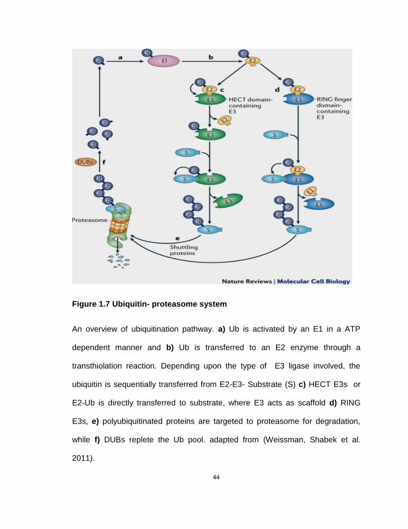
44
Figure 1.7 Ubiquitin- proteasome system
An overview of ubiquitination pathway. a) Ub is activated by an E1 in a ATP
dependent manner and b) Ub is transferred to an E2 enzyme through a
transthiolation reaction. Depending upon the type of E3 ligase involved, the
ubiquitin is sequentially transferred from E2-E3- Substrate (S) c) HECT E3s or
E2-Ub is directly transferred to substrate, where E3 acts as scaffold d) RING
E3s, e) polyubiquitinated proteins are targeted to proteasome for degradation,
while f) DUBs replete the Ub pool. adapted from (Weissman, Shabek et al.
2011).
45
consists of 5 α-helices and 4 anti-parallel β-sheets. In the catalytic groove near
the active site Cys is a "HPN" (histidine, proline and asparagine) motif that
facilitates both thioester formation between "activated" Ub from E1 and the active
site Cys of E2, and substrate ubiquitination. Both histidine and proline may play a
role in supporting the protein structure around the active site, where as
asparagine is critical for promoting isopeptide bond formation between Ub and a
substrate lysine (Wu, Hanlon et al. 2003; Eletr, Huang et al. 2005; Wenzel, Stoll
et al. 2011). The E2 Ub conjugating enzyme then catalyzes substrate
modification in conjunction with a substrate-specific E3 Ub ligase. E2s play an
important role in determining Ub chain length as well the linkage specificity and
since both these factors govern the cellular fate of the target protein, E2s are
viewed as important regulators of Ub signaling that interact with select E3
proteins rather than just carriers of activated Ub (Olsen and Lima 2013).
The E3 Ub ligases can be classified into three major types based on their
catalytic mechanism: the Homologous to E6-associated protein C-terminus
(HECT) E3s, really interesting new gene (RING) domain E3s, and structurally
related U-box-type E3s. The HECT E3 first loads the ubiquitin from the E2 to
their active site Cys and then shuttles it to the substrate (Bernassola, Karin et al.
2008). On the other hand, the RING finger domain and U-box-type E3s act as
scaffolds that brings Ub~E2 and substrate in proximity to facilitate Ub transfer
from Ub~E2 to substrate (Lipkowitz and Weissman 2011). However, this
mechanistic distinction between HECT and RING E3s has become blurred with
the identification of RING subfamily of RING-in-between-RING (RBR) E3 ligases

46
that act as RING/HECT hybrids i.e. they form a thioester~Ub intermediate via a
conserved RING domain Cys prior to Ub transfer to substrate (Wenzel,
Lissounov et al. 2011).
In mammalian cells, there are two known E1s, approximately 30-40 E2s
and more than 600 E3s, making E3s, either alone or in association with its bound
E2, the main specificity and selectivity factor of the UPS (Michelle, Vourc'h et al.
2009; Schulman and Harper 2009; Voutsadakis 2010).
1.2.2 Ubiquitin Code
A single round of the E1-E2-E3 enzymatic cascade results in mono-
ubiquitination of substrate i.e. a single Ub moiety binds to the substrate.
Additional rounds have diverse outcomes: Multi-mono-Ub chains- formed when
several single Ub moieties binds to the substrate at multiple sites and/or Poly-Ub
chains- formed through the sequential addition of several Ub moities to the
previously attached Ub. Poly-Ub chains can be homotypic, using the same lysine
residue of incoming Ub moiety or heterotypic, using more than one linkage type.
Heterotypic chains are either branched i.e. formed by ubiquitination of one Ub at
two or more sites or non-branched. The Ub amino terminal Met1 residue (M1)
can also participate in the Ub chain formation (Ikeda and Dikic 2008; Dikic,
Wakatsuki et al. 2009; Komander and Rape 2012). All eight Ub-Ub linkages have
been found to coexist in vivo (Peng, Schwartz et al. 2003; Xu, Duong et al.
2009).
47
Ub attachment to a target protein is more than just a terminal reaction, as
both the length (mono-vs. poly-Ub chain) and the linkage type (homotypic or
heterotypic) can regulate the fate, stability, activity and subcellular localization of
the modified substrates (Pickart and Fushman 2004; Li and Ye 2008).
The canonical view is that poly-ubiquitination at Lys48 (of at least four
Ub moieties) tags a protein for proteasome-mediated degradation (Komander
and Rape 2012), whereas poly-ubiquitination at Lys63 is non-degradative and
plays essential roles in DNA repair, DNA replication and signal transduction
(Chen and Sun 2009). Unlike Lys48 and Lys63 chains, not much is known about
"atypical chains" that are linked via Lys6, Lys11, Lys27, Lys29, Lys33 and Met1.
Current known functions of mono and multi-mono-Ub chains as well as atypical
chains are summarized in Fig. 1.8.

48
Histone regulation (Roest, van Klaveren et al. 1996; Robzyk, Recht et al. 2000), Endocytosis (Shih, Sloper-Mould et al. 2000; Hicke 2001), Viral Budding, DNA repair and Nuclear Export (Boyd, Tsai et al. 2000; Geyer, Yu et al. 2000; Brooks and Gu 2006).
Endosomal Trafficking (Strous and van Kerkhof 2002; Huang, Kirkpatrick et al. 2006), p53 nuclear transport (Li, Brooks et al. 2003).
DNA damage response. Reviewed in (Kulathu and Komander 2012).
Cell cycle regulation, Membrane Trafficking, Endoplasmic reticulum associated degradation (ERAD), TNFα signaling. Reviewed in (Kulathu and Komander 2012).
Mitochondrial maintenance or Mitophagy (Geisler, Holmstrom et al. 2010; Glauser, Sonnay et al. 2011), Nuclear translocation (Peng, Zeng et al. 2011) .
Ubiquitin-fusion degradation (Hwang, Shemorry et al. 2010; Metzger and Weissman 2010).
T-Cell receptor signaling (Huang, Jeon et al. 2010).
Nuclear factor-κB (NF-κB) signaling (Iwai and Tokunaga 2009), Cell cycle regulation (Wickliffe, Williamson et al. 2009).
Lys6
Mono-
Ubiquitination
Multi-mono-
Ubiquitination
Lys11
Lys27
Lys29
Lys33
Poly-
Ubiquitination
Met1-
Ubiquitination
b
c
d
a

49
Figure 1.8 The Ub code
Ub is usually conjugated to the ε-amino group of a Lys residue in a substrate,
however, Ub itself possess seven lysines residues each of which, and Met1, can
participate in chain formation and generates ubiquitin linkages of different
topologies. This figure summarizes the type of ubiquitination and cellular fate of
the protein. a) Mono-ubiquitination, b) Multi-mono- ubiquitination, c) Poly-
ubiquitination and d) Met1 ubiquitination.
50
1.2.3 The proteasome- a degradation nanomachine
The 26S proteasome is an intricate proteolytic machine that plays an
indispensible role in the degradation of misfolded, damaged or regulatory
proteins decorated with Ub. The proteasome is a large multimeric complex that is
composed of a 20S catalytic core particle (CP) flanked by one or two 19S
regulatory particles (RPs, Figure 1.9) (Coux, Tanaka et al. 1996; Baumeister,
Walz et al. 1998; Tanaka 2009).
1.2.3.1 20S catalytic core particle
The catalytic 20S core particle is the business center of proteasome. It is
assembled into a cylindrical structure formed by axial stacking of 4 hetero-
heptamaric rings. Each ring contains 7 different homologous α-subunits and 7
different β-subunits (Unno, Mizushima et al. 2002). The catalytic activities of the
20S core particle are associated with β-subunits with β1-subunit having caspase-
like (cleaves after acidic residues), β2-subunit having tryptic-like (cleaves after
basic residues) and β5-having chymotrypsin-like activity (cleaves after
hydrophobic residues) (Orlowski 1990; Kisselev, Akopian et al. 1999). The
combination of three different peptidase activities confers to the 20S core particle
the ability to degrade a broad variety of peptide sequences with high efficiency
and specificity. The inactive β-subunits are required for proper assembly and
maturation of catalytically active β-subunits (Arendt and Hochstrasser 1999).
Following degradation, the length of majority of the newly created peptides range
51
from 3-15 amino acids that are then hydrolyzed to amino acids by
oligopeptidases and/or amino carboxyl peptidases (Glickman and Ciechanover
2002; Tanaka 2009) or the short peptides are presented to class I MHC
molecules for antigen presentation to the immune system (Saeki and Tanaka
2012).
Outer α-rings serve as a gated channel to regulate the substrate entry into
the catalytic core. The substrate bound for degradation must pass through this
channel, lined by α2-, α3- and α4-subunits, to enter the catalytic chamber
(Zwickl, Grziwa et al. 1992; Lowe, Stock et al. 1995). The outer α-rings provide
binding sites for the 19S regulatory particle and their interaction results in a
conformational change in the α-subunits that allow entry of the protein substrate
into the central chamber of the 20S core particle (Smith, Chang et al. 2007).
1.2.3.2 19S regulatory particle (19S RP)
The 19S regulatory particle is a ~900 kDa complex that plays an
essential role in the recognition, deubiquitination, unfolding of ubiquitinated
substrate and in translocation of the unfolded substrate into the 20S core particle
for degradation (Coux, Tanaka et al. 1996; Baumeister, Walz et al. 1998; Tanaka
2009). The 19S regulatory particle contains at least 19 subunits that can be
subdivided into two groups: regulatory particle of non-ATPase (Rpn) and
regulatory particle of triple-ATPase (Rpt). Biochemical experiments have
organized the 19S RP into two sub-complexes: the lid and the base (Lander,
Estrin et al. 2012).

52
Figure 1.9 The 26S proteasome
a) The 26S proteasome is comprised of 20S core particle flanked by two 19S
regulatory particles. b) Individual subunit composition of 26S proteasome. The
19S regulatory particle is organized into two sub-complexes, lid and base, and
the catalytic 20S core is arranged as a cylindrical barrel made up of 2 outer α-
and 2 inner β-rings. Adapted from (Murata, Yashiroda et al. 2009).
53
The lid is composed of 9 non-ATPase subunits (Rpn3, Rpn5–Rpn9,
Rpn11, Rpn12, and Rpn15). The lid subunits, Rpn3, Rpn5, Rpn6, Rpn7, Rpn9
and Rpn12 contain protein–protein interaction motifs called PCI [proteasome–
CSN (COP9 signalosome)–eIF3 (eukaryotic translation initiation factor 3)] and
function as scaffolding subunits (Finley 2009). The best established function of
the lid is deubiquitination. Rpn11, which is a JAB1–MPN–MOV34 (JAMM)
domain containing DUB, tightly couples deubiquitination and substrate
degradation (Bar-Nun and Glickman 2012). Rpn8 acts as a non-catalytic binding
partner of Rpn11 (Finley 2009).
The base contains six ATPases (Rpt1-Rpt6), two large scaffolding
subunits (Rpn1 and Rpn2) and two Ub receptors (Rpn10 and Rpn13) that
efficiently trap Ub-conjugated substrates (Finley 2009). The 6 triple-ATPases
form a hetero-hexameric ring with the specific order of Rpt1-Rpt2-Rpt6-Rpt3-
Rpt4-Rpt5 to constitute the molecular motor of proteasome (Tomko, Funakoshi et
al. 2010). The ATPase subunits provide the energy needed for deubiquitination,
substrate unfolding, and translocation of the substrate through the narrow pore
into the degradation chamber (Martin, Baker et al. 2008; Maillard, Chistol et al.
2011). The binding of C-terminus tails of ATPases Rpt2, Rpt3 and Rpt5 on
dedicated sites on the 20S core particle (inter-subunit pockets formed by α-
subunits of the outer ring) triggers gate opening and facilitating substrate entry
(Smith, Chang et al. 2007; Gillette, Kumar et al. 2008).
54
The major activity of the lid and the base is proposed to be
deubiquitination. In mammalian cells, two DUBs, USP14 and UCHL5, are
associated with the base complex and they progressively trim poly-Ub chains
from the distal end of the chains. USP14 is associated with Rpn1 (Hu, Li et al.
2005) and UCHL5 binds to Rpn2 via Rpn13 (Hamazaki, Iemura et al. 2006; Yao,
Song et al. 2006).
1.2.4 Deubiquitination
DUBs are isopeptidases that play pivotal roles in Ub-mediated signaling
pathways. They are classified into six subclasses: Ub C-terminal hydrolases
(UCHs), Ub specific proteases (USP’s), ovarian tumor proteases (OTUs),
Josephines, JAB1/MPN/Mov34 metalloenzyme (JAMM) and monocyte
chemotactic protein-induced protein (MCPIP). Except for JAMM proteases, which
are Zn2+ metalloproteases, the rest are cysteine proteases (Fraile, Quesada et al.
2012; Lim and Baek 2013).
DUBs hydrolyze isopeptide bonds in Ub-protein conjugates and are vital
for Ub homeostasis. General cellular roles of DUBs are summarized in Fig. 1.10.
DUBs regulate ubiquitination in a spatio-temporal manner. DUBs can pair with
E3s ligases, which have inherent autoubiquitination activity, in order to prevent
their self-ubiquitination and degradation, e.g. USP8 mediated stabilization of
neuregulin receptor degradation protein 1 (Nrdp1) ligase induces degradation of
its substrate ErbB3 (Cao, Wu et al. 2007). The DUB-E3 pairing can also fine tune
editing of one Ub signal for another e.g. cellular inhibitor of apoptosis (cIAP)

55
ubiquitin ligases ubiquitinate receptor-interacting serine-threonine kinase 1
(RIPK1) at Lys63, but in order to turn off the signaling, A20 depolymerize Lys63
chains and facilitate the polymerization of Lys48-linked chains on RIPK1 resulting
in its degradation (Hymowitz and Wertz 2010).
DUBs display specificity at multiple levels as evident by their ability to
distinguish between different Ub chain linkages and chain structure (Komander,
Clague et al. 2009). Different subclasses have preference for specific chain
linkages e.g. USP14, OTUB1 and A20 process Lys48-linked chains, where as
JAMMs and CYLD prefer Lys63- linked chains (Fraile, Quesada et al. 2012).
DUBs also modulate protein stability by removing the degradative signal from
proteins meant for degradation or even reverse Ub signaling by removing even
non-degradative signal.
As mentioned before, three DUBs directly associate with 19S regulatory
particles and their activity can prevent degradation of protein even at the face of
the proteasome (Guterman and Glickman 2004). The three DUBs associated
with 19S regulatory particle are Rpn11, USP14 and UCHL5. Rpn11, a JAMM
metalloprotease, is a constituent subunit of 19S RP and is responsible for bulk of
proteasome DUB activity. Rpn11 prefers Lys63- linked chains and cleaves at the
base of the Ub chain resulting in the release of entire poly-Ub chain (Shen,
Schmitt et al. 2013). USP14 is associated with Rpn1 subunit of 19S RP and this
association increases its activity by several hundred fold (Hu, Li et al. 2005).
USP14 prefers Lys48- linked chains and to cleave at the distal end or within the

56
polymeric chain. USP14 is crucial for Ub recycling as inhibition of USP14 leads to
increased Ub degradation resulting in the reduction of monomeric Ub. Unlike
Rpn11, USP14 is not a constituent subunit of proteasome and may be involved in
other cellular processes (Hanna, Hathaway et al. 2006; Koulich, Li et al. 2008;
Shen, Schmitt et al. 2013). UCHL5 binds to 19S RP subunits, Rpn2 via Rpn13,
and this association is required for its full activation (Hamazaki, Iemura et al.
2006; Yao, Song et al. 2006). An excellent example of temporal regulation of Ub
signaling is UCHL5 that catalyzes nucleosome sliding by pairing with the inositol-
requiring 80 (INO80) chromatin-remodeling complex (Grabbe, Husnjak et al.
2011).
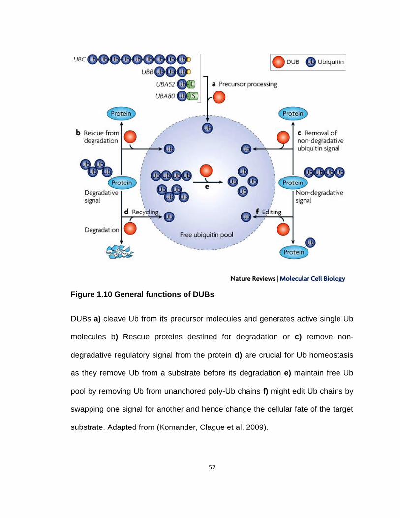
57
Figure 1.10 General functions of DUBs
DUBs a) cleave Ub from its precursor molecules and generates active single Ub
molecules b) Rescue proteins destined for degradation or c) remove non-
degradative regulatory signal from the protein d) are crucial for Ub homeostasis
as they remove Ub from a substrate before its degradation e) maintain free Ub
pool by removing Ub from unanchored poly-Ub chains f) might edit Ub chains by
swapping one signal for another and hence change the cellular fate of the target
substrate. Adapted from (Komander, Clague et al. 2009).

58
1.2.5 Targeting UPS
Regulated proteolysis by UPS controls almost all aspects of cellular
physiology and it is no wonder that defects in the components of UPS have been
implicated in the pathogenesis of several human diseases (Herrmann,
Ciechanover et al. 2004; Corn 2007; Petroski 2008; Hoeller and Dikic 2009;
Lehman 2009; Schlossarek, Frey et al. 2014). The incentives of targeting this
intricately regulated system seems obvious, and the expedited approval of
bortezomib (PS-341, Velcade ®; Millennium Pharmaceuticals), to become first
FDA approved proteasome inhibitor in clinical practice, for the treatment of
multiple myeloma and mantle cell lymphoma has corroborated UPS and its
components as legitimate therapeutic targets. (Kane, Bross et al. 2003; Fisher,
Bernstein et al. 2006).
Fast track approval of an improved proteasome inhibitor that overcomes
bortezomib resistance and has a superior toxicity profile, [Carfilzomib (Kyprolis,
Onyx Pharmaceuticals)], for multiple myeloma by the FDA and several orally
bioavailable proteasome inhibitors in clinical trials clearly demonstrate the
effectiveness of targeting this pathway (Kuhn, Chen et al. 2007; Herndon,
Deisseroth et al. 2013; Shen, Schmitt et al. 2013). Given the success of
proteasome inhibitors in the treatment of cancer and advances in our
understanding of UPS and its components, several small molecule inhibitors
have been developed or are in the pipeline that target specific components of
this pathway e.g. MLN4924 (NAE, Millenium). This is a small molecule inhibitor
for E1 enzyme of a Ub like protein NEDD8, and is in a phase 2 clinical trial
59
(Soucy, Smith et al. 2009). Several small molecules targeting E3 ligases are
either in Phase I clinical trials or are in the preclinical phase (Shen, Schmitt et al.
2013). DUB inhibitors are recent and a USP7/HAUSP inhibitor is already in active
preclinical development owing to its role in p53 regulation (Shen, Schmitt et al.
2013). A novel small molecule inhibitor b-AP15 that reversibly targets
proteasomal DUBs, USP14, and UCHL5, has anti-cancer properties and it even
induced cytotoxicity in bortezomib-resistant cells (D'Arcy, Brnjic et al. 2011).
Taken together, targeting UPS for human therapy seems promising, and
small molecule inhibitors targeting specific and smaller portions of UPS pathway
have shown encouraging results. Platelets also have components of UPS
(Burkhart, Vaudel et al. 2012) system, but the exact role of UPS in modulating
platelet function is not clear. However, older platelets have a reduced proteome
compared to newly released platelets suggesting a role of proteasome in
platelets (Ault, Rinder et al. 1992).
The studies in this thesis were conducted using small molecule inhibitors
targeting different components of the Ub-proteasome pathway in order to gain
mechanistic understanding of the potential role of this indispensible system in
modulating platelet reactivity.

60
CHAPTER II
Proteasome proteolysis supports stimulated platelet function
and thrombosis
2.1 Introduction
Proteasome inhibitors are widely used for the treatment of hematologic
cancers, specifically relapsed/refractory multiple myeloma and mantle cell
lymphoma, (Kane, Bross et al. 2003; Adams 2004; Kane, Dagher et al. 2007) yet
little is known of their impact on platelet function and hemostasis. Potentially,
these agents may also target platelets because a cyclic course of the
proteasome inhibitor bortezomib (Velcade®) induces significant cyclic
thrombocytopenia, (Lonial, Waller et al. 2005) and proteasome inhibition hastens
platelet death and turnover (Nayak, Kulkarni et al. 2013). Bortezomib therapy
also associates with reduced thrombosis (Zangari, Guerrero et al. 2008; Shen,
Zhou et al. 2011). Beyond this, there is little evidence of proteasome action on
the platelet proteome, their activation, or on thrombosis.
Platelets are recruited to the vascular wall at high shear through the GPIb-
IX-V receptor complex, (Goto, Ikeda et al. 1998) where the GPIbα subunit binds
von Willebrand factor (VWF) (Kroll, Harris et al. 1991) while its C-terminal
61
cytoplasmic domain interacts with the actin binding protein Filamin A with high
affinity (Ithychanda, Hsu et al. 2009). This interaction tethers the receptor
complex to the platelet cytoskeleton, maintaining the cytoskeletal architecture of
resting platelets and those adhering in vessels at high shear rates (Cranmer,
Ashworth et al. 2011). The GPIbα subunit also contains a high affinity binding site
for thrombin that contributes to platelet activation when thrombin concentrations
are low (Greco, Tandon et al. 1996). These interactions are essential for
thrombosis (Guerrero, Shafirstein et al. 2008). Thrombotic platelet deposition is
modeled in mice by FeCl3 induced injury of carotid arteries (Li, Febbraio et al.
2010; Li, McIntyre et al. 2013) resulting in rapid platelet adhesion and formation
of an occlusive platelet-rich thrombus at the site of injury.
Nucleated cells remove proteins from their proteome by proteasome-
catalyzed proteolysis. This multimeric complex consists of a 20S catalytic core of
non-catalytic α-subunits and three distinct β-subunits that hydrolyze peptide
bonds of unfolded protein substrates by β1 caspase-like hydrolysis, β2 tryptic
hydrolysis, and β5 chymotryptic cleavage (Chen and Hochstrasser 1996; Arendt
and Hochstrasser 1997; Dick, Nussbaum et al. 1998; Nussbaum, Dick et al.
1998). The proteasome is capped by a 19S regulatory subunit that conducts
substrate recognition, deubiquitination, unfolding, and protein translocation into
the 20S core (Dahlmann, Ruppert et al. 2000; Dahlmann, Ruppert et al. 2001)
Substrates for proteasome hydrolysis are recognized by the 19S core through
the covalent conjugation of monomeric or polymeric chains of the ~8 kDa
ubiquitin to the targeted protein (Nalepa, Rolfe et al. 2006; Jacobson, Zhang et

62
al. 2009; Xu, Duong et al. 2009).
Platelets contain several components of the 20S proteasome core
(Yukawa, Sakon et al. 1993; Ostrowska, Wojcik et al. 1997; Ostrowska,
Ostrowska et al. 2003; Avcu, Ural et al. 2008) and possess at least the
chymotryptic activity of the proteasome (Nayak, Kumar et al. 2011). In addition,
platelets have components of UPS (Burkhart, Vaudel et al. 2012) system, but the
exact role of UPS in modulating platelet function is not clear. Platelets should
contain functional E1 ubiquitin activating activity and E2/E3 ligases because
collagen activation of gpVI stimulates ubiquitination of platelet Syk kinase
(Dangelmaier, Quinter et al. 2005) through the E3 ligase Cbl-b (Daniel,
Dangelmaier et al. 2010). Furthermore, clinical inhibitor of proteasome
(bortezomib) reduced ADP-induced aggregation (Avcu, Ural et al. 2008) and
platelets isolated from patients receiving bortezomib therapy are hyporesponsive
to other stimuli (Zangari, Guerrero et al. 2008; Shen, Zhou et al. 2011).
Moreover, older platelets have a reduced proteome compared to newly released
platelets suggesting a role of proteasome in platelets (Ault, Rinder et al. 1992).
Here we show the expression of a functional ubiquitin/ proteasome
system in platelets. By investigating the impact of proteasome inhibition on a well
established mouse model of thrombosis and on a range of ex vivo activities, we
conclusively demonstrate that the platelet proteasome contributes to cellular
activation and function. Therapeutic proteasome inhibition in platelets produces a
hypothrombotic state, and might also augment anti-platelet therapy.

63
2.2 Materials and Methods
2.2.1 Chemicals and reagents
Chemicals and reagents were purchased from: sterile filtered hanks
balanced salt solution (HBSS) and sterile tissue culture plates (Falcon Labware);
endotoxin-free human serum albumin (25% human albumin solution, Baxter
Healthcare); endotoxin-free PBS, phenol-extracted LPS (Escherichia coli
O111:B4) free of lipoprotein contamination (List Biological Laboratories);
recombinant soluble CD14, recombinant lipopolysaccharide-binding protein
(LBP), Allophycocyanin (APC)-anti-human vWF (R&D Systems); anti-
polyubiquitin antibodies FK1 and FK2, anti-UBE1, UbiQapture™-Q kit (Enzo
lifesciences); anti-Filamin A, anti-Talin1 (C45f1), anti-β-actin and anti-skp1 (Cell
signaling); anti-Cul5 and anti-Asb2 (Abcam); anti-talin 1(YQ 16), anti-GPVI, anti-
p53 and SZ2 antibody (SCBT); p53 ubiquitination kit, anti-UBcH 1,5 and 7, anti-
RPN2 and anti-11S-α subunit (Boston Biochem); MG132, AYPGKF (PAR4
agonist peptide), SFLLRN (PAR1 agonist peptide) and RWJ56110 (Tocris);
Bortezomib (Eurasia); Antichrome TF® (American Diagnostica); Phycoerytherin
(PE)-anti-human CD42b (Millipore); fluorescein isothiocyanate (FITC)- CD36
(Cayman Chemicals); Calcein AM and Fura-2 AM (InvitrogenTM); Ristocetin and
Botrocetin (Sigma-Aldrich). Other chemicals were from Sigma-Aldrich or Biomol
Research Laboratories.
64
2.2.2 Platelet preparation
Human blood was drawn into acid-citrate-dextrose and centrifuged (200 ×
g, 20 min) without braking to obtain platelet-rich plasma in a protocol approved
by the Cleveland Clinic Institutional Review Board. Purified platelets were
prepared as stated (Brown and McIntyre 2011). Briefly, platelet-rich plasma was
filtered through two layers of 5-μm mesh (BioDesign) to remove nucleated cells
and recentrifuged (500 × g, 20 min) in the presence of 100 nM PGE1. The pellet
was resuspended in 50 ml PIPES/saline/glucose (5 mM PIPES, 145 mM NaCl, 4
mM KCl, 50 μM Na2HPO4, 1 mM MgCl2, and 5.5 mM glucose) containing 100 nM
PGE1. These cells were centrifuged (500 × g, 20 min) and recovered platelets
were centrifuged again before resuspension in 0.5% human serum albumin in
HBSS. For pure platelets, cells were resuspended in AutoMACS sample buffer, 5
μl anti-CD45, anti-CD15, anti-CD14, and anti-glycophorin–coated magnetic
beads (Miltenyi Biotec) per 109 cells for 25 min with constant rotation before
purification in an AutoMACS magnetic separator (Miltenyi Biotec).
2.2.3 Proteasome function
Proteasome-Glo™ 3-Substrate System (Promega) separately assayed
hydrolysis of luminogenic substrates for the three proteasome activities
[chymotryptic (Suc-LLVY-aminoluciferin), tryptic (Z-LRR-aminoluciferin), and
caspase-like or postglutamyl peptide hydrolase (Z-nLPnLD-aminoluciferin)].
Washed platelets (4 X 10⁸/ml) were pretreated with or without 30 µM MG132 for
30 min, pelleted and lysed using NP-40 lysis buffer (150 mM NaCl, 25 mM Tris
[pH 7.6], 1% nonidet P-40, 2 mM EDTA, 1 mM sodium orthovanadate, 10 mM

65
sodium fluoride, 100 µM PMSF, 1 µg/mL pepstatin, and 10 µg/mL leupeptin to
inhibit non-proteasome proteases) and incubated with luminogenic substrates for
30 min before luminescence was measured. Addition of protease inhibitors make
sure that cellular proteases don't cleave the substrate and only
2.2.4 In vivo thrombosis
Twelve week old C57BL6 mice were anesthetized with ketamine (90
mg/kg)/xylazine (15 mg/kg) and the right jugular vein and the left carotid artery
were exposed via a middle cervical incision. Platelets were labeled by injecting
100 μl of rhodamine 6G (0.5 mg/ml) in saline into the right jugular vein. MG132
(30 µM in DMSO; 6 mg/kg) or DMSO vehicle (10.5 µl) was added to the above
solution, and the drug was allowed to circulate for 15 min before FeCl3 injury (Li,
Febbraio et al. 2010; Li, McIntyre et al. 2013). Thrombosis was induced in the left
carotid artery by stripping the adventitia and placing a piece of black plastic
under the vessel to reduce background fluorescence. A 1 X 2 mm piece of filter
paper saturated with 7.5% FeCl3 was applied to the carotid artery for 1 minute,
the filter paper was removed, and the vessel rinsed with saline. Fluorescent
thrombus formation was observed in real-time under a water immersion objective
at 10 X magnification. Time to occlusive thrombosis was determined offline using
video image capture with a QImaging Retigo Exi 12-bit mono digital camera
(Surrey, Canada) and Streampix version 3.17.2 software (Norpix, Montreal,
Canada). The end points were set as either cessation of blood flow for >30
seconds or no occlusion after 30 minutes (three times longer than the average
66
occlusion time), in which case the time was recorded as 30 minutes for statistical
comparison.
2.2.5 Platelet transfusion and murine carotid artery thrombosis assay
Eight week old female platelets donor mice were anesthetized with
ketamine (100 mg/kg) and xylazine (10 mg/kg) and 700 μl blood was obtained
from inferior vena cava puncture into 1 ml syringe containing 100 μl sodium
citrate (0.109 M). Blood was transferred into a 2 ml tube containing 800 μl
Tyrode’s buffer and platelet-rich plasma (PRP) were gathered by centrifugation at
100g for 10 minutes. PRP was further centrifuged at 100g for 10 minutes in the
presence of PGE1 to pellet platelets. 109 platelets resuspended in 500 μl saline
were treated with MG132 (30 uM) or vehicle for 30 min, centrifuged and re-
suspended in 200 μl saline.
Recipient wild type mice were exposed to 11Gy of external beam
irradiation from a Cesium 137 source to induce thrombocytopenia with platelet
counts <5% of normal after 5~6 days (Chen, Febbraio et al. 2008). At sixth days
after irradiation, the thrombocytopenic WT mice (8 weeks old) were anesthetized
as before and the right jugular veins and the left carotid artery exposed via a
middle cervical incision (Li 2013). The pre-treated donor platelets were then
fluorescently stained by adding 100 μl of rhodamine 6G solution (0.5 mg/ml in
saline), and injected through the jugular vein of thrombocytopenic mice 10
minutes prior to carotid injury to allow the transfused platelets to reach
equilibrium in the circulation. Vehicle DMSO treated platelets were used as

67
controls. Details of this thrombosis model have been described previously (Li
2013).
2.2.6 p53 ubiquitination
Washed platelets (109/ml) were lysed in NP-40 lysis buffer and incubated
with components from a p53 ubiquitination kit (Boston Biochem), but lacking
recombinant E1, for 60 min before the reaction was stopped with 2X reducing
SDS sample buffer. The products were resolved by SDS-PAGE, and p53
ubiquitination status determined by western blotting with anti- p53 antibody.
2.2.7 Western blotting and liquid chromatography-mass spectrometry
Washed platelets (4 X 10⁸/ml) were treated with 30 µM MG132 or buffer
for 30 min before treatment with thrombin (0.2 U) or ADP (100 µM). After
incubation, platelets were pelleted and lysed using radioimmunoprecipitation
assay (RIPA) lysis buffer [150 mM NaCl, 25 mM Tris (pH 7.6), 1% Nonidet P-40,
0.1% sodium dodecyl sulfate, 0.5% deoxycholate, 2 mM EDTA, 1 mM EGTA, 1
mM sodium orthovanadate, 10 mM sodium fluoride, 100 µM phenylmethylsulfonyl
fluoride, 1 µg/mL pepstatin, and 10 µg/mL leupeptin]. Samples were kept on ice
for 30 min with occasional vortexing to ensure complete lysis before
centrifugation (21,000 x g, 10 min). Reducing SDS sample buffer was added
before the proteins were resolved by SDS-PAGE.
Three different LC-MS/MS experiments were performed, an identification
experiment, truncation analysis with N-terminal tagging, and truncation analysis
with chymotryptic digestion with multiple reaction monitoring (MRM). For all of
68
these experiments the SDS-PAGE bands were excised from the Coomassie
stained gels, washed, reduced and alkylated prior to in-gel digestion with either
trypsin (identification and TMT N-terminal tagging experiments) or chymotrypsin.
The N-terminal tagging was performed on extracted peptides by adding 100 µl of
the tandem mass tags (TMT, Thermo scientific) in 200 mM triethyl ammonium
bicarbonate for 1 hr. The reaction was quenched by 5% hydroxylamine for 15
min, combined, dried by speedvac, and dissolved in 1% acetic acid.
Two different LC-MS/MS systems were used in these experiments. Protein
identification and MRM analysis was carried out on a LTQ linear ion trap
equipped with an Eksigent nano-1D HPLC system. Peptides were resolved on a
self-packed Phenomenex Jupiter C18 column (8 cm x 75 µm) with an
acetonitrile/0.1% formic acid gradient. The instrument was operated in a either a
data dependent mode in which a mass scan was followed by MS/MS scans on
the most abundant ions or Multiple Reaction Monitoring (MRM) mode in which
specific ions are fragmented and assessed over the entire course of the LC
experiment. Proteins were identified by searching the LC-MS/MS spectra
against the human reference sequence database with the program Mascot. The
TMT-Tagged samples were analyzed on an LTQ-Orbitrap-Velos hybrid mass
spectrometer equipped with an Eksigent nano-1D HPLC system. Peptides were
resolved on a PicoFrit column packed with ProteoPep 2 (10 cm x 75 µm) C18
with an acetonitrile/0.1% formic acid gradient. The instrument was operated in a
data dependent mode in which all high resolution mass scans were obtained in
the Orbitrap (resolution at 60,000) to identify the five most abundant ions. These
69
ions were then subjected to five MS/MS scans in the LTQ for peptide
identification, and five HCD (higher energy collisional dissociation) scans in the
Orbitrap for TMT Tag quantitation. The resulting data was then searched
against the human reference sequence database
(http://www.ncbi.nlm.nih.gov/RefSeq/) using the program SEQUEST. The search
parameters include 10 ppm parent ion tolerance, 0.8 Da MS/MS tolerance, full
tryptic peptides, oxidized methionine as a variable modification,
carbamidomethylated cysteines and TMT tag modifications as static
modifications. Positive protein identifications were determined by setting the
FDR rate to less than 1% and requiring at least 2 peptides per protein. Peptide
TMT quantitation was performed by Proteomics Dynamics Software with an
integration window tolerance of 20 ppm and monoisotopic masses for the tabs at
126.127 and 127.131 Da.
2.2.8 Total internal reflection microscopy (TIRF) microscopy
Washed platelets (2 X 10⁸/ml) were treated with 30 µM MG132 or not for
30 min before addition onto a drop of 0.025 U thrombin placed in a glass
bottomed microwell (MatTek) dish. Imaging was performed using at 100X with a
1.46 N.A. objective in a Leica AM TIRF MC System (Leica Microsystems,
Wetzlar, Germany) equipped with an ImageEM C9100-13 EMCCD camera
(Hamamatsu, Bridgewater, N.J). The 488 nm 10-mW diode laser was used for
excitation and the penetration depth was set to 70 nm. Within 10 seconds of
being placed on the stage, the sample was focused and a time-lapse series was
70
initiated to collect images every 3 seconds for 5 minutes. At the end of the series,
five static images were collected in both TIRF and differential interference
contrast (DIC) modes of five different fields in the sample.
2.2.9 Microparticle isolation and quantitation
Washed platelets (109/ml) were pretreated with 30 µM MG132, 40 µM
bortezomib or buffer for 30 min before an overnight treatment with thrombin (0.2
U), ADP (100 µM) or 0.1 g/ml LPS with addition of 0.1 g/ml each of human
recombinant CD14 and LPS binding protein (LBP). Platelets were removed twice
by centrifugation (500 x g) before microparticles were collected by centrifugation
(100,000 x g, 90 min). The microparticle pellet was resuspended in 0.5 % human
serum albumin/HBSS MPs and were counted by flowcytometery using defined
numbers of 3-m polystyrene latex beads (Sigma) as an internal standard for
size. Forward scatter (FSC) and side scatter (SSC) were drawn to include 50,000
3-m events. Microparticle size was determined using 1-m beads (Sigma).
Flowcytometery was performed using settings where the threshold was lowered
to 200 and FSC and SSC gates were drawn to include events 1-m in size or
smaller. Microparticles express phosphatidylserine on their surface which can be
detected by annexin-V staining. Based on the flowcytometer size gating, only
microparticles (1-m ) stained with annexin-V were enumerated.
2.2.10 Peptidyl activity of tissue factor (TF) on microparticles
Tissue factor (TF)-dependent procoagulant activity was assessed with an
Actichrome TF assay (American Diagnostica) kit as previously described (Del

71
Conde, Shrimpton et al. 2005). Washed platelets (109/ml) were pretreated with
30 µM MG132 for 30 min before an overnight treatment with thrombin (0.2 U).
Platelets were removed twice by centrifugation (500 x g) before microparticles
were collected by centrifugation (100,000 x g, 90 min). The microparticle pellet
was resuspended in 0.5% human serum albumin/HBSS and were immediately
placed in 25 μl of kit assay buffer and TF was calculated. The data are displayed
as fold increase in peptidyl activity of TF on microparticles at absorbance 405
nm.
2.2.11 Clot retraction
Washed platelets (5 X 10⁸/ml) were incubated with MG132 (30 µM, 30
min) before fibrinogen was added to a final concentration of 500 µg/mL and
dispensed (500 µl) into siliconized glass tubes. Clot retraction was initiated by the
addition of human α-thrombin (0.2 U/mL) with clot volume imaged over time. The
size of the retracted clot was determined at stated times using Imagepro plus
software (Media Cybernatics, Bethesda,MD) to calculate retraction.
2.2.12 Aggregation
Washed platelets (2 X 10⁸/ml) were pretreated with combinations of 30 µM
MG132, 40 µM bortezomib, 10ng SZ2, 35 nM RWJ56110 (PAR1 receptor
inhibitor) or buffer for 30 min before stimulation with low (0.025 U) or high
concentrations of thrombin (0.1 U) or 50 µM SFLLRN (PAR1 specific agonist).
Platelet aggregation was measured by transmittance (Chronolog) with stirring
(600 rpm). For ristocetin or botrocetin induced agglutination, washed platelets (2
72
X 108/ml) were resuspended in a buffer containing 50% HBSS-A and 50%
platelet poor plasma (PPP) and were pretreated with 30 µM MG132, 40 µM
bortezomib or buffer for 30 min. Agglutination was induced with either 650 µg/ml
ristocetin or 300 ng/ml botrocetin and was measured by transmittance
(Chronolog) with stirring (1000 rpm).
2.2.13 Flow cytometry
Washed platelets (2 X 10⁸/ml) were pretreated with 30 µM MG132 or
buffer for 30 min before stimulation with 0.025 U thrombin (30 min) followed by
fixation with 2% paraformaldehyde. Anti-CD42b antibody or non-immune isotype
control (Millipore) was used to determine the surface expression of GPIbα by flow
cytometery.
2.2.14 Expression of data and statistics
All experiments were performed at least three times with cells from
different donors, and all assays were performed in triplicate. The standard errors
of the mean from all experiments are presented as error bars. Figures and
statistical analyses were generated with Prism4 (GraphPad Software). A value of
p≤ 0.05 was considered statistically significant.
2.3 Results
2.3.1 The platelet proteasome aids occlusive thrombosis
Human platelets expressed all three primary proteolytic activities of the
proteasome, effectively hydrolyzing peptides through chymotryptic, tryptic and
73
caspase-like cleavage (Fig. 2.1 A). The luminescent product was the same for
each luminogenic proteasome substrate, so the caspase-like activity was
approximately four times more efficient than either the tryptic or chymotryptic
activity. The trimeric leucinyl proteasome inhibitor MG132 at 30 M completely
blocked platelet chymotryptic and caspase-like activities, and reduced the tryptic
activity by half. MG132 at 30 µM is just optimally inhibitory (Fig. 2.1 B). Murine
platelets also hydrolyzed these three substrates at equivalent ratios (Fig. 2.1 C)
indicating that murine platelets have active proteasome.
We determined whether proteasome inhibition affected thrombosis by
systemically injecting MG132 into BL6 mice to achieve an estimated initial
circulating concentration of 30 µM along with rhodamine dye to fluorescently
label circulating platelets. We initiated occlusive thrombosis 15 min later (after
MG132 or DMSO injection) in a surgically exposed external carotid artery by a
brief ectopic application of 7.5% FeCl3 (Li, Febbraio et al. 2010; Li, McIntyre et al.
2013). This oxidative insult to the vascular wall resulted in the deposition of
fluorescently labeled platelets along the damaged vessel wall that increased over
several minutes (Fig. 2.1 D). Typically, complete occlusion of the vessel occurred
by 10 min after FeCl3 treatment, but in animals previously injected with MG132
occlusion was significantly delayed to 25 min (Fig. 2.1 E).
The delay in thrombosis after systemically administering MG132 need not
solely reflect participation of platelet proteasomes in thrombosis. We therefore
isolated and washed platelets from wild-type BL6 mice and treated these isolated
cells with 30 µM MG132 or buffer, washed the cells by centrifugation and

74
transfused these cells into mice previously rendered thrombocytopenic by
gamma irradiation. We induced carotid artery thrombosis with ectopic FeCl3 as
before to find control platelets were fully functional and occluded the carotid
artery by 10 min (Fig. 2.1 F). We again found occlusive thrombosis was delayed
in mice reconstituted with platelets treated with MG132 ex vivo. The platelet
proteasome thus participates in arterial thrombosis.

75
76
Figure 2.1 Platelets express a functional proteasome that contributes to
occlusive thrombosis
(A) Platelets contain a functional proteasome. Proteasome proteolytic activities
were individually assayed using luminogenic substrates for indicated catalytic
activities in lysates of untreated and MG132-treated platelets (n=3; **p≤ 0.01). (B)
Proteasome proteolytic activities were individually assayed as mentioned in
panel (A) in lysates from untreated platelets and platelets treated with different
concentrations of MG132 (10, 20, 30 and 40 µM), n=6; ***p<0.001, **p≤ 0.01). (C)
Mouse platelets contain a functional proteasome. Proteasome proteolytic
activities were individually assayed in mouse platelet lysates using luminogenic
substrates for indicated catalytic activities (n=4). (D) Fluorescent platelet
accretion after FeCl3 damage to carotid arteries is reduced by MG132. Video
frames at 1 min intervals of thrombus formation in FeCl3-damaged carotid
arteries (n=5). (E) MG132 lengthens the time to vascular occlusion. Time to
cessation of blood flow in mice treated as in the preceding panel determined by
cessation of platelet movement (n=8 experimental, 6 control; ***p≤ 0.001). (F) Ex
vivo MG132 pretreatment of platelets prolongs occlusion time after transfusion
into irradiated thrombocytopenic mice. Platelets from donor mice were first
pretreated for 30 min with MG132 and were washed before transfusion into
normal BL6 mice and FeCl3-induced thrombosis of carotid arteries initiated 15
min later (n=4 experimental, 3 control; *p≤ 0.05).

77
2.3.2 Platelets contain a stimulatable ubquitination system
Modification of platelet function by MG132 suggests platelets may
designate proteins as substrates for proteolysis by ubiquitination, as in nucleated
cells. We determined whether platelets contained E1, E2, E3 enzymes that
sequentially conjugate ubiquitin to target proteins. In order to ensure that we
assessed only platelet proteins, we also isolated highly purified, negatively
selected platelets that were essentially free of nucleated cells (less than 1
monocyte in 109 platelets (Shashkin, Brown et al. 2008). Western blotting
showed that these platelets contained the E1 activating enzyme UBE1, E2 and
E3 ligases, and Rpn2 and the 11S -subunit of the 19S regulatory complex (Fig.
2.2 A).
We used in vitro ubiquitination of exogenous p53, since platelets lack this
transcription factor, to determine whether platelet UBE1 was functional.
Incubation of recombinant p53 with recombinant E1 and appropriate recombinant
E2 and E3 enzymes along with ubiquitin and ATP resulted in robust p53
ubiquitination with formation of numerous slowly migrating adducts (Fig. 2.2 B).
Substitution of a platelet lysate for recombinant E1 also promoted in vitro
ubiquitination of p53, although the higher molecular weight ladder was less
prominent than produced by recombinant E1 (Fig. 2.2 B).
We next determined whether endogenous platelet proteins were modified
by ubiquitin. Western blotting with FK2 antibody that recognizes both mono- and
poly-ubiquitin chains showed that the proteome of quiescent cells contained
multiple ubiquitinated proteins (Fig. 2.2 C). The intensity of ubiquitination
78
increased after thrombin activation, although the pattern and position of the
adducted bands was unaltered compared to control cells. ADP stimulation also
increased ubiquitination of platelet proteins, but this primarily reflected an
increase in modification of very slowly migrating proteins (Fig. 2.2 C).
We investigated whether inhibiting the platelet proteasome altered the
amount of ubiquitinated proteins. Pretreatment with MG132 increased both the
abundance and the levels of ubiquitinated proteins as detected with the anti-
ubiquitin antibody (Fig. 2.2 D). In fact, MG132 was significantly more effective
than thrombin or ADP stimulation in enhancing decoration of the proteome with
ubiquitin, and the combination of agonist stimulation and MG132 was not
different from the effect of MG132 alone (Fig. 2.2 D). We observed a similar
increase in ubiquitinated proteins after treatment with bortezomib (Fig. 2.2 E).
Probing the platelet proteome with FK1 antibody that recognizes only poly-
ubiquitinated proteins revealed less abundant modification in unstimulated cells
(Fig. 2.2 F). This was increased by agonist stimulation and was again greatly
enhanced by MG132 treatment. Comparison of FK2 (Fig. 2.2 D) and FK1 (Fig.
2.2 F) immunoblots showed distinct patterns of mono-and poly-ubiquitination of
the platelet proteome.
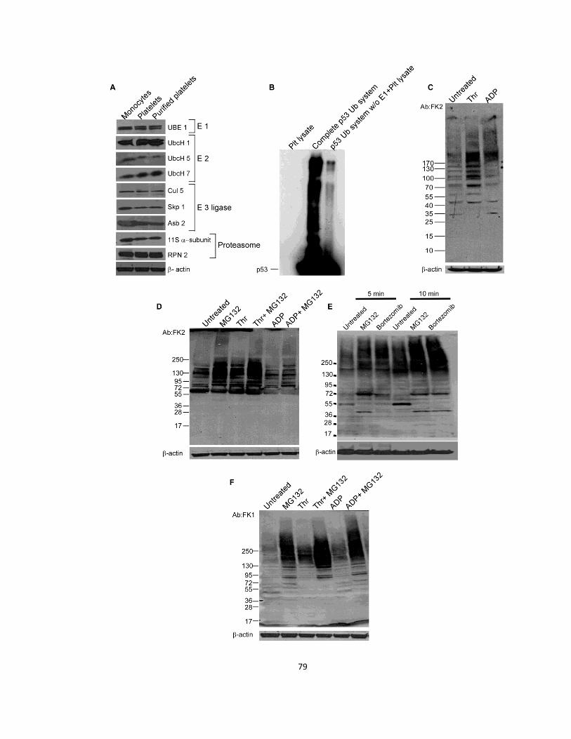
79
80
Figure 2.2 Platelets contain a functional ubiquitination system
(A) Platelets contain components of ubiquitin-proteasome system. Immunoblots
of platelet E1 activating enzyme, E2 and E3 conjugating enzymes, and
proteasome regulatory cap proteins (n=3). (B) Platelet E1 is functional. Platelet
lysates substituted for recombinant E1 and together with recombinant E2, E3,
ubiquitin and ATP ubiquitinated recombinant p53 (n=3). (C) Agonist stimulation
increases platelet proteome ubiquitination. Lysates of platelets stimulated with
buffer, thrombin (thr) or ADP were immunoblotted with FK2 antibody against
mono-and polyubiquitinated proteins (n=3). (D) MG132 increases proteome
ubiquitination. Lysates of platelets, treated or not with MG132 were stimulated, or
not, before immunoblotting with FK2 (n=3). (E) Both MG132 and bortezomib
increases proteome ubiquitination. Western blot of MG132 or bortezomib treated
platelet lysate over indicated times as probed with FK1 antibody (n=3). (F)
MG132 increases poly-ubiquitination of platelet proteins (n=3). Western blotting
of platelet lysates with FK1 antibody (n=3).
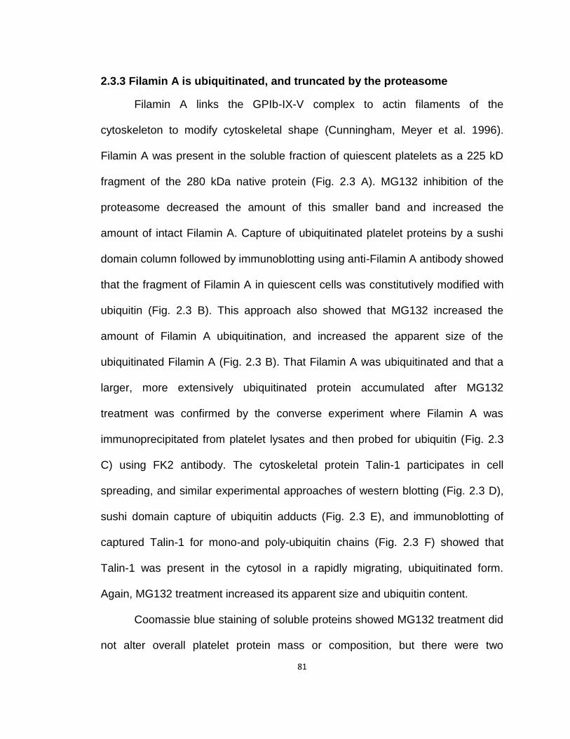
81
2.3.3 Filamin A is ubiquitinated, and truncated by the proteasome
Filamin A links the GPIb-IX-V complex to actin filaments of the
cytoskeleton to modify cytoskeletal shape (Cunningham, Meyer et al. 1996).
Filamin A was present in the soluble fraction of quiescent platelets as a 225 kD
fragment of the 280 kDa native protein (Fig. 2.3 A). MG132 inhibition of the
proteasome decreased the amount of this smaller band and increased the
amount of intact Filamin A. Capture of ubiquitinated platelet proteins by a sushi
domain column followed by immunoblotting using anti-Filamin A antibody showed
that the fragment of Filamin A in quiescent cells was constitutively modified with
ubiquitin (Fig. 2.3 B). This approach also showed that MG132 increased the
amount of Filamin A ubiquitination, and increased the apparent size of the
ubiquitinated Filamin A (Fig. 2.3 B). That Filamin A was ubiquitinated and that a
larger, more extensively ubiquitinated protein accumulated after MG132
treatment was confirmed by the converse experiment where Filamin A was
immunoprecipitated from platelet lysates and then probed for ubiquitin (Fig. 2.3
C) using FK2 antibody. The cytoskeletal protein Talin-1 participates in cell
spreading, and similar experimental approaches of western blotting (Fig. 2.3 D),
sushi domain capture of ubiquitin adducts (Fig. 2.3 E), and immunoblotting of
captured Talin-1 for mono-and poly-ubiquitin chains (Fig. 2.3 F) showed that
Talin-1 was present in the cytosol in a rapidly migrating, ubiquitinated form.
Again, MG132 treatment increased its apparent size and ubiquitin content.
Coomassie blue staining of soluble proteins showed MG132 treatment did
not alter overall platelet protein mass or composition, but there were two

82
abundant exceptions (Fig. 2.3 G). Two new, slowly migrating bands appeared in
the soluble fraction of platelets treated with MG132. Mass spectrometry showed
Filamin A to be present in the new band 1, and Talin-1 in bands 1 and 2 of the
resolved soluble proteins, corresponding to the altered mobility detected by
western blotting.
We used Tandem Mass spectrometry Tags (TMT)—chemically identical
tags differentially substituted with heavy atoms that enable quantitative
multiplexed analysis—to quantitatively compare the amount of Filamin A peptides
in tryptic digests of control and MG132 treated platelets. Untreated (band 3) and
MG132 treated (band 1) platelets were separately digested with trypsin, their
primary amines exhaustively modified with isobaric TMT tags (m/z 126 and m/z
127, respectively) before combined analysis by mass spectrometry. The peptides
confirmed both bands contained Filamin A, and the tags showed the abundance
of Filamin A tryptic peptides in band 1 and band 3 uniformly gave a m/z 126/127
ratio of 1.9. This ratio plunged to 0.08 after the tryptic peptide containing residues
1718-1745 (Fig. 2.3 H), demonstrating increased abundance of the full length
protein after MG132 treatment. Chymotryptic digestion of band 3 isolated from
control cells generated a distinct peptide map and selected reaction monitoring
identified a new Filamin A peptide with S1746 as its amino terminus. This identifies
the R1745 - S1746 bond as the site of cleavage in control cells, which maps into the
unfolded hinge 1 region of Filamin A (Fig. 2.3 I). Notably, this cleavage is tryptic-
like, and is distinct from the previously determined calpain cut site (Gorlin, Yamin
et al. 1990).
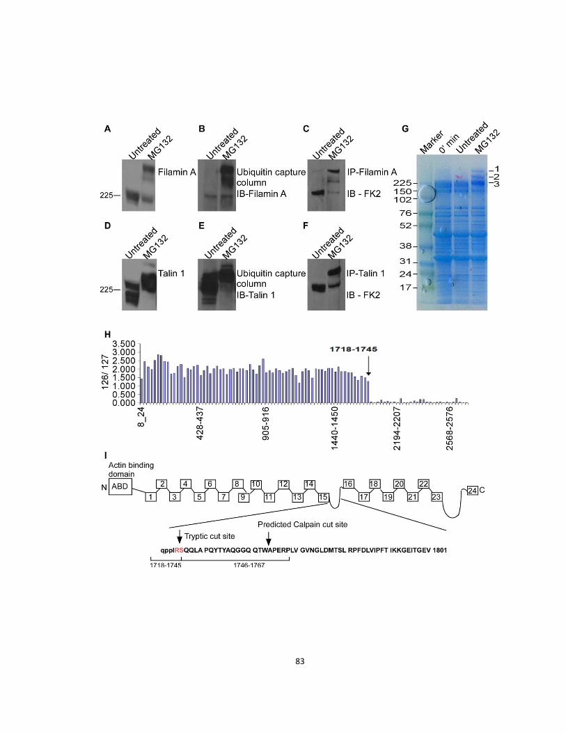
83
84
Figure 2.3 MG132 protects cytoskeletal protein cleavage
(A) Filamin A is ubiquitinated and its length increased by MG132. Western blot
of Filamin A in the soluble fraction of platelets before and after MG132 exposure
(n=3). (B) Ubiquitination and size of Filamin A are increased by MG132. Western
blot of Filamin A in eluates of ubiquitinated platelet proteins captured with sushi
columns [agarose beads conjugated to anti-ubiquitin antibody, (n=3)]. (C) Filamin
A ubiquitination and size are increased by MG132. FK2 western blot of mono-
and poly-ubiquitinated proteins immuoprecipitated with anti-Filamin A. (D) Talin-1
is ubiquitinated and its size increased by MG132. Western blot of Talin-1 before
and after MG132 exposure (n=3). (E) Ubiquitination and size of Talin-1 are
increased by MG132. Talin-1 in ubiquitinated platelet proteins captured by sushi
domain chromatography (n=3). (F) Talin-1 ubiquitination and size are increased
by MG132. FK2 western blot of mono- and poly-ubiquitinated proteins
immuoprecipitated by anti-Talin-1 (n=3). (G) MG132 increases high molecular
weight proteins. Coomassie stained gel of resolved platelet cytoplasmic proteins.
(H) Mass spectrometer determination of the mz 126/127 ratio of TMT-labeled
peptides along the Filamin A sequence (n=2). (I) Pictogram of Filamin A
structure.

85
2.3.4 Proteasome inhibition reduces cytoskeleton-dependent functions
Filamin A is a critical regulator of cytoskeletal structure and function, so
we next tested the hypothesis that proteasomal inhibition alters platelet functions
regulated by cytoskeletal dynamics, including GPIb function, microparticle
generation, and clot retraction.
First, we imaged cytoskeletal dependent spreading by Total Internal
Reflection Microscopy (TIRF) that detects only fluorophore closely opposed (≤
200 nm) to a glass matrix. Calcein-labeled platelets adhered over time after
activation by thrombin with extension of filopodia followed by lamellipodia (video
supplement) that resulted in adherent, spread cells (Fig. 2.4 A). In contrast, while
MG132-treated cells adhered and spread after thrombin stimulation, they did so
with less frequency, rapidity, and ultimately were less splayed (Fig. 2.4 B).
Stimulated platelets release pro-thrombotic microparticles from their
surface that depends on cytoskeletal rearrangement after stimulation (Morel,
Jesel et al. 2011). Thrombin induced a ~6 fold increase in microparticle shedding
(Fig. 2.4 C and D), which was significantly reduced by pre-treating the platelets
with either MG132 (Fig. 2.4 C) or bortezomib (Fig. 2.4 D). MG132 also blocked
microparticle release from LPS-stimulated platelets (Fig. 2.4 E). The majority of
these particles express phosphatidylserine on their surface which can be
detected by annexin V staining. Both MG132 (Fig. 2.4 F) and bortezomib (Fig.
2.4 G) reduced thrombin induced increase in annexin V positive microparticle
shedding. MG132 also suppressed LPS induced shedding of annexin V positive
microparticles (Fig. 2.4 H). The surface of these annexin V positive microparticles
86
is ~50-100 fold more thrombotic than even the activated platelets (Sinauridze,
Kireev et al. 2007) and they promote thrombosis through tissue factor pro-
coagulant activity (Fig. 2.4 I), which MG132 significantly inhibited.
Cytoskeletal rearrangement retracts newly formed thrombi, promoting
wound repair (Cheresh, Leng et al. 1999; Grinnell 1999). Stimulation of platelets
with thrombin induced rapid formation of a thrombus that then consolidated over
time (Fig. 2.4 J). MG132 interfered with this process, ultimately decreasing
retraction by 60%.
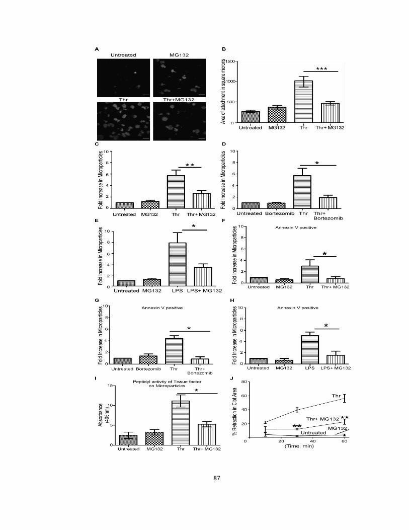
87

88
Figure 2.4 MG132 suppresses stimulated spreading, microparticle
shedding, and clot retraction
(A) MG132 disturbs thrombin stimulated adhesion and spreading. Interaction of
control or thrombin (thr, 0.025 U) stimulated platelets with a glass substrate were
imaged by Total Internal Reflection Microscopy after 5 min (n=3) (scale bar = 10
µM). (B) Platelet area after MG132 treatment. Platelet area in Panel A was
quantified by Imagepro plus software (n=3; ***p≤ 0.001). (C) Microparticle
shedding by platelets stimulated with 0.2 U thrombin with or without MG132 (n=3;
**p≤ 0.01). (D) Bortezomib (n=3; *p≤ 0.05). (E) Microparticle shedding by platelets
pretreated with or without MG132 and stimulated with lipopolysaccharide (LPS,
n=3; *p≤ 0.05). (F) Proteasome inhibitors suppress agonist-induced shedding of
procoagulant microparticles. Annexin V positive microparticle shedding by
platelets stimulated with 0.2 U thrombin with or without MG132 (n=3; *p≤ 0.05).
(G) Bortezomib (n=3; *p≤ 0.05). (H) Annexin V positive microparticle shedding by
platelets stimulated with lipopolysaccharide (LPS, n=3; *p≤ 0.05). (I) Tissue factor
procoagulant activity in platelet microparticles stimulated with 0.2 U thrombin with
or without MG132 (n=3; *p≤ 0.05). (J) MG132 represses clot retraction. Ratio of
the image surface area of images of thrombin-induced clots over time (n=3; **p≤
0.01).
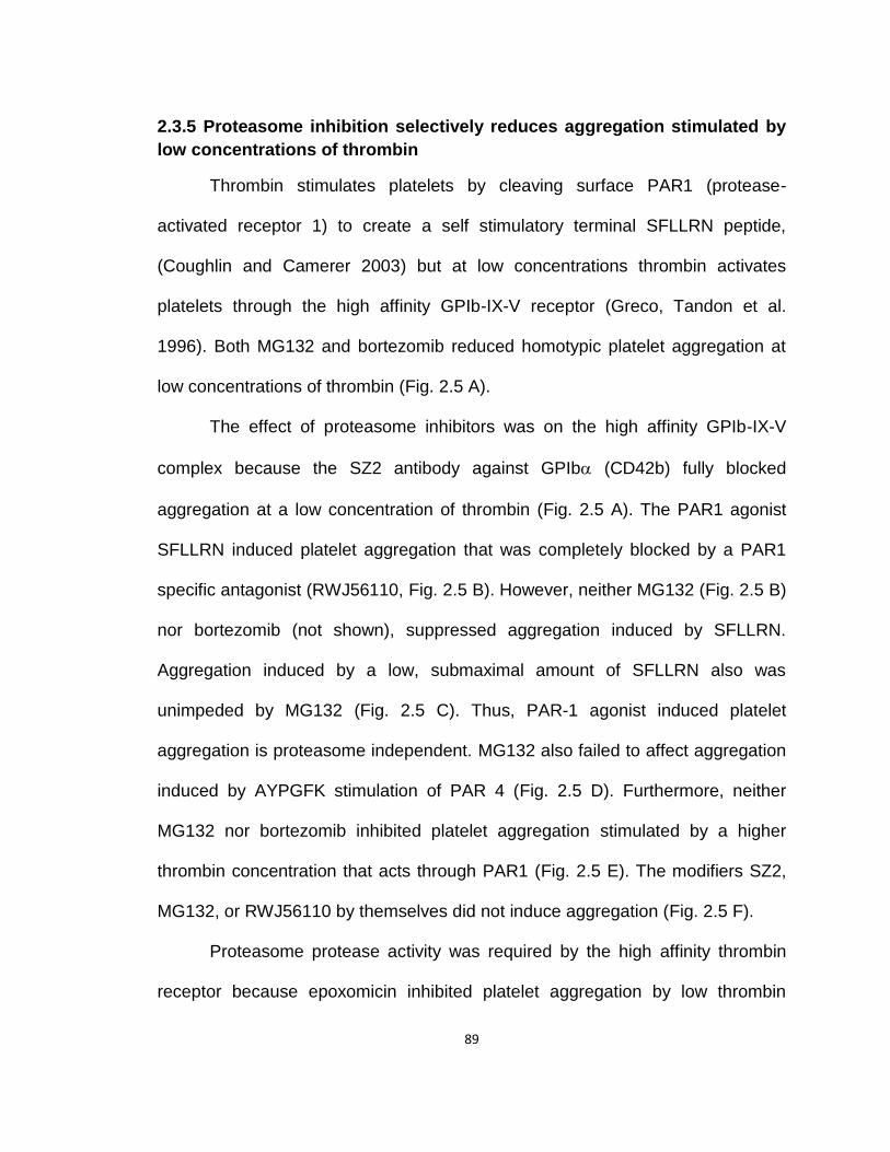
89
2.3.5 Proteasome inhibition selectively reduces aggregation stimulated by
low concentrations of thrombin
Thrombin stimulates platelets by cleaving surface PAR1 (protease-
activated receptor 1) to create a self stimulatory terminal SFLLRN peptide,
(Coughlin and Camerer 2003) but at low concentrations thrombin activates
platelets through the high affinity GPIb-IX-V receptor (Greco, Tandon et al.
1996). Both MG132 and bortezomib reduced homotypic platelet aggregation at
low concentrations of thrombin (Fig. 2.5 A).
The effect of proteasome inhibitors was on the high affinity GPIb-IX-V
complex because the SZ2 antibody against GPIb (CD42b) fully blocked
aggregation at a low concentration of thrombin (Fig. 2.5 A). The PAR1 agonist
SFLLRN induced platelet aggregation that was completely blocked by a PAR1
specific antagonist (RWJ56110, Fig. 2.5 B). However, neither MG132 (Fig. 2.5 B)
nor bortezomib (not shown), suppressed aggregation induced by SFLLRN.
Aggregation induced by a low, submaximal amount of SFLLRN also was
unimpeded by MG132 (Fig. 2.5 C). Thus, PAR-1 agonist induced platelet
aggregation is proteasome independent. MG132 also failed to affect aggregation
induced by AYPGFK stimulation of PAR 4 (Fig. 2.5 D). Furthermore, neither
MG132 nor bortezomib inhibited platelet aggregation stimulated by a higher
thrombin concentration that acts through PAR1 (Fig. 2.5 E). The modifiers SZ2,
MG132, or RWJ56110 by themselves did not induce aggregation (Fig. 2.5 F).
Proteasome protease activity was required by the high affinity thrombin
receptor because epoxomicin inhibited platelet aggregation by low thrombin

90
concentrations (Fig. 2.5 G) and this inhibition was statistically significant (Fig. 2.5
H). Epoxomicin is a fungal metabolite, structurally unrelated to either MG132 or
bortezomib, that specifically inhibits proteasome protolysis without inhibiting
calpain, trypsin, chymotrypsin, or cathepsin B at concentrations 50-times higher
than we used for blocking platelet aggregation (Meng, Mohan et al. 1999).
The GPIb-IX-V complex is displayed on the surface of quiescent cells, and is
down regulated after thrombin stimulation (Michelson, Ellis et al. 1991). I found
both MG132 (Fig. 3.5 I) and bortezomib (Fig. 3.5 J) stabilized the GPIb-IX-V
complex on the surface of stimulated cells, and actually enhanced surface GPIb
(CD42b) expression. This surface retention of the receptor in platelets treated or
not with proteasome inhibitors is significant (Fig. 2.5 K). Ristocetin increases the
affinity of GPIb for VWF, agglutinating unactivated cells, and pre-treatment of
quiescent platelets with MG132 (Fig. 2.5 L) or bortezomib (Fig. 2.5 M) reduced
ristocetin induced platelet agglutination. Thus despite the increased surface
expression of the GPIb-IX-V complex after proteasome inhibition, the complex is
less able to interact with VWF than in control platelets.
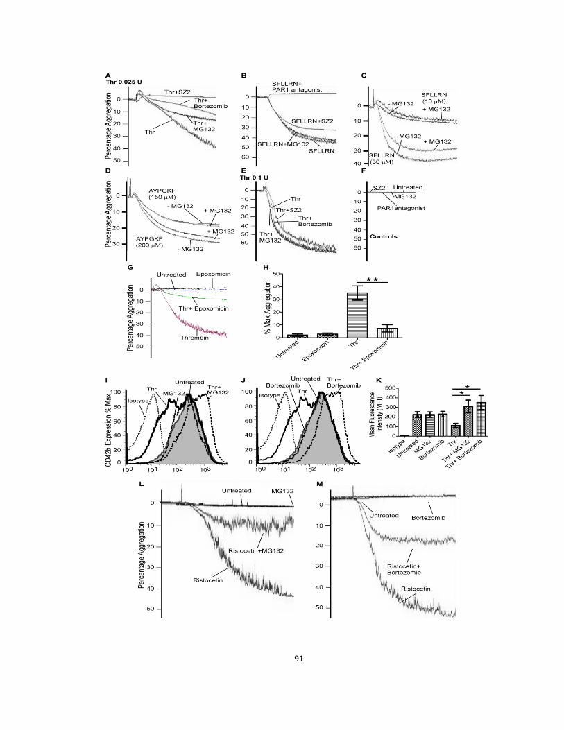
91
92
Figure 2.5 MG132 reduces aggregation in response to low dose thrombin,
and prevents GP1bα downregulation
(A) MG132 and bortezomib suppress platelet aggregation induced through the
high affinity thrombin receptor, GP1b-IX-V. Aggregation induced by 0.025 U
thrombin (thr) was reduced by MG132 and bortezomib (n=3). (B) MG132 failed to
reduce aggregation stimulated by the PAR1 (protease-activated receptor 1)
agonist peptide SFLLRN (n=3). (C) MG132 (30 µM) pretreatment minimally
reduces aggregation stimulated by low concentrations of the PAR1 agonist
peptide SFLLRN (n=3). (D) MG132 failed to reduce aggregation stimulated by
the PAR4 agonist peptide AYPGKF at both 200 µM and 150 µM concentrations
(n=3). (E) Neither MG132 nor bortezomib block aggregation from thrombin
activation of PAR1. Platelet aggregation by 0.1 U thrombin with or without SZ2
antibody (n=3). (F) GP1bα or PAR1 inhibition alone does not induce aggregation
(n=3). (G) Aggregation induced by 0.025 U thrombin (thr) was reduced in
platelets pretreated with 1 µM epoxomicin for 30 min (n=3). (H) Quantitative
representation of percentage maximum (Max) aggregation (n=3; **p≤ 0.01). (I)
MG132 repressed stimulated GPIbα down regulation. Flow cytometry of surface
CD42b (GPIbα) expression on platelets treated or not with MG132 and
stimulated or not with 0.025 U thrombin (n=3). (J) Bortezomib repressed surface
GPIbα down regulation. Flow cytometry of GPIbα expression on control and
thrombin stimulated platelets (n=3). (K) Mean fluorescence intensity (MFI) of
surface CD42b (GPIbα) expression on platelets treated either with MG132 or
bortezomib and stimulated or not with 0.025 U thrombin (n=3; *p≤ 0.05). (L)

93
MG132 suppressed ristocetin induced agglutination. Quiescent platelets
pretreated or not with MG132 were agglutinated with ristocetin (n=3). (M)
Bortezomib suppressed ristocetin induced agglutination. Quiescent platelets
pretreated or not with bortezomib were agglutinated with ristocetin (n=3).
2.4 Discussion
Multiple myeloma is associated with an increased incidence of venous
(Srkalovic, Cameron et al. 2004; Zangari, Elice et al. 2007; Kristinsson, Fears et
al. 2008) and arterial (Libourel, Sonneveld et al. 2010) thrombotic disease, and
bortezomib therapy suppresses these thrombotic states (Zangari, Guerrero et al.
2008). Platelets display proteasome chymotryptic activity that is greatly
stimulated by soluble agonists (Nayak, Kumar et al. 2011). Additionally,
bortezomib represses ADP-induced aggregation (Avcu, Ural et al. 2008) and
platelets isolated from patients receiving bortezomib are hyporesponsive to other
stimuli (Zangari, Guerrero et al. 2008; Shen, Zhou et al. 2011). A second
proteasome inhibitor, PSI, suppresses thrombosis in hypertensive animals
(Ostrowska, Wojtukiewicz et al. 2004).
I extend these observations by showing that platelets contain an intact and
functional ubiquitin/proteasome system that participates in agonist stimulated
responses, especially those aided by cytoskeletal rearrangement. I found that
inhibition of the proteasome’s proteolytic activity reduced thrombosis at sites of
oxidative damaged to murine carotid arterial walls. Since MG132 was introduced
into the circulation in this experiment, protection need not have been a direct
94
effect of MG132 on platelet function, but the target of MG132 in platelets is in fact
the proteasome. I treated purified platelets with MG132 ex vivo, washed them,
and then transfused these cells into mice rendered thrombocytopenic by prior
gamma irradiation. This platelet-specific exposure to MG132 interfered with the in
vivo function of platelets because MG132 treated platelets were deficient in their
occlusion of a FeCl3-damaged carotid artery. Remarkably, this ex vivo exposure
exactly mimicked systemic exposure to MG132. This establishes a role for the
platelet proteasome in thrombosis.
Platelets contained a functional ubiquitin system that modified cellular
proteins to mark them as proteasome substrates. The proteome of quiescent
platelets contained numerous ubiquitin-protein conjugates whose adduction was
increased upon stimulation, consistent with the prior observation that collagen
activation stimulates ubiquitination of platelet Syk kinase(Dangelmaier, Quinter et
al. 2005) through the E3 ligase Cbl-b.(Daniel, Dangelmaier et al. 2010) Two of
the proteins decorated with ubiquitin in quiescent platelets were Filamin A and
Talin-1. Agonist stimulation modestly increased the amount of ubiquitin esterified
in the platelet proteome, but blockade of the proteasome was far more effective
than agonist stimulation for this, with the result that heavily ubiquitinated Filamin
A, and Talin-1 accumulated in the cytoplasm of cells with diminished proteasome
proteolytic activity.
Filamin A is organized into an actin-binding domain (ABD), 24 tightly
compacted FERM immunoglobulin-like repeats connected with short linking
sequences, and two unstructured hinge regions. Cytoplasmic Filamin A migrated
95
more quickly than the intact protein during gel electrophoresis, suggesting it
primarily was a fragment. Quantitative comparison of tryptic peptides using
tandem mass spectrometry tags showed that MG132 protected Filamin A from
cleavage to the smaller 225 kDa fragment. Identification of Filamin A peptides
present only after MG132 treatment and identification of the new amino terminal
peptide after chymotryptic digestion by multiple reaction monitoring showed
cleavage of the Filamin A protein occurred between R1745 and S1746. This is in
a hinge region that is not compacted into FERM domains. Cleavage at this site is
distinct from the previously determined calpain cut site, (Gorlin, Yamin et al.
1990) and shows the protease was tryptic since it occurred after an arginine
residue. The proteasome subunits functionally interact with one another
(Kisselev, Callard et al. 2006) to degrade proteins to amino acids and small
peptides, but the proteasome also processes large proteins to functional
fragments, as shown by NF-κB p105 proteolysis to the p50 transcription factor
(Rape and Jentsch 2002). We now identify the longer unstructured Filamin A
loop as another site of limited proteasome proteolysis, but our data is associative
so we cannot ascribe this proteolysis as causal in MG132 inhibition of platelet
adhesion or spreading.
Platelets pretreated with MG132 or bortezomib aggregated less in
response to low concentrations of thrombin. Adhesion, spreading, microparticle
shedding, and the ability to generate the force to retract formed clots was also
reduced by proteasome inhibition. Cytoskeletal interactions aid microparticle
shedding from stimulated platelets, and proteolysis by activated calpain (Fox,
96
Austin et al. 1991; Basse, Gaffet et al. 1994) and caspase-3 (Shcherbina and
Remold-O'Donnell 1999) promote microparticle shedding through cytoskeletal
proteolysis (Basse, Gaffet et al. 1994). The reduction of microparticle shedding is
additionally revealing because MG132 suppressed shedding after stimulation by
LPS activation of TLR4, thrombin activation of PAR1, and ADP activation of
P2Y12. So, MG132 interference with stimulated platelet function is not restricted
to GPIb-IX-V activation by thrombin.
We establish that platelets, like nucleated cells, express a functional
ubiquitin/proteasome system that enables them to ubiquitinate their proteome.
This decoration increases upon stimulation, and modulates an array of
responses from several receptors that all engage the cytoskeleton. The
cytoskeletal proteins Filamin A and Talin-1 are targets of ubiquitination, and
proteasome mediated proteolysis. The ubiquitin/proteasome system of platelets
affects their response to thrombotic stimuli, and proteasome inhibition effectively
delayed arterial thrombosis. Bortezomib is already in clinical practice for multiple
myeloma and mantle cell lymphoma so elucidation of its anti-platelet activity
defines a new salutary function of these inhibitors.
97
2.5 Chapter summary
These data demonstrate that platelets, like nucleated cells, express a
functional ubiquitin/proteasome system that enables them to modify their
proteome by mono- and poly-ubiquitination. Proteasome inhibition increased
ubiquitin decoration of cytoskeletal proteins Filamin A and Talin-1 and increased
platelet content of soluble intact filamin A. Proteasome inhibition by MG132
suppressed stimulated platelet spreading as well as clot retraction. Proteasome
inhibition reduced platelet aggregation by low thrombin concentrations and
ristocetin-stimulated agglutination through the GPIb-IX-V complex. The effects of
proteasome inhibitors were not confined to a single receptor as both MG132 and
bortezomib suppressed thrombin-, ADP-, and LPS-stimulated microparticle
shedding. Systemic MG132 strongly suppressed formation of occlusive, platelet-
rich thrombi in FeCl3-damaged carotid arteries. Importantly, this is due to the
direct effect of proteasomal inhibition on platelets as transfusion of platelets
treated ex vivo with MG132 and washed prior to transfusion into
thrombocytopenic mice also reduced carotid artery thrombosis.
98
Chapter III
Unmasking platelet reactivity through deubiquitination
3.1 Introduction
Platelet activation underlies thrombotic cardiovascular disease that is
responsible for significant disability and death in the developed world (Davi and
Patrono 2007; Jackson 2011). Platelet activation is transduced from the
proteome delivered to platelets as they bud from megakaryocytes during
thrombopoiesis, augmented by limited de novo protein synthesis (Zimmerman
and Weyrich 2008). Platelets additionally contain a functional ubiquitin-
proteasome system that targets proteins to the proteasome for degradation by
decorating platelet proteins both with single molecules of the 8 kDa ubiquitin
protein and by chains of polymerized ubiquitin (Gupta, Li et al. 2014).
Polyubiquitin chains assembled by conjugation to any of the seven lysines of
ubiquitin alter protein degradation, intracellular trafficking, or protein recognition
to participate in a diverse range of cellular processes from morphogenesis to
autophagy to intercellular interactions (Kulathu and Komander 2012).
Correspondingly, proteasome proteolysis enhances platelet aggregation,

99
adhesion, and microparticle shedding (Gupta, Li et al. 2014).
Post-translational modification by covalent ligation with ubiquitin is
dynamic with six families of evolutionarily conserved deubiquitinases hydrolyzing
these mono- and polymeric ubiquitin adducts (Fraile, Quesada et al. 2012).
Deubiquitinase inhibitors alter a range of cellular functions with therapeutic
potential (Colland 2010), as anticipated from the range of processes employing
ubiquitin adduction. The general deubiquitinase inhibitor PR619 (Altun, Kramer et
al. 2011) promotes autophagy, protein aggregation, and the unfolded protein
response in nucleated cells (Seiberlich, Goldbaum et al. 2012; Seiberlich,
Borchert et al. 2013). A small molecule inhibitor of E1 ubiquitin activating
enzyme, PYR41 (Yang, Kitagaki et al. 2007), suppresses arachidonate-
stimulated adhesion and migration of tumor cells on a collagen surface (Ray,
Rogers et al. 2010), angiotensin II-mediated dendritic cell activation (Chen, Meng
et al. 2014), and NF-κB activation in tumor cells (Yang, Kitagaki et al. 2007).
However, it also leads to accumulation of ubiquitinated proteins ex vivo and in
vitro by inhibiting deubiquitinases (Kapuria, Peterson et al. 2011), allowing
ubiquitin-modified proteins to accumulate. In the context of this work I find PYR41
is a general DUB inhibitor independent of a role as an E1 inhibitor in platelets.
Recently, a small molecule inhibitor, b-AP15, was identified that
specifically inhibits proteasome-associated deubiquitinase activity independent of
numerous cytoplasmic deubiquitinases (D'Arcy, Brnjic et al. 2011). The
proteasome-associated deubiquitinase activity is contributed by three
deubiquitinase enzymes: Rpn11, USP14 and UCHL5, associated with the

100
regulatory subunit of proteasome (Lam, DeMartino et al. 1997; Borodovsky,
Kessler et al. 2001; Verma, Aravind et al. 2002). b-AP15 displays potent anti-
tumor activity both in vivo and in vitro and is highly specific for USP14 and
UCHL5, showing no inhibitory activity towards non-proteasomal deubiquitinases
(D'Arcy, Brnjic et al. 2011). b-AP15 also reduced viability of multiple myeloma
cells and overcomes the resistance to the proteasome inhibitor bortezomib (Tian,
D'Arcy et al. 2014).
Platelets express a number of deubiquitinases (Burkhart, Vaudel et al.
2012), but the potential function of these enzymes in platelets is unstudied. I
determined whether ubiquitination of the platelet proteome was dynamic and
subject to deubiquitination to recover free ubiquitin and remodel existing
polymeric ubiquitin chains. I find platelets contain several deubiquitinases that
regulate platelet thrombotic aggregation and activation ex vivo, and that
deubiquitinase inhibition reduced occlusive thrombosis in vivo. These results
suggest modulation of deubiquitinase activity is a potential new approach to anti-
platelet therapy.
3.2 Materials and Methods
3.2.1 Chemicals and reagents
Chemicals and reagents were purchased from: sterile filtered HBSS and
sterile tissue culture plates (Falcon Labware); endotoxin-free human serum
albumin (25% human albumin solution, Baxter Healthcare); PYR41 (Biogenova),
PR619 (Life Sensors), b-AP15 (Sigma-Aldrich); polyubiquitin chains (Ub 2-7)
101
K48/K63 linked, HA-Ubiquitin-Vinyl Sulfone (Boston Biochem); anti-ubiquitin
antibody (P4D1; Santa Cruz Biotechnology, Inc.); anti-HAUSP antibody (Bethyl
Laboratories); anti-Ub K48 antibody (Millipore); Calcein-AM (Molecular Probes®,
Life Technologies); DUB-GLOTM Protease Assay kit (Promega); anti-Akt, anti-
pAkt ser473, anti-pAkt thr308 and anti-pPTEN antibody (Cell Signaling). Other
chemicals were from Sigma-Aldrich or Biomol Research Laboratories.
3.2.2 Platelet preparation
Human blood was drawn into acid-citrate-dextrose and centrifuged (200 ×
g, 20 min) without braking to obtain platelet-rich plasma in a protocol approved
by the Cleveland Clinic Institutional Review Board. Purified platelets were
prepared as stated (Brown and McIntyre 2011). Briefly, platelet-rich plasma was
filtered through two layers of 5-μm mesh (BioDesign) to remove nucleated cells
and recentrifuged (500 × g, 20 min) in the presence of 100 nM PGE1. The pellet
was resuspended in 50 ml PIPES/saline/glucose (5 mM PIPES, 145 mM NaCl, 4
mM KCl, 50 μM Na2HPO4, 1 mM MgCl2, and 5.5 mM glucose) containing 100 nM
PGE1. These cells were centrifuged (500 × g, 20 min) and recovered platelets
were centrifuged again before resuspension in 0.5% human serum albumin in
HBSS.
3.2.3 Western blotting
Washed platelets (4 X 10⁸/ml) were treated 25 µM PYR41, 30 µM PR619,
10 µM b-AP15, or buffer for 30 min before platelets were pelleted (2000 X g, 10
min) and lysed using radioimmunoprecipitation assay (RIPA) lysis buffer [150
102
mM NaCl, 25 mM Tris (pH 7.6), 1% Nonidet P-40, 0.1% sodium dodecyl sulfate,
0.5% deoxycholate, 2 mM EDTA, 1 mM EGTA, 1 mM sodium orthovanadate, 10
mM sodium fluoride, 100 µM phenylmethylsulfonyl fluoride (PMSF), 1 µg/mL
pepstatin, and 10 µg/mL leupeptin]. Samples were kept on ice for 30 min with
occasional vortexing to ensure complete lysis before centrifugation (21,000 x g,
10 min). Reducing SDS sample buffer was added to the resulting supernatants
before the proteins were resolved by SDS-PAGE.
3.2.4 Deubiquitination Assay and Ubiquitin chain disassembly
DUB-Glo™ protease assay system (Promega) assayed hydrolysis of
luminogenic substrates for the deubiquitinating enzymes (DUBs). Washed
platelets (4 X 10⁸/ml) were pretreated with or without 25 µM PYR41 or 30 µM
PR619 for 30 min, pelleted and lysed in ice cold DUB buffer (50 mM Tris-HCl, pH
7.5, 0.1% NP-40, 5 mM MgCl2, 150 mM NaCl, 10 mM DTT, 1 mM sodium
orthovanadate, 10 mM sodium fluoride, 100 µM PMSF, 1 µg/ml pepstatin, and 10
µg/ml leupeptin) and incubated with luminogenic substrate for 30 min before
luminescence was measured. Ubiquitin chain disassembly was assessed by
incubating recombinant polymerized K48 or K63 linked-ubiquitin chains with
lysates (20 μg) from untreated, PYR41-(25 µM) or PR619-(30 µM) treated
platelets for 30 min at 30 ºC followed by boiling in reducing sample buffer and
resolution by SDS-PAGE. The extent of chain disassembly was assessed by
western blotting using P4D1 antibody to recognize both free and polymeric
ubiquitin.

103
Activity-based deubiquitinase labeling used washed platelets (4 X 10⁸/ml)
treated or not with 25 µM PYR41 or 30 µM PR619 for 30 min before lysis in ice
cold DUB buffer. Lysates (20 µg) cleared by centrifugation were incubated with
200 ng HA-Ubiquitin vinyl sulfone (Boston Biochem) for 30 min at 30 ºC, boiled in
reducing sample buffer, and resolved by SDS-PAGE. Proteins were transferred
to nitrocellulose membranes and immunoblotted with anti-HA antibody to detect
covalently labeled proteins.
3.2.5 In vivo thrombosis
Twelve week old C57B/6 mice were anesthetized with ketamine (90
mg/kg)/xylazine (15 mg/kg) and the right jugular vein and the left carotid artery
were exposed via a middle cervical incision. Platelets were labeled by injecting
100 μl of rhodamine 6G (0.5 mg/ml) in saline into the right jugular vein. PYR41
(25 µM in DMSO; 4.7 mg/kg) or DMSO vehicle (10.5 µl) was added to the above
solution, and the drug was allowed to circulate for 15 min before FeCl3 injury (Li,
Febbraio et al. 2010; Li, McIntyre et al. 2013). Thrombosis was induced in the left
carotid artery by stripping the adventitia and placing a piece of black plastic
under the vessel to reduce background fluorescence. A 1 X 2 mm piece of filter
paper saturated with 7.5% FeCl3 was applied to the carotid artery for 1 minute,
the filter paper was removed, and the vessel rinsed with saline. Fluorescent
thrombus formation was observed in real-time under a water immersion objective
at 10 X magnification. Time to occlusive thrombosis was determined offline using
video images captured with a QImaging Retigo Exi 12-bit mono digital camera

104
(Surrey, Canada) and Streampix version 3.17.2 software (Norpix, Montreal,
Canada). The end points were set as either cessation of blood flow for >30
seconds or no occlusion after 30 minutes (three times longer than the average
occlusion time), in which case the time was recorded as 30 minutes for statistical
comparison.
3.2.6 In vitro thrombus formation:
Microfluidics experiments were performed using the Cellix Microfluidics
System (Cellix Ltd., Dublin, Ireland). Each micro channel of a Vena8 Fluoro+
biochip was coated with 15 µl of type 1 collagen (150 µg/ml) and the biochip was
then placed in a humidified box, and incubated overnight at 4ºC. Each channel of
the Vena8Fluoro+ biochip was washed with 1X PBS using the Mirus Nanopump
before placing the biochip on the microscope stage. Images were collected using
an HC Plan Apo 20X/0.7NA lens was on a Leica DMI6000 inverted microscope
equipped with an environmental chamber and a Hamamatsu ImagEM cooled
CCD camera. Whole blood collected from healthy volunteers was fluorescently
tagged with Calcein-AM and was pretreated with or without 25 µM PYR41 or 30
µM PR619 for 30 min. After the incubation, blood was drawn through the channel
at a shear rate of 67.5 dynes for 3 minutes. Images of platelets adhering to the
collagen coating were captured every 2 seconds during that time. At the end of
the experiment, the tube containing the blood was removed and the 1X PBS in
the biochip reservoir was drawn through the channel at 2.5 dynes. Ten images
were captured along the length of the channel during that time.
105
3.2.7 Flow cytometry
Platelet rich plasma (PRPs, 2 X 10⁸/ml) was pretreated with either 25 µM
PYR41, 30 µM PR619, or 10 µM b-AP15 for 15 min. After pretreatment, PRPs
were incubated with 25 µM ADP and were stained simultaneously with PAC1-
FITC or CD62P-PE and isotype control antibody (BD Biosciences) for 20 min
room temperature. Washed platelets (2 X 10⁸/ml) were pretreated with 25 µM
PYR41, 30 µM PR619, 10 µM b-AP15, or buffer for 30 min and then were
incubated with 0.2 U thrombin or 100 ng convulxin. These cells were then stained
simultaneously with PAC1-FITC and CD62P-PE or with isotype control for 20 min
room temperature. The samples were then analyzed using flow cytometery.
3.2.8 Total internal reflection fluorescence (TIRF) microscopy
Washed platelets (2 X 10⁸/ml) were labeled with calcein-AM for 30 min
and were incubated with 25 µM PYR41, 30 µM PR619, or buffer for 30 min
before addition onto a drop of 0.2 U thrombin placed in a glass bottomed
microwell (MatTek) dish. Imaging was performed at 100X with a 1.46 N.A.
objective in a Leica AM TIRF MC System (Leica Microsystems, Wetzlar,
Germany) equipped with an ImageEM C9100-13 EMCCD camera (Hamamatsu,
Bridgewater, N.J). The 488 nm 10-mW diode laser was used for excitation and
the penetration depth was set to 70 nm. Within 10 seconds of being placed on
the stage, the sample was focused and a time-lapse series was initiated to
collect images every 3 seconds for 5 minutes. At the end of the series, five static
images were collected in both TIRF and differential interference contrast (DIC)
modes of five different fields in the sample.

106
3.2.9 Aggregation
PRPs (2 X 10⁸/ml) were pretreated with either 25 µM PYR41, 30 µM
PR619, or 10 µM b-AP15 for 15 min. After pretreatment, PRPs were stimulated
with 5 µM ADP (Chronolog). Washed platelets (2 X 10⁸/ml) were pretreated with
either 25 µM PYR41, 30 µM PR619, or 10 µM b-AP15 for 30 min before
stimulation with 0.2 U thrombin, 2 µg/ml Collagen or 100 ng convulxin. Platelet
aggregation was measured by transmittance (Chronolog) with stirring (900 rpm).
3.2.10 Expression of data and statistics
All experiments were performed at least three times with cells from
different donors, and all assays were performed in triplicate. The standard errors
of the mean from all experiments are presented as error bars. Figures and
statistical analyses were generated with Prism4 (GraphPad Software). A value of
p≤ 0.05 was considered statistically significant.
3.3 Results
3.3.1 Deubiquitination of the platelet proteome promotes thrombosis
Arterial thrombosis is modeled in vivo by FeCl3-induced injury of the wall
of murine carotid arteries that leads to rapid formation of a platelet-rich occlusive
barrier at the site of injury (Li, Febbraio et al. 2010; Li, McIntyre et al. 2013).
Typically, complete cessation of flow through the artery of DMSO injected mice
occurs 12 min after the brief exposure to ectopic FeCl3 (Fig. 3.1 A). Disruption of
ubiquitin metabolism by intravenous injection of PYR41 15 min prior to vessel
injury significantly lengthened the time to occlusion to 26 min.

107
The occlusive thrombus formed in FeCl3-damaged carotid arteries is
almost exclusively formed by platelet accretion, so I determined whether platelets
contained deubiquitinase activity that was inhibited by PYR41. Deubiquitinase
enzymes are specifically covalently adducted during hydrolysis of HA-tagged
ubiquitin vinyl sulfone (Borodovsky, Kessler et al. 2001), and this mechanism-
based inhibitor modified a number of platelet proteins (Fig. 3.1 B). PYR41 and a
structurally unrelated deubiquitinase inhibitor PR619 decreased catalytic labeling
of all platelet deubiquitinases. Direct measurement of ubiquitination hydrolytic
activity showed platelet lysates contain deubiquitinase enzymatic activity, and
that both PYR41 and PR619 showed platelets have active deubiquitinases and
these compounds are equally effective in inhibiting them (Fig. 3.1 C).
The soluble proteome of quiescent platelets contains numerous ubiquitin
conjugated proteins (Fig. 3.1 D), primarily with slow mobility during
electrophoresis. Exposing platelets to either PYR41 or the PR619 deubiquitinase
inhibitor for 30 min prior to lysis increased poly-ubiquitination of these soluble
platelet proteins. The pattern of ubiquitinated proteins after either PYR41 or
PR619 treatment recapitulated that of quiescent platelets, so platelets rapidly and
continuously cycle ubiquitin onto select proteins.
Deubiquitinase enzymes are selective for ubiquitin chain linkages (Fraile,
Quesada et al. 2012). Lysates from PYR41- or PR619-treated platelets showed
limited disassembly of K48- linked (Fig. 3.1 E) and only minor effect on K63-
linked (Fig. 3.1 F) poly-ubiquitin chains compared to lysates from untreated

108
platelets. This limited chain disassembly may account for the accumulation of
ubiquitin conjugates of high molecular weight observed in Fig. 3.1D.

109
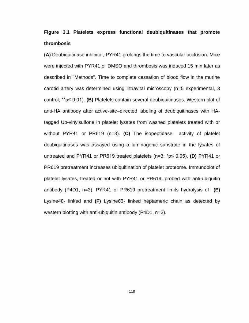
110
Figure 3.1 Platelets express functional deubiquitinases that promote
thrombosis
(A) Deubiquitinase inhibitor, PYR41 prolongs the time to vascular occlusion. Mice
were injected with PYR41 or DMSO and thrombosis was induced 15 min later as
described in "Methods". Time to complete cessation of blood flow in the murine
carotid artery was determined using intravital microscopy (n=5 experimental, 3
control; **p≤ 0.01). (B) Platelets contain several deubiquitinases. Western blot of
anti-HA antibody after active-site–directed labeling of deubiquitinases with HA-
tagged Ub-vinylsulfone in platelet lysates from washed platelets treated with or
without PYR41 or PR619 (n=3). (C) The isopeptidase activity of platelet
deubiquitinases was assayed using a luminogenic substrate in the lysates of
untreated and PYR41 or PR619 treated platelets (n=3; *p≤ 0.05). (D) PYR41 or
PR619 pretreatment increases ubiquitination of platelet proteome. Immunoblot of
platelet lysates, treated or not with PYR41 or PR619, probed with anti-ubiquitin
antibody (P4D1, n=3). PYR41 or PR619 pretreatment limits hydrolysis of (E)
Lysine48- linked and (F) Lysine63- linked heptameric chain as detected by
western blotting with anti-ubiquitin antibody (P4D1, n=2).
111
3.3.2 Deubiquitinase inhibitors suppress platelet adhesion to collagen
under shear flow
Deubiquitinase inhibition in platelets reduced hydrolysis of ubiquitin chains
and caused accumulation of ubiquitinated proteins, indicating that the ubiquitin
cycling is affected by the inhibitors and that deubiquitinase inhibition directly
alters platelet function to reduce adhesion to collagen exposed in injured arteries.
To test this, I examined platelet adhesion during the flow of whole blood through
a collagen-coated microfluidic channel that generates high shear. Fluorescently
labeled platelets in whole blood were immobilized along the chamber, as shown
in a typical video frame captured at the distal end of the chamber after 3 min of
flow (Fig. 3.2 A). Preincubating cells with either PYR41 or PR619 reduced the
number of platelets adhering in the collagen-coated well by 80% (Fig. 3.2 B).
This outcome shows the adhesive phenotype of activated platelets depends on
rapid deubiquitination of their proteome.

112

113
Figure 3.2 Deubiquitinase inhibitors suppressed in vitro thrombosis
(A) PYR41 or PR619 pretreatment blocked platelet adhesion to collagen at high
shear. Calcein-AM labeled blood, treated or not with PYR41 or PR616, was
perfused over immobilized type 1 collagen fibrils (150 µg/ml) at 67.5 dyne/cm2 for
3 min. Images are representative fields taken from three independent
experiments that yielded similar results (n=3). (B) Area of platelet attachment
after PYR41 or PR619 treatment. Platelet area in panel A was quantified by
ImagePro plus software and results are plotted as area of platelet adhesion in
square microns (n=3; ***p≤ 0.001).
114
3.3.3 Stimulated homotypic platelet aggregation, adhesion, and spreading
are suppressed by deubiquitinase inhibitors
Washed human platelets aggregated in response to thrombin (Fig. 3.3 A),
ADP (Fig. 3.3 B), or collagen (Fig. 3.3 C). Pre-incubation with either PYR41 or
PR619 suppressed aggregation in response to each of these agonists. Neither
compound alone promoted aggregate formation (Fig. 3.3 D), but inhibition by
each deubiquitinase inhibitor was statistically significant and the inhibition by the
two deubiquitinase inhibitors was equivalent (Figs. 3.3 E-G).
Outside-in signaling as platelets interact with a glass substrate induces
cytoskeletal rearrangement, extension of filopodia and lamellapodia. Thrombin
stimulation enhances these responses captured by total internal reflection
microscopy (Fig. 3.3 H). Both PYR41 and PR619 abolish the adhesion and
spreading of unstimulated platelets, while greatly reducing these responses after
thrombin stimulation (Fig. 3.3 I).
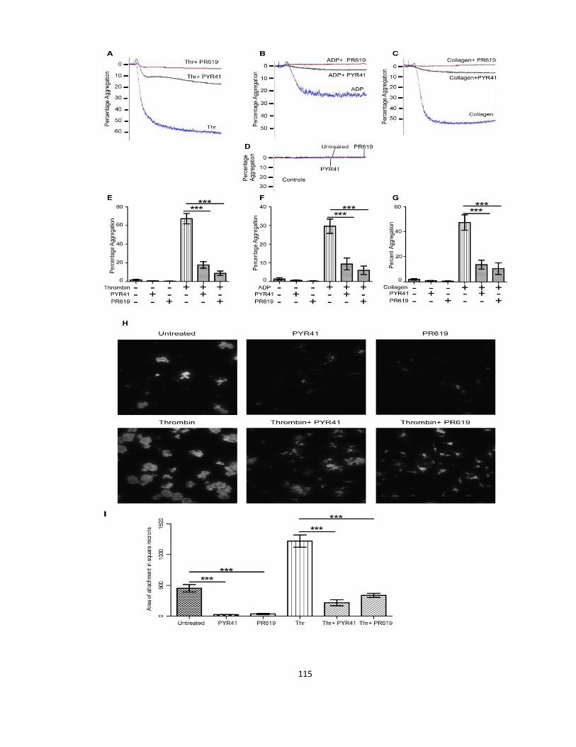
115

116
Figure 3.3 Deubiqutinase inhibition reduced platelet response to agonists
(A) PYR41 or PR619 pretreatment attenuated thrombin (thr)- (B) ADP- and (C)
collagen-induced aggregation. (D) Neither PYR41 nor PR619 stimulated platelet
aggregation in the absence of agonists. (E) PYR41 or PR619 imposed a
reduction in thrombin (thr)- (F) ADP- and (G) collagen-induced aggregation is
statistically significant (n=3; ***p≤ 0.001). (H) PYR41 or PR619 reduced thrombin
stimulated adhesion and spreading. Interaction of control or thrombin stimulated
(thr; 0.2 U) platelets with a glass substrate was imaged by total internal reflection
microscopy after 5 min (n=3). (I) Platelet area after PYR41 or PR619 treatment.
Platelet surface area in panel (A) was quantified by Imagepro plus software (n=3;
***p≤0.001).
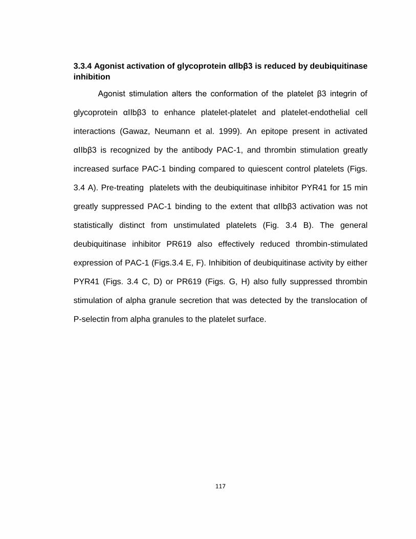
117
3.3.4 Agonist activation of glycoprotein αIIbβ3 is reduced by deubiquitinase
inhibition
Agonist stimulation alters the conformation of the platelet β3 integrin of
glycoprotein αIIbβ3 to enhance platelet-platelet and platelet-endothelial cell
interactions (Gawaz, Neumann et al. 1999). An epitope present in activated
αIIbβ3 is recognized by the antibody PAC-1, and thrombin stimulation greatly
increased surface PAC-1 binding compared to quiescent control platelets (Figs.
3.4 A). Pre-treating platelets with the deubiquitinase inhibitor PYR41 for 15 min
greatly suppressed PAC-1 binding to the extent that αIIbβ3 activation was not
statistically distinct from unstimulated platelets (Fig. 3.4 B). The general
deubiquitinase inhibitor PR619 also effectively reduced thrombin-stimulated
expression of PAC-1 (Figs.3.4 E, F). Inhibition of deubiquitinase activity by either
PYR41 (Figs. 3.4 C, D) or PR619 (Figs. G, H) also fully suppressed thrombin
stimulation of alpha granule secretion that was detected by the translocation of
P-selectin from alpha granules to the platelet surface.

118
119
Figure 3.4 Inhibitors of platelet deubiquitinases suppressed protease
activated receptor (PAR) mediated αIIbβ3 activation and degranulation
(A) PYR41 pretreatment suppressed stimulated αIIbβ3 activation. Flow cytometry
using PAC-1 antibody, that binds active αIIbβ3, on platelets treated or not with
PYR41 and with or without 0.2 U thrombin (thr, n=3). (B) Mean fluorescence
intensity (MFI) of PAC-1 binding on platelets in panel A (n=3; *p≤ 0.05). (C)
PYR41 pretreatment reduced P-selectin surface expression upon activation.
Flow cytometry using PE-conjugated-anti-P-selectin antibody on platelets treated
or not with PYR41 and with or without 0.2 U thrombin (thr, n=3). (D) Mean
fluorescence intensity (MFI) of P-selectin binding on platelets in panel C (n=3; *p≤
0.05). (E) PR619 pretreatment suppressed stimulated αIIbβ3 activation. Flow
cytometry using PAC-1 antibody, that binds active αIIbβ3, on platelets treated or
not with PR619 and with or without 0.2 U thrombin (thr, n=3). (F) Mean
fluorescence intensity (MFI) of PAC-1 binding on platelets in panel E (n=3; **p≤
0.01). (G) PR619 pretreatment reduced P-selectin surface expression upon
activation. Flow cytometry using PE-conjugated-anti-P-selectin antibody on
platelets treated or not with PR619 and with or without 0.2 U thrombin (thr, n=3).
(H) Mean fluorescence intensity (MFI) of P-selectin binding on platelets in panel
G (n=3; **p≤ 0.01).

120
3.3.5 Inhibitors of platelet deubiquitinases suppressed ADP mediated
αIIbβ3 activation and degranulation
Thrombin at the concentration used above activates platelets primarily
through the PAR1 receptor, but activation of platelets through a second receptor,
the P2Y12 receptor for ADP, was also abolished by deubiquitinase inhibition.
Thus, ADP stimulation increased binding of PAC-1 to platelets (Fig. 3.5 A),
although not to the extent induced by thrombin, and PYR41 effectively reduced
the extent of αIIbβ3 activation after ADP activation to nearly quiescent levels
(Fig. 3.5 B). PYR41 pretreatment also suppressed P-selectin exocytosis upon
ADP stimulation (Fig. 3.5 C) and this reduction was statistically significant (Fig.
3.5 D). PR619 was equally effective in suppressing platelet integrin activation
(Figs. 3.5 E, F).

121

122
Figure 3.5 Deubiquitinase inhibitors diminished ADP induced platelet
αIIbβ3 activation and degranulation
(A) PYR41 pretreatment suppressed stimulated αIIbβ3 activation. Flow
cytometry using PAC-1 antibody, that binds active αIIbβ3, on platelets treated or
not with PYR41 and with or without 25 µM ADP (n=4). (B) Mean fluorescence
intensity (MFI) of PAC-1 binding on platelets in panel A (n=4; **p≤ 0.01). (C)
PYR41 pretreatment reduced P-selectin surface expression upon activation.
Flow cytometry using PE-conjugated- anti-P-selectin antibody on platelets
treated or not with PYR41 and with or without 25 µM ADP (n=3). (D) Mean
fluorescence intensity (MFI) of P-selectin binding on platelets in panel C (n=3; *p≤
0.05). (E) PR619 pretreatment suppressed stimulated αIIbβ3 activation. Flow
cytometry using PAC-1 antibody, that binds active αIIbβ3, on platelets treated or
not with PR619 and with or without 25 µM ADP (n=3). (F) Mean fluorescence
intensity (MFI) of PAC-1 binding on platelets in panel E (n=3; *p≤ 0.05).
123
3.3.6 Proteasome associated deubiquitinases regulate platelet activation
A recent report using quantitative mass spectrometry shows platelets
contain several cytoplasmic deubiquitinase enzymes including proteasome
associated deubiquitinases USP14 and UCHL5 (Burkhart, Vaudel et al. 2012).
USP14 and UCHL5 levels are upregulated in many cancers (D'Arcy and Linder
2012) and thus are potential targets for anti-cancer therapies. A small molecule
inhibitor, b-AP15, was recently identified that specifically inhibited just these two
deubiquitinases without affecting the catalytic activities of proteasome (D'Arcy,
Brnjic et al. 2011). Both USP14 and UCHL5 are present in high copy numbers
in platelets, 3100 and 1500 copies/platelet, respectively (Burkhart, Vaudel et al.
2012), but whether these enzymes are relevant in platelets is not known. I tested
b-AP15 in platelets and observed ex vivo treatment of platelets with b-AP15
leads to accumulation of high molecular weight proteins as detected by anti-
ubiquitin P4D1 antibody (Fig. 3.6 A).
Next I ascertained the role of proteasome associated deubiquitinases,
USP14 and UCHL5, on platelet functions. Inhibition of these deubiquitinases with
b-AP15, inhibited thrombin- (Fig. 3.6 B), collagen- (Fig. 3.6 C) and ADP- (Fig. 3.6
D) induced aggregation and, like the general deubiquitinase inhibitors PYR41
and PR619, the inhibitory effect of b-AP15 on agonist-induced platelet
aggregation was significant (Fig. 3.6 E). b-AP15 treatment alone had no effect on
platelet aggregation. b-AP15 pretreatment also inhibited thrombin- (Fig. 3.6 F)
induced αIIbβ3 activation as assessed by PAC-1 binding as well as P-selectin
exocytosis from platelet alpha-granules (Fig. 3.6 G). Activation of αIIbβ3 is critical
124
for stable thrombus formation and consolidation in vivo (Shattil and Newman
2004; Gratacap, Guillermet-Guibert et al. 2011) and pretreatment with b-AP15
also blocked ADP- (Fig. 3.6 H) induced αIIbβ3 activation. USP14 and UCHL5
inhibition had a more profound inhibitory effect on collagen-induced aggregation
(Fig. 3.6 C) than the general deubiquitinase inhibitors, so I tested the effect of
inhibition of these enzymes on platelet adhesion to collagen under static
conditions. b-AP15 pretreatment reduced the number of platelets adhering to a
exposed collagen-coated surface (Fig. 3.6 I), suggesting vital role of proteasome
associated deubiquitinases in mediating platelet interactions with bound or
exposed collagen.
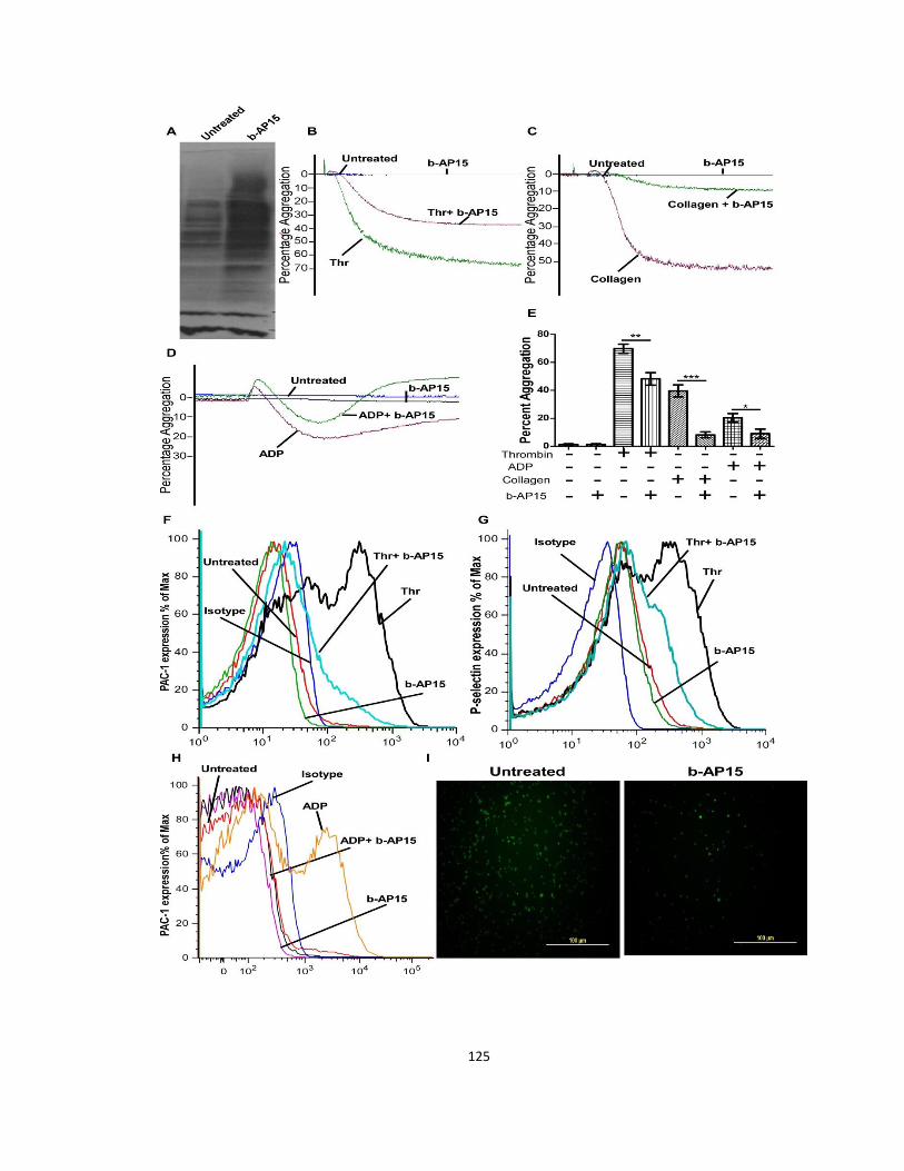
125

126
Figure 3.6 Pharmacologic inhibition of proteasome-associated
deubiquitinase enzymes, USP14 and UCHL5, reduced platelet
responsiveness to agonists
(A) b-AP15 pretreatment increases ubiquitination of the platelet proteome.
Immunoblot of platelet lysates, treated or not with b-AP15, probed with anti-
ubiquitin antibody (P4D1, n=3). (B) Proteasome-associated deubiquitinases
promote stimulated platelet aggregation. b-AP15 pretreatment inhibited thrombin-
(thr, n=3) (C) collagen- (n=3) and (D) ADP-induced aggregation (n=3). (E) b-
AP15 mediated inhibition of stimulated platelet aggregation is statistically
significant (n=3; ***p≤ 0.001, **p≤0.01, *p≤ 0.05). (F) b-AP15 pretreatment
suppressed stimulated αIIbβ3 activation. Flow cytometry using PAC-1 antibody,
that binds active αIIbβ3, on platelets treated or not with b-AP15 and with or
without 0.2 U thrombin (thr, n=3). (G) b-AP15 pretreatment reduced P-selectin
surface expression upon activation. Flow cytometry using PE-conjugated- anti-P-
selectin antibody on platelets treated or not with b-AP15 and with or without 0.2
U thrombin (thr, n=3). (H) Preincubation with b-AP15 suppressed ADP-induced
αIIbβ3 activation. Flow cytometry using PAC-1 antibody, that binds active αIIbβ3,
of platelets treated or not with b-AP15 and with or without 25 µM ADP (n=3). (I)
Proteasome-associated deubiquitinases mediate platelet adhesion to collagen
coated surface. Microscopic images of platelet adhesion on collagen under static
conditions (n=2).

127
3.3.7 Platelet deubiquitinases affect collagen mediated responses via the
gpVI receptor
Collagen binds and activates platelets via two major platelet surface
receptors, α2β1 and gpVI (Nuyttens, Thijs et al. 2011). Since all three
deubiquitinase inhibitors had a profound effect on collagen-induced aggregation,
I next looked at the effect of these inhibitors specifically on the gpVI receptor, the
main signaling receptor for collagen (Nieswandt and Watson 2003). Neither the
general deubiquitinase inhibitors, PYR41 and PR619, nor the specific
deubiquitinase inhibitor, b-AP15, alone induced platelet aggregation (Fig. 3.7 A),
but all three inhibitors suppressed aggregation to convulxin, a snake venom toxin
that activates platelets via gpVI receptor (Polgar, Clemetson et al. 1997) (Fig. 3.7
B). Convulxin stimulation increased PAC-1 binding, a marker of αIIbβ3 activation,
but pretreatment with PYR41 (Fig. 3.7 C) or PR619 (Fig. 3.7 D) or b-AP15 (Fig.
3.7 E) reduced convulxin induced increase in PAC-1 binding. This suggest that
the platelet deubiquitinases are critical for gpVI-mediated transduction of
collagen signals.
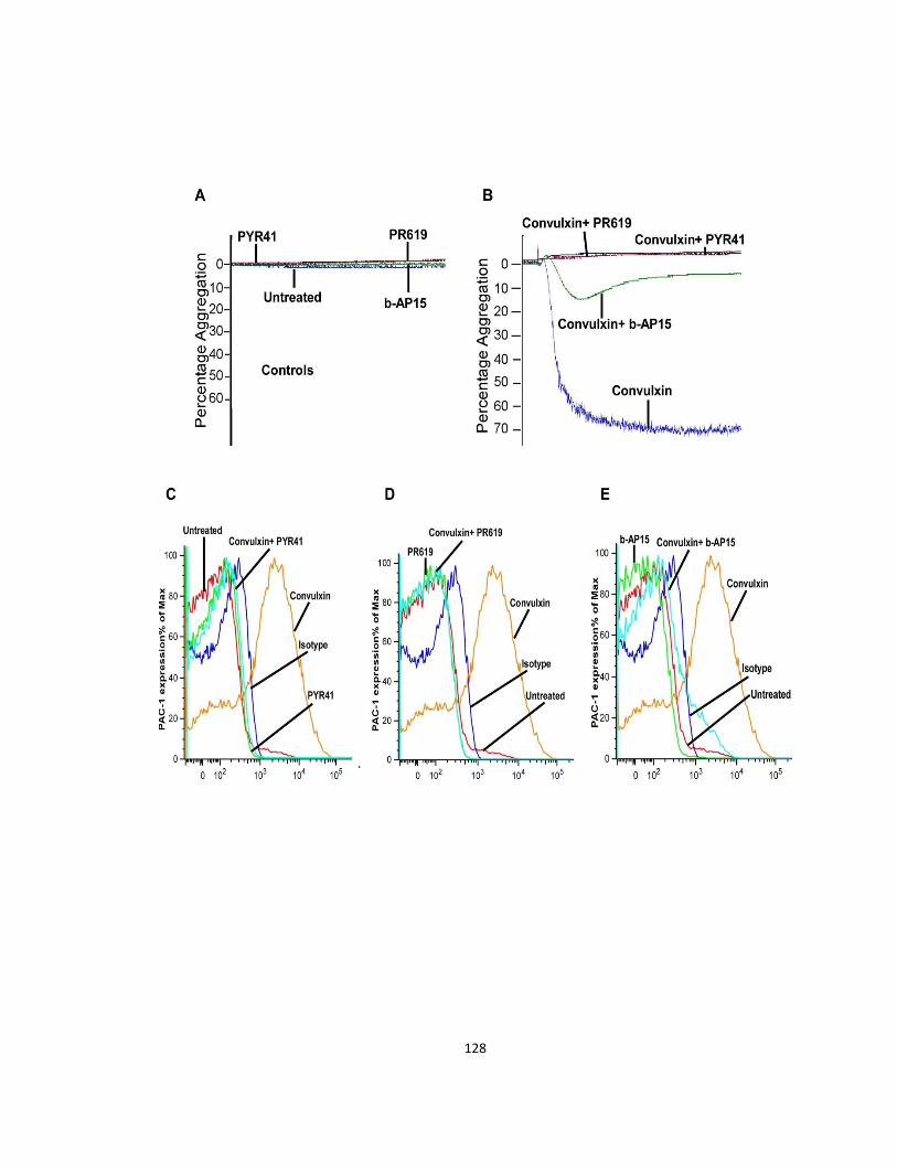
128

129
Figure 3.7 Deubiquitinase inhibitors affect collagen responses via the gpVI
receptor
(A) Neither general deubiquitinase inhibitors, PYR41 and PR619, nor b-AP15
stimulated platelet aggregation in the absence of agonists. (B) Deubiquitinase
inhibitors inhibited gpVI specific platelet responses. Platelets were pretreated
with PYR41, PR619, or b-AP15 before initiating platelet aggregation with
convulxin (n=2). (C) Convulxin-mediated increase in PAC-1 binding was
assessed using flow cytometry in platelets pretreated with PYR41 (n=2), (D)
PR619 (n=2), or (E) b-AP15 (n=2).

130
3.3.8 Inhibition of platelet deubiquitinases modulated platelet signaling
responses downstream of thrombin and collagen receptors
Akt, or protein kinase B, is a serine/ threonine kinase that is an
established downstream effector of phosphoinositide 3-kinase (PI3K) (Bhaskar
and Hay 2007). Platelets express two Akt isoforms, Akt1 and Akt2 (Kroner,
Eybrechts et al. 2000; Dittrich, Birschmann et al. 2008) and targeted deletion of
each individual isoform affects platelet function. Akt1 deletion in mice resulted in
diminished platelet responses to thrombin and collagen (Chen, De et al. 2004;
Stojanovic, Marjanovic et al. 2006) and Akt2 deletion suppressed platelet
secretion and thrombosis (Woulfe, Jiang et al. 2004).
Platelet stimulation with thrombin resulted in time dependent increase in
Akt phosphorylation at both ser473 and thr308 residues (Fig.3.8 A), but
pretreatment with either general deubiquitinase inhibitors, PYR41 and PR619, or
the specific deubiquitinase inhibitor, b-AP15, reduced thrombin-induced Akt
activation as evident from reduced Akt phosphorlylation at both ser and thr
residues (Fig. 3.8 A). An equivalent inhibition of collagen-induced Akt
phosphorylation, either at ser473 or thr308, (Fig. 3.8 B) was also observed with
all three inhibitors of deubiquitinases. The inhibition of Akt phosphorylation at
thr308, induced by either agonist, was more profound compared to
phosphorylation at ser473. In platelets, Akt activation is both PI3K dependent
and independent, but sustained Akt activation is PI3K dependent (Jackson and
Schoenwaelder 2010). Class IA PI3K isoform β (PI3K β) is an essential PI3K to
promote PLCγ-2 activation downstream of gpVI-FcRγ receptor (Gratacap,
131
Guillermet-Guibert et al. 2011). PLCγ-2 activation leads to intracellular calcium
release and DAG production and both events are critical for sustained αIIbβ3
activation and fibrin clot retraction (Canobbio, Stefanini et al. 2009; Martin,
Guillermet-Guibert et al. 2010). Collagen-induced intracellular calcium release
was inhibited when platelets were preincubated with deubiquitinase inhibitors
before stimulation by collagen (Fig.3.8 C). Deubiquitinase inhibitors also
significantly reduced fibrin clot retraction suggesting impaired outside in signaling
from integrin αIIbβ3 (Fig. 3.8 D).
Phosphatase and tensin homologue-deleted on chromosome 10 (PTEN)
is a known negative regulator of PI3K/Akt signaling (Carracedo and Pandolfi
2008) that regulates collagen-dependent platelet activation. Tissue specific pten
deletion in mice resulted in increased Akt phosphorylation and platelet
aggregation even at much lower collagen concentrations, indicating that PTEN
negatively regulates collagen mediated platelet activation (Weng, Li et al. 2010).
PTEN's phosphatase activity is negatively regulated by phosphorylation at ser
380 (Vazquez, Ramaswamy et al. 2000; Yang, Yuan et al. 2013), however this is
not studied in platelets so far. I observed both thrombin and collagen increased
PTEN phosphorylation at this residue (Fig. 3.8 E) and pretreatment with
deubiquitinase inhibitors reduced agonist dependent increase in PTEN
phosphorylation and inactivation.
Since deubiquitinases display linkage specificity for ubiquitin chains
(Komander, Clague et al. 2009), I used linkage specific antibodies (K63 or K48
specific antibodies) to determine the relative abundance of both type of linkages

132
in platelets. I observed accumulation of lysine 48 linkages (Fig. 3.8 F), but
interestingly I did not find any accumulation of lysine 63 linkages in untreated,
stimulated or deubiquitinase inhibitor treated platelets (not shown). An important
role of deubiquitinases is generation of free ubiquitin pool from unanchored
polyubiquitin chains. I observed accumulation of high molecular weight
polyubiquitin chains upon inhibition of platelet deubiquitinases, suggesting that
deubiquitinase enzyme mediated ubiquitin homeostasis is critical for platelet
responses.

133

134
Figure 3.8 Deubiquitinase inhibitors impaired platelet signaling following
agonist stimulation
(A) Deubiquitinase inhibitors reduced thrombin induced Akt activation. Platelets
treated with or without deubiquitinase inhibitors were stimulated with 0.2 U
thrombin (thr) for the stated times and immunoblotted using anti-pAKT ser473,
anti-pAKT thr308 or anti-Akt antibodies (n=3). (B) Deubiquitinase inhibitors
reduced collagen-induced Akt activation. Platelets treated with or without
deubiquitinase inhibitors were stimulated with 2 µg collagen for the stated times
and immunoblotting was done on platelet lysates using anti-pAKT ser473, anti-
pAKT thr308 or anti-Akt antibodies (n=3). (C) Deubiquitinase inhibitors blocked
collagen-induced intracellular calcium release. Platelets labeled with Fura-2 AM
were treated or not with deubiquitinase inhibitors before stimulation with 2 µg
collagen. (D) Deubiquitinase inhibition suppressed clot retraction. Ratio of the
image surface area of images of thrombin-induced clots over time (n=3; **p<
0.01, *p<0.05). (E) Deubiquitinase inhibition activates PTEN. Immunoblotting of
platelets treated or not with deubiquitinase inhibitors before stimulation with 0.2 U
thrombin or 2 µg collagen was done using anti-phospho PTEN-ser380 antibody
(n=2). (F) Deubiquitinase inhibition caused accumulation of lys-48 linked Ub
conjugates. Immunoblotting of platelets treated or not with deubiquitinase
inhibitors before stimulation with 0.2 U thrombin (thr) or 2 µg collagen was done
using anti-K48 linked ubiquitin antibody (n=2).
135
3.5 Discussion
The proteome of platelets is constitutively decorated by conjugated single
ubiquitin molecules and by ubiquitin polymeric chains, whose abundance
increases with agonist stimulation (Gupta, Li et al. 2014). Here I present
evidence showing that ubiquitin decoration of platelet proteins is dynamic.
Turnover of ubiquitin decoration was elucidated by suppressing deubiquitinase
activity by any of the general deubiquitinase inhibitors or the specific inhibitor of
proteasome-associated deubiquitinases. This greatly increased ubiquitin protein
modification within minutes. These changes were apparent in the absence of
agonist stimulation, so the protein complement of quiescent platelets undergoes
a continuous, and rapid, cycle of ubiquitination and deubiquitination.
Protein deubiquitination is, in fact, a critical step in unlocking platelet
reactivity. The general deubiquitinase inhibitors PYR41 or PR619 blocked
activation of αIIbβ3 integrin, blocked alpha granule release and hence blocked
the agonist-stimulated increase of surface P-selectin. These inhibitors also
effectively blocked ex vivo homotypic platelet aggregation. The general inhibitor
PYR41 additionally blocked formation of platelet-rich thrombi that completely
occluded blood flow in damaged carotid arteries. I conclude the complete
suppression of stimulated platelet function by deubiquitinase inhibition means
that ubiquitin modification of the proteome likely maintains platelets in an inactive
state and that deubiquitinase action may be required to release tonic ubiquitin
inhibition.

136
Deubiquitinase inhibitors affect platelet signaling downstream of platelet
receptors. Thrombin and collagen induced Akt phosphorylation was reduced by
both the pan deubiquitinase inhibitors, PYR41 and PR619, and the specific
inhibitor of proteasome associated deubiquitinase activity, b-Ap15. Akt activation
in platelets is both PI3K-dependent and PI3K-independent (Laurent, Severin et
al. 2014), however, class IA PI3K isoform β (PI3K β) is mandatory for gpVI-FcRγ
receptor-mediated Akt activation, platelet aggregation, intracellular calcium
release and degranulation (Kim, Mangin et al. 2009). I attribute these effects to
the upregulation of PTEN activity upon pretreatment with deubiquitinase
inhibitors. PTEN negatively regulates Akt activation by converting
phosphatidyinositol 3,4,5-trisphosphate (PIP3) to phosphatidylinositol 4,5-
bisphosphate (PIP2) and its been reported to negatively regulate collagen-
induced platelet activation (Weng, Li et al. 2010). Deubiquitinase inhibitors also
significantly reduced clot retraction over time, indicating impaired outside-in
integrin signaling. Phosphoinositide-dependent protein kinase 1 (PDK1) is
activated upon an increase in PIP3 following PI3K activation and activated PDK1
phosphorylates Akt at residue thr308 (Moore, Hunter et al. 2011). Platelet
specific knockdown of PDK1 and subsequent loss in Akt phosphorylation
reduced thrombin-induced platelet aggregation, clot retraction and thrombosis
(Chen, Zhang et al. 2013).
Activation of PI3K and its effector, Akt, is essential for most aspects of
collagen-induced platelet activation, and thrombin- and collagen-induced αIIbβ3
integrin outside-in signaling. The effect of PI3K/Akt axis on thrombin-induced
137
aggregation is only seen at low thrombin concentrations and is overcome at high
agonist concentrations (Martin, Guillermet-Guibert et al. 2010; Chen, Zhang et al.
2013). Additionally, the effect of thrombin-induced aggregation can be reversed
using P2Y12 inhibitors, suggesting it's not a direct effect but is due to thrombin-
induced platelet ADP secretion (Kim, Mangin et al. 2009). However, it is
controversial because P2Y12 knockout mice showed Akt phosphorylation in
response to thrombin, indicating a direct effect. In the same study it was shown
that the Src family of kinases (SFKs) act upstream of PI3K/Akt axis. The
discrepancy was attributed to nonspecific effects of P2Y12 inhibitors used in the
first study (Xiang, Zhang et al. 2010). I recognize the fact that the strong effect of
pan deubiquitinase inhibitors, PYR41 and PR619, on thrombin-induced
aggregation is independent of Akt activation, but I have seen a reduction in
thrombin induced intracellular calcium release after pretreatment with pan
deubiquitinase inhibitors (not shown) as I found when collagen was the agonist.
This did not occur with b-AP15, which also explains the strong effect of pan
deubiquitinase inhibitors and mild effect of b-AP15 on thrombin induced
aggregation.
Furthermore, I have not identified a direct target that is modulated by
deubiquitinase inhibition and that needs further experimentation. However, there
could be another possibility for the switch in platelet responsiveness since it is
notable that quiescent platelets lacked free ubiquitin monomer. Also the anti-
ubiquitin-active band with the most rapid mobility during SDS-PAGE is ~ 17 kDa
(Fig. 3.1 D), which is consistent with the anticipated mobility of di-ubiquitin. I have

138
not lost monomeric 8 kDa ubiquitin from my gels of platelet lysates since I detect
recombinant monomeric ubiquitin. Moreover, I observed accumulation of only
K48-linked substrates upon pretreatment with deubiquitinase inhibitors (Fig. 3.8
F), but this is not due to proteasome inhibition as none of the three inhibitors
inhibited the proteasome catalytic activity (Altun, Kramer et al. 2011; D'Arcy,
Brnjic et al. 2011; Kapuria, Peterson et al. 2011) that also results in K48-
polyubiquitination of the platelet proteome (Gupta, Li et al. 2014). So, it is
possible that all platelet ubiquitin is either fully adducted with protein or exists as
unanchored polymeric ubiquitin chains (Strachan, Roach et al. 2012).
Quantitative mass spectrometry reports 3600 copies of polyubiquitin B per
platelet, the trimeric ubiquitin precursor, but no free monomer (Burkhart, Vaudel
et al. 2012). Instead, I propose monomeric platelet ubiquitin would not be present
or available to conjugate to new protein targets until ubiquitin sequestered in
either protein or unanchored ubiquitin chains is first released by deubiquitinase
action. This could mean the essential role of deubiqutination in platelet activation
is either to release ubiquitin from existing depots that would allow its conjugation
to a new target protein, or the relevant change could be a required remodeling of
existing polyubiquitin chains.
The deubiquitinase inhibitors b-AP15 and PR619 strongly suppressed
stimulated platelet function ex vivo, and I suggest the effect of PYR41 similarly
was through inhibition of deubiquitinase activity. This compound was identified as
an inhibitor of ubiquitin E1 activity (Yang, Kitagaki et al. 2007), although it was
effective in suppressing E1 activity in vitro (not shown). However, it did not
139
interfere with ubiquitin activation in platelets and, like b-Ap15 and PR619, PYR41
caused a rapid and massive increase in ubiquitin decoration of soluble platelet
proteins. This is incompatible with E1 inhibition, which could indicate ubiquitin
activation through the second E1 enzyme except that this protein was not
discovered in the platelet proteome (Burkhart, Vaudel et al. 2012). However,
PYR41 is routinely used as an E1 inhibitor as evident from several publications
and it is recently shown that PYR41 at 25 µM concentration blocked cyclophilin A
dependent ubiquitination of CXCR7. This is induced by SDF-1α and its a
prerequisite for the surface externalization of CXCR7 in platelets (Chatterjee,
Seizer et al. 2014). Although I observed a strong inhibitory effect of PYR41 on
platelet deubiquitinases, I have not discounted the possibility that PYR41 might
act as a dual inhibitor of both E1 activity and deubiquitinase activity. I therefore
performed parallel to the PYR41, experiments with general deubiquitinase
inhibitor, PR619, and specific deubiquitinase inhibitor, b-AP15, and I show that
compounds inhibiting a range of deubiquitase family members suppressed
platelet function.
Taken together, I have identified a new pathway that regulates platelet
function. I have identified that platelets contain several deubiquitinases and both
the general deubiquitinase inhibitors as well as specific inhibitor of proteasome-
associated deubiquitinases modulate platelet reactivity to various agonists.
Inhibition of platelet deubiquitinases suppressed platelet aggregation, secretion
and adhesion of isolated platelets in response to all agonists. Additionally,
systemic blockade of deubiquitination also reduced arterial thrombosis in FeCl3-
140
damaged carotid arteries. I also show that the deubiquitinase inhibitors, PYR41,
PR619, and b-AP15, affect downstream signaling from the collagen receptor, as
evident from reduced Akt activation, likely via upregulation of PTEN activity. This
is the first report that establishes agonist-dependent regulation of PTEN activity
in platelets. Platelet deubiquitinases are critical for platelet responsiveness and
the specific inhibitor of proteasome associate deubiquitinases, b-AP15, is already
in active preclinical trials. This strongly makes the case that platelet
deubiquitinases can be a new class of anti-thrombotic drug targets.
3.6 Chapter summary
These data further show that platelet ubiquitin-proteasome pathway is
crucial in modulating platelet reactivity. I show that ubiquitination of platelet
proteins is dynamic, platelets express several deubiquitinases, and platelet
deubiquitinases are functional. Specific inhibition of the proteasome-associated
deubiquitinases (b-AP15) as well as pan deubiquitinase inhibitors (PYR41 and
PR619) increased mono- and poly-ubiquitination of platelet proteins. These
agents also blocked platelet degranulation upon stimulation. Deubiquitinase
inhibition suppressed αIIbβ3 activation, platelet aggregation, and adhesion/
spreading in response to diverse platelet agonists. Inhibition of platelet
deubiquitinase activity strongly suppressed formation of platelet-rich occlusive
thrombi in FeCl3-damaged murine carotid arteries and inhibition of platelet
deubiquitinase activity prevented in vitro thrombus formation on collagen-coated
surfaces at high shear rates. These inhibitors also blocked downstream signaling

141
from platelet receptors by inhibiting agonist-induced Akt phosphorylation
associated with upregulation of PTEN activity. This is the first report that
established agonist-dependent regulation of PTEN activity in platelets. These
data uncovers a new mechanism that maintains circulating platelets in a
quiescent state.

142
Chapter IV
Ubiquitin-specific protease 7 (USP7) activity promotes platelet activation
4.1 Introduction and Rationale
Arterial thrombotic occlusion causes myocardial infarction and ischemic
stroke and is the single most common cause of death and disability in the
developed world (Sanz, Moreno et al. 2012). Platelets play central roles in the
development of arterial thrombosis and are therefore the main target of anti-
thrombotic therapies. Current anti-platelet therapies, most commonly aspirin
and/or clopidogrel, are not fully effective in the prevention of arterial thrombosis,
and novel anti-platelet agents are therefore sought (Meadows and Bhatt 2007;
Michelson 2011). We have previously demonstrated that platelets contain a
functional ubiquitin-proteasome system, that proteasome mediated proteolysis is
required both for ex vivo platelet activation and for in vivo development of arterial
thrombosis (Gupta, Li et al. 2014). We have also recently established that, like
nucleated cells, the ubiquitination of platelet proteins is dynamic, so platelet
deubiquitinases that catalyze this turn-over are functional and modulate platelet
reactivity (unpublished).
Targeting ubiquitin-proteasome system in platelets seems promising, and
143
small molecule inhibitors targeting specific and smaller portions of ubiquitin-
proteasome pathway have shown encouraging results in other settings targeting
USP7 in nucleated cells. A recent quantitative mass spectrometry shows
platelets express deubiquitinase USP7 (ubiquitin-specific protease 7 or
Herpesvirus-associated ubiquitin-specific protease, HAUSP). This is interesting
as USP7 participates in the control of the cell cycle, which is irrelevant to
platelets, but quantitative mass spectrometry shows platelets contain 1300
copies of USP7 peptides per platelet (Burkhart, Vaudel et al. 2012).
USP7/HAUSP modulates proteins involved in cell cycle regulation, as well as
oncogenes and tumor suppressors and is therefore a strategic target for cancer
therapy (Reverdy, Conrath et al. 2012). USP7/HAUSP inhibitors are already in
active preclinical development owing to their role in p53 regulation (Shen,
Schmitt et al. 2013). These inhibitors induce apoptosis of tumor cells (Reverdy,
Conrath et al. 2012) and apoptosis of multiple myeloma cells that have overcome
resistance to the proteasome inhibitor bortezomib (Chauhan, Tian et al. 2012).
USP7/HAUSP is abundant in platelets and we have observed (chapter III,
Fig. 3.1 B) reduction in the intensity of the protein band at ~ 130 kD, which
corresponds to the molecular weight of USP7/HAUSP, after treatment with
generic deubiquitinase inhibitors. Whether USP7/HAUSP modulates platelet
reactivity has not been studied. Here I show that specific inhibition of
USP7/HAUSP suppresses platelet aggregation in response to collagen and
adhesion under flow. This study indentifies USP7/HAUSP inhibition as a potential
new approach to anti-platelet therapy.

144
4.2 Results
4.2.1 USP7/HAUSP activity promotes platelet activation
Active deubiquitinase enzymes are specifically covalently adducted during
hydrolysis of the suicide substrate, HA-tagged ubiquitin-vinyl sulfone
(Borodovsky, Kessler et al. 2001), and we used this approach to determine
whether platelets have active USP7/HAUSP. Platelets treated with either pan
DUB inhibitors (PYR41 and PR619) or structurally unrelated USP7/HAUSP
inhibitors (HBX 41108 and P22077) were incubated with HA-ubiquitin-vinyl
sulfone. Immunoprecipitation with anti-HA antibody was used to enrich active
deubiquitinases. Immunoblotting with anti-HAUSP antibody confirmed the
presence of active USP7/HAUSP in platelets as a full length 130 kDa protein,
and I found that its activity was suppressed by the general inhibitors PYR41 and
PR619 (Fig. 4.1 A).Selective small molecule inhibitors of USP7/HAUSP, HBX
41108 and P22077, were equally effective or better in blocking deubiquitination
by this enzyme (Fig. 4.1 B).
Selective USP7/HAUSP inhibition by HBX 41108 abolished platelet deposition
from whole blood onto the collagen-coated surface of a microfluidic channel
during flow at high shear (Fig. 4.1 C) and this inhibition was significant (Fig. 4.1
D). Neither HBX 41108 nor P22077 alone induced platelet aggregation (Fig. 4.1
E), but each inhibitor suppressed collagen induced platelet aggregation in
purified platelet preparations (Fig. 4.1 F). This inhibition was significant, and

145
equivalent (Fig. 4.1 G).

146
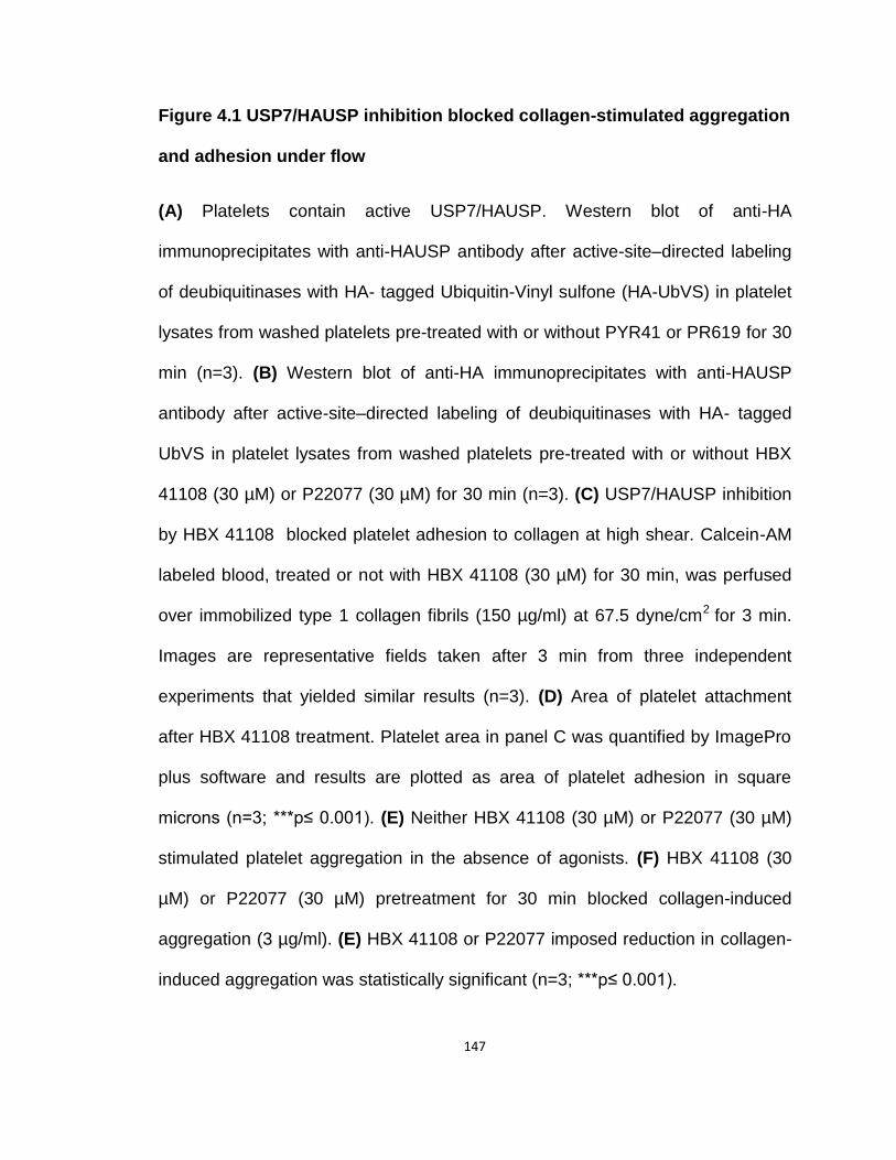
147
Figure 4.1 USP7/HAUSP inhibition blocked collagen-stimulated aggregation
and adhesion under flow
(A) Platelets contain active USP7/HAUSP. Western blot of anti-HA
immunoprecipitates with anti-HAUSP antibody after active-site–directed labeling
of deubiquitinases with HA- tagged Ubiquitin-Vinyl sulfone (HA-UbVS) in platelet
lysates from washed platelets pre-treated with or without PYR41 or PR619 for 30
min (n=3). (B) Western blot of anti-HA immunoprecipitates with anti-HAUSP
antibody after active-site–directed labeling of deubiquitinases with HA- tagged
UbVS in platelet lysates from washed platelets pre-treated with or without HBX
41108 (30 µM) or P22077 (30 µM) for 30 min (n=3). (C) USP7/HAUSP inhibition
by HBX 41108 blocked platelet adhesion to collagen at high shear. Calcein-AM
labeled blood, treated or not with HBX 41108 (30 µM) for 30 min, was perfused
over immobilized type 1 collagen fibrils (150 µg/ml) at 67.5 dyne/cm2 for 3 min.
Images are representative fields taken after 3 min from three independent
experiments that yielded similar results (n=3). (D) Area of platelet attachment
after HBX 41108 treatment. Platelet area in panel C was quantified by ImagePro
plus software and results are plotted as area of platelet adhesion in square
microns (n=3; ***p≤ 0.001). (E) Neither HBX 41108 (30 µM) or P22077 (30 µM)
stimulated platelet aggregation in the absence of agonists. (F) HBX 41108 (30
µM) or P22077 (30 µM) pretreatment for 30 min blocked collagen-induced
aggregation (3 µg/ml). (E) HBX 41108 or P22077 imposed reduction in collagen-
induced aggregation was statistically significant (n=3; ***p≤ 0.001).

148
4.3 Chapter summary
These data confirm that components of the platelet ubiquitin-proteasome
system are potential anti-thrombotic targets. I show inhibition of a specific
deubiquitinase, USP7/HAUSP, strongly suppressed formation of platelet-rich
thrombi that formed on collagen-coated surfaces at high shear rates and
suppressed platelet aggregation in response to collagen. The deubiquitinase
family contains nearly 100 members that accept a range of monomeric and
polymeric ubiquitin-conjugated protein substrates, and the polymeric ubiquitin
chains themselves are polydisperse from varied polymer length and branching
from the seven lysine residues of ubiquitin that are sites of polymerization
(Nijman, Luna-Vargas et al. 2005). Deubiquitinases, thus, generally show broadly
overlapping substrate specificities.
USP7/HAUSP, member of the DUB family, in contrast, is selective and
recognizes ubiquitinated p53 and MDM2, with a critical role in cell cycle
regulation (Hu, Gu et al. 2006; Faesen, Dirac et al. 2011). Participation of
USP7/HAUSP in deubiquitinating both p53 and its MDM2 E3 ligase, and its
participation in the cell cycle, suggests therapeutic opportunities in inhibiting
USP7/HAUSP in cancer biology (Colland, Formstecher et al. 2009; Nicholson
and Suresh Kumar 2011). This has led to the development of potent and
selective USP7/HAUSP inhibitors (Reverdy, Conrath et al. 2012).

149
In this study, I find two specific inhibitors of USP7/HAUSP that blocked
platelet aggregation by collagen and platelet adhesion under flow. Inhibition or
loss of collagen receptor, gpVI, prevented arterial thrombosis in animal models
without affecting normal hemostasis in both mice and humans (Kahn 2004),
making gpVI a suitable therapeutic target. These data define a focused role for
this deubiquitinase in response to just collagen. This shows USP7/HAUSP is
required for successful platelet interaction with collagen fibrils exposed in
damaged vessels, but does not then participate in platelet activation by soluble
agonists. This strongly makes the case that platelet USP7/HAUSP can be a new
class of anti-thrombotic drug target that specifically dampens platelet
responsiveness to collagen.

150
Chapter V
Conclusion
5.1 Summary and Conclusion
Overall, the studies presented in this thesis provide evidence that the
ubiquitin-proteasome system is an important regulator of platelet responsiveness
to diverse stimuli. Pharmacologic inhibitors against specific components of UPS
(Fig. 5.1) were used to determine the individual effect of selective inhibition of
these components on platelet function. Proteasome inhibition modulated platelet
aggregation, spreading, microparticle shedding, clot retraction, thrombus
formation and thrombus stability. The requirement for platelet deubiquitinase
activity in platelet responses varied among platelet agonists. Specific
deubiquitinase inhibitors, as well as general deubiquitinase inhibitors,
suppressed stimulated platelet aggregation, adhesion/spreading, platelet
degranulation, αIIbβ3 activation and outside-in signaling from αIIbβ3. Systemic
blockade of deubiquitination also reduced arterial thrombosis in FeCl3-damaged
carotid arteries. The inhibition of proteasome-associated deubiquitinases or
soluble deubiquitinase USP7/HAUSP, showed that the platelet responses
151
downstream of the collagen receptor were dampened. However, since pan
deubiquitinase inhibitors block stimulation by all major agonists, there are
receptor-specific as well as receptor-non-specific processes that depend on
deubiquitination.
To conclude, this study has identified a novel pathway that regulates
platelet function and reactivity. Further understanding of the mechanisms that
regulate this pathway in platelets and characterization of specific proteins that
are being modulated by this pathway seems vital for the development of new and
improved anti-platelet therapies.
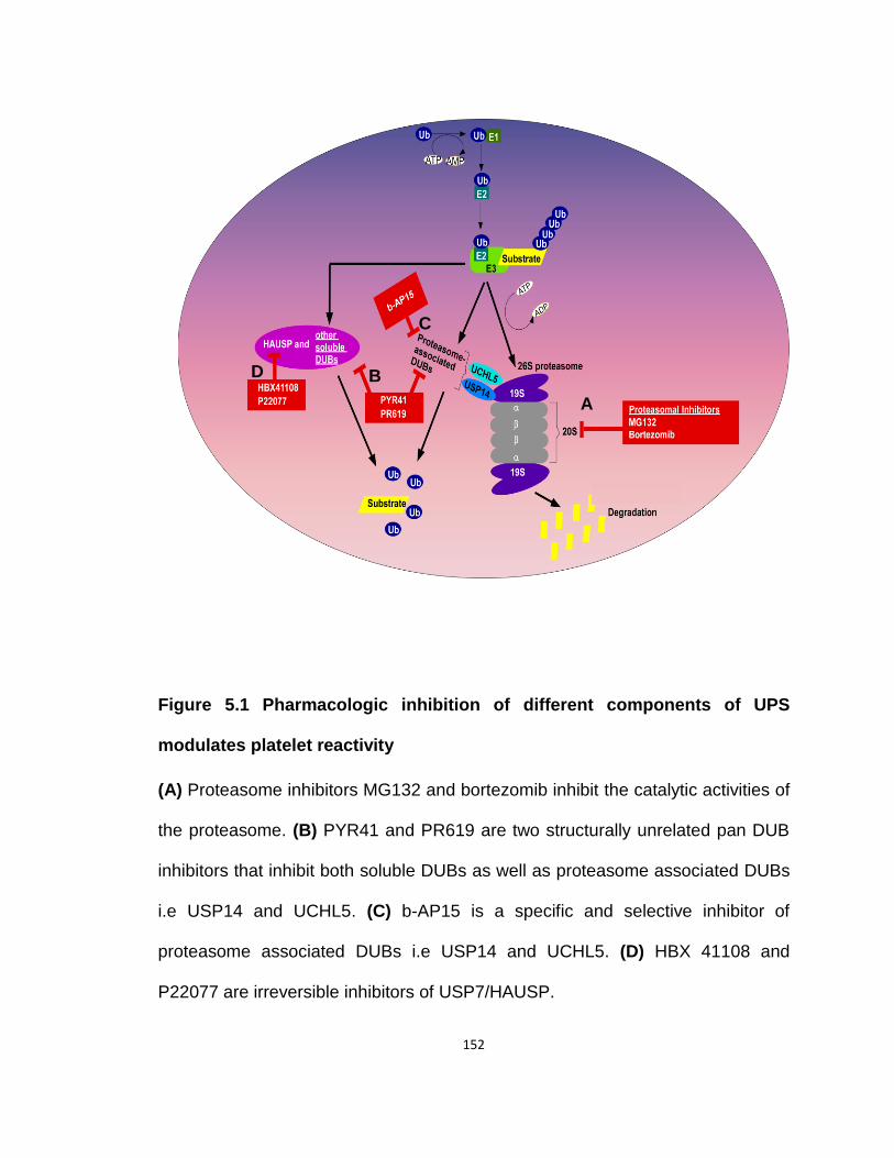
152
Figure 5.1 Pharmacologic inhibition of different components of UPS
modulates platelet reactivity
(A) Proteasome inhibitors MG132 and bortezomib inhibit the catalytic activities of
the proteasome. (B) PYR41 and PR619 are two structurally unrelated pan DUB
inhibitors that inhibit both soluble DUBs as well as proteasome associated DUBs
i.e USP14 and UCHL5. (C) b-AP15 is a specific and selective inhibitor of
proteasome associated DUBs i.e USP14 and UCHL5. (D) HBX 41108 and
P22077 are irreversible inhibitors of USP7/HAUSP.
A
B
C
D

153
Bibliography
Adam, F., M. C. Bouton, et al. (2003). "Thrombin interaction with platelet membrane glycoprotein Ib alpha." Trends Mol Med 9(11): 461-464.
Adams, J. (2004). "The proteasome: a suitable antineoplastic target." Nat Rev Cancer 4(5): 349-360.
Altun, M., H. B. Kramer, et al. (2011). "Activity-based chemical proteomics accelerates inhibitor development for deubiquitylating enzymes." Chem Biol 18(11): 1401-1412.
Alwan, A. (2011). "Burden:mortality,morbidity and risk factors. Global Status Report on Noncommunicable Diseases 2010. (WHO Press,Geneva,
Switzerland, 2011). http://www.who.int/nmh/publications/ncd_report2010/en/." Amerik, A. Y. and M. Hochstrasser (2004). "Mechanism and function of
deubiquitinating enzymes." Biochim Biophys Acta 1695(1-3): 189-207. Anderson, J. L., C. D. Adams, et al. (2013). "2012 ACCF/AHA focused update
incorporated into the ACCF/AHA 2007 guidelines for the management of patients with unstable angina/non-ST-elevation myocardial infarction: a report of the American College of Cardiology Foundation/American Heart Association Task Force on Practice Guidelines." J Am Coll Cardiol 61(23): e179-347.
Anderson, J. L., C. D. Adams, et al. (2007). "ACC/AHA 2007 guidelines for the management of patients with unstable angina/non ST-elevation myocardial infarction: a report of the American College of Cardiology/American Heart Association Task Force on Practice Guidelines (Writing Committee to Revise the 2002 Guidelines for the Management of Patients With Unstable Angina/Non ST-Elevation Myocardial Infarction): developed in collaboration with the American College of Emergency Physicians, the Society for Cardiovascular Angiography and Interventions, and the Society of Thoracic Surgeons: endorsed by the American Association of Cardiovascular and Pulmonary Rehabilitation and the Society for Academic Emergency Medicine." Circulation 116(7): e148-304.
Arendt, C. S. and M. Hochstrasser (1997). "Identification of the yeast 20S proteasome catalytic centers and subunit interactions required for active-site formation." Proc Natl Acad Sci U S A 94(14): 7156-7161.
Arendt, C. S. and M. Hochstrasser (1999). "Eukaryotic 20S proteasome catalytic subunit propeptides prevent active site inactivation by N-terminal acetylation and promote particle assembly." EMBO J 18(13): 3575-3585.
Ault, K. A., H. M. Rinder, et al. (1992). "The significance of platelets with increased RNA content (reticulated platelets). A measure of the rate of thrombopoiesis." Am J Clin Pathol 98(6): 637-646.
Avcu, F., A. U. Ural, et al. (2008). "Effects of bortezomib on platelet aggregation and ATP release in human platelets, in vitro." Thromb Res 121(4): 567-571.

154
Awtry, E. H. and J. Loscalzo (2000). "Aspirin." Circulation 101(10): 1206-1218. Baglia, F. A., K. O. Badellino, et al. (2002). "Factor XI binding to the platelet
glycoprotein Ib-IX-V complex promotes factor XI activation by thrombin." J Biol Chem 277(3): 1662-1668.
Banno, A. and M. H. Ginsberg (2008). "Integrin activation." Biochem Soc Trans 36(Pt 2): 229-234.
Bar-Nun, S. and M. H. Glickman (2012). "Proteasomal AAA-ATPases: structure and function." Biochim Biophys Acta 1823(1): 67-82.
Basse, F., P. Gaffet, et al. (1994). "Correlation between inhibition of cytoskeleton proteolysis and anti-vesiculation effect of calpeptin during A23187-induced activation of human platelets: are vesicles shed by filopod fragmentation?" Biochim Biophys Acta 1190(2): 217-224.
Baumeister, W., J. Walz, et al. (1998). "The proteasome: paradigm of a self-compartmentalizing protease." Cell 92(3): 367-380.
Behnke, O. (1970). "The morphology of blood platelet membrane systems." Ser Haematol 3(4): 3-16.
Bentfeld-Barker, M. E. and D. F. Bainton (1982). "Identification of primary lysosomes in human megakaryocytes and platelets." Blood 59(3): 472-481.
Bernassola, F., M. Karin, et al. (2008). "The HECT family of E3 ubiquitin ligases: multiple players in cancer development." Cancer Cell 14(1): 10-21.
Beumer, S., I. J. MJ, et al. (1994). "Platelet adhesion to fibronectin in flow: dependence on surface concentration and shear rate, role of platelet membrane glycoproteins GP IIb/IIIa and VLA-5, and inhibition by heparin." Blood 84(11): 3724-3733.
Bhaskar, P. T. and N. Hay (2007). "The two TORCs and Akt." Dev Cell 12(4): 487-502.
Boilard, E., P. A. Nigrovic, et al. (2010). "Platelets amplify inflammation in arthritis via collagen-dependent microparticle production." Science 327(5965): 580-583.
Borodovsky, A., B. M. Kessler, et al. (2001). "A novel active site-directed probe specific for deubiquitylating enzymes reveals proteasome association of USP14." EMBO J 20(18): 5187-5196.
Boyd, S. D., K. Y. Tsai, et al. (2000). "An intact HDM2 RING-finger domain is required for nuclear exclusion of p53." Nat Cell Biol 2(9): 563-568.
Bradford, H. N., R. A. Pixley, et al. (2000). "Human factor XII binding to the glycoprotein Ib-IX-V complex inhibits thrombin-induced platelet aggregation." J Biol Chem 275(30): 22756-22763.
Brooks, C. L. and W. Gu (2006). "p53 ubiquitination: Mdm2 and beyond." Mol Cell 21(3): 307-315.
Brown, G. T. and T. M. McIntyre (2011). "Lipopolysaccharide signaling without a nucleus: kinase cascades stimulate platelet shedding of proinflammatory IL-1beta-rich microparticles." J Immunol 186(9): 5489-5496.
Burkhart, J. M., M. Vaudel, et al. (2012). "The first comprehensive and quantitative analysis of human platelet protein composition allows the
155
comparative analysis of structural and functional pathways." Blood 120(15): e73-82.
Canobbio, I., L. Stefanini, et al. (2009). "Genetic evidence for a predominant role of PI3Kbeta catalytic activity in ITAM- and integrin-mediated signaling in platelets." Blood 114(10): 2193-2196.
Cao, Z., X. Wu, et al. (2007). "Neuregulin-induced ErbB3 downregulation is mediated by a protein stability cascade involving the E3 ubiquitin ligase Nrdp1." Mol Cell Biol 27(6): 2180-2188.
Carman, C. V. and T. A. Springer (2003). "Integrin avidity regulation: are changes in affinity and conformation underemphasized?" Curr Opin Cell Biol 15(5): 547-556.
Carracedo, A. and P. P. Pandolfi (2008). "The PTEN-PI3K pathway: of feedbacks and cross-talks." Oncogene 27(41): 5527-5541.
Chatterjee, M., P. Seizer, et al. (2014). "SDF-1alpha induces differential trafficking of CXCR4-CXCR7 involving cyclophilin A, CXCR7 ubiquitination and promotes platelet survival." FASEB J.
Chauhan, D., Z. Tian, et al. (2012). "A small molecule inhibitor of ubiquitin-specific protease-7 induces apoptosis in multiple myeloma cells and overcomes bortezomib resistance." Cancer Cell 22(3): 345-358.
Chen, C., Y. Meng, et al. (2014). "Ubiquitin-activating enzyme E1 inhibitor PYR41 attenuates angiotensin II-induced activation of dendritic cells via the IkappaBa/NF-kappaB and MKP1/ERK/STAT1 pathways." Immunology 142(2): 307-319.
Chen, H. and M. L. Kahn (2003). "Reciprocal signaling by integrin and nonintegrin receptors during collagen activation of platelets." Mol Cell Biol 23(14): 4764-4777.
Chen, J., S. De, et al. (2004). "Impaired platelet responses to thrombin and collagen in AKT-1-deficient mice." Blood 104(6): 1703-1710.
Chen, K., M. Febbraio, et al. (2008). "A specific CD36-dependent signaling pathway is required for platelet activation by oxidized low-density lipoprotein." Circulation research 102(12): 1512-1519.
Chen, P. and M. Hochstrasser (1996). "Autocatalytic subunit processing couples active site formation in the 20S proteasome to completion of assembly." Cell 86(6): 961-972.
Chen, X., Y. Zhang, et al. (2013). "PDK1 regulates platelet activation and arterial thrombosis." Blood 121(18): 3718-3726.
Chen, Z. J. and L. J. Sun (2009). "Nonproteolytic functions of ubiquitin in cell signaling." Mol Cell 33(3): 275-286.
Chen, Z. M., L. X. Jiang, et al. (2005). "Addition of clopidogrel to aspirin in 45,852 patients with acute myocardial infarction: randomised placebo-controlled trial." Lancet 366(9497): 1607-1621.
Cheresh, D. A., J. Leng, et al. (1999). "Regulation of cell contraction and membrane ruffling by distinct signals in migratory cells." J Cell Biol 146(5): 1107-1116.
156
Choi, J. and J. C. Kermode (2011). "New therapeutic approaches to combat arterial thrombosis: better drugs for old targets, novel targets, and future prospects." Mol Interv 11(2): 111-123.
Ciechanover, A., D. Finley, et al. (1984). "The ubiquitin-mediated proteolytic pathway and mechanisms of energy-dependent intracellular protein degradation." J Cell Biochem 24(1): 27-53.
Clemetson, J. M., J. Polgar, et al. (1999). "The platelet collagen receptor glycoprotein VI is a member of the immunoglobulin superfamily closely related to FcalphaR and the natural killer receptors." J Biol Chem 274(41): 29019-29024.
Cohen, I., J. M. Gerrard, et al. (1982). "Ultrastructure of clots during isometric contraction." J Cell Biol 93(3): 775-787.
Colland, F. (2010). "The therapeutic potential of deubiquitinating enzyme inhibitors." Biochem Soc Trans 38(Pt 1): 137-143.
Colland, F., E. Formstecher, et al. (2009). "Small-molecule inhibitor of USP7/HAUSP ubiquitin protease stabilizes and activates p53 in cells." Mol Cancer Ther 8(8): 2286-2295.
Coppinger, J. A., G. Cagney, et al. (2004). "Characterization of the proteins released from activated platelets leads to localization of novel platelet proteins in human atherosclerotic lesions." Blood 103(6): 2096-2104.
Corn, P. G. (2007). "Role of the ubiquitin proteasome system in renal cell carcinoma." BMC Biochem 8 Suppl 1: S4.
Cosemans, J. M., I. C. Munnix, et al. (2006). "Continuous signaling via PI3K isoforms beta and gamma is required for platelet ADP receptor function in dynamic thrombus stabilization." Blood 108(9): 3045-3052.
Coughlin, S. R. (1993). "Thrombin receptor structure and function." Thromb Haemost 70(1): 184-187.
Coughlin, S. R. (1999). "How the protease thrombin talks to cells." Proc Natl Acad Sci U S A 96(20): 11023-11027.
Coughlin, S. R. (2005). "Protease-activated receptors in hemostasis, thrombosis and vascular biology." J Thromb Haemost 3(8): 1800-1814.
Coughlin, S. R. and E. Camerer (2003). "PARticipation in inflammation." The Journal of clinical investigation 111(1): 25-27.
Coux, O., K. Tanaka, et al. (1996). "Structure and functions of the 20S and 26S proteasomes." Annu Rev Biochem 65: 801-847.
Covic, L., A. L. Gresser, et al. (2000). "Biphasic kinetics of activation and signaling for PAR1 and PAR4 thrombin receptors in platelets." Biochemistry 39(18): 5458-5467.
Cox, K., V. Price, et al. (2011). "Inherited platelet disorders: a clinical approach to diagnosis and management." Expert Rev Hematol 4(4): 455-472.
Cranmer, S. L., K. J. Ashworth, et al. (2011). "High shear-dependent loss of membrane integrity and defective platelet adhesion following disruption of the GPIbalpha-filamin interaction." Blood 117(9): 2718-2727.
Cunningham, J. G., S. C. Meyer, et al. (1996). "The cytoplasmic domain of the alpha-subunit of glycoprotein (GP) Ib mediates attachment of the entire GP Ib-IX complex to the cytoskeleton and regulates von Willebrand factor-
157
induced changes in cell morphology." Journal of Biological Chemistry 271(19): 11581-11587.
D'Arcy, P., S. Brnjic, et al. (2011). "Inhibition of proteasome deubiquitinating activity as a new cancer therapy." Nat Med 17(12): 1636-1640.
D'Arcy, P. and S. Linder (2012). "Proteasome deubiquitinases as novel targets for cancer therapy." Int J Biochem Cell Biol 44(11): 1729-1738.
Dahlmann, B., F. Kopp, et al. (1989). "The multicatalytic proteinase (prosome) is ubiquitous from eukaryotes to archaebacteria." FEBS Lett 251(1-2): 125-131.
Dahlmann, B., T. Ruppert, et al. (2001). "Subtypes of 20S proteasomes from skeletal muscle." Biochimie 83(3-4): 295-299.
Dahlmann, B., T. Ruppert, et al. (2000). "Different proteasome subtypes in a single tissue exhibit different enzymatic properties." J Mol Biol 303(5): 643-653.
Dangelmaier, C. A., P. G. Quinter, et al. (2005). "Rapid ubiquitination of Syk following GPVI activation in platelets." Blood 105(10): 3918-3924.
Daniel, J. L., C. A. Dangelmaier, et al. (2010). "Cbl-b is a novel physiologic regulator of glycoprotein VI-dependent platelet activation." J Biol Chem 285(23): 17282-17291.
Davi, G. and C. Patrono (2007). "Platelet activation and atherothrombosis." N Engl J Med 357(24): 2482-2494.
De Candia, E., S. W. Hall, et al. (2001). "Binding of thrombin to glycoprotein Ib accelerates the hydrolysis of Par-1 on intact platelets." J Biol Chem 276(7): 4692-4698.
De Marco, L., M. Mazzucato, et al. (1994). "Localization and characterization of an alpha-thrombin-binding site on platelet glycoprotein Ib alpha." J Biol Chem 269(9): 6478-6484.
Del Conde, I., C. N. Shrimpton, et al. (2005). "Tissue-factor-bearing microvesicles arise from lipid rafts and fuse with activated platelets to initiate coagulation." Blood 106(5): 1604-1611.
Demartino, G. N. and T. G. Gillette (2007). "Proteasomes: machines for all reasons." Cell 129(4): 659-662.
Denis, C. V. and D. D. Wagner (2007). "Platelet adhesion receptors and their ligands in mouse models of thrombosis." Arterioscler Thromb Vasc Biol 27(4): 728-739.
Deshaies, R. J. and C. A. Joazeiro (2009). "RING domain E3 ubiquitin ligases." Annu Rev Biochem 78: 399-434.
Dick, T. P., A. K. Nussbaum, et al. (1998). "Contribution of proteasomal beta-subunits to the cleavage of peptide substrates analyzed with yeast mutants." J Biol Chem 273(40): 25637-25646.
Dikic, I., S. Wakatsuki, et al. (2009). "Ubiquitin-binding domains - from structures to functions." Nat Rev Mol Cell Biol 10(10): 659-671.
Dittrich, M., I. Birschmann, et al. (2008). "Platelet protein interactions: map, signaling components, and phosphorylation groundstate." Arterioscler Thromb Vasc Biol 28(7): 1326-1331.
158
Dormann, D., K. J. Clemetson, et al. (2000). "The GPIb thrombin-binding site is essential for thrombin-induced platelet procoagulant activity." Blood 96(7): 2469-2478.
Ebbeling, L., C. Robertson, et al. (1992). "Rapid ultrastructural changes in the dense tubular system following platelet activation." Blood 80(3): 718-723.
Eletr, Z. M., D. T. Huang, et al. (2005). "E2 conjugating enzymes must disengage from their E1 enzymes before E3-dependent ubiquitin and ubiquitin-like transfer." Nat Struct Mol Biol 12(10): 933-934.
Elsasser, S. and D. Finley (2005). "Delivery of ubiquitinated substrates to protein-unfolding machines." Nat Cell Biol 7(8): 742-749.
Engelmann, B. and S. Massberg (2013). "Thrombosis as an intravascular effector of innate immunity." Nat Rev Immunol 13(1): 34-45.
Faesen, A. C., A. M. Dirac, et al. (2011). "Mechanism of USP7/HAUSP activation by its C-terminal ubiquitin-like domain and allosteric regulation by GMP-synthetase." Mol Cell 44(1): 147-159.
Faille, D., V. Combes, et al. (2009). "Platelet microparticles: a new player in malaria parasite cytoadherence to human brain endothelium." FASEB J 23(10): 3449-3458.
Falet, H., A. Y. Pollitt, et al. (2010). "A novel interaction between FlnA and Syk regulates platelet ITAM-mediated receptor signaling and function." J Exp Med 207(9): 1967-1979.
Finley, D. (2009). "Recognition and processing of ubiquitin-protein conjugates by the proteasome." Annu Rev Biochem 78: 477-513.
Fisher, M. and J. Loscalzo (2011). "The perils of combination antithrombotic therapy and potential resolutions." Circulation 123(3): 232-235.
Fisher, R. I., S. H. Bernstein, et al. (2006). "Multicenter phase II study of bortezomib in patients with relapsed or refractory mantle cell lymphoma." J Clin Oncol 24(30): 4867-4874.
Flaumenhaft, R. (2003). "Molecular basis of platelet granule secretion." Arterioscler Thromb Vasc Biol 23(7): 1152-1160.
Fox, J. E. (2001). "Cytoskeletal proteins and platelet signaling." Thromb Haemost 86(1): 198-213.
Fox, J. E., C. D. Austin, et al. (1991). "Evidence that agonist-induced activation of calpain causes the shedding of procoagulant-containing microvesicles from the membrane of aggregating platelets." J Biol Chem 266(20): 13289-13295.
Fox, J. E. and D. R. Phillips (1982). "Role of phosphorylation in mediating the association of myosin with the cytoskeletal structures of human platelets." J Biol Chem 257(8): 4120-4126.
Fraile, J. M., V. Quesada, et al. (2012). "Deubiquitinases in cancer: new functions and therapeutic options." Oncogene 31(19): 2373-2388.
Frojmovic, M. M., T. Wong, et al. (1992). "Platelet plasma membrane is equally distributed between surface and osmotically-evaginable surface-connecting membrane, independent of size, subpopulation or species." Nouv Rev Fr Hematol 34(1): 99-110.
159
Furie, B. C. and B. Furie (2006). "Tissue factor pathway vs. collagen pathway for in vivo platelet activation." Blood Cells Mol Dis 36(2): 135-138.
Fuster, V., L. Badimon, et al. (1992). "The pathogenesis of coronary artery disease and the acute coronary syndromes (2)." N Engl J Med 326(5): 310-318.
Gabbeta, J., X. Yang, et al. (1997). "Platelet signal transduction defect with Galpha subunit dysfunction and diminished Galphaq in a patient with abnormal platelet responses." Proc Natl Acad Sci U S A 94(16): 8750-8755.
Ganguly, P. (1977). "Binding of thrombin to functionally defective platelets: a hypothesis on the nature of the thrombin receptor." Br J Haematol 37(1): 47-51.
Gawaz, M., F. J. Neumann, et al. (1999). "Evaluation of platelet membrane glycoproteins in coronary artery disease : consequences for diagnosis and therapy." Circulation 99(1): E1-E11.
Geisler, S., K. M. Holmstrom, et al. (2010). "PINK1/Parkin-mediated mitophagy is dependent on VDAC1 and p62/SQSTM1." Nat Cell Biol 12(2): 119-131.
Gerrard, J. M., J. G. White, et al. (1978). "The platelet dense tubular system: its relationship to prostaglandin synthesis and calcium flux." Thromb Haemost 40(2): 224-231.
Gerrard, J. M., J. G. White, et al. (1976). "Localization of platelet prostaglandin production in the platelet dense tubular system." Am J Pathol 83(2): 283-298.
Geyer, R. K., Z. K. Yu, et al. (2000). "The MDM2 RING-finger domain is required to promote p53 nuclear export." Nat Cell Biol 2(9): 569-573.
Gillette, T. G., B. Kumar, et al. (2008). "Differential roles of the COOH termini of AAA subunits of PA700 (19 S regulator) in asymmetric assembly and activation of the 26 S proteasome." J Biol Chem 283(46): 31813-31822.
Glauser, L., S. Sonnay, et al. (2011). "Parkin promotes the ubiquitination and degradation of the mitochondrial fusion factor mitofusin 1." J Neurochem 118(4): 636-645.
Glickman, M. H. and A. Ciechanover (2002). "The ubiquitin-proteasome proteolytic pathway: destruction for the sake of construction." Physiol Rev 82(2): 373-428.
Go, A. S., D. Mozaffarian, et al. (2014). "Heart disease and stroke statistics--2014 update: a report from the American Heart Association." Circulation 129(3): e28-e292.
Gorlin, J. B., R. Yamin, et al. (1990). "Human endothelial actin-binding protein (ABP-280, nonmuscle filamin): a molecular leaf spring." j cell biol 111(3): 1089-1105.
Goto, S., Y. Ikeda, et al. (1998). "Distinct mechanisms of platelet aggregation as a consequence of different shearing flow conditions." J Clin Invest 101(2): 479-486.
Grabbe, C., K. Husnjak, et al. (2011). "The spatial and temporal organization of ubiquitin networks." Nat Rev Mol Cell Biol 12(5): 295-307.
160
Gratacap, M. P., J. Guillermet-Guibert, et al. (2011). "Regulation and roles of PI3Kbeta, a major actor in platelet signaling and functions." Adv Enzyme Regul 51(1): 106-116.
Greco, N. J., N. N. Tandon, et al. (1996). "Contributions of glycoprotein Ib and the seven transmembrane domain receptor to increases in platelet cytoplasmic [Ca2+] induced by alpha-thrombin." Biochemistry 35(3): 906-914.
Greene, J. C., A. J. Whitworth, et al. (2005). "Genetic and genomic studies of Drosophila parkin mutants implicate oxidative stress and innate immune responses in pathogenesis." Hum Mol Genet 14(6): 799-811.
Grinnell, F. (1999). "Signal transduction pathways activated during fibroblast contraction of collagen matrices." Curr Top Pathol 93: 61-73.
Guerrero, J. A., G. Shafirstein, et al. (2008). "In vivo relevance for platelet glycoprotein Ibalpha residue Tyr276 in thrombus formation." J Thromb Haemost 6(4): 684-691.
Gupta, N., W. Li, et al. (2014). "Proteasome proteolysis supports stimulated platelet function and thrombosis." Arterioscler Thromb Vasc Biol 34(1): 160-168.
Guterman, A. and M. H. Glickman (2004). "Deubiquitinating enzymes are IN/(trinsic to proteasome function)." Curr Protein Pept Sci 5(3): 201-211.
Hamazaki, J., S. Iemura, et al. (2006). "A novel proteasome interacting protein recruits the deubiquitinating enzyme UCH37 to 26S proteasomes." EMBO J 25(19): 4524-4536.
Hanna, J., N. A. Hathaway, et al. (2006). "Deubiquitinating enzyme Ubp6 functions noncatalytically to delay proteasomal degradation." Cell 127(1): 99-111.
Harmon, J. T. and G. A. Jamieson (1986). "The glycocalicin portion of platelet glycoprotein Ib expresses both high and moderate affinity receptor sites for thrombin. A soluble radioreceptor assay for the interaction of thrombin with platelets." J Biol Chem 261(28): 13224-13229.
Hartwig, J. and J. Italiano, Jr. (2003). "The birth of the platelet." J Thromb Haemost 1(7): 1580-1586.
Hartwig, J. H. (2006). "The platelet: form and function." Semin Hematol 43(1 Suppl 1): S94-100.
Heemskerk, J. W., E. M. Bevers, et al. (2002). "Platelet activation and blood coagulation." Thromb Haemost 88(2): 186-193.
Herndon, T. M., A. Deisseroth, et al. (2013). "U.s. Food and Drug Administration approval: carfilzomib for the treatment of multiple myeloma." Clin Cancer Res 19(17): 4559-4563.
Herrmann, J., A. Ciechanover, et al. (2004). "The ubiquitin-proteasome system in cardiovascular diseases-a hypothesis extended." Cardiovasc Res 61(1): 11-21.
Hershko, A. and A. Ciechanover (1998). "The ubiquitin system." Annu Rev Biochem 67: 425-479.
Hicke, L. (2001). "Protein regulation by monoubiquitin." Nat Rev Mol Cell Biol 2(3): 195-201.
161
Hochstrasser, M. (2000). "Evolution and function of ubiquitin-like protein-conjugation systems." Nat Cell Biol 2(8): E153-157.
Hoeller, D. and I. Dikic (2009). "Targeting the ubiquitin system in cancer therapy." Nature 458(7237): 438-444.
Horii, K., M. L. Kahn, et al. (2006). "Structural basis for platelet collagen responses by the immune-type receptor glycoprotein VI." Blood 108(3): 936-942.
http://platelets.se/platelets/. http://www.studyblue.com/notes/note/n/platelets/deck/2298597 "Microbiology &
Molecular Biology 392 with Anderson at Brigham Young University." http://www.studyblue.com/notes/note/n/platelets/deck/2298597.
Hu, M., L. Gu, et al. (2006). "Structural basis of competitive recognition of p53 and MDM2 by HAUSP/USP7: implications for the regulation of the p53-MDM2 pathway." PLoS Biol 4(2): e27.
Hu, M., P. Li, et al. (2005). "Structure and mechanisms of the proteasome-associated deubiquitinating enzyme USP14." EMBO J 24(21): 3747-3756.
Huang, D. T., H. W. Hunt, et al. (2007). "Basis for a ubiquitin-like protein thioester switch toggling E1-E2 affinity." Nature 445(7126): 394-398.
Huang, F., D. Kirkpatrick, et al. (2006). "Differential regulation of EGF receptor internalization and degradation by multiubiquitination within the kinase domain." Mol Cell 21(6): 737-748.
Huang, H., M. S. Jeon, et al. (2010). "K33-linked polyubiquitination of T cell receptor-zeta regulates proteolysis-independent T cell signaling." Immunity 33(1): 60-70.
Hurley, J. H., S. Lee, et al. (2006). "Ubiquitin-binding domains." Biochem J 399(3): 361-372.
Hwang, C. S., A. Shemorry, et al. (2010). "The N-end rule pathway is mediated by a complex of the RING-type Ubr1 and HECT-type Ufd4 ubiquitin ligases." Nat Cell Biol 12(12): 1177-1185.
Hymowitz, S. G. and I. E. Wertz (2010). "A20: from ubiquitin editing to tumour suppression." Nat Rev Cancer 10(5): 332-341.
Hynes, R. O. (2002). "Integrins: bidirectional, allosteric signaling machines." Cell 110(6): 673-687.
Ikeda, F. and I. Dikic (2008). "Atypical ubiquitin chains: new molecular signals. 'Protein Modifications: Beyond the Usual Suspects' review series." EMBO Rep 9(6): 536-542.
Israels, S. J., E. M. McMillan, et al. (1996). "The lysosomal granule membrane protein, LAMP-2, is also present in platelet dense granule membranes." Thromb Haemost 75(4): 623-629.
Italiano, J. E., Jr., P. Lecine, et al. (1999). "Blood platelets are assembled principally at the ends of proplatelet processes produced by differentiated megakaryocytes." J Cell Biol 147(6): 1299-1312.
Ithychanda, S. S., D. Hsu, et al. (2009). "Identification and characterization of multiple similar ligand-binding repeats in filamin: implication on filamin-mediated receptor clustering and cross-talk." J Biol Chem 284(50): 35113-35121.
162
Iwai, K. and F. Tokunaga (2009). "Linear polyubiquitination: a new regulator of NF-kappaB activation." EMBO Rep 10(7): 706-713.
Jackson, S. P. (2011). "Arterial thrombosis--insidious, unpredictable and deadly." Nat Med 17(11): 1423-1436.
Jackson, S. P. and S. M. Schoenwaelder (2010). "PI 3-Kinase p110beta regulation of platelet integrin alpha(IIb)beta3." Curr Top Microbiol Immunol 346: 203-224.
Jacobson, A. D., N. Y. Zhang, et al. (2009). "The lysine 48 and lysine 63 ubiquitin conjugates are processed differently by the 26 s proteasome." J Biol Chem 284(51): 35485-35494.
Jacques, S. L. and A. Kuliopulos (2003). "Protease-activated receptor-4 uses dual prolines and an anionic retention motif for thrombin recognition and cleavage." Biochem J 376(Pt 3): 733-740.
Jamieson, G. A. and T. Okumura (1978). "Reduced thrombin binding and aggregation in Bernard-Soulier platelets." J Clin Invest 61(3): 861-864.
Jandrot-Perrus, M., S. Busfield, et al. (2000). "Cloning, characterization, and functional studies of human and mouse glycoprotein VI: a platelet-specific collagen receptor from the immunoglobulin superfamily." Blood 96(5): 1798-1807.
Jardin, I., J. J. Lopez, et al. (2008). "Intracellular calcium release from human platelets: different messengers for multiple stores." Trends Cardiovasc Med 18(2): 57-61.
Jung, S. M. and M. Moroi (1998). "Platelets interact with soluble and insoluble collagens through characteristically different reactions." J Biol Chem 273(24): 14827-14837.
Jurak Begonja, A., K. M. Hoffmeister, et al. (2011). "FlnA-null megakaryocytes prematurely release large and fragile platelets that circulate poorly." Blood 118(8): 2285-2295.
Kahn, M. L. (2004). "Platelet-collagen responses: molecular basis and therapeutic promise." Semin Thromb Hemost 30(4): 419-425.
Kahn, M. L., Y. W. Zheng, et al. (1998). "A dual thrombin receptor system for platelet activation." Nature 394(6694): 690-694.
Kane, R. C., P. F. Bross, et al. (2003). "Velcade: U.S. FDA approval for the treatment of multiple myeloma progressing on prior therapy." Oncologist 8(6): 508-513.
Kane, R. C., P. F. Bross, et al. (2003). "Velcade: U.S. FDA approval for the treatment of multiple myeloma progressing on prior therapy." The Oncologist 8(6): 508-513.
Kane, R. C., R. Dagher, et al. (2007). "Bortezomib for the treatment of mantle cell lymphoma." Clinical Cancer Research 13(18 Pt 1): 5291-5294.
Kapuria, V., L. F. Peterson, et al. (2011). "Protein cross-linking as a novel mechanism of action of a ubiquitin-activating enzyme inhibitor with anti-tumor activity." Biochem Pharmacol 82(4): 341-349.
Kasirer-Friede, A., M. L. Kahn, et al. (2007). "Platelet integrins and immunoreceptors." Immunol Rev 218: 247-264.

163
Kato, K., T. Kanaji, et al. (2003). "The contribution of glycoprotein VI to stable platelet adhesion and thrombus formation illustrated by targeted gene deletion." Blood 102(5): 1701-1707.
Kauskot, A. and M. F. Hoylaerts (2012). "Platelet receptors." Handb Exp Pharmacol(210): 23-57.
Kerscher, O., R. Felberbaum, et al. (2006). "Modification of proteins by ubiquitin and ubiquitin-like proteins." Annu Rev Cell Dev Biol 22: 159-180.
Kim, S., P. Mangin, et al. (2009). "Role of phosphoinositide 3-kinase beta in glycoprotein VI-mediated Akt activation in platelets." J Biol Chem 284(49): 33763-33772.
King, S. M. and G. L. Reed (2002). "Development of platelet secretory granules." Semin Cell Dev Biol 13(4): 293-302.
Kisselev, A. F., T. N. Akopian, et al. (1999). "The sizes of peptides generated from protein by mammalian 26 and 20 S proteasomes. Implications for understanding the degradative mechanism and antigen presentation." J Biol Chem 274(6): 3363-3371.
Kisselev, A. F., A. Callard, et al. (2006). "Importance of the different proteolytic sites of the proteasome and the efficacy of inhibitors varies with the protein substrate." Journal of Biological Chemistry 281(13): 8582-8590.
Komander, D., M. J. Clague, et al. (2009). "Breaking the chains: structure and function of the deubiquitinases." Nat Rev Mol Cell Biol 10(8): 550-563.
Komander, D. and M. Rape (2012). "The ubiquitin code." Annu Rev Biochem 81: 203-229.
Koseoglu, S. and R. Flaumenhaft (2013). "Advances in platelet granule biology." Curr Opin Hematol 20(5): 464-471.
Koulich, E., X. Li, et al. (2008). "Relative structural and functional roles of multiple deubiquitylating proteins associated with mammalian 26S proteasome." Mol Biol Cell 19(3): 1072-1082.
Kovacsovics, T. J. and J. H. Hartwig (1996). "Thrombin-induced GPIb-IX centralization on the platelet surface requires actin assembly and myosin II activation." Blood 87(2): 618-629.
Kristinsson, S. Y., T. R. Fears, et al. (2008). "Deep vein thrombosis after monoclonal gammopathy of undetermined significance and multiple myeloma." Blood 112(9): 3582-3586.
Kroll, M. H., T. S. Harris, et al. (1991). "von Willebrand factor binding to platelet GpIb initiates signals for platelet activation." J Clin Invest 88(5): 1568-1573.
Kroner, C., K. Eybrechts, et al. (2000). "Dual regulation of platelet protein kinase B." J Biol Chem 275(36): 27790-27798.
Kuhn, D. J., Q. Chen, et al. (2007). "Potent activity of carfilzomib, a novel, irreversible inhibitor of the ubiquitin-proteasome pathway, against preclinical models of multiple myeloma." Blood 110(9): 3281-3290.
Kulathu, Y. and D. Komander (2012). "Atypical ubiquitylation - the unexplored world of polyubiquitin beyond Lys48 and Lys63 linkages." Nat Rev Mol Cell Biol 13(8): 508-523.
164
Kunicki, T. J. (1989). "Platelet membrane glycoproteins and their function: an overview." Blut 59(1): 30-34.
Lam, Y. A., G. N. DeMartino, et al. (1997). "Specificity of the ubiquitin isopeptidase in the PA700 regulatory complex of 26 S proteasomes." J Biol Chem 272(45): 28438-28446.
Lander, G. C., E. Estrin, et al. (2012). "Complete subunit architecture of the proteasome regulatory particle." Nature 482(7384): 186-191.
Lannan, K. L., R. P. Phipps, et al. (2014). "Thrombosis, platelets, microparticles and PAH: more than a clot." Drug Discov Today.
Lanza, F. (2006). "Bernard-Soulier syndrome (hemorrhagiparous thrombocytic dystrophy)." Orphanet J Rare Dis 1: 46.
Laurent, P. A., S. Severin, et al. (2014). "Class I PI 3-kinases signaling in platelet activation and thrombosis: PDK1/Akt/GSK3 axis and impact of PTEN and SHIP1." Adv Biol Regul 54: 162-174.
Lee, D. H. and A. L. Goldberg (1998). "Proteasome inhibitors: valuable new tools for cell biologists." Trends Cell Biol 8(10): 397-403.
Lehman, N. L. (2009). "The ubiquitin proteasome system in neuropathology." Acta Neuropathol 118(3): 329-347.
Lesurtel, M., R. Graf, et al. (2006). "Platelet-derived serotonin mediates liver regeneration." Science 312(5770): 104-107.
Li, M., C. L. Brooks, et al. (2003). "Mono- versus polyubiquitination: differential control of p53 fate by Mdm2." Science 302(5652): 1972-1975.
Li, W., M. Febbraio, et al. (2010). "CD36 participates in a signaling pathway that regulates ROS formation in murine VSMCs." J Clin Invest 120(11): 3996-4006.
Li, W., T. M. McIntyre, et al. (2013). "Ferric chloride-induced murine carotid arterial injury: A model of redoxpathology." Redox Biology 1(1): 50-55.
Li, W. and Y. Ye (2008). "Polyubiquitin chains: functions, structures, and mechanisms." Cell Mol Life Sci 65(15): 2397-2406.
Libourel, E. J., P. Sonneveld, et al. (2010). "High incidence of arterial thrombosis in young patients treated for multiple myeloma: results of a prospective cohort study." Blood 116(1): 22-26.
Lim, K. H. and K. H. Baek (2013). "Deubiquitinating enzymes as therapeutic targets in cancer." Curr Pharm Des 19(22): 4039-4052.
Lipkowitz, S. and A. M. Weissman (2011). "RINGs of good and evil: RING finger ubiquitin ligases at the crossroads of tumour suppression and oncogenesis." Nat Rev Cancer 11(9): 629-643.
Lippi, G., M. Franchini, et al. (2011). "Arterial thrombus formation in cardiovascular disease." Nat Rev Cardiol 8(9): 502-512.
Lonial, S., E. K. Waller, et al. (2005). "Risk factors and kinetics of thrombocytopenia associated with bortezomib for relapsed, refractory multiple myeloma." Blood 106(12): 3777-3784.
Lowe, J., D. Stock, et al. (1995). "Crystal structure of the 20S proteasome from the archaeon T. acidophilum at 3.4 A resolution." Science 268(5210): 533-539.
165
Lupas, A., P. Zwickl, et al. (1994). "Proteasome sequences in eubacteria." Trends Biochem Sci 19(12): 533-534.
Macfarlane, S. R., M. J. Seatter, et al. (2001). "Proteinase-activated receptors." Pharmacol Rev 53(2): 245-282.
Mackman, N. (2008). "Triggers, targets and treatments for thrombosis." Nature 451(7181): 914-918.
Mailhac, A., J. J. Badimon, et al. (1994). "Effect of an eccentric severe stenosis on fibrin(ogen) deposition on severely damaged vessel wall in arterial thrombosis. Relative contribution of fibrin(ogen) and platelets." Circulation 90(2): 988-996.
Maillard, R. A., G. Chistol, et al. (2011). "ClpX(P) generates mechanical force to unfold and translocate its protein substrates." Cell 145(3): 459-469.
Martin, A., T. A. Baker, et al. (2008). "Pore loops of the AAA+ ClpX machine grip substrates to drive translocation and unfolding." Nat Struct Mol Biol 15(11): 1147-1151.
Martin, V., J. Guillermet-Guibert, et al. (2010). "Deletion of the p110beta isoform of phosphoinositide 3-kinase in platelets reveals its central role in Akt activation and thrombus formation in vitro and in vivo." Blood 115(10): 2008-2013.
Massberg, S., M. Gawaz, et al. (2003). "A crucial role of glycoprotein VI for platelet recruitment to the injured arterial wall in vivo." J Exp Med 197(1): 41-49.
McFadyen, J. D. and S. P. Jackson (2013). "Differentiating haemostasis from thrombosis for therapeutic benefit." Thromb Haemost 110(5): 859-867.
McNicol, A. and S. J. Israels (1999). "Platelet dense granules: structure, function and implications for haemostasis." Thromb Res 95(1): 1-18.
Meadows, T. A. and D. L. Bhatt (2007). "Clinical aspects of platelet inhibitors and thrombus formation." Circ Res 100(9): 1261-1275.
Menard, M., K. M. Meyers, et al. (1990). "Demonstration of secondary lysosomes in bovine megakaryocytes and platelets using acid phosphatase cytochemistry with cerium as a trapping agent." Thromb Haemost 63(1): 127-132.
Meng, L., R. Mohan, et al. (1999). "Epoxomicin, a potent and selective proteasome inhibitor, exhibits in vivo antiinflammatory activity." Proc Natl Acad Sci U S A 96(18): 10403-10408.
Metcalf, D. G., D. T. Moore, et al. (2010). "NMR analysis of the alphaIIb beta3 cytoplasmic interaction suggests a mechanism for integrin regulation." Proc Natl Acad Sci U S A 107(52): 22481-22486.
Metzger, M. B. and A. M. Weissman (2010). "Working on a chain: E3s ganging up for ubiquitylation." Nat Cell Biol 12(12): 1124-1126.
Michelle, C., P. Vourc'h, et al. (2009). "What was the set of ubiquitin and ubiquitin-like conjugating enzymes in the eukaryote common ancestor?" J Mol Evol 68(6): 616-628.
Michelson, A. D. (2010). "Antiplatelet therapies for the treatment of cardiovascular disease." Nat Rev Drug Discov 9(2): 154-169.

166
Michelson, A. D. (2011). "Advances in antiplatelet therapy." Hematology Am Soc Hematol Educ Program 2011: 62-69.
Michelson, A. D., P. A. Ellis, et al. (1991). "Downregulation of the platelet surface glycoprotein Ib-IX complex in whole blood stimulated by thrombin, adenosine diphosphate, or an in vivo wound." Blood 77(4): 770-779.
Miller, J. and C. Gordon (2005). "The regulation of proteasome degradation by multi-ubiquitin chain binding proteins." FEBS Lett 579(15): 3224-3230.
Mistry, H., B. A. Wilson, et al. (2004). "Cullin-3 regulates pattern formation, external sensory organ development and cell survival during Drosophila development." Mech Dev 121(12): 1495-1507.
Miyata, S., S. Goto, et al. (1996). "Conformational changes in the A1 domain of von Willebrand factor modulating the interaction with platelet glycoprotein Ibalpha." J Biol Chem 271(15): 9046-9053.
Modderman, P. W., L. G. Admiraal, et al. (1992). "Glycoproteins V and Ib-IX form a noncovalent complex in the platelet membrane." J Biol Chem 267(1): 364-369.
Moore, S. F., R. W. Hunter, et al. (2011). "mTORC2 protein complex-mediated Akt (Protein Kinase B) Serine 473 Phosphorylation is not required for Akt1 activity in human platelets [corrected]." J Biol Chem 286(28): 24553-24560.
Morel, O., L. Jesel, et al. (2011). "Cellular mechanisms underlying the formation of circulating microparticles." Arterioscler Thromb Vasc Biol 31(1): 15-26.
Muniz-Lozano, A., F. Rollini, et al. (2013). "Update on platelet glycoprotein IIb/IIIa inhibitors: recommendations for clinical practice." Ther Adv Cardiovasc Dis 7(4): 197-213.
Murata, S., H. Yashiroda, et al. (2009). "Molecular mechanisms of proteasome assembly." Nat Rev Mol Cell Biol 10(2): 104-115.
Nakamura, F., R. Pudas, et al. (2006). "The structure of the GPIb-filamin A complex." Blood 107(5): 1925-1932.
Nalepa, G., M. Rolfe, et al. (2006). "Drug discovery in the ubiquitin-proteasome system." Nat Rev Drug Discov 5(7): 596-613.
Nayak, M. K., P. P. Kulkarni, et al. (2013). "Regulatory role of proteasome in determination of platelet life span." Journal of Biological Chemistry.
Nayak, M. K., K. Kumar, et al. (2011). "Regulation of proteasome activity in activated human platelets." Cell Calcium 49(4): 226-232.
Nesbitt, W. S., E. Westein, et al. (2009). "A shear gradient-dependent platelet aggregation mechanism drives thrombus formation." Nat Med 15(6): 665-673.
Nicholson, B. and K. G. Suresh Kumar (2011). "The multifaceted roles of USP7: new therapeutic opportunities." Cell Biochem Biophys 60(1-2): 61-68.
Nieswandt, B., C. Brakebusch, et al. (2001). "Glycoprotein VI but not alpha2beta1 integrin is essential for platelet interaction with collagen." EMBO J 20(9): 2120-2130.
Nieswandt, B. and S. P. Watson (2003). "Platelet-collagen interaction: is GPVI the central receptor?" Blood 102(2): 449-461.

167
Nijman, S. M., M. P. Luna-Vargas, et al. (2005). "A genomic and functional inventory of deubiquitinating enzymes." Cell 123(5): 773-786.
Nussbaum, A. K., T. P. Dick, et al. (1998). "Cleavage motifs of the yeast 20S proteasome beta subunits deduced from digests of enolase 1." Proc Natl Acad Sci U S A 95(21): 12504-12509.
Nuyttens, B. P., T. Thijs, et al. (2011). "Platelet adhesion to collagen." Thromb Res 127 Suppl 2: S26-29.
Oda, A., J. F. Daley, et al. (1992). "Heterogeneity in filamentous actin content among individual human blood platelets." Blood 79(4): 920-927.
Offermanns, S. (2006). "Activation of platelet function through G protein-coupled receptors." Circ Res 99(12): 1293-1304.
Offermanns, S., C. F. Toombs, et al. (1997). "Defective platelet activation in G alpha(q)-deficient mice." Nature 389(6647): 183-186.
Olsen, S. K. and C. D. Lima (2013). "Structure of a ubiquitin E1-E2 complex: insights to E1-E2 thioester transfer." Mol Cell 49(5): 884-896.
Orlowski, M. (1990). "The multicatalytic proteinase complex, a major extralysosomal proteolytic system." Biochemistry 29(45): 10289-10297.
Ostrowska, H., J. K. Ostrowska, et al. (2003). "Human platelet 20S proteasome: inhibition of its chymotrypsin-like activity and identification of the proteasome activator PA28. A preliminary report." Platelets 14(3): 151-157.
Ostrowska, H., C. Wojcik, et al. (1997). "Lactacystin, a specific inhibitor of the proteasome, inhibits human platelet lysosomal cathepsin A-like enzyme." Biochem Biophys Res Commun 234(3): 729-732.
Ostrowska, J. K., M. Z. Wojtukiewicz, et al. (2004). "Proteasome inhibitor prevents experimental arterial thrombosis in renovascular hypertensive rats." Thromb Haemost 92(1): 171-177.
Otsuka, F., A. V. Finn, et al. (2012). "The importance of the endothelium in atherothrombosis and coronary stenting." Nat Rev Cardiol 9(8): 439-453.
Oury, C., E. Toth-Zsamboki, et al. (2006). "The platelet ATP and ADP receptors." Curr Pharm Des 12(7): 859-875.
Paul, B. Z., J. L. Daniel, et al. (1999). "Platelet shape change is mediated by both calcium-dependent and -independent signaling pathways. Role of p160 Rho-associated coiled-coil-containing protein kinase in platelet shape change." J Biol Chem 274(40): 28293-28300.
Peng, D. J., M. Zeng, et al. (2011). "Noncanonical K27-linked polyubiquitination of TIEG1 regulates Foxp3 expression and tumor growth." J Immunol 186(10): 5638-5647.
Peng, J., D. Schwartz, et al. (2003). "A proteomics approach to understanding protein ubiquitination." Nat Biotechnol 21(8): 921-926.
Pereillo, J. M., M. Maftouh, et al. (2002). "Structure and stereochemistry of the active metabolite of clopidogrel." Drug Metab Dispos 30(11): 1288-1295.
Petroski, M. D. (2008). "The ubiquitin system, disease, and drug discovery." BMC Biochem 9 Suppl 1: S7.
Phillips, D. R. and P. P. Agin (1977). "Platelet plasma membrane glycoproteins. Evidence for the presence of nonequivalent disulfide bonds using

168
nonreduced-reduced two-dimensional gel electrophoresis." J Biol Chem 252(6): 2121-2126.
Pickart, C. M. (2001). "Mechanisms underlying ubiquitination." Annu Rev Biochem 70: 503-533.
Pickart, C. M. and M. J. Eddins (2004). "Ubiquitin: structures, functions, mechanisms." Biochim Biophys Acta 1695(1-3): 55-72.
Pickart, C. M. and D. Fushman (2004). "Polyubiquitin chains: polymeric protein signals." Curr Opin Chem Biol 8(6): 610-616.
Pinsky, D. J., M. J. Broekman, et al. (2002). "Elucidation of the thromboregulatory role of CD39/ectoapyrase in the ischemic brain." J Clin Invest 109(8): 1031-1040.
Polanowska-Grabowska, R., C. G. Simon, Jr., et al. (1999). "Platelet adhesion to collagen type I, collagen type IV, von Willebrand factor, fibronectin, laminin and fibrinogen: rapid kinetics under shear." Thromb Haemost 81(1): 118-123.
Polgar, J., J. M. Clemetson, et al. (1997). "Platelet activation and signal transduction by convulxin, a C-type lectin from Crotalus durissus terrificus (tropical rattlesnake) venom via the p62/GPVI collagen receptor." J Biol Chem 272(21): 13576-13583.
Poole, A., J. M. Gibbins, et al. (1997). "The Fc receptor gamma-chain and the tyrosine kinase Syk are essential for activation of mouse platelets by collagen." EMBO J 16(9): 2333-2341.
Pudas, R., T. R. Kiema, et al. (2005). "Structural basis for vertebrate filamin dimerization." Structure 13(1): 111-119.
Rape, M. and S. Jentsch (2002). "Taking a bite: proteasomal protein processing." Nature cell biology 4(5): E113-116.
Ravid, T. and M. Hochstrasser (2008). "Diversity of degradation signals in the ubiquitin-proteasome system." Nat Rev Mol Cell Biol 9(9): 679-690.
Ray, D. M., B. A. Rogers, et al. (2010). "Lysine 63-linked ubiquitination is important for arachidonic acid-induced cellular adhesion and migration." Biochem Cell Biol 88(6): 947-956.
Read, M. S., S. V. Smith, et al. (1989). "Role of botrocetin in platelet agglutination: formation of an activated complex of botrocetin and von Willebrand factor." Blood 74(3): 1031-1035.
Rendu, F. and B. Brohard-Bohn (2001). "The platelet release reaction: granules' constituents, secretion and functions." Platelets 12(5): 261-273.
Reverdy, C., S. Conrath, et al. (2012). "Discovery of specific inhibitors of human USP7/HAUSP deubiquitinating enzyme." Chem Biol 19(4): 467-477.
Reyes-Turcu, F. E., K. H. Ventii, et al. (2009). "Regulation and cellular roles of ubiquitin-specific deubiquitinating enzymes." Annu Rev Biochem 78: 363-397.
Robzyk, K., J. Recht, et al. (2000). "Rad6-dependent ubiquitination of histone H2B in yeast." Science 287(5452): 501-504.
Rock, K. L., C. Gramm, et al. (1994). "Inhibitors of the proteasome block the degradation of most cell proteins and the generation of peptides presented on MHC class I molecules." Cell 78(5): 761-771.
169
Roest, H. P., J. van Klaveren, et al. (1996). "Inactivation of the HR6B ubiquitin-conjugating DNA repair enzyme in mice causes male sterility associated with chromatin modification." Cell 86(5): 799-810.
Ruggeri, Z. M. (2000). "Old concepts and new developments in the study of platelet aggregation." J Clin Invest 105(6): 699-701.
Ruggeri, Z. M. (2001). "Structure of von Willebrand factor and its function in platelet adhesion and thrombus formation." Best Pract Res Clin Haematol 14(2): 257-279.
Ruggeri, Z. M. (2002). "Platelets in atherothrombosis." Nat Med 8(11): 1227-1234.
Ruggeri, Z. M. (2009). "Platelet adhesion under flow." Microcirculation 16(1): 58-83.
Ruggeri, Z. M. and G. L. Mendolicchio (2007). "Adhesion mechanisms in platelet function." Circ Res 100(12): 1673-1685.
Saeki, Y. and K. Tanaka (2012). "Assembly and function of the proteasome." Methods Mol Biol 832: 315-337.
Sandberg, H., A. P. Bode, et al. (1985). "Expression of coagulant activity in human platelets: release of membranous vesicles providing platelet factor 1 and platelet factor 3." Thromb Res 39(1): 63-79.
Sanz, J., P. R. Moreno, et al. (2012). "The year in atherothrombosis." J Am Coll Cardiol 60(10): 932-942.
Savage, B., F. Almus-Jacobs, et al. (1998). "Specific synergy of multiple substrate-receptor interactions in platelet thrombus formation under flow." Cell 94(5): 657-666.
Savage, B., E. Saldivar, et al. (1996). "Initiation of platelet adhesion by arrest onto fibrinogen or translocation on von Willebrand factor." Cell 84(2): 289-297.
Savi, P., J. M. Pereillo, et al. (2000). "Identification and biological activity of the active metabolite of clopidogrel." Thromb Haemost 84(5): 891-896.
Scarborough, R. M., M. A. Naughton, et al. (1992). "Tethered ligand agonist peptides. Structural requirements for thrombin receptor activation reveal mechanism of proteolytic unmasking of agonist function." J Biol Chem 267(19): 13146-13149.
Schlossarek, S., N. Frey, et al. (2014). "Ubiquitin-proteasome system and hereditary cardiomyopathies." J Mol Cell Cardiol 71C: 25-31.
Schulman, B. A. and J. W. Harper (2009). "Ubiquitin-like protein activation by E1 enzymes: the apex for downstream signalling pathways." Nat Rev Mol Cell Biol 10(5): 319-331.
Schulz, C. and S. Massberg (2012). "Platelets in atherosclerosis and thrombosis." Handb Exp Pharmacol(210): 111-133.
Scott, J. P., R. R. Montgomery, et al. (1991). "Dimeric ristocetin flocculates proteins, binds to platelets, and mediates von Willebrand factor-dependent agglutination of platelets." J Biol Chem 266(13): 8149-8155.
Seiberlich, V., J. Borchert, et al. (2013). "Inhibition of protein deubiquitination by PR-619 activates the autophagic pathway in OLN-t40 oligodendroglial cells." Cell Biochem Biophys 67(1): 149-160.

170
Seiberlich, V., O. Goldbaum, et al. (2012). "The small molecule inhibitor PR-619 of deubiquitinating enzymes affects the microtubule network and causes protein aggregate formation in neural cells: implications for neurodegenerative diseases." Biochim Biophys Acta 1823(11): 2057-2068.
Share, J. B. (1976). "Review of drug treatment for Down's syndrome persons." Am J Ment Defic 80(4): 388-393.
Shashkin, P. N., G. T. Brown, et al. (2008). "Lipopolysaccharide is a direct agonist for platelet RNA splicing." J Immunol 181(5): 3495-3502.
Shattil, S. J., C. Kim, et al. (2010). "The final steps of integrin activation: the end game." Nat Rev Mol Cell Biol 11(4): 288-300.
Shattil, S. J. and P. J. Newman (2004). "Integrins: dynamic scaffolds for adhesion and signaling in platelets." Blood 104(6): 1606-1615.
Shcherbina, A. and E. Remold-O'Donnell (1999). "Role of caspase in a subset of human platelet activation responses." Blood 93(12): 4222-4231.
Shen, M., S. Schmitt, et al. (2013). "Targeting the ubiquitin-proteasome system for cancer therapy." Expert Opin Ther Targets 17(9): 1091-1108.
Shen, Y., X. Zhou, et al. (2011). "Coagulation profiles and thromboembolic events of bortezomib plus thalidomide and dexamethasone therapy in newly diagnosed multiple myeloma." Leukemia research 35(2): 147-151.
Shih, S. C., K. E. Sloper-Mould, et al. (2000). "Monoubiquitin carries a novel internalization signal that is appended to activated receptors." EMBO J 19(2): 187-198.
Siedlecki, C. A., B. J. Lestini, et al. (1996). "Shear-dependent changes in the three-dimensional structure of human von Willebrand factor." Blood 88(8): 2939-2950.
Sims, P. J., E. M. Faioni, et al. (1988). "Complement proteins C5b-9 cause release of membrane vesicles from the platelet surface that are enriched in the membrane receptor for coagulation factor Va and express prothrombinase activity." J Biol Chem 263(34): 18205-18212.
Sinauridze, E. I., D. A. Kireev, et al. (2007). "Platelet microparticle membranes have 50- to 100-fold higher specific procoagulant activity than activated platelets." Thromb Haemost 97(3): 425-434.
Smith, D. M., S. C. Chang, et al. (2007). "Docking of the proteasomal ATPases' carboxyl termini in the 20S proteasome's alpha ring opens the gate for substrate entry." Mol Cell 27(5): 731-744.
Soriano, A. O., W. Jy, et al. (2005). "Levels of endothelial and platelet microparticles and their interactions with leukocytes negatively correlate with organ dysfunction and predict mortality in severe sepsis." Crit Care Med 33(11): 2540-2546.
Soucy, T. A., P. G. Smith, et al. (2009). "An inhibitor of NEDD8-activating enzyme as a new approach to treat cancer." Nature 458(7239): 732-736.
Srkalovic, G., M. G. Cameron, et al. (2004). "Monoclonal gammopathy of undetermined significance and multiple myeloma are associated with an increased incidence of venothromboembolic disease." Cancer 101(3): 558-566.

171
Stalker, T. J., E. A. Traxler, et al. (2013). "Hierarchical organization in the hemostatic response and its relationship to the platelet-signaling network." Blood 121(10): 1875-1885.
Stark, F., R. Golla, et al. (1991). "Formation and contraction of a microfilamentous shell in saponin-permeabilized platelets." J Cell Biol 112(5): 903-913.
Stojanovic, A., J. A. Marjanovic, et al. (2006). "A phosphoinositide 3-kinase-AKT-nitric oxide-cGMP signaling pathway in stimulating platelet secretion and aggregation." J Biol Chem 281(24): 16333-16339.
Strachan, J., L. Roach, et al. (2012). "Insights into the molecular composition of endogenous unanchored polyubiquitin chains." J Proteome Res 11(3): 1969-1980.
Strony, J., A. Beaudoin, et al. (1993). "Analysis of shear stress and hemodynamic factors in a model of coronary artery stenosis and thrombosis." Am J Physiol 265(5 Pt 2): H1787-1796.
Strous, G. J. and P. van Kerkhof (2002). "The ubiquitin-proteasome pathway and the regulation of growth hormone receptor availability." Mol Cell Endocrinol 197(1-2): 143-151.
Sugiyama, T., M. Okuma, et al. (1987). "A novel platelet aggregating factor found in a patient with defective collagen-induced platelet aggregation and autoimmune thrombocytopenia." Blood 69(6): 1712-1720.
Suzuki-Inoue, K., C. E. Hughes, et al. (2007). "Involvement of Src kinases and PLCgamma2 in clot retraction." Thromb Res 120(2): 251-258.
Tanaka, K. (2009). "The proteasome: overview of structure and functions." Proc Jpn Acad Ser B Phys Biol Sci 85(1): 12-36.
Thon, J. N. and J. E. Italiano (2012). "Platelets: production, morphology and ultrastructure." Handb Exp Pharmacol(210): 3-22.
Tian, Z., P. D'Arcy, et al. (2014). "A novel small molecule inhibitor of deubiquitylating enzyme USP14 and UCHL5 induces apoptosis in multiple myeloma and overcomes bortezomib resistance." Blood 123(5): 706-716.
Tomko, R. J., Jr., M. Funakoshi, et al. (2010). "Heterohexameric ring arrangement of the eukaryotic proteasomal ATPases: implications for proteasome structure and assembly." Mol Cell 38(3): 393-403.
Toti, F., N. Satta, et al. (1996). "Scott syndrome, characterized by impaired transmembrane migration of procoagulant phosphatidylserine and hemorrhagic complications, is an inherited disorder." Blood 87(4): 1409-1415.
Tsuji, M., Y. Ezumi, et al. (1997). "A novel association of Fc receptor gamma-chain with glycoprotein VI and their co-expression as a collagen receptor in human platelets." J Biol Chem 272(38): 23528-23531.
Ulrich, H. D. and H. Walden (2010). "Ubiquitin signalling in DNA replication and repair." Nat Rev Mol Cell Biol 11(7): 479-489.
Ungerer, M., K. Rosport, et al. (2011). "Novel antiplatelet drug revacept (Dimeric Glycoprotein VI-Fc) specifically and efficiently inhibited collagen-induced platelet aggregation without affecting general hemostasis in humans." Circulation 123(17): 1891-1899.
172
Unno, M., T. Mizushima, et al. (2002). "The structure of the mammalian 20S proteasome at 2.75 A resolution." Structure 10(5): 609-618.
Vassallo, R. R., Jr., T. Kieber-Emmons, et al. (1992). "Structure-function relationships in the activation of platelet thrombin receptors by receptor-derived peptides." J Biol Chem 267(9): 6081-6085.
Vazquez, F., S. Ramaswamy, et al. (2000). "Phosphorylation of the PTEN tail regulates protein stability and function." Mol Cell Biol 20(14): 5010-5018.
Verma, R., L. Aravind, et al. (2002). "Role of Rpn11 metalloprotease in deubiquitination and degradation by the 26S proteasome." Science 298(5593): 611-615.
Vinogradova, O., A. Velyvis, et al. (2002). "A structural mechanism of integrin alpha(IIb)beta(3) "inside-out" activation as regulated by its cytoplasmic face." Cell 110(5): 587-597.
Voutsadakis, I. (2010). "Ubiquitin, ubiquitination and the ubiquitin-proteasome system in cancer." Atlas of Genetics and Cytogenetics in Oncology and Haematology
Vu, T. K., D. T. Hung, et al. (1991). "Molecular cloning of a functional thrombin receptor reveals a novel proteolytic mechanism of receptor activation." Cell 64(6): 1057-1068.
Ware, J. (1998). "Molecular analyses of the platelet glycoprotein Ib-IX-V receptor." Thromb Haemost 79(3): 466-478.
Watson, S. P., J. M. Auger, et al. (2005). "GPVI and integrin alphaIIb beta3 signaling in platelets." J Thromb Haemost 3(8): 1752-1762.
Weissman, A. M. (2001). "Themes and variations on ubiquitylation." Nat Rev Mol Cell Biol 2(3): 169-178.
Weissman, A. M., N. Shabek, et al. (2011). "The predator becomes the prey: regulating the ubiquitin system by ubiquitylation and degradation." Nat Rev Mol Cell Biol 12(9): 605-620.
Weng, Z., D. Li, et al. (2010). "PTEN regulates collagen-induced platelet activation." Blood 116(14): 2579-2581.
Wenzel, D. M., A. Lissounov, et al. (2011). "UBCH7 reactivity profile reveals parkin and HHARI to be RING/HECT hybrids." Nature 474(7349): 105-108.
Wenzel, D. M., K. E. Stoll, et al. (2011). "E2s: structurally economical and functionally replete." Biochem J 433(1): 31-42.
White, J. G. (1968). "Effects of colchicine and Vinca alkaloids on human platelets. I. Influence on platelet microtubules and contractile function." Am J Pathol 53(2): 281-291.
White, J. G. (1969). "The dense bodies of human platelets: inherent electron opacity of the serotonin storage particles." Blood 33(4): 598-606.
White, J. G. (1972). "Interaction of membrane systems in blood platelets." Am J Pathol 66(2): 295-312.
White, J. G. and C. C. Clawson (1980). "The surface-connected canalicular system of blood platelets--a fenestrated membrane system." Am J Pathol 101(2): 353-364.

173
White, J. G. and W. Krivit (1967). "An ultrastructural basis for the shape changes induced in platelets by chilling." Blood 30(5): 625-635.
White, J. G. and M. Krumwiede (1987). "Further studies of the secretory pathway in thrombin-stimulated human platelets." Blood 69(4): 1196-1203.
Wickliffe, K., A. Williamson, et al. (2009). "The multiple layers of ubiquitin-dependent cell cycle control." Chem Rev 109(4): 1537-1548.
Wilkinson, K. D. (1997). "Regulation of ubiquitin-dependent processes by deubiquitinating enzymes." FASEB J 11(14): 1245-1256.
Woulfe, D., H. Jiang, et al. (2004). "Defects in secretion, aggregation, and thrombus formation in platelets from mice lacking Akt2." J Clin Invest 113(3): 441-450.
Wu, P. Y., M. Hanlon, et al. (2003). "A conserved catalytic residue in the ubiquitin-conjugating enzyme family." EMBO J 22(19): 5241-5250.
Xiang, B., G. Zhang, et al. (2010). "A G(i) -independent mechanism mediating Akt phosphorylation in platelets." J Thromb Haemost 8(9): 2032-2041.
Xu, P., D. M. Duong, et al. (2009). "Quantitative proteomics reveals the function of unconventional ubiquitin chains in proteasomal degradation." Cell 137(1): 133-145.
Xu, P. and J. Peng (2006). "Dissecting the ubiquitin pathway by mass spectrometry." Biochim Biophys Acta 1764(12): 1940-1947.
Yang, Y., J. Kitagaki, et al. (2007). "Inhibitors of ubiquitin-activating enzyme (E1), a new class of potential cancer therapeutics." Cancer Res 67(19): 9472-9481.
Yang, Z., X. G. Yuan, et al. (2013). "Reduced expression of PTEN and increased PTEN phosphorylation at residue Ser380 in gastric cancer tissues: a novel mechanism of PTEN inactivation." Clin Res Hepatol Gastroenterol 37(1): 72-79.
Yanjiang, X., H. Hongjuan, et al. (2012). "Expression patterns of ubiquitin conjugating enzyme UbcM2 during mouse embryonic development." Gene Expr 15(4): 163-170.
Yao, T., L. Song, et al. (2006). "Proteasome recruitment and activation of the Uch37 deubiquitinating enzyme by Adrm1." Nat Cell Biol 8(9): 994-1002.
Yee, D. L. and P. F. Bray (2004). "Clinical and functional consequences of platelet membrane glycoprotein polymorphisms." Semin Thromb Hemost 30(5): 591-600.
Yukawa, M., M. Sakon, et al. (1993). "Purification and characterization of endogenous protein activator of human platelet proteasome." J Biochem 114(3): 317-323.
Zangari, M., F. Elice, et al. (2007). "Thrombosis in multiple myeloma." Expert review of anticancer therapy 7(3): 307-315.
Zangari, M., J. Guerrero, et al. (2008). "Hemostatic effects of bortezomib treatment in patients with relapsed or refractory multiple myeloma." Haematologica 93(6): 953-954.
Zimmerman, G. A. and A. S. Weyrich (2008). "Signal-dependent protein synthesis by activated platelets: new pathways to altered phenotype and function." Arterioscler Thromb Vasc Biol 28(3): s17-24.

174
Zwickl, P., A. Grziwa, et al. (1992). "Primary structure of the Thermoplasma proteasome and its implications for the structure, function, and evolution of the multicatalytic proteinase." Biochemistry 31(4): 964-972.