Physiology of transition period in a neonate (Respiratory System)

195
Acute Respiratory Care of the Neonate
10 Complications of Positive Pressure Ventilation
Debbie Fraser, MN, RNC-NIC
The adaptation of mechanical ventilators for use in the neonatal population brought about
a dramatic breakthrough in the care of premature infants. Further refinements and the development of technologies such as high-frequency ventilation combined with exogenous surfactant have further pushed back the boundaries of survival. Despite these advances, mechanical ventilation is not without risk. Barotrauma and volutrauma, resulting from the mechanical effects of positive pressure, and oxygen toxicity have harmful effects on many neonatal organs, including the lungs, heart, kidneys, eyes, and brain. Of special importance to all neonatal nurses is the risk for infection and airway trauma resulting from placement and use of the endotracheal tube. This chapter begins with a general discussion of lung trauma associated with volume, pressure, atelectasis, and oxygen. A review of some of the most common complications of mechanical ventilation—air leak syndromes, airway injury, pulmonary hemorrhage, and bronchopulmonary dysplasia (BPD)—follows. Patent ductus arteriosus (PDA) and retinopathy of prematurity (ROP) and their relationship to oxygen therapy are also discussed.
AIR LEAK SYNDROMESAir leaks are produced by a rupture in the alveolus
that allows air to escape into tissue where it is not normally found.1 A review of the anatomy and physiology of the thorax and lungs will help the nurse understand why neonates are at especially high risk for developing air leak syndromes. The chest wall, or thoracic cage, consists of 12 thoracic vertebrae, 12 pairs of ribs, the sternum and diaphragm, and intercostal muscles. The
cone-shaped thoracic skeleton is quite flexible because of the presence of cartilage. The major respiratory muscle, the diaphragm, stretches across the bottom of the thorax, separating the thorax from the abdomen. Within the thorax are three subdivisions: the two lungs and the mediastinum. The mediastinum contains the thymus gland, the great vessels, the thoracic duct and small lymph nodes, the heart, a branch of the phrenic nerve, and parts of the trachea and esophagus.
The lungs and the thoracic cavity are lined by a double-layer membrane, or pleura: The parietal pleura lines the chest wall, diaphragm, and mediastinum; the visceral pleura covers each lung. These membranes lie in continuous contact with each other and form a potential space, called the pleural space, that contains a thin layer of serous fluid for lubrication and cohesion.
The elastic tissues of the lung and chest wall pull in opposite directions, creating a negative, or subatmospheric, pressure in the pleural space. These pressures are approximately –2.5 to –10 cmH2O from base to apex during respiration.2 In situations where air enters the pleural space, it interferes with the negative pressure, resulting in partial or total collapse of the lung.
Neonatal air leaks occur when large transpulmonary pressure swings, uneven alveolar ventilation, and air trapping result in alveolar overdistention and rupture. Uneven ventilation occurs, not only in neonates with immature lungs, but also in those with meconium, blood, or amniotic fluid aspiration or hypoplastic lungs. The air ruptures occur at the alveolar bases, and the air tracks along the perivascular sheaths of the pulmonary blood vessels or peribronchial tissues to the roots of the lung.

10 Complications of Positive Pressure Ventilation ARC
196
Air may then rupture into the pleura, mediastinum, pericardium, or extrathoracic areas (Table 10-1).
Air leaks occur in 1–2 percent of all newborns; however, only a small percentage of these infants (0.05–0.07 percent) are thought to demonstrate symptoms.3 Since the advent of surfactant therapy and improvements in neonatal ventilator technology, the incidence of air leaks has declined significantly. In a study done before surfactant use, Yu and associates reported that among 230 infants weighing 500–999 g, 35 percent had pulmonary interstitial emphysema (PIE), 20 percent had pneumothorax, 3 percent had pneumomediastinum, and 2 percent had pneumopericardium.4 Post surfactant reports for infants 24–32 weeks gestation found that the incidence of pneumothoraces ranged from 3.7 to 10 percent.5–7 The use of synchronized modes of ventilation has also been reported to result in lower rates of air leaks.8
In addition to decreased lung compliance resulting from inadequate surfactant production, several structural differences contribute to the premature infant’s increased risk of developing an air leak. In a seminal work published in 1935, Macklin identified the presence of alveolar pores (pores of Kohn), which allow gases to move between ventilated and nonventilated alveoli. Because these pores increase in size and number with increasing lung maturity, premature infants may
lack sufficient communication between adjacent lung units to prevent asymmetric ventilation.9
R isk factors for a i r lea k synd romes i nclude respiratory distress syndrome (RDS), meconium aspiration syndrome (MAS), hypoplastic lungs, congenital malformation, prematurity, endotracheal tube malposition, and overzealous resuscitation and suctioning.10 Neonates on mechanical ventilation or continuous positive airway pressure (CPAP) are at much higher risk for air leaks, as are low birth weight (LBW) infants.3 Sepsis and pneumonia caused by Pseudomonas or Candida have also been identified as potential risk factors.11
Mechanical ventilator factors that may increase the incidence of air leaks include positive end-expiratory pressure (PEEP), prolonged inspiratory time, high peak pressure, and breathing out of phase with the ventilator. An early study showed a 34 percent incidence of air leaks in infants receiving 3–8 cmH2O of PEEP versus a 21 percent incidence in those not receiving PEEP.12 One study reported a 50 percent incidence of air leaks when the inspiratory-to-expiratory (I:E) ratio was 1:1 or higher.13 This finding was confirmed by a Cochrane review that identified a higher incidence of air leaks in infants ventilated with a long inspiratory time.14 Prolonged inspiratory time can cause the infant to breathe against the ventilator, which can produce larger pressure and volume swings and lead to the rupture of alveoli. Studies have reported a higher incidence of air leaks with high peak inspiratory pressures (PIP) and mean airway pressures (Paw) >12 cmH2O.15,16 In a meta-analysis, patient-triggered ventilation was shown to decrease the risk of air leak compared with conventional ventilation.17
PULMONARY INTERSTITIAL EMPHYSEMA
PIE, a collection of gases in the connective tissue of the peribronchovascular sheaths, is a frequent complication in premature neonates with RDS who require mechanical ventilation.10 Neonates with meconium or amniotic f luid aspiration or infection may also develop PIE, but premature infants are more prone to develop this condition because of their increased pulmonary connective tissue, which traps extra-alveolar air. Barotrauma, usually resulting from mechanical ventilation, combined with reduced lung compliance, causes rupture of small airways and alveoli, resulting in air in the interstitial spaces along the peribronchovascular, pleural, and interlobar passages.10 This free air compromises lung ventilation
TABLE 101
Sites of Air Leak Syndromes
Site of Extraneous Air Syndrome
Pulmonary interstitium (perivascular sheaths)
Interstitial emphysema
Alveoli trabeculae-visceral pleura Pseudocysts
Pleural space Pneumothorax
Mediastinum Pneumomediastinum
Pericardial space Pneumopericardium
Perivascular sheaths (peripheral vessels)
Perivascular emphysema
Vascular lumina (blood) Air embolus
Subcutaneous tissue Subcutaneous emphysema
Retroperitoneal connective tissue Retroperitoneal emphysema
Peritoneal space Pneumoperitoneum
Intestinal wall Pneumatosis intestinalis
Scrotum Pneumoscrotum
From: Korones SB. 2011. Complications. In Assisted Ventilation of the Neonate, 5th ed., Goldsmith JP, and Karotkin EM, eds. Philadelphia: Saunders, 407. Reprinted by permission.

ARC Complications of Positive Pressure Ventilation 10
197
and pulmonary vascular circulation because it compresses alveoli and blood vessels (Figure 10-1). As a result, lung compliance decreases and pulmonary vascular resistance increases. There are case reports of PIE occurring in LBW infants receiving CPAP and in premature infants before CPAP or mechanical ventilation is initiated.18–20 A study by Verma and colleagues also noted an independent relationship between antenatal magnesium sulfate exposure and PIE in extremely low birth weight (ELBW) infants.21
There are two varieties of PIE: a localized form and a diffuse form. The localized, unilateral form may involve one or more lobes of the lung and may be accompanied by mediastinal shift. Diffuse PIE occurs more often in premature infants on mechanical ventilation, because of barotrauma. Morbidity and mortality are highest in low birth weight and lower gestational age infants who develop PIE in the first 48 hours of life.10 Premature infants with PIE are at great risk for developing BPD
and other air leak syndromes. In a study by Greenough and colleagues, 31 of 41 infants with PIE developed a pneumothorax, and 21 of these babies also developed an intraventricular hemorrhage (IVH).22
Cl i n ica l ly, neonates w ith PI E of ten ex h ibit deterioration in respiratory and cardiac status, necessitating additional ventilatory support. This can lead to a vicious cycle of increasing pressure causing more PIE.
The diagnosis of PIE is made radiographically. The classic picture is a “salt and pepper” pattern in which cyst-like radiolucent air pockets are visible against the dark background of lung parenchyma. Overinflation may be noted on the affected side (Figure 10-2). In some cases, the overinflated cysts characteristic of PIE can further enlarge to form pneumatoceles, which are visible on x-ray as cystic blebs.
When the diagnosis is in doubt, CT scanning has been shown to be of value in confirming the presence of PIE.19
TreatmentSeveral medical regimens—from conservative to
surgical interventions—have been recommended for infants with PIE. In some infants, unilateral PIE can be managed by placing the neonate with the affected side down. This position improves oxygenation in the unaffected lung and may allow a reduction in PIP, which will help to resolve the PIE. If this approach is unsuccessful, selective mainstem bronchus intubation
FIGURE 101
Pulmonary interstitial emphysema.
Adapted from: Korones S. 1986. Diseases of the lungs. In High Risk Newborn Infants: The Basis for Intensive Nursing Care, 4th ed. Philadelphia: Mosby, 252. Reprinted by permission.
FIGURE 102
Pulmonary interstitial emphysema.

10 Complications of Positive Pressure Ventilation ARC
198
and bronchial occlusion are recommended.23 The bronchus of the unaffected side is intubated for preferential ventilation while the affected lung resorbs interstitial air and becomes atelectatic. Improvement is generally seen in 3–72 hours.24,25 Complications of this treatment include difficulty in left-side intubation, bronchial mucosal damage, infection, excessive secretions, hyperinflation of the intubated lung, and further air trapping.10
High-frequency ventilation—including high-frequency positive pressure, jet, and oscillatory ventilation—has been used effectively to treat diffuse PIE. In one study of 18 premature infants, high-frequency oscillatory ventilation was effective in improving oxygenation, CO2 elimination, and circulation in infants with RDS and PIE.26 High-frequency ventilation allows for adequate minute ventilation using lower airway pressures, which may reduce the amount of air leaking into the interstitial space.
A variety of other strategies, such as percutaneous evacuation of enlarged pneumatoceles, has been
described in case reports.27,28 Surgical intervention—including pleurotomy, pneumonotomy, pneumonectomy, and lobectomy—has been utilized when the neonate does not respond to medical management.
Nursing CareBPD is a frequent sequela in neonates surviving
PIE. Nursing care of the neonate with PIE begins with close monitoring of all neonates who are intubated and mechanically ventilated. Initially, the nurse will note increasing oxygen and pressure requirements based on falling oxygen saturations and poor blood gas readings. Hypotension may also be noted.
Ventilatory management is crucial in preventing the development of further PIE. The endotracheal tube should be maintained in the proper position, above the level of the carina. Although the goal is to decrease Paw, thereby preventing further air leaks, neonates with lung disease often require higher levels of PIP and PEEP. Barotrauma can be reduced by using a synchronized mode of ventilation.17
The nurse should closely monitor oxygen saturations and blood gas levels so that ventilator changes can be made promptly. If the treatment of PIE necessitates the use of high-frequency ventilation, the nurse must be familiar with the equipment and maintain a high level of vigilance.
When treating PIE conservatively, the nurse should position the neonate on the affected side, using oxygen saturation levels and vital signs to monitor tolerance of the change in position. Follow-up x-ray examinations will determine if more aggressive therapy is needed.
Neonates who are treated with selective mainstem bronchus intubation should be monitored continuously. Adequate humidification and appropriate suctioning are vital to prevent plugging of the endotracheal tube and further development of PIE.
PNEUMOMEDIASTINUM
Pneumomediastinum occurs if the free air from a ruptured alveolus dissects along the perivascular and peribronchial tissue to the level of the hilum of the lung. At the hilum, air may accumulate in the mediastinum, causing a pneumomediastinum. If the pressure increases, air can dissect into the neck, producing subcutaneous emphysema, or into the thoracic cavity, causing a pneumothorax. Pneumomediastinum may also be an isolated air leak occurring in an otherwise healthy infant or following meconium aspiration.1
FIGURE 103
Pneumomediastinum.

ARC Complications of Positive Pressure Ventilation 10
199
In healthy infants, a pneumomediastinum is usually asymptomatic. In more compromised infants, clinical signs include respiratory distress or mild cyanosis. The sternum may be thrust forward; muffled or distant heart sounds and a crunching noise may be heard over the pericardium. Blood gas readings and oxygen saturation levels indicate hypoxia and hypercarbia as a result of the pressure of free air on the lung and blood vessels.
The diagnosis of pneumomediastinum is made radiographically. The classic finding is the “sail sign”: a windblown spinnaker sail appearance of the thymus (Figure 10-3). It may be necessary to do a lateral x-ray to clearly visualize air in the mediastinal space behind the sternum.
Medical management for pneumomediastinum involves conservative treatment. As with PIE, the goal is to maintain intrathoracic pressures as low as possible during mechanical ventilation. As with any mechanically ventilated neonate, the nurse must monitor the infant closely for respiratory deterioration. Pneumomediastinum may progress to a pneumothorax.
PNEUMOTHORAX
Spontaneous pneumothora x is est imated to occur in 1–2 percent of term and postterm infants, usually following the first few breaths after birth.3 A pneumothorax can also result from aspiration of amniotic fluid and debris or meconium, the presence of congenital anomalies, or following bag and mask resuscitation. Tension pneumothorax can be a severe complication of mechanical ventilation, with free air quickly accumulating in the pleural cavity causing the lung to collapse, shifting the mediastinum, and severely impeding venous return and cardiac output (Figure 10-4).
A study comparing 44 infants with a pneumothorax in the first 24 hours of life to 88 control infants with no pneumothorax identified the following risk factors: male sex, low birth weight, low Apgar score at one minute, vacuum extraction, meconium-stained amniotic fluid, and the use of bag and mask ventilation.29 The most significant risk factors are prematurity, respiratory distress, and high ventilatory pressures.1 Neonates with MAS are at risk for air leaks because they often require mechanical ventilation, and ball-valve airway obstruction leads to further air trapping. One study reported that 12 percent of neonates with MAS develop pneumothorax.30
Infants with RDS are at risk for air leaks because of their stiff, noncompliant lungs. One study conducted
before the use of surfactant reported the incidence of pneumothorax to be 12 percent among infants with RDS not on mechanical ventilation, 11 percent in infants on CPAP, and 26 percent among those on mechanical ventilation.31 Subsequently, a Cochrane review of surfactant administration in premature infants with RDS found that rates of pneumothorax and mortality were lower in infants receiving surfactant.32 Walker and colleagues demonstrated that by instituting a clinical protocol of prophylactic administration of natural surfactant to infants <28 weeks gestation (N = 60), they were able to reduce the incidence of pneumothorax from 26.6 percent to 10 percent.33 Development of a pneumothorax in infants with respiratory distress increases the risk of both chronic lung disease (CLD) and death.34
Premature neonates who have a pneumothorax are at high risk for developing a cerebral hemorrhage35 because of intrathoracic pressure fluctuations in association with relative overperfusion of the periventricular circulation, lack of cerebral autoregulation, and inherent weakness of the periventricular capillary beds. Often these neonates on mechanical ventilation for RDS require high ventilatory pressures, which can increase intrathoracic pressure and reduce venous return. This can increase cerebral blood pressure, causing fragile capillaries to rupture. At the time of a pneumothorax, systemic
FIGURE 104
Tension pneumothorax.
Air fills the pleural space causing a shift of the trachea, heart, and mediastinum to the opposite side.

10 Complications of Positive Pressure Ventilation ARC
200
hemodynamic changes markedly increase cerebral blood flow velocity and capillary pressure, which can lead to an IVH.36,37 In some situations there is a loss of cerebral autoregulation. Systemic hypotension caused by the increased intrathoracic pressure can result in cerebral hypotension. As a consequence of altered cerebral perfusion, ischemia and IVH have been reported.38 A similar relationship between pneumothorax and an increased risk for development of cerebral palsy has also been reported.39
Premature neonates on mechanical ventilation who have developed one pneumothorax should be monitored for bilateral pneumothoraces. Neonates at highest risk for bilateral pneumothoraces have PIE at the time of the initial pneumothorax.40 Any infant who develops a spontaneous pneumothorax should be evaluated for cardiac and renal anomalies because of a noted correlation between these anomalies and pulmonary hypoplasia.41
Signs and SymptomsThe most common sign of a pneumothorax is
respiratory distress, indicated by grunting, retractions, tachypnea, and cyanosis. In addition, there is a decrease in the pH, PaO2, and oxygen saturations. Diagnosis cannot be made by these signs alone, however, because they also often accompany other causes of respiratory deterioration.
Because the incidence of pneumothorax has decreased, NICU staff may be less familiar than in the past with the often subtle clinical signs and symptoms
of this disorder (Table 10-2). Lack of familiarity can delay diagnosis and increase the risk of hypoxia, elevated CO2 levels, and blood pressure changes.42 Unusual irritability or restlessness can be an early sign of pneumothorax. Transcutaneous CO2 trends may provide an early indication of an impending air leak,42 as can changes in the width of the QRS complex on the ECG monitor.1
Diminished breath sounds may be heard on the affected side, but this sign may be difficult to identify because of the small size of the chest and easily transmitted breath sounds in the neonate. It can be difficult to auscultate diminished breath sounds in the neonate with RDS because the lungs are stiff, noncompliant, and do not collapse in the same way as an adult’s with a tension pneumothorax. If the pneumothorax is under tension, there may be a mediastinal shift and a shift in the cardiac point of maximal impulse (PMI).
Bradycardia, increased diastolic blood pressure followed by hypotension, increased central venous pressure, and distant heart sounds indicate very high intrathoracic pressures. Clinical findings of distended abdomen and palpable liver and spleen are useful signs of a tension pneumothorax causing displacement of the diaphragm. Other findings include unequal chest wall movement (especially decreased on the affected side), increased anteroposterior (AP) chest diameter, and hyper-resonance to percussion on the affected side.
Preliminary diagnosis of pneumothorax can be made by transillumination of the chest with a high-intensity fiberoptic light. This method has been successful in diagnosing a high percentage of pneumothoraces, with a false positive rate of 5 percent.43 The nursery should be darkened as much as is safely possible and the probe placed directly on the infant’s chest—initially superior to the nipple, then inferior to the nipple.44 During transillumination of an infant with a pneumothorax, the examiner will note a larger area of illumination on the affected side than on the unaffected side. This corona of light will follow the shape of the chest
TABLE 103
Factors that Interfere with Transillumination of a Pneumothorax
Chest wall edema
Darkly pigmented skin
Chest wall dressings or tape
Monitor probes or chest lead placement
Bright room lighting
Inadequate light from the transilluminator
TABLE 102
Signs and Symptoms of Pneumothorax/Air Leaks
Profound generalized cyanosis
Bradycardia
Decrease in the height of the QRS complex on the monitor
Air hunger, including gasping and anxious facies
Diminished or shifted breath sounds
Chest asymmetry
Diminished, shifted, or muffled cardiac sound and point of maximal intensity
Severe hypotension and poor peripheral perfusion
Easily palpable liver and spleen
Subcutaneous emphysema
Cardiorespiratory arrest
Adapted from: Hagedorn MIE, et al. 2006. Respiratory diseases. In Handbook of Neonatal Intensive Care, 6th ed., Merenstein GB, and Gardner SL, eds. Philadelphia: Mosby, 625–626. Reprinted by permission.

ARC Complications of Positive Pressure Ventilation 10
201
cavity and will vary with respiration and positioning. Table 10-3 lists factors that interfere with trans-illumination of a pneumothorax.
As with all forms of air leak, the diagnosis of a pneumothorax is confirmed radiographically (Figure 10-5). Anteroposterior and lateral films are necessary to document air that has risen to the anterior part of the thorax or for smaller pneumothoraces. A pneumothorax is identified as a pocket of air impinging on the lung. A mediastinal shift toward the opposite side indicates that the pneumothorax is under tension, and immediate intervention is indicated. Other radiographic findings of a pneumothorax include widened intercostal spaces and a depressed diaphragm (Table 10-4).
TreatmentIn nonventilated infants, a pneumothorax can
cause varying degrees of respiratory distress. Infants with mildly increased work of breathing may be given supplemental oxygen and monitored until the pneumothorax has resolved spontaneously. Treatments such as nasal CPAP that have the potential to increase the infant’s end expiratory pressure should be avoided.
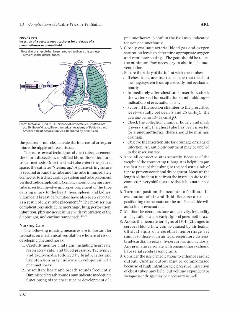
NEEDLE THORACENTESIS
The infant in severe respiratory distress with a tension pneumothorax requires immediate emergency treatment. Needle aspiration is necessary to decrease mortality and morbidity. Table 10-5 lists the equipment needed for needle aspiration. This equipment should be
kept in a clear plastic bag or container with the other emergency equipment in the nursery.
Following sterile preparation of the chest, the needle is inserted into the second or third intercostal space at the midclavicular line.45 Air is aspirated out through the syringe, then vented out the stopcock (Figure 10-6). Following removal of the needle, the insertion site is covered with a clear occlusive dressing.
CHEST TUBE INSERTION
The insertion of a chest tube is performed using sterile technique. The neonate should be positioned with the affected side up. The chest wall is prepped with bacteriostatic solution and injected with 1 percent lidocaine to provide local anesthesia. Analgesia, such as fentanyl or morphine, should be given because chest tube placement is a painful procedure.
Using the traditional superior approach, the tube is inserted into the second intercostal space on or just lateral to the midclavicular line. The lateral approach uses the fifth to sixth intercostal space just lateral to the anterior axillary line. Care must be taken not to pierce
TABLE 104
X-ray Findings: Pneumothorax
1. Increased lucency on the affected side
2. Decreased or absent pulmonary vascular markings
3. Overall increase in the size of the affected hemithorax
4. Widened intercostal spaces
5. Flattened diaphragm on the affected side
6. Sharp edge sign (The cardiac border and the diaphragm are seen in sharp contrast.)
7. With tension pneumothorax, mediastinal shift with deviation of the trachea and heart to the opposite side, decreased volume and increased opacity of the opposite lung
8. With bilateral pneumothoraces, narrow cardiac silhouette
From: Carey BE. 1999. Neonatal air leaks: Pneumothorax, pneumomediastinum, pulmonary interstitial emphysema, pneumopericardium. Neonatal Network 18(8): 81. Reprinted by permission.
FIGURE 105
Pneumothorax.
TABLE 105
Equipment for Needle Thoracentesis
Skin cleansing swabs
#18–20-gauge angiocatheter
T-connector
3-way stopcock
30–50 mL syringe
Transparent dressing
10 Complications of Positive Pressure Ventilation ARC
202
the pectoralis muscle, lacerate the intercostal artery, or injure the nipple or breast tissue.
There are several techniques of chest tube placement: the blunt dissection, modified blunt dissection, and trocar methods. Once the chest tube enters the pleural space, the catheter “steams up.” A purse-string suture is secured around the tube and the tube is immediately connected to a chest drainage system and tube placement verified radiographically. Complications following chest tube insertion involve improper placement of the tube causing injury to the heart, liver, spleen, and kidney. Significant breast deformities have also been reported as a result of chest tube placement.46 The most serious complications include hemorrhage, lung perforation, infarction, phrenic nerve injury with eventration of the diaphragm, and cardiac tamponade.47–50
Nursing CareThe following nursing measures are important for
neonates on mechanical ventilation who are at risk of developing pneumothorax: 1. Carefully monitor vital signs, including heart rate,
respiratory rate, and blood pressure. Tachypnea and tachycardia followed by bradycardia and hypotension may indicate development of a pneumothorax.
2. Auscultate heart and breath sounds frequently. Diminished breath sounds may indicate inadequate functioning of the chest tube or development of a
pneumothorax. A shift in the PMI may indicate a tension pneumothorax.
3. Closely evaluate arterial blood gas and oxygen saturation levels to determine appropriate oxygen and ventilator settings. The goal should be to use the minimum Paw necessary to obtain adequate ventilation.
4. Ensure the safety of the infant with chest tubes. If chest tubes are inserted, ensure that the chest
drainage system is set up correctly and evaluated hourly.
Immediately after chest tube insertion, check the water seal for oscillations and bubbling—indications of evacuation of air.
Set or fill the suction chamber to the prescribed level—usually between 5 and 25 cmH2O, the average being 10–15 cmH2O.
Check the collection chamber hourly and mark it every shift. If a chest tube has been inserted for a pneumothorax, there should be minimal drainage.
Observe the insertion site for drainage or signs of infection. An antibiotic ointment may be applied to the insertion site.
5. Tape all connector sites securely. Because of the weight of the connecting tubing, it is helpful to pin the first part of the tubing to the bed with a tab of tape to prevent accidental dislodgment. Measure the length of the chest tube from the insertion site to the connector every shift to assure that it has not slipped out.
6. Turn and position the neonate to facilitate the evacuation of air and f luid. Because air rises, positioning the neonate on the unaffected side will assist in air evacuation.
7. Monitor the neonate’s tone and activity. Irritability and agitation can be early signs of pneumothorax.
8. Assess the neonate for signs of IVH. (Changes in cerebral blood f low can be caused by air leaks.) Clinical signs of a cerebral hemorrhage are similar to those of an air leak: respiratory distress, bradycardia, hypoxia, hypercarbia, and acidosis. Any premature neonate with pneumothorax should have serial cerebral sonograms.
9. Consider the use of medications to enhance cardiac output. Cardiac output may be compromised because of high intrathoracic pressure. Insertion of chest tubes may help, but volume expanders or vasopressor drugs may be necessary as well.
FIGURE 106
Insertion of a percutaneous catheter for drainage of a
pneumothorax or pleural fluid.
Note that the needle has been removed and only the catheter remains in the pleural space.
From: Kattwinkel J, ed. 2011. Textbook of Neonatal Resuscitation, 6th ed. Elk Grove Village, Illinois: American Academy of Pediatrics and American Heart Association, 244. Reprinted by permission.

ARC Complications of Positive Pressure Ventilation 10
203
10. Evaluate the neonate’s level of agitation and pain, and comfort or medicate as necessary. In the past, neonates were paralyzed if they were f ighting mechanical ventilation. Sedation and analgesia are extremely important in caring for these critically ill neonates. Continuous fentanyl or morphine infusions may be helpful. Chest tube insertion and having the chest tube in place are quite painful.
11. Provide parents with accurate, honest, and understandable information rega rding the complications of mechanical ventilation and treatment of the air leak. Reassure them that the chest tube will help their baby to breathe more comfortably.
PNEUMOPERICARDIUM
Pneumopericardium is a rare complication of mechanical ventilation seen particularly in preterm neonates. PIE and pneumomediastinum often precede the entry of air into the pericardial sac (Figure 10-7). Pneumopericardium usually occurs during the first few days of life and most often occurs when high ventilatory pressures are being used.
Cardiac tamponade as a result of pneumopericardium can develop very quickly. Death can occur if this condition is not diagnosed and treated promptly. Clinical signs of pneumopericardium include bradycardia, cyanosis, muffled heart sounds, and hypotension. Chest
films using AP and lateral views reveal decreased heart size and air surrounding the heart (Figure 10-8).
A small pneumopericardium may be managed conservatively with close observation unless cardiac tamponade is evident.51 Emergency treatment for tamponade includes needling the pericardial space. Starting under the xiphoid, the angiocath is advanced at a 30- to 40-degree angle aiming at the left shoulder. A thoracotomy tube may be connected to a closed drainage system usually for two to three days. Nursing care is similar to that for the infant with pneumothorax, with specific attention to cardiac output.
PNEUMOPERITONEUM
Another rare complication of mechanical ventilation is pneumoperitoneum. Air dissects through the diaphragm into the retroperitoneal space. Clinical signs of pneumoperitoneum include a firm, shiny, and distended abdomen. The cause of the pneumoperitoneum should be investigated because this complication is also associated with necrotizing enterocolitis (NEC), gastric rupture, and a perforated ulcer that may require surgery.
FIGURE 107
Pneumopericardium.
Air fills the pericardial sac causing a tamponade of the heart.
FIGURE 108
Pneumopericardium.

10 Complications of Positive Pressure Ventilation ARC
204
In the case of a pneumoperitoneum, the x-ray shows a dark layer of air over the abdomen that blurs the normal bowel pattern. A right lateral view demonstrates the liver clearly defined from the anterior abdominal wall.
Medical treatment is indicated if the neonate’s respiratory status is severely compromised or if venous return to the heart is impeded. A soft catheter may be inserted into the peritoneum.
AIRWAY INJURYSubglottic stenosis, tracheomalacia, bronchomalacia,
tracheomegaly, necrotizing tracheobronchitis, and vocal cord injuries have been reported in infants requiring mechanical intubation and positive pressure ventilation.
Factors that appear to place intubated infants at risk for these complications include prolonged intubation, lack of an air leak around the endotracheal tube, repeated intubation, mechanical trauma from suctioning, gastroesophageal reflux, respiratory infection, hypoxia, hyperoxia, positive pressure ventilation, excessive movement of the endotracheal tube, and inadequate humidification of the endotracheal tube.52,53 These complications are more common in infants with BPD but can develop in those who required only short-term intubation and ventilation.
At a minimum, any infant who is intubated will develop edema in the airway followed by acute inflammation if intubation continues for more than a few hours. Pressure from the endotracheal tube reduces mucosal capillary perfusion, which can lead to ischemia, irritation, congestion, edema, and eventually ulceration.54,55 Progressive ulceration can lead to perichondritis, chondritis, and necrosis of the cricoid cartilage.56 Granulation tissue grows at the margins of the injured area and can persist as thick tissue, leading to narrowing of the airways. These extensive changes can lead to fibrotic, firm scar tissue, which can cause subglottic stenosis and narrowing of the airways.57 As a result, atelectasis and/or emphysema can develop. Many of these airway lesions contribute to the development of BPD.
Infants who have been intubated and ventilated for less than a week will have some edema, but their cries are normal within 24 hours after extubation. Infants who are extubated after one week to one month may have mild inspiratory and expiratory stridor lasting for a year or more.
Diagnosis of upper airway obstruction is often difficult to make in premature infants. Following extubation, the
infant may have decreased bilateral breath sounds, mild retractions, and apnea. The premature infant may not always develop stridor. Infants who develop respiratory failure will require reintubation, and if the respiratory distress immediately disappears, upper airway injuries should be suspected.
Damage to the larynx can be caused by necrosis over the arytenoid cartilage and vocal cords. Necrosis occurs because the endotracheal tube is in contact with the area. As a result, there may be persistent ulceration and/or erosion of the vocal cords. Significant damage may affect vocalization and respirations.
SUBGLOTTIC STENOSIS
Subglottic stenosis ranges from mild to severe in the intubated infant. The overall incidence of this acquired condition in ventilated preterm infants weighing <1,500 g at birth is approximately 1 percent.57 The lesion is usually associated with prolonged intubation and is diagnosed by bronchoscopy showing that the subglottic diameter (below the level of the glottic opening and above the level of the inferior margin of the cricoid cartilage) has become sufficiently narrow to cause symptoms of airway obstruction. The mildest form of subglottic stenosis is laryngeal edema.
Diagnosis of subglottic stenosis is made after physical examination, anteroposterior and lateral neck and chest x-ray films, and direct or fiberoptic laryngoscopy and bronchoscopy. In addition to respiratory distress, the infant may have mild to severe respiratory stridor that is not positional.
Treatment of mild respiratory difficulty includes elevating the head of the bed, providing humidified air, and administering racemic epinephrine. Treatment with steroids before extubation has been shown to be quite effective in premature infants.58 No significant side effects have been noted with the short-term use of dexamethasone.
The more severe form of acquired subglottic stenosis is a “hard” scar of fibrotic tissue. To extubate infants with this condition, an anterior cricoid split with or without immediate cartilage graft interposition may be required to increase the airway diameter.59 Some surgeons prefer a tracheostomy because it provides a long-term secure airway, but it too has its complications. If a tracheostomy is performed, decannulation occurs when the subglottic region has grown, usually in infants older than one year.60

ARC Complications of Positive Pressure Ventilation 10
205
TRACHEOMEGALY, TRACHEOMALACIA, AND BRONCHOMALACIA
Mechanical ventilation with positive pressure causing barotrauma can lead to dilation of the trachea and bronchi, resulting in tracheomegaly, tracheomalacia, or bronchomalacia. Tracheomegaly, diagnosed radiographically, results in an increase in the anatomic dead space, causing the infant to work harder at breathing to maintain normal carbon dioxide levels.61 Tracheomalacia and bronchomalacia develop when the cartilaginous rings in the airway soften and fail to support the round shape of the trachea, resulting in widening of the posterior airway way leading to airway collapse.62 The infant can develop expiratory stridor, wheezing, and atelectasis when the airway collapses or becomes obstructed on expiration. There are multiple factors for the pathogenesis of tracheomalacia and bronchomalacia, including barotrauma, immature airways, recurrent bacterial or viral infection, and pressure and irritation of the endotracheal tube.63 Tracheo- and bronchomalacia has been successfully treated with PEEP and ventilation or CPAP. Such treatment may place the infant at higher risk for BPD.
NECROTIZING TRACHEOBRONCHITIS
Ne c r ot i z i n g t rache o b r onch it i s , a ne c r ot ic inflammatory process involving the distal trachea and mainstem bronchi, is characterized by replacement of normal tracheal mucosa with acute inflammatory cells, mostly neutrophils. This process leads to sloughing of the mucosa, which can occlude the distal trachea. As a result of granulation, there may be impaired gas exchange, airway obstruction, and atelectasis. This lesion has been seen in neonates of all sizes and has been identified after just one day of ventilation.
Necrotizing tracheobronchitis has been associated with early work with high-frequency ventilation, but it has also been reported with conventional ventilation.64 There are various theories for the pathogenesis of necrotizing tracheobronchitis, including lack of humidification. The presence of the endotracheal tube has been suggested as a factor that causes damage by (1) direct pressure, (2) barotrauma from the ventilator-transmitted piston effect, or (3) toxins from the plastic of the endotracheal tube. Bacterial or viral infection may play a role similar to that in infants with tracheo- and bronchomalacia or subglottic stenosis. Infants with severe birth asphyxia and/or shock may develop necrotizing tracheobronchitis because of the ischemia to the airway mucosa. A disturbance in hemodynamics or
vascularization is postulated to play a role in the etiology of tracheobronchitis.64
Clinically, the infant with necrotizing tracheo-bronchitis may be asymptomatic, then suddenly deteriorate, with carbon dioxide retention that fails to respond to ventilator changes, suctioning, or reintubation. This is caused by the sloughing of the mucosa, which may occlude the distal trachea. Treatments have included excision or cauterization of the lesions, but this is difficult because of the relatively small airways of preterm infants. Obstruction can lead to lobar atelectasis or death.
Two types of lesions have been found on autopsy. Type I lesions show necrosis, mucosal hemorrhage, and ulcerations. Type II lesions, more chronic, show mucosal fibrosis and extensive squamous metaplasia.65 The long-term outcome is unknown, but follow-up is important because Type II lesions are considered to be premalignant in the area of the larynx and glottis.
NURSING CARE AND AIRWAY INJURY PREVENTION
Prevention of airway injury should be a priority for nurses caring for any mechanically ventilated infant. An endotracheal tube of the correct size should be used, and only experienced clinicians should intubate the ELBW infant or the infant who is known to be difficult to intubate. Following intubation, the tube should be stabilized to prevent excessive movement and accidental extubation.
A chest x-ray should be taken to evaluate proper endotracheal tube placement. Once tube placement is confirmed, the length of the tube in relation to the infant’s lip should be documented so that proper position can be checked every shift. When evaluating position by auscultating breath sounds, the caregiver should hear a slight air leak around the tube. Gas flow through the ventilator should be warmed and humidified sufficiently.
The nurse plays a major role in preventing airway damage from suctioning. Prior to suctioning, the nurse should select the appropriately sized suction catheter and know the exact measurement of the endotracheal tube. The suction catheter should not be passed beyond the length of the endotracheal tube. No more than 50–80 cmH2O pressure should be used when applying suction for five seconds. The frequency of suctioning should be individualized, based on the infant’s breath sounds, respiratory status, and clinical condition. Oxygen saturation and clinical status should be closely monitored while weaning the infant to appropriate ventilator settings (see Chapter 7).

10 Complications of Positive Pressure Ventilation ARC
206
Mechanically ventilated infants require continuous monitoring to maintain the fine balance between hypoxia and hyperoxia. Assessment of changes in the infant’s condition, oxygen saturation levels, and arterial blood gases is key to rapid initiation of appropriate ventilator changes to prevent complications.
Prevention of infection is a major challenge to the NICU team. The endotracheal tube prevents the cilia in the airway from clearing airway debris and potentially pathogenic bacteria or viruses. As a result, infection may develop, leading to the previously described airway lesions. Maintaining clean technique during intubation and endotracheal suctioning is important. If infection is suspected, antibiotics should be initiated and modified to specific organisms.
PULMONARY HEMORRHAGEPulmonary hemorrhage generally presents in the
first week of life in neonates who require mechanical ventilation. Before the widespread use of exogenous su r facta nt , pu l mona r y hemor rhage occu r red primarily in infants who were of low birth weight or small for gestational age, or in those with sepsis, asphyxia, or RDS.66–69 Since the introduction of exogenous surfactant, pulmonary hemorrhage rates have increased. A meta-analysis done by Raju and Langenberg in 1993 demonstrated a 47 percent increase in the risk of pulmonary hemorrhage when surfactant is given.70
The risk of pulmonary hemorrhage in surfactant-treated infants increases with decreasing gestational age and birth weight and has also been noted to be higher following vaginal delivery, in male infants, and in the presence of a patent ductus arteriosus.71,72
The incidence of pulmonary hemorrhage depends on the definition used, and there is little consistency in the grading of the severity of bleeding.73 Rates varying from 1 to 11 percent have been reported in the surfactant trials,74–76 while an incidence of less than 5 percent in infants with RDS was reported in the meta-analysis done by Raju and Langenberg.70
More than 80 percent of infants with pulmonary hemorrhage have RDS, and the incidence of pulmonary hemorrhage is inversely proportional to gestational age. At autopsy, the incidence of hemorrhage in premature infants has been found to be 80 percent.77 The extent of the hemorrhage may range from focal to massive (and fatal).
ETIOLOGY/PATHOPHYSIOLOGY
Pulmonary hemorrhage is speculated to be either the extreme result of pulmonary edema in the neonate or a consequence of increased transcapillary pore size, which allows red blood cells to enter the alveoli.77 The most common causes of pulmonary edema are increased pulmonary microvascular pressure, reduced intravascular oncotic pressure, reduced lymphatic drainage, and increased microvascular permeability.78 All result in increased fluid leakage into the pulmonary interstitium, increasing pulmonary lymphatic f luid. Pulmonary edema occurs as lung interstitial f luid increases; the fluid leaks into the alveoli after damage to the alveolar epithelium or distention caused by the interstitial f luid. Initially, only albumin leaks into the alveoli, but as the edema becomes more severe, capillary hemorrhage occurs. Pulmonary hemorrhage has been divided into three categories based on autopsy findings: (1) interstitial hemorrhages are characterized by hemorrhage in connective tissue spaces of the lung; (2) lung hematomas are accumulations of fresh blood in the interstitium of alveolar spaces; and (3) intra-alveolar hemorrhages are characterized by fresh blood filling alveoli in areas not directly adjacent to the interstitium, often extending into the bronchioles and bronchi to produce massive hemorrhage.
At-risk neonates also include those with asphyxia, shock, hypoxia, acidosis, and PDA, all of which can lead to left ventricular heart failure.
The premature infant with severe RDS who is on mechanical ventilation or a high oxygen concentration and who has heart failure secondary to increased pulmonary blood f low is at high risk for developing pulmonary edema and hemorrhage even before receiving surfactant therapy. And the pulmonary edema itself, is known to inhibit surfactant function.
Neonates with RDS who are treated with exogenous surfactant are at risk for pulmonary hemorrhage. The etiology of pulmonary hemorrhage following treatment with surfactant includes alterations in pulmonary hemodynamics because of a PDA, fragile capillaries resulting from extreme prematurity, barotrauma caused by mechanical ventilation, and a localized coagulopathy caused by the surfactant.79,80 A review of 33 treatment trials using exogenous surfactant from 1980 to 1992 focused on the association between exogenous surfactant therapy and pulmonary hemorrhage. The natural surfactant trials reported a pulmonary hemorrhage incidence of 5.87 percent in treated infants versus

ARC Complications of Positive Pressure Ventilation 10
207
5.36 percent in controls; the synthetic trials reported an incidence of 2.51 percent in treated versus 1.04 percent in control infants. Analysis revealed that surfactant treatment and lower mean birth weight had a significant influence on the risk for a pulmonary hemorrhage. Interestingly, a PDA did not have an independent effect on the risk of a pulmonary hemorrhage.70
Factors associated with pulmonary hemorrhage include intrauterine growth retardation, massive aspiration, hypothermia, infection, oxygen therapy, severe Rh hemolyt ic disease, congenital heart disease, fluid overload, and coagulopathies. Although disseminated intravascular coagulation may precede pulmonary hemorrhage, most affected infants do not have a coagulopathy but may develop it after the hemorrhage occurs.3
SIGNS AND SYMPTOMS
Clinically, an infant with a pulmonary hemorrhage may initially present with blood-tinged fluid from the endotracheal tube. With a massive hemorrhage, there may then be a sudden deterioration and simultaneous appearance of bloody secretions in the endotracheal tube and/or the infant’s mouth. The fluid has the appearance of fresh blood, but the hematocrit of the fluid is 15–20 points lower than that of the circulating blood.3
Usual ly, the in fa nt becomes pale, cya not ic, hypotensive, and hypotonic, but term infants may become agitated secondary to the hypoxemia and begin to “fight” the ventilator. Signs of heart failure may be present, including tachycardia, murmur (related to the PDA), hepatosplenomegaly, and edema. Hypotension results from the blood and fluid loss, heart failure caused by hypoxemia, and acidosis. Auscultation of the chest reveals widespread crepitus and decreased air entry.
DIAGNOSIS
A few infants may deteriorate clinically without apparent cause for an hour or two before the hemorrhage begins. Once the frank blood becomes evident, the diagnosis is made. Chest radiographic findings depend on whether the hemorrhage was focal or massive. It is often difficult to differentiate a focal hemorrhage from atelectasis or pneumonia. Massive hemorrhage reveals a “whiteout” reflecting atelectasis and opacifications with some air bronchograms (Figure 10-9).
Blood gases deteriorate rapidly following a massive hemorrhage, resulting in severe hypoxia, hypercarbia, and a marked metabolic acidosis. Although the hematocrit of the lung f luid is diluted, considerable amounts of blood may be lost. There are no specific white blood cell changes unless sepsis is present. Drawing of blood cultures is recommended following the hemorrhage. Development of disseminated intravascular coagulation is not uncommon after hemorrhage occurs.
MANAGEMENT
Control of pulmonary edema and heart failure in addition to positive pressure ventilation and oxygenation are critical in preventing pulmonary hemorrhage. Following administration of surfactant, the nurse should closely monitor the infant for signs of heart failure, hypotension, decreased air entry, and wet breath sounds.
Early detection and aggressive intervention are vital in the management of pulmonary hemorrhage. Infants experiencing pulmonary bleeding should be intubated and ventilated. They usually have severe lung diseases
FIGURE 109
Pulmonary hemorrhage x-ray.
Infant with moderate respiratory distress.
Three hours later, x-ray demonstrates a severe pulmonary hemorrhage.

10 Complications of Positive Pressure Ventilation ARC
208
that require high PEEP and PIP. An increase in PEEP may be helpful in splinting the alveoli and reducing bleeding. This may help in redistributing lung f luid back into the interstitial space, improving ventilation and perfusion.81
Transfusion of blood products, including packed red blood cells, may be necessary because of acute blood loss. Infusions of fresh frozen plasma and administration of vitamin K may be successful in correcting clotting deficiencies. Antibiotic therapy should be started if not already instituted, because sepsis is a major risk factor for pulmonary hemorrhage. Inotropes and diuretics may be needed if heart failure develops.
Administration of surfactant following a pulmonary hemorrhage has been shown to improve oxygenation significantly.82–84 It is postulated that the presence of hemoglobin in the alveoli may inhibit natural surfactant.84
Complications following pulmonary hemorrhage include air leaks and periventricular hemorrhage. The mortality rate after a pulmonary hemorrhage ranges from 30 to 90 percent, with 50–75 percent of the survivors developing CLD.77
NURSING CARE
Care of the infant with a significant pulmonary hemorrhage includes all aspects of neonatal intensive care nursing. Maintaining an open airway is a major priority. During the first few hours after the hemorrhage, the endotracheal tube may require suctioning every 10–15 minutes. There is significant risk of bloody secretions blocking the tube. Breath sounds must be evaluated frequently.
The infant is often placed on maximum ventilator settings, requiring vigilant monitoring of arterial blood gases and vital signs. Monitoring for the development of air leaks is important because of high pressure settings. Based on evaluation of blood gases, ventilator settings may be changed, and sodium bicarbonate may be ordered. If hypotension occurs, fluids will be recalculated. Blood products and vasopressors may also be necessary.
CARDIOVASCULAR COMPLICATIONSThe respiratory and cardiovascular systems work
in close harmony to provide the body with adequate oxygen and to remove waste products from the cells. The respiratory system affects cardiovascular function by altering venous return and pulmonary vascular resistance (PVR). Cardiac output depends on venous
return to the heart, which is determined in part by differences between extrathoracic and intrathoracic pressures. Subatmospheric intrapleural pressure establishes a favorable pressure gradient for blood to flow back to the right atrium.
HOW MECHANICAL VENTILATION AFFECTS THE CARDIOVASCULAR SYSTEM
The use of CPA P or posit ive pressures f rom mechanical ventilation can affect the cardiovascular system by increasing intrathoracic pressure, which decreases venous return.85 The diminished venous return along with compression of the ventricles caused by the increased intrathoracic pressure decreases cardiac output.
The impact of mechanical ventilation on cardiac output depends on the degree of pressure transmitted from the airway to the intrapleural space. This pressure is influenced by lung compliance. Neonates with RDS who have reduced lung compliance transmit significantly less pressure to the intrapleural space than do those with normal compliance, and so ventilation in these compromised neonates exerts little effect on venous return and cardiac output. The infants can generally tolerate high levels of PIP and PEEP without significant decreases in cardiac output. However, the premature infant who develops a tension pneumothorax has a sudden rise in intrathoracic pressure, which increases central venous pressure. These changes can result in IVH.
When neonates are recovering from RDS following surfactant therapy, compliance may increase rapidly along with increased intrapleural pressure. High ventilator pressures in these neonates can decrease cardiac output and increase venous pressure, leading to possible systemic hypotension, altered perfusion, and IVH.
Another potential complication of positive pres-sure ventilation and CPAP is a ventilation-to-perfusion mismatch ( ). This ratio describes the relationship between alveolar ventilation and capillary perfusion of the lung. In neonates with lung disease, even though CPAP or positive pressure is applied, areas that are atel-ectatic tend to remain so, while inflated regions tend to become further distended. The circulation responds by perfusing the areas of the lung that are distended and diminishing circulation in the atelectatic portions.81 A maximum mismatch occurs in an infant with a tension pneumothorax: Ventilation escapes into the pleural space, where no gas exchange occurs.

ARC Complications of Positive Pressure Ventilation 10
209
Mechanical ventilation increases airway pressure, which is also transmitted to the intraparenchymal pul-monary vessels. The effect is complex and depends on several factors, including the lung disease and compli-ance. In infants with RDS, there is a decrease in func-tional residual capacity (FRC), which can result in increased PVR. In infants with lung diseases treated with mechanical ventilation that overdistends the lung, the air spaces compress arterioles and capillaries, caus-ing a mismatch and leading to increased PVR.
Persistent pulmonary hypertension of the newborn (PPHN) is a well-known condition in which PVR remains elevated. During the transition to extrauterine life, PVR normally decreases. In the infant with PPHN, the PVR remains higher than the systemic blood pressure, resulting in a right-to-left shunt across the ductus arteriosus and/or foramen ovale, so blood bypasses the lungs. Clinically, neonates with this condition present with severe cyanosis, higher preductal and lower postductal oxygen saturations.
Hyperventilation with mechanical ventilation has been an important aspect of care because it has been shown to decrease PVR in infants with PPHN. Hyperventilation may not be necessary in treating milder cases and may result in complications, including air trapping. Moderate to severe PPHN may necessitate the use of high-frequency ventilation or ECMO (see Chapters 12 and 13).
PATENT DUCTUS ARTERIOSUS
Patent ductus arteriosus is a condition in which the cardiovascular system has a direct effect on ventilation and perfusion. Delayed ductal closure is inversely related to gestational age and presents a challenging problem for the team caring for the premature neonate on mechanical ventilation. The large left-to-right shunt and resulting cardiac failure aggravate preexisting pulmonary disease.
The ductus arteriosus (DA) arises from the distal dorsal sixth aortic arch and forms a bridge between the pulmonary artery and the dorsal aorta. During fetal life, it carries most of the right ventricular output and directs blood away from the fetal lungs and toward the descending aorta and placenta. Prostaglandin E2, produced by tissue in the DA, plays an important role in maintaining patency of the ductus in utero.86 The DA becomes more responsive to oxygen and less sensitive to the dilating effects of prostaglandin with increasing gestational age.
In the term neonate, the DA begins to constrict rapidly after delivery with the initiation of breathing and is usually functionally closed by 48 hours of age.87 Muscle media indent into the lumen, and the intima increases in size to form intimal mounds or cushions that begin to occlude the ductus.88 These intimal changes occur in conjunction with extensive constriction and shortening of the ductus as well as migration of smooth muscle cells from the media into the intima. Ductal constriction results from multiple factors, increased arterial oxygen tension being one of the most important.86
In preterm infants, closure of the DA is less predictable. A number of factors can delay closure, including lung disease that increases PVR, decreased ductal sensitivity to oxygen, increased circulating prostaglandins, and an increased ductal sensitivity to both prostaglandins and nitric oxide.88,89 A study of 49 preterm infants found their serum levels of prostacyclin, a vasodilatory prostaglandin, to be higher than levels in adults, especially in those infants requiring higher ventilatory support. Higher prostacyclin levels were found in those infants in the study who developed a clinically significant PDA.90
IncidenceThe incidence of PDA is 20 percent in infants born at
>32 weeks gestation but increases to 60 percent in infants born at <28 weeks gestation; the incidence increases with decreasing gestational age and birth weight and the occurrence of RDS.86 Among infants weighing <1,000 g, about 55–70 percent will have hemodynamic symptoms of a PDA.87 An early study reported that surfactant therapy may increase the incidence of symptomatic PDA in mechanically-ventilated premature infants to as high as 90 percent.91 Clinical and echocardiographic reports of these surfactant-treated infants showed that the PDA is of greater diameter, has more blood flow, and causes greater clinical deterioration.92 A subsequent meta-analysis found that surfactant treatment had no effect on the incidence of PDA.93
PathophysiologyInflation and ventilation of the lungs at birth should
decrease PVR and induce ductal constriction. The drop in PVR allows blood to flow from left to right (aorta to pulmonary artery), in the direction opposite of that fetal circulation. If PVR remains high, as it does during the acute phase of RDS, a bidirectional shunt may occur across the PDA.

10 Complications of Positive Pressure Ventilation ARC
210
The effects of the shunt through the PDA depend on several factors: diameter of the ductus, ductal tone, systemic vascular resistance and PVR, and left ventricular output. Quite often in premature neonates, especially those with RDS, the PVR remains elevated. In addition, persistent hypoxia may prevent the ductus from closing.
Before the use of surfactant, the development of a significant PDA usually corresponded to the diuresis phase of RDS. Following surfactant therapy, improved pulmonary compliance causes PVR to drop below systemic vascular resistance; a significant ductal shunt can develop rapidly as PVR drops.88 Surfactant is thought to cause the release of circulating prostaglandins, which cause relaxation of smooth muscle, including that of the DA.
As respiratory distress resolves and PVR drops, left-to-right shunting predominates, placing stress on the heart and lung. In the premature infant, the ventricles are less distensible and generate less force; therefore, this can result in left ventricular enlargement from the PDA. Elevated left ventricular end-diastolic pressure results, which increases pulmonary venous pressure and causes pulmonary congestion. As a result, the infant develops right-sided heart failure and over time may develop pulmonary hypertension.94
In addition, the infant with RDS frequently has a low plasma oncotic pressure and increased capillary permeability, both of which respond to the increased microvascular perfusion by allowing leakage of plasma proteins into the alveolar space. This leads to pulmonary edema. This leakage may inhibit surfactant function and increase surface tension, thereby worsening the disease. Additionally, immature alveoli may be more sensitive to the presence of this fluid.88 The pulmonary edema plus the continuous distention of pulmonary vessels during diastole may be factors in the development of pulmonary hemorrhage and BPD.87,95
In the presence of a left-to-right shunt such as a PDA, a term infant is capable of maintaining cardiac output by increasing left ventricular output. The premature infant’s ventricles have less muscular organization and more water content, resulting in an inability to maintain cardiac output. This may cause a redistribution of systemic blood f low to the organs. Very low birth weight (VLBW) infants with PDA have been found to have increased blood flow in the ascending aorta and decreased flow in the descending aorta, findings that have been associated with IVH and NEC.96–98
Clinical FindingsPrior to the use of surfactant, clinical signs of a PDA
did not usually appear until the third or fourth day of life, during the recovery phase of RDS. Although the ductus was patent, the elevated PVR secondary to lung disease diminished left-to-right shunting. As pulmonary functioning and oxygenation improved, PVR decreased. With the early administration of surfactant to infants with RDS, significant shunting through the ductus is seen much earlier. Infants born at less than 30 weeks gestation who have severe RDS also have a high incidence of persistent PDA.99
Moderate to large amounts of shunting through the PDA can result in congestive heart failure. Clinical signs include a hyperactive precordium, tachypnea, tachycardia, decreased urine output, increased pulse amplitude, and widened pulse pressure (difference between systolic and diastolic blood pressure is >30 mmHg). The increase in cardiac output and blood flow back to the left side of the heart cause the increased precordial activity and bounding pulses. The classic continuous murmur described in older infants is not always heard in premature infants.
Preterm infants may have a PDA that is clinically silent but hemodynamically significant. These infants can have a reduction in systolic and diastolic blood pressures severe enough to require inotropic drugs.100 Research has demonstrated that the presence of a PDA for longer than six days is associated with a longer duration of oxygen therapy and mechanical ventilation.101 Long-term effects of a PDA include poor weight gain, recurrent respiratory infections (because of increased lung fluid and left-sided heart failure), and the need for additional ventilator support.
DiagnosisThe diagnosis of PDA is based on clinical findings
plus echocardiography. A poor correlation between clinical findings alone and a PDA diagnosis has been identif ied.102 M-mode echocardiography provides measurement of the heart chambers and can be used to evaluate left ventricular function.103 A color Doppler echocardiogram can determine the degree of shunting across the ductus, and two-dimensional echocardiography provides information about the size of the ductus. With M-mode, if the ratio of the size of the aortic root to the left atrium is greater than 1:1, the presence of a PDA is confirmed. Using echocardiography and Doppler diagnosis on day 3 of life, it is possible to predict PDAs that will later become symptomatic. In

ARC Complications of Positive Pressure Ventilation 10
211
one study, a ductal diameter of >1.5 mm within the first 30 hours of life had a sensitivity of 83 percent in predicting the need for treatment of a PDA.104 A chest x-ray of an infant with a PDA may be completely normal in the absence of significant left-to-right shunting, or it may demonstrate an enlarged left atrium and alveolar edema.103
TreatmentDefinitive treatment of a PDA is closure. Conservative
methods are implemented before pharmacologic therapy or surgical ligation. There has been some debate in the recent literature both about the need to treat the ductus and about when to initiate treatment. The lack of evidence showing any long-term benefit from treatment of a PDA has led some investigators to question the need for treatment.89,105 Further, it has been suggested that although there is an association between a PDA and the morbidities discussed earlier in this section, there is little proof of causation.94,106
CONSERVATIVE APPROACHES
A study published in 2007, by Vanhaesebrouck and colleagues, demonstrated achievement of a 94 percent rate of ductal closure by employing a conservative approach of increased PEEP and fluid restriction.107
Prior to Vanhaesebrouck’s study, it was generally accepted that fluid restriction, although recommended, wa s u n l i kely to close t he PDA w it hout ot her interventions,86,106,108 but that it might confer some benefit by reducing the hemodynamic significance of the PDA.109 A combination of fluid restriction and diuretics can lead to electrolyte imbalances, dehydration, and reduced caloric intake. According to one 1983 study, administration of furosemide was associated with an increased incidence of PDA.110 It is speculated that this finding may have resulted from diuretic-induced release of renal prostaglandins.86
The use of PEEP has been shown to reduce the left-to-right shunt through the PDA.100,107 Management of mechanical ventilation for the infant with a PDA is an important issue. Infants without RDS but with a large left-to-right shunt may have increased interstitial and peribronchiolar edema. Because lung compliance is relatively normal, high inflating pressures should be avoided. These high pressures may impair venous return and cardiac output, altering pulmonary perfusion and the ratio.
PHARMACOLOGIC THERAPY: INDOMETHACIN AND IBUPROFEN
For a number of years, indomethacin has been the mainstay of pharmacologic therapy for a PDA. Debate continues regarding the criteria for initiating treatment (prophylactic vs symptomatic) and the length of treatment (short vs long course). More recently, ibuprofen has been approved for use in PDA treatment in the U.S. A number of trials comparing indomethacin and ibuprofen have been published, including three meta-analyses.111–113 Indomethacin and ibuprofen are potent inhibitors of the cyclo-oxygenase pathway, which forms the various prostaglandins, and were originally developed as anti-inflammatory agents.
Indomethacin has been proven to be clinically effective in closing PDAs in premature infants within the first seven days of life, with successful closure in approximately 66–80 percent of cases.114–116 More recent figures suggest that successful closure of a symptomatic PDA can be expected in 50 percent of 24- to 25-week-gestational-age infants receiving indomethacin and in 60 percent of infants >25 weeks gestational age.117,118
Administration of prophylactic indomethacin has been studied both in the prevention of PDA and also as a strategy to prevent IVH. Two studies have demonstrated the benef its of indomethacin prophylaxis on the incidences of PDA, PDA ligation, and severe intracranial hemorrhage (ICH).119,120 A Cochrane review reached a similar conclusion.101
Evaluation of prophylactic treatment for PDA also found that indomethacin-treated infants required more oxygen, higher mean ventilatory pressures, and more doses of surfactant.120–122 Because of the side effects of both indomethacin and ibuprofen and because prophylactic treatment has failed to demonstrate long-term benefits, this practice has been abandoned.
Use of indomethacin as a treatment for asymptomatic PDAs has also been examined. A meta-analysis done by Cooke and colleagues found a significant decrease in the incidence of symptomatic PDAs following treatment of asymptomatic PDAs with indomethacin.123 Others argue that the use of indomethacin in asymptomatic infants unnecessarily puts them at risk of side effects without proven long-term benefit.89,124
Currently, intravenous indomethacin, 0.1–0.3 mg/kg/dose, is given every 12–24 hours for a total of three doses. In most cases, a single dose has not resulted in persistent constriction of the DA. Studies looking at the efficacy of a five- or six-day course of low-dose indomethacin (0.1 mg/kg/day) have found a lower incidence of fluid

10 Complications of Positive Pressure Ventilation ARC
212
and electrolyte imbalances and also a lower rate of ductal reopening than with the traditional three-dose course.125–127 However, a meta-analysis comparing the long-course (four doses or more) approach to a short (three-dose) course found only a borderline effect on the rate of PDA closure, with a greater risk of CLD in infants receiving the long course. The long course did result in a decreased risk of renal impairment, but an increased risk of NEC. The authors concluded that a prolonged course of indomethacin could not be recommended.128
Complications from indomethacin can be significant, so infants must be screened before therapy is initiated. Serum creatinine and electrolytes should be measured before treatment is started and before each subsequent dose is given.87 Renal dysfunction can be a major complication. Indomethacin may be contraindicated if the serum creatinine is above 1.2–1.8 mg/dL or if urine output is less than 1 mL/kg/hour. If urine output decreases in an infant who has received indomethacin, the administration of low-dose dopamine has been suggested.
Platelet function may be impaired for at least a week after indomethacin administration. For this reason, indomethacin is contraindicated in infants with renal or gastrointestinal (GI) bleeding or with NEC. It is recommended that the neonate has a platelet count of at least 50,000/mm3 before initiation of indomethacin treatment.87 Although the drug has been associated with occasional intestinal perforation, there has been no evidence of increased NEC. The increased incidence of GI perforation has been reported when indomethacin and postnatal steroids are administered concurrently.129
Indomethacin has been shown to decrease cerebral blood flow by 12–40 percent in premature infants.116 There is concern that rapid infusion, which has been the standard practice, might reduce cerebral blood flow to excessively low levels, resulting in brain ischemia. Two studies have shown a significant decrease in cerebral blood f low velocities when the drug is given quickly over 5 minutes or slowly over 30 minutes. Therefore, further studies are necessary to determine the safest rate of administration.130,131
For a number of years ibuprofen has been used in Europe as an alternative to indomethacin. Studies have suggested that ibuprofen has an efficacy similar to that of indomethacin but without the significant reduction in renal function.112,132,133 In a meta-analysis, indomethacin and ibuprofen had a similar rate of PDA closure with no differences in need for surgical ligation; mortality; or the incidence of IVH, NEC, or ROP. The
review found an increased rate of CLD in infants receiving ibuprofen.134 Unlike indomethacin, ibuprofen has not been found to reduce the incidence of severe IVH.135 Ibuprofen may be the drug of choice for closure of the ductus arteriosus because it has fewer short-term side effects.117,136 No long-term follow-up data is available for ibuprofen as it was only approved for use in the United States in 2006.
Following administration of indomethacin or ibuprofen, the infant should be monitored for success of PDA closure. Significant improvements in lung compliance have been noted.137 Mechanical ventilation pressures and rates can be lowered, thus exposing infants to lower Paw. Reopening of the ductus arteriosus is a common problem in infants weighing <1,000 g.
SURGICAL LIGATION
Surgical ligation of the PDA is usually reserved for infants for whom drug therapy is contraindicated or who fail to respond to conservative and/or drug therapy. Ligation through a left lateral thoracotomy can be done in a short time either in the operating room or at the bedside. Complications of ligation include laryngeal nerve paralysis, pneumothorax, infection, and chylothorax.94,138 A significant number of postoperative infants experience hypotension, requiring inotropic support.139 A recent study found an increased incidence of CLD, ROP, and neurodevelopmental abnormalities in ELBW infants requiring PDA ligation.140 It is unclear from this study whether surgical ligation is causative or reflective of the degree of illness in this group.
A Cochrane review comparing surgical ligation with medical treatment with either ibuprofen or indomethacin found only one eligible trial to review.113 This review does state that three observational studies noted that neonates undergoing surgical ligation for PDA had an increased risk for one or more of the following outcomes; chronic lung disease, retinopathy of prematurity, and neurosensory impairment.113 That trial, from 1983, showed no difference in CLD, NEC, IVH, or mortality between the surgically and the pharmacologically treated groups.116 Other surgical techniques, including video-assisted thoracoscopic clipping and catheter coil occlusion, have been explored, but limited neonatal experience has been reported.141–143
Nursing CareNursing care of the ventilated premature infant
requires careful monitoring for signs of a PDA—especially after administration of surfactant. Changes in

ARC Complications of Positive Pressure Ventilation 10
213
vital signs that suggest heart failure should be reported. A low mean arterial blood pressure may be an early sign of the patency of the ductus arteriosus in infants weighing <1,000 g. If possible, heart sounds should be auscultated while the infant is off the ventilator, and any murmurs or clicks should be noted. An increase in precordial activity is an extremely reliable sign of a significant PDA. Infants who require increasing ventilatory support or receive surfactant should be further evaluated for PDA.
Medical treatment is based on echocardiography, clinical signs, and unit standards. Before being given indomethacin or ibuprofen, the infant should be evaluated for signs of renal and platelet dysfunction, NEC, and recent IVH. Laboratory studies should include a complete blood count (CBC) with differential, platelet count, electrolytes, blood urea nitrogen (BUN), creatinine, and bilirubin levels.
Strict measurement of urine output before and during drug treatment will reflect renal dysfunction. Assessment for clinical bleeding includes heelstick sites, gastric drainage, and blood in the stool. Auscultation for the absence of a heart murmur during treatment is important. Even after the PDA has been determined to be closed, auscultation for recurrence of a murmur is important in VLBW infants. Retreatment may be considered.
If the infant is unresponsive to drug therapy and requires increased ventilatory support, surgical ligation is considered. Preoperative care includes stabilizing fluid and electrolyte levels, oxygenation, ventilation, and the infant’s temperature. Packed red blood cells may be ordered and held for possible transfusion. The surgeon and neonatologist should discuss the benefits and risks of the surgery with the parents and obtain informed surgical consent.
Following surgical ligation, the nurse should assess the infant’s vital signs and determine the need for pain medication. The thoracotomy site should be assessed for signs of bleeding or infection. The chest tube drainage system should be checked hourly for proper functioning and any drainage.
BRONCHOPULMONARY DYSPLASIABronchopulmonary dysplasia is a CLD that develops
primarily in neonates who are born at 24–26 weeks gestation weighing <1,000 g and who receive prolonged oxygen therapy and/or positive pressure ventilation.144 The increase in survival among very premature
newborns has increased the number of infants with this disorder, challenging the health care system. In the literature the terms BPD and CLD are often used interchangeably, as they will be in this chapter.
DEFINITIONS: OLD AND NEW
In 1967, Northway, Rosan, and Porter described BPD as a type of CLD that developed in premature infants with severe RDS who were treated with positive pressure mechanical ventilation and oxygen.145 These infants had severe respiratory failure at birth; required aggressive ventilatory support; and, as a result, developed severe lung injuries and remained dependent on oxygen for long periods of time. Originally, Northway’s group postulated that oxygen toxicity caused BPD, but research has revealed that multiple complex mechanisms cause the disease. Northway and colleagues’ description of BPD is now referred to as classic, or “old,” BPD. This descriptor recognizes that in the postsurfactant, post–antenatal steroid era, the picture of BPD has changed and a “new” BPD has emerged. BPD is now known to occur in term and preterm neonates with a variety of neonatal conditions, including apnea, meconium aspiration, pneumonia, and congenital heart disease as well as primary lung disease. Today, infants with BPD may have only mild lung disease at birth and receive only brief periods of mechanical ventilation and oxygen therapy.
Northway and colleagues’ original description of BPD outlined the radiologic, pathologic, and clinical criteria associated with four stages of the disease (Table 10-6).145 Bancalari and associates, in 1979, further defined an infant with BPD as one who requires positive pressure ventilation for at least three days during the first week of life, has clinical signs of respiratory distress, requires supplemental oxygen to maintain an oxygen tension (PaO2) of 50 torr for >28 days, and shows radiographic evidence of BPD.146 Since its original description, the presentation and progression of BPD have changed, but the initial characteristics and definitions remain salient to an understanding of this disorder.
As smaller and sicker infants survive because of new technologies (including surfactant replacement therapy, high-frequency ventilation, and prenatal and postnatal steroids), new BPD has emerged. The severe form of BPD originally described was seen primarily in premature infants who were ventilated mechanically using high pressures and had prolonged exposure to high levels of inspired oxygen. New BPD is a milder form of CLD seen

10 Complications of Positive Pressure Ventilation ARC
214
in smaller infants who do not necessarily have severe lung disease at birth.147
The newer de s c r ipt ion s of BPD ref le ct ou r understanding of the disorder as one of altered lung development with decreased numbers of alveoli and abnormal blood vessel development rather than lung damage.144,148 However, a clear definition of BPD remains elusive. Some clinicians define BPD as a requirement for supplemental oxygen at day 28 of life; however, this definition may inaccurately label infants who have an acute illness at the end of the first month of life as having BPD or miss infants who subsequently develop the need for supplemental oxygen.147 Others have proposed the need for oxygen at 36 weeks postmenstrual age as a better criterion for defining BPD.149 In 2001, the National Institutes of Health (NIH) convened a consensus panel to address these inconsistencies. That panel agreed on the definition of BPD shown in Table 10-7. The NIH definition requires a minimum of 28 days of supplemental oxygen and defines the severity of the disease by the amount of oxygen required.150 It is important to note that under this definition, infants being treated with supplemental oxygen for nonpulmonary problems—for example, for congenital anomalies such as diaphragmatic hernia—are not considered to have BPD unless they also have parenchymal lung disease. To further ref ine this definition, some clinicians have suggested that infants receiving supplemental oxygen at 28 days or 36 weeks corrected age, undergo an oxygen needs test. This test
involves challenging the infant by gradually reducing the inspired oxygen to room air. Those infants with an oxygen saturation of <90 percent after 30 minutes on room air would be deemed to have BPD.151
INCIDENCE
The incidence of BPD is difficult to report because it depends both on the definition of BPD used and also on the accuracy of gestational age determination in the study population. A review by Bhandari and Panitch found the incidence of BPD, defined as oxygen need at 36 weeks postmenstrual age, to be about 30 percent among infants with birth weights <1,000 g.152 This is similar to the rate of 35 percent found by Walsh and colleagues in a study of 1,598 inborn infants weighing <1,250 g who remained hospitalized at 36 weeks postmenstrual age.151 Sahni and colleagues identified rates of BPD, defined as oxygen need at 28 and 36 weeks, to be 21.1 and 7.4 percent, respectively.153 Ehrenkranz and colleagues applied the NIH definition of BPD in a retrospective review of 4,866 infants (birth weight ≤1,000 g, gestational age <32 weeks, alive at 36 weeks postmenstrual age) born between 1995 and 1999 and found that 77 percent of the infants met the criteria for BPD, with 30 percent having moderate disease and 16 percent severe BPD. Of those who met the NIH criteria and were seen in follow-up at 18–22 months corrected age, 35 percent had required rehospitalization for respiratory illnesses and 40 percent had received medications for a pulmonary condition.154
TABLE 106
Stages of Bronchopulmonary Dysplasia (Classic)
Stage Time Pathologic Findings Radiologic Findings Clinical Features
I (mild) 2–3 days Patchy loss of cilia; bronchial epithelium intact; profuse hyaline membranes
Air bronchograms; diffuse reticulogranularity (identical to RDS)
Identical to RDS
II (moderate) 4–10 days Loss of cilia; fewer hyaline membranes; necrosis of alveolar epithelium; regeneration of bronchial epithelium; ulceration in bronchioles
Opacification; coarse, irregularly shaped densities containing small vacuolar radiolucencies
Increased O2 requirements and increasing ventilatory support when recovery is expected; rales, retractions
III (severe) 10–20 days Advanced alveolar epithelial regeneration; extensive alveolar collapse; bronchiolar metaplasia and interstitial fibrosis; bronchial muscle hypertrophy
Small radiolucent cysts in generalized pattern
Prolonged O2 dependency; PaCO2 retention; retractions; early barrel chest; severe acute episodes of bronchospasm
IV (advanced-chronic)
1 month Obliterative bronchiolitis; active epithelial proliferation; peribronchial and some interstitial fibrosis; severe bronchiolar metaplasia
Dense fibrotic strands; generalized cystic areas; large or small heart; hyperinflated lungs; hyperlucency at bases
Increased chest anteroposterior diameter; cor pulmonale; frequent respiratory infection; prolonged O2 dependency; failure to thrive
From Korones SB. 2011. In Assisted Ventilation of the Neonate, 5th ed., Goldsmith JP, and Karotkin EH, eds. Philadelphia: Saunders, 390. Reprinted by permission.

ARC Complications of Positive Pressure Ventilation 10
215
PATHOGENESIS
Bronchopulmonary dysplasia has been attributed to oxygen toxicity, barotrauma, volutrauma, lung immaturity, inflammation, and infection (Figure 10-10). The causes are multifactorial and likely include acute lung injury, arrested lung development, as well as
abnormal repair processes that occur in the lung. Normal lung growth and development are disrupted by a premature birth. The immature lung, already deficient in surfactant, is then exposed to adverse stimuli.
TABLE 107
Definition of Diagnostic Criteria for BPD
Gestational age <32 weeks ≥32 weeks
Time point of assessment
36 week PCA or discharge to home, whichever comes first >28 days, but <56 days postnatal age or discharge to home, whichever comes first
Treatment with oxygen >21% for at least 28 days plus
Mild BPD Breathing room air at 36 weeks PCA or discharge, whichever comes first
Breathing room air by 56 days postnatal age or discharge, whichever comes first
Moderate BPD Need* for <30% oxygen at 36 weeks PCA or discharge, whichever comes first
Need* for <30% oxygen at 56 days postnatal age or discharge, whichever comes first
Severe BPD Need* for <30% oxygen and/or positive pressure, (PPV or NCPAP) at 36 weeks PCA or discharge, whichever comes first
Need* for ≥30% oxygen and/or positive pressure (PPV or NCPAP) at 56 days postnatal age or discharge, whichever comes first
Key: NCPAP = nasal continuous positive airway pressure; PCA = postconceptional age; PPV = positive-pressure ventilation.
*A physiologic test confirming that the oxygen requirement at the assessment time point remains to be defined. This assessment may include a pulse oximetry saturation range. BPD usually develops in neonates being treated with oxygen and PPV for respiratory failure, most commonly RDS. Persistence of clinical features of respiratory disease (tachypnea, retractions, rales) are considered common to the broad description of BPD and have not been included in the diagnostic criteria describing the severity of BPD. Infants treated with oxygen >21 percent and/or positive pressure for nonrespiratory disease (e.g., central apnea or diaphragmatic paralysis) do not have BPD unless they also develop parenchymal lung disease and exhibit clinical features of respiratory distress. A day of treatment with oxygen >21 percent means that the infant received oxygen >21 percent for more than 12 hours on that day. Treatment with oxygen >21 percent and/or positive pressure at 36 weeks PMA, or at 56 days postnatal age or discharge, should not reflect an “acute” event, but should rather reflect the infant’s usual daily therapy for several days preceding and following 36 weeks PMA, 56 days postnatal age, or discharge.
From: Jobe AH, and Bancalari E. 2001. Bronchopulmonary dysplasia. American Journal of Respiratory Critical Care Medicine 163(7): 1726. Reprinted by permission.
FIGURE 1010
Factors contributing to the development of BPD.
PrematurityInflammation/
Infection
PDA/Fluid Management
Vascular Maldevelopment
Arrested Development
Oxygen?Genetics
Nutrition
Mechanical Ventilation
Surfactant Deficiency/Dysfunction
In UteroEnvironment
BPD
From: Chess PR, et al. 2006. Pathogenesis of bronchopulmonary dysplasia. Seminars in Perinatology 30(4): 172. Reprinted by permission.
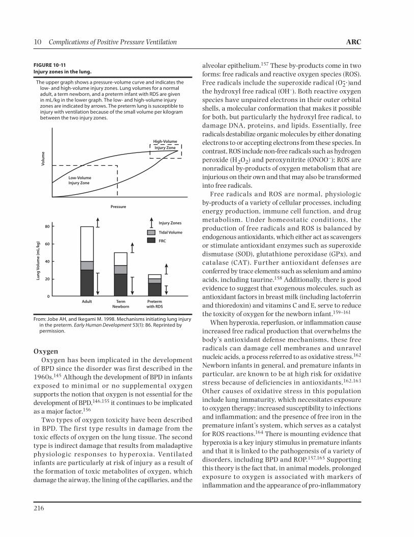
10 Complications of Positive Pressure Ventilation ARC
216
OxygenOxygen has been implicated in the development
of BPD since the disorder was first described in the 1960s.145 Although the development of BPD in infants exposed to minimal or no supplemental oxygen supports the notion that oxygen is not essential for the development of BPD,146,155 it continues to be implicated as a major factor.156
Two types of oxygen toxicity have been described in BPD. The f irst type results in damage from the toxic effects of oxygen on the lung tissue. The second type is indirect damage that results from maladaptive physiologic responses to hyperoxia. Venti lated infants are particularly at risk of injury as a result of the formation of toxic metabolites of oxygen, which damage the airway, the lining of the capillaries, and the
alveolar epithelium.157 These by-products come in two forms: free radicals and reactive oxygen species (ROS). Free radicals include the superoxide radical and the hydroxyl free radical (OH–). Both reactive oxygen species have unpaired electrons in their outer orbital shells, a molecular conformation that makes it possible for both, but particularly the hydroxyl free radical, to damage DNA, proteins, and lipids. Essentially, free radicals destabilize organic molecules by either donating electrons to or accepting electrons from these species. In contrast, ROS include non-free radicals such as hydrogen peroxide (H2O2) and peroxynitrite (ONOO–); ROS are nonradical by-products of oxygen metabolism that are injurious on their own and that may also be transformed into free radicals.
Free radicals and ROS are normal, physiologic by-products of a variety of cellular processes, including energy production, immune cell function, and drug metabolism. Under homeostatic conditions, the production of free radicals and ROS is balanced by endogenous antioxidants, which either act as scavengers or stimulate antioxidant enzymes such as superoxide dismutase (SOD), glutathione peroxidase (GPx), and catalase (CAT). Further antioxidant defenses are conferred by trace elements such as selenium and amino acids, including taurine.158 Additionally, there is good evidence to suggest that exogenous molecules, such as antioxidant factors in breast milk (including lactoferrin and thioredoxin) and vitamins C and E, serve to reduce the toxicity of oxygen for the newborn infant.159–161
When hyperoxia, reperfusion, or inflammation cause increased free radical production that overwhelms the body’s antioxidant defense mechanisms, these free radicals can damage cell membranes and unravel nucleic acids, a process referred to as oxidative stress.162 Newborn infants in general, and premature infants in particular, are known to be at high risk for oxidative stress because of deficiencies in antioxidants.162,163 Other causes of oxidative stress in this population include lung immaturity, which necessitates exposure to oxygen therapy; increased susceptibility to infections and inflammation; and the presence of free iron in the premature infant’s system, which serves as a catalyst for ROS reactions.164 There is mounting evidence that hyperoxia is a key injury stimulus in premature infants and that it is linked to the pathogenesis of a variety of disorders, including BPD and ROP.157,165 Supporting this theory is the fact that, in animal models, prolonged exposure to oxygen is associated with markers of inflammation and the appearance of pro-inflammatory
FIGURE 1011
Injury zones in the lung.
The upper graph shows a pressure-volume curve and indicates the low- and high-volume injury zones. Lung volumes for a normal adult, a term newborn, and a preterm infant with RDS are given in mL/kg in the lower graph. The low- and high-volume injury zones are indicated by arrows. The preterm lung is susceptible to injury with ventilation because of the small volume per kilogram between the two injury zones.
Injury Zones
Tidal Volume
FRC
Adult Term
Newborn
Preterm
with RDS
80
60
40
20
0
Pressure
Lu
ng
Vo
lum
e (
mL
/kg
)
Vo
lum
e
Low-Volume
Injury Zone
High-Volume
Injury Zone
From: Jobe AH, and Ikegami M. 1998. Mechanisms initiating lung injury in the preterm. Early Human Development 53(1): 86. Reprinted by permission.
ARC Complications of Positive Pressure Ventilation 10
217
cytokines such as interleukin (IL)-1 .166,167 Lung abnormalities such as decreased septation and decreased lung surface area have been found to persist even after recovery from hyperoxic exposure.167 Furthermore, SOD and CAT decrease lung injuries associated with oxygen toxicity.168 The appearance of the antioxidant SOD coincides with the onset of surfactant synthesis by Type II pneumocytes.169
Reactive oxygen species’ injuries to epithelial and endothelial cells result in pulmonary edema and activation of inflammatory cells.157 As pulmonary edema progresses, proteins leak into the alveoli, inhibiting the surface tension properties of surfactant, thereby exacerbating the surfactant deficiency of prematurity.162 The resulting cycle of worsening atelectasis, decreased lung compliance, and increased mismatch leads to the need for higher oxygen and ventilator settings, which increases oxidative stress.
In response to the direct damage to cells caused by oxygen free radicals, a second phase of damage occurs. This phase is characterized by the proliferation of alveolar Type II cells and, ultimately, tissue fibrosis.157 A number of markers of peroxidation (oxidative damage) have been found in the tracheal f luid and urine of neonates who later develop BPD. These changes are often seen only a few hours or a day after birth, supporting the theory that prenatal inflammation is also important in the development of BPD.170
Lung TraumaPositive pressure ventilation is known to be important
in the pathogenesis of BPD because of the contribution of pressure (barotrauma) and volume (volutrauma) to initiation of the inflammatory cascade. Dreyfuss and Saumon report that mechanical ventilation in animals using volumes greater than lung capacity injures the alveoli, resulting in leukocyte migration into the lungs, increased tissue permeability, and leakage of fluid into the interstitial tissue and alveoli.171 Equally damaging is ventilation using volumes below FRC, which results in cyclic collapse of the alveoli (atelectotrauma).172 The lung injury zones are illustrated in Figure 10-11. Without adequate tools to measure FRC, it can be very difficult to avoid injury when mechanically ventilating a premature infant.
In some cases, it is likely that injury to the lungs begins during the initial resuscitation of the neonate as clinicians try to establish ventilation quickly. This finding was illustrated by Bjorklund and colleagues who found that as few as six breaths at high tidal volumes
prior to surfactant administration resulted in significant lung injury in preterm lambs.173
Several studies have also demonstrated an inverse relationship between the development of BPD and PCO2 levels.174,175 In fact, Garland and colleagues reported that low PCO2 before surfactant administration was a stronger predictor of BPD than was the severity of lung disease. This finding supports the theory that hyperventilation plays a significant role in lung injury and subsequent development of BPD.172
Inf lammation and InfectionInf lam mation is now recogni zed as playing
a sign i f ica nt role i n the development of BPD. Chorioamnionitis and the presence of elevated cytokine levels in utero initiate a pulmonary inf lammatory response that is thought to alter wound healing, alveolarization, and vascular development in immature lungs.176–178
The pulmonary vasculature contains numerous neutrophils that can trigger an inflammatory response, resulting in the release of enzymes that ultimately disrupt the extracellular matrix of the lung.179 This response can also be initiated after birth as a result of alveolar injury secondary to oxidative stress or mechanical injury.176 Young and colleagues found a significant increase in BPD in infants weighing between 700 and 1,000 g with positive initial endotracheal cultures compared with those with cultures that did not grow bacteria.180 The same association was not found for infants less than 700 g. Similarly, Watterberg and colleagues demonstrated that neonates born following chorioamnionitis experienced mild initial respiratory distress but needed more ventilatory support in the second week of life.181
Several infectious agents have been implicated in the development of BPD, among them Chlamydia and adenovirus.182 Ureaplasma urealyticum has been identified as being associated with the development of BPD,183 but it is unclear whether treating Ureaplasma with antibiotics reduces the incidence of BPD.184 Studies have demonstrated that this organism may cause a chronic subclinical pneumonia, increasing ventilation and oxygen requirements. It has been suggested that infection may act as an additional stimulus in the inflammatory response, with recruitment of neutrophils and activation of the arachidonic acid cascade ultimately leading to BPD.

10 Complications of Positive Pressure Ventilation ARC
218
NutritionCompromised nutritional status may also exacerbate
the development of BPD in the premature infant.185 Adequate caloric and protein intake is required for cell growth and division. Copper, zinc, iron, manganese, and selenium are required cofactors for antioxidant enzymes and may be necessary for repair of elastin and collagen. Vitamin E may provide antioxidant protection, but research findings are inconclusive. Vitamin A deficiency may also play a significant role in the pathogenesis of BPD because this vitamin is essential for differentiation, integrity, and repair of respiratory epithelial cells.185,186 Vitamin A supplementation in neonates has been shown to reduce the production of pro-inflammatory cytokines.187 Malnutrition in the premature infant can impair macrophages and neutrophil and lymphocyte function, which protect the lung against infection.188
Other Risk FactorsOther factors that have been correlated with the
pathogenesis of BPD include a genetic predisposition, excessive fluid intake, lipid infusion, and gas temperature and humidification in the ventilator circuit.189 Research suggests that infants are more likely to develop BPD if there is a family history of airway reactivity (including asthma).190,191 Fluid overload can cause pulmonary edema. Several studies have shown that a persistently patent ductus arteriosus increases the risk of developing BPD.95,192
PATHOLOGY
The pathologic features of BPD first described by Northway and colleagues are divided into four stages (see Table 10-6). This classic, or “old,” BPD described by Northway and colleagues begins with an exudative and early repair stage. That stage is followed, in severe cases, by a chronic fibroproliferative phase marked by widespread fibrosis with atelectasis and emphysema, as well as capillary vascular damage resulting in reduced alveolar development.145
In the original descriptions of BPD, characteristics of mild disease included patchy loss of cilia accompanied by mucosal breakdown of the airway lining followed by edema of the bronchi, blood vessels, and alveolar septa. Infants with moderate BPD experienced extensive loss of cilia in the bronchial lining cells and had evidence of inflammatory cells. Areas of atelectasis and metaplasia of cells lining the conductive airways also occurred. Infants with severe BPD developed necrosis of the airway lining resulting in excessive amounts of debris containing necrotic epithelial cells, mucus, and inflammatory cells. Areas of atelectasis and hyperinflation caused a mismatch.193,194
Upper airway damage in infants with traditional BPD included tracheal, subglottic, and bronchial stenosis; polyps; granulomas; and tracheo- and bronchomalacia. Airway hyperactivity was commonly found in infants with BPD and often persisted into childhood.
Lungs affected by BPD in the postsurfactant era are less likely to have significant fibrosis, airway or smooth muscle hypertrophy, or epithelial metaplasia.195 Under the definition of BPD, lungs show uniform inflation with fewer but larger alveoli. Other findings include a disruption of the collagen network around the saccules and dysplastic Type II cells in the saccules.196
The proposed mechanism for these findings is a disruption in alveolarization occurring as a result of damage to the developing capillaries and the alveolar
TABLE 108
Potentially Better Practices for Reducing CLD in LBW Infants
Practice
Level of
Evidence*
Provide vitamin A supplementation Level 1
Decrease fluid administration Level 3
Administer postextubation CPAP Level 1
Institute permissive hypercarbia Level 2
Decrease supraphysiologic corticosteroid exposure in premature infants
Level 1
Provide prophylactic surfactant for infants with birth weights <1,000 g or delivery room CPAP for infants with birth weights >1,000 g
Level 1
Reduce ventilator days Level 1–5
Use high-frequency ventilation or low tidal volume ventilation
Level 1–2
Provide gentle ventilation in the delivery room Level 2–3
* Muir Gray Classification System 70:
Level 1—Strong evidence from at least one systematic review of multiple well-designed randomized controlled trials
Level 2—Strong evidence from at least one properly randomized controlled trial of appropriate size
Level 3—Evidence from well-designed trials without randomization including single group, prepost, cohort, time series, or matched case controls
Level 4—Evidence from well-designed nonexperimental studies preferably from more than one center or research group
Level 5—Opinion of respected authorities, based on clinical evidence, descriptive studies, or reports of expert committees
Adapted from: Sharek PF, et al. 2003. Evaluation and development of potentially better practices to prevent chronic lung disease and reduce lung injury in neonates. Pediatrics 111(4): e428. Reprinted by permission.

ARC Complications of Positive Pressure Ventilation 10
219
crest cells.150 Before 36 weeks gestation, the functional respiratory units in the lung consist primarily of saccules. During the late saccular stage (after 20 weeks gestation), septal crest cells infiltrate these saccules, dividing each saccule into multiple alveoli. This septation process is accompanied by proliferation of alveolar capillaries that nourish the developing alveoli. Much of this development takes place in the relative hypoxemia found in the normal human fetus. Animal research suggests that inflammatory damage to the crest cells and the alveolar vasculature arrests septation, resulting in the findings of fewer, larger alveoli.197
Some researchers have proposed a three-stage model for the new definition of BPD, which might be useful in designing research studies.198 The perinatal and early postnatal stage (Stage 1) represents opportunities to prevent BPD and is characterized by injury caused by inf lammation. Evolving BPD (Stage 2) occurs at 7–14 days of age; interventions at this stage are aimed at diminishing the severity of the disease. In established BPD (Stage 3), which occurs at 21–35 days of age, characteristics include over-reactive airways, pulmonary edema, and oxygen dependency.
PREVENTION
Prevention of BPD begins with the elimination of preterm birth. If this is not possible, attempts should be made to accelerate lung maturity through administration of antenatal corticosteroids. The benefit of antenatal steroids in lessening the severity of RDS and, subsequently, BPD has been clearly shown.199
Numerous preventive strategies have been proposed, but few have been shown to significantly reduce the incidence of BPD in LBW infants. Using a research-to-practice translation process, representatives from nine member hospitals in the Neonatal Intensive Care Quality Collaborative developed a list of nine evidence-based potentially better practices (PBPs) aimed at reducing the incidence and severity of CLD in LBW infants.200 Table 10-8 lists these practices. The levels of evidence supporting these PBPs varied. A report on the challenges and successes in implementing these PBPs was published.201 The evidence supporting selected PBPs is presented in the following sections.
Avoid VentilationIt was hoped that the introduction of surfactant
therapy in the 1990s would reduce CLD. Although survival rates for LBW infants did increase following the
introduction of this therapy, rates of BPD remained the same or increased.202
Assisted venti lation has come under intense scrutiny. Efforts have been made to reduce the impact of mechanical ventilation by avoiding intubation altogether, reducing the number of days on mechanical venti lat ion, and reducing the barotrauma and volutrauma l inked to conventional mechanical ventilation. To this end, a variety of devices has been developed to support the LBW infant with respiratory disease. These include nasal CPAP (NCPAP) and nasal intermittent positive pressure ventilation (NIPPV), high-frequency ventilation (HFV), and synchronized mechanical ventilation (SIMV). None of these modes of ventilation have been shown to prevent BPD, but some results are promising.202
In a groundbreaking paper published in 1987, Avery and colleagues compared survival rates and rates of BPD in eight U.S. NICUs.203 They found that, despite similar survival rates for LBW infants among the facilities, the incidence of BPD was significantly lower at Columbia Presbyterian Medical Center in New York. The most striking difference between Columbia and the other centers was the early use of nasal prong CPAP with less dependence on intubation and mechanical ventilation at Columbia. Similarly, Sahni and colleagues reported that in infants weighing <1,250 g managed primarily with bubble CPAP, the incidence of BPD was 7.4 percent.153 In a study done in New Zealand, Meyer and colleagues examined the preferential use of bubble CPAP and noted an incidence of CLD of 19 percent compared with an average of 45 percent in 28 other centers. These investigators also noted a trend toward a decrease in late-onset sepsis.204 Studies are now examining the early use of surfactant followed by rapid extubation to CPAP, an approach dubbed INSURE.205 The use of the INSURE protocol at one institution in Sweden resulted in a 50 percent reduction in the number of infants requiring mechanical ventilation.205
A 2007 Cochrane review compared early surfactant replacement therapy followed by extubation to nasal CPAP with the use of rescue surfactant replacement and mechanical ventilation. The authors found that prophylactic surfactant and extubation to CPAP is associated with a reduction in the need for mechanical ventilation, fewer air leaks, and a lower incidence of BPD.206
In randomized controlled studies using NIPPV, a trend to lower rates of BPD among infants treated with NIPPV was noted, although the numbers did not reach

10 Complications of Positive Pressure Ventilation ARC
220
statistical significance.207,208 A more recent study published by Kugelman and colleagues found that in the 84 infants 28–33 weeks gestational age in their study, those randomized to NIPPV for the initial treatment of RDS were significantly less likely to require ventilation than were those in the CPAP group (25 percent vs 49 percent). Those infants also had significantly lower rates of BPD (5 percent vs 33 percent, p<.05, for infants <1,500 g).209
Permissive HypercapniaPermissive hypercapnia (PaCO2 45–55 mmHg) is
another strategy that has been suggested to ensure a more gentle approach to ventilation aimed at reducing volutrauma and barotrauma. In addition to reduced barotrauma, animal data have demonstrated that lambs exposed to supplemental CO2 to levels of 100 mmHg had fewer markers of pulmonary inflammation than did control lambs with normal levels of CO2.210 Clinical trials of permissive hypercapnia in human infants have been limited and have failed to demonstrate a consistent reduction in CLD.211,212 The Cochrane review of this topic also failed to find a significant benefit to permissive hypercapnia.213
Fluid RestrictionExcessive lung water has been shown to be a risk
factor for the development of BPD.214 Lung injury and inf lammation result in capillary leak, leading to pulmonary edema. Restricting fluid intake to the minimum necessary to provide adequate calories for growth has been recommended for infants at risk of developing BPD.155,215 A meta-analysis examining fluid intake showed that restricted intake in preterm infants was associated with a lower risk of mortality and a trend toward a lower incidence of BPD.109 Excessive sodium administration has also been shown to contribute to fluid retention and should be avoided.216
Antioxidant TherapyVitamin A is one of the only preventive strategies
that has been clearly shown to reduce the incidence of BPD.202 A randomized controlled trial of vitamin A supplementation in VLBW infants found that infants in the treatment group had significantly lower rates of oxygen dependency at 36 weeks than did control infants.217 A Cochrane review of vitamin A in the prevention of BPD concurred with these findings.218 Despite these conclusions, a survey of 207 Level III NICUs found that only 20 percent of training units and
13 percent of nontraining units routinely give vitamin A supplements to VLBW infants.219
Antioxidant agents other than vitamin A have been explored in the quest to reduce the incidence of BPD. These agents include intratracheal superoxide dismutase220 and N-acetylcysteine.221 Neither agent demonstrated a significant difference in BPD rates between study and control populations.
SIGNS AND SYMPTOMS
Early clinical signs of BPD may begin within the first week of life, when recovery from the initial disease (for example, RDS) is anticipated. Most LBW infants who develop “new” BPD have a relatively mild course of respiratory distress, but apnea or poor respiratory drive may delay extubation from mechanical ventilation.95 These infants are weaned quickly to low ventilator settings and low concentrations of inspired oxygen. Following a honeymoon period with minimal or no supplemental oxygen, these infants progressively deteriorate, requiring increased ventilatory support. This deterioration may coincide with the onset of a symptomatic PDA or the diagnosis of a bacterial or viral infection.155 Clinically, the infant may have retractions, diminished breath sounds, and fine crackles.
During the early phases of mild to moderate CLD, the changes on x-ray and in pulmonary function are usually mild. Persistent diffuse haziness may be the only change evident on x-ray.155 As BPD progresses, a fine lacy pattern may develop in the parenchyma, and some hyperinflation with occasional large cysts may be seen on x-ray.222
A number of scoring systems have been developed to assist clinicians in predicting which infants with RDS will develop BPD.223,224 The purpose of these scoring systems is, in part, to determine which infants to enroll in clinical trials investigating BPD treatments.223 In these clinical trials, criteria found to be predictive of BPD development included the following two: a logistic regression analysis combining birth weight, five-minute Apgar scores, and PIP at 12 hours of age;223 with FiO2 >.30 and ventilation index <0.51 (10,000/peak pressure × rate × PCO2) at 14 days.224
TREATMENT
The etiology and pathophysiology of BPD are mult i factor ia l; the t reat ment i s mult i faceted. Management of the infant with BPD requires a multidisciplinary team in which all members are aware of the infant’s response to various treatments. The

ARC Complications of Positive Pressure Ventilation 10
221
goal is to promote growth and maintain homeostasis in all systems, while keeping the infant free from infection and gradually weaning the ventilator and oxygen. Prevention and early recognition of the many complications associated with BPD are essential.
Oxygen TherapyThe clinician, knowing that oxygen therapy is
necessary but also causes further damage to the infant with BPD, must strive for a fine balance. As the Supplemental Therapeutic Oxygen for Prethreshold Retinopathy of Prematurity (STOP-ROP) study demonstrated, infants maintained at a higher oxygen saturation range had more severe BPD than did those in a lower range.225 Although optimal oxygen levels for LBW infants are unknown, targeting an oxygen saturation of 88–92 should help to decrease the severity of BPD.225 At the same time, adequate oxygen levels should be maintained to avoid pulmonary hypertension and promote tissue growth. Oxygen should be reduced gradually based on the infant’s tolerance.
Using pulse oximetry and physical findings, the nurse should continuously assess the infant with BPD to determine oxygen requirements. During activities that may stress the infant—bathing; feeding; painful procedures, including laboratory work; and endotracheal tube suctioning—additional oxygen may be required. Following these activities, the infant should be given time to stabilize before the oxygen is reduced to baseline levels.
Parents should be taught to maintain adequate oxygen saturations at all times: while the infant is awake, feeding, and asleep. They also need to be taught to observe for respiratory distress, cyanosis, irritability, and early signs of respiratory infections.
Mechanical VentilationLike oxygen, mechanical ventilation is a known risk
factor for infants with BPD. It is important to use the most modest settings possible to maintain appropriate levels of gas exchange. It is also important to set reasonable targets for PCO2 and PO2 and to work toward the shortest possible duration of mechanical ventilation. Few prospective randomized trials have been conducted to determine the combination of ventilator settings that either limits the severity or prevents the development of BPD; however, some general principles can be applied. To minimize both barotrauma and atelectotrauma, the lowest peak airway pressure that provides an adequate tidal volume should be used.226 A PEEP adequate to
avoid airway collapse is also important. Higher PEEP levels (6–8 cmH2O) may be needed in infants with floppy airways secondary to prolonged ventilation.
Synchronized intermittent mandatory ventilation and pressure support ventilation (assist-control) are designed to improve interaction and reduce antagonism between infant-generated and mechanically generated breaths (see Chapter 9). The ability of SIMV to limit lung overdistention reduces the need for the neonate to fight the ventilator breaths. Intuitively, synchronized ventilation should be of benefit in reducing the incidence and severity of BPD; however, a large multicenter study failed to show any difference in the incidence of BPD.227 A Cochrane review of synchronized ventilation did find a reduction of air leaks and a shorter duration of ventilation but did not find a reduction in the incidence of CLD with synchronized ventilators.17 A Cochrane review of volume-targeted versus pressure limited ventilation did demonstrate a reduction in BPD, duration of ventilation, and airleaks for those infants receiving volume ventilation.228 Studies that are more recent have demonstrated a positive effect of volume-targeted ventilation on lung inflammation in premature infants.229,230 Longer-term follow-up is needed to assess for a reduction in BPD rates.
High-frequency ventilation (see Chapter 12) has been evaluated to determine its impact on BPD, again with mixed results. Thome and colleagues found that in infants 24–30 weeks, use of HFV with high lung volumes did not confer any benefit in preventing BPD compared with conventional ventilation.231 A meta-analysis of two trials using high-frequency oscillatory ventilation (HFOV) concluded that the use of HFOV may reduce CLD rates somewhat, but the findings were inconsistent.232 The Cochrane review of high-frequency jet ventilation (HFJV) found that the use of HFJV moderately reduces CLD rates but may increase the risk of IVH.233
MedicationsLong-term management of infants with BPD often
includes treatment with many drugs such as vitamins, diuretics, bronchodilators, and in some cases, steroids. The decision to use each drug should be individualized. The addition of each drug to the infant’s management plan should be closely monitored to prevent the “polypharmacy” phenomenon associated with BPD care. For a detailed review of all medications used for the infant with BPD, see Chapter 11.

10 Complications of Positive Pressure Ventilation ARC
222
NUTRITION
Despite improvements in parenteral and enteral nutrition, postnatal growth failure is still seen in LBW infants, especially those with BPD.234 For infants with BPD, the incidence of growth failure in the immediate postdischarge period is estimated to be between 30 and 67 percent.235 Studies have shown that children with BPD have increased resting energy levels and that growth failure in this population may contribute to adverse pulmonary and developmental outcomes.236,237 More specifically, infants with BPD have a metabolic rate approximately 25 percent higher than infants without BPD, leading to a caloric requirement 20–40 percent higher than age-matched infants without BPD.188
The increased work of breathing resulting from decreased lung compliance, increased airway resistance, and tachypnea is one of the factors interfering with normal growth. Another concern is the delay in initiating and advancing enteral feedings in the presence
of respiratory disease, feeding intolerance, and other complications.188
Parenteral NutritionProviding adequate nutrition to the VLBW neonate
at risk for BPD is often quite challenging. The overriding goal is to provide adequate calorie and protein intake to support a rate of growth similar to that seen in utero. It is important to address the neonate’s nutritional needs beginning on the first day of life. Initially, nutrition is provided in parenteral form, with enteral feedings started as soon as the infant is medically stable, ideally within the first few days of life. Recommended parenteral intake on the first day of life is 80–100 mL/kg/day of fluid, 2–3 g/kg of protein, 0.5–1 g/kg of fat emulsion, and 4–6 mg/kg/minute of glucose.188 Proteins, fats, and carbohydrates are gradually increased to 4 g/kg, 3 g/kg, and 10–12 mg/kg/minute, respectively. Some clinicians have been reluctant to provide protein in the first few days of life because of concerns regarding side effects such as metabolic acidosis; however, a study by Thureen and others demonstrated that a protein intake as high as 3 g/kg on day 1 of life was well tolerated by VLBW infants.238
Enteral FeedingsBreast milk or formula is introduced as soon as
possible after delivery in the form of trophic feedings. Commonly, however, infants with respiratory distress have periods of feeding intolerance or medical complications such as a PDA or sepsis, that delay their progression to full enteral feedings. Feedings are started at small volumes and increased slowly to decrease the risk of NEC. Human breast milk, although the preferred source of nutrition, does not provide adequate calories, protein, or minerals to meet the increased metabolic needs of premature neonates. Fortification with human milk fortifier, liquid formula concentrate, or protein powder may be used to address the deficits.
Multivitamin supplementation, including vitamin D and iron, is indicated for infants receiving breast milk. Some support also exists for carnitine supplements as a mechanism for enhancing weight gain and catch-up growth.239 Gastroesophageal reflux disease (GERD) often makes it difficult to provide adequate nutrition to infants with BPD. Reflux may alter lung function in these patients by causing aspiration of stomach contents and triggering bronchial reactivity. Neonates with suspected swallowing dysfunction should be evaluated by a feeding specialist. Conservative measures for managing reflux
TABLE 109
Recommendations for Monitoring the Nutritional Status of
Enterally Fed Hospitalized VLBW Infants
Intake/Output Monitoring
Fluid intake (mL/kg/day) Daily
Urine output (mL/kg/day) Daily
Nutrient Intake
Energy (kcal/kg/day) Daily
Proteins (g/kg/day) Daily if weight gain is poor
Anthropometric Monitoring
Body weight (g) Twice daily until stable, then daily
Length (cm) Weekly
Head circumference Weekly
Biochemical Monitoring
Complete blood counts including platelet and reticulocyte count
Every 2 weeks
Serum electrolytes and blood urea nitrogen
Weekly if on diuretics or with fluid restriction; every 2 weeks when stable
Calcium, phosphorus, and alkaline phosphatase
Every 2 weeks
Total protein, albumin, prealbumin
Consider if weight gain is poor or blood urea nitrogen is low
Liver function tests At 2 weeks, then every 2 weeks if there is evidence of cholestasis
Adapted from: Biniwale MA, and Ehrenkranz RA. 2006. The role of nutrition in the prevention and management of bronchopulmonary dysplasia. Seminars in Perinatology 30(4): 204. Reprinted by permission.

ARC Complications of Positive Pressure Ventilation 10
223
include thickening the feedings, decreasing the volume of the feeding by increasing the number of feedings, and elevating the head of the bed at a 30-degree angle. Nipple feeding the infant with BPD can be problematic because of the negative oral stimulation from the endotracheal tube, gastric feeding tubes, and frequent oral suctioning.
E le c t r oly t e a nd m i ne r a l i m b a la nc e s o f t e n accompany BPD as a result of fluid restriction, diuretics, dexamethasone, and other medications. Monitoring of serum electrolyte levels is important to determine appropriate amounts of supplementation. Table 10-9 displays a suggested schedule for nutritional monitoring.
NURSING CARE
Caring for the infant with BPD can be a frustrating experience for even the most experienced nurse. Although infants with the newer form of BPD are less likely to develop profound bronchospasms, or BPD “snits,” they may still have episodes of increased airway resistance or hypoxia in response to stress, handling, or discomfort. They may not respond as readily to the usual soothing techniques such as rocking, patting, holding, or talking.240 Learning to interpret the infant’s behavior is essential in supporting the infant and family. Major considerations in caring for infants with BPD include limiting environmental demands when the infant loses control and identifying early signs of loss of control. In addition, staff members understanding when intervention is needed, and knowing strategies to reduce stress is invaluable. Each infant should have an individualized developmental plan constructed by the neonatal team.
Infants with BPD are at increased risk of neuro-developmental delays beyond those associated with low birth weight alone. In a follow-up study done at eight years of age comparing term infants and VLBW infants with and without BPD, children from the BPD group demonstrated deficits in intelligence, reading, mathematics, and gross motor skills. Children in the BPD group were more likely to be enrolled in special education classes (54 percent) than were children in the VLBW-without-BPD group (37 percent) or term birth group (25 percent).241 The risk for disabilities increases as birth weight decreases and more complications develop. Factors that appear to place infants with BPD at higher risk include moderate to severe IVH and low socioeconomic and parent education levels.242
Maximizing neurodevelopmental outcome in infants with BPD requires a multidisciplinary team approach. A team consisting of developmental specialists, physical
therapists, speech therapists, neonatologists, and nurses should develop a plan of care to maximize neurologic growth and development. Goals include maximizing the environmental conditions in the NICU, reducing stress, and maintaining normal oxygen levels. Proper positioning in natural flexion using rolls and blankets is helpful. Minimizing stimulation and evaluating the infant’s response to procedures using oxygen saturation monitoring as a guide are important. Evaluating and reporting the infant’s responses to the various therapists help in developing an individualized plan of care.
Discharge planning should ensure that the family understands the plan of care, including handling, positioning, stimulation, and stress reactions. Teaching and supporting families who have an infant with BPD is a challenge for NICU nurses. The nurse is often coordinator of the various disciplines involved in the infant’s care. Understanding their baby’s treatment regimen and the importance of close medical follow-up may help with compliance and assist the parents in coping with the complexity of their infant’s care. Parents need to be aware of the high risk of lower respiratory tract infections and rehospitalization despite optimal care and appropriate precautions. Preparing the infant and family for discharge is critical to a successful transition from the NICU environment to home. To assist NICUs in appropriate discharge planning, the American Academy of Pediatrics (AAP) has developed guidelines for the discharge of high-risk infants.243 Before discharge, infants with BPD should be free from apnea, be taking all of their feedings by breast or bottle, and have oxygen requirements and medication regimens that are stable and manageable outside the hospital setting.244 Care and attention must also be given to ensuring that the family is ready for discharge. Offering parents the opportunity to stay in the hospital overnight with their infant may help to ease the transition home.
PROGNOSIS
Infants with BPD have an increased hospital readmission rate than their non-BPD counterparts, and respiratory abnormalities that persistent into adolescence.245 The prognosis for infants with BPD is dependent on the severity of the disease and the infant’s overall health status. A 1996 study reported that 50 percent of infants with severe BPD were readmitted to the hospital in the first year of life for lower respiratory tract infections.246 This number has been reduced somewhat with the advent of respiratory syncytial virus

10 Complications of Positive Pressure Ventilation ARC
224
prophylaxis, the main cause of respiratory infections requiring hospitalization in prematurely born infants.
Long-term follow-up of infants with BPD suggests that these children experience progressive normalization of lung mechanics and, to some extent, lung volumes; however, abnormalities of the small airways persist.152 Other follow-up studies have found that children who had BPD as infants have no difference in exercise capacity and no difference in FRC but do have a decrease in forced expiratory volumes and forced vital capacity.247 It has also been reported that at five years of age, BPD survivors have a higher incidence of asthma than the general population.248
RETINOPATHY OF PREMATURITYOne of the most common complications ascribed
to the use of oxygen and mechanical ventilation in LBW infants is ROP. ROP is a disorder of retinal vascularization in the retina of a premature infant that causes a proliferation of abnormal vasculature, leading to visual loss from cicatrization (scarring) and retinal detachment. Immature retinal vessels are a prerequisite for the development of ROP. Initiated by an injury, ROP represents the healing process.
HISTORY
ROP was f irst described in 1942 by Terry, who called the disease retrolental fibroplasia because of the formation of scar tissue behind the lens of the eye in premature infants.249 In the late 1940s and 1950s, the amount of blindness from this disorder was considered epidemic. Following a report of a controlled study by Patz and associates in the early 1950s showing that high concentrations of oxygen contributed to the development of retrolental fibroplasia, the use of oxygen was greatly curtailed.250 Kinsey and Hemphill reported an ROP incidence of 71 percent with liberal use of oxygen and of 33 percent when oxygen use was curtailed.251 Although the incidence of blindness in preterm infants decreased significantly with decreased use of oxygen, mortality and morbidity, including such conditions as cerebral palsy and lung disease, increased.252
The disorder was renamed retinopathy of prematurity in the 1980s because of its association with prematurity and its occurrence despite excellent oxygen monitoring and the absence of high oxygen blood levels. Cases of ROP have been reported in term infants and premature infants who did not receive oxygen, leading investigators to the current conclusion that ROP is multifactorial.1
INCIDENCE
The incidence of ROP is inversely related to birth weight and gestational age. There is a wide variation in the reported incidence of ROP, in part because of variations in screening criteria as well as differences in populations and the gestational ages of infants included in the studies. There is also a difference of opinion as to whether the incidence of ROP is increasing with greater survival rates among ELBW infants253,254 or decreasing as a result of interventions such as the use of surfactant.255
I n 1981, Phelp s e st i mat e d t hat i n t he U. S. approximately 600 infants a year were at risk for blindness from ROP.256 In 1990, the Cryotherapy for Retinopathy of Prematurity (CRYO-ROP) study, which involved 23 U.S. institutions and followed 4,009 infants, reported the incidence of ROP to be 47 percent in infants weighing 1,000–1,250 g, 78 percent in infants weighing 750–999 g, and 90 percent in infants weighing <750 g at birth. The overall rate of severe ROP—greater than threshold (Stage 3+ or greater)—was 22 percent. Multiple births and gender had no significant effect on the incidence. Caucasian infants had a higher chance of progressing to severe ROP than did African American infants.257 In 2005, Good and colleagues from the Early Treatment for Retinopathy of Prematurity Cooperative Group compared then-current rates of ROP to those identified by the original CRYO-ROP study and found that, at 68 percent in 2005, rates of ROP had changed little since 1990.257,258 A review of infants <1,250 g born between 1994 and 2000 found an increase in ROP over that time period from 40 to 54 percent, with a 2–5 percent rate of threshold ROP.251 On the other hand, Hussain and colleagues reviewed 2,528 infants born between 1989 and 1997 at <37 weeks gestation and reported an overall incidence of ROP of 21 percent.255 However, when the statistics were broken down by gestational age, the ROP rate for infants ≤28 weeks was 40.1 percent, with 9.8 percent of this group experiencing severe (Grade 3 or worse) ROP. Studies of ROP in infants born at >30 weeks gestation or ≥1,250 g demonstrated rates of ROP of 2–4.2 percent, with no infant >1,500 g developing ROP that needed to be treated.255,259,260
In the U.S., ROP is the second leading cause of childhood blindness, accounting for 6–20 percent of all cases.261,262 In the United Kingdom, a follow-up study of infants with Stage 3 ROP found that 13 percent had a severe vision deficit.263 Many middle-income areas such as South America and Eastern Europe are experiencing

ARC Complications of Positive Pressure Ventilation 10
225
an exponential increase in the number of cases of severe ROP, with an estimated 50,000 cases of ROP-induced blindness being reported.264 Infants of higher birth weights are at greater risk for ROP in these countries compared to those in high-income countries.265
PATHOPHYSIOLOGY
Knowledge of the development of retinal vasculature assists in understanding the disease, diagnosis, and treatment of ROP. Normal ocular development occurs in the hypoxic intrauterine environment. At 6 weeks gestation, the hyaloid artery enters the eye and begins to fill the vitreous cavity with blood vessels. The retina remains avascular until approximately 16 weeks gestation, when capillary precursor cells (spindle cells) start branching out from the tissue around the fetal optic nerve and slowly grow centrifugally toward the nasal edge of the optic disc. Spindle cells normally disappear by 21 weeks gestation. By 16–18 weeks, new blood vessels begin forming at the ora serrata, the central edge of the retina, reaching the temporal ora serrata at approximately 40–44 weeks gestation. The blood vessels behind the leading edge of capillaries gradually form arteries and veins.
In utero the fetus is exposed to a hypoxic environment with a PaO2 of 30 mmHg.266 This hypoxia is thought to be important in stimulating growth factors, which regulate normal vascular development in utero.267 Two growth factors known to be important in both normal vessel growth and in the development of ROP are vascular endothelial growth factor (VEGF) and insulin-like growth factor (IGF-1). VEGF production occurs in response to hypoxia. Insulin-like growth factor is transported across the placenta and regulates neovascularization. Following premature delivery, IGF-1 levels fall quickly and remain low until endogenous production occurs. IGF-1 is postulated to be one of the factors not related to hypoxia that plays a role in ROP.268,269 Studies have shown that IGF-1 levels are lowest in infants who later develop ROP.270,271
Phase 1 ROPThe first or acute phase of ROP occurs between
birth and 30–32 weeks postmenstrual age and may progress slowly or rapidly.266 Following premature birth, exposure to oxygen levels that are higher than those in the intrauterine environment suppresses VEGF, leading to a cessation of blood vessel development, vasoconstriction of existing immature retinal vessels, and ultimately to the destruction of some of the newly
developed capillaries.268,272 Falling levels of IGF-1 also play a role in the cessation of vascular growth. This cessation of vascular growth marks the beginning of Phase 1 ROP. In its early stages, this vasoconstriction may be reversible, but if it continues, there is tissue ischemia.
Phase 2 ROPAs the ret i na cont i nues to g row, metabol ic
demands increase, and without adequate vascular development, local hypoxia occurs. This tissue hypoxia leads to the secretion of VGEF and new vessel growth (neovascularization). These vessels form at the junction of the avascular and vascularized retina in response to an increase in VEGF. If vascularization succeeds in reestablishing circulation to the central retina and to the peripheral avascular retina, ROP regresses, and any excess vessels are absorbed. In other cases, neovascularization erupts into the vitreous, and vessel growth is uncontrolled. These vessels may regress and heal, but the process can result in residual scarring that causes a neovascular ridge. As the scar tissue hardens and shrinks, it places traction on the retina, which can lead to detachment.266 Phase 2 ROP occurs between 32 and 34 weeks postmenstrual age.264
Classification of ROPAn international classification system for ROP has
been developed by ophthalmologists from six countries. First published in 1984 and updated in 1987 and 2005, this system describes ROP in terms of four characteristics or measures: the retinal location of the disease, the extent of involvement of the developing vasculature, the stage of the disease, and the presence or absence of dilated and tortuous vessels.273
LOCATION: ZONE I, II, OR IIIRetinal vessel development occurs from the optic
nerve in the posterior area of the eye toward the periphery. The location of the disease in one of three zones is a measure of how the progression of blood vessels has developed (Figure 10-12).
Zone I is a small area extending from the optic nerve to twice the distance from the center of the optic nerve to the center of the macula.273 Zone I disease is the most dangerous because progression to extensive scar tissue and total retinal detachment is most likely in this location.

10 Complications of Positive Pressure Ventilation ARC
226
Zone II extends from the edge of Zone I to the nasal ora serrata found at the 3-o’clock position in the right eye and the 9-o’clock position in the left eye.273
Zone III is the remainder of the temporal retina, the last to be vascularized during fetal development.
EXTENT OF VASCULATURE INVOLVEMENT
The extent of the disease is defined by how many clock hours of the eye’s circumference are diseased. Figure 10-12 illustrates both the zones and clock hours of ROP.
ROP STAGING
It is common for more than one stage of ROP to be present in the eye. However, staging of the eye as a whole is based on the most severe stage present.273
Stage 1 ROP is characterized by a sharp white line that lies within the plane of the retina and separates the vascular and avascular retina.
Stage 2 ROP displays a rolled ridge of scar tissue in the region of the white demarcation line. The ridge may be limited to a small area of the retina or may encircle the eye. Small tuffs of new blood vessels, called popcorn, may be found behind the ridge.
Stage 3 ROP is characterized by neovascularization proliferating from the posterior aspect of the ridge out of the retina into the vitreous. Stage 3 is further subdivided into mild, moderate, or severe amounts of tissue reaching into the vitreous.
Stage 4 ROP is subtotal retinal detachment caused by the scar tissue formed in Stages 1–3. Stage 4a is a partial detachment in the periphery of the retina.
Stage 4b is a subtotal or total detachment involving the macula and fovea, usually with a fold extending through Zones I, II, and III.
Stage 5 ROP involves a complete retinal detachment, with the retina assuming a closed or partially closed funnel from the optic nerve to the front of the eye.
Plus disease is a designation given to ROP when, at any of the stages, the posterior veins are enlarged and the arterioles are tortuous; it is expressed by adding a plus sign following the stage number (for example, Stage 3+). The plus sign indicates extensive ROP changes that may signify a rapidly progressive course. Other findings of plus disease include poor papillary dilation and vitreous haze.273 In the CRYO-ROP study, the presence of plus disease significantly increased the chances of an unfavorable outcome.274
Pre-plus disease is a category that was added during the 2005 revision of the ROP classification system. This identifier designates the presence of abnormal dilation and tortuosity of the posterior pole vessels that are not yet severe enough to be classified as plus disease. Over time, as dilation increases, pre-plus disease may progress to plus disease.273
Aggressive posterior ROP, or AP-ROP, an unusually aggressive pattern of ROP development, has been reported in infants weighing <1,000 g.273 Formerly termed Rush disease, AP-ROP develops earlier than usual (at three to five weeks after birth) and can progress rapidly to severe ROP with retinal detachment.1
THRESHOLD AND PRETHRESHOLD ROP Threshold ROP is a term used to designate Zone I or
II changes found in a minimum of five uninterrupted clock hours or involvement of a total of eight clock hours.1
Prethreshold ROP describes ROP changes that do not meet threshold ROP criteria in Zone I but that reach Stage 2+ or Stage 3 in Zone II. Infants with prethreshold disease require more frequent ROP examinations.275
OutcomesThe visual outcome for infants with ROP depends on
the stage of the disease, whether the macula is involved, and the results of treatment. Because Stages 1–3 ROP generally occur in the peripheral retina, the macula is
FIGURE 1012
Zones and clock hours of retinopathy of prematurity.
From: Phelps DL. 2006. Retinopathy of prematurity. In Neonatal-Perinatal Medicine: Diseases of the Fetus and Infant, 8th ed., Martin RJ, Fanaroff AA, and Walsh MC, eds. Philadelphia: Mosby, 1750. Reprinted by permission.

ARC Complications of Positive Pressure Ventilation 10
227
not affected. For most infants with Stage 1 or 2 ROP, the disease resolves spontaneously without scarring.276 Although these infants do not require treatment, they are at greater risk for developing myopia, amblyopia, astigmatism, strabismus, and glaucoma.272,277 In other studies, late peripheral retinal degeneration and late retinal detachment have been reported in teens and adults with ROP that regressed without treatment (regressive ROP).278,279
If they are not treated, infants with moderate to severe Stage 3 or Stage 4 ROP are at risk for reduced vision because scar tissue shrinks and then exerts traction on the retina. Moderate traction results in the distortion of the macula. In severe cases of ROP (Stages 4b and 5), the retina and macula may be totally detached, resulting in blindness. Untreated threshold disease leads to retinal detachment in about 50 percent of cases.280
RISK FACTORS
Since the 1950s, investigators have been looking at the many factors that may be associated with the development of ROP. Early studies identified prematurity and hyperoxia as the most important correlates with ROP.264 During the resurgence of ROP that occurred in the 1970s and 1980s, the disorder occurred despite stringent oxygen controls, and gestational age was again noted to be the leading risk factor. Oxygen exposure continues to be a risk factor for the development of ROP but does not completely explain its occurrence.
In recognition of the complexity of ROP, many other risk factors have been studied, including blood transfusions, IVH, pneumothorax, mechanical ventilation, apnea, infection, hypercarbia, hypocarbia, PDA, administration of prostaglandin synthetase inhibitors (indomethacin), vitamin E deficiency, prenatal complications, and genetic factors. After controlling for immaturity, the sicker the infant, the more likely it is that serious ROP will develop. Because of the complexity and severity of illness in these infants who develop ROP, it remains difficult to determine precisely which factors increase the risk of ROP. For example, prenatal use of steroids appears to protect against ROP, but infants whose mothers received antenatal steroids are also less likely to be ill than are those who were not exposed to steroids.281
In most studies, birth weight and gestational age as well as the degree of serious illness, are among the primary risk factors for ROP.282,283 Kim and colleagues also identified surfactant therapy and apnea as risk factors, noting that apnea not only increased the
risk of ROP but also worsened preexisting ROP.283 In a study of 88 infants <34 weeks gestation at birth, Akkoyun and colleagues found that risk factors for severe ROP in their study population included birth weight, blood transfusions, and duration of mechanical ventilation.284 Blood transfusions were also cited as a risk factor by Dutta and colleagues.285 In another study of 159 infants with birth weights <1,600 g, significant risk factors for the development of ROP included birth weight ≤1,000 g, IVH, sepsis, and the use of dopamine or glucocorticoids.286 Another review found that the primary risk factors for threshold ROP were maternal preeclampsia, birth weight, presence of pulmonary hemorrhage, and duration of ventilation.287 In most cases, these risk factors are measures of the degree of illness experienced by LBW infants and may or may not play a direct role in the development of ROP.
Ethnic origin and country of birth have been found to correlate with the development of ROP. South Asian infants are more likely to develop severe ROP than are Caucasian infants,288 and African American infants are less likely to develop ROP than are Caucasian infants.289 Infants born in countries classified as middle income (for example, Latin America, Eastern Europe, and Thailand) were at increased risk of developing serious ROP and of becoming blind as a result.262,290
PREVENTION
Oxygen LevelsFor many years, oxygen use has been the focus of
attention as the primary cause of ROP. Early studies demonstrated a correlation between the duration of oxygen therapy and the development of ROP.250,291,292 Other studies did not identify a clear correlation. An early multicenter study designed to assess the relationship between arterial oxygen levels and ROP failed to find any significant differences between study groups.293 Another early study comparing transcutaneous oxygen (TcPO2) levels demonstrated no difference in the incidence or severity of ROP at various TcPO2 levels in infants weighing <1,000 g.294 One 1992 study supports an association between the incidence and severity of ROP and the duration of exposure to transcutaneous oxygen levels of >80 mmHg.295 This finding is supported by later work showing that lowering oxygen saturation alarm limits and implementing oxygen targeting guidelines and educational programs can reduce the incidence of severe ROP. In 2006, Vanderveen and colleagues reported that lowering oxygen alarm limits to the

10 Complications of Positive Pressure Ventilation ARC
228
range of 85–93 percent in infants <1,250 g decreased prethreshold ROP from 17.5 percent to 5.6 percent.296
Ambient Light LevelsExposure to high levels of ambient light has been
suggested as a cause of progression of ROP. It is proposed that light can generate free radicals, which damage the developing retinal blood vessels. In 1985, Glass and colleagues reported a reduced incidence of ROP in neonates—especially those weighing <1,000 g—exposed to reduced (150 lux) compared with standard (600 lux) lighting in the nursery.297 Two studies reported in 1988 and 1989 conflict with Glass and colleagues’ findings, however.298,299 A large multicenter, randomized clinical trial also failed to find a reduction in ROP as a result of reducing ambient light, a finding confirmed by a later Cochrane review.300,301
Dietary SupplementsA number of pharmacologic agents and dietary
supplements have been examined to determine if they might play a role in the prevention of ROP. Several clinical and experimental studies have indicated that vitamin E (tocopherol) deficiency may cause ROP.302–304 It has been theorized that premature infants have a deficiency of vitamin E, a naturally occurring antioxidant. Without sufficient amounts of vitamin E to protect the spindle cells in the retina, the free oxygen radicals destroy the cells.305 Several infant trials using vitamin E reported mixed results in terms of lowering the incidence and severity of ROP.302,304,306
Even though vitamin E has been effective in some instances, there are concerns about the risks of sepsis, NEC, IVH, and retinal hemorrhage associated with its use.304,307–309 Many of these problems were reported with use of intravenous pharmacologic megadoses or unapproved formulations of the vitamin. If vitamin E is administered, serum levels should be monitored to prevent overdose. More recently, a meta-analysis done by Brion and associates found that vitamin E supplementation did reduce the risk of both ROP and IVH in VLBW infants but increased the risk of sepsis.310 These reviewers suggest that supplementation at high doses or producing levels >3.5 mg/dL is not supported by evidence.
Another supplement being studied is D-penicillamine, a potent antioxidant. Interest in this substance originated from work done by Lakatos and colleagues, who noted a low incidence of ROP among infants receiv i ng D -penici l la m i ne to prevent or t reat
hyperbilirubinemia.311 A meta-analysis of this and one other study concluded that D-penicillamine may reduce the incidence of acute ROP and should be further investigated.312,313 More recently, Christensen and colleagues compared15 premature infants treated with D-penicillamine to 34 matched controls and found that a 14-day course of D-penicillamine decreased the odds of developing ROP from 60 percent to 21 percent with no short-term adverse effects noted.314 The D-penicillamine treatment did not, however, reduce the need for ROP surgery in treated infants who did develop ROP.
A 1992 study reported beneficial effects of inositol, a dietary supplement.315 A Cochrane review of two trials315,316 using inositol to reduce morbidity in premature infants with respiratory distress reported a significant reduction in Stage 4 ROP and suggested the need for further randomized controlled trials.317
SCREENING
Screening guidelines for ROP have been published by the AAP, the American Academy of Ophthalmology, and the American Association for Pediatric Ophthalmology and Strabismus.318 Recommendations from these guidelines identify the need for a skilled ophthalmologist to screen all infants with a gestational age of <32 weeks or a birth weight of <1,500 g, as well as selected infants with a birth weight of 1,500–2,000 g and an unstable clinical course. Examinations should be started when infants born at ≤27 weeks reach 31 weeks gestational age or at 4 weeks for infants born between 28 and 32 weeks gestational age.319 The examining ophthalmologist determines the timing of subsequent examinations based on retinal findings. Generally, follow-up is required in one week or less for infants with Stage 1 or 2 ROP in Zone I or Stage 3 ROP in Zone II.
Some researchers have suggested that the AAP criteria be modified. Subhani and colleagues found that ELBW infants may develop significant ROP earlier than 31 or 32 weeks and that delays in screening in this population may miss prethreshold disease.320 Others have suggested that the screening criteria could be safely modified to include only those infants born at <1,251 g and <30 weeks gestational age.321,322 In a review of 205 LBW infants screened over an eight-year period, Mathew and colleagues identified ROP in five babies with birth weights >1,250 g and in eight babies born at >30 weeks gestation. In all of these babies, ROP was either Stage 1 or 2.321
Every NICU should have a program in place to identify infants who meet screening criteria and

ARC Complications of Positive Pressure Ventilation 10
229
to schedule eye examinations for them. When an at-risk infant is discharged or transferred to another center, consideration must be given to the availability of appropriate follow-up.
To prepare the infant for a screening examination, the nurse administers mydriatic agents prescribed by the ophthalmologist. Drugs containing phenylephrine should be used cautiously if the infant has hypertension. Prior to and during the examination, a nurse should be at the bedside to monitor the infant’s responses to the medication and examination. Resuscitative equipment should be available because neonates can react to the examination with apnea, bradycardia, increased blood pressure, and oxygen desaturation.
Using an indirect ophthalmoscope with a lid speculum and scleral depressors, the ophthalmologist examines each eye, evaluating all zones to determine retinal vascularization. If the infant does not tolerate the exam, it should be stopped and the infant treated appropriately. Administering atropine to an unstable infant has been recommended—especially if the examination is critical. Communication between members of the health care team and the family is particularly important in the screening and management of ROP. In addition to being aware of their infant’s risk for developing ROP, parents should be counseled regarding the importance of ongoing follow-up with the ophthalmologist until retinal vascularization is complete.318
TREATMENT
The criteria for determining the need for intervention in ROP have been the subject of ongoing debate and much research.
Nonsurgical TreatmentProvision of supplemental oxygen to infants with
prethreshold ROP has been studied as a method of preventing progression of the disease. The large multicenter STOP-ROP trial evaluated the safety and efficacy of maintaining oxygen saturations at between 96 and 99 percent in LBW infants with ROP. The investigators found that this therapy did not reduce the severity of ROP but did result in more adverse pulmonary complications. A small benefit was noted in infants with prethreshold ROP without plus disease compared with infants with plus disease, but this finding was not significant.225 The systematic review conducted by Lloyd and colleagues in 2003 echoed this finding.323
Surgical TreatmentThe AAP recommends surgical treatment for ROP
when any of the following conditions exist: Zone I ROP at any stage when plus disease is present; Zone I ROP at Stage 3 in the absence of plus disease; and Stage 2 or 3 ROP in Zone II with plus disease.318 Two effective treatments are available for infants with severe progressive ROP: cryotherapy and laser therapy.
Cryotherapy, which involves freezing the avascular area of the retina anterior to the area of disease, was originally the first-line treatment for ROP. It has largely been replaced by laser therapy and is today reserved for cases where blood or corneal haziness obscures the view of the retina.324
Despite the fact that its use is now limited, early evaluation of cryotherapy identified a significant benefit in infants with threshold ROP. In a multicenter trial of cryotherapy for ROP termed the CRYO-ROP study, 6 percent of infants weighing <1,250 g developed threshold ROP, 90 percent of whom were between 33 and 42 weeks gestation. Within 72 hours after threshold disease was detected, the infants were treated with cryotherapy. Cryotherapy was shown to improve visual outcome by 50 percent compared to those receiving no treatment.276
In the 10-year follow-up of the original CRYO-ROP study, similar results persisted, with 62 percent of the control eyes having unfavorable distance visual acuity compared with 44 percent of the treated eyes. Eyes that had disease in Zone I had a poor outcome at 10 years, whether treated or part of the control group,
FIGURE 1013
Laser therapy.
Courtesy of Coherent, Inc., Santa Clara, California.

10 Complications of Positive Pressure Ventilation ARC
230
with 94 percent (15 of 16) having an unfavorable visual outcome.325
Laser photocoagulation (Figure 10-13), now the preferred treatment for ROP, acts in the same way as cryotherapy in destroying avascular tissue. The advantages of laser therapy include ease in treating more difficult locations such as Zone I, less trauma to the tissue, less discomfort for the infant, and fewer ocular and systemic complications such as intraocular hemorrhage and bradycardia.326 A number of studies have evaluated laser therapy for ROP, and longer term follow-up data are now becoming available. Studies have shown that laser therapy is effective in preserving both distance and near vision in eyes with threshold ROP.327 A ten-year follow-up of a randomized trial comparing laser treatment with cryotherapy found that eyes treated with laser therapy had better corrected visual acuity and less myopia than did cryotherapy-treated eyes.328,329 A small risk of cataract development has been reported with laser therapy.280
If laser therapy is unsuccessful in preventing retinal detachment, scleral buckling may be indicated. Under general anesthesia, a silicone band is placed around the eye. This reduces the circumference of the eye enough that the retina can re-attach itself to the wall. However, if the scar continues to contract, the retina will again pull away from the posterior wall toward the center of the eye. Scleral buckling has been reported to have a 50–100 percent anatomic success rate for patients with Stage 4a or 4b ROP.330 Favorable visual acuity of 20/60 to 18/400 has been reported with this technique, but if surgery is delayed, blindness may result.331
When retinal detachment progresses to Stage 5, a vitrectomy may be indicated. Two techniques, closed and open eye, have been reported, with comparable results. During this three- to four-hour procedure, the lens and vitreous humor are removed from the eye. The scar tissue is removed, and the retina is laid back against the eye wall. A study by Hartnett and colleagues demonstrated that after one procedure, anatomic attachment was more likely to be achieved in infants undergoing vitrectomy than in those receiving a scleral buckling procedure, but by the end of the follow-up period (six months) there was no difference in retinal attachment rates.332
In a follow-up analysis of infants in the Early Treatment for Retinopathy of Prematurity (ETROP) study333 who developed retinal detachment, vitreoretinal surgery (vitrectomy and/or scleral buckling) resulted in macular attachment in 16 of 48 eyes. Macular
attachment at the nine-month follow-up was achieved in 30 percent of the eyes treated with vitrectomy with or without buckling, in 60 percent of the eyes treated with scleral buckling alone, and in 17 percent of the 12 eyes followed with no surgery. The five eyes in which normal acuity was maintained after retinal surgery all had Stage 4a disease. In 11 eyes with Stage 5 ROP, vitreoretinal surgery resulted in some structural successes but poor functional outcomes, with 6 eyes having no light perception, 3 having light perception only, and the remaining 2 having low vision.334
Anti-VEGF TherapyFollowing the recognition of the role that growth
factors such as VEGF plays in the pathogenesis of ROP, work has been ongoing to develop more targeted treat-ments for ROP. One such treatment is the angiogenesis inhibitor, bevacizumab (Avastin, Genentech), which is given as an intraocular injection. Originally studied in oncology, bevacizumab has been successfully used to treat diabetic retinopathy in adults.335 Bevacizumab is a monoclonal antibody that suppresses neovascular-ization of the retina by inhibiting vascular endothelial growth factor.336,337 Bevacizumab has a large molecular weight and is therefore less likely to penetrate the retina and cause systemic effects; however, concerns remain regarding the safety and efficacy of anti-VEGF use in premature neonates.337 In a systematic review of beva-cizumab and ROP, Micieli and colleagues noted consid-erable variation in the dose and timing of treatment.338
A large multicenter randomized controlled trial done by Mintz-Hittner and colleagues compared the use of bevacizumab to laser therapy in 150 neonates <1,500 g at birth with Stage 3+ ROP. The rate of recurrence of ROP was significantly higher with the conventional laser therapy group than with the intravitreal bevacizumab (42 percent vs 6 percent).339 This study was not powered to establish the safety of the treatment. The follow-up phase of this study is ongoing until 2012.
NURSING CARE
Caring for the mechanically ventilated infant who is at risk for ROP requires collaborative practice. Because of the multitude of current theories regarding the etiology of the disease, the NICU nurse needs to be aware of all the potential risk factors.
Until the role of oxygen in the development of ROP is clearly defined, current recommendations are that oxygen saturation levels for LBW infants be maintained between 85 and 93 percent.296,340,341 Although a large

ARC Complications of Positive Pressure Ventilation 10
231
randomized controlled trial has yet to be conducted, the studies by Chow and colleagues, Vanderveen and others, and Coe and associates demonstrate a clear reduction in ROP rates when lower oxygen saturation protocols are adopted.296,340,331 When the infant’s oxygen saturations ref lect changes in oxygenation and ventilation, adjustments in the oxygen and ventilator settings should be made to keep oxygen saturations within acceptable levels while avoiding frequent changes in oxygen in response to brief variations in saturation levels.340 Fluctuating PaO2 levels have been found to increase the risk of threshold ROP in vulnerable infants.342
Each N IC U should develop a screen i ng a nd tracking program to ensure that at-risk infants are screened at appropriate intervals.280 When infants are back-transported, appropriate arrangements for ROP screening should be in place, and the transfer documentation should communicate the infant’s ROP status at transfer.
Nurses should educate and support parents of infants at risk for developing ROP. The neonatologist generally informs the parents of the possibility of ROP and the prognosis based on the infant’s gestational age and early NICU course. Parents vary in their understanding of this complication. Following the initial ophthalmologic screening, parents are given information that may be difficult to understand. Their infant may have passed through the critical phase and may be off the ventilator, so they are not prepared for new problems. The nurse should assist the parents in understanding the diagnosis and possible treatment by giving them pamphlets and arranging meetings with the ophthalmologist and neonatologist. Parents need to be made aware of the critical importance of ongoing monitoring and follow-up, especially if discharge occurs before retinal vascularization is complete.280
Nursing care for the infant requiring ocular surgery is similar to that for any preoperative patient. All systems should be stabilized and baseline laboratory values and vital signs documented. The parents should understand the reason for the surgery, its potential risks, and its possible outcomes. Cryotherapy and laser therapy can be done in the NICU, but appropriate safety procedures, particularly for laser therapy, must be in place.
Following cryotherapy or laser therapy, all vital signs should be assessed as the infant is recovering. Respiratory status should be carefully monitored, especially in premature infants and infants with BPD. Eyelid edema and nasal stuffiness usually resolve in a few days. Eyedrops should be administered as ordered.
The infant should be monitored for level of comfort and receive appropriate pain medication.
SUMMARYAs nurses, we are aware of the many benefits of
mechanical ventilation, but we must be diligent in monitoring for the adverse effects. Nurses play a key role in recognizing early signs of potential complications and alerting the neonatal team. Support of the family through this stressful time is important.
REFERENCES 1. Korones SB. 2011. Complications. In Assisted Ventilation of the Neonate, 5th
ed., Goldsmith JP, and Karotkin EH, eds. Philadelphia: Saunders, 389–425. 2. Wood BR. 2003. Physiologic principles. In Assisted ventilation of the Neonate,
4th ed., Goldsmith JP, and Karotkin EH, eds. Philadelphia: Saunders, 15–40 3. Whitsett JA, et al. 2005. Acute respiratory disorders. In Avery’s Neonatology:
Pathophysiology and Management of the Newborn, 6th ed., MacDonald MG, Mullett MD, and Seshia MMK, eds. Philadelphia: Lippincott Williams & Wilkins, 553–577.
4. Yu VY, et al. 1986. Pulmonary air leaks in extremely low birth weight infants. Archives of Disease in Childhood 61(3): 239–241.
5. Bloom BB, et al. 1997. Comparison of Infasurf (calf lung surfactant extract) to Survanta (beractant) in the treatment and prevention of respiratory distress syndrome. Pediatrics 100(1): 31–38.
6. Gortner L, et al. 1998. Early versus late surfactant treatment in preterm infants of 27 to 32 weeks’ gestational age: A multicenter controlled clinical trial. Pediatrics 102(5): 1153–1160.
7. Kendig JW, et al. 1998. Comparison of two strategies for surfactant prophylaxis in very premature infants: A multicenter randomized trial. Pediatrics 101(6): 1006–1012.
8. Ozkan H, et al. 2004. Synchronized ventilation of very-low-birth-weight infants; report of 6 years’ experience. Journal of Maternal-Fetal and Neonatal Medicine 15(4): 261–265.
9. Macklin CC. 1935. Pulmonic alveolar vents. Journal of Anatomy 69(part 2): 188–192.
10. Bhatt AJ, and Ryan RM. 2009. Pulmonary interstitial emphysema. Emedicine. Retrieved November 11, 2009, from http://emedicine.medscape.com/article/976801-overview.
11. Briassoulis GC, et al. 2000. Air leaks from the respiratory tract in mechanically ventilated children with severe respiratory disease. Pediatric Pulmonology 29(2): 127–134.
12. Berg TJ, et al. 1975. Bronchopulmonary dysplasia and lung rupture in hyaline membrane disease: Influence of continuous distending pressure. Pediatrics 55(1): 51–54.
13. Tarnow-Mordi WO, Narang A, and Wilkinson AR. 1985a. Lack of association between barotrauma and air leak in hyaline membrane disease. Archives of Disease in Childhood 60(6): 555–559.
14. Kamlin CO, and Davis PG. 2004. Long versus short inspiratory times in neonates receiving mechanical ventilation. Cochrane Database of Systematic Reviews (4): CD004503.
15. Greenough A, et al. 1984. Pancuronium prevents pneumothoraces in ventilated premature babies who actively expire against positive pressure inflation. Lancet 1(8367): 1–3.
16. T a r n o w - M o r d i W O , S u t t o n P, a n d W i l k i n s o n A R . 1 9 8 5 b . Inspiratory:expiratory ratio and pulmonary interstitial emphysema. Archives of Disease in Childhood 60(5): 496–497.
17. Greenough A, et al. 2008. Synchronized mechanical ventilation for respiratory support in newborn infants. Cochrane Database of Systematic Reviews (1): CD000456.
18. Al-Abdi SY, and Singhal N. 2005. Pulmonary interstitial emphysema and continuous positive airway pressure in a premature infant. Saudi Medical Journal 26(10): 1627–1629.
19. Berk DR, and Varich LJ. 2005. Localized persistent pulmonary interstitial emphysema in a preterm infant in the absence of mechanical ventilation. Pediatric Radiology 35(12): 1243–1245.
20. Arioni C, et al. 2006. Pulmonary interstitial emphysema in preterm twins on continuous positive airway pressure. Journal of Maternal-Fetal and Neonatal Medicine 19(10): 671–673.

10 Complications of Positive Pressure Ventilation ARC
232
21. Verma RP, et al. 2006. Risk factors and clinical outcomes of pulmonary interstitial emphysema in extremely low birth weight infants. Journal of Perinatology 26(3): 197–200.
22. Greenough A, Dixon AK, and Robertson NR. 1984. Pulmonary interstitial emphysema. Archives of Disease in Childhood 59(11): 1046–1051.
23. Chalak LF, Kaiser JR, and Arrington RW. 2007. Resolution of pulmonary interstitial emphysema following selective left main stem intubation in a premature newborn: An old procedure revisited. Paediatric Anaesthesia 17(2): 183–186.
24. Weintraub Z, and Oliven A. 1988. Successful resolution of unilateral pulmonary interstitial emphysema in a premature infant by selective bronchial balloon catheterization. Journal of Pediatric Surgery 23(11): 1005–1006.
25. Weintraub Z, et al. 1990. A new method for selective left main bronchus intubation in premature infants. Journal of Pediatric Surgery 25(6): 604–606.
26. Nelle M, Zilow EP, and Linderkamp O. 1997. Effects of high-frequency oscillatory ventilation on circulation in neonates with pulmonary interstitial emphysema or RDS. Intensive Care Medicine 23(6): 671–676.
27. Arias-Camison JM, et al. 2001. Decompression of multiple pneumatoceles in a premature infant by percutaneous catheter placement. Journal of Perinatology 21(8): 553–555.
28. Fujii AM, and Moulton S. 2003. Percutaneous catheter evacuation of a pneumatocele in an extremely premature infant with respiratory failure. Journal of Perinatology 23(6): 516–518.
29. Ngerncham S, Kittiratsatcha P, and Pacharn P. 2005. Risk factors of pneumothorax during the f irst 24 hours of life. Journal of the Medical Association of Thailand 88(supplement 8): S135–S141.
30. Wiswell TE, Tuggle JM, and Turner BS. 1990. Meconium aspiration syndrome: Have we made a difference? Pediatrics 85(5): 715–721.
31. Madansky DL, et al. 1979. Pneumothorax and other forms of pulmonary air leaks in newborns. American Review of Respiratory Disease 120(4): 729–737.
32. Soll RF, and Blanco F. 2001. Natural surfactant extract versus synthetic surfactant for neonatal respiratory distress syndrome. Cochrane Database of Systematic Reviews (2): CD000144.
33. Walker MW, et al. 2002. Clinical process improvement: Reduction of pneumothorax and mortality in high-risk preterm infants. Journal of Perinatology 22(8): 641–645.
34. Powers WF, and Clemens JD. 1993. Prognostic implications of age at detection of air leak in very low birth weight infants requiring ventilatory support. Journal of Pediatrics 123(4): 611–617.
35. Linder N, et al. 2003. Risk factors for intraventricular hemorrhage in very low birth weight premature infants: A retrospective case-control study. Pediatrics 111(5 part 1): e590–e595.
36. Hill A, Perlman JM, and Volpe JJ. 1982. Relationship of pneumothorax to occurrence of intraventricular hemorrhage in the premature newborn. Pediatrics 69(2): 144–149.
37. Hillman K. 1987. Intrathoracic pressure f luctuation and periventricular haemorrhage in the newborn. Australian Pediatrics Journal 23(6): 343–346.
38. Mehrabani D, Gowen CW Jr, and Kopelman AE. 1991. Association of pneumothorax and hypotension with intraventricular hemorrhage. Archives of Disease in Childhood 66(1 spec. no.): 48–51.
39. Laptook AR, et al. 2005. Adverse neurodevelopmental outcomes among extremely low birth weight infants with a normal head ultrasound: Prevalence and antecedents. Pediatrics 115(3): 673–680.
40. Ryan CA, et al. 1987. Contralateral pneumothoraces in the newborn: Incidence and predisposing factors. Pediatrics 79(3): 417–421.
41. Katar S, et al. 2006. Symptomatic spontaneous pneumothorax in term newborns. Pediatric Surgery International 22(9): 755–758.
42. McIntosh N, et al. 2000. Clinical diagnosis of pneumothorax is late: Use of trend data and decision support might allow preclinical detection. Pediatric Research 48(3): 408–415.
43. Wyman ML, and Kuhns LR. 1977. Accuracy of transillumination in the recognition of pneumothorax and pneumomediastinum in the neonate. Clinical Pediatrics 16(4): 323–327.
44. Kuhns LR, et al. 1975. Diagnosis of pneumothorax pneumomediastinum in the neonate by transillumination. Pediatrics 56(3): 355–360.
45. Gardner SL, Enzman-Hines M, Dickey LA. 2011. Respiratory diseases. In Merenstein and Gardner’s Handbook of Neonatal Intensive Care, 7th ed., Gardner SL, et al., eds. St. Louis: Mosby Elsevier, 581–677.
46. Rainer C, et al. 2003. Breast deformity in adolescence as a result of pneumothorax drainage during neonatal intensive care. Pediatrics 111(1): 80–86.
47. Marinelli PV, Ortiz A, and Alden ER. 1981. Acquired eventration of the diaphragm: A complication of chest tube placement in neonatal pneumothorax. Pediatrics 67(4): 552–554.
48. Stahly TL, and Tench WD. 1977. Lung entrapment and infarction by chest tube suction. Radiology 122(2): 307–309.
49. Quak JM, Szatmari A, and van den Anker JN. 1993. Cardiac tamponade in a preterm neonate secondary to a chest tube. Acta Paediatrica 82(5): 490–491.
50. Ary AH, et al. 1991. Neonatal paralysis caused by chest drains. Archives of Disease in Childhood 66(4 spec. no.): 441.
51. Pomerance JJ, et al. 1974. Pneumopericardium complicating respiratory distress syndrome: Role of conservative management. Journal of Pediatrics 84(6): 883–886.
52. Benjamin B. 1993. Prolonged intubation injuries of the larynx: Endoscopic diagnosis, classification and treatment. Annals of Otology, Rhinology and Laryngology Supplement 160: 1–15.
53. Sprecher RC, and Arnold JE. 2005. Upper airway lesions. In Neonatal-Perinatal Medicine: Diseases of the Fetus and Infant, 7th ed., Fanaroff AA, and Martin RJ, eds. Philadelphia: Mosby, 1146–1155.
54. McCulloch TM, and Bishop MJ. 1991. Complications of translaryngeal intubation. Clinics in Chest Medicine 12(3): 507–521.
55. Carlo WA, Martin RJ, Fanarof f AA. 2005. Assisted ventilation and complications of respiratory distress. In Neonatal-Perinatal Medicine: Diseases of the Fetus and Infant, 8th ed, Martin RJ, Fanaroff AA, and Walsh MC, eds. Philadelphia: Mosby, 1108–1122.
56. Donnelly WH. 1969. Histopathology of endotracheal intubation. An autopsy study of 99 cases. Archives of Pathology 88(5): 511–520.
57. Ratner I, and Whitfield J. 1983. Acquired subglottic stenosis in the very-low-birth-weight infant. American Journal of Diseases of Children 137(1): 40–43.
58. Davis PG, and Henderson-Smart DJ. 2001. Intravenous dexamethasone for extubation of newborn infants. Cochrane Database of Systematic Reviews (4): CD000308.
59. Richardson MA, and Inglis AF Jr. 1991. A comparison of anterior cricoid split with and without costal cartilage graft for acquired subglottic stenosis. International Journal of Pediatric Otorhinolaryngology 22(2): 187–193.
60. Sidman JD, Jaguan A, and Couser RJ. 2006. Tracheotomy and decannulation rates in a Level 3 neonatal intensive care unit: A 12-year study. Laryngoscope 116(1): 136–139.
61. Bhutani VK, Ritchie WG, and Shaffer TH. 1986. Acquired tracheomegaly in very preterm neonates. American Journal of Diseases of Children 140(5): 449–452.
62. Bye MR. 2009. Tracheomalacia. E Medicine. Accessed Nov 12 2009 from http://emedicine.medscape.com/article/1004463-overview
63. Sotomayor JL, et al. 1986. Large-airway col lapse due to acquired tracheobronchomalacia in infancy. American Journal of Diseases of Children 140(4): 367–371.
64. Gaugler C, et al. 2004. Neonatal necrotizing tracheobronchitis: Three case reports. Journal of Perinatology 24(4): 259–260.
65. Hwang WS, et al. 1988. The histopathology of upper airway in the neonate following mechanical ventilation. Journal of Pathology 156(3): 189–195.
66. Rowe S, and Avery ME. 1966. Massive pulmonary hemorrhage in the newborn. Part 2: Clinical considerations. Journal of Pediatrics 69(1): 12–20.
67. Sly PD, and Drew JH. 1981. Massive pulmonary hemorrhage: A cause of sudden unexpected deaths in severely growth retarded infants. Australian Paediatrics Journal 17(1): 32–34.
68. Yeung CY. 1976. Massive pulmonary hemorrhage in neonatal infection. Canadian Medical Association Journal 114(2): 135–137.
69. Coffin CM, et al. 1993. Neonatal and infantile pulmonary hemorrhage: An autopsy study with clinical correlation. Pediatric Pathology 13(5): 583–589.
70. Raju TN, and Langenberg P. 1993. Pulmonary hemorrhage and exogenous surfactant therapy: A meta analysis. Journal of Pediatrics 123(4): 603–610.
71. Long W, et al. 1991c. Pulmonary hemorrhage after Exosurf Neonatal. In Proceedings of the Exosurf Neonatal Treatment IND Investigators’ Meeting, Long WA, and Tilson HH, eds. Langhorn, Pennsylvania: Addis, 65–74.
72. Garland J, Buck R, and Weinberg M. 1994. Pulmonary hemorrhage risk in infants with a clinically diagnosed patent ductus arteriosus: A retrospective cohort study. Pediatrics 94(5): 719–723.
73. Pandit PB, et al. 1999. Outcome following pulmonary haemorrhage in very low birthweight neonates treated with surfactant. Archives of Disease in Childhood. Fetal and Neonatal Edition 81(1): F40–F44.
74. Long W, et al. 1991a. A controlled trial of synthetic surfactant in infants weighing 1250 g or more with respiratory distress syndrome. New England Journal of Medicine 325(24): 1696–1703.

ARC Complications of Positive Pressure Ventilation 10
233
75. Long W, et al. 1991b. Effects of two rescue doses of a synthetic surfactant on mortality rate and survival without bronchopulmonary dysplasia in 700- to 1350-gram infants with respiratory distress syndrome. Journal of Pediatrics 118(4 part 1): 595–605.
76. Leichty EA, et al. 1991. Reduction of neonatal mortality after multiple doses of bovine surfactant in low birth weight neonates with respiratory distress syndrome. Pediatrics 88(1): 19–28.
77. Raju TNK. 2006. Neonatal pulmonary hemorrhage. In Manual of Neonatal Respiratory Care, 2nd ed., Donn S, and Sinha SK, eds. Philadelphia: Mosby, 456–465.
78. Bland RD. 1982. Edema formation in the newborn lung. Clinics in Perinatology 9(3): 593–611.
79. Pramanik AK, Holtzman RB, and Merritt TA. 1993. Surfactant replacement therapy for pulmonary diseases. Pediatric Clinics of North America 40(5): 913–936.
80. Pappin A, et al. 1994. Extensive intraalveolar pulmonary hemorrhage in infants dying after surfactant therapy. Journal of Pediatrics 124(4): 621–626.
81. Malo J, Ali J, and Wood LD. 1984. How does positive end-expiratory pressure reduce intrapulmonary shunt in canine pulmonary edema? Journal of Applied Physiology 57(4): 1002–1010.
82. Greenough A. 2000. Expanded use of surfactant replacement therapy. European Journal of Pediatrics 159(9): 635–640.
83. Pandit PB, Dunn MS, and Colucci EA. 1995. Surfactant therapy in neonates with respiratory deterioration due to pulmonary hemorrhage. Pediatrics 95(1): 32–36.
84. Finer NN. 2004. Surfactant use for neonatal lung injury: Beyond respiratory distress syndrome. Paediatric Respiratory Reviews 5(supplement A): S289–S297.
85. Sherman TI, et al. 2003. Physiologic effects of CPAP: Application and monitoring. Neonatal Network 22(6): 7–16.
86. Wyllie J. 2003. Treatment of patent ductus arteriosus. Seminars in Neonatology 8(6): 425–432.
87. Hermes-DeSantis ER, and Clyman RI. 2006. Patent ductus arteriosus: Pathophysiology and management. Journal of Perinatology 26(supplement 1): S14–S18.
88. Clyman RI. 2004. Mechanisms regulating closure of the ductus arteriosus. In Fetal and Neonatal Physiology, 3rd ed, Polin RA, Fox WW, Abman SH, eds. Philadelphia: Saunders, 743–748.
89. Laughon MM, Simmons MA, and Bose CL. 2004. Patency of the ductus arteriosus in the premature infant: Is it pathologic? Should it be treated? Current Opinion in Pediatrics 16(2): 146–151.
90. Kluckow M, et al. 1999. Prostacyclin concentrations and transitional circulation in preterm infants requiring mechanical ventilation. Archives of Disease in Childhood. Fetal and Neonatal Edition 80(1): F34–F37.
91. Fujiwara T, et al. 1980. Artificial surfactant therapy in hyaline membrane disease. Lancet 1(8159): 55–59.
92. Heldt GP, et al. 1989. Closure of the ductus arteriosus and mechanics of breathing in preterm infants after surfactant replacement therapy. Pediatric Research 25(3): 305–310.
93. Soll RF, and McQueen MC. 1992. Respiratory distress syndrome. In Effective Care of the Newborn Infant, Sinclair JC, and Bracken MB, eds. Oxford: Oxford University Press, 325–368.
94. Clyman RI, and Chorne N. 2007. Patent ductus arteriosus: Evidence for and against treatment. Journal of Pediatrics 150(3): 216–219.
95. Rojas MA, et al. 1995. Changing trends in the epidemiology and pathogenesis of neonatal chronic lung disease. Journal of Pediatrics 126(4): 605–610.
96. Cotton RB, et al. 1981. Early prediction of symptomatic patent ductus arteriosus from perinatal risk factors: A discriminant analysis model. Acta Paediatrica Scandinavica 70(5): 723–727.
97. Martin CG, et al. 1982. Abnormal cerebral blood flow patterns in preterm infants with a large patent ductus arteriosus. Journal of Pediatrics 101(4): 587–593.
98. Fowlie PW, and Davis PG. 2003. Prophylactic indomethacin for preterm infants: A systematic review and meta-analysis. Archives of Disease in Childhood. Fetal and Neonatal Edition 88(6): F464–F466.
99. Clyman RI. 2005. Patent ductus arteriosus in the premature infant. In Avery’s Diseases of the Newborn, 8th ed., Taeusch HW, Ballard RA, and Gleason CA, eds. Philadelphia: Saunders, 816–826.
100. Teixeira LS, and McNamara PJ. 2006. Enhanced intensive care for the neonatal ductus arteriosus. Acta Paediatrica 95(4): 394–403.
101. Fowlie PW, Davis PG, and McGuire W. 2010. Prophylactic intravenous indomethacin for preventing mortality and morbidity in preterm infants. Cochrane Database of Systematic Reviews (7): CD000174.
102. A laga rsa my S, et a l. 2005. Compa r ison of cl i n ica l cr iter ia w ith echocardiographic findings in diagnosing PDA in preterm infants. Journal of Perinatal Medicine 33(2): 161–164.
103. Schneider DJ, and Moore JW. 2006. Patent ductus arteriosus. Circulation 114(7): 1873–1882.
104. Kluckow M, and Evans N. 1995. Early electrocardiographic prediction of symptomatic patent ductus arteriosus in preterm infants undergoing mechanical ventilation. Journal of Pediatrics 127(5): 774–779.
105. Fowlie PW. 2005. Managing the baby with a patent ductus arteriosus. More questions than answers? Archives of Disease in Childhood. Fetal and Neonatal Edition 90(3): F190.
106. Knight DB. 2001. The treatment of patent ductus arteriosus in preterm infants. A review and overview of randomized trials. Seminars in Neonatology 6(1): 63–73.
107. Vanhaesebrouck S, et al. 2007. Conservative treatment for patent ductus arteriosus in the preterm. Archives of Disease in Childhood. Fetal and Neonatal Edition 92(4): F244–F247.
108. Bell EF, et al. 1980. Effect of f luid administration on the development of symptomatic patent ductus arteriosus and congestive heart failure in premature infants. New England Journal of Medicine 302(11): 598–604.
109. Bell EF, and Acarregui MJ. 2007. Restricted versus liberal water intake for preventing morbidity and mortality in preterm infants. Cochrane Database of Systematic Reviews (1): CD000503.
110. Green TP, et al. 1983. Furosemide promotes patent ductus arteriosus in premature infants with respiratory distress syndrome. New England Journal of Medicine 308(13): 743–748.
111. Ohlsson A, and Shah S. 2011. Ibuprofen for the prevention of patent ductus arteriosus in preterm and/or low birth weight infants. Cochrane Database of Systematic Reviews (7): CD000503.
112. Ohlsson A, and Shah S. 2011. Ibuprofen for the prevention of patent ductus arteriosus in preterm and/or low birth weight infants. Cochrane Database of Systematic Reviews (7): CD004213.
113. Malviya M, Ohlsson A, and Shah SS. 2008. Surgical versus medical treatment with cyclooxygenase inhibitors for symptomatic patent ductus arteriosus in preterm infants. Cochrane Database of Systematic Reviews (1): CD003951.
114. Merritt TA, et al. 1981. Early closure of the patent ductus arteriosus in very low-birth-weight infants: A controlled trial. Journal of Pediatrics 99(2): 281–286.
115. Yanagi RM, et al. 1981. Indomethacin treatment for symptomatic patent ductus arteriosus: A double-blind control study. Pediatrics 67(5): 647–652.
116. Gersony WM, et al. 1983. Effects of indomethacin in premature infants with patent ductus arteriosus: Results of a national collaborative study. Journal of Pediatrics 102(6): 895–906.
117. Koch J, et al. 2006. Prevalence of spontaneous closure of the ductus arteriosus in neonates at a birth weight of 1000 grams or less. Pediatrics 117(4): 1113–1121.
118. Cordero L, et al. 2007. Indomethacin prophylaxis or expectant treatment of patent ductus arteriosus in extremely low birth weight infants? Journal of Perinatology 27(3): 158–163.
119. Ment L R, et a l. 1994. Low- dose i ndomethaci n a nd prevent ion of intraventricular hemorrhage: A multicenter randomized trial. Pediatrics 93(4): 543–550.
120. Schmidt B, et al. 2006. Indomethacin prophylaxis, patent ductus arteriosus, and the risk of bronchopulmonary dysplasia: Further analyses from the Trial of Indomethacin Prophylaxis in Preterms (TIPP). Journal of Pediatrics 148(6): 730–734.
121. Van Overmeire B, et al. 2001. Early versus late indomethacin treatment for patent ductus arteriosus in premature infants with respiratory distess syndrome. Journal of Pediatrics 138(2): 205–211.
122. Yaseen H, et al. 1997. Effects of early indomethacin administration on oxygenation and surfactant requirement in low birth weight infants. Journal of Tropical Pediatrics 143(1): 42–46.
123. Cooke L, Steer P, and Woodgate P. 2003. Indomethacin for asymptomatic patent ductus arteriosus in preterm infants. Cochrane Database of Systematic Reviews (2): CD003745.
124. Benitz WE. 2011. Learning to live with patency of the ductus arteriosus in preterm infants. Journal of Perinatology 31(supplement 1): S42–S48.
125. Hammerman C, and Aramburo MJ.1990. Prolonged indomethacin therapy for the prevention of recurrences of patent ductus arteriosus. Journal of Pediatrics 117(5): 771–776.
126. Rennie JM, and Cooke RW. 1991. Prolonged low dose indomethacin for persistent ductus arteriosus of prematurity. Archives of Disease in Childhood 66(1 spec. no.): 55–58.

10 Complications of Positive Pressure Ventilation ARC
234
127. Quinn D, Cooper B, and Clyman RI. 2000. Factors associated with permanent closure of the ductus arteriosus: A role for prolonged indomethacin therapy. Pediatrics 110(1 part 1): e10.
128. Herrera C, Holberton J, and Davis P. 2007. Prolonged versus short course of indomethacin for the treatment of patent ductus arteriosus in preterm infants. Cochrane Database of Systematic Reviews (2): CD003480.
129. Watterberg, KL, et al. 2004. Prophylaxis of early adrenal insufficiency to prevent bronchopulmonary dysplasia: A multicenter trial. Pediatrics 114(6): 1649–1657.
130. Mardoum R, et al. 1991. Controlled study of the effects of indomethacin on cerebral blood flow velocities in newborn infants. Journal of Pediatrics 118(1): 112–115.
131. Simko A, et al. 1994. Effects on cerebral blood flow velocities of slow and rapid infusion of indomethacin. Journal of Perinatology 14(1): 29–35.
132. Su PH, et al. 2003. Comparison of ibuprofen and indomethacin therapy for patent ductus arteriosus in preterm infants. Pediatrics International 45(6): 665–670.
133. Thomas RL, et al. 2005. A meta-analysis of ibuprofen versus indomethacin for closure of patent ductus arteriosus. European Journal of Pediatrics 164(3): 135–140.
134. Jones LJ, et al. 2011. Network meta-analysis of indomethacin versus ibuprofen versus placebo for PDA in preterm infants. Archives of Disease in Childhood. Fetal and Neonatal Edition 96(1): F45–F52.
135. Aranda JV, and Thomas R. 2006. Systematic review: Intravenous ibuprofen in preterm newborns. Seminars in Perinatology 30(3): 114–120.
136. Sekar KC, and Corff KE. 2008. Treatment of patent ductus arteriosus: Indomethacin or ibuprofen? Journal of Perinatology 28 (supplement 1): S60–S62.
137. Stefano J, et al. 1991. Closure of the ductus arteriosus with indomethacin in ventilated neonates with respiratory distress syndrome. Effects of pulmonary compliance and ventilation. American Review of Respiratory Disease 143(2): 236–239.
138. Mavroudis C, Backer CL, and Gevitz M. 1995. Forty-six years of patent ductus arteriosus division at Children’s Memorial Hospital of Chicago. Standards for comparison. Annals of Surgery 220(3): 402–410.
139. Moin, F, Kennedy KA, and Moya FR. 2003. Risk factors predicting vasopressor use after patent ductus arteriosus ligation, American Journal of Perinatology 20(6): 313–320.
140. Kabra NS, et al. 2007. Neurosensory impairment after surgical closure of patent ductus arteriosus in extremely low birth weight infants: Results for the Trial of Indomethacin Prophylaxis in Preterms. Journal of Pediatrics 150(3): 229–234.
141. Mortier E, et al. 1996. Operative closure of patent ductus arteriosus in the neonatal intensive care unit. Acta Chirurgica Belgica 96(6): 266–268.
142. Hines MH, et al. 1998. Video-assisted thoracoscopic ligation of patent ductus arteriosus: Safe and outpatient. Annals of Thoracic Surgery 66(3): 853–858.
143. Forster R. 1993. Thoracoscopic clipping of patent ductus arteriosus in premature infants. Annals of Thoracic Surgery 56(6): 1418–1420.
144. Coalson JJ. 2006. Pathology of bronchopulmonary dysplasia. Seminars in Perinatology 30(4): 179–184.
145. Northway WH Jr, Rosan RC, and Porter DY. 1967. Pulmonary disease following respiratory therapy of hyaline-membrane disease. Bronchopulmonary dysplasia. New England Journal of Medicine 276(7): 357–368.
146. Bancalari E, et al. 1979. Bronchopulmonary dysplasia: Clinical presentation. Journal of Pediatrics 95(5 part 2): 819–823.
147. Bancalari E, and Claure N. 2006. Definitions and diagnostic criteria for bronchopulmonary dysplasia. Seminars in Perinatology 30(4): 164–170.
148. Jobe AJ. 1999. The new BPD: An arrest of lung development. Pediatric Research 46(6): 641–643.
149. Shennan AT, et al. 1988. Abnormal pulmonary outcomes in premature infants: Prediction from oxygen requirement in the neonatal period. Pediatrics 82(4): 527–532.
150. Jobe AH, and Bancalari E. 2001. Bronchopulmonary dysplasia. American Journal of Respiratory and Critical Care Medicine 163(7): 1723–1729.
151. Wa l s h MC , e t a l . 20 0 4. I m p ac t o f a phy s io lo g ic d e f i n i t io n o n bronchopulmonary dysplasia rates. Pediatrics 114(5): 1305–1311.
152. B h a nd a r i A , a nd Pa n it c h H B . 20 06. P u l m o n a r y o ut c o m e s i n bronchopulmonary dysplasia. Seminars in Perinatology 30(4): 219–226.
153. Sahni R, et al. 2005. Is the new definition of bronchopulmonary dysplasia more useful? Journal of Perinatology 25(1): 41–46.
154. Ehrenkranz RA, et al. 2005. National Institutes of Child Health and Human Development Neonatal Research Network. Validation of the National Institutes of Health consensus definition of bronchopulmonary dysplasia. Pediatrics. 116(6): 1353–1360.
155. Bancalari E. 2001. Changes in the pathogenesis and prevention of chronic lung disease of prematurity. American Journal of Perinatology 18(1): 1–9.
156. Chess PR, et al. 2006. Pathogenesis of bronchopulmonary dysplasia. Seminars in Perinatology 30(4): 171–178.
157. Weinberger B, et al. 2002. Oxygen toxicity in premature infants. Toxicology and Applied Pharmacology 181(1): 60–67.
158. Tubman TR, Halliday HL, and McMaster D. 1990. Glutathione peroxidase and selenium levels in the preterm infant. Biology of the Neonate 58(6): 305–310.
159. Asikainen TM, and White CW. 2004. Pulmonary antioxidant defenses in the preterm newborn with respiratory distress and bronchopulmonary dysplasia in evolution: Implications for antioxidant therapy. Antioxidants and Redox Signaling 6(1): 155–167.
160. Todoroki Y, et al. 2005. Concentrations of thioredoxin, a redox-regulating protein, in umbilical cord blood and breast milk. Free Radical Research 39(3): 291–297.
161. Friel JK, et al. 2007. Impact of iron and vitamin C–containing supplements on preterm human milk: In vitro. Free Radical Biology and Medicine 42(10): 1591–1598.
162. Gitto E, et al. 2002. Causes of oxidative stress in the pre- and perinatal period. Biology of the Neonate 81(3): 146–157.
163. Higgins RD, et al. 2007. Executive summary of the workshop on oxygen in neonatal therapies: Controversies and opportunities for research. Pediatrics 119(4): 790–796.
164. Saugstad OD. 2003. Bronchopulmonary dysplasia-oxidative stress and antioxidants. Seminars in Neonatology 8(1): 39–49.
165. O’Donovan DJ, and Fernandes CJ. 2004. Free radicals and diseases in premature infants. Antioxidants and Redox Signaling 6(1): 169–176.
166. Deng H, Mason SN, and Auten RL Jr. 2000. Lung inflammation in hyperoxia can be prevented by antichemokine treatment in newborn rats. American Journal of Respiratory and Critical Care Medicine 162(6): 2316–2323.
167. Warner BB, et al. 1998. Functional and pathological effects of prolonged hyperoxia in neonatal mice. American Journal of Physiology 275(1 part 1): L110–L117.
168. Wilborn AM, Evers LB, and Canada AT. 1996. Oxygen toxicity to the developing lung of the mouse: Role of reactive oxygen species. Pediatric Research 40(2): 225–232.
169. Kotecha S. 2000. Lung growth: Implications for the newborn infant. Archives of Disease in Childhood. Fetal and Neonatal Edition 82(1): F69–F74.
170. Yoon BH, et al. 1999. A systemic fetal inf lammatory response and the development of bronchopulmonary dysplasia. American Journal of Obstetrics and Gynecology 181(4): 773–779.
171. Dreyfuss D, and Saumon G. 1998. Ventilator-induced lung injury. American Journal of Respiratory Diseases 157(1): 294–323.
172. Jobe AH, and Ikegami M. 1998. Mechanisms initiating lung injury in the preterm. Early Human Development 53(1): 81–94.
173. Bjorklund LJ, et al. 1997. Manual ventilation with a few large breaths at birth compromises the therapeutic effect of subsequent surfactant replacement in immature lambs. Pediatric Research 42(3): 348–355.
174. Kraybill EN, et al. 1989. Risk factors for chronic lung disease in infants with birth weights of 751 to 1000 grams. Journal of Pediatrics 115(1): 115–120.
175. Garland JS, et al. 1995. Hypocarbia before surfactant therapy appears to increase bronchopulmonary dysplasia risk in infants with respiratory distress syndrome. Archives of Pediatrics and Adolescent Medicine 49(6): 617–622.
176. Van Marter LJ, et al. 2002. Chorioamnionitis, mechanical ventilation, and postnatal sepsis as modulators of chronic lung disease in preterm infants. Journal of Pediatrics 140(2): 171–176.
177. Jobe AH. 2003. Antenatal factors and the development of bronchopulmonary dysplasia. Seminars in Neonatology 8(1): 9–17.
178. Speer CP. 2006. Inf lammation and bronchopulmonary dysplasia: A continuing story. Seminars in Fetal and Neonatal Medicine 11(5): 354–362.
179. Sweet DG, Halliday HL, and Warner JA. 2002. Airway remodelling in chronic lung disease of prematurity. Paediatric Respiratory Reviews 3(2): 140–146.
180. Young KC, et al. 2005. The association between early tracheal colonization and bronchopulmonary dysplasia. Journal of Perinatology 25(6): 403–407.
181. Watterberg KL, et al. 1996. Chorioamnionitis and early lung inflammation in infants in whom bronchopulmonary dysplasia develops. Pediatrics 97(2): 210–215.
182. Couroucli XI, et al. 2000. Detection of microorganisms in the tracheal aspirates of preterm infants by polymerase chain reaction: Association of adenovirus infection with bronchopulmonary dysplasia. Pediatric Research 47(2): 225–232.
183. Benstein BD, et al. 2003. Ureaplasma in lung. Part 2. Association with bronchopulmonary dysplasia in premature newborns. Experimental and Molecular Pathology 75(2): 171–177.

ARC Complications of Positive Pressure Ventilation 10
235
184. Schelonka RL, and Waites KB. 2007. Ureaplasma infection and neonatal lung disease. Seminars in Perinatology 31(1): 2–9.
185. Young TE. 2007. Nutritional support and bronchopulmonary dysplasia. Journal of Perinatology 27(supplement 1): S75–S78.
186. Van Marter LJ. 2006. Progress in discovery and evaluation of treatments to prevent bronchopulmonary dysplasia. Biology of the Neonate 89(4): 303–312.
187. Bessler H, et al. 2007. A comparison of the effect of vitamin A on cytokine secretion by mononuclear cells of preterm newborns and adults. Neonatology 91(3): 196–202.
188. Biniwale MA, and Ehrenkranz RA. 2006. The role of nutrition in the prevention and management of bronchopulmonary dysplasia. Seminars in Perinatology 30(4): 200–208.
189. Abman SH, and Groothius JS. 1994. Pathophysiology and treatment of bronchopulmonary dysplasia. Pediatric Clinics of North America 41(2): 277–315.
190. Smith J. 2003. An update on bronchopulmonary dysplasia: Is there a relationship to the development of childhood asthma? Medical Hypotheses 61(4): 495–502.
191. Evans M, et al. 1998. Associations between family history of asthma, bronchopulmonary dysplasia, and childhood asthma in very low birth weight children. American Journal of Epidemiology 148(5): 460–466.
192. Bancalari E, Claure N, and Gonzalez A. 2005. Patent ductus arteriosus and respiratory outcome in premature infants. Biology of the Neonate 88(3): 192–201.
193. Stocker J T. 1986. Pathologic feat u res of long-sta nd i ng “hea led” bronchopulmonary dysplasia: A study of 28 3 to 40-month-old infants. Human Pathology 17(9): 943–961.
194. Bonikos DS, and Bensch KG. 1988. Pathogenesis of bronchopulmonary dysplasia. In Bronchopulmonary Dysplasia, Merritt TA, Northway WH, and Boynton BR, eds. Boston: Blackwell Scientific Publications, 33–58.
195. Coalson JJ, Winter V, and deLemos RA. 1995. Decreased alveolarization in baboon survivors with bronchopulmonary dysplasia. American Journal of Respiratory and Critical Care Medicine 152(2): 640–646.
196. Jobe AH. 2006. The new BPD. NeoReviews 7(10): e531–e545.197. Coalson JJ, et al. 1999. Neonatal chronic lung disease in extremely immature
baboons. American Journal of Respiratory and Critical Care Medicine 160(4): 1333–1346.
198. Walsh MC, et al. 2006. Summary Proceedings from the bronchopulmonary dysplasia group. Pediatrics 117(3 part 2): S52–S56.
199. Van Marter LJ, et al. 1990. Maternal glucocorticoid therapy and reduced risk of bronchopulmonary dysplasia. Pediatrics 86(3): 331–336.
200. Sharek PJ, et al. 2003. Evaluation and development of potentially better practices to prevent chronic lung disease and reduce lung injury in neonates. Pediatrics 111(4 part 2): e426–e431.
201. Burch K, et al. 2003. Implementing potentially better practices to reduce lung injury in neonates. Pediatrics 111(4 part 2): e432–e436.
202. Van Marter LJ. 2005. Strategies for preventing bronchopulmonary dysplasia. Current Opinion in Pediatrics 17(2): 174–180.
203. Avery ME, et al. 1987. Is chronic lung disease in low birth weight infants preventable? A survey of eight centers. Pediatrics 79(1): 26–30.
204. Meyer M, Mildenhall L, and Wong M. 2004. Outcomes for infants weighing less than 1000 grams cared for with a nasal continuous positive airway pressure-based strategy. Journal of Paediatrics and Child Health 40(1-2): 38–41.
205. Bohlin K, et al. 2007. Implementation of surfactant treatment during continuous positive airway pressure. Journal of Perinatology 27(7): 422–427.
206. Stevens TP, et al. 2007. Early surfactant administration with brief ventilation vs selective surfactant and continued mechanical ventilation for preterm infants with or at risk for respiratory distress syndrome. Cochrane Database of Systematic Reviews (4): CD003063.
207. Barrington KJ, Bull D, and Finer NN. 2001. Randomized trial of nasal synchronized intermittent mandatory ventilation compared with continuous positive airway pressure after extubation of very low birth weight infants. Pediatrics 107(4): 638–641.
208. Khalaf MN, et al. 2001. A prospective randomized, controlled trial comparing synchronized nasal intermittent positive pressure ventilation versus nasal continuous positive airway pressure as modes of extubation. Pediatrics 108(1): 13–17.
209. Kugelman A, et al. 2007. Nasal intermittent mandatory ventilation versus nasal continuous positive airway pressure for respiratory distress syndrome: a randomized, controlled, prospective study. Journal of Pediatrics 150(5): 521–526.
210. Strand M, Ikegami M, and Jobe AH. 2003. Effects of high PCO2 on ventilated preterm lamb lungs. Pediatric Research 53(3): 468–472.
211. Mariani G, Cifuentes J, and Carlo WA. 1999. Randomized trial of permissive hypercapnia in preterm infants. Pediatrics 104(5 part 1): 1082–1088.
212. Carlo WA, et al. 2002. Minimal ventilation to prevent bronchopulmonary dysplasia in extremely-low-birth-weight infants. Journal of Pediatrics 141(3): 370–374.
213. Woodgate PG, and Davies MW. 2001. Permissive hypercapnia for the prevention of morbidity and mortality in mechanically ventilated newborn infants. Cochrane Database of Systematic Reviews (2): CD002061.
214. Adams EW, et al. 2004. Increased lung water and tissue damage in bronchopulmonary dysplasia. Journal of Pediatrics 145(4): 503–507.
215. Oh W, et al. 2005. Association between fluid intake and weight loss during the first ten days of life and risk of bronchopulmonary dysplasia in extremely low birth weight infants. Journal of Pediatrics 147(6): 786–790.
216. Hartnoll G, Betremieux P, and Modi N. 2001. Randomised controlled trial of postnatal sodium supplementation in infants of 25–30 weeks gestational age: Effects on cardiopulmonary adaptation. Archives of Disease in Childhood. Fetal and Neonatal Edition 85(1): F29–F32.
217. Tyson JE, et al.1999. Vitamin A supplementation for extremely-low-birth-weight infants. New England Journal of Medicine 340(25): 1962–1968.
218. Darlow BA, and Graham PJ. 2002. Vitamin A supplementation for preventing morbidity and mortality in very low birthweight infants. Cochrane Database of Systematic Reviews (4): CD000501.
219. Ambalavanan N, et al. 2003. A comparison of three vitamin A dosing regimens in extremely-low-birth-weight infants. Journal of Pediatrics 142(6): 656–661.
220. Davis JM, et al. 2003. Pulmonary outcome at 1 year corrected age in premature infants treated at birth with recombinant human CuZn superoxide dismutase. Pediatrics 111(3): 469–476.
221. Ahola T, et al. 2003. N-acetylcysteine does not prevent bronchopulmonary dysplasia in immature infants: A randomized controlled trial. Journal of Pediatrics 143(6): 713–719.
222. Toce SS, et al. 1984. Clinical and roentgenographic scoring systems for assessing bronchopulmonary dysplasia. American Journal of Diseases of Children 138(6): 581–585.
223. Sinkin RA, Cox C, and Phelps DL. 1990. Predicting risk for bronchopulmonary dysplasia: Selection criteria for clinical trials. Pediatrics 86(5): 728–736.
224. Rozycki HJ, and Narla L. 1996. Early versus late identification of infants at high risk of developing moderate to severe bronchopulmonary dysplasia. Pediatric Pulmonology 21(6): 345–352.
225. STOP-ROP Multicenter Study Group. 2000. Supplemental therapeutic oxygen for prethreshold retinopathy of prematurity (STOP-ROP), a randomized, controlled trial. Part 1: Primary outcomes. Pediatrics 105(2): 295–310.
226. Bancalari E, Wilson-Costello D, and Iben SC. 2005. Management of infants with bronchopulmonary dysplasia in North America. Early Human Development 81(2): 171–179.
227. Baumer JH. 2000. International randomised controlled trial of patient triggered ventilation in neonatal respiratory distress syndrome. Archives of Disease in Childhood. Fetal and Neonatal Edition 82(1): F5–F10.
228. Wheeler KI, et al. 2011. Volume-targeted versus pressure-limited ventilation for preterm infants. Cochrane Database of Systematic Reviews (11): CD003666.
229. Lista G, et al. 2004. Impact of targeted-volume ventilation on lung inflammatory response in preterm infants with respiratory distress syndrome (RDS). Pediatric Pulmonology 37(6): 510–514.
230. Lista G, et al. 2008. Volume guarantee versus high frequency ventilation: Lung inflammation in preterm infants. Archives of Disease in Childhood. Fetal and Neonatal Edition 93(4): F252–F256.
231. Thome U, et al. 1999. Randomized comparison of high-frequency ventilation with high-rate intermittent positive pressure ventilation in preterm infants with respiratory failure. Journal of Pediatrics 135(1): 39–46.
232. Cools F, et al. 2009. Elective high frequency oscillatory ventilation versus conventional ventilation for acute pulmonary dysfunction in preterm infants. Cochrane Database of Systematic Reviews (3): CD000104.
233. Bhuta T, and Henderson-Smart DJ. 2000. Elective high frequency jet ventilation versus conventional ventilation for respiratory distress syndrome in preterm infants. Cochrane Database of Systematic Reviews (2): CD000328.
234. Reynolds RM, and Thureen PJ. 2007. Special circumstances: Trophic feeds, necrotizing enterocolitis and bronchopulmonary dysplasia. Seminars in Fetal and Neonatal Medicine 12(1): 64–70.
235. Johnson DB, Cheney C, and Monsen ER. 1998. Nutrition and feeding in infants with bronchopulmonary dysplasia after initial hospital discharge: Risk factors for growth failure. Journal of the American Dietetic Association 98(6): 649–656.
236. Bott L, et al. 2006. Nutritional status at 2 years in former infants with bronchopulmonary dysplasia influences nutrition and pulmonary outcomes during childhood. Pediatric Research 60(3): 340–344.

10 Complications of Positive Pressure Ventilation ARC
236
237. Bhatia J, and Parish A. 2009. Nutrition and the lung. Neonatology 95(4): 362–367.
238. Thureen PJ, et al. 2003. Effect of low versus high intravenous amino acid intake on very low birth weight infants in the early neonatal period. Pediatric Research 53(1): 24–32.
239. Crill CM, et al. 2006. Carnitine supplementation in premature neonates: effect on plasma and red blood cell total carnitine concentrations, nutrition parameters and morbidity. Clinical Nutrition 25(6): 886–896.
240. Lund CL, and Collier SB. 1990. Nutrition and bronchopulmonary dysplasia. In Bronchopulmonary Dysplasia: Strategies for Total Patient Care, Lund CH, ed. Santa Rosa, California: NICU INK, 75.
241. Shor t E J, et a l . 2003. C og n it ive a nd academ ic con s e quenc e s of bronchopulmonary dysplasia and very low birth weight: 8-year-old outcomes. Pediatrics 112(5): e359.
242. Bregman J, and Farrell EE. 1992. Neurodevelopmental outcome in infants with bronchopulmonary dysplasia. Clinics in Perinatology 19(3): 673–694.
243. American Academy of Pediatrics Committee on Fetus and Newborn. 1998. Hospital discharge of the high-risk neonate—Proposed guidelines. Pediatrics 102(2 part 1): 411–417.
244. Gracey K, et al. 2003. The changing face of bronchopulmonary dysplasia. Part 2: Discharging an infant home on oxygen. Advances in Neonatal Care 3(2): 88–98.
245. Kinsella JP, Greenough A, and Abman SH. 2006. Bronchopulmonary dysplasia. Lancet 367(9520): 1421–1431.
246. Furman L, et al. 1996. Hospitalization as a measure of morbidity among very low birth weight infants with chronic lung disease. Journal of Pediatrics 128(4): 447–452.
247. Allen JL, and Panitch HB. 2001. Lung function testing: Chronic lung disease of infancy. Pediatric Pulmonology 26 (supplement 23): 138–140.
248. Ng DK, Lau WY, and Lee SL. 2000 Pulmonary sequelae in long-term survivors of bronchopulmonary dysplasia. Pediatrics International 42(6): 603–607.
249. Terry TL. 1942. Extreme prematurity and f ibroblastic overgrowth of persistent vascular sheath behind each crystalline lens. Part 1: Preliminary report. American Journal of Ophthalmology 25(2): 203–204.
250. Patz A, Hoeck LE, and DeLaCruz E. 1952. Studies on the effect of high oxygen administration in retrolental fibroplasia. I. Nursery observations. American Journal of Ophthalmology 35(9): 1248–1253.
251. Kinsey VE, and Hemphill FM. 1955. Etiology of retrolental f ibroplasia and preliminary report of cooperative study of retrolental f ibroplasia. Transactions—American Academy of Ophthalmology and Otolaryngology 59(1): 15–24.
252. Cross KW. 1973. Cost of preventing retrolental fibroplasia? Lancet 2(7835): 954–956.
253. Todd DA, Wright A, and Smith J. 2007. Severe retinopathy of prematurity in infants <30 weeks’ gestation in New South Wales and the Australian Capital Territory from 1992 to 2002. Archives of Disease in Childhood. Fetal and Neonatal Edition 92(4): F251–F254.
254. O’Connor MT, et al. 2003. Is retinopathy of prematurity increasing among infants less than 1250 g birth weight? Journal of Perinatology 23(8): 673–678.
255. Hussain N, Clive J, and Bhandari V. 1999. Current incidence of retinopathy of prematurity, 1989–1997. Pediatrics 104(3): e26.
256. Phelps DL. 1981. Retinopathy of prematurity: An estimate of visual loss in the United States—1979. Pediatrics 67(6): 924–925.
257. Cryotherapy for Retinopathy of Prematurity Cooperative Group. 1990. Multicenter trial of cryotherapy for retinopathy of prematurity: One-year outcome—Structure and function. Archives of Ophthalmology 108(10): 1408–1416.
258. Good WV, et al. 2005. The incidence and course of retinopathy of prematurity: Findings from the early treatment for retinopathy of prematurity study. Pediatrics 116(1): 15–23.
259. Ahmed MA, Duncan M, and Kent A. 2006. Incidence of retinopathy of prematurity requiring treatment in infants born greater than 30 weeks’ gestation and with a birthweight greater than 1250 g from 1998 to 2002: A regional study. Journal of Paediatrics and Child Health 42(6): 337–340.
260. Yanovitch TL, et al. 2006. Retinopathy of prematurity in infants with birth weight ≥1250 grams—Incidence, severity, and screening guideline cost-analysis. Journal of the American Association for Pediatric Ophthalmology and Strabismus 10(2): 128–134.
261. Steinkuller PG, et al. 1999. Childhood blindness. Journal of the American Association for Pediatric Ophthalmology and Strabismus 3(1): 26–32.
262. Gilbert C, et al. 1997. Retinopathy of prematurity in middle-income countries. Lancet 350(9070): 12–14.
263. Haines L, et al. 2005. UK population based study of severe retinopathy of prematurity: Screening, treatment, and outcome. Archives of Disease in Childhood. Fetal and Neonatal Edition 90(3): F240–F244.
264. Saugstad OD. 2006. Oxygen and retinopathy of prematurity. Journal of Perinatology 26(supplement 1): S46–S50.
265. Fleck BW, and McIntosh N. 2008. Pathogenesis of retinopathy of prematurity and possible preventative strategies. Early Human Development 84(2): 83–88.
266. Chen J, and Smith LE. 2007. Retinopathy of prematurity. Angiogenesis 10(2): 133–140.
267. Chan-Ling T, Gock B, and Stone J. 1995. The effect of oxygen on vasoformative cell division. Evidence that “physiological hypoxia” is the stimulus for normal retinal vasculogenesis. Investigative Ophthalmology and Visual Science 36(7): 1201–1214.
268. Smith LE. 2003. Pathogenesis of retinopathy of prematurity. Seminars in Neonatology 8(6): 469–473.
269. Smith LE. 2005. IGF-1 and retinopathy of prematurity in the preterm infant. Biology of the Neonate 88(3): 237–244.
270. Smith LE, et al. 1999. Regulation of vascular endothelial growth factor–dependent retinal neovascularization by insulin-like growth factor-1 receptor. Nature Medicine 5(12): 1390–1395.
271. Hellstrom A, et al. 2001. Low IGF-1 suppresses VEGF-a survival signaling in retinal endothelial cells: Direct correlation with clinical retinopathy of prematurity. Proceedings of the National Academy of Sciences of the United States of America 98(10): 5804–5808.
272. Stout AU, and Stout JT. 2003. Retinopathy of prematurity. Pediatric Clinics of North America 50(1): 77–87.
273. International Committee for the Classification of Retinopathy of Prematurity. 2005. The international classification of retinopathy of prematurity revisited. Archives of Ophthalmology 123(7): 991–999.
274. Schaffer DB, et al. 1993. Prognostic factors in the natural course of retinopathy of prematurity. Ophthalmology 100(2): 230–237.
275. Phelps DL. 2001. Retinopathy of prematurity: History, classifications and pathophysiology. NeoReviews 2(7): e153–e166.
276. Cryotherapy for Retinopathy of Prematurity Cooperative Group. 1996. Multicenter trial of cryotherapy for retinopathy of prematurity. Snellen visual acuity and structural outcome at 5½ years after randomisation. Archives of Ophthalmology 114(4): 417–442.
277. O’Connor AR, et al. 2002. Long-term ophthalmic outcome of low birth weight children with and without retinopathy of prematurity. Pediatrics 109(1): 12–18.
278. Tasman W, and Brown CC. 1988. Progressive visual loss in adults with retinopathy of prematurity. Trans American Ophthalmologic Society 86(1): 367–375.
279. Cats BP, and Tan K. 1989. Prematures with and without regressed retinopathy of prematurity: Comparison of long term (6–10 years) ophthalmological morbidity. Journal of Pediatric Ophthalmology and Strabismus 26(6): 271–275.
280. Phelps DL. 2001b. Retinopathy of prematurity: Practical clinical approach. NeoReviews 2(7): e174–e179.
281. Higgins RD, et al. 1998. Antenatal dexamethasone and decreased severity of retinopathy of prematurity. Archives of Ophthalmology 116(5): 601–605.
282. Quinn GE. 2005. The “ideal” management of retinopathy of prematurity. Eye 19(10): 1044–1049.
283. Kim TI, et al. 2004. Postnatal risk factors of retinopathy of prematurity. Paediatric and Perinatal Epidemiology 18(2): 130–134.
284. Akkoyun I, et al. 2006. Risk factors in the development of mild and severe retinopathy of prematurity. Journal of the American Association for Pediatric Ophthalmology and Strabismus (AAPOS) 10(5): 449–453.
285. Dutta S, et al. 2004. Risk factors of threshold retinopathy of prematurity. Indian Pediatrics 41(7): 665–671.
286. Liu PM, et al. 2005. Risk factors of retinopathy of prematurity in premature infants weighing less than 1600 g. American Journal of Perinatology 22(2): 115–120.
287. Shah VA, et al. 2005. Incidence, risk factors of retinopathy of prematurity among very low birth weight infants in Singapore. Annals of the Academy of Medicine, Singapore 34(2): 169–178.
288. Ng YK, et al. 1988. Epidemiology of retinopathy of prematurity. Lancet 2(8632): 1235–1238.
289. Saunders RA, et al, 1997. Racial variation in retinopathy of prematurity. Archives of Ophthalmology 115(5): 604–608.
290. Fielder AR, and Reynolds JD. 2001. Retinopathy of prematurity: Clinical aspects. Seminars in Neonatology 6(6): 461–475.
291. Campbell K. 1951. Intensive oxygen therapy as a possible cause of retrolental fibroplasia: A clinical approach. Medical Journal of Australia 2(2): 48–50.

ARC Complications of Positive Pressure Ventilation 10
237
292. Ashton N, and Cook C. 1954. Direct observation of the effect of oxygen on developing vessels: Preliminary report. British Journal of Ophthalmology 38(7): 433–440.
293. Kinsey VE, et al. 1977. PaO2 levels and retrolental fibroplasia: A report of the cooperative study. Pediatrics 60(5): 655–668.
294. Bancalari E, et al. 1987. Influence of transcutaneous monitoring on the incidence of retinopathy of prematurity. Pediatrics 79(5): 663–669.
295. Flynn JT, et al. 1992. A cohort study of transcutaneous oxygen tension and the incidence and severity of retinopathy of prematurity. New England Journal of Medicine 326(16): 1050–1054.
296. Vanderveen DK, Mansfield TA, and Eichenwald EC. 2006. Lower oxygen saturation alarm limits decrease the severity of retinopathy of prematurity. Journal of the American Association for Pediatric Ophthalmology and Strabismus 10(5): 445–448.
297. Glass P, et al. 1985. Effect of bright light in the hospital nursery on the incidence of retinopathy of prematurity. New England Journal of Medicine 313(7): 401–404.
298. Hommura S, et al. 1988. Ophthalmic care of very low birthweight infants. Report 4. Clinical studies of the inf luence of light on the incidence of retinopathy of prematurity. Nippon Ganka Gakkai Zasshi 92(3): 456–461.
299. Ackerman B, Sherwonit E, and Williams J. 1989. Reduced incidental light exposure effect on the development of retinopathy of prematurity in low birth weight infants. Pediatrics 83(6): 958–962.
300. Reynolds JD, et al. 1998. Lack of efficacy of light reduction in preventing retinopathy of prematurity. New England Journal of Medicine 338(22): 1572–1576.
301. Phelps DL, and Watts JL. 2001. Early light reduction for preventing retinopathy of prematurity in very low birth weight infants. Cochrane Database of Systematic Reviews (1): CD000122. 302. M i l ne r R A , et a l . 1981. Vitamin E supplement in under 1,500 gram neonates. In Retinopathy of Prematurity Conference. Columbus, Ohio: Ross Laboratories, 703–716.
303. Finer NN, et al. 1982. Effect of intramuscular vitamin E on frequency and severity of retrolental fibroplasia. A controlled trial. Lancet 1(8281): 1087–1091.
304. Johnson L, et al. 1989. Effect of sustained pharmacologic vitamin E levels on incidence and severity of retinopathy of prematurity: A controlled clinical trial. Journal of Pediatrics 114(5): 827–838.
305. Kretzer FL, et al. 1983. Spindle cells as vasoformative elements in the developing human retina: Vitamin E modulation. In Developing and Regenerating Vertebrate Nervous Systems, Coates PW, Markwald RR, and Kenney AD, eds. New York: Alan R. Liss, 199–210.
306. Raju TN, et al. 1997. Vitamin E prophylaxis to reduce retinopathy of prematurity: A reappraisal of published trials. Journal of Pediatrics 131(6): 844–850.
307. Rosenbaum AL, et al. 1985. Retinal hemorrhage in retinopathy of prematurity associated with tocopherol treatment. Ophthalmology 92(8): 1012–1014.
308. Phelps DL, et al. 1987. Tocopherol eff icacy and safety for preventing retinopathy of prematurity: A randomized, controlled, double-masked trial. Pediatrics 79(4): 489–500.
309. Ehrenkranz RA. 1989. Vitamin E and retinopathy of prematurity: Still controversial. Journal of Pediatrics 114(5): 801–803.
310. Brion LP, Bell EF, and Raghuveer TS.2003. Vitamin E supplementation for prevention of morbidity and mortality in preterm infants. Cochrane Database of Systematic Reviews (4): CD003665.
311. Lakatos L, et al. 1986. Controlled trial of D-penicillamine to prevent retinopathy of prematurity. Acta Paediatrica Hungary 27(1): 47–56.
312. Lakatos L, et al. 1987. Controlled trial of use of D-penicillamine to prevent retinopathy of prematurity in very low-birth-weight infants. In Physiologic Foundations of Perinatal Care, Stern L, Oh W, and Friis-Hansen B, eds. Philadelphia: Elsevier, 9–23.
313. Phelps DL, Lakatos L, and Watts JL. 2001. D-penicillamine for preventing retinopathy of prematurity in preterm infants. Cochrane Database of Systematic Reviews (1): CD001073.
314. Christensen RD, et al. 2007. D-penicillamine administration and the incidence of retinopathy of prematurity. Journal of Perinatology 27(2): 103–111.
315. Hallman M, et al. 1992. Inositol supplementation in premature infants with respiratory distress syndrome. New England Journal of Medicine 326(19): 1233–1239.
316. Hallman M, Jarvanpaa AL, and Pohjavuori M. 1986. Respiratory distress syndrome and inositol supplementation in preterm infants. Archives of Disease in Childhood 61(11): 1076–1083.
317. Howlett A, and Ohlsson A. 2003. Inositol for respiratory distress syndrome in preterm infants. Cochrane Database of Systematic Reviews (4): CD000366.
318. Section on Ophthalmology American Academy of Pediatrics, American Academy of Ophtha l molog y, A merica n Associat ion for Pediatr ic Ophthalmology and Strabismus. 2006. Screening examination of premature infants for retinopathy of prematurity. Pediatrics 117(2): 572–576.
319. Reynolds JD, et al. 2002. Evidence-based screening criteria for retinopathy of prematurity: Natural history data from the CRYO-ROP and LIGHT-ROP studies. Archives of Ophthalmology 120(11): 1470–1476.
320. Subhani M, et al. 2001. Screening guidelines for retinopathy of prematurity: the need for revision in extremely low birth weight infants. Pediatrics 107(4): 656–659.
321. Mathew MR, Fern AI, and Hill R. 2002. Retinopathy of prematurity: Are we screening too many babies? Eye 16(5): 538–542.
322. Ho SF, et al. 2005. Retinopathy of prematurity: An optimum screening strategy. Journal of the American Association for Pediatric Ophthalmology and Strabismus 9(6): 584–588.
323. Lloyd J, et al. 2003. Supplemental oxygen for the treatment of prethreshold retinopathy of prematurity. Cochrane Database of Systematic Reviews (2): CD003482.
324. Hutcheson KA. 2003. Retinopathy of prematurity. Current Opinion in Ophthalmology 14(5): 286–290.
325. Cr yotherapy for Ret inopathy of Prematu rit y Cooperat ive Group. 2001. Multicenter trial of cryotherapy for retinopathy of prematurity: Ophthalmalogical outcomes at 10 years. Archives of Ophthalmology 119(8): 1110–1118.
326. Subramanian KNS, Bahri M, Vincente G. 2009. Retinopathy of Prematurity. Emedicine. Accessed Nov 11 2009 from http://emedicine.medscape.com/article/976220-overview
327. McLoone E, et al. 2006. Long term functional and structural outcomes of laser therapy for retinopathy of prematurity. British Journal of Ophthalmology 90(6): 754–759.
328. Connolly BP, et al. 2002. A comparison of laser photocoagulation with cryotherapy for threshold retinopathy of prematurity at 10 years. Part 2: Refractive outcome. Ophthalmology 109(5): 936–941.
329. Ng EY, et al. 2002. A comparison of laser photocoagulation with cryotherapy for threshold retinopathy of prematurity at 10 years. Part 1: Visual function and structural outcome. Ophthalmology 109(5): 928–934.
330. Hunter DG, and Mukai S. 1992. Retinopathy of prematurity: Pathogenesis, diagnosis, and treatment. International Ophthalmology Clinics 32(1): 163–184.
331. Topilow HW, and Ackerman AL. 1989. Cryotherapy for Stage 3+ retinopathy of prematurity: Visual and anatomic results. Ophthalmic Surgery 20(12): 864–871.
332. Hartnett ME, et al. 2004. Comparison of retinal outcomes after scleral buckle or lens-sparing vitrectomy for Stage 4 retinopathy of prematurity. Retina 24(5): 753–757.
333. Early Treatment for Retinopathy of Prematurity Cooperative Group. 2003. Revised indications for the treatment of retinopathy of prematurity: Results of the early treatment for retinopathy of prematurity randomized trial. Archives of Ophthalmology 121(12): 1684–1694.
334. Repka MX, et al. 2006. Outcome of eyes developing retinal detachment during the Early Treatment for Retinopathy of Prematurity Study (ETROP). Archives of Ophthalmology 124(1): 24–30.
335. Arevalo JF, et al. 2011. Intravitreal bevacizumab (Avastin) for diabetic retinopathy: The 2010 GLADROF lecture. Journal of Ophthalmology. doi: 10.1155/2011/584238.
336. Hubbard GB. 2008. Surgical management of retinopathy of prematurity. Current Opinion in Ophthalmology 19(5): 384–390.
337. Salvin JH, et al. 2010. Update on retinopathy of prematurity: Treatment options and outcomes. Current Opinion in Ophthalmology 21(5): 329–334.
338. Micieli JA, Surkont M, and Smith AF. 2009. A systematic analysis of the off-label use of bevacizumab for severy retinopathy of prematurity. American Journal of Ophthalmology 148(4): 536–543.
339. Mintz-Hittner H, Kennedy K, and Chuang AZ. 2011. Efficacy of intravitreal bevacizumab for stage 3 retinopathy of prematurity. New England Journal of Medicine 364(7): 603–615.
340. Chow LC, Wright KW, and Sola A. 2003. Can changes in clinical practice decrease the incidence of severe retinopathy of prematurity in very low birth weight infants? Pediatrics 111(2): 339–345.
341. Coe K, et al. 2006. Special Premie Oxygen Targeting (SPOT): A program to decrease the incidence of blindness in infants with retinopathy of prematurity. Journal of Nursing Care Quality 21(3): 230–235.
342. York JR, et al. 2004. Arterial oxygen f luctuation and retinopathy of prematurity in very-low-birth-weight infants. Journal of Perinatology 24(2): 82–87.

10 Complications of Positive Pressure Ventilation ARC
238
NOTES