Acute Lung Injury and Acute Respiratory Distress Syndrome · tiple organ dysfunction syndrome...
17
1410 CHAPTER 109 Acute Lung Injury and Acute Respiratory Distress Syndrome ETTORE CRIMI, CARL W. PETERS, MIHAE YU, ROBERT N. SLADEN, ANDREA GABRIELLI, and A. JOSEPH LAYON OVERVIEW The acute respiratory distress syndrome (ARDS) is a devas- tating injury to the lungs, characterized by diffuse pulmonary inflammation, hypoxemia, and respiratory distress (1). In 1994, the American–European Consensus Committee (AECC) on ARDS defined diagnostic criteria to include acute onset; bilateral radiographic infiltrates; pulmonary artery occlusion pressure (PAOP) 18 mmHg or less, or no evidence of left atrial hypertension; and PaO 2 /FiO 2 ratio of 300 mmHg or less for ALI and 200 mmHg or less for ARDS (2). Most of the clinical investigation done on ARDS in the past decade is based upon this far from perfect definition. Respira- tory distress is common to many pulmonary processes. Bilateral radiographic infiltrates may be seen with cardiogenic pulmo- nary edema, pneumonitis, and several other entities. The PaO 2 / FiO 2 ratio may be influenced by therapy, especially positive end- expiratory pressure (PEEP) and the FiO 2 itself. It seems specious to separate “acute lung injury” (ALI) from ARDS when the two terms reflect only a different severity of the—apparently—same processes. Heart failure may be present at a PAOP less than 18 mmHg and may coexist with ARDS, but heart failure may not be present with a PAOP of 18 mmHg or higher. The new consensus Berlin definition addresses the limits of the AECC definition, by excluding the ALI term, removing the PAOP criteria, introducing PEEP level criteria and including the use of computed tomography (CT) images and bedside echocar- diography for the assessment of the pulmonary edema (3). The new criteria include (a) onset within 1 week of a known clini- cal insult or new or worsening respiratory system; (b) bilateral opacities secondary to pulmonary edema and not explained by effusions, lobar and lung collapse or pulmonary nodules, pres- ent on chest radiograph or CT scan; (c) respiratory failure not explained by cardiac failure or fluid overload; echocardiogra- phy can be used as objective assessment to exclude hydrostatic pulmonary edema; (d) severity of hypoxemia defined as mild (PaO 2 /FiO 2 > 200 but <300 mmHg with PEEP or continuous positive airway pressure (CPAP) ≥ 5 cm H 2 O); moderate (PaO 2 / FiO 2 > 100 but <200 mmHg with PEEP ≥ 5 cm H 2 O); and severe (PaO 2 /FiO 2 ≤ 100 mmHg with PEEP ≥ 5 cm H 2 O). To better define the range of lung injury, Murray et al. (4) in 1988 described the Lung Injury Score (LIS) based on chest radiographic findings, degree of hypoxemia (using PaO 2 / FiO 2 values), compliance of the pulmonary system (if venti- lated), and PEEP levels (Table 109.1). A patient was consid- ered to have ARDS if the score was more than 2.5. Whether this scoring system contributes additional descriptive value is debatable, since the mortality rate is impacted more by the comorbidities, such as sepsis or cirrhosis (5,6), than by the LIS value, and the LIS does not add accuracy either to the AECC (7) or Berlin definitions (8). Multiple risk factors for ARDS have been identified, with sepsis syndrome having the highest prevalence (30% to 50%) (9–12). The pathogenesis for pulmonary and extrapulmonary causes for ALI may be different (13); the Consensus Com- mittee categorized ARDS into direct versus indirect causes (Tables 109.2 and 109.3) (2). Secondary predisposing factors described in the literature are alcohol abuse, chronic lung dis- ease, and a low systemic pH (10). ARDS should be considered the final common pathway of a very heterogeneous group of insults. Although the pulmonary injury is widespread, it does not uniformly affect lung tissue; this nonuniformity has important therapeutic consequences. There are also two broad etiologies of ARDS (13): In pulmo- nary ARDS (generally corresponding to “direct” disease), there is primary lung injury (e.g., pneumonia) that involves the alve- olar epithelium, and may be confined to single organ failure. In extrapulmonary ARDS (generally corresponding to “indirect” disease), the inflammatory effect of a remote insult—usually sepsis—reaches the capillary endothelium via a SIRS phenom- enon, and lung failure becomes one more component of mul- tiple organ dysfunction syndrome (MODS). Although there are important differences in pathophysiology, the outcome between ARDS of pulmonary and extrapulmonary origin does not appear to differ greatly. While the vast majority of studies reviewed here consider ARDS to be a single entity, questions remain as to whether this is true. The reported incidence of ARDS is variable. In 1972, the National Heart and Lung Institute Task Force on Respiratory Disease estimated the incidence to be 150,000 cases per year, or 71 patients per 100,000 people. Although the “true” inci- dence of ARDS as defined by the LIS may be lower—1.5 to 8 cases per 100,000 people—the incidence of ALI was found to be 89 cases per 100,000, which approximates the previous value (11,14). A more recent study reported the incidence of ALI to be 78.8 cases per 100,000 and for ARDS to be 58.7 cases per 100,000 (12). OUTCOME The cause of death in ARDS patients is more often associated with MODS than deficient oxygenation. The overall mortal- ity rate has declined from 68% in the 1980s to 36% in 1993 (14), and presently ranges widely from 30% to 58% (4–19), depending on the specific patient group—based on age and etiology of lung injury—being studied. A recent observational study showed that, even with the use of lung-protective strat- egy, overall ICU and hospital mortality can be still higher LWBK1580_C109_p1410-1426.indd 1410 31/07/17 4:40 PM
-
Upload
phungkhanh -
Category
Documents
-
view
222 -
download
0
Transcript of Acute Lung Injury and Acute Respiratory Distress Syndrome · tiple organ dysfunction syndrome...
1410
CHAPTER
109Acute Lung Injury and Acute Respiratory Distress SyndromeEttorE Crimi, Carl W. PEtErs, mihaE Yu, robErt N. sladEN, aNdrEa GabriElli, and a. JosEPh laYoN
OveRvIew
The acute respiratory distress syndrome (ARDS) is a devas-tating injury to the lungs, characterized by diffuse pulmonary inflammation, hypoxemia, and respiratory distress (1). In 1994, the American–European Consensus Committee (AECC) on ARDS defined diagnostic criteria to include acute onset; bilateral radiographic infiltrates; pulmonary artery occlusion pressure (PAOP) 18 mmHg or less, or no evidence of left atrial hypertension; and PaO2/FiO2 ratio of 300 mmHg or less for ALI and 200 mmHg or less for ARDS (2).
Most of the clinical investigation done on ARDS in the past decade is based upon this far from perfect definition. Respira-tory distress is common to many pulmonary processes. Bilateral radiographic infiltrates may be seen with cardiogenic pulmo-nary edema, pneumonitis, and several other entities. The PaO2/FiO2 ratio may be influenced by therapy, especially positive end-expiratory pressure (PEEP) and the FiO2 itself. It seems specious to separate “acute lung injury” (ALI) from ARDS when the two terms reflect only a different severity of the—apparently—same processes. Heart failure may be present at a PAOP less than 18 mmHg and may coexist with ARDS, but heart failure may not be present with a PAOP of 18 mmHg or higher.
The new consensus Berlin definition addresses the limits of the AECC definition, by excluding the ALI term, removing the PAOP criteria, introducing PEEP level criteria and including the use of computed tomography (CT) images and bedside echocar-diography for the assessment of the pulmonary edema (3). The new criteria include (a) onset within 1 week of a known clini-cal insult or new or worsening respiratory system; (b) bilateral opacities secondary to pulmonary edema and not explained by effusions, lobar and lung collapse or pulmonary nodules, pres-ent on chest radiograph or CT scan; (c) respiratory failure not explained by cardiac failure or fluid overload; echocardiogra-phy can be used as objective assessment to exclude hydrostatic pulmonary edema; (d) severity of hypoxemia defined as mild (PaO2/FiO2 > 200 but <300 mmHg with PEEP or continuous positive airway pressure (CPAP) ≥ 5 cm H2O); moderate (PaO2/FiO2 > 100 but <200 mmHg with PEEP ≥ 5 cm H2O); and severe (PaO2/FiO2 ≤ 100 mmHg with PEEP ≥ 5 cm H2O).
To better define the range of lung injury, Murray et al. (4) in 1988 described the Lung Injury Score (LIS) based on chest radiographic findings, degree of hypoxemia (using PaO2/FiO2 values), compliance of the pulmonary system (if venti-lated), and PEEP levels (Table 109.1). A patient was consid-ered to have ARDS if the score was more than 2.5. Whether this scoring system contributes additional descriptive value is debatable, since the mortality rate is impacted more by the comorbidities, such as sepsis or cirrhosis (5,6), than by the LIS
value, and the LIS does not add accuracy either to the AECC (7) or Berlin definitions (8).
Multiple risk factors for ARDS have been identified, with sepsis syndrome having the highest prevalence (30% to 50%) (9–12). The pathogenesis for pulmonary and extrapulmonary causes for ALI may be different (13); the Consensus Com-mittee categorized ARDS into direct versus indirect causes (Tables 109.2 and 109.3) (2). Secondary predisposing factors described in the literature are alcohol abuse, chronic lung dis-ease, and a low systemic pH (10).
ARDS should be considered the final common pathway of a very heterogeneous group of insults. Although the pulmonary injury is widespread, it does not uniformly affect lung tissue; this nonuniformity has important therapeutic consequences. There are also two broad etiologies of ARDS (13): In pulmo-nary ARDS (generally corresponding to “direct” disease), there is primary lung injury (e.g., pneumonia) that involves the alve-olar epithelium, and may be confined to single organ failure. In extrapulmonary ARDS (generally corresponding to “indirect” disease), the inflammatory effect of a remote insult—usually sepsis—reaches the capillary endothelium via a SIRS phenom-enon, and lung failure becomes one more component of mul-tiple organ dysfunction syndrome (MODS). Although there are important differences in pathophysiology, the outcome between ARDS of pulmonary and extrapulmonary origin does not appear to differ greatly. While the vast majority of studies reviewed here consider ARDS to be a single entity, questions remain as to whether this is true.
The reported incidence of ARDS is variable. In 1972, the National Heart and Lung Institute Task Force on Respiratory Disease estimated the incidence to be 150,000 cases per year, or 71 patients per 100,000 people. Although the “true” inci-dence of ARDS as defined by the LIS may be lower—1.5 to 8 cases per 100,000 people—the incidence of ALI was found to be 89 cases per 100,000, which approximates the previous value (11,14). A more recent study reported the incidence of ALI to be 78.8 cases per 100,000 and for ARDS to be 58.7 cases per 100,000 (12).
OutcOme
The cause of death in ARDS patients is more often associated with MODS than deficient oxygenation. The overall mortal-ity rate has declined from 68% in the 1980s to 36% in 1993 (14), and presently ranges widely from 30% to 58% (4–19), depending on the specific patient group—based on age and etiology of lung injury—being studied. A recent observational study showed that, even with the use of lung-protective strat-egy, overall ICU and hospital mortality can be still higher
LWBK1580_C109_p1410-1426.indd 1410 31/07/17 4:40 PM

CHAPTER 109 acute lung injury and acute respiratory distress syndrome 1411
the year after hospital discharge, but pulmonary gas exchange abnormalities may persist (22). Spirometry is likely to be nor-mal at 6 months, but the Short Form General Health Survey (SF-36) score was low in one study (23). Mild-to-moderate deterioration in health-related quality of life (QOL), as mea-sured by the Sickness Impact Profile, has been reported (24). Thus, ARDS appears to add a functional burden of reduced
TABLE 109.1 Lung Injury Score
Chest roentgenogram score0 No alveolar consolidation
1 alveolar consolidation in one quadrant
2 alveolar consolidation in two quadrants
3 alveolar consolidation in three quadrants
4 alveolar consolidation in four quadrants
Hypoxemia (PaO2/FiO2) score0 ≥300
1 225–299
2 175–224
3 100–174
4 <100
Respiratory system compliance score (mL/cm H2O)0 ≥80
1 60–79
2 40–59
3 20–39
4 ≤19
PEEP score (cm H2O)0 ≤5
1 6–8
2 9–11
3 12–14
4 ≥15
Final value0 No lung injury
1–2.5 acute lung injury
>2.5 severe lung injury (acute respiratory distress syndrome)
TABLE 109.2 Major Categories of Acute Respiratory Distress Syndrome Risk
Direct•Pneumonia•aspiration•Pulmonary contusion•Fat emboli•Near-drowning• inhalational injury•reperfusion after lung transplant or pulmonary embolectomy
Indirect•sepsis•severe trauma with shock•multiple transfusions•Cardiopulmonary bypass•drug overdose•acute pancreatitis•multiple transfusions
ards, acute respiratory distress syndrome.From bernard Gr, artigas a, brigham Kl, et al. the american-European Consensus
Conference on ards. Am J Respir Crit Care Med. 1994;149:818.
than 40% (20). ARDS patients who leave the hospital seem to have no increased risk of subsequent death when matched for comorbidities (21).
Families and intensive care unit (ICU) patients frequently ask about the long-term outcomes and quality of life after ARDS. As with all heterogeneous diseases, outcome varies. Lung mechanics in ARDS survivors may return to normal in
TABLE 109.3 Conditions Associated with Acute Respiratory Distress Syndrome
Shock•hemorrhagic•septic•Cardiogenic•anaphylactic
Trauma•burns•Fat emboli•lung contusion•Nonthoracic trauma (especially head trauma)•Near-drowning
Infection•Viral pneumonia•bacterial pneumonia•Fungal pneumonia•Gram-negative sepsis•tuberculosis
Inhalation of toxic gases•oxygen•smoke•No2, Nh3, Cl2•Cadmium•Phosgene
Drug ingestion•Cocaine•heroin•methadone•barbiturates•Ethchlorvynol•thiazides•Fluorescein•Propoxyphene•salicylates•Chlordiazepoxide•Colchicine•dextran 40
Metabolic•uremia•diabetic ketoacidosis
Miscellaneous•Pancreatitis•Postcardiopulmonary bypass•Postcardioversion•multiple transfusions•diC•leukoagglutinin reaction•Eclampsia•air or amniotic fluid emboli•bowel infarction•Carcinomatosis•aspiration of gastric contents (especially with ph < 2.5)
ards, acute respiratory distress syndrome; No2, nitrogen dioxide; Nh3, ammonia; Cl2, chlorine; diC, disseminated intravascular coagulation.
From taylor rW, duncan Ca. the adult respiratory distress syndrome. Res Med. 1983;1:17.
LWBK1580_C109_p1410-1426.indd 1411 31/07/17 4:40 PM

1412 SECTion 11 rEsPiratorY disordErs
QOL to survivors compared to non-ARDS patients who sur-vived a major illness (25,26); nonetheless, as many as 78% of patients return to work (23). Cognitive dysfunction, depres-sion, anxiety, and post-traumatic stress disorder can com-monly occur in ARDS survivors (27,28). Determining whether the quality of life is “good” after a devastating illness is likely a personal decision.
Patients with ARDS who die within the first several days do so because of the underlying condition and respiratory fail-ure. Many of those who survive the original insult succumb to sepsis or MODS. Of those who survive ARDS, most return to their premorbid state of respiratory function by about 6 months after extubation (24). Long-term outcomes are quite good even among patients who developed life-threatening hypoxemia, if they survived their hospitalization (29). This relatively optimistic view of post-ARDS outcome has been seriously challenged and is likely a gross oversimplification (see Chapter 12).
PAthOPhySIOLOgy
The inciting process in ALI is the pathologic loss of integrity of the alveolar–capillary membrane complex associated with exuberant inflammation, with increased endothelial and epi-thelial permeability and leakage of proteinaceous edema and cellular components into the interstitial and alveolar spaces. This occurs in response to some provocative stimulus, which may arise from various disease processes or physical or chemi-cal insults, including primary pulmonary or extrapulmonary events (see Tables 109.2 and 109.3). While the details of lung injury may differ between primary and secondary causes (30), the differences in overt clinical consequences are diffi-cult to identify when comparing patients from either general category.
The initial acute event that induces disruption of the alveolar epithelial or capillary endothelial cells in the exudative phase of ARDS yields denuded alveolar basement membrane and dysfunctional or destroyed surfactant and types 1 and 2 pneu-mocytes (Table 109.4). Demarginated “activated” neutrophils within the pulmonary circulation release inflammatory media-tors, degrading the integrity of capillary endothelial cell junctions and allowing the influx of proteinaceous plasma fluid, eryth-rocytes, and inflammatory cells into the interstitium (31,32). Interstitial fluid volume eventually exceeds lymphatic clearance
capabilities, flooding the alveoli with hemorrhagic plasma. Thickened interstitium is “stiffer” and worsens pulmonary com-pliance, yielding a scenario of restrictive physiology. Loss and dysfunction of surfactant (33) increases alveolar surface tension, thus producing alveolar collapse. Ongoing inflammation initi-ates the coagulation cascade within the microcapillaries, with platelet deposition (34) obliterating the capillary luminal cross-sectional area, disrupting blood flow, and raising pulmonary artery pressure. Further recruitment of activated neutrophils into the interstitium (31,32) augments the inflammatory cycle of capillary permeability, interstitial edema, and continuous alveo-lar macrophage activation (Fig. 109.1) (35).
Accumulation of proinflammatory mediators such as tumor necrosis factor-α (TNF-α), interleukin-1β (IL-1β), and IL-8 in the alveolar fluid of ARDS patients (36) portends the amplified production of cytokine and toxic reactive oxygen and nitro-gen radical species (37,38) (Table 109.5). The highly complex network mediating inflammation in ARDS also includes sig-naling pathways activated by Toll-like receptor, mitochondrial derived products, and posttranslational modification (i.e., ubiquitination), all potential therapeutic targets (39).
Activated complement components accumulate with fibrin and immunoglobulins to form alveolar hyaline membranes, further worsening compliance. Fibroproliferation and accel-erated collagen deposition may begin early in the inflamma-tory sequence and continue into the proliferative phase (7 to 21 days) (40,41), with thickening of the alveolar walls already denuded of type 1 pneumocytes (35,42).
While the original inciting event may resolve, judicious correction of persistent metabolic and infectious issues, and meticulous attention to appropriate ventilatory techniques, must continue in order to minimize iatrogenic contributions to self-sustaining inflammation (see Ventilator-Induced Lung Injury below). Evolution into the fibrotic phase occurs, gen-erally, after 3 to 4 weeks. Variable degrees of fibrosis and parenchymal tissue loss (40) yield “diffuse alveolar damage,” the histologic correlate of advanced ARDS, characterized by widespread and severe damage to the alveolar–capillary unit (40). Microcystic and macrocystic areas abut dilated ectatic bronchi, with fibrotic noncompliant septa and col-lapsed alveoli—no longer tethered open by healthy surround-ing tissue—and interwoven with thrombosed capillaries that provide no capacity for gas exchange (i.e., dead-space venti-lation) (40). Hypoxemia from tenaciously collapsed, fibrotic, shunt- producing alveoli accompanies the hypercarbia and
TABLE 109.4 Histopathologic Changes in Acute Respiratory Distress Syndrome
Exudative Phase Proliferative Phase Fibrotic Phase
macroscopic heavy, rigid, dark heavy, gray Cobblestoned
microscopic hyaline membranes barrier disruption Fibrosis
Edema Edema macrophages
Neutrophils alveolar type ii cell proliferation lymphocytes
Epithelial > endothelial damage myofibroblast infiltration matrix organization
Neutrophils deranged acinar architecture
alveolar collapse Patchy emphysematous change
alveoli filled with cells and organizing matrix
Epithelial apoptosis
Fibroproliferation
Vasculature local thrombus loss of capillaries myointimal thickening
Pulmonary hypertension tortuous vessels
LWBK1580_C109_p1410-1426.indd 1412 31/07/17 4:40 PM

CHAPTER 109 acute lung injury and acute respiratory distress syndrome 1413
respiratory acidosis of large dead-space fractions from non-gas exchanging overdistended alveoli, dilated cystic areas, and thrombosed non–CO2-excreting pulmonary capillaries.
Clinical Presentation Physical Examination
After the inciting event, several hours to a day may pass before clinically apparent respiratory failure ensues. Based on work
by Gomez (43), the clinical findings in ARDS may be roughly grouped into four phases (Table 109.6). Tachypnea and tachy-cardia usually develop during the first 12 to 24 hours. The skin may appear moist and cyanotic; intercostal and acces-sory respiratory muscles become actively involved in support-ing ventilation. A dramatic increase in work of breathing can be appreciated at a glance from the bedside. High-pitched end-expiratory crackles are heard throughout all lung fields. Increasing agitation, lethargy, and obtundation may occur as the syndrome progresses. Because these clinical findings may become apparent long after hypoxemia develops, careful attention to arterial blood gas analysis is warranted in patients at risk for ARDS.
Disease initiation
Acute respiratory distress syndrome
(e.g., Sepsis, Burns, Acute pancreatitis, Hemorrhage, Trauma)
Cytokines, Chemokines, Adhesion molecules, Lipid mediators,Neuropeptides (?), C5a (?)
Epithelial and endothelial cells Neutrophils Monocytes
Reactive Oxygen and Nitrogen Species
Leukocyte activation, Chemotaxis, Leukocyte adhesion, VascularInstability—vasodilation and capillary leakFIguRe 109.1 Pathogenesis of acute respiratory
distress syndrome. (data from bhatia m, mooch-hala s. role of inflammatory mediators in the pathophysiology of acute respiratory distress syn-drome. J Pathol. 2004;202(2):145–156.)
TABLE 109.5 Inflammatory Mediators in Acute Respiratory Distress Syndrome
Inflammatory Mediator Function
tNF-α Proinflammatory; neutrophil activation
il-1β Proinflammatory; neutrophil activation
il-6 leukocyte growth/activation; prolifera-tion of myeloid progenitor cells; acute-phase response; pyrexia
il-10 anti-inflammatory; inhibits release of proinflammatory cytokines
tGF-β resolution of tissue injury; proinflammatory
Gm-CsF host defense; hematologic growth factor
PaF Platelet activation; neutrophil activation and chemotaxis
iCam-i Neutrophil adhesion
C5a leukocyte chemoattractant; dual pro- and anti-inflammatory role
substance P Proinflammatory
Chemokines leukocyte activation and chemotaxis
VEGF Endothelial cytokine; plays a role in angiogenesis and vascular permeability
iGF-i alveolar macrophage–derived growth factor; profibrotic
KGF Epithelial-specific growth factor; impor-tant for lung development repair
reactive oxygen and nitrogen species
regulation of vascular tone, antimicro-bial action
TABLE 109.6 Progression of Clinical Findings in Acute Respiratory Distress Syndrome
Phase 1: Acute injury•Normal physical examination and chest radiograph•tachycardia, tachypnea, and respiratory alkalosis develop
Phase 2: Latent period•lasts approximately 6–48 hr after injury•Patient appears clinically stable•hyperventilation and hypocapnia persist•mild increase in work of breathing•Widening of the alveolar-arterial oxygen gradient•minor abnormalities on physical examination and chest
radiograph
Phase 3: Acute respiratory failure•marked tachypnea and dyspnea•decreased lung compliance•diffuse infiltrates on chest radiograph•high-pitched crackles heard throughout all lung fields
Phase 4: Severe abnormalities•severe hypoxemia unresponsive to therapy• increased intrapulmonary shunting•metabolic and respiratory acidosis
From taylor rW. the adult respiratory distress syndrome. in: Kirby rr, taylor rW, eds. Respiratory Failure. Chicago, il: Year book medical Publishers; 1986:208.
LWBK1580_C109_p1410-1426.indd 1413 31/07/17 4:40 PM

1414 SECTion 11 rEsPiratorY disordErs
Lung Imaging
The changes seen on the chest radiograph in ARDS are char-acteristic but nonspecific, rarely revealing the etiology of the syndrome. Acutely, pulmonary edema is seen; intersti-tial infiltrates progress to a diffuse, fluffy, panacinar pattern (Fig. 109.2). Although it may be difficult to differentiate from cardiogenic pulmonary edema, there is generally an absence of pulmonary vascular redistribution, pleural effusion, or cardiomegaly. The panacinar infiltrates may consolidate and, with time, take on a patchy or nodular pattern. If the patient improves, radiographic results may revert to normal. If the disorder progresses, a pattern of diffuse interstitial fibrosis may ensue (Fig. 109.3). Therapeutic interventions may alter the radiographic findings. Pulmonary infiltrates may increase with injudicious fluid administration. Positive pressure ventila-tion and PEEP may lead to hyperinflation, and subcutaneous, mediastinal, retroperitoneal, and intraperitoneal emphysema, or pneumothorax. Mainstem bronchus intubation may lead
to ipsilateral pneumothorax or contralateral lung collapse (Fig. 109.4).
Whereas a two-dimensional chest radiograph may suggest diffuse homogeneous infiltrates, the chest CT scan usually demonstrates remarkably inhomogeneous lung involvement. Dependent regions of the lung appear to be much more involved than nondependent regions. Although chest CT scan-ning is not always practical in the day-to-day management of patients with ARDS, in investigational trials, it has provided a vivid image of dramatically reduced lung volumes. The chest CT also may be useful in demonstrating the presence and mag-nitude of pneumothoraces and pleural effusions not well visu-alized on the standard chest radiograph. It is also useful for the positioning of thoracostomy tubes in patients with loculated pneumothoraces.
tReAtment
General Therapeutic Measures
Nutritional Support
The gut serves a critical function beyond the absorption and transport of nutrients. Enteral nutrition seems to have an advantage over parenteral nutrition in preventing gastroin-testinal atrophy, maintaining normal gut flora, and preserving immune function in surgical patients (44). Enteral nutrition should be started within 24 to 48 hours. The ARDS Network EDEN multicenter trial showed no difference in the short-term functional outcome measures, including mortality and days without mechanical ventilation, for the initial 6 days of either full feeding (about 80% of caloric goals achieved) or trophic feeding (about 20% of caloric goals achieved) (45). Longer-term outcomes were subsequently assessed and there were no differences at either 6- or 12-month follow-up in physi-cal parameters (anthropometrics, muscle strength, pulmonary function, and 6-minute walking distance) or cognitive assess-ment (46). Chapter 150 details the importance of nutrition in the critically ill.
Fluid Management
Fluid management in ARDS has been controversial. As the permeability of the alveolar–capillary membrane increases,
FIguRe 109.4 this 70-year-old patient with acute respiratory distress syn-drome has a right tension pneumothorax and right mainstem intubation.
FIguRe 109.3 a pattern of diffuse interstitial fibrosis can be seen in this 52-year-old patient with acute respiratory distress syndrome.
FIguRe 109.2 diffuse interstitial and panacinar infiltrates are seen in a 36-year-old patient with acute respiratory distress syndrome. also notice one of the complications of the respiratory support—a right mainstem intubation.
LWBK1580_C109_p1410-1426.indd 1414 31/07/17 4:40 PM

CHAPTER 109 acute lung injury and acute respiratory distress syndrome 1415
pulmonary edema develops at lower pulmonary capillary pressures. When a strategy of fluid restriction and diuresis is undertaken, extravascular lung water (EVLW) is decreased, as is the duration of mechanical ventilation. While mortality in ARDS seems to be associated with net fluid gain, adequate intravascular volume must be maintained to avoid tissue hypo-perfusion; we recommend that the minimal amount of fluid be given, and that judicious attempts at diuresis be undertaken in the hemodynamically stable patient (47). A large study conducted by the National Heart, Lung, and Blood Institute (NHLBI) Acute Respiratory Distress Syndrome Clinical Tri-als Network (48) found no difference in 60-day mortality when comparing liberal and conservative fluid management strategies. Although the time allowed to enrollment was long (48 hours) and may not have captured the initial resuscitation, in light of the shorter ventilator and ICU days with conser-vative fluid management and associated improvement in pul-monary function when compared to liberal use of fluid, our routine practice is a conservative fluid strategy. There is some recent evidence that a fluid-conservative approach increases the risk of long-term dysfunction (27).
Bronchodilators
Multiple factors may lead to airflow obstruction in patients with ARDS, including mucosal and interstitial edema, air-way secretions, and atelectasis. Airway hyperreactivity also contributes to increased airflow resistance in many patients with ARDS, in both the acute and chronic phases. Aerosol-ized β-agonists can decrease airway resistance, even in patients without underlying chronic obstructive pulmonary disease or asthma. By reducing airway resistance, the work of breath-ing can be decreased. Aerosolized β-agonists might have anti-inflammatory activity and promote alveolar fluid absorp-tion, but have not shown clinical benefit in prevention and treatment of ARDS (49–51). We recommend a therapeutic trial of inhaled bronchodilators in patients with wheezing, in those with increased resistance as measured directly, or in patients with high peak airway pressures (52).
Steroids
The use of corticosteroids in the treatment of the various phases of ARDS is the basis of controversy and ongoing inves-tigation. The cytokine-mediated inflammatory response to an inciting event in ARDS intuitively suggests that suppression of that response would be therapeutic, but studies are equivocal in reporting benefit. Steroid use in different phases of ARDS has been meticulously investigated, but the dynamic nature of the inflammatory process has made the findings in individual studies difficult to extrapolate to varying illnesses at varying times. Furthermore, infectious risks of corticosteroid use aside, their prolonged use risks profoundly detrimental neuromus-cular effects, even further compounded when employed with nondepolarizing neuromuscular blocking agents—often used in ARDS patients to facilitate efficient mechanical ventilation (53). Thus, routine use of corticosteroids is not advocated, especially in the acute phase of ARDS. During the late phase, fibroproliferation often occurs in response to tissue injury and is associated with persistent inflammation. In this setting, fever and SIRS are present in the absence of infection. A small uncontrolled trial suggested that improvement in “late” ARDS patients—those mechanically ventilated for approximately 15 days—with progressive fibroproliferation may be seen when
corticosteroid treatment begins during that period (54). Propo-nents of this therapy recommend that a trial of corticosteroids be instituted in such patients after infection has been excluded. The NHLBI ARDS Clinical Trials Network (55) conducted a randomized multicenter controlled trial of steroid use in 180 patients with ARDS of at least 7 days’ duration. While there was no difference overall in mortality at 60 and 180 days, steroids imparted a higher number of ventilator-free days and earlier departure from the ICU in the first 28 days. Those given methylprednisolone after day 13 of ARDS, however, had a higher mortality than controls. Meduri et al (56) found reduc-tions in length of mechanical ventilation, and ICU stay and in mortality in early septic/ARDS patients receiving “low-dose” methylprednisolone infusions. While there have been several recent reviews and meta-analyses addressing corticosteroid use in ARDS (52–61), varying population groups and treatment regimens and differing end points and definitions of “success” in the studies make broadly inclusive recommendations diffi-cult to formulate. Even the impact of steroids on mortality var-ies positively or negatively with different groups of patients. In general, corticosteroids are not effective in ameliorating cyto-kine-induced inflammation in ARDS in a clinically significant way and routine use of corticosteroids is, therefore, not advo-cated, especially in the acute phase. There are, however, some subgroups of patients in whom corticosteroids may have a pos-itive effect. One example may be the late phase, during which fibroproliferation often occurs in response to tissue injury. This response is damaging to the lung and is associated with per-sistent cytokine-mediated inflammation (62). Lung injury is characterized by endothelial and epithelial damage, as well as augmented fibroblast proliferation, which may be lessened by steroid treatment. Proponents of this therapy recommend that a trial of corticosteroids be instituted in patients with severe ARDS after infection has been excluded (54).
Neuromuscular Blockade
The use of neuromuscular blockade in ARDS patients has been controversial. A recent trial showed that early admin-istration of short term (48 hours) infusion of cisatracurium besylate in patients with severe ARDS (e.g., PaO2/FiO2 ≤ 120 mmHg) reduces in-hospital mortality without increasing mus-cle weakness (63). The use of neuromuscular blockade in the early phase of severe ARDS can minimize patient–ventilator dyssynchrony, the implementation of a lung-protective strat-egy and ultimately limit the VILI (64,65).
Monitoring
Monitoring the patient with ARDS is similar to that performed on other critically ill patients (Table 109.7). Detailed descrip-tions of monitoring techniques that are essential to reduce or prevent the occurrence of significant complications are found in earlier chapters. Careful titration of therapy is best guided by monitoring clinical, laboratory, and cardiorespiratory vari-ables. Our practice is to use, minimally an arterial line, pulse oximetry, and capnography in patients with ARDS. More inva-sive devices—central venous pressure (CVP), pulmonary artery catheter—may be required based on the clinical situation.
Standard Management
Progress has been made in the management of ARDS, as suggested by the number of large studies and meta-analyses
LWBK1580_C109_p1410-1426.indd 1415 31/07/17 4:40 PM

1416 SECTion 11 rEsPiratorY disordErs
published in the last decade. Over this time, data have been gathered addressing modes of therapy and ancillary support techniques previously initiated and practiced empirically. Despite considerable progress, however, many questions await definitive resolution, as will become apparent in the discussion that follows. Due to the complex metabolic and pulmonary aberrations that characterize ARDS, treatment strategies can be divided into those directed toward respiratory support and all other therapeutic measures.
Respiratory Support
Mechanical ventilatory support, most often via an endotra-cheal tube, is fundamental to the management of ARDS, as perturbations of gas exchange and respiratory mechanics asso-ciated with this syndrome exceed the limits of compensation that most individuals are able to muster without mechanical assistance; it is as fundamental and integral to the manage-ment of the patient with ARDS as is exogenous insulin to the diabetic or antibiotics to the treatment of infections. The indi-cations for respiratory support include hemodynamic insta-bility, protection and maintenance of the airway, inability to maintain PaO2 above 55 mmHg on an FiO2 of 0.6 or less, the need for positive airway pressure, and progressive ventila-tory insufficiency with rising respiratory rate and hypercarbia.
The presence of several or all of these features in most indi-viduals with ARDS mandates endotracheal intubation and mechanical ventilation to optimize gas exchange and minimize work of breathing. Noninvasive positive pressure ventilation (NIPPV) has been employed in some instances for those with less severe pulmonary impairment and preserved mental status (66), although studies are few, with fairly high rates of even-tual endotracheal intubation (67,68). Broad recommendations regarding the use of NIPPV in ARDS are difficult to make in the absence of large prospective studies due to the heterogene-ity of patient populations, comorbidities, and diversity of the inciting pathophysiology (66).
Lung CT studies have demonstrated the distribution of areas of alveolar collapse and distension characteristic of ARDS to be regional rather than diffuse. Alveolar collapse pre-dominates in dependent areas, producing venous admixture and hypoxemia, while nondependent areas manifest airway destruction with hyperinflation, often to the point of exclusion of pulmonary capillary blood flow (dead space) (69,70). These alveolar morphologies, however, are not strictly related to dependency within the chest cavity, as is clearly visible in Fig-ure 109.5. Areas of atelectasis, producing shunt and airway/alveolar destruction, and areas of overdistension, producing dead space, may be randomly distributed and interspersed with areas of spared pulmonary tissue, thereby generating pro-found ventilation/perfusion mismatch.
The use of mechanical ventilation in ARDS has evolved dramatically over the last 30 years. Techniques of mechani-cal ventilation commonly employed through the decade of the 1980s led to use of what would now be described by most practitioners and investigators as “high” tidal volumes (Vt), with FiO2 supplemented well above ambient. Subsequently, the observation was made (71,72) that ventilation of healthy laboratory animals with high Vt induced profound clini-cal and histologic deterioration that was difficult to distin-guish from those of ARDS. In a retrospective study in 1990, Hickling et al. observed an apparent improved mortality in
TABLE 109.7 Monitoring the Patient with Acute Respiratory Distress Syndromea
Level Itemperature, heart rate, respiratory rate, arterial blood pressure,
pulse oximetry, capnographyWeightintake and outputCaloric intakePhysical examination, with special emphasis on:
skin (texture, turgor, perspiration, emphysema)respiratory (breathing pattern, lung examination)Cardiovascular (heart examination, peripheral pulses)abdominalNeurologic (mental status)
Continuous ECG monitoringChest radiographylaboratory (CbC, electrolytes)arterial pressure monitoring/blood gasesVital capacityNegative inspiratory pressuredead space/tidal volume ratio (Vdb/bVt)tracheal tube cuff pressuresVentilator settingsPressure–volume relationship: lung and chest wall compliance
and airways resistance
Level IIminimally invasive Co/Ci with pulse waveform variability evaluation
of preload
Pulmonary artery catheter: pulmonary artery pressures, PaoP waveforms, Ci, mixed venous blood gases, stroke volume index, ventricular stroke work indices, systemic and pulmonary vas-cular resistance, arterial and mixed venous oxygen content, oxygen transport, arteriovenous content difference, oxygen consumption, oxygen extraction, venous admixture.
ECG, electrocardiographic; CbC, complete blood cell count; Co/Ci, cardiac output/cardiac index; PaoP, pulmonary artery occlusion pressure.
aVarious monitoring techniques have been divided into three arbitrary levels. the levels are roughly ordered in terms of increasing invasiveness and sophistication. the exact monitoring modalities selected and the frequency with which mea-surements are made must be individualized.
From taylor rW. the adult respiratory distress syndrome. in: Kirby rr, taylor rW, eds. Respiratory Failure. Chicago, il: Year book medical Publishers; 1986:208.
FIguRe 109.5 Computed tomography scan of the chest of a patient with acute respiratory distress syndrome. Solid arrows show dense paren-chymal opacification resulting in shunt. the broken lines show relatively “normal”-appearing lung but that can suffer from overdistension, result-ing in dead space. (From desai sr. acute respiratory distress syndrome: imaging of the injured lung. Clin Radiol. 2002;57(1):8–17; with permission.)
LWBK1580_C109_p1410-1426.indd 1416 31/07/17 4:40 PM

CHAPTER 109 acute lung injury and acute respiratory distress syndrome 1417
ARDS patients with lower than “traditional” Vt (73). Subse-quent investigations yielding conflicting results mandated the ARMA (acute respiratory distress syndrome network low tidal volume) trial of mechanical ventilation, with limited Vt and plateau pressures compared to the higher values in common use at the time (74). The result was a reduction in mortality from 40% to 31% with the experimental protocol parameters. While the ARMA study has been criticized from a number of standpoints (75), none is sufficiently compelling to negate the persuasiveness of its results. In our practice, low Vt ventilation (Vt = 6–8 mL/kg ideal body weight) is considered standard, maintaining a plateau pressure—as a surrogate of transpul-monary pressure—30 cm H2O or less. Tidal volumes exceed-ing these parameters have been implicated in generating lung injury caused by mechanical ventilation itself. This phenom-enon, termed ventilator-induced lung injury (VILI), is a by-product of the interaction of mechanical ventilation and the cytokine proliferation that is a fundamental pathophysiologic feature of ARDS (76,77). As described below, components of VILI include (a) barotrauma, the appearance of air outside the airways and alveoli, attributed to airway pressures that exceed certain thresholds; (b) volutrauma, increased alveolar and cap-illary permeability due to alveolar overdistension and leading to pulmonary edema; and (c) atelectrauma, the destructive repetitive opening and closing of stiff, collapsed, surfactant-depleted, fibrotic alveoli with thickened interstitium that are associated with cyclic positive pressure ventilation. Excessive alveolar stretch is associated with inflammatory cytokine pro-liferation, in particular during excursions into ranges of tidal volume that induce VILI (78,79) (Fig. 109.6). Of note, this cytokine proliferation can be limited by using a lung- protective ventilation strategy (79).
The importance of Vt limitation as a guide to appropriate mechanical ventilation in the acutely injured lung may be more
easily understood when viewed within a conceptual frame-work of patchy, unevenly distributed alveolar injury. When such an injured lung receives a positive pressure breath, the gas distribution is impacted by the variability of compliance and resistance in the injured and healthy areas. Flow prefer-entially enters unaffected (i.e., low resistance, relatively high compliance) pulmonary tissue, risking unintentional overdis-tension and injury of these normal areas (80) despite inflation with an “appropriate” Vt based on body weight. This is often termed the baby lung phenomenon (81), since the volume of unaffected lung parenchyma within the ARDS patient’s tho-rax more closely approximates that of a child than an adult. Delivered Vt, therefore, must more closely approximate those appropriate for a smaller lung, usually on the order of 6 to 8 mL/kg; exceeding these volumes risks iatrogenic perpetua-tion of lung injury, since a positive pressure breath inflates a smaller volume of lung tissue than would be predicted by ideal body weight.
The importance of PEEP and recruitment maneuvers in providing efficient ventilator management of ARDS patients warrants further discussion. While the traditional approach of oxygen supplementation may improve the PaO2 within the limits of a marginal FRC, such supplementation should be looked upon only as a temporizing measure. Prolonged high FiO2 use risks toxicity and absorption atelectasis, while leav-ing the underlying cause of hypoxemia neither identified nor corrected. Recovery of FRC by reinflation of atelectatic areas using recruitment maneuvers and PEEP will restore gas flow to previously nonaerated areas of lung (82–86). These modali-ties of treatment are commonly utilized in the modern strategy of ARDS treatment (87,88). Areas of particularly tenacious atelectasis will often require an inspiratory time (Ti) equal to several inspiratory time constants (one “time constant” equals product of compliance and resistance, both easily
TNFα IL-1β
IFNγMIP-2
IL-6
IL-10
MV
HP
MV
ZP
HV
ZPC
1,400
1,200
200
100
0
MV
HP
MV
ZP
HV
ZPC
1,400
1,200
200
100
0
MV
HP
MV
ZP
HV
ZPC
2,000
1,600
1,200
100
50
0
MV
HP
MV
ZP
HV
ZPC
1,400
1,200
1,000
400
200
0
MV
HP
MV
ZP
HV
ZPC
600
500
400
20
10
0
MV
HP
MV
ZP
HV
ZPC
800
600
400
200
0
Ventilation Strategy
Co
nce
ntr
atio
n (
pg
/mL
)
FIguRe 109.6 the effect of ventilation strategy on inflammatory mediator concentration. high tidal volume strat-egy resulted in higher levels. C, control: Vt = 7 ml/kg, PEEP = 3 cm h2o; mVhP, moderate-volume, high PEEP: Vt = 15 ml/kg, PEEP = 10 cm h2o; hVZP, high-volume, zero PEEP: Vt = 40 ml/kg; mVZP, moderate-volume, zero PEEP: Vt = 15 ml/kg. tNF-α, tumor necrosis factor-α; il-1β, interleukin-1β; il-6, interleukin-6; miP-2, macrophage inflammatory pro-tein 2; iFNγ, interferon-γ. (adapted from tremblay l, Valenza F, ribeiro sP, et al. injurious ventilatory strategies increase cytokines and c-fos m-rNa expression in an isolated rat lung model. J Clin Invest. 1997;99(5):944–952.)
LWBK1580_C109_p1410-1426.indd 1417 31/07/17 4:40 PM

1418 SECTion 11 rEsPiratorY disordErs
measurable by current ventilators) to achieve inflation. Insuf-ficient Ti may leave such areas persistently collapsed, worsen-ing shunt fraction and compromising FRC and oxygenation. Despite the most heroic efforts, a substantial percentage of ARDS patients harbor lung tissue that is only variably “PEEP recruitable” (85). The benefit of recruitment maneuvers and PEEP can be understood within the context of the Law of LaPlace (actually the Young–LaPlace equation), which states that the pressure difference across a fluid interface is equal to the surface tension times the mean curvature of the surface. In pulmonary physiology and ARDS, this means that the pres-sure difference between alveolar gas and alveolar epithelium contracts the alveolus inward unless counteracted by surfac-tant. Furthermore, the relationship between surface tension and alveolar radius is inverse. Thus, the smaller the alveolar radius, the greater the force contracting it even further inward (i.e., toward collapse). Since surfactant decreases surface ten-sion, the inward force within a collapsed alveolus is greater than that within its surfactant-replete, healthy, “noncollapsed” neighbor with lower surface tension, resulting in a temporary high-pressure requirement to open a collapsed alveolus. The alveolus may then be maintained open, with PEEP exceeding the alveolar closing pressure. Because low Vt (6 mL/kg) fol-lowed by PEEP in itself is generally ineffective in expanding collapsed alveoli, a “recruitment maneuver,” the temporary application of airway pressure far above any possible alveolar retractive force, may be warranted to open and stabilize col-lapsed alveoli (89–91), preventing exposure to repetitive cyclic collapse and associated destructive shear forces by maintain-ing an “open lung” (Fig. 109.7); while there are several ways to perform this maneuver, our preferred method is to utilize esophageal pressure as an inference of pleural pressure, and push the transpulmonary pressure to 20 to 25 cm H2O.
The benefit of PEEP was recently addressed and showed, surprisingly, that no difference was achieved in discharge or survival between ICU patients receiving high- or low-PEEP regimens (87). Flaws in the study may have contributed to this result, as the table defining PEEP levels was altered after the study had begun. More recently, no significant improvement in all-cause mortality rates was noted in two studies of ARDS patients managed with an “open-lung” approach, involving the addition of modestly higher levels of PEEP and recruitment maneuvers compared to a regimen utilizing a tidal volume of 6 mL/kg and approximately 10 cm H2O PEEP (88). There
are still controversies on the best method to select the ade-quate level of PEEP that guarantees lung recruitment without overdistension (92). The use of a PEEP/FiO2 table approach achieves more effective lung recruitment than methods based on lung mechanics (93,94). Nonetheless, in the patient with a noncompliant abdominal or chest wall, use of the PEEP table may be dramatically inadequate. In these cases, in which there is significant difficulty with oxygenation—and these comprise about 20% of our ARDS population—the measurement of transpulmonary pressure using esophageal pressure as infer-ential of pleural pressure is mandatory. Patients who would have been placed prone or started on extracorporeal mem-brane oxygenation were kept from these procedures using this technique to measure transpulmonary pressure and optimize pulmonary mechanics.
The selection of Vt is intimately linked to the pressure–vol-ume curve. Optimal gas exchange with minimal alveolar injury is achieved when the lung is positioned on the vertical portion of the pressure–volume curve (Fig. 109.8). This minimizes collapse in areas of high time constants and overdistension in normal areas. Once alveolar re-expansion is optimized, which may take several hours of vigilance to titrate Vt and mean air-way pressure, optimal inflation is maintained with PEEP as ventilation is then conducted along the expiratory limb of the curve, lowering mean pressures overall (see Fig. 109.8). There is wide acceptance of the use of low Vt/limited plateau pressure ventilation techniques, directed toward gas exchange along the expiratory curve once inflation has been achieved, with the goal of preserving the integrity of pulmonary paren-chyma not yet affected by inflammation and to allow healing of diseased areas. Since compliance varies between individual alveoli, a given inspiratory pressure may hold some in overdis-tension while others are minimally opened; the curve depicted in Figure 109.8 actually represents an averaged compliance. A not uncommon observation when monitoring gas exchange in ARDS is hypercarbia with mild acidemia, often more uncomfortable for the clinician to observe than the patient to experience. However, “permissive hypercapnia” is safe and acceptable (95) when not contraindicated by underlying medi-cal condition (e.g., elevated intracranial pressure), though it
B
C
A
FIguRe 109.7 atelectrauma and the interdependence of lung units. (adapted from moloney Ed, Griffiths mJ. Protective ventilation of patients with acute respiratory distress syndrome. Br J Anaesth. 2004;92(2):261–270.)
ExpiratoryLimb Upper
InflectionPoint
InspiratoryLimb
LowerInflectionPoint
30
20
10
00 10 20 30 40 50 60 70
Volu
me
Pressure
FIguRe 109.8 Pressure–volume curve of an idealized lung, showing both the inspiratory and expiratory limbs as well as the upper and lower inflection points.
LWBK1580_C109_p1410-1426.indd 1418 31/07/17 4:40 PM

CHAPTER 109 acute lung injury and acute respiratory distress syndrome 1419
often warrants protocol-delivered sedation. This may be under-stood by visualizing a variety of pressure–volume curves depict-ing compliance curves for variously distensible alveoli. The pressure to aerate sufficient numbers of tenaciously collapsed alveoli may overdistend more compliant areas such that the increased dead space precludes adequate ventilation.
The mode of mechanical ventilation used in ARDS is likely more dependent on the comfort level of the practitioner than on “best evidence.” Pressure control ventilation (PCV) offers the theoretical advantages of limiting peak airway pressure, a component that may be associated with VILI (77). PCV may decrease work of breathing, possibly due to the variable flow rate (96). PCV is a ventilatory mode that is time initiated, pres-sure limited, and time cycled. PCV delivers a square pressure wave that provides tight control of the inflation pressure equal to the applied pressure plus PEEP. This mode also allows pre-cise adjustment of inspiratory time at the expense of expira-tory time—that is, increased inspiratory to expiratory (I:E) ratio, or “inverse ratio ventilation” (IRV). Mean airway pres-sure is substantially increased without an increase in peak air-way pressure, promoting alveolar recruitment while—again, theoretically—attenuating barotrauma and volutrauma. With PC–IRV, mean airway pressures are typically increased from less than 10 to between 20 and 30 cm H2O; inspiratory time—if the ventilator is set, for example, at 10 breaths/min—between 3 and 5 seconds; and I:E ratio between 1:1 and 3:1. Indeed, IRV may be considered an alternative (or adjunct) to PEEP in providing airway pressure therapy during inspiration instead of expiration, and with limited peak airway pressure.
To date, the hypothesis that PC–IRV results in a better outcome than standard volume–limited ventilation has not been rigorously tested (97,98). Moreover, IRV may result in inadequate exhalation time, air trapping, and the generation of intrinsic PEEP (“auto-PEEP”), leading to barotrauma and CO2 retention. Paradoxically, hypercarbia occurring during PC–IRV may be improved by decreasing the ventilator rate to allow additional time for CO2 elimination. Nonetheless, high time-constant, low-compliance lung segments may benefit from the ability to control the inspiratory time and the pro-longed, but controlled, plateau pressures that PCV allows (99).
Airway pressure release ventilation (APRV), also known as invasive bi-level ventilation, combines the advantages of improved alveolar recruitment, lung protection, and sponta-neous ventilation. In this mode, a sustained 3- to 4-second high airway pressure—the upper PEEP level—of 20 to 30 cm H2O is intermittently released for one-half to one second to the lower level of PEEP (5 to 10 cm H2O), while allowing spontaneous breathing to occur throughout the cycle (100). This technique optimizes alveolar recruitment by increasing mean airway pressure while restricting the peak airway pres-sure to the upper PEEP level, and can maintain oxygenation and ventilation at lower airway pressures than conventional ventilation (100). This mode is useful in the transition from PC–IRV to ventilatory weaning with intermittent mandatory ventilation (IMV) or pressure support, but it has not been sub-jected to randomized outcome trials (101,102).
Advantageous aspects of both volume- and pressure- controlled ventilation can be combined in advanced circuitry ventilators in a mode termed pressure-regulated volume control (VC+). This mode allows the practitioner to select the mechani-cal rate, Vt, inspiratory time, pressure support level (if desired), FiO2, PEEP level, and maximal values for PIP and Vt. When
VC+ is selected, the ventilator adjusts the pressure to deliver the desired Vt, changing the pressure by about 3 cm H2O every third breath or so. As compliance worsens, Vt is maintained up to the maximal set PIP, which will not be exceeded. When compliance improves, the ventilator automatically decreases the inspiratory pressure to keep the Vt within the set range. A single Vt delivered above the set maximal value generates a ventilator alarm. Thus, the potential problems one might see with standard volume ventilation (excessive peak pressure to deliver the target Vt) or PC ventilation (improving compliance, producing a dangerously high Vt) are obviated with this mode of ventilation.
Prone Positioning
A progressive decrease in transpulmonary pressure—the force distending the alveoli, defined as the difference between alveo-lar pressure (PA) and pleural pressure (Ppl)—with dependency manifests itself as airway collapse in the dependent portions of the inflamed lung. When proceeding from ventral to dor-sal areas in the supine position, transpulmonary pressure—the outward traction force keeping the airways “tethered” open—no longer exceeds alveolar surface tension, and col-lapse occurs in dependent areas. In the absence of adequate PEEP, inflation of dependent alveoli, once achieved, cannot be maintained, and inspiratory volume is preferentially directed into nondependent areas of the lung (103). Preferential distri-bution of perfusion to dependent areas with collapsed alveoli contributes to ventilation/perfusion mismatch and intrapul-monary shunt (104). A logical step to realign distribution of inflated alveoli with pulmonary perfusion is to turn the patient prone, alleviating many factors contributing to airway col-lapse. These factors include the position of cardiac mass that no longer impinges on the retrocardiac lung parenchyma, pat-terns of diaphragm movement, gravitational redistribution of perfusion, and chest wall mechanics (103). Prone positioning at high PEEP levels in ARDS patients decreases hyperinflation, alveolar instability, and increases alveolar recruitment (105). It also shows beneficial hemodynamic effects, reducing right ventricular afterload and increasing cardiac preload (106).
Although few large studies have documented improvements in oxygenation without significant improvement in outcomes (107–109), a recent multicenter study clearly showed that early application of prolonged prone position (more than 16 hours per day) in patients with severe ARDS significantly reduces mortality (16% prone group vs. 32.8% nonprone group) (110). Considerable skill and experience are needed to pronate to prevent potential pressure-bearing ventral body structures—face, eyes, chest, and knees—and accidental device removal.
High-Frequency Ventilation
High-frequency ventilation (HFV) is a technique that mini-mizes the risk of ventilator associated lung injury (VALI) and atelectrauma by avoiding both excessive inspiratory volumes and repetitive airway collapse produced by conventional cyclic ventilation in the noncompliant ARDS lung, while maintain-ing higher mean airway pressures (Fig. 109.9). Subcategories of HFV include high-frequency positive pressure ventilation, high-frequency oscillatory ventilation, and high-frequency jet ventilation. Respiratory rates range from 50 to 2,400 breaths/min, the latter rate produced in oscillatory ventilation.
High-frequency oscillation (HFO) potentially provides lung protection in ARDS by avoiding alveolar distension
LWBK1580_C109_p1410-1426.indd 1419 31/07/17 4:40 PM

1420 SECTion 11 rEsPiratorY disordErs
and collapse (111). Oscillation is provided at rates of 180 to 900 cycles/min—or 3 to 15 Hz, where 1 Hz = 60 cycles/min or 1 cycle/s—with below dead-space Vt (0.1 to 0.3 mL/kg), high gas flow, and an active expiratory phase. High levels of PEEP are necessary to support the mean airway pressure and main-tain alveolar recruitment. HFO provides a number of man-agement challenges, including the necessity for deep sedation, muscle paralysis, a firm bed surface, with increased risk of pres-sure injury, and difficulty with humidification of inspired gas.
Despite its theoretical benefits and some initial favorable data from small clinical trials (112–114), two recent large multicenter trials showed no benefits of early application of HFO in patients with moderate-to-severe ARDS (115,116), with one study stopped early due to an increased in-hospi-tal mortality in the HFO group when compared to a lung- protective ventilation strategy with low Vt and higher levels of PEEP (115). There may still be some utility for HVO as a salvage technique.
Extracorporeal Life Support
ARDS-related respiratory failure particularly refractory to the most aggressive support measures may warrant the temporary use of mechanical gas exchange devices for pulmonary sup-port while native lung tissue recovers. The process, known as extracorporeal life support (ECLS) or ECMO (extracorporeal membrane oxygenation), employs a membrane oxygenator, a blood warmer, and pump systems in parallel with components of the central circulation, depending on the intensity of sup-port required. Blood is withdrawn from the venous system— typically via the internal jugular vein—anticoagulated, oxy-genated, decarbonated, adjusted to appropriate temperature, and then returned via the femoral vein (venovenous ECLS) or the right carotid or femoral artery (venoarterial ECLS), depending on the physiologic system(s) requiring support. Either function can be supported alone or together. ECLS is supportive only, bridging the patient’s vital cardiopulmonary functions until definitive therapy is instituted. ECLS should not be used as a salvage procedure once irreversible loss of organ function is thought to have occurred (117). During the actual functioning of the bypass circuit, ventilator settings are turned to minimal, thereby avoiding the additional pulmonary insult that VALI would impart.
Initial studies, such as the U.S. ECMO trial (1974 to 1977), used ECMO with complete lung collapse; the unfortunate result was dismal survival (9%). Over the next 10 years, Gatti-noni et al. (118) demonstrated the effectiveness of maintaining
low levels of lung ventilation (pressure limit 35 cm H2O, rate 3 to 5 breaths/min) by utilizing low-flow venovenous ECMO for CO2 removal. In Gattinoni’s hands, this approach, termed low-frequency positive pressure ventilation with extracorpo-real CO2 removal (LFPPV-ECCO2R), was associated with a 49% survival in very severe ARDS patients (118). In survivors, lung function improved within 48 hours. In a subsequent ran-domized study carried out in the United States, Morris et al. (119) compared LFPPV–ECCO2R with PC–IRV using com-puterized protocols in 40 patients. There was no statistical sig-nificance in 30-day survival—33% versus 42% (p = 0.8)—but the study size was small.
At present, ECLS is well established in neonatology and pediatrics (120,121), but use in the adult population is less widespread. In the most experienced center, the University of Michigan at Ann Arbor (http//www.med.umich.edu/ecmo/intro.htm), consideration is given to use of ECLS when all maximally supportive measures yield an arterial–alveolar O2 difference of more than 600 with hypercarbia and persistently reduced compliance (117). Survival may exceed 50%, despite several potential complications, including coagulopathy with bleeding, stroke, pulmonary thromboembolism, ischemic bowel, sepsis, and MODS. Considerable experience and exper-tise in this complex, expensive, and resource-intensive proce-dure is required to maximize outcome. Venovenous ECMO may be a lifesaving intervention in selected patients with pri-mary ARDS, especially ischemic–perfusion injury after double lung transplantation. A salutary outcome is predicated on good cardiovascular function, the absence of MODS, and rela-tively rapid (less than 72 hours) improvement in lung function.
Inhaled Vasodilators
Fundamental to the pathophysiology of ARDS is the phenom-enon of ventilation/perfusion mismatch–induced shunt-related hypoxemia. The phenomenon of hypoxic pulmonary vaso-constriction (HPV), which may be viewed evolutionarily as a mechanism to “isolate and exclude” the pathologic hypoxic collapsed alveoli, carries a price of right heart pressure ele-vation. The influence of HPV extends beyond the collapsed areas; well-aerated alveoli may abut remotely constricted ves-sels, increasing dead-space ventilation and further straining the right ventricle. When airway inflation and stabilization via optimization of mechanical ventilation do not suffice to alleviate collapse, vasodilators may be employed. Intrave-nous agents, such as sodium nitroprusside, affect all vessels, frequently worsening hypoxemia by dilating and perfusing
Time
Pressure(cm H2O)
mPaw - PCV
mPaw - HFOV30
20
10
40
PCV tracing
HFOV tracing
FIguRe 109.9 depiction of airway pres-sures—peak and mean—in high- frequency oscillatory ventilation (hFoV) as com-pared to pressure control ventilation (PCV). (adapted from Chan KP, stewart tE, mehta s. high-frequency oscillatory venti-lation for adult patients with ards. Chest. 2007;131(6):1907–1916.)
LWBK1580_C109_p1410-1426.indd 1420 31/07/17 4:40 PM
CHAPTER 109 acute lung injury and acute respiratory distress syndrome 1421
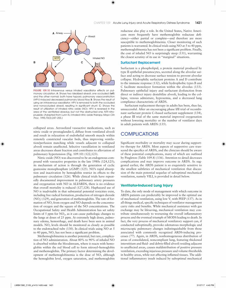
collapsed areas. Aerosolized vasoactive medications, such as nitric oxide or prostaglandin-I, diffuse from ventilated alveoli and result in relaxation of endothelial smooth muscle within remotely constricted vascular beds, thus improving ventila-tion/perfusion matching while vessels adjacent to collapsed alveoli remain unaffected. Selective vasodilation in ventilated areas decreases shunt fraction and contributes to alleviation of pulmonary hypertension (Fig. 109.10) (122,123).
Nitric oxide (NO) was discovered to be an endogenous com-pound with vasoactive properties in the late 1980s (124,125); its mechanism of action is through the generation of cyclic guanosine monophosphate (cGMP) (125). NO’s rapid absorp-tion and inactivation by hemoglobin restrict its effects to the pulmonary circulation (126). While clinical trials have repeat-edly documented improvement in pulmonary artery pressures and oxygenation with NO in ALI/ARDS, there is no evidence that overall mortality is reduced (127,128). Haphazard use of NO is inadvisable in that substantial potential toxicities exist, including free radical formation, production of nitrogen dioxide (NO2) (129), and generation of methemoglobin. The rate of for-mation of NO2 from oxygen and NO depends on the concentra-tion of oxygen and the square of the NO concentrations. The Occupational Safety and Health Administration has set safety limits of 5 ppm for NO2, as it can cause pathologic changes to the lungs at doses of 25 ppm. At extremely high doses, pulmo-nary edema, hemorrhage, and death have been seen in animal models. NO2 levels should be monitored as closely as possible to the endotracheal tube (130). In clinical trials using NO at 5 to 40 ppm, NO2 has not been a significant problem.
Methemoglobinemia is another potential, but rare, complica-tion of NO administration. About 80% to 90% of inhaled NO is absorbed within the bloodstream, where it reacts with hemo-globin within the red blood cell to form nitrosyl– hemoglobin and methemoglobin. The primary factor determining the devel-opment of methemoglobinemia is the dose of NO, although the hemoglobin level, oxygen saturation, and methemoglobin
reductase also play a role. In the United States, Native Ameri-cans more frequently have methemoglobin reductase defi-ciency—either partial or complete—and therefore are more susceptible to methemoglobinemia. Closer monitoring of such patients is warranted. In clinical trials using NO at 5 to 40 ppm, methemoglobinemia has not been a significant problem. Finally, the cost of inhaled NO is surprisingly steep (131), warranting the closest scrutiny of its use in “marginal” situations.
Surfactant Replacement
Surfactant is a phospholipid, a protein material produced by type II epithelial pneumocytes, secreted along the alveolar sur-face and acting to decrease surface tension to prevent alveolar collapse. Hydrophilic surfactant proteins A and D contribute to the immune response (132), while hydrophobic types B and C facilitate monolayer formation within the alveolus (133). Pulmonary epithelial injury and surfactant dysfunction from direct or indirect injury destabilize alveoli, leading to the col-lapse, venous admixture, hypoxemia, and a decreased lung compliance characteristic of ARDS.
Surfactant replacement therapy in adults has been, thus far, unsuccessful. After an encouraging phase I/II trial of recombi-nant surfactant protein C–based surfactant supplement (134), a phase III trial of the same material improved oxygenation without lowering mortality or the number of ventilator days in adult patients with ARDS (135).
cOmPLIcAtIOnS
Significant morbidity or mortality may occur during support-ive therapy for ARDS. Most aspects of supportive care tran-scend the specifics of ARDS, and the clinician should be aware of these potential complications, many of which are outlined by Pingleton (Table 109.8) (136). Attention to detail decreases complications and may improve outcome in ARDS. As sug-gested earlier, the ARDS patient is so exquisitely sensitive to the smallest subtleties of mechanical ventilation that discus-sion of the main potential sequelae of suboptimal mechanical ventilation, namely VILI, is provided in detail below.
Ventilator-Induced Lung Injury
To date, the only mode of management with which outcome in ARDS patients can predictably be improved is the optimal use of mechanical ventilation, using low Vt with PEEP (137). As in all things medical, specific techniques of ventilator management carry risks and benefits. While mechanical assistance with gas exchange may be lifesaving, mechanical ventilation may con-tribute simultaneously to worsening the overall inflammatory process and the eventual triumph of MODS leading to death. In fact, the very process of mechanical ventilatory support can, if conducted suboptimally, provoke edematous morphologic and microscopic pulmonary changes indistinguishable from those associated with commonly recognized ARDS-inducing pro-cesses (77). Again, in ARDS, nonhomogeneous distribution of areas of consolidated, noncompliant lung, featuring thickened interstitium and fluid- and debris-filled alveoli residing adjacent to unaffected areas, causes maldistribution of positive pressure ventilation, exceeding injurious pressure and volume thresholds in healthy areas, while not affecting inflamed tissues. The addi-tional inflammatory insult induced by suboptimal mechanical
AlveoliAirway
NTP
NTPNTP
Inhaled NO
CB
A
NONO
NO
FIguRe 109.10 intravenous versus inhaled vasodilator effects on pul-monary circulation. A: shows two idealized alveoli, one occluded (left) and the other normal; both have hypoxic pulmonary vasoconstriction (hPV)-induced decreased pulmonary blood flow. B: shows the result of using an intravenous vasodilator: hPV is removed to both the occluded and nonoccluded alveoli, resulting in significant shunt. c: shows the result of utilization of inhaled nitric oxide (No): hPV is reversed in the area of the ventilated alveolus, but not the obstructed one. NtP, nitro-prusside. (adapted from lunn rJ. inhaled nitric oxide therapy. Mayo Clin Proc. 1995;70(3):247–255.)
LWBK1580_C109_p1410-1426.indd 1421 31/07/17 4:40 PM

1422 SECTion 11 rEsPiratorY disordErs
ventilation technique is termed ventilator-associated lung injury. The ARMA study (74) revealed the importance of low-volume ventilation in ARDS, with substantial improvement in several indices using 6 mL/kg rather than 12 mL/kg Vt. While criticized, the findings document the importance of avoiding several putative mechanisms of pathologic effect:• Excess alveolar hyperinflation with associated increased
permeability and cytokine release• Escape of alveolar air outside the confines of alveoli• Destructive sheer-stress influence of repetitive inflation/
collapse of unstable alveoli.Each of these phenomena contributes to pulmonary dys-
function and perpetuation of the inflammatory response in the ARDS patient, and thus each has been classified. Barotrauma refers to the presence of air outside the alveoli when receiving positive pressure ventilation. Air leaks track along the peri-vascular sheath to the mediastinum and pleural cavities, or along fascial planes to extrathoracic areas (138). It seems intu-itive that such occurrences are related to pressures exceeding the limits of tissue structural integrity, but the issue is clearly more complicated, since musicians are repeatedly able to gen-erate 150 cm H2O airway pressure with no sequelae (139). It is speculated that barotrauma represents regional overin-flation in areas of diseased lung, such areas thereby being particularly at risk for structural failure and air leak (139). Volutrauma occurs when excessive inspiratory volumes induce microvascular edema (140); the offending agent appears to be excessive volume, rather than the excessive pressure required to supply that volume. Resultant mechanical stretch triggers changes in the alveolar–capillary barrier (79), and in prolif-eration of inflammatory cytokines (75), resulting in interstitial and alveolar proteinaceous edema, decreased compliance, and hyaline membrane formation. Compromised surfactant pro-duction and function leads to increased surface tension, pro-voking alveolar collapse with increased venous admixture and subjecting alveolar epithelium to the tissue-destructive shear stresses of recruitment/derecruitment in the process known as atelectrauma. While the use of PEEP to maintain diseased distal alveoli splinted “open” has not definitively been dem-onstrated to improve outcome (79, and earlier discussion, above), improvements in oxygenation and pulmonary compli-ance with PEEP mandate its routine use in ARDS. Of note is the complex relationship between mechanical ventilation and patchy maldistribution throughout the lung in areas of varying ratios of ventilation and perfusion, with atelectatic areas abut-ting hyperinflated bullous and cystic areas. While collapsed noncompliant airways require high initial opening pressures consistent with the Law of LaPlace (within the context of pulmonary physiology, the following formula is appropriate: P = 2t/R, where P = pressure, t = tension, and R = radius), such high pressure is transmitted throughout the lung, over-distending more compliant airways both through direct influ-ence and by an unequally distributed traction force upon the adjacent airways, as depicted in Figure 109.7. Implicit in such heterogeneous patterns of gas distribution is the initiation of the destructive sequelae associated with inflation of each sub-region of lung, as delineated above.
Furthermore, over the last few years, there has been rec-ognition of the inflammatory cytokine release, as well as alveolar and interstitial neutrophil infiltration associated with ventilator-related pulmonary disruption, leading to MODS (76,141). While it is indisputable that “excessive” Vt
TABLE 109.8 Complications Associated with Acute Respiratory Distress Syndrome
Pulmonary•Pulmonary emboli•Pulmonary barotrauma•Pulmonary fibrosis•oxygen toxicity
Gastrointestinal•Gastrointestinal hemorrhage• ileus•Gastric distension•Pneumoperitoneum
Renal•renal failure•Fluid retention
Cardiovascular• invasive catheters•arrhythmia•hypotension•low cardiac output
Infection•sepsis•Nosocomial pneumonia
Hematologic•anemia•thrombocytopenia•disseminated intravascular coagulation
Other•hepatic•Endocrine•Neurologic•Psychiatric•malnutrition
Complications attributable to intubation and extubation•Prolonged attempt at intubation• intubation of a mainstem bronchus•Premature extubation•self-extubation
Complications associated with endotracheal/tracheostomy tubes•tube malfunction•Nasal necrosis•Paranasal sinus infection•tracheal stenosis•tracheomalacia•Polyps•Erosion•Fistulae•airway obstruction•hoarseness
Complications attributable to operation of the ventilator•machine failure•alarm failure•alarms silenced• inadequate nebulization or humidification
Complications occurring during positive airway pressure therapy•alveolar hypoventilation•alveolar hyperventilation•massive gastric distension•barotrauma•atelectasis•Pneumonia•hypotension
From taylor rW. the adult respiratory distress syndrome. in: Kirby rr, taylor rW, eds. Respiratory Failure. Chicago, il: Year book medical Publishers; 1986:208.
LWBK1580_C109_p1410-1426.indd 1422 31/07/17 4:40 PM

CHAPTER 109 acute lung injury and acute respiratory distress syndrome 1423
ventilation augments systemic cytokine levels (79), the specific causal relationship with worse outcome has yet to be vali-dated. The ALVEOLI study (87), which examined the varia-tion of short-term indices—28-day mortality and number of ventilator-free days—with modulation of PEEP, was discontin-ued early based on lack of improvement in outcome with high PEEP levels. Thus, the optimal inspiratory pressure, or level of PEEP for a given ARDS patient’s pressure–volume curve, can be exceedingly difficult to identify despite the potentially severe consequences of failure to do so. While dependent areas of tenaciously collapsed, high time-constant alveoli may require the equivalent of repeated and prolonged high-pressure recruitment maneuvers to achieve inflation and avoid atelectrauma, simultaneous transmission of such pressure to compliant alveoli incurs the risk of inducing volutrauma and inciting inflammation. Clearly, in those with advanced lung injury, the “optimal” inspiratory pressure, in reality, reflects a statistical bell curve of widely variable individual alveolar compliances. The clinician must vary inspiratory time, plateau pressure, PEEP, and tidal volume to inflate stiff alveoli while not persistently overdistending the normal ones. Such impor-tant actions are required because of the dynamic and changing compliance profiles of the inflamed lung.
References 1. Kollef MH, Schuster D. The acute respiratory distress syndrome. N Engl J
Med. 1995;332:27. 2. Bernard GR, Artigas A, Brigham KL, et al. The American-European Con-
sensus Conference on ARDS. Am J Respir Crit Care Med. 1994;149:818. 3. Ranieri VM, Rubenfeld GD, Thompson BT, et al. Acute respiratory dis-
tress syndrome: the Berlin definition. JAMA. 2012;307:2526–2533. 4. Murray JF, Matthay MA, Luce JM, et al. An expanded definition of the
adult respiratory distress syndrome. Am Rev Respir Dis. 1988;138:720. 5. Matthay MA. Conference summary—acute lung injury. Chest. 1999;116:
119S–126S. 6. Zilberberg MD, Epstein SK. Acute lung injury in the medical ICU: comor-
bid conditions, age, etiology, and hospital outcome. Am J Respir Crit Care Med. 1998;157(Pt 1):1159–1164.
7. Moss M, Goodman PL, Heinig M, et al. Establishing the relative accuracy of three new definitions of the adult respiratory distress syndrome. Crit Care Med. 1995;23:1629.
8. Kangelaris KN, Calfee CS, May AK, et al. Is there still a role for the lung injury score in the era of the Berlin definition ARDS? Ann Intensive Care. 2014;4:4.
9. Hudson LD, Milberg JA, Anardi D, et al. Clinical risks of the develop-ment of the acute respiratory distress syndrome. Am J Respir Care. 1995; 151:293.
10. Hudson LD, Steinberg KP. Epidemiology of acute lung injury and ARDS. Chest. 1999;116:74S.
11. Ware LB, Matthay MA. The acute respiratory distress syndrome. N Engl J Med. 2000;342:1334.
12. Rubenfield GD, Caldwell E, Peabody E, et al. Incidence and outcomes of acute lung injury. N Engl J Med. 2005;353:1685.
13. Rocco PRM, Zin WA. Pulmonary and extrapulmonary acute respiratory distress syndrome: are they different? Curr Opin Crit Care. 2005;11:10.
14. Milberg JA, Davis DR, Steinberg KP, et al. Improved survival of patients with acute respiratory distress syndrome (ARDS): 1983–1993. JAMA. 1995; 273(4):306–309.
15. Abel SJC, Finney SJ, Brett SJ, et al. Reduced mortality in association with acute respiratory distress syndrome (ARDS). Thorax. 1998;53:292.
16. Taylor RW, Duncan CA. The adult respiratory distress syndrome. Res Med. 1983;1:17.
17. Brun-Buisson C, Minelli C, Bertolini G, et al. Epidemiology and outcome of acute lung injury in European intensive care units: results from the ALIVE study. Intensive Care Med. 2004;30:51–61.
18. Luhr OR, Antonsen K, Karlsson M, et al. Incidence and mortality after acute respiratory failure and acute respiratory distress syndrome in Sweden, Denmark, and Iceland: the ARF Study Group. Am J Respir Crit Care Med. 1999;159:1849–1861.
19. Bersten AD, Edibam C, Hunt T, et al. Incidence and mortality of acute lung injury and the acute respiratory distress syndrome in three Australian states. Am J Respir Crit Care Med. 2002;165:443–448.
20. Villar J, Blanco J, Añón JM, et al. The ALIEN study: incidence and out-come of acute respiratory distress syndrome in the era of lung protective ventilation. Intensive Care Med. 2011;37:1932–1941.
21. Davidson TA, Rubenfeld GD, Caldwell ES, et al. The effect of acute respi-ratory distress syndrome on long-term survival. Am J Respir Crit Care Med. 1999;160:1838.
22. Luce JM. Acute lung injury and the acute respiratory distress syndrome. Crit Care Med. 1998;26:369–376.
23. Herridge MS, Cheung AM, Tansey CM, et al. One-year outcomes in sur-vivors of the acute respiratory distress syndrome. N Engl J Med. 2003; 348:683.
24. McHugh LG, Milberg JA, Whitecomb ME, et al. Recovery of function in survivors of the acute respiratory distress syndrome. Am J Respir Crit Care Med. 1994;150:90.
25. Davidson TA, Caldwell ES, Curtis JR, et al. Reduced quality of life in survivors of acute respiratory distress syndrome compared with critically ill control patients. JAMA. 1999;281:354.
• ARDS is commonly seen with the systemic inflamma-tory response syndrome (SIRS) and the MODS.
• Risk factors for developing ARDS include SIRS, sepsis, pulmonary contusion, aspiration, inhalation of toxic sub-stances, near-drowning, long-bone fractures, pancreati-tis, diffuse pneumonia, and multiple blood transfusions.
• Most patients with ARDS demonstrate similar clinical and pathologic features, irrespective of the cause of the ALI.
• The lung’s response to injury can be divided into an exu-dative phase, a proliferative phase, and a fibrotic phase.
• A variety of inflammatory mediators have been impli-cated in the pathogenesis of ALI.
• The neutrophil plays a central role in ALI.• The severe hypoxemia associated with this syndrome
is caused by intrapulmonary shunting that occurs with interstitial edema, and alveolar flooding and collapse.
• A reduction in functional residual capacity (FRC) and lung compliance are the hallmarks of ARDS.
• Radiographic changes seen in patients with ARDS are characteristic but nonspecific, and rarely reveal the eti-ology of the syndrome.
Essential Diagnostic Tests and Procedures
• History and physical examination• Chest radiograph• Arterial blood gas measurements• Further diagnostic tests based on the clinical circum-
stances
Initial Therapy
• Most patients require early endotracheal intubation and positive pressure ventilation.
Key Points
• The goal of mechanical ventilation is to provide ade-quate oxygenation and carbon dioxide elimination while keeping complications, such as oxygen toxicity, ventilator-associated lung injury, and hemodynamic compromise, to a minimum.
LWBK1580_C109_p1410-1426.indd 1423 31/07/17 4:40 PM

1424 SECTion 11 rEsPiratorY disordErs
26. Weinert CR, Gross CR, Kangas JR, et al. Health-related quality of life after acute lung injury. Am J Respir Crit Care Med. 1997;156:1120.
27. Mikkelsen ME, Christie JD, Lanken PN, et al. The adult respiratory dis-tress syndrome cognitive outcomes study: long-term neuropsychological function in survivors of acute lung injury. Am J Respir Crit Care Med. 2012;185:1307–1315.
28. Bienvenu OJ, Colantuoni E, Mendez-Tellez PA, et al. Cooccurrence of and remission from general anxiety, depression, and posttraumatic stress disorder symptoms after acute lung injury: a 2-year longitudinal study. Crit Care Med. 2015;43:642–653.
29. Khandelwal N, Hough CL, Bansal A, et al. Long-term survival in patients with severe acute respiratory distress syndrome and rescue therapies for refractory hypoxemia. Crit Care Med. 2014;42:1610–1618.
30. Pelosi P, D’Onofrio D, Chiumello D, et al. Pulmonary and extrapulmonary acute respiratory distress syndrome are different. Eur Respir J (Suppl). 2003;42:48s–56s.
31. Lee WL, Downey GP. Neutrophil activation and acute lung injury. Curr Opin Crit Care. 2001;7(1):1–7.
32. Abraham E. Neutrophils and acute lung injury. Crit Care Med. 2003;31 (4 Suppl):S195–199.
33. Frerking I, Günther A, Seeger W, et al. Pulmonary surfactant: functions, abnormalities and therapeutic options. Intensive Care Med. 2001;27(11): 1699–1717.
34. Idell S. Coagulation, fibrinolysis, and fibrin deposition in acute lung injury. Crit Care Med. 2003;31(4 Suppl):S213–S220.
35. Pittet JF, Mackersie RC, Martin TR, et al. Biological markers of acute lung injury: prognostic and pathogenetic significance. Am J Respir Crit Care Med. 1997;155(4):1187–1205.
36. Park WY, Goodman RB, Steinberg KP, et al. Cytokine balance in the lungs of patients with acute respiratory distress syndrome. Am J Respir Crit Care Med. 2001;164:1896–1903.
37. Fink MP. Role of reactive oxygen and nitrogen species in acute respiratory distress syndrome. Curr Opin Crit Care. 2002;8:6–11.
38. Bhatia M, Moochhala S. Role of inflammatory mediators in the pathophysi-ology of acute respiratory distress syndrome. J Pathol. 2004;202:145–156.
39. Han S, Mallampalli RK. The acute respiratory distress syndrome: from mechanism to translation. J Immunol. 2015;194:855–860.
40. Tomashefski JF Jr. Pulmonary pathology of acute respiratory distress syn-drome. Clin Chest Med. 2000;21:435–466.
41. Bellingan GJ. The pulmonary physician in critical care: the pathogenesis of ALI/ARDS. Thorax. 2002;57:540–546.
42. Marshall RP, Bellingan G, Webb S, et al. Fibroproliferation occurs early in the acute respiratory distress syndrome and impacts on outcome. Am J Respir Crit Care Med. 2000;162:1783–1788.
43. Gomez AC: Pulmonary insufficiency in non-thoracic trauma. J Trauma. 1968;8:666.
44. Moore FA, Feliciano DV, Andrassy RJ, et al. Early enteral feeding, com-pared with parenteral, reduces postoperative septic complications. Ann Surg. 1992;216:172.
45. Rice TW, Wheeler AP, Thompson BT, et al. Initial trophic vs full enteral feeding in patients with acute lung injury: the EDEN randomized trial. JAMA. 2012; 307:795–803.
46. Needham DM, Dinglas VD, Bienvenu OJ, et al. One year outcomes in patients with acute lung injury randomised to initial trophic or full enteral feeding: prospective follow-up of EDEN randomised trial. Br Med J. 2013; 346:f1532
47. Schuster D. Fluid management in ARDS: “keep them dry” or does it mat-ter? Intensive Care Med. 1995;21:101.
48. National Heart, Lung, and Blood Institute Acute Respiratory Distress Syn-drome (ARDS) Clinical Trials Network. Comparison of two fluid-management strategies in acute lung injury. N Engl J Med. 2006;354(24):2564–2575.
49. Matthay MA, Brower RG, Carson S, et al. National Heart, Lung, and Blood Institute Acute Respiratory Distress Syndrome (ARDS) Clinical Tri-als Network. Randomized, placebo-controlled clinical trial of an aerosol-ized b2-agonist for treatment of acute lung injury. Am J Respir Crit Care Med. 2011;184:561–568.
50. Gao Smith F, Perkins GD, Gates S, et al.; BALTI-2 study investigators. Effect of intravenous b-2 agonist treatment on clinical outcomes in acute respiratory distress syndrome (BALTI-2): a multicentre, randomised con-trolled trial. Lancet. 2012;379:229–235.
51. Perkins GD, Gates S, Park D et al. The beta agonist lung injury trial prevention. A randomized controlled trial. Am J Respir Crit Care Med. 2014;189:674–683.
52. Wright P, Carmichael L, Bernard G. Effect of bronchodilators on lung mechanics in the acute respiratory distress syndrome (ARDS). Chest. 1994;106:157.
53. Stoelting RK. Pharmacology and Physiology in Anesthetic Practice. 3rd ed. Philadelphia, PA: Lippincott–Raven; 1999:196.
54. Meduri GU, Chinn AJ, Leeper KV, et al. Corticosteroid rescue treatment of progressive fibroproliferation in late ARDS. Patterns of response and predictors of outcome. Chest. 1994;105:1516–1527.
55. Steinberg KP, Hudson LD, Goodman RB, et al.; The National Heart, Lung, and Blood Institute Acute Respiratory Distress Syndrome (ARDS) Clinical Trials Network. Efficacy and safety of corticosteroids for persistent acute respiratory distress syndrome. N Engl J Med. 2006;354:1671–1684.
56. Meduri GU, Golden E, Freire AX, et al. Methylprednisolone infusion in early severe ARDS: results of a randomized controlled trial. Chest. 2007; 131:954–963.
57. Meduri GU, Marik PE, Chrousos GP, et al. Steroid treatment in ARDS: a critical appraisal of the ARDS network trial and the recent literature. Intensive Care Med. 2008;34:61–69.
58. Peter JV, John P, Graham PL, et al. Corticosteroids in the prevention and treatment of acute respiratory distress syndrome (ARDS) in adults: meta-analysis. BMJ. 2008;336:1006–1009.
59. Calfee CS, Matthay MA. Nonventilatory treatments for acute lung injury and ARDS. Chest. 2007;131:913–920.
60. Hudson LD, Hough CL. Therapy for late-phase acute respiratory distress syndrome. Clin Chest Med. 2006;27:671–677.
61. Bream-Rouwenhorst HR, Beltz EA, Ross MB, et al. Recent developments in the management of acute respiratory distress syndrome in adults. Am J Health Syst Pharm. 2008;65:29–36.
62. Meduri GU, Headley S, Tolley E, et al. Plasma and BAL cytokine response to corticosteroid rescue treatment in late ARDS. Chest. 1995;103:1315.
63. Papazian L, Forel JM, Gacouin A, et al. Neuromuscular blockers in early acute respiratory distress syndrome. N Engl J Med. 2010;363:1107–1116.
64. Alhazzani W, Alshahrani M, Jaeschke R, et al. Neuromuscular blocking agents in acute respiratory distress syndrome: a systematic review and meta-analysis of randomized controlled trials. Crit Care. 2013;17:R43.
65. Hraiech S, Dizier S, Papazian L. The use of paralytics in patients with acute respiratory distress syndrome. Clin Chest Med. 2014;35:753–763.
66. Antonelli M, Conti G, Esquinas A, et al. A multiple-center survey on the use in clinical practice of noninvasive ventilation as a first-line intervention for acute respiratory distress syndrome. Crit Care Med. 2007;35:18–25.
67. Ferrer M, Esquinas A, Leon M, et al. Noninvasive ventilation in severe hypoxemic respiratory failure: a randomized clinical trial. Am J Respir Crit Care Med. 2003;168:1438–1444.
68. Keenan SP, Sinuff T, Cook DJ, et al. Does noninvasive positive pressure ventilation improve outcome in acute hypoxemic respiratory failure? A systematic review. Crit Care Med. 2004;32:2516–2523.
69. Rouby JJ, Puybasset L, Cluzel P, et al. Regional distribution of gas and tissue in acute respiratory distress syndrome. II. Physiological correlations and definition of an ARDS Severity Score. CT Scan ARDS Study Group. Intensive Care Med. 2000;26:1046–1056.
70. Puybasset L, Gusman P, Muller JC, et al. Regional distribution of gas and tissue in acute respiratory distress syndrome. III. Consequences for the effects of positive end-expiratory pressure. CT Scan ARDS Study Group. Intensive Care Med. 2000;26:1215–1227.
71. Kolobow T, Moretti MP, Fumagalli R, et al. Severe impairment in lung function induced by high peak airway pressure during mechanical ventila-tion: an experimental study. Am Rev Respir Dis. 1987;135:312–315.
72. Tsuno K, Prato P, Kolobow T. Acute lung injury from mechanical ventilation at moderately high airway pressures. J Appl Physiol. 1990;69:956–961.
73. Hickling KG, Henderson SJ, Jackson R. Low mortality associated with low volume pressure limited ventilation with permissive hypercapnia in severe adult respiratory distress syndrome. Intensive Care Med. 1990;16:372–377.
74. Acute Respiratory Distress Syndrome Network. Ventilation with lower tidal volumes as compared with traditional tidal volumes for acute lung injury and the acute respiratory distress syndrome. N Engl J Med. 2000; 342:1301–1308.
75. Tremblay LN, Slutsky AS. Ventilator-induced lung injury: from the bench to the bedside. Intensive Care Med. 2006;32:24–33.
76. Tremblay L, Valenza F, Ribeiro SP, et al. Injurious ventilatory strategies increase cytokines and c-fos m-RNA expression in an isolated rat lung model. J Clin Invest. 1997;99:944–952.
77. Slutsky AS, Ranieri VM. Ventilator-induced lung injury. N Engl J Med. 2013;369:2126–2136.
78. Belperio JA, Keane MP, Lynch JP III, et al. The role of cytokines during the pathogenesis of ventilator-associated and ventilator-induced lung injury. Semin Respir Crit Care Med. 2006;27:350–364.
79. Ranieri VM, Suter PM, Tortorella C, et al. Effect of mechanical ventila-tion on inflammatory mediators in patients with acute respiratory distress syndrome: a randomized controlled trial. JAMA. 1999;282:54–61.
LWBK1580_C109_p1410-1426.indd 1424 31/07/17 4:40 PM

CHAPTER 109 acute lung injury and acute respiratory distress syndrome 1425
80. Terragni PP, Rosboch G, Tealdi A, et al. Tidal hyperinflation during low tidal volume ventilation in acute respiratory distress syndrome. Am J Respir Crit Care Med. 2007;175:160–166.
81. Gattinoni L, Pesenti A. The concept of “baby lung.” Intensive Care Med. 2005;31:776–784.
82. Barbas CS, de Matos GF, Pincelli MP, et al. Mechanical ventilation in acute respiratory failure: recruitment and high positive end-expiratory pressure are necessary. Curr Opin Crit Care. 2005;11:18–28.
83. Amato MB, Barbas CS, Medeiros DM, et al. Effect of a protective- ventilation strategy on mortality in the acute respiratory distress syndrome. N Engl J Med. 1998;338:347–354.
84. Lachmann B. Open up the lung and keep the lung open. Intensive Care Med. 1992;18:319–321.
85. Gattinoni L, Caironi P, Cressoni M, et al. Lung recruitment in patients with the acute respiratory distress syndrome. N Engl J Med. 2006;354: 1775–1786.
86. Dueck R. Alveolar recruitment versus hyperinflation: a balancing act. Curr Opin Anaesthesiol. 2006;19:650–654.
87. Brower RG, Lanken PN, MacIntyre N, et al.; National Heart, Lung, and Blood Institute ARDS Clinical Trials Network. Higher versus lower posi-tive end-expiratory pressures in patients with the acute respiratory distress syndrome. N Engl J Med. 2004;351:327–336.
88. Meade MO, Cook DJ, Guyatt GH, et al.; Lung Open Ventilation Study Investigators. Ventilation strategy using low tidal volumes, recruitment maneuvers, and high positive end-expiratory pressure for acute lung injury and acute respiratory distress syndrome: a randomized controlled trial. JAMA. 2008;299:637–645.
89. Borges JB, Okamoto VN, Matos GF, et al. Reversibility of lung collapse and hypoxemia in early acute respiratory distress syndrome. Am J Respir Crit Care Med. 2006;174(3):268–278.
90. Lapinsky SE, Mehta S. Bench-to-bedside review: recruitment and recruit-ing maneuvers. Crit Care. 2005;9:60–65.
91. Medoff BD, Harris RS, Kesselman H, et al. Use of recruitment maneuvers and high-positive end-expiratory pressure in a patient with acute respira-tory distress syndrome. Crit Care Med. 2000;28:1210–1216.
92. Gattinoni L, Carlesso E, Cressoni M. Selecting the ‘right’ positive end-expiratory pressure level. Curr Opin Crit Care. 2015;21:50–57.
93. Chiumello D, Cressoni M, Carlesso E, et al. Bedside selection of positive end-expiratory pressure in mild, moderate, and severe acute respiratory distress syndrome. Crit Care Med. 2014;42:252–264.
94. Gulati G, Novero A, Loring SH, et al. Pleural pressure and optimal posi-tive end-expiratory pressure based on esophageal pressure versus chest wall elastance: incompatible results. Crit Care Med. 2013;41:1951–1957.
95. Laffey JG, O’Croinin D, McLoughlin P, et al. Permissive hypercapnia: role in protective lung ventilatory strategies. Intensive Care Med. 2004; 30:347–356.
96. Kallet RH, Campbell AR, Alonso JA, et al. The effects of pressure control versus volume control assisted ventilation on patient work of breathing in acute lung injury and acute respiratory distress syndrome. Respir Care. 2000;45:1085–1096.
97. Shanholtz C, Brower R. Should inverse ratio ventilation be used in adult respiratory distress syndrome. Am J Respir Crit Care Med. 1994;149: 1354–1358.
98. Mercat A, Graïni L, Teboul JL, et al. Cardiorespiratory effects of pressure-controlled ventilation with and without inverse ratio in the adult respira-tory distress syndrome. Chest. 1993;104:871–875.
99. Esteban A, Alía I, Gordo F, et al. Prospective randomized trial comparing pressure-controlled ventilation and volume-controlled ventilation in ARDS. For the Spanish Lung Failure Collaborative Group. Chest. 2000;117: 1690–1696.
100. Habashi NM. Other approaches to open-lung ventilation: airway pressure release ventilation. Crit Care Med. 2005;33:S228–S240.
101. Kaplan LJ, Bailey H, Formosa V. Airway pressure release ventilation increases cardiac performance in patients with acute lung injury/adult respiratory distress syndrome. Crit Care. 2001;5:221–226.
102. Putensen C, Zech S, Wrigge H, et al. Long-term effects of spontaneous breathing during ventilatory support in patients with acute lung injury. Am J Respir Crit Care Med. 2001;164:43–49.
103. Pelosi P, Brazzi L, Gattinoni L. Prone position in acute respiratory distress syndrome. Eur Respir J. 2002;20:1017–1028.
104. West JB, Dollery CT, Naimark A. Distribution of blood flow in isolated lung; relation to vascular and alveolar pressures. J Appl Physiol. 1964; 19:713–724.
105. Cornejo RA, Díaz JC, Tobar EA, et al. Effects of prone positioning on lung protection in patients with acute respiratory distress syndrome. Am J Respir Crit Care Med. 2013;188:440–448.
106. Jozwiak M, Teboul JL, Anguel N, et al. Beneficial hemodynamic effects of prone positioning in patients with acute respiratory distress syndrome. Am J Respir Crit Care Med. 2013;188:1428–1433.
107. Gattinoni L, Tognoni G, Pesenti A, et al.; Prone-Supine Study Group. Effect of prone positioning on the survival of patients with acute respira-tory failure. N Engl J Med. 2001;345:568–573.
108. Guerin C, Gaillard S, Lemasson S, et al. Effects of systematic prone posi-tioning in hypoxemic acute respiratory failure: a randomized controlled trial. JAMA. 2004;292:2379–2387.
109. Mancebo J, Fernández R, Blanch L, et al. A multicenter trial of prolonged prone ventilation in severe acute respiratory distress syndrome. Am J Respir Crit Care Med. 2006;173:1233–1239.
110. Guérin C, Reignier J, Richard JC, et al Prone positioning in severe acute respiratory distress syndrome. N Engl J Med. 2013;368:2159–2168.
111. Derdak S. High-frequency oscillatory ventilation for acute respiratory distress syndrome in adult patients. Crit Care Med. 2003;31(4 Suppl): S317–S323.
112. Derdak S, Mehta S, Stewart TE, et al.; Multicenter Oscillatory Ventilation For Acute Respiratory Distress Syndrome Trial (MOAT) Study Investiga-tors. High-frequency oscillatory ventilation for acute respiratory distress syndrome in adults: a randomized, controlled trial. Am J Respir Crit Care Med. 2002;166:801–808.
113. Bollen CW, van Well GT, Sherry T, et al. High frequency oscillatory ventilation compared with conventional mechanical ventilation in adult respiratory distress syndrome: a randomized controlled trial. Crit Care. 2005;9:R430–R439.
114. Sud S, Sud M, Friedrich JO, et al. High frequency oscillation in patients with acute lung injury and acute respiratory distress syndrome (ARDS): systematic review and meta-analysis. BMJ. 2010;340:c2327.
115. Ferguson ND, Cook DJ, Guyatt GH, et al. High-frequency oscillation in early acute respiratory distress syndrome. N Engl J Med. 2013;368: 795–805.
116. Young D, Lamb SE, Shah S, et al. High-frequency oscillation for acute respiratory distress syndrome. N Engl J Med. 2013;368:806–813.
117. Brown JK, Haft JW, Bartlett RH, et al. Acute lung injury and acute respira-tory distress syndrome: extracorporeal life support and liquid ventilation for severe acute respiratory distress syndrome in adults. Semin Respir Crit Care Med. 2006;27:416–425.
118. Gattinoni L, Pesenti A, Mascheroni D, et al. Low-frequency positive-pres-sure ventilation with extracorporeal CO2 removal in severe acute respira-tory failure. JAMA. 1986;256:881–886.
119. Morris AH, Wallace CJ, Menlove RL, et al. Randomized clinical trial of pressure-controlled inverse ratio ventilation and extracorporeal CO2 removal for adult respiratory distress syndrome. Am J Respir Crit Care Med. 1994;149:295–305.
120. Lequier L. Extracorporeal life support in pediatric and neonatal critical care: a review. J Intensive Care Med. 2004;19:243–258.
121. Skinner SC, Hirschl RB, Bartlett RH. Extracorporeal life support. Semin Pediatr Surg. 2006;15:242–250.
122. Pison U, López FA, Heidelmeyer CF, et al. Inhaled nitric oxide reverses hypoxic pulmonary vasoconstriction without impairing gas exchange. J Appl Physiol. 1993;74:1287–1292.
123. Rossaint R, Falke KJ, López F, et al. Inhaled nitric oxide for the adult respiratory distress syndrome. N Engl J Med. 1993;328:399–405.
124. Palmer RM, Ferrige AG, Moncada S. Nitric oxide release accounts for the biological activity of endothelium-derived relaxing factor. Nature. 1987; 327:524–526.
125. Moncada S, Palmer RM, Higgs EA. Nitric oxide: physiology, pathophysi-ology, and pharmacology. Pharmacol Rev. 1991;43:109–142.
126. Cooper CE. Nitric oxide and iron proteins. Biochim Biophys Acta. 1999; 1411:290–309.
127. Taylor RW, Zimmerman JL, Dellinger RP, et al.; Inhaled Nitric Oxide in ARDS Study Group. Low-dose inhaled nitric oxide in patients with acute lung injury: a randomized controlled trial. JAMA. 2004;291(13):1603–1609.
128. Adhikari NK, Burns KE, Friedrich JO, et al. Effect of nitric oxide on oxy-genation and mortality in acute lung injury: systematic review and meta-analysis. BMJ. 2007;334:779.
129. Lowson SM. Inhaled alternatives to nitric oxide. Crit Care Med. 2005; 33:S188–S195.
130. Puybasset L, Rouby JJ, Mourgeon E, et al. Factors influencing cardiopul-monary effects of inhaled nitric oxide in acute respiratory failure. Am J Respir Crit Care Med. 1995;152(1):318–328.
131. Pierce CM, Peters MJ, Cohen G, et al. Cost of nitric oxide is exorbitant [Letter to the Editor]. BMJ. 2002;325:336.
132. Wright JR. Immunoregulatory functions of surfactant proteins. Nat Rev Immunol. 2005;5:58–68.
LWBK1580_C109_p1410-1426.indd 1425 31/07/17 4:40 PM

1426 SECTion 11 rEsPiratorY disordErs
133. Stevens TP, Sinkin RA. Surfactant replacement therapy. Chest. 2007; 131:1577–1582.
134. Spragg RG, Lewis JF, Wurst W, et al. Treatment of acute respiratory dis-tress syndrome with recombinant surfactant protein C surfactant. Am J Respir Crit Care Med. 2003;167:1562–1566.
135. Spragg RG, Lewis JF, Walmrath HD, et al. Effect of recombinant surfac-tant protein C-based surfactant on the acute respiratory distress syndrome. N Engl J Med. 2004;351:884–892.
136. Pingleton SK. Complications associated with the adult respiratory distress syndrome. Clin Chest Med. 1982;5:143.
137. Villar J, Kacmarek RM, Pérez-Méndez L, et al. A high positive end- expiratory pressure, low tidal volume ventilatory strategy improves
outcome in persistent acute respiratory distress syndrome: a randomized, controlled trial. Crit Care Med. 2006;34(5):1311–1318.
138. Gammon RB, Shin MS, Buchalter SE. Pulmonary barotrauma in mechani-cal ventilation: patterns and risk factors. Chest. 1992;102:568–572.
139. Bouhuys A. Physiology and musical instruments. Nature. 1969;221: 1199–2004.
140. Dreyfuss D, Soler P, Saumon G. Mechanical ventilation-induced pulmo-nary edema. Interaction with previous lung alterations. Am J Respir Crit Care Med. 1995;151:1568–1575.
141. Kawano T, Mori S, Cybulsky M, et al. Effect of granulocyte depletion in a ventilated surfactant-depleted lung. J Appl Physiol. 1987;62:27–33.
LWBK1580_C109_p1410-1426.indd 1426 31/07/17 4:40 PM


![Lecture Lung Oedema.PPT [Read-Only]ocw.usu.ac.id/.../emd166_slide_pulmonary_oedema.pdf · NonNon--Cardiogenic CardiogenicLung Lung OedemaOedema A.R.D.S (Acute Respiratory Distress](https://static.fdocuments.us/doc/165x107/5e617e8300003419542fef3a/lecture-lung-read-onlyocwusuacidemd166slidepulmonaryoedemapdf-nonnon-cardiogenic.jpg)