ACUTE LEUKAEMIA by DR. FATIMA AL-QAHTANI CONSULTANT HAEMATOLOGIST.
62
ACUTE LEUKAEMIA ACUTE LEUKAEMIA by by DR. FATIMA AL-QAHTANI DR. FATIMA AL-QAHTANI CONSULTANT HAEMATOLOGIST CONSULTANT HAEMATOLOGIST
-
Upload
hannah-page -
Category
Documents
-
view
225 -
download
5
Transcript of ACUTE LEUKAEMIA by DR. FATIMA AL-QAHTANI CONSULTANT HAEMATOLOGIST.

ACUTE LEUKAEMIAACUTE LEUKAEMIA
byby
DR. FATIMA AL-QAHTANIDR. FATIMA AL-QAHTANICONSULTANT HAEMATOLOGISTCONSULTANT HAEMATOLOGIST
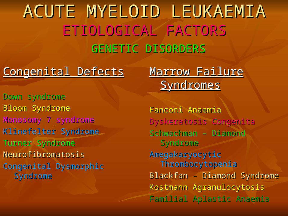
ACUTE MYELOID LEUKAEMIAACUTE MYELOID LEUKAEMIAETIOLOGICAL FACTORSETIOLOGICAL FACTORS
GENETIC DISORDERSGENETIC DISORDERS
Congenital DefectsCongenital Defects
Down syndromeDown syndrome
Bloom SyndromeBloom Syndrome
Monosomy 7 syndromeMonosomy 7 syndrome
Klinefelter SyndromeKlinefelter Syndrome
Turner SyndromeTurner Syndrome
NeurofibromatosisNeurofibromatosis
Congenital Dysmorphic SyndromeCongenital Dysmorphic Syndrome
Marrow Failure SyndromesMarrow Failure Syndromes
Fanconi AnaemiaFanconi Anaemia
Dyskeratosis CongenitaDyskeratosis Congenita
Schwachman Schwachman –– Diamond Syndrome Diamond Syndrome
Amegakaryocytic ThrombocytopeniaAmegakaryocytic Thrombocytopenia
Blackfan Blackfan –– Diamond Syndrome Diamond Syndrome
Kostmann AgranulocytosisKostmann Agranulocytosis
Familial Aplastic AnaemiaFamilial Aplastic Anaemia
ACUTE MYELOID LEUKAEMIAACUTE MYELOID LEUKAEMIAETIOLOGICAL FACTORSETIOLOGICAL FACTORS
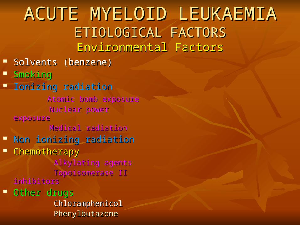
Environmental FactorsEnvironmental Factors Solvents (benzene)Solvents (benzene) SmokingSmoking Ionizing radiationIonizing radiation Atomic bomb exposureAtomic bomb exposure
Nuclear power exposureNuclear power exposure Medical radiationMedical radiation Non ionizing radiationNon ionizing radiation ChemotherapyChemotherapy Alkylating agentsAlkylating agents Topoisomerase II inhibitorsTopoisomerase II inhibitors Other drugsOther drugs ChloramphenicolChloramphenicol PhenylbutazonePhenylbutazone

Acute Myeloid LeukaemiaAcute Myeloid LeukaemiaCLINICAL FEATURESCLINICAL FEATURES
Increasing incidence with age. Median age at Increasing incidence with age. Median age at diagnosis is 50 years.diagnosis is 50 years.
Symptoms are due to marrow failure (anaemia, Symptoms are due to marrow failure (anaemia, infection, haemorrhage) or hyperleucocytosis.infection, haemorrhage) or hyperleucocytosis.
Rarely presenting symptoms are due to chloromas Rarely presenting symptoms are due to chloromas or CNS involvement.or CNS involvement.
Acute Lymphoblastic Leukaemia (ALL)Acute Lymphoblastic Leukaemia (ALL) Clinical FeaturesClinical Features
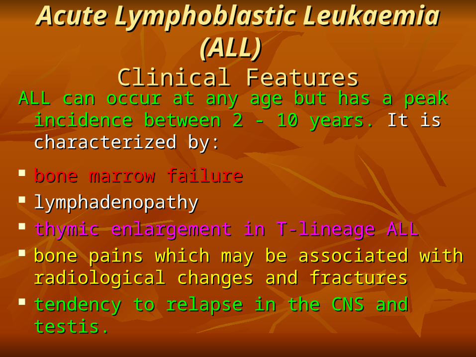
ALL can occur at any age but has a peak incidence ALL can occur at any age but has a peak incidence between 2 - 10 years.between 2 - 10 years. It is characterized by: It is characterized by:
bone marrow failurebone marrow failure lymphadenopathy lymphadenopathy thymic enlargement in T-lineage ALLthymic enlargement in T-lineage ALL bone pains which may be associated with bone pains which may be associated with
radiological changes and fracturesradiological changes and fractures tendency to relapse in the CNS and testis.tendency to relapse in the CNS and testis.

Acute Myeloid LeukaemiaAcute Myeloid Leukaemia DEFINITION DEFINITION
Acute Myeloid Leukaemia (AML) is currently Acute Myeloid Leukaemia (AML) is currently defined by the defined by the FAB criteria. criteria.
The percentage of blastsThe percentage of blasts, , the presence of the presence of cytochemical myeloperoxidasecytochemical myeloperoxidase, , the major cell types the major cell types present defined by morphology and esterase present defined by morphology and esterase cytochemistry & the immunophenotypecytochemistry & the immunophenotype define the 8 define the 8 FAB subtypesFAB subtypes
Acute Myeloid LeukaemiaAcute Myeloid LeukaemiaLABORATORY DIAGNOSISLABORATORY DIAGNOSIS
MORPHOLOGYMORPHOLOGY
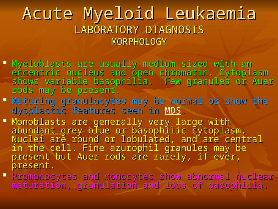
Myeloblasts are usually medium sized with an eccentric Myeloblasts are usually medium sized with an eccentric nucleus and open chromatin. Cytoplasm shows variable nucleus and open chromatin. Cytoplasm shows variable basophilia. Few granules or Auer rods may be present.basophilia. Few granules or Auer rods may be present.
Maturing granulocytes may be normal or show the Maturing granulocytes may be normal or show the dysplastic features seen indysplastic features seen in MDS..
Monoblasts are generally very large with abundant grey-Monoblasts are generally very large with abundant grey-blue or basophilic cytoplasm. Nuclei are round or lobulated, blue or basophilic cytoplasm. Nuclei are round or lobulated, and are central in the cell. Fine azurophil granules may be and are central in the cell. Fine azurophil granules may be present but Auer rods are rarely, if ever, present.present but Auer rods are rarely, if ever, present.
Promonocytes and monocytes show abnormal nuclear Promonocytes and monocytes show abnormal nuclear maturation, granulation and loss of basophilia.maturation, granulation and loss of basophilia.
Acute Myeloid LeukaemiaAcute Myeloid LeukaemiaLABORATORY DIAGNOSISLABORATORY DIAGNOSIS
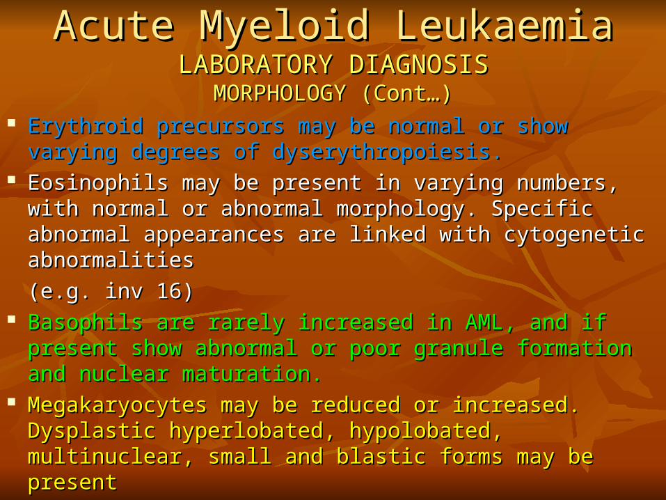
MORPHOLOGY (ContMORPHOLOGY (Cont……)) Erythroid precursors may be normal or show varying Erythroid precursors may be normal or show varying
degrees of dyserythropoiesis.degrees of dyserythropoiesis. Eosinophils may be present in varying numbers, with Eosinophils may be present in varying numbers, with
normal or abnormal morphology. Specific abnormal normal or abnormal morphology. Specific abnormal appearances are linked with cytogenetic abnormalities appearances are linked with cytogenetic abnormalities
(e.g. inv 16)(e.g. inv 16) Basophils are rarely increased in AML, and if present show Basophils are rarely increased in AML, and if present show
abnormal or poor granule formation and nuclear maturation.abnormal or poor granule formation and nuclear maturation. Megakaryocytes may be reduced or increased. Dysplastic Megakaryocytes may be reduced or increased. Dysplastic
hyperlobated, hypolobated, multinuclear, small and blastic hyperlobated, hypolobated, multinuclear, small and blastic forms may be presentforms may be present

Acute Myeloid LeukaemiaAcute Myeloid LeukaemiaFAB SubtypesFAB Subtypes


Acute Myeloid LeukaemiaAcute Myeloid LeukaemiaFAB SubtypesFAB Subtypes
M0-M5:M0-M5: >20% myelo/monoblasts by>20% myelo/monoblasts by morphology or immunophenotype.morphology or immunophenotype.
M6: >50% erythroid precursors, >20% blasts in non-erythroid cells.M6: >50% erythroid precursors, >20% blasts in non-erythroid cells.
M7: >20% megakaryoblasts present.M7: >20% megakaryoblasts present.
NoteNote:: CytogeneticsCytogenetics, , molecular geneticsmolecular genetics, , previous MDS or MPDprevious MDS or MPD, ,
previous chemo- or radiotherapy and previous chemo- or radiotherapy and the presence of trilineage the presence of trilineage myelodysplasiamyelodysplasia do not have any place in the FAB system. do not have any place in the FAB system. All these All these features which contribute to the definition of prognostically features which contribute to the definition of prognostically important sub-groups is included in the important sub-groups is included in the new classification system..

ACUTE MYELOID LEUKAEMIASACUTE MYELOID LEUKAEMIAS WHOWHO CLASSIFICATIONCLASSIFICATION
Acute myeloid leukaemia with reccurent geneticAcute myeloid leukaemia with reccurent genetic abnormalitiesabnormalities
Acute myeloid leukaemia withAcute myeloid leukaemia with t(8;21)(q22;q22);t(8;21)(q22;q22); (AML1(CBFa)/ETO)(AML1(CBFa)/ETO)
Acute myeloid leukaemia with abnormal bone marrow eosinophilsAcute myeloid leukaemia with abnormal bone marrow eosinophils inv (16)(p13q22) or t(16; 16)(p13;q22);inv (16)(p13q22) or t(16; 16)(p13;q22); (CBFb/MYH11)(CBFb/MYH11)
Acute promyelocytic leukaemiaAcute promyelocytic leukaemia (AML with t(15; 17)(q22;q12)(AML with t(15; 17)(q22;q12)(PML/RARa)(PML/RARa) & & variantsvariants
Acute myeloid leukaemia withAcute myeloid leukaemia with 11q2311q23 (MLL)(MLL) abnormalities abnormalities
Acute myeloid leukaemia with multilineage dysplasiaAcute myeloid leukaemia with multilineage dysplasia Acute myeloid leukaemia and myelodysplastic syndromes, Acute myeloid leukaemia and myelodysplastic syndromes,
therapy relatedtherapy related

ACUTE MYELOID LEUKAEMIASACUTE MYELOID LEUKAEMIAS
WHO CLASSIFICATIONWHO CLASSIFICATION Acute myeloid leukaemia not otherwise categorizedAcute myeloid leukaemia not otherwise categorized Acute myeloid leukaemia minimally differentiatedAcute myeloid leukaemia minimally differentiated Acute myeloid leukaemia without maturationAcute myeloid leukaemia without maturation Acute myeloid leukaemia with maturationAcute myeloid leukaemia with maturation Acute myelomonocytic leukaemiaAcute myelomonocytic leukaemia Acute monoblastic and monocytic leukaemiaAcute monoblastic and monocytic leukaemia Acute erythroid leukaemiasAcute erythroid leukaemias Acute megakaryoblastic leukaemiaAcute megakaryoblastic leukaemia Acute basophilic leukaemiaAcute basophilic leukaemia Acute panmyelosis with myelofibrosisAcute panmyelosis with myelofibrosis Myeloid sarcomaMyeloid sarcoma Acute leukaemia of ambiguous lineageAcute leukaemia of ambiguous lineage Undifferentiated acute leukaemiaUndifferentiated acute leukaemia Bilineal acute leukaemiaBilineal acute leukaemia
Biphenotypic leukaemiaBiphenotypic leukaemia

M0 M1 M2
M3 M4
M5b
M5a
M6 M7

Acute Myeloid LeukaemiaAcute Myeloid LeukaemiaLABORATORY DIAGNOSISLABORATORY DIAGNOSIS
CYTOCHEMISTRYCYTOCHEMISTRY
Myeloperoxidase (MPO): identify blast cells as myeloid. Myeloperoxidase (MPO): identify blast cells as myeloid. Auer rods are detected twice as frequently as on Auer rods are detected twice as frequently as on Romanowsky stains. Dysplastic neutrophils may be Romanowsky stains. Dysplastic neutrophils may be negative. Eosinophil granules are always positive. negative. Eosinophil granules are always positive. Monoblasts and promonocytes may be negative. Sudan Monoblasts and promonocytes may be negative. Sudan Black B gives identical results.Black B gives identical results.
Myeloperoxidase (MPO)

Acute Myeloid LeukaemiaAcute Myeloid LeukaemiaLABORATORY DIAGNOSISLABORATORY DIAGNOSIS
CYTOCHEMISTRYCYTOCHEMISTRY
Chloroacetate Esterase (CAE) specifically identifies Chloroacetate Esterase (CAE) specifically identifies cells of the granulocyte lineage, from the early cells of the granulocyte lineage, from the early promyelocyte stage to mature neutrophils.promyelocyte stage to mature neutrophils.
Chloroacetate Esterase
Acute Myeloid LeukaemiaAcute Myeloid LeukaemiaLABORATORY DIAGNOSISLABORATORY DIAGNOSIS
CYTOCHEMISTRYCYTOCHEMISTRY
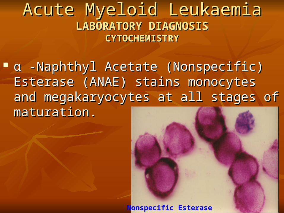
αα -Naphthyl Acetate (Nonspecific) Esterase -Naphthyl Acetate (Nonspecific) Esterase (ANAE) stains monocytes and megakaryocytes at (ANAE) stains monocytes and megakaryocytes at all stages of maturation.all stages of maturation.
Nonspecific Esterase
Acute Myeloid LeukaemiaAcute Myeloid LeukaemiaLABORATORY DIAGNOSISLABORATORY DIAGNOSIS
CYTOCHEMISTRYCYTOCHEMISTRY
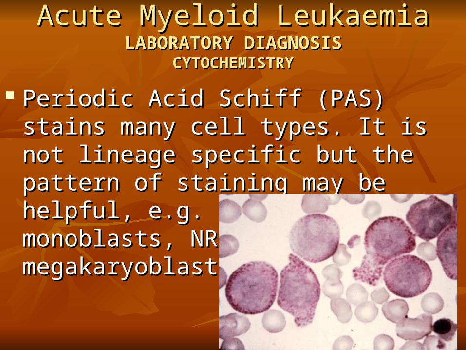
Periodic Acid Schiff (PAS) stains many cell Periodic Acid Schiff (PAS) stains many cell types. It is not lineage specific but the pattern types. It is not lineage specific but the pattern of staining may be helpful, e.g. positive of staining may be helpful, e.g. positive monoblasts, NRBC, megakaryoblastsmonoblasts, NRBC, megakaryoblasts……
Acute Lymphoblastic LeukaemiaAcute Lymphoblastic LeukaemiaLaboratory DiagnosisLaboratory Diagnosis
FAB ClassificationFAB Classification
Acute Lymphoblastic LeukaemiaAcute Lymphoblastic LeukaemiaLaboratory DiagnosisLaboratory Diagnosis
Acute Lymphoblastic LeukaemiaAcute Lymphoblastic LeukaemiaLaboratory DiagnosisLaboratory Diagnosis
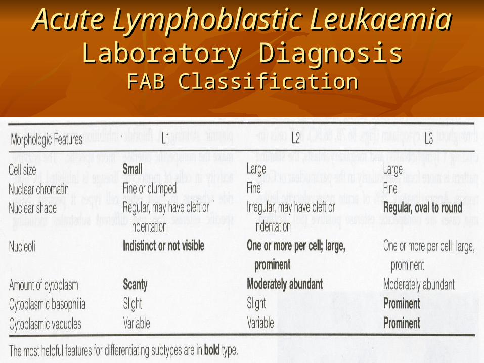
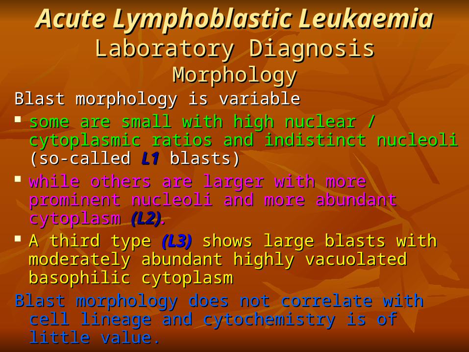
MorphologyMorphologyBlast morphology is variable Blast morphology is variable some are small with high nuclear / cytoplasmic some are small with high nuclear / cytoplasmic
ratios and indistinct nucleoliratios and indistinct nucleoli (so-called (so-called L1L1 blasts) blasts) while others are larger with more prominent while others are larger with more prominent
nucleoli and more abundant cytoplasmnucleoli and more abundant cytoplasm (L2)(L2).. A third type A third type (L3)(L3) shows large blasts with shows large blasts with
moderately abundant highly vacuolated basophilic moderately abundant highly vacuolated basophilic cytoplasmcytoplasm
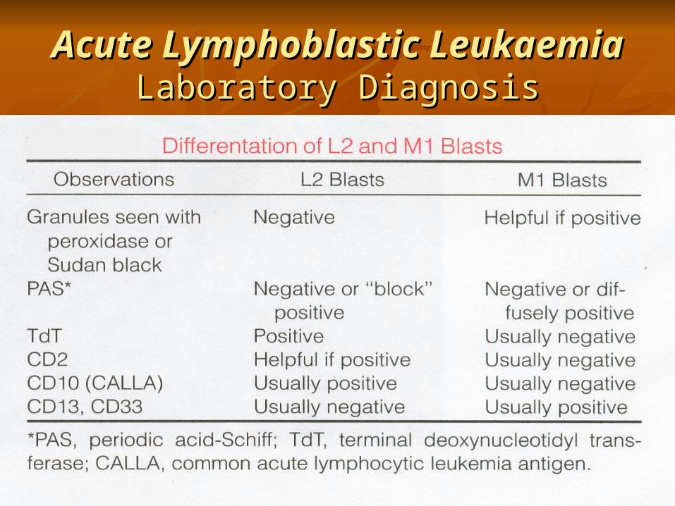
Blast morphology does not correlate with cell lineage Blast morphology does not correlate with cell lineage and cytochemistry is of little value.and cytochemistry is of little value.

ALL (L2)
ALL (L3)
Acute Lymphoblastic LeukaemiaAcute Lymphoblastic LeukaemiaLaboratory Diagnosis - FAB ClassificationLaboratory Diagnosis - FAB Classification
ALL (L1)

PAS Ac. Phos (T-ALL)
Acute Lymphoblastic LeukaemiaAcute Lymphoblastic LeukaemiaLaboratory DiagnosisLaboratory Diagnosis
CYTOCHEMISTRYCYTOCHEMISTRY

Acute Myeloid LeukaemiaAcute Myeloid Leukaemia LABORATORY DIAGNOSISLABORATORY DIAGNOSIS
TREPHINE BIOPSY HISTOLOGYTREPHINE BIOPSY HISTOLOGY
Helpful when there is severe cytopenia and a dry Helpful when there is severe cytopenia and a dry tap, e.g. in megakaryoblastic AML.tap, e.g. in megakaryoblastic AML.
Useful for identifying megakaryocyte dysplasia and Useful for identifying megakaryocyte dysplasia and dyserythropoiesis.dyserythropoiesis.
Immunophenotyping is possible when the diagnosis Immunophenotyping is possible when the diagnosis is in doubt.is in doubt.
Not suitable for fine classification which is based on Not suitable for fine classification which is based on percentages of cell types and cytological detail.percentages of cell types and cytological detail.
Acute Myeloid LeukaemiaAcute Myeloid LeukaemiaLABORATORY DIAGNOSISLABORATORY DIAGNOSIS
FLOW CYTOMETRYFLOW CYTOMETRY
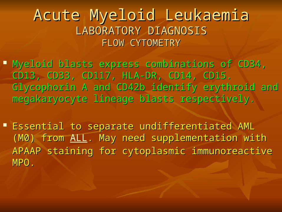
Myeloid blasts express combinations of CD34, CD13, Myeloid blasts express combinations of CD34, CD13, CD33, CD117, HLA-DR, CD14, CD15. Glycophorin A and CD33, CD117, HLA-DR, CD14, CD15. Glycophorin A and CD42b identify erythroid and megakaryocyte lineage blasts CD42b identify erythroid and megakaryocyte lineage blasts respectively.respectively.
Essential to separate undifferentiated AML (M0) from Essential to separate undifferentiated AML (M0) from ALL. . May need supplementation with APAAP staining for May need supplementation with APAAP staining for cytoplasmic immunoreactive MPO.cytoplasmic immunoreactive MPO.

Acute Myeloid LeukaemiaAcute Myeloid LeukaemiaLABORATORY DIAGNOSISLABORATORY DIAGNOSIS
FLOW CYTOMETRY (ContFLOW CYTOMETRY (Cont……))
Essential for diagnosing biphenotypic leukaemias and Essential for diagnosing biphenotypic leukaemias and detecting aberrant (promiscuous) antigen expression.detecting aberrant (promiscuous) antigen expression.
Can identify patient specific unique blast cell phenotypes.Can identify patient specific unique blast cell phenotypes.
Can identify blasts of megakaryocyte, erythroid, monocyte Can identify blasts of megakaryocyte, erythroid, monocyte and granulocyte lineages.and granulocyte lineages.
Can identify leukaemic contamination of 'remission' Can identify leukaemic contamination of 'remission' marrow harvests.marrow harvests.
Can identify early relapse.Can identify early relapse.

Acute Lymphoblastic LeukaemiaAcute Lymphoblastic LeukaemiaLaboratory DiagnosisLaboratory Diagnosis
FlowcytometryFlowcytometry ALL is derived from precursor lymphocytes that are undergoing ALL is derived from precursor lymphocytes that are undergoing
antigen receptor gene (Ig and TCR) rearrangement.antigen receptor gene (Ig and TCR) rearrangement.
B-lineage ALLB-lineage ALL
The precursor nature of the cells is established by demonstrating The precursor nature of the cells is established by demonstrating lack of surface Ig, the presence of nuclear TDT and sometimes the lack of surface Ig, the presence of nuclear TDT and sometimes the expression of CD34.expression of CD34.
Sub classification is as follows:Sub classification is as follows:
Pre-pre B-ALLPre-pre B-ALL: CD19+ CD10- cytoplasmic mu heavy chain negative.: CD19+ CD10- cytoplasmic mu heavy chain negative.
Common ALLCommon ALL: CD19+ CD10+ cytoplasmic mu present in <20% of cells: CD19+ CD10+ cytoplasmic mu present in <20% of cells
Pre B-ALLPre B-ALL: CD19+ CD10+ cytoplasmic mu present in >20% of cells : CD19+ CD10+ cytoplasmic mu present in >20% of cells
Blasts of all subgroups will express cytoplasmic CD22 and CD79b.Blasts of all subgroups will express cytoplasmic CD22 and CD79b.

Acute Lymphoblastic LeukaemiaAcute Lymphoblastic LeukaemiaLaboratory DiagnosisLaboratory Diagnosis
FlowcytometryFlowcytometry
T-lineage ALLT-lineage ALL
The precursor nature of the cells is established by demonstrating The precursor nature of the cells is established by demonstrating TDT and sometimes CD34 positivity and the lack of surface TDT and sometimes CD34 positivity and the lack of surface TCR/CD3.TCR/CD3.
T-cell lineage is demonstrated by the expression of CD7 and/or T-cell lineage is demonstrated by the expression of CD7 and/or CD1a.CD1a.
Expression of the other pan-T cell markers is variable.Expression of the other pan-T cell markers is variable.
Acute Myeloid LeukaemiaAcute Myeloid LeukaemiaLABORATORY DIAGNOSISLABORATORY DIAGNOSIS
IMMUNOHISTOCHEMISTRY / IMMUNOFLUORESCENCEIMMUNOHISTOCHEMISTRY / IMMUNOFLUORESCENCE
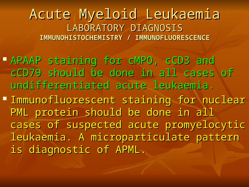
APAAP staining for cMPO, cCD3 and cCD79 APAAP staining for cMPO, cCD3 and cCD79 should be done in all cases of undifferentiated should be done in all cases of undifferentiated acute leukaemia.acute leukaemia.
Immunofluorescent staining for nuclear PML Immunofluorescent staining for nuclear PML protein should be done in all cases of suspected protein should be done in all cases of suspected acute promyelocytic leukaemia. A acute promyelocytic leukaemia. A microparticulate pattern is diagnostic of APML.microparticulate pattern is diagnostic of APML.

Acute Myeloid LeukaemiaAcute Myeloid LeukaemiaLABORATORY DIAGNOSISLABORATORY DIAGNOSIS
CYTOGENETICSCYTOGENETICS
An abnormal karyotype is found in 50-60% of AML cases at An abnormal karyotype is found in 50-60% of AML cases at presentation.presentation.
t(15;17), t(8;21) and inv/del/t(16) are associated with specific t(15;17), t(8;21) and inv/del/t(16) are associated with specific morphology, younger patients and a good prognosis.morphology, younger patients and a good prognosis.
Complex karyotypes and abnormalities of chromosomes 5 and Complex karyotypes and abnormalities of chromosomes 5 and 7 are associated with older patients, trilineage dysplasia and a 7 are associated with older patients, trilineage dysplasia and a poor response to treatment.poor response to treatment.
Other karyotypic abnormalities are prognostically neutral.Other karyotypic abnormalities are prognostically neutral.

Acute Myeloid LeukaemiaAcute Myeloid LeukaemiaLABORATORY DIAGNOSISLABORATORY DIAGNOSIS
MOLECULAR GENETICSMOLECULAR GENETICS
The 3 major good prognosis translocations are all detectable The 3 major good prognosis translocations are all detectable by RT-PCR, which can be used to detect up to 40% more by RT-PCR, which can be used to detect up to 40% more cases than are found by metaphase cytogenetics.cases than are found by metaphase cytogenetics.
All translocations with cloned breakpoint genes are All translocations with cloned breakpoint genes are theoretically detectable by RT-PCR.theoretically detectable by RT-PCR.
Real time multiplex PCR may become available for routine Real time multiplex PCR may become available for routine diagnosis.diagnosis.
RT-PCR is used for monitoring speed of response to RT-PCR is used for monitoring speed of response to
treatment and may be predictive for relapsetreatment and may be predictive for relapse

Acute Lymphoblastic LeukaemiaAcute Lymphoblastic LeukaemiaLaboratory DiagnosisLaboratory Diagnosis
CYTOGENETICSCYTOGENETICS Hyperdiploidy is common. A number of balanced translocations Hyperdiploidy is common. A number of balanced translocations
have been identified in ALL:have been identified in ALL:
t(12;21) - this is the commonest translocation in ALL (30% of cases). It t(12;21) - this is the commonest translocation in ALL (30% of cases). It results in the TEL-AML fusion gene and is primarily associated with the results in the TEL-AML fusion gene and is primarily associated with the common phenotype.common phenotype.
t(9;22) - this is commoner in adults and is associated with a very poor t(9;22) - this is commoner in adults and is associated with a very poor prognosis.prognosis.
t(4;11) - this translocation results in the MLL-AF4 fusion gene. It is t(4;11) - this translocation results in the MLL-AF4 fusion gene. It is associated with pre-pre B-ALL and is associated with a poor prognosis.associated with pre-pre B-ALL and is associated with a poor prognosis.
t(1;19) - associated with pre-B ALL and results in the formation of the E2A-t(1;19) - associated with pre-B ALL and results in the formation of the E2A-PBX fusion gene.PBX fusion gene.
These translocations are demonstrable by RT-PCR techniquesThese translocations are demonstrable by RT-PCR techniques . .
TAL-1 deregulation is the commonest genetic abnormality in T-ALL. This TAL-1 deregulation is the commonest genetic abnormality in T-ALL. This may occur as the result of the t(1;14) or more commonly due to chromosome may occur as the result of the t(1;14) or more commonly due to chromosome 1p32 deletions.1p32 deletions.

Acute Lymphoblastic LeukaemiaAcute Lymphoblastic LeukaemiaPROGNOSTIC FACTORSPROGNOSTIC FACTORS
The following are poor prognostic factors in ALL:The following are poor prognostic factors in ALL:
age <1 year and >10 yearsage <1 year and >10 years male sexmale sex CNS disease at presentationCNS disease at presentation high white cell count high white cell count t(9;22)t(9;22) t(4;11)t(4;11) hypodiploidyhypodiploidy

Acute Lymphoblastic LeukaemiaAcute Lymphoblastic LeukaemiaOUTCOME AND THERAPYOUTCOME AND THERAPY
The treatment of ALL consists of the following "phases":The treatment of ALL consists of the following "phases":
Remission inductionRemission induction - vincristine, prednisolone, daunorubicin, - vincristine, prednisolone, daunorubicin, asparaginase.asparaginase.
ConsolidationConsolidation - various combinations of chemotherapeutic agents. - various combinations of chemotherapeutic agents.
CNS directed therapyCNS directed therapy - high dose systemic and intrathecal - high dose systemic and intrathecal methotrexate.methotrexate.
Maintenance therapyMaintenance therapy - vincristine, prednisolone, mercaptopurine and - vincristine, prednisolone, mercaptopurine and methotrexate for 2 years.methotrexate for 2 years.
Childhood ALL is associated with 75% long term survival. Childhood ALL is associated with 75% long term survival. Minimal residual Minimal residual diseasedisease assessment using assessment using PCR based strategiesPCR based strategies appear to be able to appear to be able to predict predict relapserelapse although they are not yet in routine clinical use. although they are not yet in routine clinical use. Allogeneic transplantation is the treatment of choice at relapse. Allogeneic transplantation is the treatment of choice at relapse.
The outlook in adult ALL is poor with approximately 20% long-term The outlook in adult ALL is poor with approximately 20% long-term survivors. Allogeneic transplantation is advisable in first remission.survivors. Allogeneic transplantation is advisable in first remission.

ACUTE LEUKAEMIAACUTE LEUKAEMIA CLASSIFICATIONCLASSIFICATION
Acute lymphoblastic leukaemiaAcute lymphoblastic leukaemia Early pre-B-cell ALLEarly pre-B-cell ALL Pre-B-cell ALLPre-B-cell ALL B-cell ALLB-cell ALL T-cell ALLT-cell ALL Acute nonlymphocytic leukaemiaAcute nonlymphocytic leukaemia Acute myelocytic leukaemia, minimally differentiated (AML-M0)Acute myelocytic leukaemia, minimally differentiated (AML-M0)
Acute myelocytic leukaemia, without maturation (AML-M1)Acute myelocytic leukaemia, without maturation (AML-M1) Acute myelocytic leukaemia, with maturation (AML-M2)Acute myelocytic leukaemia, with maturation (AML-M2) Acute promyelocytic leukaemia (APL, AML-M3)Acute promyelocytic leukaemia (APL, AML-M3) Acute myelomonocytic leukaemia, (AMMoL, AML-M4)Acute myelomonocytic leukaemia, (AMMoL, AML-M4) Acute monocytic leukaemia, (AMoL, AML-M5)Acute monocytic leukaemia, (AMoL, AML-M5) Acute erythroleukaemia, (AEL, AML-M6)Acute erythroleukaemia, (AEL, AML-M6) Acute megakaryocytic leukaemia, (AMegL, AML-M7)Acute megakaryocytic leukaemia, (AMegL, AML-M7) Biphenotypic (mixed lineage) leukaemiaBiphenotypic (mixed lineage) leukaemia Acute undifferentiated leukaemiaAcute undifferentiated leukaemia
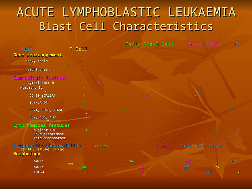
ACUTE LYMPHOBLASTIC LEUKAEMIAACUTE LYMPHOBLASTIC LEUKAEMIABlast Cell CharacteristicsBlast Cell Characteristics
Early Pre-B CellEarly Pre-B Cell Pre-B CellPre-B Cell B CellB Cell T CellT CellGene rearrangementGene rearrangement Heavy chainHeavy chain ++** + + ++ - - Light chainLight chain +/-+/- +/-+/- ++ --
Immunologic featuresImmunologic features Cytoplasmic Cytoplasmic UU -- ++ - - - - Membrane IgMembrane Ig -- - - ++ - - CD 10 (CALLA)CD 10 (CALLA) ++ +/- +/- +/- +/- +/-+/- Ia/HLA-DRIa/HLA-DR ++ + + + + - - CD24, CD19, CD20CD24, CD19, CD20 + + + + ++ - - CD2, CD5, CD7CD2, CD5, CD7 - - - - - - ++
Cytochemical featuresCytochemical features Nuclear TDTNuclear TDT + + ++ - - + + 5’-Nucleotidase5’-Nucleotidase + + + + - - + + Acid phosphataseAcid phosphatase - - - - - - + +
Cytogenetic abnormalitiesCytogenetic abnormalities t(12;21)t(12;21) t(1;19)t(1;19) t(8;14), t(2;8), t(8;22)t(8;14), t(2;8), t(8;22) t(1;14), t(11;14), del(9p)t(1;14), t(11;14), del(9p)
MorphologyMorphology FAB L1FAB L1 90%90% 90%90% 10%10% 95%95% FAB L2FAB L2 10%10% 10%10% 15%15% 5%5%
FAB L3FAB L3 0 0 0 0 75%75% 00
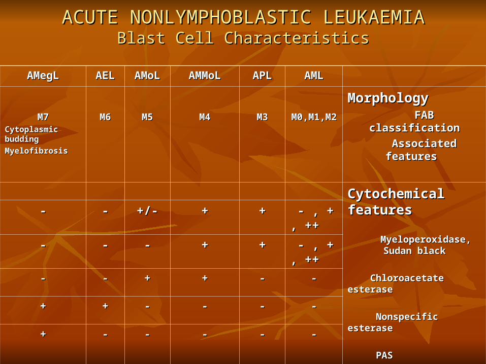
ACUTE NONLYMPHOBLASTIC LEUKAEMIAACUTE NONLYMPHOBLASTIC LEUKAEMIABlast Cell CharacteristicsBlast Cell Characteristics
AMLAMLAPLAPLAMMoLAMMoLAMoLAMoLAELAELAMegLAMegL
MorphologyMorphology FAB classificationFAB classification
Associated features Associated features
M0,M1,M2M0,M1,M2M3M3M4M4M5M5M6M6M7M7Cytoplasmic buddingCytoplasmic budding
MyelofibrosisMyelofibrosis
Cytochemical featuresCytochemical features
Myeloperoxidase, Sudan blackMyeloperoxidase, Sudan black
Chloroacetate esteraseChloroacetate esterase
Nonspecific esteraseNonspecific esterase
PASPAS
Platelet peroxidasePlatelet peroxidase
- , + , ++- , + , +++++++/-+/-----
- , + , ++- , + , ++++++------
----++++----
--------++++
----------++
CytogeneticCytogenetic
abnormalitiesabnormalities
t(8;21)t(8;21)t(15;17)t(15;17)del(16)(q22)del(16)(q22)
inv(16)(p13;q22)inv(16)(p13;q22)t(11q)t(11q)
del(11q)del(11q)
Abnormalities ofAbnormalities of
5, 7, 215, 7, 21

Myelodysplastic Syndrome (MDS)Myelodysplastic Syndrome (MDS)WHO ClassificationWHO Classification
There are 8 categories of MDS in the WHO system. There are 8 categories of MDS in the WHO system.
1)1) Refractory anemia (RA)Refractory anemia (RA)2)2) Refractory anemia with ringed sideroblasts (RARS) Refractory anemia with ringed sideroblasts (RARS) 3)3) Refractory cytopenia with multilineage dysplasia (RCMD) Refractory cytopenia with multilineage dysplasia (RCMD) 4)4) Refractory cytopenia with multilineage dysplasia and Refractory cytopenia with multilineage dysplasia and
ringed sideroblasts (RCMD-RS)ringed sideroblasts (RCMD-RS)5)5) Refractory anemia with excess blasts-1(RAEB-1) Refractory anemia with excess blasts-1(RAEB-1) 6)6) Refractory anemia with excess blasts-2(RAEB-2) Refractory anemia with excess blasts-2(RAEB-2) 7)7) Myelodysplastic syndrome, unclassified (MDS-U) Myelodysplastic syndrome, unclassified (MDS-U) 8)8) MDS associated with isolated del(5q) MDS associated with isolated del(5q)

Myelodysplastic Syndrome (MDS)Myelodysplastic Syndrome (MDS)WHO ClassificationWHO Classification
TypeType% % BloodBloodBone MarrowBone MarrowConvert to Convert to AML AML
RARA
ICD-O ICD-O codecode
9980/39980/3
5-10%5-10%AnaemiaAnaemia
No or rare blasts No or rare blasts
Only erythroid dysplasiaOnly erythroid dysplasia
<5% blasts<5% blasts
<15% ringed sideroblasts<15% ringed sideroblasts
6%6%
RARSRARS
ICD-O ICD-O codecode
9982/39982/3
10-12%10-12%AnaemiaAnaemia
No blastsNo blasts
Only erythroid dysplasiaOnly erythroid dysplasia
<5% blasts<5% blasts
≥≥15% ringed sideroblasts15% ringed sideroblasts
1-2%1-2%
RCMDRCMD
ICD-O ICD-O codecode
9985/39985/339% 39%
(24/15)(24/15)
Cytopenias (2-3)Cytopenias (2-3)
No or rare blasts, No No or rare blasts, No Auer rodsAuer rods
<1<1××101099/L mono./L mono.
≥≥10% dysplasia of 2 or more myeloid cell 10% dysplasia of 2 or more myeloid cell lineslines
<5% blasts, No Auer rods<5% blasts, No Auer rods
<15% ringed sideroblasts<15% ringed sideroblasts11%11%
RCMDRCMD-RS-RS
Cytopenias (2-3)Cytopenias (2-3)
No or rare blasts, No No or rare blasts, No Auer rodsAuer rods
<1<1××101099/L mono./L mono.
≥≥10% dysplasia of 2 or more myeloid cell 10% dysplasia of 2 or more myeloid cell lineslines
<5% blasts, No Auer rods<5% blasts, No Auer rods
≥≥15% ringed sideroblasts15% ringed sideroblasts

Myelodysplastic Syndrome (MDS)Myelodysplastic Syndrome (MDS)WHO ClassificationWHO Classification
RAEB IRAEB I
ICD-O codeICD-O code
9983/39983/3
40% 40% (22/18)(22/18)
(affect(affect
pts > pts > 50 50
years)years)
CytopeniasCytopenias
<5% blasts, No <5% blasts, No Auer rodsAuer rods
<1<1××101099/L mono./L mono.
Uni/Multilineage dysplasiaUni/Multilineage dysplasia
5-9%blasts, No Auer rods5-9%blasts, No Auer rods
25%25%
RAEB IIRAEB IICytopeniasCytopenias
5-19% blasts, Auer 5-19% blasts, Auer rods +/-rods +/-
<1<1××101099/L mono./L mono.
Uni/Multilineage dysplasiaUni/Multilineage dysplasia
10-19%blasts, Auer rods +/-10-19%blasts, Auer rods +/-
33%33%
MDS-UMDS-U
ICD-O codeICD-O code
9989/39989/3
? (0-? (0-18%)18%)
CytopeniasCytopenias
No or rare blasts, No or rare blasts, No Auer rodsNo Auer rods
Unilineage dysplasiaUnilineage dysplasia
<5% blasts, No Auer rods<5% blasts, No Auer rods
??
MDS-del(5q)MDS-del(5q)
ICD-O codeICD-O code
9986/39986/3
(predo(predominantlminantl
y in y in middle middle age to age to older older
women)women)
Anaemia Anaemia
Normal or High Normal or High Platelet CountPlatelet Count
<5% blasts<5% blasts
Normal to increased megakaryocytes Normal to increased megakaryocytes with hypolobated nucleiwith hypolobated nuclei
<5% blasts<5% blasts
Isolated del(5q) cytogenetic Isolated del(5q) cytogenetic abnormalityabnormality
No Auer rodsNo Auer rods
UncommonUncommon

Etiologic Factors in Acute
Leukaemia ---------------------------------------------------
-
Chromosome Abnormalities
Down’s syndrome
Bloom’s syndrome
Fanconi’s anaemia
Radiation Exposure

Etiologic Factors in Acute Leukaemia (continued) -------------------------------------------------------------------------------------------------------------------------------------
Marrow Toxins
1. Benzene
2. Chloramphenicol
3. Phenylbutazone
4. Anticancer chemotherapeutic drugs, especially alkylating agents.
Antecedent Hematopoietic Disorders
1. Paroxysmal nocturnal hemoglobinuria
2. Myelodysplastic syndromes (preleukemias)
3. Myeloproliferative disorders (chronic myelogenous leukaemia, polycythemia vera)
-------------------------------------------------------------------------------------------------------------------------------------

Clinical Manifestations- General- Related to anaemia, decrease WBC, decrease Plt.- Lymphadenopathy, hepatosplenomegaly: - Uncommon
Big Spleen: AML blast crisis of CML- Organ infiltration- Rare: Granulocytic sarcomas- Uric Acid: Nephropathy, Gout- DIC: All forms especially promyelocytic (M3)

Presenting manifestations of acute leukaemia----------------------------------------------------------------------------------------------------------------------------------------------------
CommonAnaemia
Fever, malaise
Haemorrhagic manifestations
Less commonInfection of the mouth and pharynx
Pains in bones and joints (childhood especially)
Upper respiratory tract infection (childhood especially)
Superficial lymph node enlargement

Presenting manifestations of acute leukaemia (continued)
--------------------------------------------------------------------------------------------------------------------------------------------
OccasionalDiarrhoea and/or vomiting
Acute abdominal pain
Mediastinal pressure (childhood)
Nervous system manifestations
Skin rash

Acute Myelogenous Leukaemia (AML)
Definition & Epidemiology
----------------------------------------------------- It is also known as:
Acute non-lymphoblastic leukaemia A clonal malignant disease characterised by:
- Proliferation of abnormal blasts in the BM.
- Impaired production of normal blood cells.

Acute Myelogenous Leukaemia (AML)Definition & Epidemiology (Continued)
----------------------------------------------------- A neoplastic proliferation of:
Haemopoietic stem cell. Constitutes around 80% of adult cases. There is no major differences in geographical
incidence between urban and rural areas. Slightly more common in males than females.

Acute Myelogenous Leukaemia (AML)
Diagnosis
----------------------------------------------------- - Complete Blood Count (CBC):
* WBC: High, Normal, Low
30% of cases > 10 X 109/L
Hypogranular neutrophils
Blasts are usually seen
* RBC & Hb: Low
* Plat: Low

Acute Leukaemia
Morphological (FAB) Classification ------------------------------------------------------------------------------------------------------------------------------------------------------------
A. Lymphoblastic (ALL):
L1: Small, Monomorphic
L2: Large, heterogenous
L3: Burkitt-cell type
B. Myeloid (AML):
M1: Myeloblastic without maturation.
M2: Myeloblastic with maturation.
M3: Hypergranular promyelocytic
M4: Myelomonocytic
M5: Monocytic:
Poorly differentiated (M5a)
Well differentiated (M5b)
M6: Erythroleukaemia.
M7: Megakaryoblastic
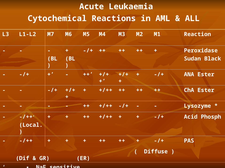
Acute Leukaemia
Cytochemical Reactions in AML & ALL
ReactionM1M2M3M4M5M6M7L1-L2L3
Peroxidase
Sudan Black
+++++++-/++
(BL)
-
(BL)
--
ANA Ester-/+++/+++/++’++’-+’-/+-
ChA Ester+++++++/++++/++-/+--
Lysozyme *---/++/++++----
Acid Phosph-/++++/++++++-/++▪
(Local.)
-
PAS-/+++++++++-/++-
( Diffuse ) (Dif & GR) (ER)
‘ ▪ NaF sensitive * ▪ T-ALL
* ▪ Cytobacterial Test & /Or Serum Level
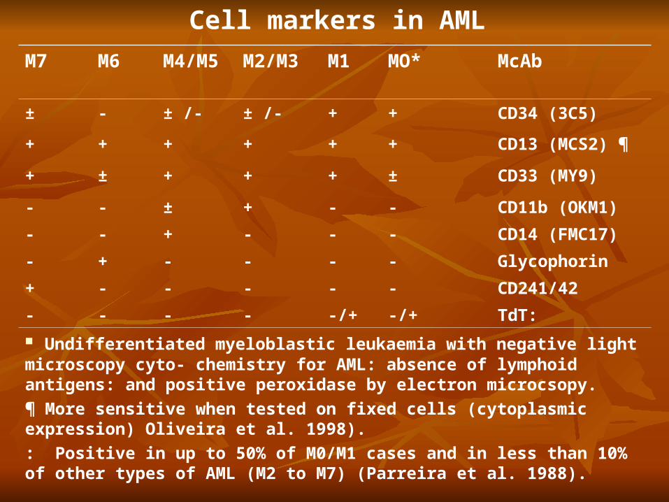
Cell markers in AML
McAbMO*M1M2/M3M4/M5M6M7
CD34 (3C5)++± /-± /--±
CD13 (MCS2) ¶++++++
CD33 (MY9)±+++±+
CD11b (OKM1)--+±--
CD14 (FMC17)---+--
Glycophorin----+-
CD241/42-----+
TdT:-/+-/+----
Undifferentiated myeloblastic leukaemia with negative light microscopy cyto- chemistry for AML: absence of lymphoid antigens: and positive peroxidase by electron microcsopy.
¶ More sensitive when tested on fixed cells (cytoplasmic expression) Oliveira et al. 1998).
: Positive in up to 50% of M0/M1 cases and in less than 10% of other types of AML (M2 to M7) (Parreira et al. 1988).
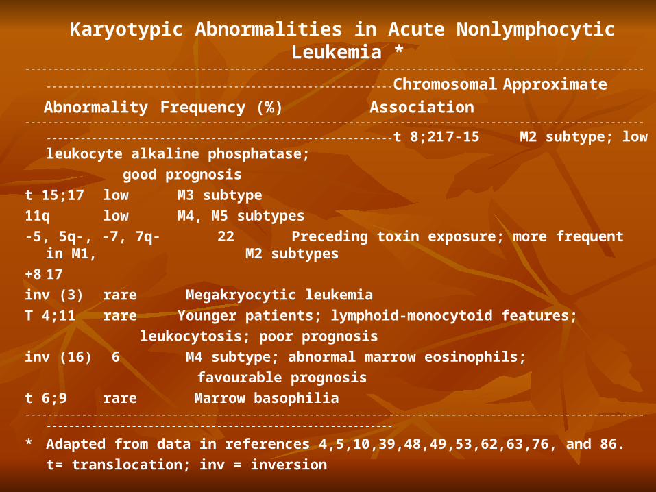
Karyotypic Abnormalities in Acute Nonlymphocytic Leukemia *
--------------------------------------------------------------------------------------------------------------------------------------------------------------------------
Chromosomal Approximate
Abnormality Frequency (%) Association-------------------------------------------------------------------------------------------------------------------------------------------------------------------------- t
8;21 7-15 M2 subtype; low leukocyte alkaline phosphatase;
good prognosis
t 15;17 low M3 subtype
11q low M4, M5 subtypes
-5, 5q-, -7, 7q- 22 Preceding toxin exposure; more frequent in M1, M2 subtypes
+8 17
inv (3) rare Megakryocytic leukemia
T 4;11 rare Younger patients; lymphoid-monocytoid features;
leukocytosis; poor prognosis
inv (16) 6 M4 subtype; abnormal marrow eosinophils;
favourable prognosis
t 6;9 rare Marrow basophilia--------------------------------------------------------------------------------------------------------------------------------------------------------------------------
* Adapted from data in references 4,5,10,39,48,49,53,62,63,76, and 86.
t= translocation; inv = inversion

Features of hypergranular promyelocytic leukaemia
------------------------------------------------------------ -
* Incidence : 7% of all AML.
* Age : predominantly < 40 years
* Characteristic morphology (M3 of the FAB classification)
* Pancytopenia common.
* High incidence of D.I.C.
* High levels of serum vitamin B12 binding protein (Transcobalamin I)
* Unique chromosome abnormality: t(15q+; 17q-) all ≈ cases
* Prolonged survival : 16% > 6 years

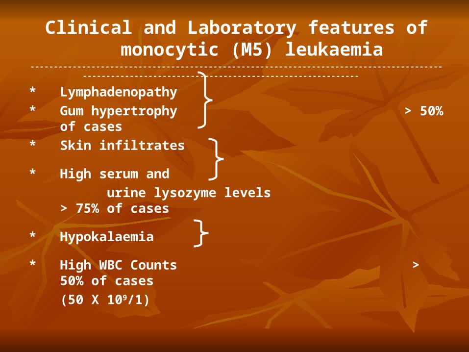
Clinical and laboratory features of monocytic (M5) leukaemia
----------------------------------------------------- * Lymphadenopathy
* Gum hypertrophy > 50% of cases
* Skin infiltrates
* High serum and > 75% of cases
urine lysozyme levels
* Hypokalaemia
* High WBC counts > 50% of cases
(≥ 50 X 109/1)
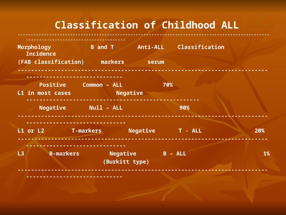
Classification of Childhood ALL--------------------------------------------------------------------------------------------------------------------------------------
Morphology B and T Anti-ALL Classification Incidence
(FAB classification) markers serum
--------------------------------------------------------------------------------------------------------
Positive Common – ALL 70%
L1 in most cases Negative ----------------------------------------------------
Negative Null – ALL 90%
---------------------------------------------------------------------------------------------------------
L1 or L2 T-markers Negative T - ALL 20%
---------------------------------------------------------------------------------------------------------
L3 B-markers Negative B – ALL 1%
(Burkitt type)
--------------------------------------------------------------------------------------------------------

Classification of Adult ALL
Morphology (FAB
classification)
B and T markers
Anti-ALL serum
Ph1
ChromosomeClassificationIncidence
L2 in most casesNegative
PositiveNegativeCommon
ALL30%
PositivePh1(+)-ALL20%
NegativeNegativeNull-ALL30%
L1 or L2T-markersNegativeNegative T-ALL8%
L2 or L3B-markersNegativeNegativeLeukaemic phase of B-
cell lymphomas
12%

-----------------------------------------------------------------------------------------------------------------------------------------
Clinical and Haematological features of T-ALL-----------------------------------------------------------------------------------------------------------------------------------------
1. Blast cell count ≥ 100 X 109/1 50% of cases
2. Anterior mediastinal mass
3. Median age : 6.5 years
4. Male : female ratio 2.5 : 1
5. Relatively less anaemia and thrombocytopenia than in common-ALL
6. Short – lived complete remissions
7. High incidence of meningeal relapse
-----------------------------------------------------------------------------------------------------------------------------------------
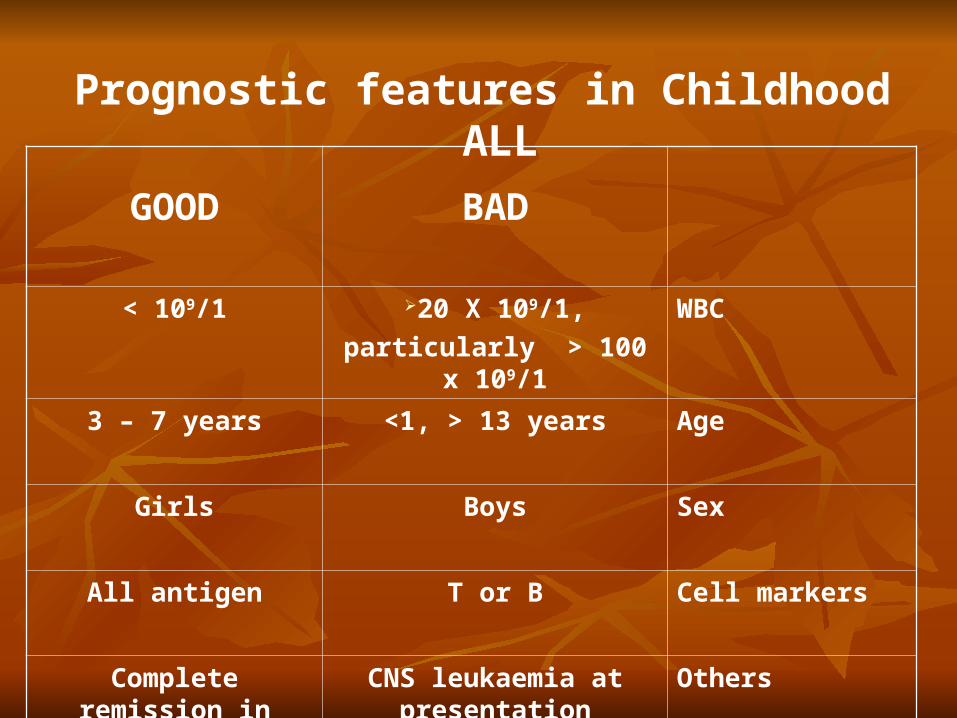
Prognostic features in Childhood ALL
BADGOOD
WBC20 X 109/1,
particularly > 100 x 109/1
< 109/1
Age <1, > 13 years3 – 7 years
SexBoysGirls
Cell markersT or BAll antigen
OthersCNS leukaemia at presentation
Complete remission in first 3 – 4 weeks
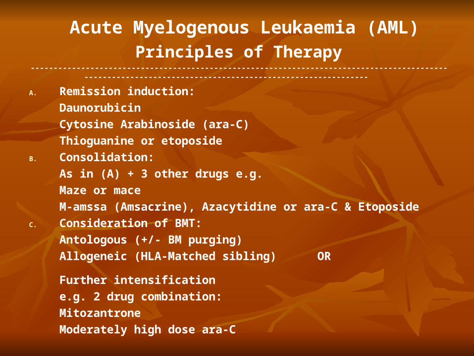
Acute Myelogenous Leukaemia (AML)
Principles of Therapy ---------------------------------------------------------------------------------------------------------------------------------------------------------
A. Remission induction:
Daunorubicin
Cytosine Arabinoside (ara-C)
Thioguanine or etoposide
B. Consolidation:
As in (A) + 3 other drugs e.g.
Maze or mace
M-amssa (Amsacrine), Azacytidine or ara-C & Etoposide
C. Consideration of BMT:
Antologous (+/- BM purging)
Allogeneic (HLA-Matched sibling) OR
Further intensification
e.g. 2 drug combination:
Mitozantrone
Moderately high dose ara-C

Acute Lymphoblastic Leukaemia (ALL)
Principles of Therapy-------------------------------------------------------------------------------------------------------------------------------------------------------------
1. Remission induction ( 4 weeks)
2. Consolidation
(5 days)
3. Consideration of BMT
(High Risk Cases)
Vincristine
Prednisolone
Daunorubicin
L- Asparaginase
Vincristine, Daunorubicin
Prednisolone, ara-C
Etoposide, Thioguanine
Allogenic Or Autologous

Acute Lymphoblastic Leukaemia (ALL)Principles of Therapy (Continued)
-------------------------------------------------------------------------------------------------------------------------------------------------------------
4. CNS Prophylaxix
5. Late Intensification
6. Maintenance Therapy
(2 years)
Cranial Irradiation
Intrathecal Methotrexate
(6 injections)
Cyclophosphamide
Methotrexate Intermediate
Or High dose (etc.)
Methotrexate
6-Mercaptopurine
Vincristine
Predinisolone
Clinical and Laboratory features of monocytic (M5)
leukaemia---------------------------------------------------------------------------------------------------------------------------------------------------
* Lymphadenopathy
* Gum hypertrophy > 50% of cases
* Skin infiltrates
* High serum and
urine lysozyme levels > 75% of cases
* Hypokalaemia
* High WBC Counts > 50% of cases
(50 X 109/1)