Acceleration of Bone Formation and Adhesion Ability on ...
13
metals Article Acceleration of Bone Formation and Adhesion Ability on Dental Implant Surface via Plasma Electrolytic Oxidation in a Solution Containing Bone Ions Mosab Kaseem 1 and Han-Cheol Choe 2, * Citation: Kaseem, M.; Choe, H.-C. Acceleration of Bone Formation and Adhesion Ability on Dental Implant Surface via Plasma Electrolytic Oxidation in a Solution Containing Bone Ions. Metals 2021, 11, 106. https://doi.org/10.3390/met11010106 Received: 12 November 2020 Accepted: 24 November 2020 Published: 7 January 2021 Publisher’s Note: MDPI stays neu- tral with regard to jurisdictional clai- ms in published maps and institutio- nal affiliations. Copyright: © 2021 by the authors. Li- censee MDPI, Basel, Switzerland. This article is an open access article distributed under the terms and con- ditions of the Creative Commons At- tribution (CC BY) license (https:// creativecommons.org/licenses/by/ 4.0/). 1 Department of Nanotechnology and Advanced Materials Engineering, Sejong University, Seoul 05006, Korea; [email protected] 2 Advanced Functional Surface & Biomaterials Research Lab, Department of Dental Materials and Research Center of Nano-Interface Activation for Biomaterials, College of Dentistry, Chosun University, Gwangju 61452, Korea * Correspondence: [email protected] Abstract: The present study examined the in vitro and in vivo bone formation and adhesion ability on the surface of a titanium dental implant made by plasma electrolytic oxidation (PEO) in electrolytes containing bioactive ions. To achieve this goal, screw-shaped fabricated Ti-6Al-4V alloy implants were processed via PEO using an electrolyte solution containing calcium (Ca), phosphorous (P), magnesium (Mg), zinc (Zn), strontium (Sr), silicon (Si), and manganese (Mn) species. The screw implants doped with bioactive elements via PEO were placed in rabbit tibia, and the results were compared to the sand-blasted Ti-6Al-4V alloy implants. At eight-week post-surgery, there was no significant difference in the values of removal torque between sand-blasted and PEO-treated implants. However, it was observed that the PEO treatment of dental implants led to the formation of more periphery bone as compared to the case of sand-blasted implants. Accordingly, the PEO-treated implants have the potential to be used as promising materials for dental applications. Keywords: dental implant; porous TiO 2 ; bone adhesion; rabbit tibia; removal torque 1. Introduction Titanium-based materials have been considered as promising metallic implants owing to their high strength/density ratios and low corrosion rate, favorably meeting the majority of the fundamental aspects in the development of metallic biomaterials [1–3]. However, an implant with a higher elastic modulus as compared to the bone might lead to stress or the shielding effect, allowing osteoporosis or poor osseointegration. In contrast, the implant with a low elastic modulus share would load with the bone to promote bone growth [4,5]. In the oral and maxillofacial area, therefore, efforts have been made for aesthetic, functional, and mental recovery by restoring lost teeth from restoration of single teeth to reconstruction of the maxillary defects after maxillofacial trauma or tumor surgery. Bone adhesion is necessary for the safety and long-term success of the implants, and the degree of new bone formation around the implant is considered to be very important in the prognosis. Therefore, several surface modification approaches, such as acid etching, sand blasting, plasma spray coating and anodizing have been largely studied [6–10]. In particular, plasma electrolytic oxidation (PEO) has been suggested by many research groups to overcome the aforementioned drawbacks [11]. Porous titanium oxide (TiO 2 ) coating made on Ti and its alloys via PEO in aqueous electrolytes has been reported to have acceptable mechanical properties and osteogenic ability [12,13]. Besides, the osteointegration of Ti-based materials can be significantly enhanced through the decoration of their surfaces with nanoscale coatings [14]. Considering the composition and crystal structure that is similar to the human bone, nanoscale hydroxyapatite (HA) was considered a promising material in biomedical appli- Metals 2021, 11, 106. https://doi.org/10.3390/met11010106 https://www.mdpi.com/journal/metals
Transcript of Acceleration of Bone Formation and Adhesion Ability on ...

metals
Article
Acceleration of Bone Formation and Adhesion Ability onDental Implant Surface via Plasma Electrolytic Oxidation in aSolution Containing Bone Ions
Mosab Kaseem 1 and Han-Cheol Choe 2,*
�����������������
Citation: Kaseem, M.; Choe, H.-C.
Acceleration of Bone Formation and
Adhesion Ability on Dental Implant
Surface via Plasma Electrolytic
Oxidation in a Solution Containing
Bone Ions. Metals 2021, 11, 106.
https://doi.org/10.3390/met11010106
Received: 12 November 2020
Accepted: 24 November 2020
Published: 7 January 2021
Publisher’s Note: MDPI stays neu-
tral with regard to jurisdictional clai-
ms in published maps and institutio-
nal affiliations.
Copyright: © 2021 by the authors. Li-
censee MDPI, Basel, Switzerland.
This article is an open access article
distributed under the terms and con-
ditions of the Creative Commons At-
tribution (CC BY) license (https://
creativecommons.org/licenses/by/
4.0/).
1 Department of Nanotechnology and Advanced Materials Engineering, Sejong University, Seoul 05006, Korea;[email protected]
2 Advanced Functional Surface & Biomaterials Research Lab, Department of Dental Materials and ResearchCenter of Nano-Interface Activation for Biomaterials, College of Dentistry, Chosun University,Gwangju 61452, Korea
* Correspondence: [email protected]
Abstract: The present study examined the in vitro and in vivo bone formation and adhesion abilityon the surface of a titanium dental implant made by plasma electrolytic oxidation (PEO) in electrolytescontaining bioactive ions. To achieve this goal, screw-shaped fabricated Ti-6Al-4V alloy implantswere processed via PEO using an electrolyte solution containing calcium (Ca), phosphorous (P),magnesium (Mg), zinc (Zn), strontium (Sr), silicon (Si), and manganese (Mn) species. The screwimplants doped with bioactive elements via PEO were placed in rabbit tibia, and the results werecompared to the sand-blasted Ti-6Al-4V alloy implants. At eight-week post-surgery, there was nosignificant difference in the values of removal torque between sand-blasted and PEO-treated implants.However, it was observed that the PEO treatment of dental implants led to the formation of moreperiphery bone as compared to the case of sand-blasted implants. Accordingly, the PEO-treatedimplants have the potential to be used as promising materials for dental applications.
Keywords: dental implant; porous TiO2; bone adhesion; rabbit tibia; removal torque
1. Introduction
Titanium-based materials have been considered as promising metallic implants owingto their high strength/density ratios and low corrosion rate, favorably meeting the majorityof the fundamental aspects in the development of metallic biomaterials [1–3]. However, animplant with a higher elastic modulus as compared to the bone might lead to stress or theshielding effect, allowing osteoporosis or poor osseointegration. In contrast, the implantwith a low elastic modulus share would load with the bone to promote bone growth [4,5].
In the oral and maxillofacial area, therefore, efforts have been made for aesthetic,functional, and mental recovery by restoring lost teeth from restoration of single teeth toreconstruction of the maxillary defects after maxillofacial trauma or tumor surgery. Boneadhesion is necessary for the safety and long-term success of the implants, and the degree ofnew bone formation around the implant is considered to be very important in the prognosis.Therefore, several surface modification approaches, such as acid etching, sand blasting,plasma spray coating and anodizing have been largely studied [6–10]. In particular, plasmaelectrolytic oxidation (PEO) has been suggested by many research groups to overcome theaforementioned drawbacks [11]. Porous titanium oxide (TiO2) coating made on Ti and itsalloys via PEO in aqueous electrolytes has been reported to have acceptable mechanicalproperties and osteogenic ability [12,13]. Besides, the osteointegration of Ti-based materialscan be significantly enhanced through the decoration of their surfaces with nanoscalecoatings [14].
Considering the composition and crystal structure that is similar to the human bone,nanoscale hydroxyapatite (HA) was considered a promising material in biomedical appli-
Metals 2021, 11, 106. https://doi.org/10.3390/met11010106 https://www.mdpi.com/journal/metals

Metals 2021, 11, 106 2 of 13
cations due to its good ability to bond with bone tissue in vivo as well as stable chemicalproperties [15,16]. It was also claimed that HA can promote angiogenesis, which assiststhe osseointegration [17]. Although Ca and P elements, the major constitutes of HA, canbe easily incorporated and distributed homogeneously throughout the thickness of PEOcoating, their impacts on the surface morphology of the oxide layer were insignificant [18].However, the presence of a sufficient amount of those elements on the outmost surfacewould be necessary to induce the formation of nanoscale HA [19].
To promote the osteointegration, therefore, considerable interest has been given toform the endogenous nanoscale HA on the surface of the TiO2 made via PEO along withthe inclusion of bioactive elements into such oxide layers. Although the individual ordual incorporation of various combination of zinc (Zn), magnesium (Mg), silicon (Si),strontium (Sr), and manganese (Mn) elements into the TiO2 coating via wet and dry plasmatreatment helped to improve the bone formability in vitro [20–24], the impacts of Zn, Mg,Si, Sr, and Mn species on the in vivo osteointegration of dental implants have been littleinvestigated. In our recent study [24], the effects of the addition of Zn, Mg, Sr, Si, and Mnions into the solution were compared with the case when the electrolyte only contains Caand P ions. The in vitro examination assessment revealed that the PEO treatment in anelectrolyte containing Ca, P, Zn, Mg, Sr, Si, and Mn was an effective procedure to inducethe proliferation of osteoblasts on the surface of Ti alloy samples when compared to thecase of PEO treatment in solution only containing Ca and P ions [24]. Therefore, thedeveloping Zn-, Mg-, Sr-, Si-, and Mn-doped TiO2 coatings would be a promising strategyto improve the implant’s osseointegration due to the combined effects originating fromthe porous structure of PEO coatings and the roles of those elements in the enhancementof the osteointegration. Well-known bioactive elements are the elements that facilitatebiomineralization, which helps to reduce a typical adverse host response and facilitate theformation of direct bonding with the bone [11]. As demonstrated in [24], Ca, P, Mg, Zn,Sr, Si, and Mn are found to be bioactive elements. Therefore, the aim of this work was toimprove the osseointegration activity of Ti-6Al-4V alloy through the incorporation of Ca,P, Mg, Zn, Sr, Si, and Mn into the porous TiO2 coating made on the surface of Ti-6Al-4Valloy via PEO treatment. Accordingly, Ca-, P-, Zn-, Mg-, Sr-, Si-, and Mn-incorporated TiO2was fabricated by PEO treatment utilizing an electrolyte with ions of those elements. Sincethe sand-blasted implants are usually used in dental clinical application, we comparedthe osseointegration activity of sand-blasted implants with the implants treated via PEO.The removal torque and surface analysis of sand-blasted Ti-6Al-4V alloy and bioactiveion-doped Ti-6Al-4V surface implants in rabbit tibia were compared. The results provide asimple procedure to improve the bone adhesion at the surface of dental implants, increasingthe contact of the bone and implant and improving clinical success.
2. Experimental2.1. Fabrication and Modification of Ti Implants
Screw-shaped Ti-6Al-4V implants (2.9 mm in diameter and 7 mm in length) werefabricated and used as substrates in the present study. The surface of Ti-6Al-4V implantswas either sand blasted or treated via PEO. For sand-blasting treatment, the implantswere sand blasted with 250 µm alumina particles and passivated with HF/HNO3 acid. Toincorporate bone ions into implant material via PEO, implants made of Ti-6Al-4V alloywere treated via PEO in an electrolyte solution consisting of Ca, P, Zn, Mg, Sr, and Mnspecies. To prepare 7 ions solution, calcium acetate (0.12 M), calcium glycerophosphate(0.019 M), zinc acetate (0.0075 M), magnesium chloride (0.0075 M), manganese acetate(0.0075 M), and sodium metasilicate (0.001 M) were mixed together in 1000 mL of distilledwater. The applied voltage was fixed to be 280 V for 3 min during the PEO process, whichwas conducted under direct current mode. Following PEO treatment, all coated sampleswere rinsed with distilled water and dried in hot air. Here, Ti64/SA describes the Ti-6Al-4Vimplants subjected to sand blasting, while Ti64/7 ions labels the Ti-6Al-4V implants treatedvia PEO in a solution containing ions of 7 elements.
Metals 2021, 11, 106 3 of 13
2.2. Surface Characterization
The surface and cross-sectional morphologies of the samples were analyzed by scan-ning electron microscopy (SEM-HITACHI, S4800, Hitachi, Tokyo, Japan), with equippedenergy-dispersive X-ray spectroscopy (EDS) for identifying chemical compositions of coat-ings. The measurements of the pore size and porosity were performed by SEM observationstaken from at least ten different areas for each condition with the aid of image analyzersoftware (version 1.47 for Windows, 64 bit, free software, National Institutes of Health,Bethesda, MD, USA). Atomic force microscopy (AFM, ToscaTM Analysis, Anton Paar, Graz,Austria) was used to determine the surface roughness of the samples. The contact anglesof the cleaned samples (sand-blasted and PEO-coated) were determined by a water contactangle goniometer (Kruss DSA100, Hamburg, Germany) with one drop (5 µL) of distilledwater. It is worth mentioning that the sand-blasted and PEO-coated samples were notstored and aged before the contact angle measurements. The surface energy (solid-liquid)(γs) of the samples was calculated using Equation (1) [23]:
cosθ = 2(γs
γL)
0.5exp[−β
(γL − γs)
2]− 1 (1)
where β = 0.0001247 (m2/mJ)2, γL is the surface energy of water (72.8 mJ/m2), and θ is theaverage of the five measurements of contact angle.
2.3. Cell Culture
The MC3T3-E1 mouse osteoblasts (ATCC, USA) were used for the in vitro experimentsto characterize the cells attachment on the surface of the tested implants. The cells wereincubated in an atmosphere with a temperature of 37 ◦C and 95% O2 and 5% CO2, whileminimum essential medium (MEM, WELGENE, Gyeongsan, Korea) was used as a culturemedium. The sub-culture medium was altered every three days. The cultured cells wereplaced in phosphate-buffered saline (PBS) and then cultured in trypsin–ethylenediaminetetraacetic acid (EDTA) solution (0.05% trypsin, 0.53 mM EDTA, phenol red in Hank’sbalanced salt solution) at 37 ◦C for 10 min to separate the cells. Cells were seeded on a Tialloy at a concentration of 9.2 × 10⁶ cells/well on a 24-well plate and grown on the coatedsurface for 24 h. The samples were washed with PBS and fixed with 10% formaldehyde at4 ◦C for 12 h. After fixing, the specimens were dehydrated with ethanol. The morphologyof the attached cells was observed by using FE-SEM.
2.4. In Vivo Experiments2.4.1. Surgical Procedures
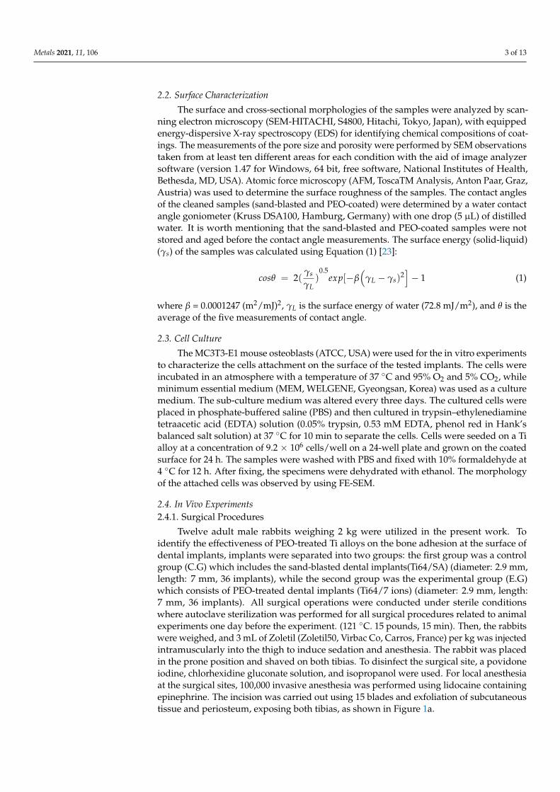
Twelve adult male rabbits weighing 2 kg were utilized in the present work. Toidentify the effectiveness of PEO-treated Ti alloys on the bone adhesion at the surface ofdental implants, implants were separated into two groups: the first group was a controlgroup (C.G) which includes the sand-blasted dental implants(Ti64/SA) (diameter: 2.9 mm,length: 7 mm, 36 implants), while the second group was the experimental group (E.G)which consists of PEO-treated dental implants (Ti64/7 ions) (diameter: 2.9 mm, length:7 mm, 36 implants). All surgical operations were conducted under sterile conditionswhere autoclave sterilization was performed for all surgical procedures related to animalexperiments one day before the experiment. (121 ◦C. 15 pounds, 15 min). Then, the rabbitswere weighed, and 3 mL of Zoletil (Zoletil50, Virbac Co, Carros, France) per kg was injectedintramuscularly into the thigh to induce sedation and anesthesia. The rabbit was placedin the prone position and shaved on both tibias. To disinfect the surgical site, a povidoneiodine, chlorhexidine gluconate solution, and isopropanol were used. For local anesthesiaat the surgical sites, 100,000 invasive anesthesia was performed using lidocaine containingepinephrine. The incision was carried out using 15 blades and exfoliation of subcutaneoustissue and periosteum, exposing both tibias, as shown in Figure 1a.
Metals 2021, 11, 106 4 of 13
Metals 2021, 11, x FOR PEER REVIEW 4 of 14
in the prone position and shaved on both tibias. To disinfect the surgical site, a povidone iodine, chlorhexidine gluconate solution, and isopropanol were used. For local anesthesia at the surgical sites, 100,000 invasive anesthesia was performed using lidocaine containing epinephrine. The incision was carried out using 15 blades and exfoliation of subcutaneous tissue and periosteum, exposing both tibias, as shown in Figure 1a.
Figure 1. Surgical procedures showing (a) incision and peeling; (b) preparation of implantation sites using a guide drill and twist drill; (c) implant replacement where Ti64/SA implants were placed on the right side, while Ti64/7 ion implants were placed in the left side; and (d) the scarified sample taken after 8 weeks of the experiments.
When the tibia was exposed, bone formation for implant placement under douche using a guide drill and twist drill was performed (Figure 1b). When the bone formation was completed, the control group (Ti64/SA implants) was inserted on the right side, the PEO-treated experimental group (Ti64/7 ion implants) was inserted on the left side, and three implants were implanted using an implant kit (Figure 1c). Periosteal sutures were performed using 2-0 vicryl, muscle sutures were used with 3-0 vicryl, and subcutaneous and skin closures were performed with 5-0 vicryl and 6-0 nylon. Before surgery, the site was sterilized using povidone-iodine, chlorhexidine gluconate solution, and isopropanol, and injected with first-generation cephalosporin antibiotics and Keromin. The rabbit′s general condition, wound healing, and infection at the surgical site were checked daily and disinfected. After 8 weeks, the rabbits were sacrificed using a KCl solution, and bone fragments were collected using fracture diagrams with a margin of 7 mm or more around the implant, as shown in Figure 1d.
2.4.2. Histological and Histomorphological Analysis After the tissue was fixed with plastic, the tissue was made using a grinder and sub-
jected to Haemotoxylin and Eosin staining. Afterward, eighteen specimens were prepared for each of the control (Ti64/SA) and experimental group (Ti64/7 ions) implants. As a re-sult of histological observation, the amount of new bone formation around the implant surface of the experimental group was increased compared to the control group. The BIC (bone-to-implant contact ratio), which is the ratio of the contact surface between the im-plant fixture and the bone, was measured. Four different measurements utilizing the Im-age J (1.52a, National Institutes of Health, Bethesda, MD, USA ) program for each implant type were carried to calculate the bone-to-implant contact area ratio (BIC%) as well as new bone formation area ratio.
2.4.3. Removal Torque Value
Figure 1. Surgical procedures showing (a) incision and peeling; (b) preparation of implantation sitesusing a guide drill and twist drill; (c) implant replacement where Ti64/SA implants were placed onthe right side, while Ti64/7 ion implants were placed in the left side; and (d) the scarified sampletaken after 8 weeks of the experiments.
When the tibia was exposed, bone formation for implant placement under doucheusing a guide drill and twist drill was performed (Figure 1b). When the bone formationwas completed, the control group (Ti64/SA implants) was inserted on the right side, thePEO-treated experimental group (Ti64/7 ion implants) was inserted on the left side, andthree implants were implanted using an implant kit (Figure 1c). Periosteal sutures wereperformed using 2-0 vicryl, muscle sutures were used with 3-0 vicryl, and subcutaneousand skin closures were performed with 5-0 vicryl and 6-0 nylon. Before surgery, the sitewas sterilized using povidone-iodine, chlorhexidine gluconate solution, and isopropanol,and injected with first-generation cephalosporin antibiotics and Keromin. The rabbit’sgeneral condition, wound healing, and infection at the surgical site were checked dailyand disinfected. After 8 weeks, the rabbits were sacrificed using a KCl solution, and bonefragments were collected using fracture diagrams with a margin of 7 mm or more aroundthe implant, as shown in Figure 1d.
2.4.2. Histological and Histomorphological Analysis
After the tissue was fixed with plastic, the tissue was made using a grinder andsubjected to Haemotoxylin and Eosin staining. Afterward, eighteen specimens wereprepared for each of the control (Ti64/SA) and experimental group (Ti64/7 ions) implants.As a result of histological observation, the amount of new bone formation around theimplant surface of the experimental group was increased compared to the control group.The BIC (bone-to-implant contact ratio), which is the ratio of the contact surface betweenthe implant fixture and the bone, was measured. Four different measurements utilizingthe Image J (1.52a, National Institutes of Health, Bethesda, MD, USA) program for eachimplant type were carried to calculate the bone-to-implant contact area ratio (BIC%) aswell as new bone formation area ratio.
2.4.3. Removal Torque Value
A specially designed fixture mount for connecting the experimental implant andthe torque meter was fastened to 35 Ncm and connected to a torque meter (MGT12,ELECTROMATIC Equipment Co. Inc., Oakland, CA, USA). The maximum torsionalremoval value at which the separation of the implant and bone occurred by rotating in thereverse direction of the implant placement was measured and recorded. Three values wereobtained for each rabbit to calculate the mean value.

Metals 2021, 11, 106 5 of 13
2.4.4. Statistical Analysis
Statistical analysis of the data (mean ± SD) was performed using an unpaired Stu-dent’s t-test with Graph Pad Prism4 software Version 5.04 (GraphPad Software, San Diego,CA, USA). Values of P less than 0.05 were considered statistically significant.
3. Results3.1. Surface Characterization In Vitro
Figure 2 shows the SEM images of the Ti64/SA and Ti64/7 ion implants. The Ti64/SAimplants were characterized by a rough surface characterized by irregular cavities uni-formly alternated with peaks and valleys (Figure 2a-2). The surface of Ti64/7 ion implantsexhibited a porous structure with different characteristics in terms of the shape and size ofpores (Figure 2b-2). The mean pore size and porosity in the surface of Ti64/7 ion implantswere measured to be 1.13 ± 0.4 µm and 9.98 ± 1.1%, respectively. The pores are ascribed tothe presence of different ions in the electrolyte during PEO which can affect the gas evolu-tion during PEO coating [25]. The presence of small particles on the Ti64/7 ion implantssuggested the inclusion of electrolyte species, namely, Ca, P, Zn, Mg, Mn, Sr, and Si into theoxide layer during PEO. As displayed in Figure 3, the Ti64/7 ion implants exhibited lowerroughness value with an average value of mean surface roughness (Ra = 1.43 ± 0.40 µm)as compared to the Ti64/SA implant, which showed a rougher surface with a value of(Ra = 1.61 ± 0.60 µm). The thickness of the oxide layer formed on the Ti64/7 ion implantwas found to be ~3 µm, as revealed by the cross-section image shown in Figure 4a.
Metals 2021, 11, x FOR PEER REVIEW 5 of 14
A specially designed fixture mount for connecting the experimental implant and the torque meter was fastened to 35 Ncm and connected to a torque meter (MGT12, ELEC-TROMATIC Equipment Co. Inc., Oakland, CA, USA). The maximum torsional removal value at which the separation of the implant and bone occurred by rotating in the reverse direction of the implant placement was measured and recorded. Three values were ob-tained for each rabbit to calculate the mean value.
2.4.4. Statistical Analysis Statistical analysis of the data (mean ± SD) was performed using an unpaired Stu-
dent′s t-test with Graph Pad Prism4 software Version 5.04 (GraphPad Software, San Di-ego, CA, USA). Values of P less than 0.05 were considered statistically significant.
3. Results 3.1. Surface Characterization In Vitro
Figure 2 shows the SEM images of the Ti64/SA and Ti64/7 ion implants. The Ti64/SA implants were characterized by a rough surface characterized by irregular cavities uni-formly alternated with peaks and valleys (Figure 2a-2). The surface of Ti64/7 ion implants exhibited a porous structure with different characteristics in terms of the shape and size of pores (Figure 2b-2). The mean pore size and porosity in the surface of Ti64/7 ion im-plants were measured to be 1.13 ± 0.4 μm and 9.98 ± 1.1%, respectively. The pores are ascribed to the presence of different ions in the electrolyte during PEO which can affect the gas evolution during PEO coating [25]. The presence of small particles on the Ti64/7 ion implants suggested the inclusion of electrolyte species, namely, Ca, P, Zn, Mg, Mn, Sr, and Si into the oxide layer during PEO. As displayed in Figure 3, the Ti64/7 ion implants exhibited lower roughness value with an average value of mean surface roughness (Ra = 1.43 ± 0.40 μm) as compared to the Ti64/SA implant, which showed a rougher surface with a value of (Ra = 1.61 ± 0.60 μm). The thickness of the oxide layer formed on the Ti64/7 ion implant was found to be ~3 μm, as revealed by the cross-section image shown in Figure 4a.
Figure 2. The appearance and SEM images of the Ti-6Al-4V alloy implants treated via (a,a-1,a-2) sand blasting and (b,b-1,b-2) plasma electrolytic oxidation (PEO) in a solution containing ions of seven elements.
Figure 2. The appearance and SEM images of the Ti-6Al-4V alloy implants treated via (a,a-1,a-2)sand blasting and (b,b-1,b-2) plasma electrolytic oxidation (PEO) in a solution containing ions ofseven elements.
Metals 2021, 11, x FOR PEER REVIEW 6 of 14
Figure 3. Atomic force microscopy (AFM) results of the implants: (a) Ti64/SA implants and (b) Ti64/7 ion implants.
Figure 4. (a) Cross-sectional image of the Ti64/7 ion implant; (b) EDS area spectrum for the Ti64/7 ion implant. Ca, P, O, Ti, V, Al, Si, Mn, Mg, Sr, and Zn were identified.
According to the EDS area results shown in Figure 4b and Table 1, Ca, P, O, Ti, V, Al, Si, Mn, Mg, Sr, and Zn elements were identified. The Ti, Al, and V originated from sub-strate alloy, while the presence of the other elements was attributed to the electrolyte so-lution. In our previous work, we found that anatase, rutile, and HA were the main phases in seven ion implants [24]. The wettability measurements revealed that the surface of Ti64/7 ion implants had a lower contact angle (85.9 ± 8.5) than Ti64/SA implants (99.8 ± 6.8), suggesting that a significant improvement in the hydrophilicity of the Ti-6Al-4V alloy implants was achieved via PEO. The surface energies for Ti64/SA and Ti64/7 ions surfaces were 3.53 ± 0.9 and 4.57 ± 0.5 mJ/m2, respectively. In this study, the values of contact angle were somewhat in accordance with those obtained in other works. For example, contact angles in the range between ~ 66.8° and ~ 77.4° and the range between ~76.9° and ~ 82.1° were reported by Pegueroles et al. [26] for the pure titanium implants sanded by either SiC or Al2O3 particles, respectively. Raphel and coworkers [27] measured the values of contact angles of Ti-6Al-4V substrates irradiated with UV light in the presence of phos-phate-buffered saline. This UV exposure resulted in a significant increase in surface hy-drophilicity, with the contact angle decreasing from 50.83° to 26.33°, consistent with an increase in surface hydroxyls. On the other hand, lower values of contact angle below 15° were reported by Li et al. [28] for pure titanium treated via PEO in tetraborate electrolytes. As for surface energy, Kaseem and Choe [23] reported recently higher surface energies (~7.44 and ~8.13 mJ/m2) than those obtained in the present work for Ti-6Al-4V alloy treated via PEO in solutions containing Zn and Mg ions. Higher surface energy values in the
Figure 3. Atomic force microscopy (AFM) results of the implants: (a) Ti64/SA implants and (b)Ti64/7 ion implants.

Metals 2021, 11, 106 6 of 13
Metals 2021, 11, x FOR PEER REVIEW 6 of 14
Figure 3. Atomic force microscopy (AFM) results of the implants: (a) Ti64/SA implants and (b) Ti64/7 ion implants.
Figure 4. (a) Cross-sectional image of the Ti64/7 ion implant; (b) EDS area spectrum for the Ti64/7 ion implant. Ca, P, O, Ti, V, Al, Si, Mn, Mg, Sr, and Zn were identified.
According to the EDS area results shown in Figure 4b and Table 1, Ca, P, O, Ti, V, Al, Si, Mn, Mg, Sr, and Zn elements were identified. The Ti, Al, and V originated from sub-strate alloy, while the presence of the other elements was attributed to the electrolyte so-lution. In our previous work, we found that anatase, rutile, and HA were the main phases in seven ion implants [24]. The wettability measurements revealed that the surface of Ti64/7 ion implants had a lower contact angle (85.9 ± 8.5) than Ti64/SA implants (99.8 ± 6.8), suggesting that a significant improvement in the hydrophilicity of the Ti-6Al-4V alloy implants was achieved via PEO. The surface energies for Ti64/SA and Ti64/7 ions surfaces were 3.53 ± 0.9 and 4.57 ± 0.5 mJ/m2, respectively. In this study, the values of contact angle were somewhat in accordance with those obtained in other works. For example, contact angles in the range between ~ 66.8° and ~ 77.4° and the range between ~76.9° and ~ 82.1° were reported by Pegueroles et al. [26] for the pure titanium implants sanded by either SiC or Al2O3 particles, respectively. Raphel and coworkers [27] measured the values of contact angles of Ti-6Al-4V substrates irradiated with UV light in the presence of phos-phate-buffered saline. This UV exposure resulted in a significant increase in surface hy-drophilicity, with the contact angle decreasing from 50.83° to 26.33°, consistent with an increase in surface hydroxyls. On the other hand, lower values of contact angle below 15° were reported by Li et al. [28] for pure titanium treated via PEO in tetraborate electrolytes. As for surface energy, Kaseem and Choe [23] reported recently higher surface energies (~7.44 and ~8.13 mJ/m2) than those obtained in the present work for Ti-6Al-4V alloy treated via PEO in solutions containing Zn and Mg ions. Higher surface energy values in the
Figure 4. (a) Cross-sectional image of the Ti64/7 ion implant; (b) EDS area spectrum for the Ti64/7 ion implant. Ca, P, O, Ti,V, Al, Si, Mn, Mg, Sr, and Zn were identified.
According to the EDS area results shown in Figure 4b and Table 1, Ca, P, O, Ti, V, Al, Si,Mn, Mg, Sr, and Zn elements were identified. The Ti, Al, and V originated from substratealloy, while the presence of the other elements was attributed to the electrolyte solution.In our previous work, we found that anatase, rutile, and HA were the main phases inseven ion implants [24]. The wettability measurements revealed that the surface of Ti64/7ion implants had a lower contact angle (85.9 ± 8.5) than Ti64/SA implants (99.8 ± 6.8),suggesting that a significant improvement in the hydrophilicity of the Ti-6Al-4V alloyimplants was achieved via PEO. The surface energies for Ti64/SA and Ti64/7 ions surfaceswere 3.53 ± 0.9 and 4.57 ± 0.5 mJ/m2, respectively. In this study, the values of contactangle were somewhat in accordance with those obtained in other works. For example,contact angles in the range between ~66.8◦ and ~77.4◦ and the range between ~76.9◦
and ~82.1◦ were reported by Pegueroles et al. [26] for the pure titanium implants sandedby either SiC or Al2O3 particles, respectively. Raphel and coworkers [27] measured thevalues of contact angles of Ti-6Al-4V substrates irradiated with UV light in the presence ofphosphate-buffered saline. This UV exposure resulted in a significant increase in surfacehydrophilicity, with the contact angle decreasing from 50.83◦ to 26.33◦, consistent with anincrease in surface hydroxyls. On the other hand, lower values of contact angle below 15◦
were reported by Li et al. [28] for pure titanium treated via PEO in tetraborate electrolytes.As for surface energy, Kaseem and Choe [23] reported recently higher surface energies(~7.44 and ~8.13 mJ/m2) than those obtained in the present work for Ti-6Al-4V alloy treatedvia PEO in solutions containing Zn and Mg ions. Higher surface energy values in therange between ~65 to ~70 mJ/m2 were reported by Marques and co-workers [29] for puretitanium coating via PEO in electrolytes containing Ca, P, and Si ions. Here, it is belie vedthat the differences in the morphologies and the composition of treated implants wouldexplain the variation in the values of contact angle and surface energy obtained in thepresent work in comparison to those described in the literature [23,26–29].
Figure 5 shows the SEM images of MC3T3-E1 cells cultured on the surface of Ti64/SAand Ti64/7 ion implants. Cells show the spinning form of filopodia (Ti64/SA implants) inlamellipodia (Ti64/7 ion implants) and grow in association with the cells. As can be seenfrom Figure 5b-1, the circular-type lamellipodia are noted near the pores as displayed inFigure 5b. Moreover, cell growth is actively found through interconnection with cells whencompared with the Ti64/SA implants, as shown in Figure 5a.

Metals 2021, 11, 106 7 of 13
Table 1. EDS results of the Ti64/7 ion implant showing the incorporation of Ca, P, Mg, Zn, Si, Sr, andMn into the coating.
Element Weight. % Atomic. %
O K 43.12 67.68Mg K 0.29 0.30Al K 1.92 1.79Si K 0.41 0.35P K 5.53 4.49
Ca K 6.61 4.14Ti K 33.84 17.74V K 1.56 0.77
Mn K 3.53 1.61Zn L 2.03 0.78Sr L 1.16 0.35Total 100.00 100.00
Metals 2021, 11, x FOR PEER REVIEW 7 of 14
range between ~65 to ~70 mJ/m2were reported by Marques and co-workers [29] for pure titanium coating via PEO in electrolytes containing Ca, P, and Si ions. Here, it is belie ved that the differences in the morphologies and the composition of treated implants would explain the variation in the values of contact angle and surface energy obtained in the present work in comparison to those described in the literature [23,26–29].
Table 1. EDS results of the Ti64/7 ion implant showing the incorporation of Ca, P, Mg, Zn, Si, Sr, and Mn into the coating.
Element Weight. % Atomic. % O K 43.12 67.68
Mg K 0.29 0.30 Al K 1.92 1.79 Si K 0.41 0.35 P K 5.53 4.49
Ca K 6.61 4.14 Ti K 33.84 17.74 V K 1.56 0.77
Mn K 3.53 1.61 Zn L 2.03 0.78 Sr L 1.16 0.35 Total 100.00 100.00
Figure 5 shows the SEM images of MC3T3-E1 cells cultured on the surface of Ti64/SA and Ti64/7 ion implants. Cells show the spinning form of filopodia (Ti64/SA implants) in lamellipodia (Ti64/7 ion implants) and grow in association with the cells. As can be seen from Figure 5b-1, the circular-type lamellipodia are noted near the pores as displayed in Figure 5b. Moreover, cell growth is actively found through interconnection with cells when compared with the Ti64/SA implants, as shown in Figure 5a.
Figure 5. SEM images of the osteoblast cells cultured on the surface of Ti-6Al-4V alloy implants: (a,a-1) Ti64/SA implants and (b,b-1) Ti64/7 ion implants.
3.2. Clinical, Histological, and Histomorphologic Findings
Figure 5. SEM images of the osteoblast cells cultured on the surface of Ti-6Al-4V alloy implants:(a,a-1) Ti64/SA implants and (b,b-1) Ti64/7 ion implants.
3.2. Clinical, Histological, and Histomorphologic Findings
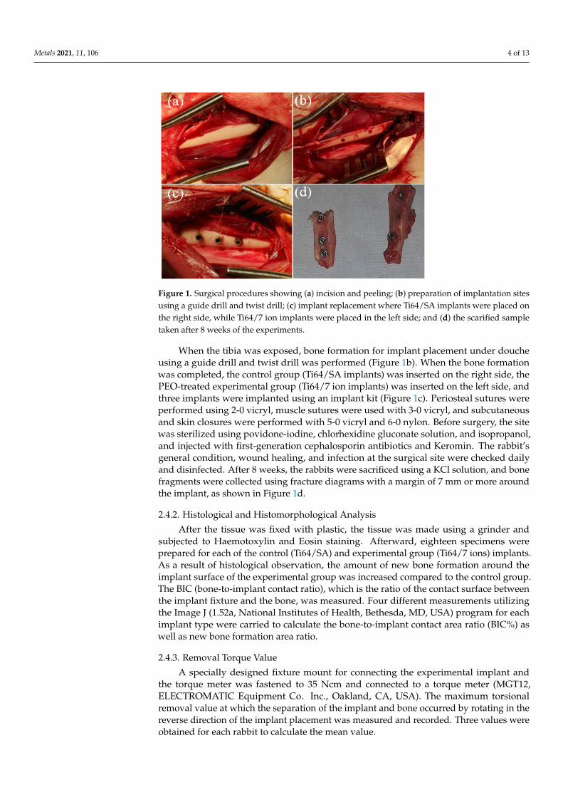
The visual observations were as follows. When the implants were detached and theimplants were observed, the fluctuations of all implant fixtures were hardly observed.The formation of inflammatory and granulation tissues around the implant fixture wasnot observed, and an almost smooth bone surface was confirmed. Figure 6 shows thetissue photograph eight weeks after implantation of the Ti64/SA and Ti64/7 ion implants,respectively. Eight weeks after implantation, there were no signs of inflammation at thebone–implant interface where all implants in both groups were histologically in directcontact with the surrounding cortical bone (red color) and bone marrow (gray color part)along existed implant threads, as shown in Figure 7a,b, after removing the implant frombone. Interestingly, the amount of newly formed bone (blue color) around the surface ofthe experimental group (Ti64/7 ions) was higher than those in those around the experi-mental groups (Ti64/SA), as shown in Figure 7c,d. However, in both groups, the bonefixation of the implant fixture was similarly high. From the results of histomorphometricanalysis listed in Table 2, the ratio of new bone created nearby the implant fixture wasmore than doubled in the experimental group (Ti64/7 ions) in comparison to the control
Metals 2021, 11, 106 8 of 13
group (Ti64/SA implants) (p = 0.029). As reported earlier [30], the bone formation wouldhave originated from the implant surface properties, namely, chemical and physical design,which is referred to as “contact osteogenesis”, as well as from the bony margin of thesurgical defect, i.e., “distance osteogenesis”. Therefore, we thought that the porous struc-ture containing bioactive elements would be responsible for the higher amounts of boneformed in the case of Ti64/7 ion implants. However, there was no considerable differencebetween implant types in the values of BIC% (~69.3 for Ti64/SA implants vs. ~69.0 forTi64/7 ion implants). This result was consistent with those obtained by He et al. [31]and Zhang et al. [32], who found that the incorporation of bioactive elements, such asZn or Mg or Sr, into titanium implants would accelerate the surrounding bone formationand improve the amount of new bone formation in the rabbit model of osteoporosis. Assuch, Fernández-Yagüe et al. [33] found that the bioactive implants produced by plasma orthermochemical processes would exhibit more new bone formation when compared to thenon-bioactive implants treated by shot blasting and acid etching. Furthermore, Aparicioet al. [34] demonstrated that the created microroughness by Al2O3 grit blasting acceleratedin vitro apatite nucleation on alkali- and heat-treated titanium plates.
Metals 2021, 11, x FOR PEER REVIEW 8 of 14
The visual observations were as follows. When the implants were detached and the implants were observed, the fluctuations of all implant fixtures were hardly observed. The formation of inflammatory and granulation tissues around the implant fixture was not observed, and an almost smooth bone surface was confirmed. Figure 6 shows the tissue photograph eight weeks after implantation of the Ti64/SA and Ti64/7 ion implants, respec-tively. Eight weeks after implantation, there were no signs of inflammation at the bone–implant interface where all implants in both groups were histologically in direct contact with the surrounding cortical bone (red color) and bone marrow (gray color part) along existed implant threads, as shown in Figure 7a,b, after removing the implant from bone. Interestingly, the amount of newly formed bone (blue color) around the surface of the experimental group (Ti64/7 ions) was higher than those in those around the experimental groups (Ti64/SA), as shown in Figure 7c,d. However, in both groups, the bone fixation of the implant fixture was similarly high. From the results of histomorphometric analysis listed in Table 2, the ratio of new bone created nearby the implant fixture was more than doubled in the experimental group (Ti64/7 ions) in comparison to the control group (Ti64/SA implants) (p = 0.029). As reported earlier [30], the bone formation would have originated from the implant surface properties, namely, chemical and physical design, which is referred to as “contact osteogenesis”, as well as from the bony margin of the surgical defect, i.e., “distance osteogenesis”. Therefore, we thought that the porous struc-ture containing bioactive elements would be responsible for the higher amounts of bone formed in the case of Ti64/7 ion implants. However, there was no considerable difference between implant types in the values of BIC% (~69.3 for Ti64/SA implants vs. ~69.0 for Ti64/7 ion implants). This result was consistent with those obtained by He et al. [31] and Zhang et al. [32], who found that the incorporation of bioactive elements, such as Zn or Mg or Sr, into titanium implants would accelerate the surrounding bone formation and improve the amount of new bone formation in the rabbit model of osteoporosis. As such, Fernández-Yagüe et al. [33] found that the bioactive implants produced by plasma or ther-mochemical processes would exhibit more new bone formation when compared to the non-bioactive implants treated by shot blasting and acid etching. Furthermore, Aparicio et al. [34] demonstrated that the created microroughness by Al2O3 grit blasting accelerated in vitro apatite nucleation on alkali- and heat-treated titanium plates.
Figure 6. (a) Ti64/SA implants after 8 weeks of implantation, and (b) Ti64/7 ion implants after 8 weeks of implantation. As shown in (a,b), the implant fixture is well connected with the surround-ing bone.
Figure 6. (a) Ti64/SA implants after 8 weeks of implantation, and (b) Ti64/7 ion implants after 8weeks of implantation. As shown in (a,b), the implant fixture is well connected with the surroundingbone.
3.3. Removal Torque Analysis
The removal torque values measured in each animal group on Ti64/SA and Ti64/7 ionsurface-treated implants are summarized in Table 3. As reported earlier [3], the interfacialshear stress can be reflected by the removal torque value strength. The degree of biome-chanical anchorage determined by removal torque testing indicates the strength of implantintegration in bone tissue [35]. As shown in Table 3, the average removal torque followingeight weeks of implant placement was 49.7 Ncm for the control implants and 51.5 Ncmfor the experimental implants. The values of removal torque reported by the present workwere higher than those obtained in other studies. For instance, Cho and Park [36] measuredthe removal torque of dual acid-etched titanium screw implants inserted in rabbit tibiaand reported that the average removal torque value was 38.7 Ncm. Cordioli et al. [37]reported that the average removal torque value was 25.28 Ncm for the machined implants,26.85 Ncm for the grit-blasted implants, 29.57 Ncm for the plasma-sprayed implants, and40.85 Ncm for the acid-etched implants under similar conditions. Thus, the greater torquerotation forces required to remove the implants in the present study would suggest higherstrengths of osseointegration.

Metals 2021, 11, 106 9 of 13Metals 2021, 11, x FOR PEER REVIEW 9 of 14
Figure 7. Histological images of tissue 8 weeks after implantation showing no signs of inflamma-tion at the bone–implant interface where (a) Ti64/SA implants and (b) Ti64/7 ion implants are used. These images were observed after removing the implant from rabbits. By observing the tis-sue images of Ti64/SA and Ti64/7 ion implants, it can be seen that the dense bone in (b) is well-formed. The black part is judged as marrow, and the red represents bone. (c,d) represent the amount of newly formed bone around the surfaces of Ti64/SA and Ti64/7 ion implants, respec-tively. From high-magnification images shown as insets, the Ti64/7 ion implants were found to exhibit greater amounts of newly formed bone from original cortical bone compared with the Ti64/SA implants.
Table 2. Average bone-to-implant contact area (BIC) ratio and new bone formation rate measured in Ti64/SA and Ti64/7 ion implants.
Implants Bone-to-Implant Contact
Ratio (%) New Bone Formation Ratio
(%) Ti64/SA 69.3 ± 6.9 28.4 ± 3.2
Ti64/7 ions 69.0 ± 7.1 58.4 ± 4.7
3.3. Removal Torque Analysis The removal torque values measured in each animal group on Ti64/SA and Ti64/7
ion surface-treated implants are summarized in Table 3. As reported earlier [3], the inter-facial shear stress can be reflected by the removal torque value strength. The degree of biomechanical anchorage determined by removal torque testing indicates the strength of implant integration in bone tissue [35]. As shown in Table 3, the average removal torque following eight weeks of implant placement was 49.7 Ncm for the control implants and 51.5 Ncm for the experimental implants. The values of removal torque reported by the present work were higher than those obtained in other studies. For instance, Cho and Park [36] measured the removal torque of dual acid-etched titanium screw implants inserted in rabbit tibia and reported that the average removal torque value was 38.7 Ncm. Cordioli et al. [37] reported that the average removal torque value was 25.28 Ncm for the machined implants, 26.85 Ncm for the grit-blasted implants, 29.57 Ncm for the plasma-sprayed im-plants, and 40.85 Ncm for the acid-etched implants under similar conditions. Thus, the
Figure 7. Histological images of tissue 8 weeks after implantation showing no signs of inflammationat the bone–implant interface where (a) Ti64/SA implants and (b) Ti64/7 ion implants are used. Theseimages were observed after removing the implant from rabbits. By observing the tissue images ofTi64/SA and Ti64/7 ion implants, it can be seen that the dense bone in (b) is well-formed. The blackpart is judged as marrow, and the red represents bone. (c,d) represent the amount of newly formedbone around the surfaces of Ti64/SA and Ti64/7 ion implants, respectively. From high-magnificationimages shown as insets, the Ti64/7 ion implants were found to exhibit greater amounts of newlyformed bone from original cortical bone compared with the Ti64/SA implants.
Table 2. Average bone-to-implant contact area (BIC) ratio and new bone formation rate measured inTi64/SA and Ti64/7 ion implants.
Implants Bone-to-Implant Contact Ratio (%) New Bone Formation Ratio (%)
Ti64/SA 69.3 ± 6.9 28.4 ± 3.2
Ti64/7 ions 69.0 ± 7.1 58.4 ± 4.7
Table 3. Removal torque value measured in each animal group on Ti64/SA and Ti64/7 ion implants.
Rabbits 1 2 3 4 5 6
Ti64/SA 49.7 ± 14.5 50.4 ± 17.5 51.5 ± 16.7 46.3 ± 18.5 50.6 ± 14.2 49.7 ± 15.7
Ti64/7ions 50.0 ± 18.5 52.5 ± 18.3 51.3 ± 15.4 49.5 ± 19.6 52.3 ± 19.5 53.4 ± 13.9
4. Discussion
The implant material is usually characterized as bio-tolerant (gold, cobalt-chromium,stainless steel, poly(methyl methacrylate), bio-inert (Ti), and bio-active (Ti-active surface)material. For bio-tolerant material, the bone formation is only on the bone surface andwe observe only distant osteogenesis, while in other cases, it is found close to implantsurface for bio-inert materials. The phenomenon of both distant osteogenesis and contactosteogenesis will be observed for bio-inert and bio-active material, but the formation rateis considered higher in the case of bioactive material. Bioactive surfaces are active because

Metals 2021, 11, 106 10 of 13
of the presence of bio-bonding materials, such as calcium crystals, phosphate crystals, andhydroxyl appetite crystals. These crystals will act as nucleation sites to the newly formedbone cell and it will be bonded with the calcium ions of the implant interface. For thisreason, we see a higher response of bone cell formation on the surface of the experimentalgroup (Ti64/7 ions), as can be seen in Figure 7. The results of the present work revealed thatTi64/7 ion implants exhibited enhanced osteoblast differentiation as compared to Ti64/SAimplants. In Figure 8, we simply explained the assumed mechanism for bone developmentin both cases of C.G (Ti64/SA), and E.G (Ti64/7 ions). During the initial few minutes afterthe implant is successfully checked for its primary stability, the blood irrigates the areaaround the implant. The white blood cells within the blood provide necessary resourcesfor healing. Ions, proteins, and platelets start to adhere to the implant surface. Shortly, thebleeding stops due to the action of the platelets which fills the wounds by clotting andcreating blood vessels and a fibrin-based provisional matrix that adheres to the implant.Days after surgery, fibroblasts migrate into the wound, and components such as collagenare synthesized which stabilize and protect the extracellular matrix of perivascular cells,which have characteristics of stem cells. These cells also migrate to wounded areas nearimplant surfaces forming new blood vessels that will restore oxygen supply and therebypromote tissue healing. After the first week, osteoclasts and osteoblasts adhere to theresidual bone, resorbing it and creating space for the formation of new bone. Perivascularcells of progenitive nature also migrate to the implant surface where they will form newosteoblasts; these osteoblasts will form a mineralized matrix by incorporating calciumand phosphate. This collagen-free calcified matrix with proteoglycans makes the wovenbone. However, this woven bone has very limited mechanical stability, but it permeatesthe growth of lamellar bone and remodeling and gives secondary stability due to goodosseointegration [38].
Surface roughness, hydrophilicity, and surface activity of implants increase the bonehealing response. Its higher reverse torque value and histomorphometric analysis suggest aquicker healing rate. On the basis of this, we propose that the bone formation shows contactosteogenesis and distant osteogenesis at a faster pace, as shown in Figure 8a, compared toits counterpart shown in Figure 8b. The faster bone response was attributed to the activeionic crystals which provided nucleation sites to the newly formed bone cells, and thesebone cells find it easier to bond with the calcium and phosphorous ions of the implantinterface. One of the main advantages of activating the interfacial surface with activatingions over the simple sand-blasting sample is the obtention of a better bone response inbodies that show a difficult healing response. This is because the bone formation indifferent patients of diabetes, cancers, and patients of radiotherapy is very slow; for asimple Ti-implant to complete osseointegration, 8–16 months are required; and in somecases, it is considerably challenging. Thus, the surface modification and incorporatingthese active seven ions will provide the active surface where the distant osteogenesis andcontact osteogenesis rate will speed up, and, thus, we experience higher bone formation.
As mentioned above, the surface roughness, hydrophilicity, and surface activity ofimplants are the three important factors for their binding ability with fibrin-based matrixleading to good osseointegration and bone formation. Increased surface roughness wouldenhance the osseointegration of titanium implants [39–41], where Buser et al. [42] reporteda direct relationship between surface roughness and bone formation.
In addition to surface roughness, Wennerberg et al. [43], reported that the osseointe-gration activity would be affected by other factors related to surface roughness, such asthe pattern, size, and distribution of peaks and valleys. As the roughness values of boththe studied implants are almost identical with the value of Ra~1.43 µm of the Ti64/7 ionimplant and the value of Ra~1.61 µm of the Ti64/SA implant, we conclude that the fasterbone formation is not mainly dependent on surface roughness values in our study. Theother factor of improved osteoblast attachment on the surface of Ti64/7 ion implants wouldbe related to its better hydrophilic property. Previous reports also suggest that hydrophilicsurfaces promote stronger and faster osteoblasts attachment on their surfaces as compared

Metals 2021, 11, 106 11 of 13
to the hydrophobic surfaces [44–46]. The calculation of surface energy also indicated thatthe highest surface hydrophilicity observed in the case of Ti64/7 ion implants would yieldaccelerated healing and an early osseointegration process [47,48].
Metals 2021, 11, x FOR PEER REVIEW 11 of 14
Figure 8. The proposed mechanism for the new bone formation in (a) Ti64/7 ion implants and (b) Ti64/SA implants.
Surface roughness, hydrophilicity, and surface activity of implants increase the bone healing response. Its higher reverse torque value and histomorphometric analysis suggest a quicker healing rate. On the basis of this, we propose that the bone formation shows contact osteogenesis and distant osteogenesis at a faster pace, as shown in Figure 8a, com-pared to its counterpart shown in Figure 8b. The faster bone response was attributed to the active ionic crystals which provided nucleation sites to the newly formed bone cells, and these bone cells find it easier to bond with the calcium and phosphorous ions of the implant interface. One of the main advantages of activating the interfacial surface with activating ions over the simple sand-blasting sample is the obtention of a better bone re-sponse in bodies that show a difficult healing response. This is because the bone formation in different patients of diabetes, cancers, and patients of radiotherapy is very slow; for a simple Ti-implant to complete osseointegration, 8–16 months are required; and in some cases, it is considerably challenging. Thus, the surface modification and incorporating these active seven ions will provide the active surface where the distant osteogenesis and contact osteogenesis rate will speed up, and, thus, we experience higher bone formation.
As mentioned above, the surface roughness, hydrophilicity, and surface activity of implants are the three important factors for their binding ability with fibrin-based matrix leading to good osseointegration and bone formation. Increased surface roughness would enhance the osseointegration of titanium implants [39–41], where Buser et al. [42] reported a direct relationship between surface roughness and bone formation.
In addition to surface roughness, Wennerberg et al. [43], reported that the osseointe-gration activity would be affected by other factors related to surface roughness, such as the pattern, size, and distribution of peaks and valleys. As the roughness values of both the studied implants are almost identical with the value of Ra~1.43 μm of the Ti64/7 ion implant and the value of Ra~1.61 μm of the Ti64/SA implant, we conclude that the faster bone formation is not mainly dependent on surface roughness values in our study. The other factor of improved osteoblast attachment on the surface of Ti64/7 ion implants would be related to its better hydrophilic property. Previous reports also suggest that hy-drophilic surfaces promote stronger and faster osteoblasts attachment on their surfaces as compared to the hydrophobic surfaces [44–46]. The calculation of surface energy also in-dicated that the highest surface hydrophilicity observed in the case of Ti64/7 ion implants would yield accelerated healing and an early osseointegration process [47,48].
Figure 8. The proposed mechanism for the new bone formation in (a) Ti64/7 ion implants and (b) Ti64/SA implants.
Hence, we can argue that the increased surface hydrophilicity and the surface chem-istry of the Ti64/7 ion implant due to the incorporation of Ca, P, Sr, Mg, Zn, Si, and Mnelements would be the main factors affecting the performance of these implants duringboth in vitro and vivo tests. The effect of each ion separately is also important to brieflydiscuss, as we discussed previously the role of Ca and P ions in mineralization of the extra-cellular matrix. For example, Si ions are crucial for the promotion of osteoblasts maturation,while osteoclast proliferation can be prevented in the presence of Sr ions. Similarly, Mgand Mn ions would also improve the cell adhesion at the interface, thus enhancing theinteraction with host bone tissues as both Mg and Mn ions can substitute Ca sites where itis required during the healing process. The small amounts of Zn and Si elements can leadto improvements in the corrosion performance of the oxide layers.
5. Conclusions
In the present study, the influence of sand blasting and PEO treatments on the surfaceproperties of Ti-6Al-4V alloy was examined in vitro and in vivo. Ti64/SA and Ti64/7 ionimplants were successfully inserted into the tibia of rabbits. The results showed that bothTi64/SA and Ti64/7 ions had high bone adhesion around implants. However, it wasconfirmed that the Ti64/7 ion implants formed more periphery bone than those treatedvia sand blasting, which was explained by the differences in surface chemistry owing tothe incorporation of Ca, P, Mg, Zn, Si, Sr, and Mn elements. Therefore, the PEO treatmentof Ti-6Al-4V alloy via PEO in a solution containing ions of seven elements would be aneffective approach for improving the performance of dental implants since it combines thebenefits of both a porous structure and bioactive elements incorporated into the oxide layergrown on the implant surface.
Author Contributions: M.K.; Formal analysis, writing-review and editing, H.-C.C.; funding acqui-sition, study design, data collection, data analysis, data interpretation, resources, supervision. Allauthors have read and agreed to the published version of the manuscript.
Funding: This research was supported by National Research Foundation of South Korea (No.NRF2016R1D1A1B01016542, No. NRF17GJ1006, and No. NRF2019R1G1A1099335).

Metals 2021, 11, 106 12 of 13
Institutional Review Board Statement: The animal protocols used in this work were evaluatedand approved by the Chosun University Institutional Animal Care and Use Committee(CIACUC).The study was performed according to Korea legislations on animal experimentation. They are inaccordance with animal protection law (Law no. 16977) and the National law for Laboratory AnimalAct (Law no. 15944).
Informed Consent Statement: Informed consent was obtained from all subjects involved in thestudy. The data presented in this study are available on request from the corresponding author.
Data Availability Statement: The data presented in this study are available on request from thecorresponding author. The data are not publicly available as the data also forms part of an ongoingstudy.
Conflicts of Interest: The authors declare no conflict of interest
References1. Chen, Q.; Thouas, G.A. Metallic implant biomaterials. Mater. Sci. Eng. R Rep. 2015, 87, 1–57. [CrossRef]2. Liu, X.; Chu, P.K.; Ding, C. Surface modification of titanium, titanium alloys, and related materials for biomedical applications.
Mater. Sci. Eng. R Rep. 2004, 47, 49–121. [CrossRef]3. Park, J.W.; Kim, H.K.; Kim, Y.J.; Jang, J.H.; Song, H.; Hanawa, T. Osteoblast response and osseointegration of a Ti–6Al–4V alloy
implant incorporating strontium. Acta Biomater. 2010, 6, 2843–2851. [CrossRef] [PubMed]4. Lin, D.J.; Chuang, C.C.; Lin, J.H.C.; Lee, J.W.; Ju, C.P.; Yin, H.S. Bone formation at the surface of low modulus Ti–7.5Mo implants
in rabbit femur. Biomaterials 2007, 28, 2582–2589. [CrossRef]5. Summer, D.R.; Turner, T.M.; Igloria, R.; Urban, R.M.; Galante, J.O. Functional adaptation and ingrowth of bone vary as a function
of hip implant stiffness. J. Biomech. 1998, 31, 909–917. [CrossRef]6. Wang, G.; Liu, X.; Zreiqat, H.; Ding, C. Enhanced effects of nano-scale topography on the bioactivity and osteoblast behaviors of
micron rough ZrO2 coatings. Colloids Surf. B 2011, 86, 267–274. [CrossRef] [PubMed]7. Hotchkiss, K.M.; Reddy, G.B.; Hyzy, S.L.; Schwartz, Z.; Boyan, B.D.; Olivares-Navarrete, R. Titanium surface characteristics,
including topography and wettability, alter macrophage activation. Acta Biomater. 2016, 31, 425–434. [CrossRef] [PubMed]8. Szmukler-Moncler, S.; Bischof, M.; Nedir, R.; Ermrich, M. Titanium hydride and hydrogen concentration in acid-etched commer-
cially pure titanium and titanium alloy implants: A comparative analysis of five implant systems. Clin. Oral Implant. Res. 2010,21, 944–950. [CrossRef] [PubMed]
9. Wang, C.S.; Chen, K.K.; Tajima, K.K.; Nagamatsu, Y.; Kakigawa, H.; Kozono, Y. Effects of sandblasting media and steam cleaningon bond strength of titanium-porcelain. Dent. Mater. J. 2010, 29, 381–391. [CrossRef] [PubMed]
10. Sul, Y.T. The significance of the surface properties of oxidized titanium to the bone response: Special emphasis on potentialbiochemical bonding of oxidized titanium implant. Biomaterials 2003, 24, 3893–3907. [CrossRef]
11. Kaseem, M.; Fatimah, S.; Nashrah, N.; Ko, Y.G. Recent progress in surface modification of metals coated by plasma electrolyticoxidation: Principle, structure, and performance. Prog. Mater. Sci. 2020, 100735. [CrossRef]
12. Kim, S.P.; Kaseem, M.; Choe, H.-C. Plasma electrolytic oxidation of Ti-25Nb-xTa alloys in solution containing Ca and P ions. Surf.Coat. Technol. 2020, 395, 125916. [CrossRef]
13. Zhou, R.; Wei, D.; Cao, J.; Feng, W.; Cheng, S.; Du, Q.; Li, B.; Wang, Y.; Jia, D.; Zhou, Y. Synergistic effects of surface chemistry andtopologic structure from modified microarc oxidation coatings on Ti implants for improving osseointegration. ACS Appl. Mater.Interfaces 2015, 7, 8932–8941. [CrossRef] [PubMed]
14. Murphy, W.L.; McDevitt, T.C.; Engler, A.J. Materials as stem cell regulators. Nat. Mater. 2014, 13, 547–557. [CrossRef] [PubMed]15. Kandasamy, S.; Narayanan, V.; Shanmugam, S. Zinc and manganese substituted hydroxyapatite/CMC/PVP electrospun
composite for bone repair applications. Int. J. Biol. Macromol. 2020, 145, 1018–1030. [CrossRef]16. Yuan, H.; Fernandes, H.; Habibovic, P.; Boer, J.D.; Barradas, A.M.C.; Ruiter, A.D.; Walsh, W.R.; van Blitterswijk, C.A.; de Bruijn, J.D.
Osteoinductive ceramics as a synthetic alternative to autologous bone grafting. Proc. Nat. Acad. Sci. USA 2010, 107, 13614–13619.[CrossRef]
17. Malhotra, A.; Habibovic, P. Calcium phosphates and angiogenesis: Implications and advances for bone regeneration. TrendsBiotechnol. 2016, 34, 983–992. [CrossRef]
18. Song, W.H.; Jun, Y.K.; Hong, S.H. Biomimetic apatite coatings on micro-arc oxidized titania. Biomaterials 2004, 25, 3341–3349.[CrossRef]
19. Bai, L.; Wu, R.; Wang, Y.; Wang, X.; Zhang, X.; Huang, X.; Qin, L.; Hang, R.; Zhao, L.; Tang, B. Osteogenic and angiogenic activitiesof silicon-incorporated TiO2 nanotube arrays. J. Mater. Chem. B 2016, 4, 5548–5559.
20. Santos-Coquillat, A.; Tenorio, R.G.; Mohedano, M.; Martinez-Campos, E.; Arrabal, R.; Matykina, E. Tailoring of antibacterial andosteogenic properties of Ti6Al4V by plasma electrolytic oxidation. Appl. Surf. Sci. 2018, 454, 157–172. [CrossRef]
21. Yu, J.M.; Choe, H.C. Morphology changes and bone formation on PEO-treated Ti-6Al-4V alloy in electrolyte containing Ca, P, Sr,and Si ions. Appl. Surf. Sci. 2019, 477, 121–130. [CrossRef]

Metals 2021, 11, 106 13 of 13
22. Hu, H.; Zhang, W.; Qiao, Y.; Jiang, X.; Liu, X.; Ding, C. Antibacterial activity and increased bone marrow stem cell functions ofZn-incorporated TiO2 coatings on titanium. Acta Biomater. 2012, 8, 904–9158. [CrossRef] [PubMed]
23. Kaseem, M.; Choe, H.C. Triggering the hydroxyapatite deposition on the surface of PEO-coated Ti-6Al-4V alloy via the dualincorporation of Zn and Mg ions. J. Alloys Compd. 2020, 819, 153038. [CrossRef]
24. Yu, J.M.; Kim, H.J.; Ahn, S.G.; Choe, H.C. Plasma electrolytic oxidation of Ti-6Al-4V alloy in electrolytes containing bone formationions. Appl. Surf. Sci. 2020, 513, 145776. [CrossRef]
25. Kaseem, M.; Ko, Y.G. A novel hybrid composite composed of albumin, WO3, and LDHs film for smart corrosion protection of Mgalloy. Compos. Part B 2020, 204, 108490. [CrossRef]
26. Pegueroles, M.; Aparicio, C.; Bosio, M.; Engel, E.; Gil, F.J.; Planell, J.A.; Altankov, G. Spatial organization of osteoblast fibronectinmatrix on titanium surfaces: Effects of roughness, chemical heterogeneity and surface energy. Acta Biomater. 2010, 6, 291–301.[CrossRef]
27. Raphel, J.; Karlsson, J.; Galli, S.; Wennerberg, A.; Lindsay, C.; Haugh, M.G.; Pajarinen, J.; Goodman, S.B.; Jimbo, R.; Andersson,M.; et al. Engineered protein coatings to improve the osseointegration of dental and orthopaedic implants. Biomaterials 2016, 83,269–282. [CrossRef]
28. Li, Y.; Wang, W.; Liu, H.; Lei, J.; Zhang, J.; Zhou, H.; Qi, M. Formation and in vitro/in vivo performance of “cortex-like”micro/nano-structured TiO2 coatings on titanium by micro-arc oxidation. Mater. Sci. Eng. C 2018, 87, 90–103. [CrossRef]
29. Marques, I.D.S.V.; da Cruz, N.C.; Landers, R.; Yuan, J.C.-C.; Mesquita, M.F.; Sukotjo, C.; Mathew, M.T.; Barão, V.A.R. Incorporationof Ca, P, and Si on bioactive coatings produced by plasma electrolytic oxidation: The role of electrolyte concentration andtreatment duration. Biointerphases 2015, 10, 041002. [CrossRef]
30. Folkman, M.; Becker, A.; Meinster, I.; Masri, M.; Ormianer, Z. Comparison of bone-to-implant contact and bone volume aroundimplants placed with or without site preparation: A histomorphometric study in rabbits. Sci. Rep. 2020, 10, 12446. [CrossRef]
31. He, J.; Zhang, B.; Shao, L.; Feng, W.; Jiang, L.; Zhao, B. Biomechanical and histological studies of the effects of active zinc-coatedimplants by plasma electrolytic oxidation method on osseointegration in rabbit osteoporotic jaw. Surf. Coat. Technol. 2020, 396,125848. [CrossRef]
32. Zhang, J.; Liu, L.; Zhao, S.; Wang, H.; Yang, G. Characterization and in vivo evaluation of trace element-loaded implant surfacesin ovariectomized rats. Int. J. Oral Maxillofac. Implant. 2015, 30, 1105–1112. [CrossRef] [PubMed]
33. Fernández-Yagüe, M.; Antoñanzas, R.P.; Roa, J.J.; Biggs, M.; Gil, F.J.; Pegueroles, M. Enhanced osteoconductivity on electricallycharged titanium implants treated by physicochemical surface modifications methods. Nanomedicine 2019, 18, 1–10. [CrossRef]
34. Aparicio, C.; Manero, J.M.; Conde, F.; Pegueroles, M.; Planell, J.A.; Gil, F.J. Acceleration of apatite nucleation on microroughbioactive titanium for bone-replacing implants. J. Biomed. Mater. Res. A 2007, 82, 521–529. [CrossRef] [PubMed]
35. Sul, Y.T.; Kang, B.S.; Johansson, C.; Um, H.S.; Park, C.J.; Albrektsson, T. The roles of surface chemistry and topography in thestrength and rate of osseointegration of titanium implants in bone. J. Biomed. Mater. Res. A 2009, 89, 942–950. [CrossRef]
36. Cho, S.A.; Park, K.T. The removal torque of titanium screw inserted in rabbit tibia treated by dual acid etching. Biomaterials 2003,24, 3611–3617. [CrossRef]
37. Cordioli, G.; Majzoub, Z.; Piattelli, A.; Scarano, A. Removal torque and histomorphometric investigation of 4 different titaniumsurfaces: An experimental study in the rabbit tibia. Int. J. Oral. Maxillofac. Implants. 2000, 15, 668–674.
38. Yeo, I.-L. Modifications of dental implant surfaces at the Micro- and Nano-level for enhanced osseointegration. Materials 2020, 13,89. [CrossRef]
39. Guéhennec, L.; Soueidan, L.; Layrolle, P.; Amouriq, Y. Surface treatments of titanium dental implants for rapid osseointegration.Dent. Mater. 2007, 23, 844–854. [CrossRef]
40. Lee, H.J.; Yang, I.H.; Kim, S.K.; Yeo, I.S.; Kwon, T.K. In vivo comparison between the effects of chemically modified hydrophilicand anodically oxidized titanium surfaces on initial bone healing. J. Periodontal Implant Sci. 2015, 45, 94–100. [CrossRef]
41. Velasco-Ortega, E.; Ortiz-García, I.; Jiménez-Guerra, J.; Monsalve-Guil, L.; Muñoz-Guzón, F.; Perez, R.A.; Gil, F.J. Comparisonbetween sandblasted-acid etched and oxidized titanium dental implants: In vivo study. Int. J. Mol. Sci. 2019, 20, 3267. [CrossRef]
42. Buser, D.; Schenk, R.K.; Steinemann, S.; Fiorellini, J.P.; Fox, C.H.; Stich, H. Influence of surface characteristics on bone integrationof titanium implants. A histomorphometric study in miniature pigs. J. Biomed. Mater. Res. 1991, 25, 889–902. [CrossRef]
43. Wennerberg, A.; Albrektsson, T.; Lausmaa, J. Torque and histomorphometric evaluation of c.p. titanium screws blasted with 25-and 75-microns-sized particles of Al2O3. J. Biomed. Mater. Res. 1996, 30, 251–260. [CrossRef]
44. Shibata, Y.; Hosaka, M.; Kawai, H.; Miyazaki, T. Glow discharge plasma treatment to titanium plates enhances adhesion ofosteoblast-like cells to the plates through the integrin-mediated mechanism. Int. J. Oral Maxillofac. Implant. 2002, 17, 771–777.
45. Park, J.W.; Jang, J.H.; Lee, C.S.; Hanawa, T. Osteoconductivity of hydrophilic microstructured titanium implants with phosphateion chemistry. Acta Biomater. 2009, 5, 2311–2321. [CrossRef]
46. Eriksson, C.; Nygren, H.; Ohlson, K. Implantation of hydrophilic and hydrophobic titanium discs in rat tibia: Cellular reactionson the surfaces during the first 3 weeks in bone. Biomaterials 2004, 25, 4759–4766. [CrossRef]
47. Zhu, X.; Chen, J.; Scheideler, L.; Reichl, R.; Geis-Gerstorfer, J. Effects of topography and composition of titanium surface oxides onosteoblast responses. Biomaterials 2004, 25, 4087–4103. [CrossRef]
48. Gittens, R.A.; Scheideler, L.; Rupp, F.; Hyzy, S.L.; Geis-Gerstorfer, J.; Schwartz, Z.; Boyan, B.D. A review on the wettability ofdental implant surfaces II: Biological and clinical aspects. Acta Biomater. 2014, 10, 2907–2918. [CrossRef]