Abdominal wall. UMBILICUS DISEASES OF THE UMBILICUS 1 - Infection of the stump of the umbilical cord...
22
Abdominal wall
-
Upload
asher-bailey -
Category
Documents
-
view
224 -
download
0
Transcript of Abdominal wall. UMBILICUS DISEASES OF THE UMBILICUS 1 - Infection of the stump of the umbilical cord...

Abdominal wall
UMBILICUSDISEASES OF THE UMBILICUS1 - Infection of the stump of the umbilical cord
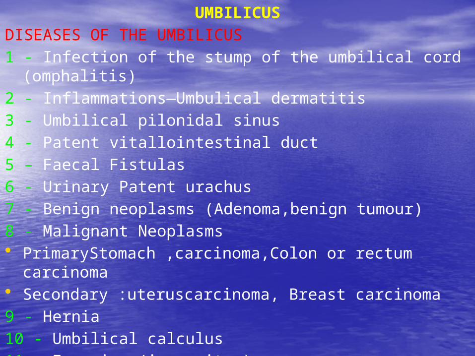
(omphalitis)2 - Inflammations—Umbulical dermatitis3 - Umbilical pilonidal sinus4 - Patent vitallointestinal duct5 – Faecal Fistulas6 - Urinary Patent urachus7 - Benign neoplasms (Adenoma,benign tumour)8 - Malignant Neoplasms• PrimaryStomach ,carcinoma,Colon or rectum carcinoma• Secondary :uteruscarcinoma, Breast carcinoma9 - Hernia10 - Umbilical calculus11 - Eversion (in ascites)
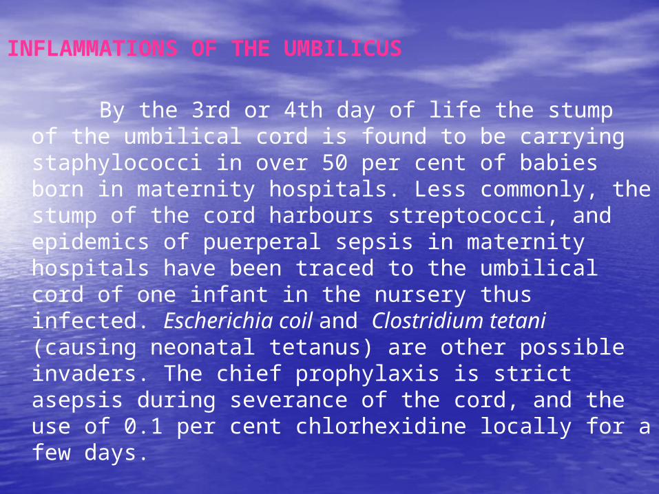
INFLAMMATIONS OF THE UMBILICUS By the 3rd or 4th day of life the stump of the
umbilical cord is found to be carrying staphylococci in over 50 per cent of babies born in maternity hospitals. Less commonly, the stump of the cord harbours streptococci, and epidemics of puerperal sepsis in maternity hospitals have been traced to the umbilical cord of one infant in the nursery thus infected. Escherichia coil and Clostridium tetani (causing neonatal tetanus) are other possible invaders. The chief prophylaxis is strict asepsis during severance of the cord, and the use of 0.1 per cent chlorhexidine locally for a few days.
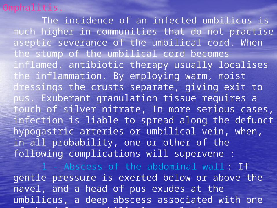
Omphalitis. The incidence of an infected umbilicus is much higher
in communities that do not practise aseptic severance of the umbilical cord. When the stump of the umbilical cord becomes inflamed, antibiotic therapy usually localises the inflammation. By employing warm, moist dressings the crusts separate, giving exit to pus. Exuberant granulation tissue requires a touch of silver nitrate, In more serious cases, infection is liable to spread along the defunct hypogastric arteries or umbilical vein, when, in all probability, one or other of the following complications will supervene :
1 - Abscess of the abdominal wall : If gentle pressure is exerted below or above the navel, and a head of pus exudes at the umbilicus, a deep abscess associated with one of the defunct umbilical vessels is present. This must be opened.
Extensive ulceration of the abdominal wall :due to a synergic infection, is treated in the same way as postoperative subcutaneous gangrene.
Septicaemia : can occur from organisms entering the bloodstream and liver via the umbilical vein. Jaundice is often the first sign. An abscess in the abdominal wall above the umbilicus should be searched for.
Jaundice in the newborn : Infection reaching the liver via the umbilical vein may cause a stenosing intrahepatic cholangiolitis, appearing some 3—6 weeks after birth.
Portal vein thrombosis and subsequent portal hypertension.
Peritonitis carries a particularly bad prognosis.Umbilical hernia
umbilical granuloma. Chronic infection of the umbilical cicatrix which continues for weeks causes granulation tissue to pout at the umbilicus. There is no certain means of distinguishing this condition from an adenoma. Usually an umbilical granuloma can be destroyed by one application of a silver nitrate stick followed by dry dressings but an adenoma soon recurs in spite of these measures.
Dermatitis of and around the umbilicus is common at all times of life. Fungus and parasitic infections are more difficult to eradicate from the umbilicus than from the skin of the abdomen. Sometimes the dermatitis is consequent upon a discharge from the umbilicus, as is the case when an umbilical fistula or a sinus is present.
Pionidal sinus (a sinus containing a sheaf of hairs) is sometimes encountered. It should be excised.
Umbilical calculus (umbolith) often black in colour, is composed of desquamated epithelium which becomes inspissated and collects in the deep recess of the umbilicus. Eventually it gives rise to inflammation, and often a bloodstained discharge. The treatment is to dilate the orifice and extract the calculus, but, to prevent recurrence, it may he better to excise the umbilicus.
UMBILICAL FISTULAS
The umbilicus being a central abdominal scar, it is understandable that a slow leak from any viscus is liable to track to the surface at this point. Added to this, very occasionally the vitellointestinal duct or the urachus remains patent; consequently it has been aptly remarked that the umbilicus is a creek into which many flstulous streams may open.
For instance, an enlarged inflamed gallbladder perforating at its fundus may discharge gallstones through the umbilicus.
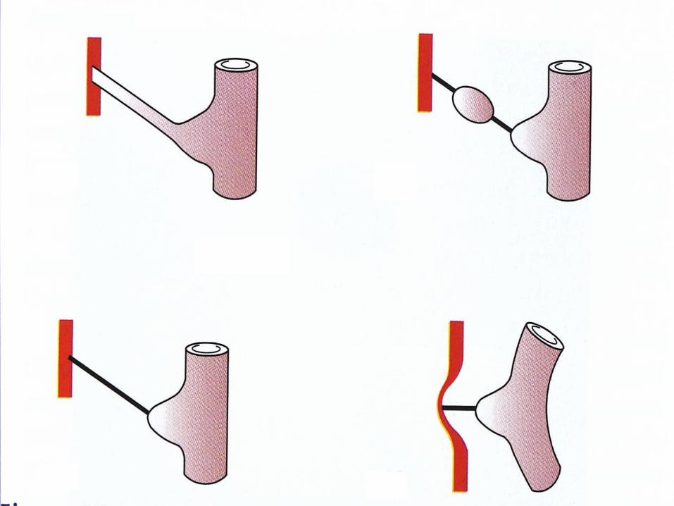
The vitellointestinal tract : The vitellointestinal duct occasionally persists, and
gives rise to one of the following conditions:1 - It remains patent . The resulting umbilical fistula
discharges mucus and, rarely, faeces. Often a small portion only of the duct near the umbilicus remains unobliterated. This gives rise to a sinus that discharges mucus. The epithelial lining of the sinus often becomes everted to form an adenoma.
2 - Sometimes both the umbilical and the intestinal ends of the duct close, but the mucous membrane of the intervening portion remains, and an intra-abdominal cyst develops.
3 - With its lumen obliterated or unobliterated, the vitellointestinal duct provides an intraperitoneal band which is a potential danger, for intestinal obstruction is liable to occur. The obstruction results from a coil of small intestine passing under or over, or becoming twisted around the band.
4 - Such a band may contract and pull a Meckel’s diverticulum into a congenital umbilical hernia.
5 - A viteilointestinal cord connected to Meckel’s diverticulum. but not attached to the umbilicus, becomes adherent to, or knotted around, another loop of small intestine, and so causes intestinal obstruction.
6 - Sometimes a band extending from the umbilicus is attached to the mesenterv near its junction with a distal part of the ileum, In this case the band is probably an obliterated vitelline artery, and is not necessarily associated with a Meckels diverticulum.
Treatment : A patent vitellointestinal duct should be excised, together with a Meckel’s diverticulum, if such be present, preferably when the child is about 6 months old. When a vitellointestinal band gives rise to acute intestinal obstruction, after removing the obstruction by division of the band, it is expedient, when possible, to excise the band and to bury the cut ends.
•A patent urachus seldom reveals itself until maturity, or even old age. This is because the contractions of the bladder commence at the apex of the organ and pass towards the base. A patent urachus, because it opens into the apex of the bladder, is closed temporarily during micturition. and so the potential urinary stream to the umbilicus is cut off. Therefore the fistula remains unobtrusive until a time when the organ is overfull, often due to some form of urinary obstruction
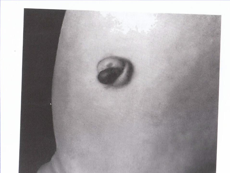
NEOPLASMS OF THE UMBILICUSUmbilical adenoma or raspberry tumour is commonly seen in infants, but only occasionally
later in life. It is due to a partially (occasionally a completely) unobliterated vitellointestinal duct. Mucosa prolapsing through the umbilicus gives rise to a raspberry-like tumour, which is moist with mucus arid tends to bleed.
Treatment If the tumour is pedunculated1 a ligature is tied around it, and in a few days the polypus drops off. Should the tumour reappear after this procedure, umbiledomy is indicated. Sometimes a patent vitellointestinal duct, or more often a vitellointestintal band, will be found associated with a Meckel’s diverticulum. The Meckel’s diverticulum and the attached cord or duct should be excised at the same time as the umbilicus. Histologically, the tumour at the umbilicus consists of columnar epithelium rich in goblet cells.
Endometrioma occurs in women between the ages of 21 and 45. On
histological examination it is found to consist of endometrial glands occupying the dermis and opening on to the surface. The umbilicus becomes painful and bleeds at each menstruation, when the small fleshy tumour between the folds of the umbilicus becomes more apparent. Occasionally an umbilical endometrioma is accompanied by endometriomas in the uterus or ovary. When, as is usually the case, the tumour is solitary. umbilectomy will cure the condition,
Secondary carcinoma at the umbilicus (Sister Joseph’s nodule)
Is not very uncommon, but it is always a late manifestation of the disease. The primary neoplasm is often situated in the stomach, pancreas. colon or ovary, but a metastasis from the breast, probably transmitted along the lymphatics of the round ligament of the liver, is sometimes located here.
NEOPLASMS OF THE ABDOMINAL WALLDesmoid tumour is a tumour arising in the mcsculoaponeurotic
structures of the abdominal wall, especially below the level of the umbilicus. It is a completely unencapsulated fibroma, and is so hard that it creaks when it is cut. Some cases recur repeatedly in spite of apparently adequate excision.
Aetiology. Eighty per cent of cases occur in women, many of whom have borne children, and the neoplasm occurs occasionally in scars of old hernia or other abdominal operation wounds. Consequently. trauma — e.g. the stretching of the muscle fibers during pregnancy or possibly a small haematoma of the abdominal wall —appears to be an aetiological factor. They can occur in cases of familial adenomatous polyposis (FAB)
Pathology The tumour is composed of fibrous tissue containing multinucleated plasmodial masses resembling foreign-body giant cells. Usually of very slow growth, it tends to infiltrate muscle in the immediate neighbourhood. Eventually it undergoes a myxomatous change: it then increases in size more rapidly. Metastasis doss not occur. Unlike fibroma elsewhere, no sarcomatotis change occurs.
Treatment Unless the tumour is excised widely, with a surrounding margin of at least 2.5 cm of healthy tissue, recurrence commonly takes place. After removal of a large tumour, repair of the defect in the abdominal wall by a mesh is required. These tumors are moderately radiosensitive. (lntraperitoneal desmoids are best left alone when possible.)
INFECTIONS
Cellulitis can occur in any of the planes of the abdominal wall
Superficial cellulitis is usually discovered when an abdominal wound is inspected following pyrexia. The earliest sign is when the stitches become embedded in the oedematous skin. Later there is a blush extending for a variable distance from the incision or the stitch holes. On palpation with the gloved hand usually one area is found to be more indurated and tender than the remainder. A stitch should be removed from the immediate vicinity, and if pus or seropus escapes it is sent for bacteriological examination; meanwhile a broad-spectrum antibiotic is administered.
Deep cellulitis is characterised by brawny oedema towards one or
both flanks, and not infrequently of the scrotum or vulva as well. Antibiotic therapy is the mainstay of treatment. When tenderness persists, an anatomical incision dividing the muscles carefully, layer by layer, until pus or purulent fluid is encountered, is often advisable.
Progressive postoperative bacterial synergistic gangrene is fortunately a rare complication after laparotomy,
usually for a perforated viscus (notably perforated appendicitis). It has also occurred after gallbladder operations, colectomy for ulcerative colitis, and even after drainage of an empyema thorcis. The condition is due to the synergic action of microaerophilic nonhaemolytic streptococci and, usually, a staphylococcus.
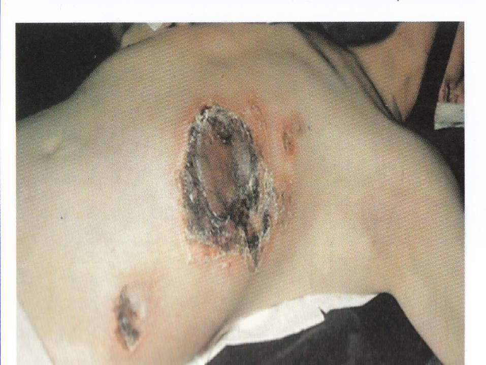
The skin in the immediate vicinity of the wound exhibits signs of cellulitis. Within a few days a central purplish zone with an outer brilliant red zone can be distinguished, and the whole region is extremely tender. Slowly the condition advances.The gangrenous skin liquefies, exposing underlying granulation tissue, If the condition persists, the general condition deteriorates.
Treatment: Identification of the organisms and a report on their
sensitivity to antibiotics is essential. Metronidazole should be given together with a powerful broad-spectrum antibiotic. Without vigorous and effective treatment the gangrene spreads to the flanks and the patient may die of toxaemia, Hyperbaric oxygen, if available, can be life-saving. Cellulitis due to bacteroides may give no bacteriological growth by conventional techniques and may be missed.

THANK YOU