A single mutation induces the substrate-binding ... · package programs DENZO and SCALEPACK ......
26
Crystal structure of P450 BM3 A264E -1- A single mutation in P450 BM3 induces the conformational rearrangement seen upon substrate-binding in wild-type enzyme M. Gordon Joyce, Hazel M. Girvan, Andrew W. Munro, and David Leys* Department of Biochemistry, University of Leicester, The Adrian Building, University Road, Leicester LE1 7RH, UK Author for correspondence: E mail [email protected], Phone: 0044 116 252 3484; Fax: 0044 116 252 3369 Running title: Crystal structure of P450 BM3 A264E . JBC Papers in Press. Published on March 12, 2004 as Manuscript M401717200 Copyright 2004 by The American Society for Biochemistry and Molecular Biology, Inc. by guest on June 27, 2018 http://www.jbc.org/ Downloaded from
Transcript of A single mutation induces the substrate-binding ... · package programs DENZO and SCALEPACK ......

Crystal structure of P450 BM3 A264E
-1-
A single mutation in P450 BM3 induces theconformational rearrangement seen upon
substrate-binding in wild-type enzyme
M. Gordon Joyce, Hazel M. Girvan, Andrew W. Munro, and DavidLeys*
Department of Biochemistry, University of Leicester, The Adrian
Building, University Road, Leicester LE1 7RH, UK
Author for correspondence: E mail [email protected], Phone: 0044 116 252
3484; Fax: 0044 116 252 3369
Running title: Crystal structure of P450 BM3 A264E
.
JBC Papers in Press. Published on March 12, 2004 as Manuscript M401717200
Copyright 2004 by The American Society for Biochemistry and Molecular Biology, Inc.
by guest on June 27, 2018http://w
ww
.jbc.org/D
ownloaded from

Crystal structure of P450 BM3 A264E
-2-
Summary
The multidomain fatty acid hydroxylase flavocytochrome P450 BM3 has been studied
as a paradigm model for eukaryotic microsomal P450s, due to its homology with
eukaryotic family 4 P450s and its use of a eukaryotic-like diflavin reductase redox
partner. High-resolution crystal structures have led to the proposal that substrate-
induced conformational changes lead to removal of the 6th ligand water ligand to the
heme iron. Concomitant changes in heme iron spin-state and heme iron reduction
potential help to trigger electron transfer from the reductase and initiate catalysis.
Surprisingly, the crystal structure of the substrate-free A264E heme domain mutant
reveals the enzyme to be in the conformation observed for the substrate-bound wild-
type P450, but with the iron in the low-spin state. This provides strong evidence that
the spin-state shift observed upon substrate-binding in wild-type P450 BM3 is not
only caused indirectly by structural changes in the protein, but is a direct consequence
of the presence of the substrate itself, similar to that observed for P450 cam. The
crystal structure of the palmitoleate-bound A264E mutant reveals that substrate
binding promotes heme ligation by Glu 264 with little other difference from the wild-
type palmitoleate-bound structure observable. Despite having a protein-derived 6th
heme ligand in the substrate-bound form, the A264E mutant is catalytically active,
providing further indication for structural rearrangement of the active site upon
reduction of the heme iron, including displacement of the glutamate ligand to allow
binding of dioxygen.
by guest on June 27, 2018http://w
ww
.jbc.org/D
ownloaded from

Crystal structure of P450 BM3 A264E
-3-
Introduction
Cytochrome P450s (P450s) are among the most studied enzymes, in no small part due
to the pivotal roles that hepatic P450s play in mammalian drug-metabolism (1).
Recent years have seen an explosion in the structural data available for these systems,
a substantial proportion of which is on cytochrome P450 BM3 (2). This multidomain
enzyme is isolated from Bacillus megaterium and contains a N-terminal fatty acid-
binding P450 domain fused to its redox partner, an NADPH-dependent diflavin
cytochrome P450 reductase (CPR, 3). It has been used as the paradigm model for
studying the similar, but membrane-associated, eukaryotic microsomal P450 systems.
This has been primarily due to the similarity in its heme domain to the eukaryotic
fatty acid hydroxylases from P450 family 4, the fact that the enzyme is soluble, that it
uses a eukaryotic-like CPR as the redox partner (as opposed to the two component
ferredoxin reductase and ferredoxin systems found in many other bacterial systems)
and since it is a convenient, catalytically self-sufficient fusion protein enzyme (2-4).
Cytochrome P450 BM3 is a fatty acid hydroxylase that displays an unsually high rate
of oxygenation of long chain fatty acids (e.g. >15,000 turnovers/min with arachidonic
acid, 5), likely due to the efficient electron transfer between the different redox
modules afforded by their covalent linkage and hence close spatial organization (2). A
sophisticated mechanism to avoid the unwanted generation of reactive oxygen species
through futile cycling has been found in many P450s studied to date. The binding of
oxygen only occurs to the reduced (ferrous) heme and the reduction of the ferric heme
iron by electron transfer from the redox partner is in turn dependent on the binding of
substrate, effectively gating initiation of the reaction by substrate-binding (6, 7). In
P450 BM3 and the Pseudomonas putida camphor hydroxylase P450 cam (the mosty
intensively studied P450 enzymes) substrate-binding induces a heme iron spin-state
by guest on June 27, 2018http://w
ww
.jbc.org/D
ownloaded from

Crystal structure of P450 BM3 A264E
-4-
shift and concomitant increase in reduction potential of the heme iron, favouring the
one electron reduction that commits the enzyme to the catalytic cycle (6, 7). The
molecular mechanism whereby substrate-binding induces this shift seems to be
somewhat different in P450s studied to date, although the substrate-binding induced
displacement of water as the 6th ligand to the heme iron is a common feature (e.g. 8,
9). In P450 cam, the binding of substrate does not effect any large-scale changes in
the protein structure, and the displacement of water is due to direct steric hindrance
with the camphor molecule (10). In contrast, P450 BM3 undergoes large-scale
conformational changes upon binding of fatty acids, and these changes have been
proposed to drive the conversion of a hexa- to a penta-coordinated heme group.
Indeed, there is no direct interaction observed between bound fatty acids and the
water molecule in the 6th ligand position in the available crystal structures of the P450
BM3 heme domain. The conformational change in P450 BM3 involves a
reorganisation of the I helix and it has been proposed that this creates a new water
binding position (11). This position is mutually exclusive with the 6th ligand binding
position, and is proposed to have greater affinity, leading to an effective switch in
heme coordination by transfer of the water to the new position. The majority of the
P450 structures indeed show a conserved bend in the I helix and this mechanism
might therefore be of a general nature (e.g. 12). However, certain P450s do not
contain any deformation of the I helix in the resting state (e.g. 13), while P450 cam
has a bent I helix that does not significantly change conformation upon substrate
binding (14).
We here present the crystal structures of both substrate-free and palmitoleic acid-
bound forms of the A264E mutant in the P450 BM3 heme domain. The alanine
by guest on June 27, 2018http://w
ww
.jbc.org/D
ownloaded from

Crystal structure of P450 BM3 A264E
-5-
occupies a key position in the I helix of the P450. The backbone of this residue
hydrogen bonds to the 6th ligand water molecule in the unbound form and is
substantially shifted upon substrate binding (8, 15). Interestingly, in several of the
CYP 4 family of fatty acid oxygenase P450s, a conserved glutamate residue at
position 264 (BM3 numbering) is known to covalently ligate the heme macrocycle
through autocatalytic, turnover-dependent attachment to the 5-methyl group of the
porphyrin (16). To investigate the possibility of creating a similar protein-heme link
in the related P450 BM3 heme domain, the A264E mutant was created. Although no
covalent modification of the heme was observed, this mutant has several unique
features (see accompanying manuscript, 17). In the fatty acid-free form, Glu 264
ligates the heme iron in a proportion of the molecules, creating a novel thiolate-
carboxylate ligation that is pushed towards full ligation by binding of the substrate.
Surprisingly, the crystal structure of the substrate-free form of this mutant reveals the
protein to be in the conformation previously considered to be induced by substrate-
binding, despite the fact that the P450 is still in a low-spin state and free of fatty acid.
We show that binding of fatty acid does not then introduce any further gross
conformational change in the protein structure, although a change in the proportion of
molecules in which glutamate ligates the heme iron is effected. The implications of
these observations on the mechanism of substrate-binding induced heme iron spin-
state shift of P450-BM3 and cytochromes P450 in general are discussed, along with
the ramifications for understanding conformational equilibria in cytochromes P450
and how binding of substrate impacts on these equilibria.
Experimental procedures
Mutagenesis, expression and purification of mutant P450 BM3.
by guest on June 27, 2018http://w
ww
.jbc.org/D
ownloaded from

Crystal structure of P450 BM3 A264E
-6-
The A264E mutant of P450 BM3 heme domain was created, expressed and purified as
described in the accompanying manuscript (17). A final purification step by FPLC
using Q-Sepharose resin (under the same conditions as those used for the low-
pressure chromatographic purification described in the accompanying manuscript, 17)
was used to produce homogeneous enzyme for crystallographic studies.
Crystallization
The P450 BM3 A264E heme domain was crystallized in both the palmitoleic acid-
bound and substrate-free forms by the sitting drop method at 4 ºC. Sitting drops were
prepared by adding 2 µl of mother liquor to 2 µl of 15 mg/ml enzyme. Palmitoleic
acid-bound crystals were obtained by co-crystallizing the enzyme with 1.2 µM
palmitoleic acid (approximately 6 x the Kd value obtained from spectral binding
titration data, and a well solution of 100 mM magnesium acetate, 20 % PEG 2000
MME and 100 mM cacodylic acid at pH 6.3. Substrate-free crystals were obtained
with a well solution of 10mM manganese sulfate, 20% PEG 2000MME and 100 mM
cacodylic acid at pH 6.3. Crystals were formed in both cases after approximately 7
days. Crystals were immersed in 10 % PEG 200 in mother liquor as a cryoprotectant,
before being mounted on a nylon loop and flash-cooled in liquid nitrogen.
Data collection, structure elucidation and refinement
Data used for refinement were collected at ESRF, Grenoble, France on ID14-EH1
using an ADSC Q4R CCD detector. Crystals were cooled at 100 K and diffraction
data collected in oscillations of 1°. Data were processed and scaled using the HKL
package programs DENZO and SCALEPACK (18). The substrate-free crystal
structures were solved via molecular replacement using the program AMoRE and the
by guest on June 27, 2018http://w
ww
.jbc.org/D
ownloaded from

Crystal structure of P450 BM3 A264E
-7-
high-resolution P450 BM3 wild-type crystal structure (PDB code 2HPD) as a search
model (19). The wild-type palmitoleate-bound crystal structure was used a starting
model for the palmitoleate-bound mutant form. In both cases positional and B-factor
refinement were carried out using REFMAC5 with manual rebuilding of the model at
regular intervals in TURBO-FRODO (20, 21). Only in the case of the low resolution
substrate-bound crystal form were strong NCS-restraints imposed throughout
refinement. Data collection and final refinement statistics are given in Table 1.
Structure factors and final coordinates for both crystal structures have been deposited
in the PDB with codes 1SMI (substrate free A264E mutant) and 1SMJ (substrate
bound A264E mutant).
Results and Discussion
Crystal structure of the substrate-free form
Initial crystallization trials for the substrate-free form of the A264E heme domain of
P450 BM3, using the published conditions for the wild-type heme domain, proved
unsuccessful. The use of manganese sulphate to substitute for magnesium acetate led
to related conditions that generated large, diffraction quality crystals. The structure
was solved to 2.0 Å and contained 2 molecules in the asymmetric unit. The overall
conformation of both molecules is similar (r.m.s.d. 0.46 Å for all Cα atoms), with
molecule B having a significant higher average B-factor (39.1 Å2 compared to 47.6
Å2) due to less packing constraints. In this manuscript, molecule A will be used for
discussion and calculations unless mentioned otherwise.
by guest on June 27, 2018http://w
ww
.jbc.org/D
ownloaded from

Crystal structure of P450 BM3 A264E
-8-
Due to the different space groups of the orthorhombic A264E mutant and the wild-
type monoclinic crystal form, comparison of the structures was made after overlay
using the structurally invariant residues, as described by Haines et al. (11),
representing approximately 62% of the structure (11). Surprisingly, it was found that
the A264E structure is structurally similar (r.m.s.d. 0.54 Å for all Cα) with the
substrate-bound form of the wild-type enzyme (hereafter referred to as conformation
SB) and shows a significant difference (r.m.s.d. 1.37 Å for all Cα) with the substrate-
free wild-type structure (hereafter referred to as conformation SF) (8, 15) (Figure
1a,b). However, no substrate was added to the A264E mutant during either
purification or crystallization, nor could any substrate be observed in the electron
density maps. Similar to the changes seen upon substrate-binding in wild-type P450
BM3, the majority of residues that are in significantly different positions between the
substrate-free structures of A264E and the wild-type heme domain are located in the
"lid domain" of the substrate access channel, which consists of the F and G helices,
the loop between them and the B'-helix (8). Several of these residues are less well
defined in A264E molecule B, indicating substantial plasticity of this region in
absence of the substrate. However, there is no significant large-scale difference
between both A264E molecules in the asymmetric unit.
In contrast, upon closer inspection of the active site of the A264E enzyme, there is a
marked difference observed in the vicinity of the heme iron between the two
molecules in the asymmetric unit. Molecule B has the side chain of Glu 264 pointing
away from the heme, its carboxylate group stacking with the aromatic group of Phe
87 (Figure 2a). In contrast, molecule A has the carboxylate group coordinating the
heme iron (Figure 2b). This heterogeneity of glutamate ligation was predicted from
by guest on June 27, 2018http://w
ww
.jbc.org/D
ownloaded from

Crystal structure of P450 BM3 A264E
-9-
solution spectrophotometric studies, and from EPR analysis (see accompanying paper,
17) and the thiolate-carboxylate ligation is novel to cytochromes P450, and (as far as
we are aware to date) to cytochromes in general. There do not seem to be any
significant conformational changes to the overall P450 structure associated with the
“switch” of Glu 264 between its two detected conformations on a) the heme iron, or
b) stacking with Phe 87. It is likely that, in solution, the carboxylate continually
switches between the “on” and “off” heme iron states without major accompanying
protein reorganization. According to spectroscopic data, in solution state the
equilibrium is poised at ~ 3-4:1 in favour of the heme iron ligand “off” form (17). The
interaction with Phe 87 is particularly interesting, given the fact that this residue is
absent from the CYP4 enzymes in which covalent ligation of the heme methyl group
has been demonstrated. Phe 87 interacts with the ω-methyl group of fatty acid
substrate(s) in wild-type P450 BM3, and is considered to be a critical regulatory
residue that controls regioselectivity of substrate oxygenation (8, 22, 23). A particular
difference in the behaviour of P450 BM3 with respect to eukaryotic CYP4 enzymes is
the inability of the former to hydroxylate at the ω-position (24).
Close examination of the A264E structure reveals no clear direct structural
explanation for the fact that this mutant is mimicking the conformation of the
substrate-bound form of the wild-type enzyme. In particular, no extra stabilizing
features involving the newly introduced glutamate side chain can be found in
comparison to available wild-type structures. We therefore tried to place the Glu 264
side chain in the corresponding substrate-free wild-type structure. All of the
conformations available to the glutamate result in severe steric clashes with several
other residues nearby (e.g. Phe 87, Thr260 and Ile263, in addition to the heme
by guest on June 27, 2018http://w
ww
.jbc.org/D
ownloaded from

Crystal structure of P450 BM3 A264E
-10-
macrocycle itself), clearly resulting in the need for a protein conformational
rearrangement to accommodate for the increased bulk of residue 264. It therefore
seems likely that the A264E mutation does not particularly stabilize conformation SB,
but rather destabilises the SF conformation of the enzyme, to the extent that the SB
conformation, even in absence of the substrate, is preferred.
In light of the above observations, it is interesting to note that in the substrate-bound
wild-type P450 BM3 structures the substrates are in close contact with the side chain
of Ala 264. This suggests the mechanism by which substrate-binding switches P450
BM3 to a different conformational state does not involve simply the expulsion of
water molecules from the substrate-binding cavity, but in addition exploits the force
exerted by the substrate on Ala 264, a residue that acts as a sensitive trigger for the
conformational conversion. In the A264E mutant, unfavourable steric interactions of
the glutamate side chain induce the switch to the SB conformation, without necessity
for the substrate interaction. An alternative explanation that could be put forward is
that the P450 BM3 heme domain is in rapid conformational equilibrium between SF
and SB forms, with the equilibrium being strongly favoured towards the SF form for
wild-type P450 BM3 in the absence of substrate. In this model, the substrate binds
preferentially to the SB conformation, effectively shifting the equilibrium towards this
form as substrate concentration is increased. This model is also consistent with the
behaviour observed for the A264E heme domain. For several substrates tested, the
apparent binding constants (Kd values) determined are considerably lower that those
for the wild-type heme domain, indicating much tighter binding (17). Given that the
SB conformation is favoured in the fatty acid-free form of the A264E heme domain,
by guest on June 27, 2018http://w
ww
.jbc.org/D
ownloaded from

Crystal structure of P450 BM3 A264E
-11-
the “tight-binding” form of the protein is over-represented in solution with respect to
that seen for wild-type P450 BM3 (Figure 3).
A substrate-binding induced spin-state shift has been observed in a large number of
P450s studied to date (e.g. 25, 26). It is generally accepted that this behaviour serves
to avoid the potentially dangerous generation of active oxygen species that would
occur through binding and subsequent reduction of molecular oxygen in the absence
of substrate. Spin-state change induces a positive change in the heme iron reduction
potential, favouring electron transfer from the redox partner (6, 7). The molecular
mechanism underlying this change in heme iron spin-state has been proposed to either
be a direct displacement of the 6th ligand water molecule (as in P450 cam) or a more
indirect displacement via substrate-induced changes in the protein structure (as for
P450 BM3, 11). We have shown that the structure of fatty acid-free A264E does show
all the structural hallmarks of a substrate-bound BM3 enzyme, but in the absence of
substrate. An intriguing difference regarding A264E is the fact that, in contrast to
substrate-bound wild type-enzyme, A264E does not show any significant high-spin
character, in either solution or crystalline state. A low-spin configuration would be
expected for the glutamate-ligated species, but both spectroscopic and
crystallographic studies show that there is a large population of non glutamate-ligated
protein in substrate-free A264E heme domain, and that this species does not lose the
aqua ligand and convert to high-spin despite the change to the SB conformation. This
suggests strongly that even in wild-type P450 BM3 the spin-state shift is a direct,
rather than an indirect, result of substrate binding. The high-resolution N-palmitoyl-
glycine-bound wild-type structure has lead to the suggestion that substrate-binding-
induced protein rearrangement creates a new water-binding site (designated site H)
by guest on June 27, 2018http://w
ww
.jbc.org/D
ownloaded from

Crystal structure of P450 BM3 A264E
-12-
adjacent to the heme (11). The proposed higher water affinity of site H over the heme
iron ligation site L, and the fact that these sites are mutually exclusive (so that only a
single water molecule can bind at either site L or H at any given time) led to the to
proposal that site H effectively pulls the water molecule away from site L, leading to
the observed shift in heme iron coordination state and hence conversion to the high-
spin form (11). The A264E mutant heme domain structure shows all ligands to the H
site to be in identical position as for the N-palmitoyl-glycine-bound wild-type P450
BM3 structure (Figure 4a). However, the water molecule is still occupying site L (the
heme iron) for molecule B, while in molecule A Glu 264 ligates the heme iron. In
both cases the H site remains unoccupied due to steric hindrance with either the water
(in molecule B) or the Glu 264 side chain (in molecule A) at site L. It can therefore be
concluded that site L (heme iron) remains the higher affinity site for water in the
absence of substrate in conformation SB.
As has been pointed out previously, there are no direct steric clashes between the
bound fatty acid substrates of P450 BM3 and the water molecule at site L. The
palmitoleic acid-bound structure of wild-type P450 BM3 indicates that secondary
conformational changes of protein/substrate must occur following reduction of the
heme iron, since substrate is too distant from the heme iron for oxidative attack at
catalytically relevant positions on the fatty acid chain (8). NMR studies of the
substrate-bound form of ferrous, fatty acid-bound wild-type P450 BM3 are consistent
with a significant reorientation of the substrate in this enzyme form (27). It is,
however, clear that upon binding of the fatty acid analogue N-palmitoyl glycine the
surroundings of site L become more hydrophobic, decreasing the water affinity and
ultimately shifting the water molecule to predominantly occupy site H. Spectroscopic
studies of wild-type P450 BM3 at catalytically-relevant temperatures (by both
by guest on June 27, 2018http://w
ww
.jbc.org/D
ownloaded from

Crystal structure of P450 BM3 A264E
-13-
electronic absorption and resonance Raman) have shown that, even in presence of
apparently saturating concentrations of substrate, an equilibrium exists between the
high-spin, five-coordinate, and the low-spin, six-coordinate states of the heme iron.
Depending on nature of the fatty acid substrate used, varying amounts of low-spin
heme iron are detected, with shorter chain saturated fatty acids (e.g. lauric acid) being
less effective than longer chain ones (e.g. palmitic acid) at effecting the shift in spin-
state equilibrium towards high-spin (28, 29). This clearly indicates how, even in the
presence of substrate, the possibility exists for water remaining bound at the heme
iron (site L), and the water affinity of this site is strongly dependent on the nature of
the substrate, although all drive the L-H equilibrium towards the H site.
Crystal structure of the substrate-bound form
In contrast to the requirement for screening novel conditions to obtain suitable
crystals of the substrate-free A264E heme domain, the palmitoleate-bound form of
A264E was found to crystallise in the same space group as that reported previously
for the substrate-bound wild-type P450 BM3 (8). As observed for the wild-type
enzyme, the resolution and the quality of the data obtained for this particular crystal
form is rather poor by comparison with the substrate-free enzyme. Nevertheless,
electron density clearly indicates no major changes between the palmitoleate-bound
A264E and palmitoleate-bound wild-type P450 BM3 structures. The single exception
is the fact that in all 4 molecules in the asymmetric unit of the A264E structure, the
Glu 264 side chain ligates the heme iron (Figure 4b). This finding indicates that the
presence of substrate induces movement of the glutamate onto the iron to replace
water as the 6th heme ligand, and is completely consistent with the spectroscopic
by guest on June 27, 2018http://w
ww
.jbc.org/D
ownloaded from

Crystal structure of P450 BM3 A264E
-14-
studies reported in the accompanying paper (17). Addition of long chain fatty acids
perturbs the UV-visible absorption spectrum of the A264E heme domain, inducing
red shifts of the heme Soret band towards 426 nm at apparent substrate saturation.
This is in contrast to the blue shifts observed following substrate addition to wild-type
P450 BM3 (towards 390 nm) and reflects the increasing proportion of the glutamate-
ligated, low-spin form of the A264E mutant following substrate addition, as opposed
to the pentacoordinated, high-spin form of the wild-type that accumulates through
substrate-induced displacement of the water ligand from the heme iron (17, 28). From
the substrate-free A264E structure, it is clear that two conformations are possible for
the Glu 264 side chain and solution studies indicate the equilibrium ratio between
both states to be strongly dependent on the solution conditions. Upon palmitoleate
binding, the substrate effectively occupies the volume of the non heme iron-ligating
conformation, driving the enzyme towards a completely ligated state, as observed in
solution studies. Specifically, palmitoleate interacts with Phe 87, preventing Glu 264
from occupying the position observed in molecule A in the substrate-free A264E
structure. Despite any significant further structural rearrangement induced following
palmitoleic acid binding to the SB conformation of A264E, the substrate does
influence directly the heme iron ligation state by minimising the degrees of freedom
available to the Glu 264 sidechain. Strong heme ligands such as azoles function as
potent inhibitors for P450s, and many are used as antifungal drugs to inactivate the
sterol demethylase P450 CYP51 (30). It is therefore surprising to note that, despite the
fact that the substrate-bound oxidised A264E structure shows fully hexa-coordinated
heme iron, catalytic turnover for this mutant can still be observed, albeit at lower
levels than those observed for wild-type P450 BM3 (17). This is a further indication
that, upon reduction of ferric iron to ferrous, the position of the substrate with respect
by guest on June 27, 2018http://w
ww
.jbc.org/D
ownloaded from

Crystal structure of P450 BM3 A264E
-15-
to the heme and perhaps the structure of the enzyme itself change dramatically,
releasing the strong conformational lock on the glutamate side-chain and allowing
oxygen to bind to the iron. Presumably this commits the enzyme to its “regular”
catalytic cycle, and prevents coordination of the glutamate to the iron until it returns
to a ferric form following product formation. A further interesting aspect of this study
arises from the structural change and its effect on thermodynamic properties of the
P450. In both the substrate-free and arachidonate-bound forms of the A264E mutant,
the reduction potential of the heme iron is ~ –315 mV (see accompanying paper). By
contrast, the reduction potential of the conformationally different substrate-free form
of P450 BM3 is –427 mV, rising to –289 mV on binding of arachidonate and an
extensive switch in spin-state equilibrium towards high spin (31). In A264E, the heme
iron remains predominantly low-spin in both substrate-free and substrate-bound
states. Thus, a possibility that arises is that the conformational change and its effects
on the electronics of the heme system is of considerable importance in controlling the
reduction potential of the heme iron. This is under further study, using A264 variants
in which the sidechain of the introduced amino acid does not ligate the heme iron in
substrate-free or substrate-bound forms.
Further scrutiny of both the substrate-free and substrate-bound A264E structures
reveal other important features of the P450 BM3 structure that relate to attempts to
engineer covalent ligation of the heme macrocycle via the interaction of Glu 264 with
the heme 5-methyl group (Figure 5). The active site organization in P450 BM3 is such
that Phe 87, and likely the I helix residue Thr 260, obstruct access of Glu 264 to the
relevant position on the porphyrin ring. The failure to obtain any significant degree of
by guest on June 27, 2018http://w
ww
.jbc.org/D
ownloaded from

Crystal structure of P450 BM3 A264E
-16-
covalent ligation in the A264E mutant may thus be explicable through steric
restrictions in the active site. To address these restrictions, and in work to enable
covalent heme ligation by Glu 264 and produce a more robust and biotechnologically
exploitable form of P450 BM3, we are currently generating secondary mutations at
these locations that might facilitate access of Glu 264 to the relevant methyl group
and could thus allow autocatalytic linkage to occur.
Conclusions
Crystallographic studies of the A264E variant of P450 BM3 confirm the proposals
based on spectroscopic studies that the glutamate is able to ligate to the ferric heme
iron of the mutant in the substrate-free form, and that substrate addition “forces” on
the ligand – producing a completely hexa-coordinated low-spin species, as opposed to
the extensively high-spin penta-coordinated form seen for the wild-type P450 BM3
(17). Structural studies explain clearly why substrate has this effect in the A264E
enzyme, since palmitoleic acid occupies one of the two favoured positions for the Glu
264 sidechain. Glu 264 can no longer form an interaction with the key
regiospecificity-determining residue Phe 87 in the palmitoleate-bound form, and is
thus induced to move towards its only other acceptable position – coordinating to the
heme iron.
An unexpected finding, but one with enormous ramifications for understanding the
conformational changes that occur in P450 BM3 (and P450 systems in general) and
their consequences, is the fact that both substrate-free and palmitoleic acid-bound
forms of the A264E heme domain have overall structural conformations that are
virtually identical to those found for the substrate-bound forms of wild-type P450
by guest on June 27, 2018http://w
ww
.jbc.org/D
ownloaded from

Crystal structure of P450 BM3 A264E
-17-
BM3, but are distinct from that of the substrate-free wild-type heme domain (8, 11,
15). This “SB” conformation is not dependent on whether Glu 264 ligates the heme
iron or is positioned against Phe 87, and the enzyme is low-spin in both forms and
aqua-coordinated in the latter form for substrate-free A264E. The most obvious
explanations are 1) that the SB conformation in wild-type P450 BM3 is a
consequence of substrate-induced deformation of the I helix in the region of Ala 264,
and that the A264E mutation favours this conformational rearrangement independent
of substrate due to steric restrictions to movement of the glutamate side chain in the
SF conformation, and/or 2) that P450 BM3 is in a continual dynamic equilibrium
between SF and SB conformations, and that the A264E mutation forces this
equilibrium toward the SB form. For both cases, the fact that the mutant remains in a
low-spin form in the SB conformation in the fatty acid-free structure suggests that the
spin-state conversion observed in wild-type P450 BM3 on substrate association (and
the concomitant change in reduction potential) is a consequence of the physical
presence of the lipid in the environment of the heme, and not a result of the adoption
of the SB conformation per se. Moreover, the fact that the SB conformation is clearly
accessible in the substrate-free A264E mutant also suggests that the binding of fatty
acid might not be essential for inducing this conformational rearrangement in the
wild-type enzyme, and that the adoption of the SB conformation in the palmitoleate-
bound wild-type structure could merely be a consequence of favourable binding of the
substrate to this conformer. This conclusion is supported by the fact that much tighter
Kd values are observed for binding of several long chain fatty acids to the A264E
variant than to the wild-type P450 BM3 (17). The SB conformation predominates in
the mutant. In ongoing work, we aim to validate further the hypotheses that arise from
these findings through creation other variants at position 264 – specifically
by guest on June 27, 2018http://w
ww
.jbc.org/D
ownloaded from

Crystal structure of P450 BM3 A264E
-18-
investigating A264 variants that induce the conformational switch to the SB
conformation, but which do not in addition give rise to coordination to the heme iron.
Acknowledgements
DL is a Royal Society University Research Fellow. AWM and DL are grateful for the
continued support from the BBSRC and EPSRC for their research. MGJ is a BBSRC-
funded PhD student. HMG is an EPSRC-funded PhD student. We are grateful for
access to beamlines X11 and BW7A at the EMBL-outstation, DESY, Hamburg that
were essential to crystal improvement and for access to ID14.1 at ESRF, Grenoble
where final data were collected. We acknowledge the support of the European
Community - Access to Research Infrastructure Action of the Improving Human
Potential Programme to the EMBL Hamburg Outstation, contract number: HPRI-CT-
1999-00017 .
References
1) Guengerich, F.P. (2002). Drug Met. Rev. 34, 7-15.
2) Munro, A.W., Leys, D., McLean, K.J., Marshall, K.R., Ost, T.W., Daff, S.,
Miles, C.S., Chapman, S.K., Lysek, D.A., Moser, C.C., Page, C.C. and
Dutton, P.L. (2002). Trends Biochem. Sci. 27, 250-257.
3) Narhi, L.O. and Fulco, A.J. (1987). J. Biol. Chem. 262, 6683-6690.
4) Fulco, A.J. (1991). Annu. Rev. Pharmacol. Toxicol. 31, 177-203.
5) Noble, M.A., Miles, C.S., Chapman, S.K., Lysek, D.A., MacKay, A.C., Reid,
G.A., Hanzlik, R.P. and Munro, A.W. (1999). Biochem. J. 339, 371-379.
6) Daff, S.N., Chapman, S.K., Turner, K.L., Holt, R.A., Govindaraj, S., Poulos,
T.L. and Munro, A.W. (1997). Biochemistry 36, 13816-13823.
7) Sligar, S.G. (1976). Biochemistry 15, 5399-5406.
8) Li, H. and Poulos, T.L. (1997). Nat. Struct. Biol. 4, 140-146.
9) Cupp-Vickery, J., Anderson, R. and Hatziris, Z. (2000). Proc. Natl. Acad. Sci.
USA 97, 3050-3055.
by guest on June 27, 2018http://w
ww
.jbc.org/D
ownloaded from
Crystal structure of P450 BM3 A264E
-19-
10) Poulos,T.L., Finzel, B.C. and Howard, A.J. (1987). J. Mol. Biol. 195, 687-700.
11) Haines, D.C., Tomchick, D.R., Machius, M. and Peterson, J.A. (2001).
Biochemistry 40, 13456-13465.
12) Podust, L.M., Poulos, T.L. and Waterman, M.R. (2001). Proc. Natl. Acad. Sci.
USA 98, 3068-3073.
13) Leys, D., Mowat, C.G., McLean, K.J., Richmond, A., Chapman, S.K.,
Walkinshaw, M.D. and Munro, A.W. (2003). J. Biol. Chem. 278, 5141-5147.
14) Raag, R. and Poulos, T.L. (1991). Biochemistry 30, 2674-2684.
15) Ravichandran, K.G., Boddupalli, S.S., Hasermann, C.A., Peterson, J.A. and
Deisenhofer, J. (1993). Science 261, 731-736.
16) Colas, C. and Ortiz de Montellano, P.R. (2003). Chem. Rev. 103, 2305-2332.
17) Girvan, H.M., Leys, D., Joyce, M.G., Marshall, K.R., Lawson, R.J., Clarkson,
J., Smith, W.E., Cheesman, M.R. and Munro, A.W. (2004). Flavocytochrome
P450 BM3 mutant A264E undergoes substrate-dependent formation of a novel
heme iron ligand set (accompanying paper). J. Biol. Chem. in press
18) Otwinoswki, Z., and Minor, W. (1997). Methods Enzymol. 276, 307-326.
19) Navazza, J. (2001). Acta Crystallogr. D. 57, 1367-1372.
20) Murshudov, G.N., Vagin, A.A., and Dodson, E.J. (1997) Acta Crystallogr. D
53, 240-255.
21) Roussel, A., and Cambillau, C. (1992) TURBO-FRODO, Biographics,
AFMB. Marseille, France.
22) Oliver, C.F., Modi, S., Sutcliffe, M.J., Primrose, W.U., Lian, L.Y., Roberts,
G.C.K. (1997). Biochemistry 36, 1567-1572.
23) Munro, A.W., Noble, M.A., Miles, C.S., Daff, S.N., Green, A.J., Quaroni, L.,
Rivers, S., Ost, T.W., Reid, G.A. and Chapman, S.K. (1999). Biochem. Soc.
Trans. 27, 190-196.
24) Okita, R.T. and Okita, J.R. (2001). Curr. Drug Metab. 2, 265-281.
25) Lee, D.S., Yamada, A., Sugimoto, H., Matsunaga, I., Ogura, H., Ichihara, K.,
Adachi, S., Park, S.Y. and Shiro, Y. (2003). J. Biol. Chem. 278, 9761-9767.
26) Modi, S., Gilham, D.E., Sutcliffe, M.J., Lian, L.Y., Primrose, W.U., Wolf,
C.R. and Roberts, G.C.K. (1997). Biochemistry. 36, 4461-70.
27) Modi, S., Sutcliffe, M.J., Primrose, W.U., Lian, L.Y. and Roberts, G.C.K.
(1996). Nat. Struct. Biol. 3, 414-417.
by guest on June 27, 2018http://w
ww
.jbc.org/D
ownloaded from

Crystal structure of P450 BM3 A264E
-20-
28) Miles, J.S., Munro, A.W., Rospendowski, B.N., Smith, W.E., McKnight, J.
and Thomson, A.J. (1992). Biochem. J. 288, 503-509.
29) Boddupalli, S.S., Pramanik, B.C., Slaughter, C.A., Estabrook, R.W. and
Peterson, J.A. (1992). Arch. Biochem. Biophys. 292, 20-28.
30) Georgopapadakou, N.H. (1998). Curr. Opin. Microbiol. 1, 547-557.
31) Ost, T.W.B., Miles, C.S., Munro, A.W., Murdoch, J., Reid, G.A. and
Chapman, S.K. (2001). Biochemistry 40, 13421-13429.
by guest on June 27, 2018http://w
ww
.jbc.org/D
ownloaded from

Crystal structure of P450 BM3 A264E
-21-
Figure legends
Figure 1: Stereoview of an overlay of substrate-free A264E heme domain
(coloured in green ribbons). Panel a) with the substrate-free (in yellow) form of
wild-type P450 BM3 heme domain Panel b) with the substrate-bound (in blue) form
of wild-type P450 BM3 heme domain.
Figure 2: Conformations of Glu 264 in molecules A and B of the substrate-free
A264 heme domain of flavocytochrome P450 BM3. The heme macrocycle along
with residues Cys 400 and Glu 264 is displayed for molecule B (panel a) and
molecule A (panel b) with the 2Fo-Fc-map contoured at 1σ superposed.
Figure 3: A scheme representing a model for the conformational equilibrium of
P450-BM3 in solution and the influence of substrate binding. The left panel
displays the wild-type equilibrium; the right panel the A264E equilibrium. The
equilibrium in absence of substrate is drastically changed by introduction of the
A264E mutation. In both cases, large grey arrows indicate the apparent shift on
substrate binding.
Figure 4: Stereo view of the active site of A264E. Panel a) An overlay of the active
site of A264E (in blue) and the N-palmitoylglyine bound wild-type BM3 (in green)
(PDB-code 1JPZ). For clarity, the heme macrocycle is only displayed for the A264E
mutant. Hydrogen bonding pattern to the H-site is indicated by dotted black lines. The
ligating water molecule occupying the L-site in the A264E mutant is coloured in red.
Panel b) Active site structure of the palmitoleic acid-bound form of the A264E heme
by guest on June 27, 2018http://w
ww
.jbc.org/D
ownloaded from

Crystal structure of P450 BM3 A264E
-22-
domain. Residues are coloured according to residue type, the substrate is depicted in
purple.
Tables
Table 1: Crystallographic statistics
Protein A264E mutant of BM-3 A264E mutant withsubstrate bound
Data collectionSpace group P 21 21 21 C 2 2 21Cell parameters (Å)a 61.2 106.5b 118.9 165.7c 146.3 224.7X-ray source ESRF ID14-EH1 ESRF ID14-EH1Resolution 15.0-2.0 15.0-2.75Number of ObservationsTotal 686004 787601Unique 68550 53600Completeness (%) 94.0 (97.1) 99.9 (99.6)Rmerge 5.1 (44.1) 6.9 (49.1)I/σI 28.2 (2.41) 30.9 (3.7)RefinementRwork 0.227 0.257Rfree 0.288 0.338R.m.s. deviations from idealBonds (Å) 0.018 0.034Angle (o) 1.719 2.933
by guest on June 27, 2018http://w
ww
.jbc.org/D
ownloaded from
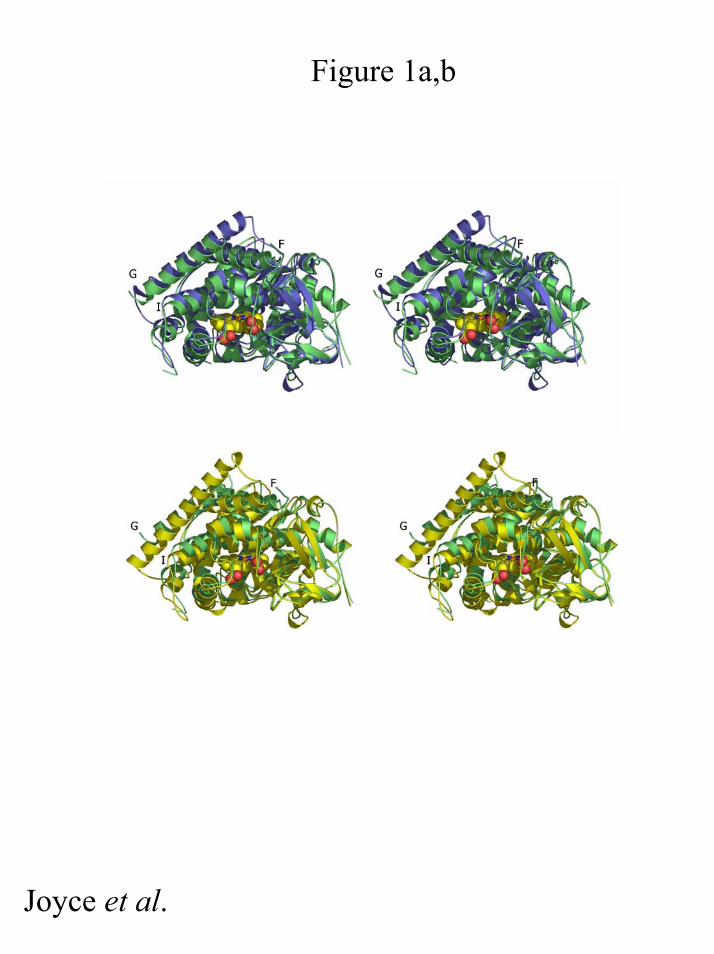
Figure 1a,b
Joyce et al.
by guest on June 27, 2018http://w
ww
.jbc.org/D
ownloaded from

S S
Wild Type
S S
A264E
+S+S +S +S
Figure 2a and b, 3
Joyce et al.
by guest on June 27, 2018http://w
ww
.jbc.org/D
ownloaded from

Figure 4a,b
Joyce et al.
by guest on June 27, 2018http://w
ww
.jbc.org/D
ownloaded from

M. Gordon Joyce, Hazel M. Girvan, Andrew W. Munro and David Leysupon substrate-binding in wild-type enzyme
A single mutation in P450 BM3 induces the conformational rearrangement seen
published online March 12, 2004J. Biol. Chem.
10.1074/jbc.M401717200Access the most updated version of this article at doi:
Alerts:
When a correction for this article is posted•
When this article is cited•
to choose from all of JBC's e-mail alertsClick here
by guest on June 27, 2018http://w
ww
.jbc.org/D
ownloaded from