A Simple Biosynthesis of Silver Nanoparticles from...
7
Journal of Applied Pharmaceutical Science Vol. 8 (01), pp 073-079, January, 2018 Available online at http://www.japsonline.com DOI: 10.7324/JAPS.2018.8111 ISSN 2231-3354 © 2018 E. Chandra Sekhar et al. This is an open access article distributed under the terms of the Creative Commons Attribution License -NonCommercial- ShareA- likeUnported License (http://creativecommons.org/licenses/by-nc-sa/3.0/). * Corresponding Author K. S. V. Krishna Rao, Assistant Professor, Department of Chemistry, Yogi Vemana University, Kadapa, Andhra Pradesh, India-516 003. E-mail: ksvkr @ yogivemanauniversity.ac.in A Simple Biosynthesis of Silver Nanoparticles from Syzygium cumini Stem Bark Aqueous Extract and their Spectrochemical and Antimicrobial Studies E. Chandra Sekhar 1 , K. S. V. Krishna Rao 2,* , K. Madhu Sudana Rao 3 , S. Bahadur Alisha 4 1 Department of Chemistry, Rajeev Gandhi Memorial college of Engineering and Technology, Kurnool, Nandyal, Andhra Pradesh India-518501. 2 Polymer Biomaterial Design and Synthesis Laboratory, Department of Chemistry, Yogi Vemana University, Kadapa. Andhra Pradesh, India-516003. 3 School of Chemical Engineering, Yeungnam University, 280 Daehak-Ro,Gyeongsan, Gyeongbuk, 38541, South of Korea. 4 Department of Chemistry, S.L.S. Degree College, Pullareddypeta - 516 175, Andhra Pradesh, India. ARTICLE INFO ABSTRACT Article history: Received on: 09/10/2017 Accepted on: 12/12/2017 Available online: 28/01/2018 This paper describes a simple biosynthesis of silver nanoparticles (SNs) from aqueous stem bark extract of Syzygium cumini (SC), a natural product. The formation of SNs was confirmed and optimized by measuring surface plasmon resonance (SPR) peak around 420 nm by using UV-visible spectroscopy (UV-Vis). The possible functional groups of SC bark extract and their changes after treating with aqueous silver nitrate were evaluated by Fourier transform infrared spectroscopy (FTIR). The size of synthesized SNs was measured by dynamic light scattering (DLS) analysis, and morphology was examined by transmission electron microscopy (TEM) in nano range. The average size of SNs is ~14 nm and monodispersed. The effect of variation of bark extract amount, silver nitrate concentration, time and pH on the synthesis of SNs was investigated at different volumes. Antibacterial activity of SC-SNs was studied taking into account Bacillus subtilis (bacillus) and Escherichia coli (E. coli). The characteristics of the facial SC-SNs formed suggest for biomedical application as chemical sensors in the future. Key words: Biosynthesis, Syzygium cumini, Natural product, Silver nanoparticles, and Antimicrobial activity INTRODUCTION In recent years, an escalating percentage of nano- materials have emerged, providing advancement in diverse fields (Qu, 2016; Ahmad et al., 2010; Sarkar et al., 2010; Lird- prapamongkol et al., 2010; Nune et al., 2009). As nature makes optimum use of materials and space, many inorganic materi- als are produced in biological systems (Qu, 2016). Similar to such natural processes, plants (Ahmad et al., 2010; Sarkar et al., 2010), fungi (Binupriya et al., 2010), and bacteria (Reddy et al., 2010) are found to be of powerful sources for production of nano-sized metal particles (Sinha et al., 2009). Biomolecules are the sources of reductants that are found to have a poten- tial gain over their protecting group counterparts (Huang et al., 2007). The highest challenge in the nanoparticles study is to get the desired size and shape of the metal nanoparticles using benign starting materials. To achieve these results, scientists made several efforts to develop beneficial and green chemistry compound inspection protocols for the synthesis of nanomateri- als with tailor-made structural properties. Currently there is a great need to derive an environmen- tal friendly nanoparticle synthesis process that does not involve any toxic chemicals. However polymeric metal nanoparticles are synthesized by reducing agents such as hydrazine, 1, 2-hexade- canediol, NaBH 4 and ascorbic acid (Rao et al., 2012; Praveena et al., 2014; Rao et al., 2010; Deepali et al., 2015; Bilal et al., 2017) for reduction of silver ions to SNs (Ag + to Ag o ), which are extremely toxic in nature. Also, natural plant extracts are used extensively as reducing agents as well as capping agents to inhibit the agglomeration and stability (Kharissova et al., 2013; Malar- kodi et al., 2014; Sharma et al., 2009) of the synthesized nanopar- ticles. Interestingly the noble metallic nanomaterials are receiving
Transcript of A Simple Biosynthesis of Silver Nanoparticles from...

Journal of Applied Pharmaceutical Science Vol. 8 (01), pp 073-079, January, 2018Available online at http://www.japsonline.comDOI: 10.7324/JAPS.2018.8111
ISSN 2231-3354
© 2018 E. Chandra Sekhar et al. This is an open access article distributed under the terms of the Creative Commons Attribution License -NonCommercial- ShareA-likeUnported License (http://creativecommons.org/licenses/by-nc-sa/3.0/).
*Corresponding AuthorK. S. V. Krishna Rao, Assistant Professor, Department of Chemistry, Yogi Vemana University, Kadapa, Andhra Pradesh, India-516 003.E-mail: ksvkr @ yogivemanauniversity.ac.in
A Simple Biosynthesis of Silver Nanoparticles from Syzygium cumini Stem Bark Aqueous Extract and their Spectrochemical and Antimicrobial Studies
E. Chandra Sekhar1, K. S. V. Krishna Rao2,*, K. Madhu Sudana Rao3, S. Bahadur Alisha4
1Department of Chemistry, Rajeev Gandhi Memorial college of Engineering and Technology, Kurnool, Nandyal, Andhra Pradesh India-518501.2Polymer Biomaterial Design and Synthesis Laboratory, Department of Chemistry, Yogi Vemana University, Kadapa. Andhra Pradesh, India-516003.3School of Chemical Engineering, Yeungnam University, 280 Daehak-Ro,Gyeongsan, Gyeongbuk, 38541, South of Korea.4Department of Chemistry, S.L.S. Degree College, Pullareddypeta - 516 175, Andhra Pradesh, India.
ARTICLE INFO ABSTRACTArticle history:Received on: 09/10/2017Accepted on: 12/12/2017Available online: 28/01/2018
This paper describes a simple biosynthesis of silver nanoparticles (SNs) from aqueous stem bark extract of Syzygium cumini (SC), a natural product. The formation of SNs was confirmed and optimized by measuring surface plasmon resonance (SPR) peak around 420 nm by using UV-visible spectroscopy (UV-Vis). The possible functional groups of SC bark extract and their changes after treating with aqueous silver nitrate were evaluated by Fourier transform infrared spectroscopy (FTIR). The size of synthesized SNs was measured by dynamic light scattering (DLS) analysis, and morphology was examined by transmission electron microscopy (TEM) in nano range. The average size of SNs is ~14 nm and monodispersed. The effect of variation of bark extract amount, silver nitrate concentration, time and pH on the synthesis of SNs was investigated at different volumes. Antibacterial activity of SC-SNs was studied taking into account Bacillus subtilis (bacillus) and Escherichia coli (E. coli). The characteristics of the facial SC-SNs formed suggest for biomedical application as chemical sensors in the future.
Key words:Biosynthesis, Syzygium cumini, Natural product, Silver nanoparticles, and Antimicrobial activity
INTRODUCTIONIn recent years, an escalating percentage of nano-
materials have emerged, providing advancement in diverse fields (Qu, 2016; Ahmad et al., 2010; Sarkar et al., 2010; Lird-prapamongkol et al., 2010; Nune et al., 2009). As nature makes optimum use of materials and space, many inorganic materi-als are produced in biological systems (Qu, 2016). Similar to such natural processes, plants (Ahmad et al., 2010; Sarkar et al., 2010), fungi (Binupriya et al., 2010), and bacteria (Reddy et al., 2010) are found to be of powerful sources for production of nano-sized metal particles (Sinha et al., 2009). Biomolecules are the sources of reductants that are found to have a poten-tial gain over their protecting group counterparts (Huang et al.,
2007). The highest challenge in the nanoparticles study is to get the desired size and shape of the metal nanoparticles using benign starting materials. To achieve these results, scientists made several efforts to develop beneficial and green chemistry compound inspection protocols for the synthesis of nanomateri-als with tailor-made structural properties.
Currently there is a great need to derive an environmen-tal friendly nanoparticle synthesis process that does not involve any toxic chemicals. However polymeric metal nanoparticles are synthesized by reducing agents such as hydrazine, 1, 2-hexade-canediol, NaBH4 and ascorbic acid (Rao et al., 2012; Praveena et al., 2014; Rao et al., 2010; Deepali et al., 2015; Bilal et al., 2017) for reduction of silver ions to SNs (Ag+ to Ago), which are extremely toxic in nature. Also, natural plant extracts are used extensively as reducing agents as well as capping agents to inhibit the agglomeration and stability (Kharissova et al., 2013; Malar-kodi et al., 2014; Sharma et al., 2009) of the synthesized nanopar-ticles. Interestingly the noble metallic nanomaterials are receiving
Sekhar et al. / Journal of Applied Pharmaceutical Science 8 (01); 2018: 073-079074
more interest in recent research, due to their interesting properties exhibiting wholly innovative and improved characteristic prop-erties compared to the voluminous particles of the bulk materi-als with specific characteristics, such as unique size distribution and required morphology (Ankanna et al., 2010; Mahdieha et al., 2012).
The potentiality of a plant system in biologically assisted synthesis of metal nanoparticles called “green synthesis” and has now achieved a great role in the field of nano science and tech-nology (Jha et al., 2009). Green synthesis is a chemistry which is helps in the design, development and implementation of chemical products; also it facilitates in reduction or elimination the sub-stances that are hazardous to human health and the environment (Kumar et al., 2008). Recently, there is a renewed interest in using a green chemistry approach to synthesize metal nanoparticles (Geethalakshmi et al., 2010; Christensen et al., 2011; Arokiyaraj et al., 2014; Gandhi et al., 2014). Sastry and coworkers (Ahmad et al., 2003; Shankar et al., 2004) employed an in silico approach to investigate the synthesis of stable Ag, Au and Ag-Au bimetallic nanoparticles using Azadirachta indica leaf broth and suggested that the flavanone and terpenoids are prime in responsibility for the stabilization of nanoparticles. Recent reports also suggested biosynthesis of Ag and Au nanoparticles from apiin (Kasthuri et al., 2009), Cinnamon zeylanicum bark extract (Sathishkumar et al., 2009), mushroom extract (Philip, 2009), Acalypha indica leaf extracts (Krishnaraj et al., 2010), glucose and starch (Raveendran et al., 2006). The biosynthesized nanoparticles such as Au, Ag, and Pt are widely used in medical and pharmaceutical applications (Singh et al., 2013; Galdiero et al., 2011) for catalysis (Burda et al., 2005), anti-bacterial, antiviral agents (Singh et al., 2013) and sensor applications (Nirlipta et al., 2016).
Syzygium cumini (SC) is distributed on the Indian sub-continent, and in many others regions of South Asia and it is traditionally used ethno-medicinal plant of Myrtaceae family. The stem bark of SC is rich in many pyto-chemicals such as betu-linic acid, friedelin, epi-friedelanol, β-sitosterol, eugenin and fatty acid ester of epi-friedelanol, β-sitosterol, quercetin kaempferol, myricetin, gallic acid and ellagic acid, bergenins, flavonoids and tannins (Ayyanar et al., 2012). It is used as folk medicine. Also, the Ayurvedic Pharmacopoeia of India recommends the SC bark for diseases like acute diarrhea and haemorrhagic (https://sites.google.com/site/blackberries7890/). In addition to the above, SC bark is commonly used as a raw medicine for the treatment of sore throat, bronchitis, asthma, thirst, biliousness, dysentery and ulcers and it has a nature that is acrid, sweet, digestive, astringent to the bowels, anthelmintic (Kumar et al., 2015).
In this paper, we report a simple biosynthesis of Ag metallic nanoparticles by the reduction of aqueous Ag+ ions through a green approach using the bark extract of SC at room temperature. Due to the nutritional, medicinal and environmental properties (Prasad et al., 2014; Rao et al., 2015; Prabhakaran et al., 2011; Sharma et al., 2009) and also bioavailability of SC, it is chosen to produce nanoparticles with bark extracts. Rapid biosyn-thesis of nanoparticles using SC stem bark extract has been inves-tigated. It is the easiest, most cost efficient, non-toxic, eco-friendly and efficient method for exploiting SC stem barks. So the bark of SC has advantages over the other reducing agents used in synthe-sis of metal nanoparticles. To the best of author’s knowledge there
have been no reports on the biosynthesis of nano silver from the aqueous extracts of the SC bark, also their spectro-chemical and anti-bacterial studies.
MATERIALS AND METHODS
MaterialsSilver nitrate (AgNO3; 99.9%, Sigma Aldrich Chemie,
Germany). Syzygium cumini (SC) stem bark was collected from the premises of Yogi Vemana University, Kadapa, India. All other chemicals used are analytical grade and received from Merck, India. Throughout the experiment double distilled water was used.
Extraction procedureAqueous Syzygium cumini stem bark extract was pre-
pared by a green process technique, using the standard procedure described by Sekhar et al. (Sekhar et al., 2016). Collected SC stem bark was washed several times with double distilled water to remove the dust and foreign particles on the surface of bark. 20 gms of bark samples were finely chopped and boiled in 100 mL double distilled water in 250 mL conical flask for 30 min. The SC extract was filtered with Whatmann grade No. 1 filter paper. The extract was centrifuged at 7500 rpm to remove the heavy molec-ular weight constituents. The pH of the SC aqueous extract was found to be 7.5. The collected extract was preserved at 4oC for further experimental analysis.
Biosynthesis of silver nanoparticles (SNs) 5 mL of aqueous SC stem bark extract was added to 100
mL of 1 × 10⁻³ aqueous solution of AgNO3 at room temperature. The color change from pale yellow to dark brown was observed within 30 minutes, indicating the formation of SNs. UV-Visible spectra showed the plasmon resonance peak at around 420 nm.
Analytical characterizationTo confirm the formation of SNs in the extract, absorp-
tion studies of developed SNs were carried out on a UV-visible spectrophotometer (LAB INDIA, UV-3092) for well-dispersed extract SNs solution in the wavelength range 200-800 nm. Fou-rier transforms infrared spectroscopy (FTIR) is only one of the superlative analytical tools, which provides the possibility to iden-tify the functional groups in the aqueous bark extract and pro-duced SNs. The IR spectra of the SC-SNs were measured using a FTIR spectrophotometer (Perkin Elmer Spectrum Two, UK). Mean diameter of the SNs was determined by a dynamic light scattering (DLS) method using a Brookhaven model BI-9000 AT (Brookhaven Instruments Corporation, USA). To measure the transmission electron microscopy (TEM) (HR-TEM, JEOL JEM-2010, accelerating voltage of 200 kV) samples of the green-syn-thesized SNs were prepared by placing a drop of the SN solution on carbon-coated copper grids and allowed the grid for dry prior to measurement.
Antibacterial activityAn antimicrobial activity of SNs was performed by
Bauer-Kirby’s disc diffusion method (Rahman et al., 2014). The Muller Hinton Agar (M173Himedia, India) media was sterilized at 121oC and 15 lbs for 15 min in an autoclave. The cultured

Sekhar et al. / Journal of Applied Pharmaceutical Science 8 (01); 2018: 073-079 075
plates were prepared with a depth of about 4 mm. The cultures were transferred to the center of an agar plate independently and homogeneously speeded the culture on surface of the plate with a sterile bent-glass rod (purveyor). 10 µL of pure Syzygium cumini stem bark extract along with biosynthesized SNs solution and 1 × 10⁻³ M AgNO3 were impregnated onto filter paper discs of ~5 mm diameter (which were prepared using Whatmann grade No. 1 filter paper) under aseptic conditions, then placed onto cultured plates using sterile forceps. The plates were then incubated for 24 h at 37oC in an incubation chamber. The antimicrobial activity was evaluated in replicates by quantifying the zone of inhibition (ZoI) for the test organisms viz. Bacillus subtilis and Escherichia coli respectively.
RESULTS AND DISCUSSIONSThe bioactive chemical constituents, which are pres-
ent in Syzygium cumini (SC) bark extract and are responsible for biosynthesis SNs, are identified in this study. Reduction of Ag nanoparticles during exposure to bark extract could be detected by the color change. A color change from pale yellow to brown was observed within 5 min and for dark brown within 30 min. It may be due to the addition of aqueous AgNO3 solution into SC bark extract, that the Ag+ ions were attracted by the -O- group of bio-molecules to form silver complex then after it was reduced to zero valence of silver (Ago) by free electrons which were produced in the reduction process. The SNs are produced and stabilized by carboxyl (-COO-), hydroxyl (OH-) groups present in the aqueous extract of bark (Pal et al., 2007). This phenomenon is the result of the surface plasmon vibrations of SNs. It is due to the free elec-trons which are present at SNs. These surface plasmon vibrations of SNs produced a peak at 420 nm, which indicates the reduction of AgNO3 into SNs. It is a well known fact that the optical prop-erties of the metal nanoparticles are strongly dependent on their shape and size (Man et al., 2007).
UV-visible spectra of band peaks produced by SNs with biosynthesizing bark extract clearly shows that there is no signif-icant peak showing a sign of pure extract as shown in Figure 1C. However, soon after the addition of AgNO3 solution to the SC bark extract, surface plasmon resonance (SPR) spectra of silver was obtained at 420 nm and increased the time of reaction in the peak wavelength devoid of any changes. Similarly after 100 min the increase in the number and size of the SNs gets completely stopped. This may be due to the depletion of the Ag+ ions in the SC bark extract (Figure 1A). Interestingly Figure1B and Figure 1C clearly depict the optimized concentrations of SC bark extract and volume of AgNO3 solution for enhanced production of SNs. Based on the above data 500 µL of SC bark extract would require 1.5 µL of AgNO3 solution, to reduce 1.5 µL of aqueous AgNO3 solu-tion of Ag+ complex. It is interesting to note that higher intensity peaks are obtained due to the depletion of silver ions in the extract. However variations in both AgNO3 and SC bark extract confirmed that formation of SNs with the optimized concentrations showed superior plasmon resonance absorbance at 420 nm.
Kinetic studies of formation of SNs form SC bark extractA kinetic study of the reduction process shows (Figure
1A) that the synthesis of SNs starts within a 30 sec and reaches the flat terrain after 30 min. In the initial 30 sec there is a grad-ual increase in absorbance followed by a slow increase in the
absorbance values which finally, become linear after 30 min thus suggesting very fast reaction kinetics of SNs synthesis from SC bark extract. A control solution with the same compositions was wrapped with aluminum foil and kept in a dark place for 2 weeks. The control did not show much color formation, suggesting that light plays a vital role in the biosynthesis of SNs. Similar results were observed in photochemical synthesis of SNs in the presence -NH2 and -COO- terminated PAMAM dendrimers by keki et al., (keki et al., 2000).
Fig. 1: UV-visible absorption spectra of SNs at different time intervals. (Visual observations and UV-visible Spectral Studies). A: Time variation, B: volume of AgNO3 variation and C: volume of PE variation.
In the present study, it is proposed that after the addi-tion of aqueous AgNO3 into SC bark extract, the Ag+ ions were attracted by the -O- group of biomolecules. The SNs thus formed are stabilized by -COO- groups present in the phytochemicals. The exposure of the reaction mixture to sunlight enhanced the speed in transfer of electrons in the reduction process i.e. the transfer of electrons from -O- to Ag+ ion resulting in the complete reduction of Ag+ ions to Ago. A plot is drawn between the log(At − A∞)/(Ao − A∞) vs. time t and a slope obtained as a pseudo first order rate constant (kobs), the same is fitted in to the equation At = A∞ + (Ao − A∞)exp(−kobst), where Ao and A∞ are the initial and final absorbance of SNs respectively (Nadagouda et al., 2008). The kobs was found to be 4.79 × 10⁻² min−1 with R = 0.9908 (Figure 2).
Dilute HCl and NaOH solutions were used to adjust the pH of reaction mixture of SC bark extract and AgNO3. The SNs did not form properly at acidic pH (pH-3.0) and increased with increasing of pH. However SNs were rapidly formed at basic pH (pH-9.0) rather than the acidic pH (Figure 3). Ag+ ions more effec-tively interact with phytochemicals which are present in the bark extract at higher pH conditions (>pH-7). At lower acidic pH the SPR band intensity and wavelength were low indicating the SNs are smaller in size and give a lesser yield. The basic pH-9.0 was the optimized pH for the production of SNs with SC bark extract at room temperature. SNs were showed a sharp SPR band with a wavelength (λmax) 420 nm at basic pH-9.0 and thus formed more extensively. Finally, the results indicate that the pH-9.0 induces more nucleation and increasing growth of SNs, which is attributed to the activation of phytochemicals present in the SC bark extract involved in formation of SNs in the basic conditions.

Sekhar et al. / Journal of Applied Pharmaceutical Science 8 (01); 2018: 073-079076
Fig. 2: Kinetic study of biosynthesized SNs.
Fig. 3: UV-visible absorption spectra of SNs at different pH values.
FTIR analysisFigure (4) shows that the FTIR analysis of bark aque-
ous SC bark extract was carried out to identify possible functional groups which may be attributed to the process of bio-reduction of (Ag+) silver ions to silver nanoparticles (Ago). The analysis of SNs displayed several absorption peaks at 2931, 1519, and 1043 cm−1 in Figure (4.B). The peak shift of 3389 to 3415 cm−1 showed that the -OH stretching groups of a phenolic compound may be involved in biosynthesis of SNs. The absorption band at 2931 cm−1 indicated that -CH2- and -CH3 symmetrical vibration of aliphatic hydrocarbons and the bands at 1727 cm−1 could be assigned to the -C=O stretching of aliphatic esters and an intense band at 1043 cm−1 indicates the -C-N- stretching vibrations of aliphatic amines. Figure (4.A) depicts a peak at around 1520 cm−1 which is N-O symmetrical stretching of -C=C-NO2, that was observed whereas a total disappearance of this significant band in Figure (4.B) clearly shows that the functional groups are mainly involved in the bio-reduction of silver ions and formation of SNs in the green synthesis. The fingerprint information evidenced that the carbonyl groups were bound to silver ions and could act as cap-ping agents to prevent the agglomeration and provides the stability to the nanoparticles.
Fig. 4: FTIR spectra of (A) bark extract and (B) SNs.
The hydroxyl groups of phenolic compounds are present in the aqueous SC bark extract of SC chelates Ag+ ions forming as ring (Nadagouda et al., 2008). It is also interesting to note that the chelated groups were oxidized to quinones with the reduction of Ag+ to Ago, as a result of excitation of free electrons present in the phenolic compounds (Scheme I). The crash of the neighbor-ing Ag+ ion leads to the formation of SNs (Husen et al., 2014). Hydroxyl and carboxyl groups present in the plant extract bind to metals like silver, aurum, platinum, iron (Zhu et al., 2008; Link et al., 2000; Sekhar et al., 2016) etc. These hydroxyl and car-boxyl groups inactivate the metal ions by chelating. When metal salts come in contact with the polyphenols present in the SC bark extract the phenolic hydroxyl and ester oxygen atoms form a con-jugation effect. The esterification of the carboxyl and hydroxyl group of polyphenol makes the phenolic hydroxyl lose the hydro-gen atoms easily forming a semi-Quinone structure. Thus scheme I explains that the quercetin has an easy electron loosing capacity which results in the formation of hydrogen radicals, which pro-duced the nanosized Ag particles.
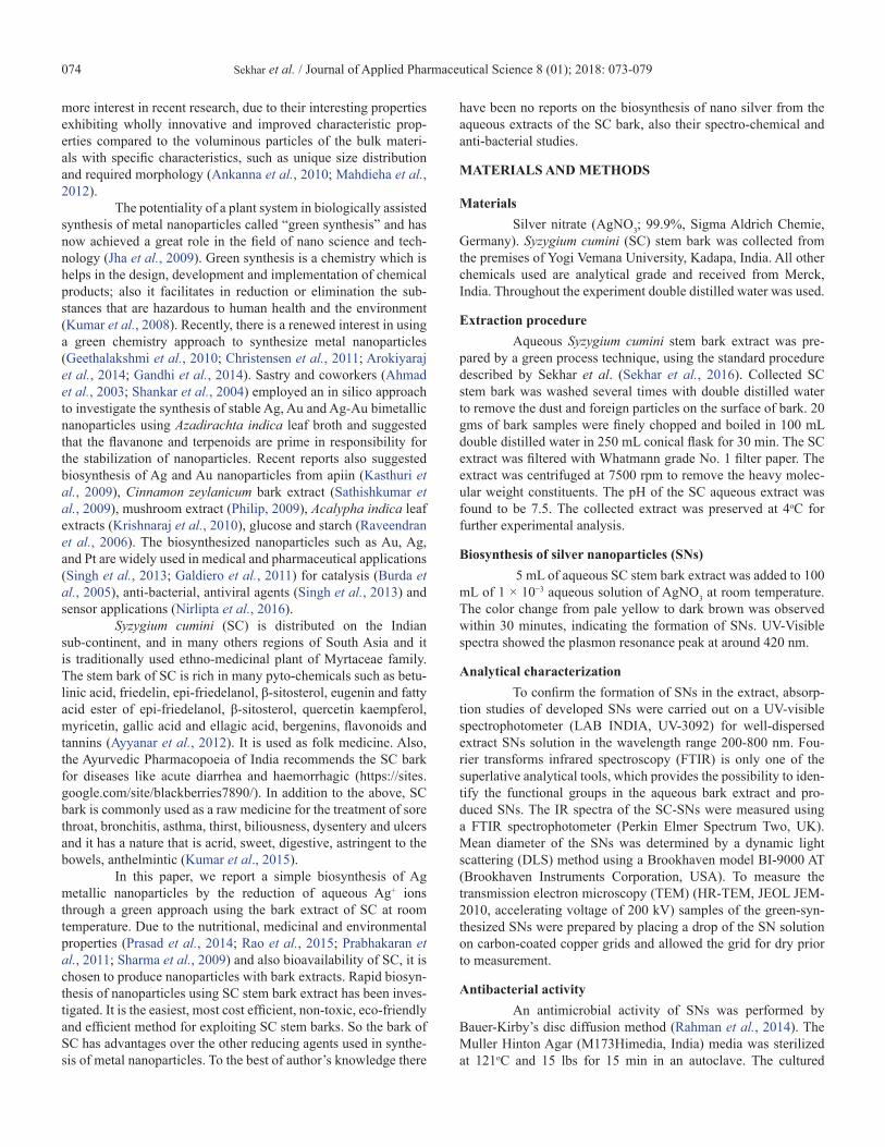
Particle size distribution analysis of biosynthesized SNsParticle size experiment of SNs was carried out by
means of dynamic light scattering. Particle size ascertainment of the produced nanoparticles was shown under heading like size distribution by intensity. The size of SNs dispersed was drifted widely from 10 nm to 20 nm (Figure (5)), the average particle size is expected to be in the range of ~14 nm and the peaks width was found to be 2.5. Allocation of particle size by magnitude gives a spherical shaped group. In an earlier study, SNs size distribu-tions were obtained in the range of 18.03 nm to 148.7 nm by using Saururus chinensis leaf extract (Morones et al., 2005).
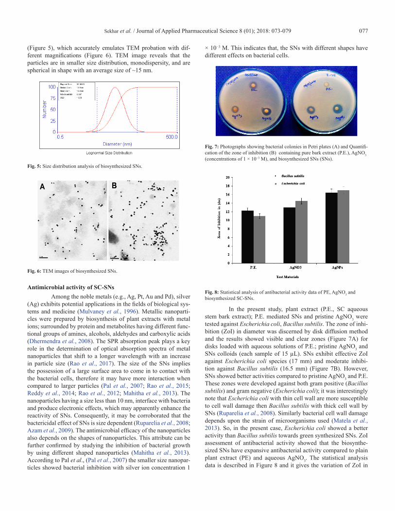
Transmission electron microscopic (TEM) analysisTEM images are apprehended with a dry sample; before
analysis of SNs using TEM and dynamic light scattering (DLS) was carried out to dictate the size of SNs in an aqueous solution using DLS. Size of SNs, size distribution, morphology of nano sized particles and surface morphology in solution are important factors in evaluating the biosynthesized SNs toxicity (Kreibig, 1995). Synthesized SNs showed with mean diameter of ~14 nm
Sekhar et al. / Journal of Applied Pharmaceutical Science 8 (01); 2018: 073-079 077
(Figure 5), which accurately emulates TEM probation with dif-ferent magnifications (Figure 6). TEM image reveals that the particles are in smaller size distribution, monodispersity, and are spherical in shape with an average size of ~15 nm.
Fig. 5: Size distribution analysis of biosynthesized SNs.
Fig. 6: TEM images of biosynthesized SNs.
Antimicrobial activity of SC-SNsAmong the noble metals (e.g., Ag, Pt, Au and Pd), silver
(Ag) exhibits potential applications in the fields of biological sys-tems and medicine (Mulvaney et al., 1996). Metallic nanoparti-cles were prepared by biosynthesis of plant extracts with metal ions; surrounded by protein and metabolites having different func-tional groups of amines, alcohols, aldehydes and carboxylic acids (Dhermendra et al., 2008). The SPR absorption peak plays a key role in the determination of optical absorption spectra of metal nanoparticles that shift to a longer wavelength with an increase in particle size (Rao et al., 2017). The size of the SNs implies the possession of a large surface area to come in to contact with the bacterial cells, therefore it may have more interaction when compared to larger particles (Pal et al., 2007; Rao et al., 2015; Reddy et al., 2014; Rao et al., 2012; Mahitha et al., 2013). The nanoparticles having a size less than 10 nm, interface with bacteria and produce electronic effects, which may apparently enhance the reactivity of SNs. Consequently, it may be corroborated that the bactericidal effect of SNs is size dependent (Ruparelia et al., 2008; Azam et al., 2009). The antimicrobial efficacy of the nanoparticles also depends on the shapes of nanoparticles. This attribute can be further confirmed by studying the inhibition of bacterial growth by using different shaped nanoparticles (Mahitha et al., 2013). According to Pal et al., (Pal et al., 2007) the smaller size nanopar-ticles showed bacterial inhibition with silver ion concentration 1
× 10−3 M. This indicates that, the SNs with different shapes have different effects on bacterial cells.
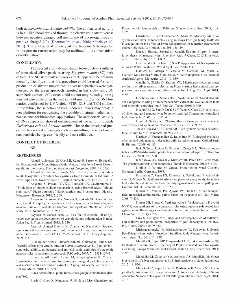
Fig. 7: Photographs showing bacterial colonies in Petri plates (A) and Quantifi-cation of the zone of inhibition (B) containing pure bark extract (P.E.), AgNO3 (concentrations of 1 × 10⁻³ M), and biosynthesized SNs (SNs).
Fig. 8: Statistical analysis of antibacterial activity data of PE, AgNO3 and biosynthesized SC-SNs.
In the present study, plant extract (P.E., SC aqueous stem bark extract); P.E. mediated SNs and pristine AgNO3 were tested against Escherichia coli, Bacillus subtilis. The zone of inhi-bition (ZoI) in diameter was discerned by disk diffusion method and the results showed visible and clear zones (Figure 7A) for disks loaded with aqueous solutions of P.E.; pristine AgNO3 and SNs colloids (each sample of 15 µL). SNs exhibit effective ZoI against Escherichia coli species (17 mm) and moderate inhibi-tion against Bacillus subtilis (16.5 mm) (Figure 7B). However, SNs showed better activities compared to pristine AgNO3 and P.E. These zones were developed against both gram positive (Bacillus subtilis) and gram negative (Escherichia coli); it was interestingly note that Escherichia coli with thin cell wall are more susceptible to cell wall damage then Bacillus subtilis with thick cell wall by SNs (Ruparelia et al., 2008). Similarly bacterial cell wall damage depends upon the strain of microorganisms used (Matela et al., 2013). So, in the present case, Escherichia coli showed a better activity than Bacillus subtilis towards green synthesized SNs. ZoI assessment of antibacterial activity showed that the biosynthe-sized SNs have expansive antibacterial activity compared to plain plant extract (PE) and aqueous AgNO3. The statistical analysis data is described in Figure 8 and it gives the variation of ZoI in
Sekhar et al. / Journal of Applied Pharmaceutical Science 8 (01); 2018: 073-079078
both Escherichia coli, Bacillus subtilis. The antibacterial activity is in all likelihood derived through the electrostatic attentiveness between negative charged cell membrane of microorganism and positive charged NPs (Dhermendra et al., 2008; Matela et al., 2013). The antibacterial potency of the biogenic SNs reported in the present retrospection may be attributed to the mechanism described above.
CONCLUSIONThe present study demonstrates bio-reductive synthesis
of nano sized silver particles using Syzygium cumini (SC) bark extract. The SC stem bark aqueous extracts appear to be environ-mentally friendly, so that this procedure could be used for rapid production of silver nanoparticles. Silver nanoparticles were syn-thesized by the green approach reported in this study using SC stem bark extracts. SC extract could act not only reducing the Ag ions but also controlling the size i.e. ~14 nm. Further the SNs for-mation conformed by UV-Visible, FTIR, DLS and TEM studies. In the future, the selection of such medicinal plants may create a new platform for recognizing the probable of natural medicines in nanoscience for biomedical applications. The antibacterial activity of SNs suspension showed enhancement of the activity towards Escherichia coli and Bacillus subtilis. Hence the developed pro-cedure has several advantages such as controlling the size of metal nanoparticles being, eco-friendly and cost effective.
CONFLICT OF INTERESTNil.
REFERENCESAhmad A, Senapati S, Khan MI, Kumar R, Sastry M, Extracellu-
lar Biosynthesis of Monodisperse Gold Nanoparticles by a Novel Extrem-ophilic Actinomycete, Thermomonospora sp. Langmuir. 2003; 19: 3550.
Ahmad N, Sharma S, Singh, VN. Shamsi, Fatma SFA, Meh-ta BF, Biosynthesis of Silver Nanoparticles from Desmodium triflorum: A Novel Approach Towards Weed Utilization. Biotech. Res. Int.; 2010; 2011.
Ankanna S, Prasad TNVKV, Elumalai EK, Savithramma N, “Production of biogenic silver nanoparticles using Boswellaovali foliolata stem bark,” Digest Journal of Nanomaterials and Biostructures, Digest J. Nanomater. Biostruct. 2010; 5: 369.
Arokiyaraj S, Arasu MV, Vincent S, Prakash NU, Choi SH, Oh YK, Kim KH, Rapid green synthesis of silver nanoparticles from Chrysan-themum indicum L and its antibacterial and cytotoxic effects: an in vitro study. Int. J. Nanomed. 2014; 9: 379.
Ayyanar M, Subash-Babu P, The effect of essential oil of Syz-ygium cumini on the development of granulomatous inflammation in mice. Asian Pac. J. Trop. Biomed. 2012; 2: 240.
Azam A, Ahmed F, Arshi N, Chaman M, Naqvi AH, One step synthesis and characterization of gold nanoparticles and their antibacteri-al activities against E. coli (ATCC 25922 strain). Int. J. Theory Appl. Sci. 2009; 1: 1.
Bilal Haider Abbasi, Sumaira Anjuma, Christophe Hanob, Dif-ferential effects of in vitro cultures of Linum usitatissimum L. (Flax) on bio-synthesis, stability, antibacterial and antileishmanial activities of zinc oxide nanoparticles: a mechanistic approach. RSC Adv., 2017; 7: 15931-15943.
Binupriya AR, Sathishkumar M, Vijayaraghavan K, Yun SI, Bioreduction of trivalent aurum to nano-crystalline gold particles by active and inactive cells and cell-free extract of Aspergillus oryzae var. viridis. J. Hazard. Mater. 2010; 177: 539.
Black berries/black plum. https://sites.google.com/site/blackber-ries7890/.
Burda C, Chen X, Narayanan R, El-Sayed MA, Chemistry and
Properties of Nanocrystals of Different Shapes. Chem. Rev. 2005; 105: 1025.
Christensen L, Vivekanandhan S, Misra M, Mohanty AK, Bio-synthesis of silver nanoparticles using murraya koenigii (curry leaf): An investigation on the effect of broth concentration in reduction mechanism and particle size. Adv. Mater. Let. 2011; 2: 429.
Deepali Sharma, Suvardhan Kanchi, Krishna Bisetty, Biogen-ic synthesis of nanoparticles: A review. Arab J Chem, 2015 https://doi.org/10.1016/j.arabjc.2015.11.002
Dhermendra K, Behari TJ, Sen P, Application of Nanoparticles in Waste Water Treatment. World Appl. Sci.; 2008; 3: 417.
Galdiero S, Falanga A, Vitiello M, Cantisani M, Marra V, Galdiero M, Veronica Marra, Galdiero M, Silver Nanoparticles as Potential Antiviral Agents. Molecules. 2011; 16: 8894.
Gandhi N, Sirisha D, Sharma VC, Microwave-mediated green synthesis of silver nanoparticles using Ficus elastica leaf extract and ap-plication in air pollution controlling studies. Int. J. Eng. Res. Appl. 2014; 4: 61.
Geethalakshmi R, Sarada DVL, Synthesis of plant-mediated sil-ver nanoparticles using Trianthemadecandra extract and evaluation of their anti-microbial activities. Int. J. Eng. Sci. Techn. 2010; 2: 970.
Huang J, Li Q, Sun D, Lu Y, Su Y, Yang X, Chen C, Biosynthesis of silver and gold nanoparticles by novel sundried Cinnanonum camphora leaf, Nanotechn. 2007; 18: 105104.
Husen A, Siddiqi KS, Phytosynthesis of nanoparticles: concept, controversy and application. Nanoscale Res. Lett. 2014; 9: 229.
Jha AK, Prasad K, Kulkarni AR, Plant system: nature’s nanofac-tory. Colloid Surf. B: Biointerf. 2009; 73: 219.
Kasthuri J, Veerapandian S, Rajendran N, Biological synthesis of silver and gold nanoparticles using apiin as reducing agent. Colloid Surf. B: Biointerf. 2009; 68: 55.
Keki S, Torok J, Deak G, Daroczi L, Zsuga MJ, ) Silver nanopar-ticles by PAMAM-assisted photochemical reduction of Ag?. J Colloid In-terface Sci. 2000; 229: 550.
Kharissova OV, Dias HV, Kharisov BI, Perez BO, Perez VMJ, The greener synthesis of nanoparticles. Trends in Biotechn. 2013; 31: 240.
Kreibig U, Vollmer M, Optical Properties of Metal Clusters. Springer. Berlin, Germany. 1995.
Krishnaraj C, Jagan EG, Rajasekar S, Selvakumar P, Kalaichel-van PT, Mohan N, Synthesis of silver nanoparticles using Acalypha indica leaf extracts and its antibacterial activity against water borne pathogens. Colloid Surf. B: Biointerf. 2010; 76: 50.
Kumar A., Vemula PK, Ajayan PM, John G, Silver-nanopar-ticle-embedded antimicrobial paints based on vegetable oil. Nat. Mater. 2008; 7: 236.
Kumar SH, Prasad C, Venkateswarlu S, Venkateswarlu P, Jyothi NVV, Green synthesis of silver nanoparticles using aqueous solution of Syz-ygium cumini flowering extract and its antimicrobial activity, Indian J. Adv. Chem. Sci. 2015; 3(4): 299.
Link S, El-Sayed MA, Shape and size dependence of radiative, non-radiative and photothermal properties of gold nanocrystals. Int. Rev. Phy. Chem. 2000; 19:409-453.
Lirdprapamongkol K, Warisnoicharoen W, Soisuwan S, Svasti. Eco-Friendly Synthesis of Fucoidan-Stabilized Gold Nanoparticles. Ameri-can J. Appl. Sci. 2010; 7: 1038.
Mahitha B, Raju BDP, Durgamaha CHN, Lakshmi, Sushma NJ, Evaluation of antimicrobial Efficiency of Phyto Fabicated Gold Nanoparti-cles Using Bacopa MonnieraPlant Extract. Indian J. Adv. Chem. Sci. 2013; 1: 94.
Mahdieha M, Zolanvarib A, Azimeea AS, Mahdiehc M, Green biosynthesis of silver nanoparticles by Spirulina platensis, Scientia Iranica.; 2012; 19: 926.
Malarkodi C, Rajeshkumar S, Paulkumar K, Vanaja M, Gnana-jobitha G, Annadurai G, Biosynthesis and Antimicrobial Activity of Semi-conductor Nanoparticles against Oral Pathogens. Bioin. Chem. Appl. 2014; 2014.
Sekhar et al. / Journal of Applied Pharmaceutical Science 8 (01); 2018: 073-079 079
Man WL, Kwong HK, Lam WW, Xiang J, Wong TW, Lam WH, Lau TC, General synthesis of (salen) ruthenium (III) complexes via N-N coupling of (salen)ruthenium(VI) nitrides. Inorg. Chem. 2008; 47: 5936.
Matela G, Aman R, Sharma C, Chaudhary S, Synthesis, Charac-terization and Antimicrobial Evaluation of Diorganotin(IV) Complexes of Schiff Base. Indian J. Adv. Chem. Sci.; 2013; 1: 157.
Morones JR, Elechiguerra JL, Camacho A, Holt K, Kouri JB, Ramirez JT, Yacaman MJ, The bactericidal effect of silver nanoparticles. Nanotechnology. 2005; 16: 2346.
Mulvaney P, Surface Plasmon Spectroscopy of Nanosized Metal Particles. Langmuir.; 1996; 12:788.
Nadagouda MN, Varma RS, Green synthesis of silver and palla-dium nanoparticles at room temperature using coffee and tea extract. Green Chem. 2008; 10: 859.
Nirlipta Saha, Priyanka Trivedi S. Dutta Gupta. Surface Plasmon Resonance (SPR) Based Optimization of Biosynthesis of Silver Nanoparti-cles from Rhizome Extract of Curculigo orchioides Gaertn. and Its Antioxi-dant Potential. J Clust Sci. 2016; DOI 10.1007/s10876-016-1050-7.
Nune SK, Chandra N, Shukla R, Katti K, Kulkarni RR, Thilaka-vathy, Mekapothula S, Kannan R, Katti KV, Green Nanotechnology from Tea: Phytochemicals in Tea as Building Blocks for Production of Biocom-patible Gold Nanoparticles. J. Mat. Chem. 2009; 19: 2912.
Pal S, Tak YK, Song JM, Does the Antibacterial Activity of Sil-ver Nanoparticles Depend on the Shape of the Nanoparticle? A Study of the Gram-Negative Bacterium Escherichia coli. Appl. Env. Micr. 2007; 73: 1712.
Philip D, Biosynthesis of Au, Ag and Au-Ag nanoparticles using edible mushroom extract. Acta A Mol. Biomol. Spectrosc. 2009; 73: 374.
Prabhakaran S, Gothandam KM, Sivashanmugam K, Phyto-chemical and antimicrobial properties of Syzygium cumini an ethanomedic-inal plant of Javadhu hills Res. Pharm. 2011; 1: 22.
Prasad Ch, Venkateswarlu P, Soybean seeds extract based green synthesis of SNPs, Indian J. Adv. Chem. Sci. 2014; 2: 208.
Praveena VD, Kumar KV, Green synthesis of Silver Nanoparti-cles from Achyranthes Aspera Plant Extract in Chitosan Matrix and Eval-uation of their Antimicrobial Activities. Indian J. Adv. Chem. Sci.; 2014; 2: 171.
Rahman MM, Hattori N, Nakagawa Y, Lin X, Yagai S, Sakai M, Yamamoto K, Preparation and characterization of silver nanoparticles on localized surface plasmon-enhanced optical absorption. J. Appl. Phy. 2014; 53 (11S): 11RE01.
Rao Ch V, Viswanathan B, Monodispersed Platinum Nanopar-ticle Supported Carbon Electrodes for Hydrogen Oxidation and Oxygen Reduction in Proton Exchange Membrane Fuel Cells. J. Phys. Chem. C. 2010; 114: 8661.
Rao Ch V, Viswanathan B, Microemulsion synthesis and electro-catalytic properties of carbon-supported Pd–Co–Au alloy nanoparticles. J. Colloid Inte. Sci. 2012; 367: 337.
Rao KM, Sekhar EC, Rao KSVK. Biosynthesis of Microbial Resistance Au-nanoparticles from Aqueous Extract of Tridax procumbens Leaves. Indian J. Adv. Chem. Sci. 2017; 5(1): 24.
Rao KSVK, Rao KM, Reddy PRS, Reddy NS, Shchipunov Y, Ha CS, Chitosan–poly(aminopropyl/phenylsilsesquioxane) hybrid nanocom-posite membranes for antibacterial and drug delivery applications. Polym. Int.; 2015; 64: 293.
Rao KSVK, Reddy PRS, Rao KM, Kumar SP, A Green Approach to Synthesize Silver Nanoparticles from Natural Polymer for Biomedical Application, Indian J. Adv. Chem. Sci. 2015; 3: 340.
Rao KSVK, Reddy PRS, Yong-Ill Lee, Kim C, Synthesis and
Characterization of Chitosan-PEG-Ag Nanocomposites for Antimicrobial Application. Carbohyd. Polym. 2012; 87: 920.
Raveendran P, Fu J, Wallen SL, A simple and “green” method for the synthesis of Au, Ag, and Au–Ag alloy nanoparticles. Green Chem. 2006; 8: 34.
Reddy AS, Chen CY, Chen CC, Jean JS, Chen HR, Tseng MJ, Fan CW, Wang JC, Biological synthesis of gold and silver nanoparticles mediated by the bacteria Bacillus subtilis. J. Nanosci. Nanotechnol. 2010; 10: 6567.
Reddy PRS, Rao KM, Rao KSVK, Shchipunov Y, -Ha CS, Syn-thesis of alginate based silver nanocomposite hydrogels for biomedical ap-plications. Macromol. Res. 2014; 22: 832.
Ruparelia JP, Chatterjee AK, Duttagupta SP, Mukherji S, Strain specificity in antimicrobial activity of silver and copper nanoparticles. Acta Biomater.; 2008; 4: 707.
Sarkar R, Kumbhakar P, Mitra AK, Green Synthesis of Silver Nanoparticles and Its Optical Properties. Digest J. Nanomater. Biostruct. 2010; 5: 491-496.
Sathishkumar M, Sneha K, Won SW, Cho CW, Kim S, Yun YS, Cinnamon zeylanicum bark extract and powder mediated green synthesis of nano-crystalline silver particles and its bactericidal activity. Colloid Surf. B: Biointerf.; 2009; 73: 332.
Shankar SS, Rai A, Ahmad A, Sastry MJ, Rapid synthesis of Au, Ag, and bimetallic Au core-Ag shell nanoparticles using Neem (Azadirach-ta indica) leaf broth. Colloid. Interf. Sci. 2004; 275: 496.
Sharma A, Patel VK, Chaturvedi AN, Vibriocidal activity of cer-tain medicinal plants used in Indian folklore medicine by tribals of Maha-koshal region of central India. Ind. J. Pharmacology. 2009; 41: 129.
Sharma VK, Yngard RA, Lin Y, Silver nanoparticles: green syn-thesis and their antimicrobial activities. Adv. Colloid. Interf. Sci. 2009; 145: 83.
Sekhar EC, Rao KM, Rao KSVK, A green approach to synthe-size controllable silver nanostructures from Limonia acidissima for inacti-vation of pathogenic bacteria, Cogent Chemistry. 2016; 2: 1144296.
Sekhar EC, Rao KSVK, Rao KM, Bio-synthesis and character-ization of silver nanoparticles using Terminalia chebula leaf extract and evaluation of its antimicrobial potential. Materials Letters. 2016; 174: 129.
Singh M, Kalaivani R, Manikandan S, Sangeetha N, Kumaragu-ru AK, Facile green synthesis of variable metallic gold nanoparticle using Padina gymnospora, a brown marine macroalga. Appl. Nanosci. 2013; 3: 145.
Sinha S, Pan I, Chanda P, Sen SK, Nanoparticles fabrication us-ing ambient biologicalresources. J. Appl. Biosci. 2009; 19: 1113.
Xiaogang Qu, Journal of Materials Chemistry B top picks web collection: “seeing the unseen: advances in bioimaging and biosensors” J. Mater. Chem. B, 2016; 4: 4500-4501.
Zhu H, Han J, Xiao JQ, Jin YJ, Uptake, translocation, and ac-cumulation of manufactured iron oxide nanoparticles by pumpkin plants. Environ. Monit. 2008; 10: 713.
How to cite this article: Sekhar EC, Rao KSVK, Rao KMS, Alisha SB. A simple biosyn-thesis of silver nanoparticles from Syzygium cumini stem bark aqueous extract and their spectrochemical and antimicrobial studies. J App Pharm Sci, 2018; 8 (01): 073-079.