A rapid sampling technique for isolating highly productive ... · Biofuel Research Journal 21...
7
* Corresponding author at: Tel.: +98 9374997785 E-mail address: [email protected] Please cite this article as: Huang S.T., Goh J.L., Ahmadzadeh H., Murry M.A. A rapid sampling technique for isolating highly productive lipid-rich algae strains from environmental samples. Biofuel Research Journal 21 (2019) 920-926. DOI: 10.18331/BRJ2019.6.1.3 Biofuel Research Journal 21 (2019) 920-926 Original Research Paper A rapid sampling technique for isolating highly productive lipid-rich algae strains from environmental samples Suting T. Huang 1 , Jo L. Goh 2 , Hossein Ahmadzadeh 3, *, Marcia A. Murry 1 1 Department of Biological Sciences, California State Polytechnic University, Pomona, CA. 2 Department of Chemistry, California State Polytechnic University, Pomona, CA. 3 Department of Chemistry, Ferdowsi University of Mashhad, Mashhad 1436-91779, Iran. HIGHLIGHTS Selection of lipid-rich native strains for biofuel production. Single cell sampling techniques is used for strain selection. Rapid growth by direct selection of product-rich species is achieved. The enrichment step can be manipulated to select for strains for specific technological applications. Direct sampling of lipid-rich cells avoids tedious task of screening isolates while using relatively inexpensive equipment. GRAPHICAL ABSTRACT ARTICLE INFO ABSTRACT Article history: Received 20 September 2018 Received in revised form 12 January 2019 Accepted 12 January 2019 Available online 1 March 2019 Keywords: Strain selection Algae isolation Single cell sampling Lipid-rich microalgae Algae screening Biofuel production Strain selection and isolation of lipid-rich microalgae are among the most important steps for screening isolates with maximum biofuel productivity. In this work, we introduce a novel direct sampling technique that allows native strains to be selected for rapid growth under defined conditions followed by direct selection of product-rich species, two desirable characteristics of algae for mass culture. This sampling strategy directly selects the lipid-rich strains visualized under an inverted fluorescence microscope using an X-Y-Z micromanipulator. The enrichment step can be manipulated to select for strains with specific technological applications. Direct sampling of lipid-rich cells avoids the tedious task of screening isolates while using relatively inexpensive equipment. © 2019 BRTeam. All rights reserved. Journal homepage: www.biofueljournal.com
Transcript of A rapid sampling technique for isolating highly productive ... · Biofuel Research Journal 21...

* Corresponding author at: Tel.: +98
9374997785
E-mail address: [email protected]
Please cite this article as: Huang S.T., Goh J.L., Ahmadzadeh H., Murry
M.A. A rapid sampling technique for isolating highly productive lipid-rich algae
strains from environmental samples.
Biofuel Research Journal 21
(2019) 920-926. DOI: 10.18331/BRJ2019.6.1.3
Biofuel Research Journal 21 (2019) 920-926
Original Research Paper
A rapid sampling technique for isolating highly productive lipid-rich algae strains from
environmental samples
Suting T. Huang1, Jo L. Goh2, Hossein Ahmadzadeh3,*, Marcia A. Murry1
1Department of Biological Sciences, California State Polytechnic University, Pomona, CA.
2Department of Chemistry, California State Polytechnic University, Pomona, CA.
3Department of Chemistry, Ferdowsi University of Mashhad, Mashhad 1436-91779, Iran.
HIGHLIGHTS
Selection of lipid-rich native strains for biofuel
production.
Single cell sampling techniques is used for strain
selection.
Rapid growth by direct selection of product-rich
species is achieved.
The enrichment step can be manipulated to select
for strains for specific technological applications.
Direct sampling of lipid-rich cells avoids tedious
task of screening isolates while using relatively
inexpensive equipment.
GRAPHICAL ABSTRACT
ARTICLE INFO ABSTRACT
Article history:
Received
20
September
2018
Received in revised form 12
January 2019
Accepted
12 January 2019
Available online
1 March
2019
Keywords:
Strain selection
Algae isolation
Single cell sampling
Lipid-rich microalgae
Algae screening
Biofuel production
Strain selection and isolation of lipid-rich microalgae are among the most important steps for screening isolates with maximum
biofuel productivity. In this work, we introduce a novel direct sampling technique
that allows native strains to be selected for
rapid growth under defined conditions followed by direct selection of product-rich species, two desirable characteristics of algae
for mass culture. This sampling strategy directly selects the lipid-rich strains visualized under an inverted fluorescence
microscope using an X-Y-Z micromanipulator. The enrichment step can be manipulated to select for strains with specific
technological applications. Direct sampling of lipid-rich cells avoids the tedious task of screening isolates while using relatively
inexpensive equipment.
© 2019
BRTeam.
All rights reserved.
Journal homepage: www.biofueljournal.com

Huang et al. / Biofuel Research Journal 21 (2019) 920-926
Please cite this article as: Huang S.T., Goh J.L., Ahmadzadeh H., Murry M.A. A rapid sampling technique for isolating highly productive lipid-rich algae strains
from environmental samples. Biofuel Research Journal 21 (2019) 920-926. DOI: 10.18331/BRJ2019.6.1.3
1. Introduction
The quest for economically and environmentally viable alternatives to
petroleum-based fuels has been the driving force behind the rapid growth of the
biofuels industry. Biodiesel, an alternative to diesel fuel, is produced from oils via transesterification producing Fatty Acid Methyl Esters (FAMEs) that can
be used directly as a transportation fuel. Because biodiesel is renewable,
nontoxic, and biodegradable, it has the potential to replace conventional diesel fuel. However, first generation biofuels including biodiesel produced from
plant oils extracted from corn, soy, and palm oil and bioethanol derived from
the fermentation and distillation of traditional crops such as sugar cane and corn, have been associated with serious negative impacts. More specifically,
they have hindered global food supplies (with regards to soy and corn) and have
resulted in the destruction of tropical rain forest with regards to sugar cane and oil palm. In response to the difficulties associated with the first generation
biofuels, algae-based biofuels have gained considerable attention in recent
years. Many species of microalgae have properties well suited for commercial
scale biodiesel production including rapid growth rate, high lipid content, and
the ability to grow on marginal lands in saline or waste waters not suitable for
agricultural irrigation (Murry and Benemann, 1980; Sheenan et al., 1998). The extensive work carried out by the DOE/NREL sponsored Aquatic
Species Program to explore large-scale algae production for biodiesel
production provided a number of recommendations for further work (Sheenan et al., 1998). Key among these was to isolate native strains for mass cultivation
to ensure adaptation to local conditions. We are particularly interested in native
strains that grow rapidly on animal wastes to allow bioremediation and nutrient recovery from animal wastes, a serious problem in intensive meat and dairy
operations. We are also interested in strains that can grow in waters high in
salinity and/or alkalinity. An early, but key analysis of the economics of algae-based biofuels considered the importance of water quality and availability to
the economics, design, and operation of large-scale ponds (Weissmann and
Goebel, 1987). They considered low cost, high salt or alkaline waters and agricultural waste waters as prime candidates for economically feasible
ponding scenarios in sunny areas where cheap land is available, notably in the
southwest U.S. It has also been suggested that algae isolated from fluctuating and adverse environments, such as tide pools and estuaries, would be
opportunistic fast growing strains with an ability to accumulate storage lipids
as a survival mechanism (Duong et al., 2012). Microalgae are a phylogenetically diverse group of photosynthetic
microorganisms that vary greatly in their metabolic capabilities, environmental
adaptations, and growth rate. Microalgae have oil content that varies from 15 to 77% of the dry weight (Chisti, 2007) and in general, lipid biosynthesis is
regulated by environmental variables as recently reviewed (Lari et al., 2016).
Although many culture collections of microalgae have been established, the variety of unknown strains present in the environment with potential
application in the production of biofuels and for other biotechnological uses is
likely very high and has been poorly explored to date. “Bio-prospecting” for useful strains requires rapid high throughput screening procedures in order to
isolate novel species that are adapted to production location and for specific
purposes. Although several techniques for microalgae isolation have been described
previously, including single-cell isolation using serial dilutions, micromanipulation, gravimetric separation, and atomized cell spray (Anderson
et al., 2005; Mutanda et al., 2011), traditional means of isolating algal strains
are time consuming and require further screening of axenic cultures to identify growth and lipid biosynthesis patterns. Flow cytometry coupled to cell sorting
has led to rapid selection of lipid rich strains (Pereira et al., 2011; Cabanelas et
al., 2014 and 2016) but the cost of these instruments restricts use of this technology to larger well-funded labs. To facilitate isolation of desirable strains
for both rapid growth and high oil productivity, we have developed an
enrichment strategy, coupled with a capillary aided sampling procedure. This allows for the direct selection of oil-rich strains from a heterogeneous
population. The objective is to select individual algal cell candidates for biofuel
and feed production coupled to bioremediation of agricultural wastes.
2. Materials and Methods
2.1. Strains and culture conditions
Environmental samples were collected from livestock fields and fecal
contaminated marine, freshwater, and estuarine sites in Southern California
(Table 1) and transferred to defined media at approximately 1:10 dilution. Conductivity was measured at sampling sites using a Beckman Coulter
meter (Model pHi 410; Fullerton, CA) and samples were incubated in
media with matching salinity. Enrichment cultures were incubated at 24 oC on a rotary shaker (100 rpm) under constant light at a photon flux density
of 59 µmol m-2 s-1 for 4 to 7 d to enrich for fast growing strains.
2.2. Enrichment media
Basal media (B) contained KH2PO4 7 mM; H3PO4, 3.4 mM; MgSO4, 0.1
mM; CaCl2, 0.07 mM; KCl, 2.35 mM; FeNaEDTA, 10 mg/L; and trace
elements (Ripka et al., 1979). As source of nitrogen, NH4Cl, 5 mM (BN) or
filter sterilized urea, 5 mM (BUN), was used. For an organic media with all essential elements, a duck pond sample was supplemented with 10% Luria
broth (LB) and B (DP). An artificial sewage (AS) media (OECD, 2001)
contained in tap water: peptone, 160 mg; meat extract, 110 mg; urea, 30
mg; K2HPO4, 28 mg; NaCl, 7 mg; CaCl2.2H2O, 4 mg; and MgSO4.7H20, 2
mg. Artificial seawater (SW) media (containing NaCl, 0.4 M; MgSO4, 13.3
mM; MgCl2, 25 mM; CaCl2, 8.2 mM; KCl, 9.4 mM; NH4Cl and/or urea (filter sterilized), 5 mM; Na2CO3, 0.16 mM; KH2PO4, 0.5mM; FeNaEDTA,
10 mg/L), was used. All media were supplemented with F/2 vitamins
(Guillard and Ryther, 1962), Tris pH 7.5 (10mM) to control pH, and either streptomycin, vancomycin or kanamycin at 25µg/mL to decrease bacterial
contamination. For solid media, agar was added at 1.5% w:v and dispensed
into petri dishes or microtiter dishes for cell isolation. To enrich for strains with a “N-trigger” for lipid biosynthesis, actively growing mixed
enrichment cultures were harvested by centrifugation (5,000 rpm, 10 min)
and the pellet washed in N-free media, re-suspended in low nitrogen media (0.5 mM NH4Cl), and incubated for 3-4 d before sampling. Axenic strains
were tested for microbial contamination and for the ability to grow
heterotrophically by spotting a 10-µL sample on solid LB media and incubating in the dark at room temperature for 5 d.
2.3. Screening for growth rates and lipid content
Axenic cultures were inoculated (10% v:v) with log phase cultures
grown in the same media and grown as described above. The light intensity was increased up to 100 µmol/m2.s after 2 to 3 d of incubation to avoid light
limitation. Experiments were carried out either in duplicate or in most cases
in triplicate. Fv/Fm, an indicator of photosynthetic efficiency (Parkhill et al., 2001), was measured in triplicate 1 mL aliquots transferred to an OS1p
cuvette and adapted for 20 to 30 min in the dark before measurements were
made using an OS1p-Fl Modulated Chlorophyll Fluorometer (Opti-Sciences, Hudson N.H.). Biomass accumulation was measured by vacuum
filtration of 10 to 25 mL of culture using MFS mixed cellulose ester 47 mm
diameter, 0.45 µm pore size filters. The cells were washed with several volumes of distilled water and were vacuum dried at 60 oC overnight and
cooled in a desiccation chamber under vacuum before weighing. Dry
weight was correlated with optical density at 650 nM in triplicate 200 µL
aliquots harvested daily using a Multiskan FC with incubator (Thermo
Scientific) with SkanIt Software. The growth rate was calculated during logarithmic growth phase by N=No e
kt where N is the biomass at end of
logarithmic growth and No is the biomass at time zero (to), the beginning of
logarithmic growth phase (Fogg, 1987). From this, the mean doubling time G in days (the mean generation time if the cells divide into two) is as
follows (Eq. 1):
G =0.301
k k =
logeN−logeN0
t Eq. 1
2.4. In-vivo lipid staining
To identify individual cells with high lipid content, mixed enrichment cultures of both N-replete and N-deficient cultures were stained with a
lipophilic fluorescent dye, Nile Red. Nile red (9-(diethyl amino)
benzo[a]phenoxazin-5(5H)-one, Sigma– Aldrich) was prepared at 250
921

Huang et al. / Biofuel Research Journal 21 (2019) 920-926
Please cite this article as: Huang S.T., Goh J.L., Ahmadzadeh H., Murry M.A. A rapid sampling technique for isolating highly productive lipid-rich algae strains
from environmental samples. Biofuel Research Journal 21
(2019) 920-926. DOI: 10.18331/BRJ2019.6.1.3
mg/L
in acetone and stored in darkness at -20
oC. Nile Red (NR) staining
procedure was performed using 1 µg/mL
of NR and 10% DMSO to allow cell
penetration (Chen et al.,
2009). Cells were incubated in the dark at 37
oC for 10
min and observed microscopically using the DAPI filter system.
For monitoring lipid content in axenic cultures, BODIPY505/515 (4, 4-
difluro-1, 3, 5, 7-tetramethyl-4-bora-3a, 4adiaza-
s-indacene, Invitrogen Molecular Probes, Carlsbad, CA) was prepared in DMSO to give a stock
solution of 5
mM and stored in darkness at -20
oC. An incubation time of 30
min at room temperature was used for all assays. The stained algae were analyzed on a Varian Cary Eclipse Fluorescence 96-well Microplate
Spectrophotometer (XP900, Varian Australia Pty Lt) with a 490 nm narrow band (10
nm) excitation filter,
a 525 nm emission filter (10
nm),
and
photomultiplier at 400 V. Relative fluorescence intensity of stained cells was
calculated by subtraction of autofluorescence of the unstained algae and correlated with gravimetric lipid measurements of each strain (R2
of correlation
ranged from 0.909 to 0.997).
2.5. Lipid analysis
The
lipid contents of microalgae cultures were analyzed by modifications to the gravimetric method of Bligh and Dyer (1959). Briefly, 1 g of wet algal
biomass was mixed with 2 mL
of methanol and 1
mL
of chloroform and
disrupted using 0.5
mm glass beads for 2 min at high speed in a mini-bead-
beater (Biospec, Inc. Bartlesville, OK). The mixture was vortexed for 2 min
and centrifuged at 12,000
rpm for 10 min. The chloroform layer was collected
carefully and the extraction process was repeated three times. The organic extracts were combined and dried by N2
airflow. The lipid residues were dried
in an oven at 60
°C for 50 min and cooled in a vacuum desiccator before
weighing.
2.6. Sampling
An Olympus IX-71 inverted fluorescence microscope (Olympus
Corporation, Tokyo, Japan) equipped with a U-MWB
cube with a band pass 450-480 nm excitation filter and a dichroic mirror, DM500,
with high
transmission above 500 nm, was used to view NR-stained cells. The barrier
filter was a BA515, with a steep slope below 515 nm. A clear Plexiglas capillary holder was mounted between the light condenser and the objective
lens, without blocking light transmission for observations (Ahmadzadeh et
al., 2004). The capillary holder was mounted on a micromanipulator (Soma Scientific, Irvine, CA) to position the capillary over cells of interest. A
flexible fused silica capillary tubing (150 m Outer Diameter (OD) and a
50
m Inner Diameter (ID) (Polymicro Technologies, Phoenix, AZ ) was held vertically by
the Plexiglas holder and sampling was achieved with
pressure applied by a 1 mL
disposable syringe coupled to the tubing (Fig.
1). The capillary was sterilized by flushing with 70% ethanol and cleaned
with sterile media before sampling. The sampling end of
the capillary was
trimmed using a sapphire cutter to obtain a clean straight cut.
To identify individual cells with high lipid content, cells
were diluted to
approximately 105
cells/mL
and a 10
µL
sample was placed on a silanized
coated glass cover slip and observed using bypass filters to visually identify lipid-rich cells. Using the X and Y manipulation knobs, the lumen of the
capillary was positioned directly above the target cell. Using the Z
manipulation knob, the micromanipulator was lowered to position the target
cell within the capillary lumen. Negative pressure was applied on the
opposite side of the capillary to capture the cell that was then deposited on
the surface of solid media for growth and colony formation.
To determine the viability of cells stained with NR, serial dilutions of
stained cells
were
plated
on solid
media in
triplicates
after
staining and
Table 1. Collection sites in Southern California of isolated strains, enrichment media, and conductivity (in Siemens) of sites. H: heterotrophic growth. Strain Identification was based on morphology and 100%
similarity between amplified ITS sequences, and the closest sequence in the National Center for Biotechnology Information (NCBI) using the Basic Local Alignment Search Tool (BLAST).
Isolate Species Gene Bank# of related
spp.
Enrichment
media* Collection site**
Conductivity
(S/cm) H
CP214 Auxenochlorella protothecoides LN610701.1 BN Lyle Pond, Pomona 880 uS Yes
CP215 Desmodesmus sp. AB917110.1 BN Lyle Pond, Pomona 880 uS Yes
CPP4 Pseudochlorella sp. KY364701.1 BUN Animal lot, Pomona - Yes
CPP5 Auxenochlorella pyrenoidosa KX752082,1 BUN Animal lot, Pomona - Yes
CPP7 Chlorella sorokiniana KY303731.1 BUN Animal lot, Pomona - Yes
CPP9 Auxenochlorella protothecoides LN610701.1 BUN Animal lot, Pomona - Yes
CPP201 Scenedesmus sp. KU170547.1 BUN Lyle Pond, Pomona 880 uS Yes
CPP210 Micractinium sp. AB917104.1 BUN Lyle Pond, Pomona 880 uS Yes
CPP13 Coelastrum sp. GQ375097.1 BUNQ Animal lot, Pomona - Yes
CPP98 Chlorella vulgaris MF686487.1 BUNQ Animal lot, Pomona - Yes
CPP82 Chlorella sp. KP726221.1 SWU Salton Sea 71.5 mS Yes
CPP 18 Chlorella sp. KM061458.1 BUNQ Malibu Lagoon 22.1 mS Yes
CPP67 Desmodesmus sp. DQ417525.1 BUNQ Malibu Lagoon 22.1 mS Yes
CPP33 Dictyosphaerium sp. GQ477066.1 BUNQ Salton Sea 71.5 mS Yes
CPP60 Chlorella vulgaris MF686487.1 BUNQ Animal lot, Pomona - Yes
CPP98 Pseudochlorella sp. KY364701.1 BUNQ Animal lot, Pomona - Yes
CPP215 Scenedesmus obliquus FR865731.1 BUNQ Lagoon, San Onofre 26.6 mS Yes
CPP171 Heterochlorella luteoviridis LN610702 SW Ocean, San Onofre 54.6 mS Yes
CPP153 Tetraselmis sp. HE610131.1 SW Ocean, San Onofre 54.6 mS -
*
BN: basal media enriched with NH4Cl
(5 mM) as nitrogen source; BUN: basal media enriched with filter sterilized urea
(5 mM) as source of nitrogen; BUNQ: quarter strength
BUN;
SW:
artificial
seawater;
SWU:
SW
enriched with filter sterilized urea
(5 mM) as source of nitrogen.
** Coordinates: Lyle Pond and animal fields at Cal Poly Pomona N34
2’ 57.19; W117
49’ 25.60; Malibu Lagoon N34.03453°; E-118.6852°;
Salton Sea N33015’12.7; W1150 42.6; San Onofre
N33 22’ 51.64; W117
34’ 42.83.
922
Huang et al. / Biofuel Research Journal 21 (2019) 920-926
Please cite this article as: Huang S.T., Goh J.L., Ahmadzadeh H., Murry M.A. A rapid sampling technique for isolating highly productive lipid-rich algae strains
from environmental samples. Biofuel Research Journal 21 (2019) 920-926. DOI: 10.18331/BRJ2019.6.1.3
colony forming units (CFU) were compared between treated and untreated cells. To assess the effect of exposure to fluorescent light during the sampling
procedure, the percentage of cells sampled that formed viable colonies after 10
d of incubation in the light was measured. 2.7. Strain identification
Genomic DNA was extracted as described by Fawley and Fawley (2004).
The ITS region (~650bp) from the 3’end of the 18S small subunit to the 5’end of the 28S large subunit was amplified using ITS 4 (5’
TCCTCCGCTTATTGATATGC3) and ITS 5 (5’GGAAGTAAAAGTCGT AACAAGG3) primers developed by White et al. (1990). Amplifications were done in a 50 μL reaction mixture containing 5× GoTaq colorless buffer
(Promega, WI), 2 mM MgCl2, 250 μM of each dNTP, 0.1 μM of each primer,
1 unit of GoTaq Taq polymerase, and 1 μL of template DNA. The PCR cycle was as follows: an initial cycle of denaturation, 95 °C for 5 min; followed by
35 cycles of denaturation, 95 °C for 1 min; annealing, 55 °C for 1 min; and
extension, 72 °C for 1 min; and a final extension step of 72 °C for 5min. PCR products were visualized under UV illumination on 2% agarose gel containing ethidium bromide (1µg/mL). PCR products were purified using an UltraClean
PCR clean-up DNA purification kit (MO BIO, Carlsbad, CA) and were commercially sequenced in both directions (Retrogen Inc, San Diego CA).
Sequence analysis, alignment, and phylogenetic trees were constructed using Geneious software (Drummond et al., 2010).
3. Results and Discussion
Algae strains isolated for biotechnical applications need to grow rapidly in
large-scale systems to ensure an economically sustainable process. This is especially true in selecting strains for low value commodity products including
biodiesel and animal feeds in which the economics of production dictates the
use of outdoor ponds. Fast growing strains, well adapted to the local environment and culture media, have the potential to out compete invading
competitors as well as providing high biomass productivity which also favors
the economics of downstream processing (Borowitzka, 1997).
Environmental samples collected from sites in Southern California with
significant fecal contamination, including marine sites, estuaries, fresh water,
and animal fields (Table 1), were grown in nitrogen-rich defined media with varying combinations of salinity and nitrogen sources to amplify proportionally
the fastest growing strains under specific media conditions. The strains fell
within a cluster of Chlorophytes associated with eutrophic waters and most were heterotrophic. Aliquots of each culture were transferred to nitrogen-free
media to identify strains in which nitrogen limitation triggers lipid biosynthesis.
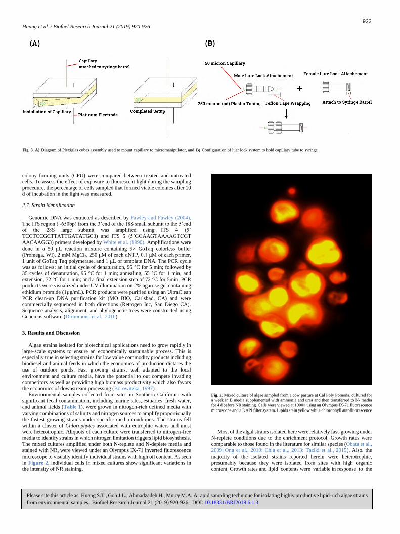
The mixed cultures amplified under both N-replete and N-deplete media and stained with NR, were viewed under an Olympus IX-71 inverted fluorescence
microscope to visually identify individual strains with high oil content. As seen
in Figure 2, individual cells in mixed cultures show significant variations in the intensity of NR staining.
Most of the algal strains isolated here were relatively fast-growing under
N-replete conditions due to the enrichment protocol. Growth rates were
comparable to those found in the literature for similar species (Obata et al.,
2009;
Ong et al., 2010; Chia et al., 2013; Taziki et al., 2015). Also, the
majority of the isolated strains reported herein
were heterotrophic,
presumably because they were isolated from sites with high organic content. Growth rates and lipid
contents
were
variable in response
to
the
Fig. 2. Mixed culture of algae sampled from a cow pasture at Cal Poly Pomona, cultured for
a week in B media supplemented with ammonia and urea and then transferred to N- media
for 4 d before NR staining. Cells were viewed at 1000× using an Olympus IX-71 fluorescence
microscope and a DAPI filter system. Lipids stain yellow while chlorophyll autofluorescence
Diagram of Plexiglas cubes assembly used to mount capillary to micromanipulator, and Configuration of luer lock system to hold capillary tube to syringe.Fig. 3. A) B)
923

Huang et al. / Biofuel Research Journal 21 (2019) 920-926
Please cite this article as: Huang S.T., Goh J.L., Ahmadzadeh H., Murry M.A. A rapid sampling technique for isolating highly productive lipid-rich algae strains
from environmental samples. Biofuel Research Journal 21 (2019) 920-926. DOI: 10.18331/BRJ2019.6.1.3
Table 2.
Growth rate during exponential growth, lipid content as percentage of dry weight, and Fv/Fm
values of select isolates.
media used for cultivation. In most cases, growth rates increased
significantly when basal inorganic media was supplemented with organics (DP media) and often lipid content in log phase was enhanced over those
seen in N-deplete media (Table 2). In contrast, AS media promoted early
rapid growth relative to inorganic media but was followed by declining growth, Fv/Fm values, and enhanced lipid biosynthesis, similar to the
response in N-deplete cultures. Because of the high organic N levels in AS,
it is unlikely that N limited growth and impacted photosynthetic efficiency, but rather that other essential nutrients such as iron and trace minerals were
growth limiting leading to enhanced lipid biosynthesis as described earlier
(Spoehr and Milner, 1949; Shifrin and Chisholm, 1981). To isolate oil-rich cells from a heterogeneous suspension, the field was
viewed at low magnification under dim bright field illumination to focus
and to roughly position the capillary, and then under 400× magnification, the cells were illuminated with fluorescence light briefly to identify cells
with high lipid content. The light was returned to bright field illumination
and the micromanipulator was positioned directly over the target cell (Fig.
3a). Using the Z manipulation knob, the micromanipulator was slowly
lowered to penetrate the suspension and position the target cell within the
capillary lumen (Fig. 3b). A light pressure was applied to the syringe plunger to pull the cell into the lumen of the capillary tube and then
displaced onto the surface of solid agar (Fig. 3c). Following capture, the
agar microtiter plate was incubated in light for 7 to 10 d until visible colonies appeared (Fig. 3d).
Fig. 3. A)
Identification of target cells. Small inset at 400×
magnification; Larger inset, same
cells viewed at 1,000× magnification;
B)
Small inset, view of targeted cells inside the lumen
of the capillary viewed at 400×
magnification; large inset, viewed at 1000× magnification;
C)
Cells deposited on the surface of solid media, at 400 and 1,000× magnifications; and
D)
10 d
old colony from target cells (10×) and under 1,000× magnification.
3.1. Viability of stained cells
The viability of cells stained with NR for 10 min in 10% DMSO was
estimated using plate counts of stained cells and compared to cells stained, viewed under fluorescent light and deposited on solid media. Viability of
two axenic strains, CPP 2.1.98, a freshwater strain, and CPP 2.2.27, a
brackish water isolate, showed an
87% and 85.8% survival rate, respectively, compared to unstained cells. Of 178 NR-stained cells
subjected to the sampling protocol (which included brief exposure to
incoming blue excitation light to identify lipid-rich strains), 139 (78%)
survived following transfer to solid media.
3.2. Characterization of selected isolates
Figure 4
shows a typical growth curve of isolates cultured under both
N-replete (A) and N-deplete (B) conditions in
basal media. Light
intensity
Strain Media*
Lipid (SD) (%) Fv/Fm (SD)
Auxenchlorella CPP214
B 1.53 19.26 (0.52) 0.78 (.012)
B-N - 38.2 (.864) 0.553 (.004)
BQ-N - 46.56 (1.22) 0.44 (.008)
DP 0.7 49.7 (.534) 0.778 (.015)
Desmodesmus CPP215
B 0.657 15.55 (.416) 0.776 (.026)
B-N - 39.53 (.41) 0.539 (.029)
DP 0.556 44.33 (1.02) 0.697 (.06)
Pseudochlorella CPP4
B 0.66 21.13 (.98) 0.766 (.02)
B-N - 48.1 (1.6) 0.419 (.025)
DP 0.55 34.13 (2.1) 0.77 (.025)
Auxenochlorella CPP5
B 0.6257 19.5 (.408) 0.776 (.017)
B-N - 38.53 (.411) 0.416 (.024)
DP 0.556 29.55 (.415) 0.763 (.033)
Chlorella sorokiniana
CPP7
B 1.06 20.1 (.804) 0.713 (.026)
B-N 28.7 (.496) 0.506 (.032)
DP 0.726 35.33 (1.69) 0.743 (.042)
Auxenochlorella
protothecoides CPP9
B 0.633 20.7 (.922) 0.73 (.032)
B-N - 40.06 (.82) 0.402 (.012)
DP 0.553 36.66 (1.2) 0.763 (.0124)
Scenedesmus CPP201
B 0.876 17.56 (.41) 0.76 (.043)
B-N - 43.53 (1.22) 0.38.5 (.0147)
DP 0.62 46.63 (.449) 0.726 (.032)
Micractinium sp.
CPP210
B 0.97 17.95 (.05) 0.743 (.03)
B-N - 42.6 (2.05) 0.396 (.036)
DP 0.653 33.33 (1.24) 0.743 (.031)
Coelastrum sp.CPP13
B 0.96 16.5 (1.08) 0.759 (.042)
BQ 0.648 20.83 (.845) 0.776 (.017)
BQ-N - 45.36 (.59) 0.52 (.022)
DPQ 0.579 38.8 (.216) 0.752 (.038)
Chlorella vulgaris
CPP98
BQ 0.8 21.06 (.82) 0.683 (.016)
BQ-N - 49.13 (.837) 0.496 (.007)
DPQ 0.67 41.33 (1.24) 0.736 (.033)
Chlorella sp. CPP82
SW 0.45 21.73 (1.26) 0.757 (.043)
SW-N - 44.23 (.555) 0.4026 (.013)
AS - 44.2 (1.07) 0.396 (.012)
Chlorella sp. CPP18
B 0.7 46.4 (1.1) 0.778 (.019)
B-N - 28.7 (2.4) 0.416 (.021)
DP 0.556 51.1 (.828) 0.759 (.043)
AS - 49.06 (.899) 0.468 (.025)
Desmodesmus sp.CPP67
BQ 0.7 27.86 (1.64) 0.753 (.046)
BQ-N - 41.1 (.828) 0.472 (.0288)
DPQ 0.656 45.4 (1.74) 0.68 (.066)
AS 42.46 (.49) 0.405 (.03)
Dictyosphaerium
sp.CPP33
BQ 0.75 28.5 (1.04) 0.736 (.028)
BQ-N - 40.1 (.94) 0.48 (.024)
DPQ 0.65 45.1 (.94) 0.616 (.032)
Chlorella vulgaris
CPP60
BQ 0.746 37.4 (.864) 0.736 (.028)
BQ-N - 30.5 (3.67) 0.455 (.03)
DPQ 0.55 34.3 (1.73) 0.703 (.012)
AS - 36.6 (2.05) 0.473 (.025)
Pseudochlorella CPP57
BQ 0.97 21.33 (1.69) 0.7233 (.036)
BQ-N - 46 (1.63) 0.398 (.0082)
AS - 41.6 (2.4) 0.407 (.014)
Scenedesmus obliquus
CPP215
SW 2.1 23.8 (1.28) 0.773 (.018)
BQ 0.696 27.66 (1.69) 0.763 (.025)
BQ-N - 48.1 (.94) 0.452 (.027)
Heterochlorella
luteoviridis CPP171
SW 0.913 30.2 (1.6) 0.676 (.017)
SW-N - 41.33 (1.24) 0.409 (.022)
Tetraselmis spp.
CPP153
SW 0.657 19.55 (.416) 0.783 (.016)
SW-N - 39.8 (.169) 0.525 (.033) * B: basal media; B-N: nitrogen-depleted B; BQ-N: quarter strength B-N; DP: duck pond
sample supplemented with 10% Luria broth (LB) and B; DPQ: quarter strength DP; SW:
artificial seawater; SW-N: : nitrogen-depleted SW; AS: artificial sewage.
924
Growth rate (d)
Huang et al. / Biofuel Research Journal 21 (2019) 920-926
Please cite this article as: Huang S.T., Goh J.L., Ahmadzadeh H., Murry M.A. A rapid sampling technique for isolating highly productive lipid-rich algae strains
from environmental samples. Biofuel Research Journal 21 (2019) 920-926. DOI: 10.18331/BRJ2019.6.1.3
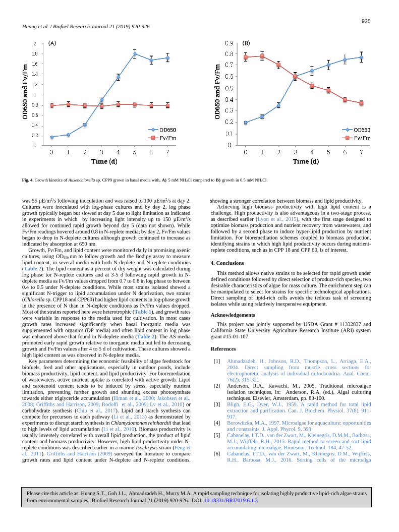
was 55 µE/m2/s following inoculation and was raised to 100 µE/m2/s at day 2.
Cultures were inoculated with log-phase cultures and by day 2, log phase
growth typically began but slowed at day 5 due to light limitation as indicated in experiments in which by increasing light intensity up to 150 µE/m2/s
allowed for continued rapid growth beyond day 5 (data not shown). While
Fv/Fm readings hovered around 0.8 in N-replete media; by day 2, Fv/Fm values began to drop in N-deplete cultures although growth continued to increase as
indicated by absorption at 650 nm.
Growth, Fv/Fm, and lipid content were monitored daily in promising axenic cultures, using OD650 nm to follow growth and the Bodipy assay to measure
lipid content, in several media with both N-deplete and N-replete conditions
(Table 2). The lipid content as a percent of dry weight was calculated during log phase for N-replete cultures and at 3-5 d following rapid growth in N-
deplete media as Fv/Fm values dropped from 0.7 to 0.8 in log phase to between
0.4 to 0.5 under N-deplete conditions. While most strains isolated showed a significant N-trigger to lipid accumulation under N deprivation, two strains
(Chlorella sp. CPP18 and CPP60) had higher lipid contents in log-phase growth
in the presence of N than in N-deplete conditions as Fv/Fm values dropped. Most of the strains reported here were heterotrophic (Table 1), and growth rates
were variable in response to the media used for cultivation. In most cases
growth rates increased significantly when basal inorganic media was supplemented with organics (DP media) and often lipid content in log phase
was enhanced above that found in N-deplete media (Table 2). The AS media
promoted early rapid growth relative to inorganic media but led to decreasing growth and Fv/Fm values after 4 to 5 d of cultivation. These cultures showed a
high lipid content as was observed in N-deplete media. Key parameters determining the economic feasibility of algae feedstock for
biofuels, feed and other applications, especially in outdoor ponds, include
biomass productivity, lipid content, and lipid productivity. For bioremediation of wastewaters, active nutrient uptake is correlated with active growth. Lipid
and carotenoid content tends to be induced by stress, especially nutrient
limitation, preventing further growth and shunting excess photosynthate towards either triglyceride accumulation (Illman et al., 2000; Jakobsen et al.,
2008; Griffiths and Harrison, 2009; Rodolfi et al., 2009; Lv et al., 2010) or
carbohydrate synthesis (Chiu et al., 2017). Lipid and starch synthesis can compete for precursors to each pathway (Li et al., 2013) as demonstrated by
experiments to disrupt starch synthesis in Chlamydomonas reinhardtii that lead
to high levels of lipid accumulation (Li et al., 2010). Biomass productivity is
usually inversely correlated with overall lipid production, the product of lipid
content and biomass productivity. However, high lipid productivity under N-
replete conditions was described earlier in a marine Isochrysis strain (Feng et al., 2011). Griffiths and Harrison (2009) surveyed the literature to compare
growth rates and lipid content under N-deplete and N-replete conditions,
Achieving high biomass productivity with high lipid content is a
challenge. High productivity is also advantageous in a two-stage process,
as described earlier (Lyon et al., 2015), with the first stage designed to optimize biomass production and nutrient recovery from wastewaters, and
followed by a second phase to induce hyper-lipid production by nutrient
limitation. For bioremediation schemes coupled to biomass production, identifying strains in which high lipid productivity occurs during nutrient-
replete conditions, such as in CPP 18 and CPP 60, is of interest.
4. Conclusions
This method allows native strains to be selected for rapid growth under defined conditions followed by direct selection of product-rich species, two
desirable characteristics of algae for mass culture. The enrichment step can
be manipulated to select for strains for specific technological applications. Direct sampling of lipid-rich cells avoids the tedious task of screening
isolates while using relatively inexpensive equipment.
Acknowledgements
This project was jointly supported by USDA Grant # 11332837 and California State University Agriculture Research Institute (ARI) system
grant #15-01-107
References
[1] Ahmadzadeh, H., Johnson, R.D., Thompson, L., Arriaga, E.A.,
2004. Direct sampling from muscle cross sections for
electrophoretic analysis of individual mitochondria. Anal. Chem. 76(2), 315-321.
[2] Anderson, R.A., Kawachi, M., 2005. Traditional microalgae
isolation techniques, in: Anderson, R.A. (ed.), Algal culturing techniques. Elsevier, Amsterdam, pp. 83-100.
[3] Bligh, E.G., Dyer, W.J., 1959. A rapid method for total lipid
extraction and purification. Can. J. Biochem. Physiol. 37(8), 911-917.
[4] Borowitzka, M.A., 1997. Microalgae for aquaculture: opportunities
and constraints. J. Appl. Phycol. 9, 393.
[5] Cabanelas, I.T.D., van der Zwart, M., Kleinegris, D.M.M., Barbosa,
M.J., Wijffels, R.H., 2015. Rapid method to screen and sort lipid
accumulating microalgae. Bioresour. Technol. 184, 47-52. [6] Cabanelas, I.T.D., van der Zwart, M., Kleinegris, D.M., Wijffels,
R.H., Barbosa, M.J., 2016. Sorting cells of the microalga
showing a stronger correlation between biomass and lipid productivity.
Fig. 4. Growth kinetics of Auxenchlorella sp. CPP9 grown in basal media with, 5 mM NH4Cl compared to growth in 0.5 mM NH4Cl. A) B)
925

Huang et al. / Biofuel Research Journal 21 (2019) 920-926
Please cite this article as: Huang S.T., Goh J.L., Ahmadzadeh H., Murry M.A. A rapid sampling technique for isolating highly productive lipid-rich algae strains
from environmental samples. Biofuel Research Journal 21 (2019) 920-926. DOI: 10.18331/BRJ2019.6.1.3
Chlorococcum littorale with increased triacylglycerol productivity.
Biotechnol. Biofuels. 9(1), 183. [7] Chen, W., Zhang, C.W., Song, L.R., Sommerfeld, M., Hu, Q., 2009. A
high throughput Nile red method for quantitative measurement of
neutral lipids in microalgae. J. Microbiol. Meth. 77(1), 41-47. [8] Chia, M.A., Lombardi, A.T., Melão, M.D.G.G., 2013. Growth and
biochemical composition of Chlorella vulgaris in different growth
media. Anais da Academia Brasileira de Ciências. 85(4), 1427-1438. [9] Chisti, Y., 2007. Biodiesel from microalgae. Biotechnol. Adv. 25, 294-
306.
[10] Collyer, D.M., Fogg, G.E., 1955. Studies on fat accumulation by algae. J. Exp. Bot. 6(17), 256-275.
[11] Duong, V.T., Li, Y., Nowak, E., Schenk, P.M., 2012. Microalgae
isolation and selection for prospective biodiesel production. Energies. 5(6), 1835-1849.
[12] Fawley, K.P., Fawley, M.W., 2007. Observations on the diversity and
ecology of freshwater Nannochloropsis (Eustigmatophyceae), with
descriptions of new taxa. Protist. 158(3), 325-336.
[13] Feng, D., Chen, Z., Xue, S., Zhang, W., 2011. Increased lipid
production of the marine oleaginous microalgae Isochrysis zhangjiangensis (Chrysophyta) by nitrogen supplement. Bioresour.
Technol. 102(12), 6710-6716.
[14] Fogg, G.E., Thake, B., 1987. Algal cultures and phytoplankton ecology. The University of Wisconsin Press.
[15] Griffiths, M.J., Harrison, S.T., 2009. Harrison. Lipid productivity as a
key characteristic for choosing algal species for biodiesel production. J. Appl. Phycol. 21(5), 493-507.
[16] Guillard, R.R., Ryther, J.H., 1962. Studies of marine planktonic
diatoms: I. Cyclotella nana Hustedt and Detonula confervacea (Cleve) Gran. Can. J. Microbiol. 8(2), 229-239.
[17] Ho, S.H., Shimada, R., Ren, N.Q., Ozawa, T., 2017. Rapid in vivo
lipid/carbohydrate quantification of single microalgal cell by Raman spectral imaging to reveal salinity-induced starch-to-lipid shift.
Biotechnol. Biofuels. 10(1), 9.
[18] Illman, A.M., Scragg, A.H., Shales, S.W., 2000. Increase in Chlorella strains calorific values when grown in low nitrogen medium. Enzyme
Microb. Technol. 27(8), 631-635.
[19] Jakobsen, A.N., Aasen, I.M., Josefsen, K.D., Strom, A.R., 2008. Accumulation of docosahexaenoic acid-rich lipid in thraustochytrid
Aurantiochytrium sp strain T66: effects of N and P starvation and O2
limitation. Appl. Microbiol. Biotechnol. 80(2), 297-306. [20] Lari, Z., Moradi-kheibari, N., Ahmadzadeh, H., Abrishamchi, P.,
Moheimani, N.R, Murry, M.A., 2016. Bioprocess engineering of
microalgae to optimize lipid production through nutrient management. J. Appl. Phycol. 28(6), 3235-3250.
[21] Li, Y., Han, D., Hu, G., Sommerfeld, M., Hu, Q., 2010. Inhibition of
starch synthesis results in overproduction of lipids in Chlamydomonas reinhardtii. Biotechnol. Bioeng. 107(2), 258-268.
[22] Li, Y., Han, D., Yoon, K., Zhu, S., Sommerfeld, M., Hu, Q., 2013.
Molecular and cellular mechanisms for lipid synthesis and accumulation in microalgae: biotechnological implications, Second ed,
in: Richmond, A., Hu, Q. (Eds.), Handbook of Microalgal Culture. Appl. Phycol. Biotechnol. John Wiley & Sons, Ltd, Oxford, UK.
[23] Lyon, S.R., Ahmadzadeh, H., Murry, M.A., 2015. Algae-based
wastewater treatment for biofuel production: processes, species, and extraction methods, in: Moheimani, N., McHenry, M., de Boer, K.,
Bahri, P. (Eds.), Biomass and Biofuels from Microalgae. Biofuel and
Biorefinery Technologies, Springer, Cham. 95-115.
Lv, J.M., Cheng, L.H., Xu, X.H., Zhang, L., Chen, H.L., 2010.
Enhanced lipid production of Chlorella vulgaris by adjustment of cultivation conditions. Bioresour. Technol. 101(17), 6797-6804.
[25] Murry, M.A., Benemann, J.R., 1980. Fresh and brackish water
aquatic plant resources, in: Zaborsky, O. (Ed.), Handbook of Biosolar resources, vol II. CRC Press, Boca Raton. 407-470
[26] Mutanda, T., Ramesh, D., Karthikeyan, S., Kumari, S., Anandraj,
A., Bux, F., 2011. Bioprospecting for hyper-lipid producing microalgal strains for sustainable biofuel production. Bioresour.
Technol. 102(1), 57-70.
[27] OECD, 2001. Test No. 303: simulation test-aerobic sewage treatment--A: activated sludge units; B: biofilms. OECD
Publishing, Paris.
[28] Obata, M., Toda, T., Taguchi, S., 2009. Using chlorophyll fluorescence to monitor yields of microalgal production. J. Appl.
Phycol. 21(3), 315-319.
[29] Ong, S., Kao, C.Y., Chiu, S.Y., Tsai, M.T., Lin, C.S., 2010.
Characterization of the thermal-tolerant mutants of Chlorella sp.
with high growth rate and application in outdoor photobioreactor
cultivation. Bioresour. Technol. 101(8), 2880-2883. [30] Parkhill, J.P., Maillet, G., Cullen, J.J., 2001. Fluorescence-based
maximum quantum yield for PSII as a diagnosis of nutrient stress.
J. Phycol. 37(4), 517-529. [31] Pereira, H., Barreira, L., Mozes, A., Florindo, C., Polo, C., Duarte,
C.V., Custódio, L., Varela, J., 2011. Microplate-based high
throughput screening procedure for the isolation of lipid-rich marine microalgae. Biotechnol. Biofuels. 4(1), 61.
[32] Rippka, R., Deruelles, J. Waterbury, J.B., Herdman, M., Stanier,
R.Y., 1979. Generic assignments, strain histories and properties of pure cultures of cyanobacteria. Microbiology. 111(1), 1-61.
[33] Rodolfi, L., Chini Zittelli, G., Bassi, N., Padovani, G., Biondi, N.,
Bonini, G., Tredici, M.R., 2008. Microalgae for oil: strain selection, induction of lipid synthesis and outdoor mass cultivation in a low-
cost photobioreactor. Biotechnol. Bioeng. 102(1), 100-112.
[34] Roessler, P.G., 1990. Environmental control of glycerolipid metabolism in microalgae: commercial implications and future
research directions. J. Phycol. 26(3), 393-399.
[35] Sheehan, J., Dunahay, T., Benemann, J.R., Roessler, P., 1998. A look back at the U.S. department of energy’s aquatic species
program: biodiesel from algae. National Renewable Energy Lab.
NREL/TP-580-24190. [36] Shifrin, N.S., Chisholm, S.W., 1981. Phytoplankton lipids:
interspecific differences and effects of nitrate, silicate and light‐dark cycles. J. Phycol. 17(4), 374-384.
[37] Spoehr, H.A., Milner, H.W., 1949. The chemical composition of Chlorella; effect of environmental conditions. Plant physiol. 24(1),
120-149.
[38] Takia, M., Ahmadzadeh, H., Murry, M.A., 2015. Growth of
Chlorella vulgaris in high concentrations of nitrate and nitrite for
wastewater treatment. Current Biotechnol. 4(4), 441-447.
[39] Taziki, M., Ahmadzadeh, H., Murry, M.A., Lyon, S.R., 2015.
Nitrate and nitrite removal from wastewater using algae. Curr.
Biotechnol. 4(3), 426-440.
[40] Weissman, J.C., Goebel, R.P., 1987. Factors affecting the
photosynthetic yield of microalgae. FY 1986 Aquatic Species
Program Annual Report. Solar Energy Research Institute. Golden,
Colorado, SERI/SP-231-3071, 139-168.
926
[24]