A Nucleolin-Targeted Multimodal Nanoparticle Imaging Probe for
9
A Nucleolin-Targeted Multimodal Nanoparticle Imaging Probe for Tracking Cancer Cells Using an Aptamer Do Won Hwang* 1–3 , Hae Young Ko* 2–4 , Jung Hwan Lee 5 , Hyungu Kang 5 , Sung Ho Ryu 5 , In Chan Song 6 , Dong Soo Lee 1,2,7 , and Soonhag Kim 8 1 Programs in Neuroscience, Seoul National University, Seoul, Korea; 2 Department of Nuclear Medicine, Seoul National University College of Medicine, Seoul, Korea; 3 Cancer Research Institute, Seoul National University College of Medicine, Seoul, Korea; 4 Institute of Radiation Medicine, Medical Research Center, Seoul, Korea; 5 Aptamer Unit, Postech Biotech Center, Pohang University of Science and Technology, Pohang, Kyungbuk, Korea; 6 Department of Radiology, Seoul National University College of Medicine, Seoul, Korea; 7 Department of Nuclear Medicine and Department of Molecular Medicine and Biopharmaceutical Science, Seoul National University College of Medicine, Seoul, Korea; and 8 Laboratory of Molecular Imaging, CHA Stem Cell Institute, CHA University, Seoul, South Korea The recent advances in molecular imaging techniques, using cancer-targeting nanoparticle probes, provide noninvasive tracking information on cancer cells in living subjects. Here, we report a multimodal cancer-targeted imaging system capable of concurrent fluorescence imaging, radionuclide imaging, and MRI in vivo. Methods: A cobalt–ferrite nanoparticle surrounded by fluorescent rhodamine (designated MF) within a silica shell matrix was synthesized with the AS1411 aptamer (MF-AS1411) that targets nucleolin (a cellular membrane protein highly expressed in cancer) using N-(3-dimethylaminopropyl)-N- ethylcarbodiimide (EDC). This purified MF-AS1411 particle was bound with 2-( p-isothio-cyanatobenzyl)-1,4,7-triazacyclonane- 1,4,7-triacetic acid ( p-SCN-bn-NOTA) chelating agent and fur- ther labeled with 67 Ga-citrate (MFR-AS1411). The shape and size distribution of MFR-AS1411 were characterized by trans- mission electron microscope (TEM). The cellular distribution of the nucleolin protein using the MFR-AS1411 nanoparticle was detected by fluorescence confocal microscopy. Phantom MR images were obtained as the concentration of MFR-AS1411 in- creased, using a 1.5-T MRI scanner. In vivo 67 Ga radionuclide im- aging and MRI were performed using a g-camera and a 1.5-T MR imager, respectively. Results: TEM imaging revealed MF and MFR-AS1411 to be spheric and well dispersed. The purified MFR-AS1411 nanoparticle showed specific fluorescence signals in nucleolin-expressing C6 cells, compared with MFR-AS1411 mutant (MFR-AS1411mt)–treated C6 cells. The rhodamine fluo- rescence intensity and 67 Ga activity of MFR-AS1411 were en- hanced in a dose-dependent manner as the concentration of MFR-AS1411 was increased. The 67 Ga radionuclide was de- tected in both thighs of the mice injected with MFR-AS1411, whereas the MFR-AS1411 mutant (MFR-AS1411mt) admini- stration revealed rapid clearance via the bloodstream, demon- strating that MFR-AS1411 specifically targeted cancer cells. Bioluminescence images in the C6 cells, stably expressing the luciferase gene, illustrated the in vivo distribution. T2-weighted MR images of the same mice injected with MFR-AS1411 showed dark T2 signals inside the tumor region, compared with the MRI signal of the tumor region injected with MFR-AS1411mt parti- cles. Conclusion: We developed a nanoparticle-based cancer- specific imaging probe using the AS1411 aptamer in vivo and in vitro. This multimodal targeting imaging strategy, using a can- cer-specific AS1411 aptamer, can be used as a versatile imaging tool for specific cancer diagnosis. Key Words: multimodal image; cancer targeting; nanoparticles; aptamer; optical and radionuclide image J Nucl Med 2010; 51:98–105 DOI: 10.2967/jnumed.109.069880 The study of biologic imaging for cancer-specific targeting, using nanosized materials, has been an important part of theranostics use in drug development, nanosurgery, or medical nanosciences by visualization of target objects in animal subjects in vivo and at the molecular and cellular levels in vitro (1–4). Recently, a wide range of studies using multifunctional imaging probes has provided more precise imaging interpretation by overcoming the limitations of a single imaging modality. Several bi- or trimodal imaging probes have been devised and reported in the literature as successfully monitoring intracellular molecular activity and tracking stem cells in vivo (5–7). Surface modifications of nanoparticles with antibodies, aptamers, peptides, or small molecules that bind to antigens present on the target cells or tissues have resulted in the development of sensitive and specific targeted imaging and diagnostic modalities for in vitro and in vivo applications (8,9). Until now, monoclonal antibodies have been broadly used as ligands for molecular targeting because of their Received Aug. 27, 2009; revision accepted Oct. 7, 2009. For correspondence or reprints contact: Soonhag Kim, Laboratory of Molecular Imaging, Department of Applied Bioscience, CHA Stem Cell Institute, CHA University, 605-21 Yoeksam 1-dong, Gangnam-gu, Seoul (135-081), Korea. E-mail: [email protected] COPYRIGHT ª 2010 by the Society of Nuclear Medicine, Inc. *Contributed equally to this work. 98 THE JOURNAL OF NUCLEAR MEDICINE • Vol. 51 • No. 1 • January 2010 by on December 27, 2018. For personal use only. jnm.snmjournals.org Downloaded from
Transcript of A Nucleolin-Targeted Multimodal Nanoparticle Imaging Probe for
A Nucleolin-Targeted MultimodalNanoparticle Imaging Probe for TrackingCancer Cells Using an Aptamer
Do Won Hwang*1–3, Hae Young Ko*2–4, Jung Hwan Lee5, Hyungu Kang5, Sung Ho Ryu5, In Chan Song6,Dong Soo Lee1,2,7, and Soonhag Kim8
1Programs in Neuroscience, Seoul National University, Seoul, Korea; 2Department of Nuclear Medicine, Seoul National UniversityCollege of Medicine, Seoul, Korea; 3Cancer Research Institute, Seoul National University College of Medicine, Seoul, Korea;4Institute of Radiation Medicine, Medical Research Center, Seoul, Korea; 5Aptamer Unit, Postech Biotech Center, Pohang Universityof Science and Technology, Pohang, Kyungbuk, Korea; 6Department of Radiology, Seoul National University College of Medicine,Seoul, Korea; 7Department of Nuclear Medicine and Department of Molecular Medicine and Biopharmaceutical Science, SeoulNational University College of Medicine, Seoul, Korea; and 8Laboratory of Molecular Imaging, CHA Stem Cell Institute, CHAUniversity, Seoul, South Korea
The recent advances in molecular imaging techniques, usingcancer-targeting nanoparticle probes, provide noninvasivetracking information on cancer cells in living subjects. Here, wereport a multimodal cancer-targeted imaging system capableof concurrent fluorescence imaging, radionuclide imaging, andMRI in vivo. Methods: A cobalt–ferrite nanoparticle surroundedby fluorescent rhodamine (designated MF) within a silica shellmatrix was synthesized with the AS1411 aptamer (MF-AS1411)that targets nucleolin (a cellular membrane protein highlyexpressed in cancer) using N-(3-dimethylaminopropyl)-N-ethylcarbodiimide (EDC). This purified MF-AS1411 particle wasbound with 2-(p-isothio-cyanatobenzyl)-1,4,7-triazacyclonane-1,4,7-triacetic acid ( p-SCN-bn-NOTA) chelating agent and fur-ther labeled with 67Ga-citrate (MFR-AS1411). The shape andsize distribution of MFR-AS1411 were characterized by trans-mission electron microscope (TEM). The cellular distribution ofthe nucleolin protein using the MFR-AS1411 nanoparticle wasdetected by fluorescence confocal microscopy. Phantom MRimages were obtained as the concentration of MFR-AS1411 in-creased, using a 1.5-T MRI scanner. In vivo 67Ga radionuclide im-aging and MRI were performed using a g-camera and a 1.5-T MRimager, respectively. Results: TEM imaging revealed MF andMFR-AS1411 to be spheric and well dispersed. The purifiedMFR-AS1411 nanoparticle showed specific fluorescence signalsin nucleolin-expressing C6 cells, compared with MFR-AS1411mutant (MFR-AS1411mt)–treated C6 cells. The rhodamine fluo-rescence intensity and 67Ga activity of MFR-AS1411 were en-hanced in a dose-dependent manner as the concentration ofMFR-AS1411 was increased. The 67Ga radionuclide was de-tected in both thighs of the mice injected with MFR-AS1411,whereas the MFR-AS1411 mutant (MFR-AS1411mt) admini-stration revealed rapid clearance via the bloodstream, demon-strating that MFR-AS1411 specifically targeted cancer cells.
Bioluminescence images in the C6 cells, stably expressing theluciferase gene, illustrated the in vivo distribution. T2-weightedMR images of the same mice injected with MFR-AS1411 showeddark T2 signals inside the tumor region, compared with the MRIsignal of the tumor region injected with MFR-AS1411mt parti-cles. Conclusion: We developed a nanoparticle-based cancer-specific imaging probe using the AS1411 aptamer in vivo andin vitro. This multimodal targeting imaging strategy, using a can-cer-specific AS1411 aptamer, can be used as a versatile imagingtool for specific cancer diagnosis.
Key Words: multimodal image; cancer targeting; nanoparticles;aptamer; optical and radionuclide image
J Nucl Med 2010; 51:98–105DOI: 10.2967/jnumed.109.069880
The study of biologic imaging for cancer-specifictargeting, using nanosized materials, has been an importantpart of theranostics use in drug development, nanosurgery,or medical nanosciences by visualization of target objectsin animal subjects in vivo and at the molecular and cellularlevels in vitro (1–4). Recently, a wide range of studies usingmultifunctional imaging probes has provided more preciseimaging interpretation by overcoming the limitations ofa single imaging modality. Several bi- or trimodal imagingprobes have been devised and reported in the literature assuccessfully monitoring intracellular molecular activity andtracking stem cells in vivo (5–7).
Surface modifications of nanoparticles with antibodies,aptamers, peptides, or small molecules that bind to antigenspresent on the target cells or tissues have resulted in thedevelopment of sensitive and specific targeted imaging anddiagnostic modalities for in vitro and in vivo applications(8,9). Until now, monoclonal antibodies have been broadlyused as ligands for molecular targeting because of their
Received Aug. 27, 2009; revision accepted Oct. 7, 2009.For correspondence or reprints contact: Soonhag Kim, Laboratory of
Molecular Imaging, Department of Applied Bioscience, CHA Stem CellInstitute, CHA University, 605-21 Yoeksam 1-dong, Gangnam-gu, Seoul(135-081), Korea.
E-mail: [email protected] ª 2010 by the Society of Nuclear Medicine, Inc.*Contributed equally to this work.
98 THE JOURNAL OF NUCLEAR MEDICINE • Vol. 51 • No. 1 • January 2010
by on December 27, 2018. For personal use only. jnm.snmjournals.org Downloaded from

high affinity, specificity, and varied targeting availability.However, because of the long residence in the blood ofantibodies, in vivo imaging using monoclonal antibodies asligands deteriorates in quality over time. Therefore, weinvestigated the use of an aptamer for specific targeting ofcancer cells, instead of an antibody using a multimodalnanoimaging probe.
Aptamers are synthetic DNA or RNA oligonucleotidesthat bind to target molecules with high affinity andspecificity by a 3-dimensional structure. Because aptamershave low molecular weights, lack immunogenicity, and arereadily available, these advantages make them good can-didates for targeted cancer imaging and therapy. Recently, 2reports on cancer cell imaging using the tenascin-C aptamerconjugated with a radioisotope and a prostate-specificmembrane antigen aptamer conjugated with quantum dotsshowed specific targeting of cancer cells in vivo (10,11).
In this study, we used a DNA aptamer (AS1411, alsoknown as AGRO100) that binds to nucleolin in the plasmamembrane (12). Nucleolin is highly expressed in continu-ously proliferating cells, such as cancer cells (13), and has aremarkable multifunction in the nucleus and cytoplasm andon the cell surface (14–16). The most representative func-tion of AS1411 is its antiproliferative activity that blocksthe antiapoptotic pathway by combining with nuclearfactor-kB essential modulator in cancer cells (12).
The aim of this study was to develop an imaging systemusing a cancer-specific aptamer, by examining the in vivodistribution of a multimodal nanoparticle imaging probein living subjects. An AS1411-laden nanoparticle imagingprobe was constructed using a magnetic fluorescence nano-particle for the investigation of an aptamer-based multimodalcancer imaging probe, using AS1411 aptamers that bind tonucleolin proteins in the cellular membrane of cancer cells.Here, we report an experimental investigation of this multi-modal imaging system with fluorescence imaging, radionu-clide imaging, and MRI modalities in living animals.
MATERIALS AND METHODS
Construction of MF-AS1411 ParticleMNP@SiO2(RITC)-(PEG)/COOH/pro-N/NH2 nanoparticles
(MF, 2 mg/mL) were purchased from Biterials (Seoul, Korea) andprepared as previously described (17). Carboxyl moieties (1.1 ·104/nanoparticle) of the MF particles (size, ;50 nm; hydrody-namic diameter, 58.1 nm) were covalently linked to a 59-NH2–modified AS1411 aptamer (59-TTGGTGGTGGTGGTTGTGGTGGTGGTGG-39) using N-(3-dimethylaminopropyl)-N-ethylcarbo-diimide (EDC) (MF:aptamer molar ratio in conjugation reaction,1:3) (Sigma) for 1 h at room temperature (18). The MF-AS1411conjugates were washed off by centrifugation at 22,250g for 10min and resuspended in selection buffer solution (50 mM Tris-HCl, pH 7.4). Amine groups (6.4 · 104/nanoparticle) protected bythe Fmoc group were released by 20% piperidine (Sigma) in anN,N-dimethylformamide solution (Sigma). After 1 h of incubation,the MF-AS1411 particles were washed off twice with Tris buffer(pH 7.4) and briefly sonicated. To confirm the conjugation patternof MF-AS1411, the conjugated MF-AS1411 and MF particle were
loaded onto each well of 0.5% agarose gel with electrophoresisbuffer (0.5· Tris–acetate–ethylenediaminetetraacetic acid) andrun at 100 V.
Transmission Electron Microscope (TEM) AnalysisNegatively stained specimens were prepared on a formvar-
coated grid, and the AS1411 aptamer was stained with 2%aqueous uranyl acetate. TEM study was performed using a JEM1010 (JEOL) at 80 kV. 67Ga-labeled MF-AS1411 particles (MFR-AS1411) (n 5 3) were fixed using 2% formaldehyde for 10 min.MF particle was also prepared as a control (n 5 3) by being fixedusing 2% formaldehyde. Electron microscope digital images wererecorded using a Gatan cooled charge-coupled device camera.
Cell CultureC6 rat glioma cells were cultured in Dulbecco’s modified
Eagle’s medium (Invitrogen), supplemented with 10% heat-in-activated (65�C for 20 min) fetal bovine serum (Invitrogen) with1% antibiotics (Invitrogen), in a standard incubator (5% CO2
atmosphere at 37�C). The cells were split at regular intervals. C6stably expressing luciferase gene regulated by cytomegaloviruspromoter was generated by the selection with several concentra-tions of geneticin (Invitrogen) over 2 wk.
Confocal Microscopy of MF-AS1411 Targetedto C6 Cells
C6 cells (1 · 105 cells/well) were seeded on 25-mm-diameterglass cover slips, and the cells were grown for 24 h at 37�C. Beforetreatment with MF-AS1411 conjugates, the cells were incubated for30 min at 4�C and then washed with phosphate-buffered saline(PBS). The conjugated particles were added to cells under Trisbuffer (pH 7). After incubation for 30 min at 4�C, the cells werefixed by gently shaking for 20 min with 4% formaldehyde solution(Sigma). Cells were then washed 3 times with PBS for 10 min andcover-slipped with mounting medium containing 49,6-diamidino-2-phenylindole dihydrochloride solution (Vector Laboratories, Inc.).The images were acquired with confocal laser scanning microscopy(LSM 510; Carl Zeiss) with an excitation wavelength of 556 nmand an emission wavelength of 570 nm.
In Vitro Fluorescence Analysis and Stability Testing ofRhodamine Fluorescence in MFR-AS1411 Particle
Each concentration (3, 6, 9, 12, 15, and 18 pmol) of MF particlewas first conjugated with the AS1411 aptamer at a 1:3 ratio of MFto AS1411 aptamer. The MF-AS1411 nanoparticles were added tothe C6 cells and incubated for 1 h. The cells treated with the MF-AS1411 particle were harvested by trypsinization after rinsingwith PBS. The collected cells were transferred into dark 96-wellmicroplates, and their fluorescence intensities were measuredusing the Infinite M200 (Tecan, GmbH, SZ). For fluorescencestability testing, 5 pmol of MF particles were conjugated with 15pmol of the AS1411 aptamer using EDC. After the MF-AS1411was synthesized with 2-(p-isothio-cyanatobenzyl)-1,4,7-triazacy-clonane-1,4,7-triacetic acid (p-SCN-bn-NOTA), the same amountof the conjugated MFR-AS1411 particle was added to Eppendorftubes containing PBS solution and measured using the fluores-cence imager (Tecan, GmbH, SZ).
Radiolabeling of MF-AS1411 with 67Ga-Citrate and InVitro Experiment with MFR-AS1411
To react the MF-AS1411 particle with p-SCN-Bn-NOTA (500gmol21; concentration, 5 mg/mL), the amino moieties of MF-
CANCER-TARGETED MULTIMODAL IMAGING • Hwang et al. 99
by on December 27, 2018. For personal use only. jnm.snmjournals.org Downloaded from
AS1411 were synthesized using a p-SCN-Bn-NOTA adduct withNaHCO3 buffer solution, at a MF–to–p-SCN-Bn-NOTA reactionratio of 1:5 under agitation at 4�C overnight (19). MF-AS1411synthesized with p-SCN-Bn-NOTA was labeled with 67Ga-citrate(37 MBq [1 mCi]/400 mL) under 0.2 M Na2HPO4/NaH2PO4
(Sigma) buffer (pH 6.5) and incubated for 1 h. The particles ofMF-AS1411-NOTA labeled with 67Ga-citrate (MFR-AS1411)were checked by thin-layer chromatography (TLC) to analyzethe labeling efficiency. The reaction mixture of radiolabeled MF-AS1411 was applied to instant thin-layer chromatography–silicagel (Pall, Inc.) and separated in a simple beaker containing 0.1 Mcitric acid solvent. The radioactivity was measured using an AR-2000 TLC imaging scanner (Bioscan, Inc.). The labeling effi-ciency for the radioactivity bound to the MF-AS1411 particleswas measured as percentage of ROIs on TLC paper silica gel.
To examine the specific targeting of MFR-AS1411 nanoparticleto the C6 cells by measuring radioactivity of 67Ga-citrate in MFR-AS1411, the synthesized MF-AS1411-NOTA particles at the dif-ferent concentrations (MF particle, 3, 6, 9, 12, 15, and 18 pmol)were labeled with 67Ga-citrate (37 MBq for 18 pmol of MFconcentration) through a serial dilution method. Radioactivity of67Ga-citrate was measured for 1 min at each group using a NaIwell counter (Cobra II; Canberra Packard).
In Vitro Phantom MRI AnalysisPhantom MRI studies were performed using mixtures of MFR-
AS1411 at 6 different concentrations (0.2, 0.4, 0.8, 1.6, 3.2, and6.4 mM). Six samples in a variety of concentrations of MFR-AS1411mt were also prepared. One day after 5 · 105 C6 cellswere seeded into 24-well plates, each MFR-AS1411 sample wasadded to the C6 cells and incubated for 1 h. The collected cellswere transferred into Eppendorf tubes after several washing stepswith PBS solution. The T2 MRI signal intensities of phantomexperiments, for the different concentrations of the MFR-AS1411particles, were measured using 1.5 T on a whole-body MRIscanner (GE Healthcare).
Scintigraphic Imaging of MFR-AS1411 Particle inTumor-Bearing Nude Mice
The C6 cells (1 · 106) stably expressing the luciferase genewere harvested in PBS solution, for in vivo cellular tracking of C6cells, and injected into the left and right thighs of nude mice. Thesubcutaneously implanted C6 cells were grown until a tumor sizeof approximately 1–2 cm was reached (observed maximum tumorsize, 1.6 cm). MFR-AS1411 was separated from free 67GaCl3 bycentrifugation at 22,250g for 10 min, was thoroughly washed inTris buffer, and underwent a brief sonication. Two weeks after theimplantation of C6-expressing luciferase, MFR-AS1411 was sys-temically administered into C6-bearing nude mice. For the in vivonegative control group, MFR-AS1411mt (AS1411 mutant formfor which the core nucleotides G in AS1411 aptamer weresubstituted with C) was injected into other nude mice via the tailvein. BALB/c nude mice (10 wk old; n 5 3) were used for MFR-AS1411–treated mice and the MFR-AS1411mt (control) group,respectively. All mice received intraperitoneal injections of 50 mLof a ketamine and xylazine (2:1) solution for anesthesia. For theacquisition of 67Ga scintigraphic images, the mice injected withMFR-AS1411 were placed in spread-prone position, and whole-body scintigraphic images were acquired using a g-scintillationcamera (ON 410; Ohio Nuclear) equipped with a pinhole colli-mator. The accumulation of 67Ga radioactivity was examined in
each thigh of the mice, with the acquisition time for 10 min. Forthe in vivo cellular tracking of C6 cells, bioluminescence imageswere acquired after 3 mg/0.1 mL of luciferin were intraperitone-ally injected. Animals were positioned in an IVIS-200 equippedwith a cooled charge-coupled device camera (Xenogen), andbioluminescence images were acquired for 3 min. ROIs weredrawn around the area of uptake in the right and left thighs on theg-camera images. The average counts per pixel were recorded forboth thighs of the mouse. All experimental animals were housedunder specific pathogen-free conditions and handled in accordancewith the guidelines issued by the Institutional Animal Care andUse Committee of Seoul National University Hospital.
Acquisition of MR Images in Nude MiceTo determine whether the transplanted cells were visible by
MRI, as soon as scintigraphic images for the 24-h group wereacquired in mice, then the T2*-weighted images were obtained inthe same mice using a fast low-angle shot sequence. During MRIexperiments, the animals were sedated with 2% isoflurane ina mixture of O2 and N2O at a flow rate of 1 L/min through the nosecone. The temperature and respirations were monitored by a rectalthermistor and pillow. Mice were positioned in an animal coil box(n 5 3, mice), and in vivo MR images were acquired using a 1.5-TMR imager (GE Healthcare).
Statistical AnalysisData are represented as means 6 SEM and were calculated
using the Student t test. Statistical significance was accepted at Pvalues of less than 0.05.
RESULTS
Design of Probe and In Vitro Evaluation of AS1411Aptamer Specificity
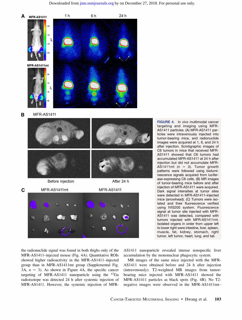
To construct the cancer-targeting multimodal imagingprobe, 67Ga-MNP@SiO2(RITC)-PEG/NH2-AS1411 (MFR-AS1411) was designed (Fig. 1). The MFR-AS1411 nano-particle was composed of magnetic cobalt ferrite in thecentral core and rhodamine B isothiocyanate fluorescencedye (MF) coated with a silica shell. In addition, poly-ethylene glycol (PEG) (1 · 104/nanoparticle), Fmoc-pro-tected amine moieties, and a carboxyl group weresurrounded with the surface of particles, which were furtherlabeled with the AS1411 aptamer and p-SCN-bn-NOTAchelator (Fig. 1). TEM imaging of the MF particles showeda homogeneous distribution of the magnetic cobalt–ferritecoated with rhodamine fluorescence dye (Fig. 2A; hydro-dynamic particle size, 58.1 nm). After the removal of theFmoc-protection group from the MF-AS1411 particles, theMF-AS1411 particles were labeled with 67Ga-citrate afterthe introduction of the NOTA chelating agents (MFR-AS1411). TEM images of the MFR-AS1411 particles alsoshowed that the AS1411 aptamer in the MFR-AS1411 wasdetected as a dark frame line on 2% uranyl acetate–filledspecimens and measured about 68.1 nm (hydrodynamic size)(Fig. 2A). In addition, when the MFR-AS1411 particleswere incubated with PBS, in a time-dependent manner (0, 1,3, 6, and 24 h) to measure the stability of the rhodamine dyein the MFR-AS1411, the fluorescence intensity ofMFR-AS1411 was maintained up to 24 h (Supplemental
100 THE JOURNAL OF NUCLEAR MEDICINE • Vol. 51 • No. 1 • January 2010
by on December 27, 2018. For personal use only. jnm.snmjournals.org Downloaded from
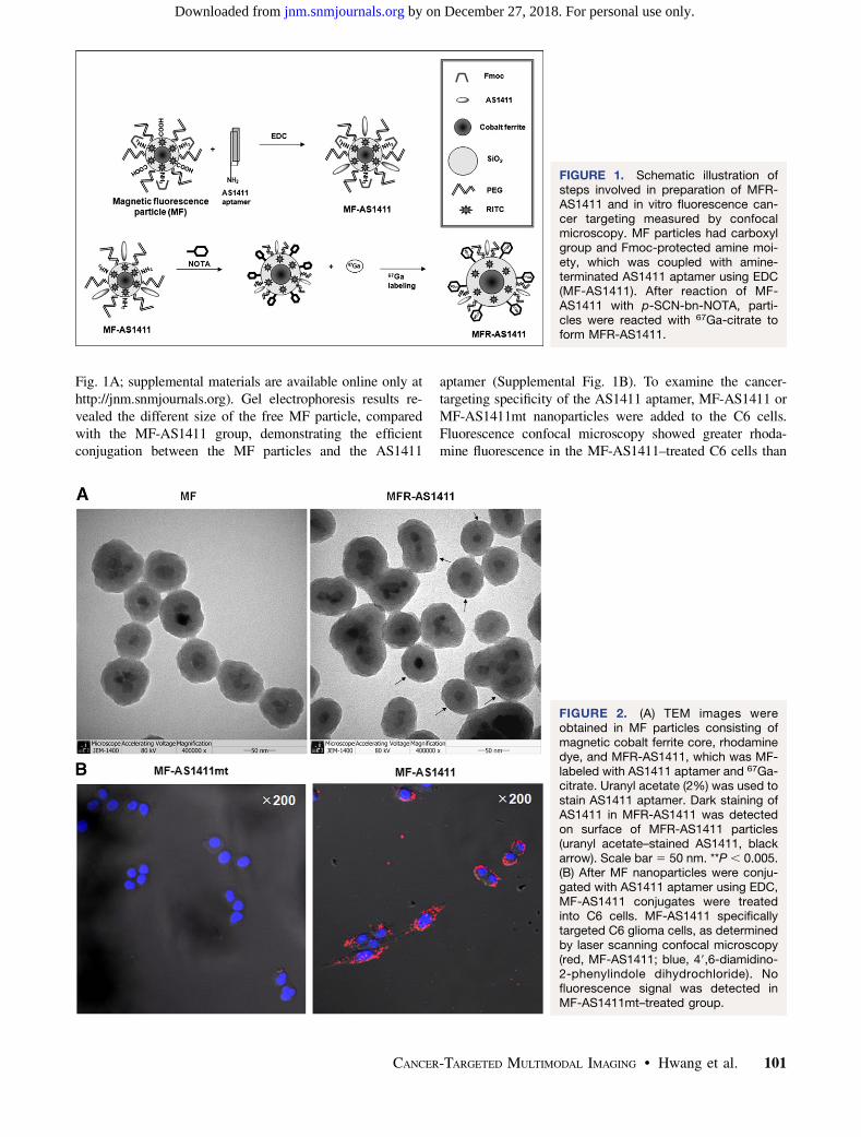
Fig. 1A; supplemental materials are available online only athttp://jnm.snmjournals.org). Gel electrophoresis results re-vealed the different size of the free MF particle, comparedwith the MF-AS1411 group, demonstrating the efficientconjugation between the MF particles and the AS1411
aptamer (Supplemental Fig. 1B). To examine the cancer-targeting specificity of the AS1411 aptamer, MF-AS1411 orMF-AS1411mt nanoparticles were added to the C6 cells.Fluorescence confocal microscopy showed greater rhoda-mine fluorescence in the MF-AS1411–treated C6 cells than
FIGURE 1. Schematic illustration ofsteps involved in preparation of MFR-AS1411 and in vitro fluorescence can-cer targeting measured by confocalmicroscopy. MF particles had carboxylgroup and Fmoc-protected amine moi-ety, which was coupled with amine-terminated AS1411 aptamer using EDC(MF-AS1411). After reaction of MF-AS1411 with p-SCN-bn-NOTA, parti-cles were reacted with 67Ga-citrate toform MFR-AS1411.
FIGURE 2. (A) TEM images wereobtained in MF particles consisting ofmagnetic cobalt ferrite core, rhodaminedye, and MFR-AS1411, which was MF-labeled with AS1411 aptamer and 67Ga-citrate. Uranyl acetate (2%) was used tostain AS1411 aptamer. Dark staining ofAS1411 in MFR-AS1411 was detectedon surface of MFR-AS1411 particles(uranyl acetate–stained AS1411, blackarrow). Scale bar 5 50 nm. **P , 0.005.(B) After MF nanoparticles were conju-gated with AS1411 aptamer using EDC,MF-AS1411 conjugates were treatedinto C6 cells. MF-AS1411 specificallytargeted C6 glioma cells, as determinedby laser scanning confocal microscopy(red, MF-AS1411; blue, 49,6-diamidino-2-phenylindole dihydrochloride). Nofluorescence signal was detected inMF-AS1411mt–treated group.
CANCER-TARGETED MULTIMODAL IMAGING • Hwang et al. 101
by on December 27, 2018. For personal use only. jnm.snmjournals.org Downloaded from

in the MF-AS1411mt–treated C6 cells (Fig. 2B). In vitrofluorescence microscopy revealed detailed information onthe location of nucleolin in cells using the AS1411 aptamer,with good resolution in the C6 cells (Fig. 2B).
Multimodal Probe Activity of MFR-AS1411Nanoparticle in C6 Cells
To quantify the optical specificity for targeting cancercells using MF-AS1411 or MF-AS1411mt, serially increasedconcentrations of MF-AS1411 were added to the C6 cells.The results showed a dose-dependent and significant in-crease in the fluorescence signal of the MF-AS1411–treatedC6 cells, compared with the MF-AS1411mt–treated C6cells (Fig. 3A).
The 67Ga-labeling efficiency of MFR-AS1411 was mea-sured using quantitative TLC analysis. The results showedthat approximately 40% of the MFR-AS1411 incorporated67Ga-citrate (Supplemental Fig. 2A). The binding stabilitybetween p-SCN-bn-NOTA and 67Ga-citrate, measured byTLC analysis over time, was relatively stable up to 12 h inmouse serum and PBS solution (data not shown).
After the MF-AS1411 particles labeled with 67Ga-citrateusing NOTA chelator (MFR-AS1411) at various concen-trations (MF particle, 3, 6, 9, 12, 15, and 18 pmol) wereincubated with the C6 cells for 1 h, 67Ga radioactivity of thecollected C6 cells was gradually increased dependingon the concentration of the MFR-AS1411 particle, showinghigher radioactivity than the MFR-AS1411mt–treated group(Fig. 3B). The radioactivity of MFR-AS1411–treated C6cells was higher than that of MFR-AS1411mt–treated C6cells at each concentration point. For the in vitro assay ofMFR-AS1411 in the cells, the synthesized MF-AS1411-
NOTA particles at the different concentrations were labeledwith 67Ga-citrate (37 MBq for 18 pmol of MF concentra-tion) through a serial dilution method. 67Ga-citrate reactionactivities were measured using a NaI well counter. PhantomMR images were obtained at 6 different concentrations(0.2, 0.4, 0.8, 1.6, 3.2, and 6.4 mM) of the MFR-AS1411nanoparticle. The same concentrations of MFR-AS1411mtwere prepared as 6 samples. The prepared MFR-AS1411(or MFR-AS1411mt) particle was incubated with the C6cells for 1 h. The T2-weighted MR images showed a gradualdecrease in the magnetic signal intensity as the concentra-tion of MFR-AS1411 increased. The specific relaxivity was9.04 mM21s21 (Fig. 3C). The magnetic signals of theMFR-AS1411 particles were also gradually decreased as theconcentration of MFR-AS1411 increased, showing a specificrelaxivity of 82.1 mM21s21 (Supplemental Fig. 2B).
In Vivo Radionuclide Detection and MRI ofSystemic MFR-AS1411
Twenty-four hours after the purified MFR-AS1411 parti-cles were systemically administered into the nude micebearing C6 tumors, 67Ga radioactivity was detected in theMFR-AS1411–administered mice at the site of the admin-istration; the MFR-AS1411mt–administered mice showedrapid clearance through the bloodstream, indicating that theMFR-AS1411 specifically targeted the tumors (Fig. 4A). Forthe in vivo cellular tracking of C6 cells, C6 cells that expressconstant firefly luciferase were used as an internal controlfor the in vivo normalization of the radioactivity intensities.The bioluminescence images of C6 cells stably expressingluciferase in both thighs of MFR-AS1411– or MFR-AS1411mt–injected mice were clearly visualized, whereas
FIGURE 3. In vitro cancer targetingusing different imaging modalities. (A)C6 cells were treated with dilutedconcentration of MF-AS1411 (or MF-AS1411mt). Gradual increase offluorescence intensity in MF-AS1411–treated group was observed, comparedwith MF-AS1411mt group. (B) Aftersynthesizing MF-AS1411 (or MF-AS1411mt) using p-SCN-bn-NOTA andincubating overnight, MF-AS1411 andMF-AS1411mt were labeled with 67Ga-citrate in buffer solution for 1 h. Con-centration of MFR-AS1411 particleswas critically dependent on 67Ga radio-activity in C6 cells. *P , 0.05. **P ,
0.005. (C) Phantom studies were per-formed at various concentrations ofMFR-AS1411 (or MFR-AS1411mt) mix-ture. Gradual decrease of MRI signals inMFR-AS1411–treated group were de-tected, compared with MFR-AS1411mt–treated group, on 1.5-T MRI system.
102 THE JOURNAL OF NUCLEAR MEDICINE • Vol. 51 • No. 1 • January 2010
by on December 27, 2018. For personal use only. jnm.snmjournals.org Downloaded from
the radionuclide signal was found in both thighs only of theMFR-AS1411–injected mouse (Fig. 4A). Quantitative ROIsshowed higher radioactivity in the MFR-AS1411–injectedgroup than in MFR-AS1411mt group (Supplemental Fig.3A, n 5 3). As shown in Figure 4A, the specific cancertargeting of MFR-AS1411 nanoparticle using the 67Garadioisotope was detected 24 h after systemic injection ofMFR-AS1411. However, the systemic injection of MFR-
AS1411 nanoparticle revealed intense nonspecific liveraccumulation by the mononuclear phagocytic system.
MR images of the same mice injected with the MFR-AS1411 were obtained before and 24 h after injection(intravenously). T2-weighted MR images from tumor-bearing mice injected with MFR-AS1411 showed theMFR-AS1411 particles as black spots (Fig. 4B). No T2-negative images were observed in the MFR-AS1411mt–
FIGURE 4. In vivo multimodal cancertargeting and imaging using MFR-AS1411 particles. (A) MFR-AS1411 par-ticles were intravenously injected intotumor-bearing mice, and radionuclideimages were acquired at 1, 6, and 24 hafter injection. Scintigraphic images ofC6 tumors in mice that received MFR-AS1411 showed that C6 tumors hadaccumulated MFR-AS1411 at 24 h afterinjection but did not accumulate MFR-AS1411mt (n 5 3). Tumor growthpatterns were followed using biolumi-nescence signals acquired from lucifer-ase-expressing C6 cells. (B) MR imagesof tumor-bearing mice before and afterinjection of MFR-AS1411 were acquired.Dark signal intensities at tumor siteswere detected in MFR-AS1411–injectedmice (arrowhead). (C) Tumors were iso-lated and their fluorescence verifiedusing IVIS200 system. Fluorescencesignal at tumor site injected with MFR-AS1411 was detected, compared withtumors injected with MFR-AS1411mt.Isolated organs in order from upper leftto lower right were intestine, liver, spleen,muscle, fat, kidney, stomach, righttumor, left tumor, heart, lung, and tail.
CANCER-TARGETED MULTIMODAL IMAGING • Hwang et al. 103
by on December 27, 2018. For personal use only. jnm.snmjournals.org Downloaded from
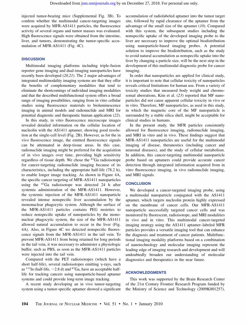
injected tumor-bearing mice (Supplemental Fig. 3B). Toconfirm whether the multimodal cancer-targeting imageswere acquired by MFR-AS1411 particles, the fluorescenceactivity of several organs and tumor masses was evaluated.High fluorescence signals were obtained from the intestine,liver, and tumors, demonstrating the tumor-specific accu-mulation of MFR-AS1411 (Fig. 4C).
DISCUSSION
Multimodal imaging platforms including triple-fusionreporter gene imaging and dual-imaging nanoparticles haverecently been developed (20,21). The 2 major advantages ofintegrated multimodality imaging systems are that they offerthe benefits of complementary modalities that tend toeliminate the shortcomings of individual imaging modalitiesand that the described multifunctional system offers a broadrange of imaging possibilities, ranging from in vitro cellularstudies using fluorescence materials to bioluminescenceimaging in animal models and radionuclide and MRI forpotential diagnostic and therapeutic human application (22).
In this study, in vitro fluorescence microscope imagesrevealed detailed information on the cellular location ofnucleolin with the AS1411 aptamer, showing good resolu-tion at the single-cell level (Fig. 2B). However, as for the invivo fluorescence image, the emitted fluorescence signalscan be attenuated in deep-tissue areas. In this case,radionuclide imaging might be preferred for the acquisitionof in vivo images over time, providing high sensitivityregardless of tissue depth. We chose the 67Ga radioisotopefor cancer-targeting radionuclide imaging because of itscharacteristics, including the appropriate half-life (78.2 h),to enable longer image tracking. As shown in Figure 4A,the specific cancer targeting of MFR-AS1411 nanoparticlesusing the 67Ga radioisotope was detected 24 h aftersystemic administration of the MFR-AS1411. However,the systemic injection of the MFR-AS1411 nanoparticlerevealed intense nonspecific liver accumulation by themononuclear phagocytic system. Although the surface ofthe MFR-AS1411 particles contains PEG moieties toreduce nonspecific uptake of nanoparticles by the mono-nuclear phagocytic system, the size of the MFR-AS1411allowed natural accumulation to occur in the liver (Fig.4A). Also, in Figure 4C we detected nonspecific fluores-cence signals from the MFR-AS1411 in the tail vein. Toprevent MFR-AS1411 from being retained for long periodsin the tail vein, it was necessary to administer a physiologicbuffer, such as PBS, as soon as the MFR-AS1411 particleswere injected into the tail vein.
Compared with the PET radioisotopes (which have ashort half-life), several radioisotopes emitting g-rays, suchas 111In (half-life, ;2.8 d) and 67Ga, have an acceptable half-life for tracking cancers using nanoparticle-based aptamersystems and could provide long-term image tracking.
A recent study developing an in vivo tumor-targetingsystem using a tumor-specific aptamer showed a significant
accumulation of radiolabeled aptamer into the tumor targetsite, followed by rapid clearance of the aptamer from theadvantage of the small size of the aptamer (10). Comparedwith this system, the subsequent studies including thenonspecific uptake of the developed imaging probe to theliver are necessary to improve the optimal biodistributionusing nanoparticle-based imaging probes. A potentialsolution to improve the biodistribution, such as the studyto avoid natural accumulation as nonspecific uptake into theliver by changing a particle size, will be the next step in thedevelopment of this multimodal diagnostic probe for cancerimaging.
In order that nanoparticles are applied for clinical study,it is important to note that cellular toxicity of nanoparticlesreveals critical limitations for human use. From a variety oftoxicity studies that measured body weight and chromo-somal aberrations, Kim et al. (23) reported that MF nano-particles did not cause apparent cellular toxicity in vivo orin vitro. Therefore, MF nanoparticles, as used in this study,in which the magnetic core of the MF nanoparticle issurrounded by a stable silica shell, might be acceptable forclinical studies in humans.
In the present study, the MFR particles consistentlyallowed for fluorescence imaging, radionuclide imaging,and MRI in vitro and in vivo. These findings suggest thatMFR-AS1411 nanoparticles are useful for the multimodalimaging of disease, theranostics (including cancer andneuronal diseases), and the study of cellular metabolism.In addition, this cancer-targeting multimodal nanoparticleprobe based on aptamers could provide accurate cancerdetection through integrated information acquired from invitro fluorescence imaging, in vivo radionuclide imaging,and MRI signals.
CONCLUSION
We developed a cancer-targeted imaging probe, usinga multimodal nanoparticle conjugated with the AS1411aptamer, which targets nucleolin protein highly expressedon the membrane of cancer cells. Our MFR-AS1411nanoparticle successfully targeted cancer cells and wasmonitored by fluorescent, radioisotope, and MRI modalitiesin vivo and in vitro. This multimodal cancer-targetedimaging strategy using the AS1411 aptamer–labeled MFRparticles provides a versatile imaging tool that can enhancethe diagnosis and treatment of cancer patients. Multifunc-tional imaging modality platforms based on a combinationof nanotechnology and molecular imaging represent theleading edge of imaging research and development and willundoubtedly broaden our understanding of moleculardiagnostics and therapeutics in the near future.
ACKNOWLEDGMENTS
This work was supported by the Brain Research Centerof the 21st Century Frontier Research Program funded bythe Ministry of Science and Technology (2009K001257),
104 THE JOURNAL OF NUCLEAR MEDICINE • Vol. 51 • No. 1 • January 2010
by on December 27, 2018. For personal use only. jnm.snmjournals.org Downloaded from

the WCU project of the MEST and the NRF (R31-2008-000-10103-0), the National R&D Program for Cancer Control ofMinistry of Health and Welfare (0820320), the NationalResearch Foundation of Korea (no. 20090084640), anda grant of the Korea Healthcare Technology R&D Project,Ministry for Health, Welfare & Family Affairs, Republic ofKorea (A085136).
REFERENCES
1. Leary SP, Liu CY, Yu C, Apuzzo ML. Toward the emergence of nano-
neurosurgery: part II—nanomedicine: diagnostics and imaging at the nanoscale
level. Neurosurgery. 2006;58:805–823.
2. Suri SS, Fenniri H, Singh BL. Nanotechnology-based drug delivery systems. J
Occup Med Toxicol. 2007;2:16.
3. Liu Y, Miyoshi H. Nakamura M. Nanomedicine for drug delivery and imaging:
a promising avenue for cancer therapy and diagnosis using targeted functional
nanoparticles. Int J Cancer. 2007;120:2527–2537.
4. Sharma P, Brown S, Walter G, Santa S, Moudgil B. Nanoparticles for
bioimaging. Adv Colloid Interface Sci. 2006;123–126:471–485.
5. Doubrovin M, Serganova I, Mayer-Kuckuk P, Ponomarev V, Blasberg RG.
Multimodality in vivo molecular-genetic imaging. Bioconjug Chem. 2004;15:
1376–1388.
6. Love Z, Wang F, Dennis J, et al. Imaging of mesenchymal stem cell transplant by
bioluminescence and PET. J Nucl Med. 2007;48:2011–2020.
7. Wang W, Ke S, Kwon S, et al. A new optical and nuclear dual-labeled imaging
agent targeting interleukin 11 receptor alpha-chain. Bioconjug Chem. 2007;18:
397–402.
8. Cai W, Chen X. Preparation of peptide-conjugated quantum dots for tumor
vasculature-targeted imaging. Nat Protoc. 2008;3:89–96.
9. Chu TC, Shieh F, Lavery LA, et al. Labeling tumor cells with fluorescent
nanocrystal-aptamer bioconjugates. Biosens Bioelectron. 2006;21:1859–1866.
10. Hicke BJ, Stephens AW, Gould T, et al. Tumor targeting by an aptamer. J Nucl
Med. 2006;47:668–768.
11. Farokhzad OC. Quantum dot-aptamer conjugates for synchronous cancer
imaging, therapy, and sensing of drug delivery based on bi-fluorescence
resonance energy transfer. Nano Lett. 2007;7:3065–3070.
12. Girvan AC, Teng Y, Casson LK, et al. AGRO100 inhibits activation of nuclear
factor-kB (NF-kB) by forming a complex with NF-kB essential modulator
(NEMO) and nucleolin. Mol Cancer Ther. 2006;5:1790–1799.
13. Derenzini M, Sirri V, Trere D, Ochs RL. The quantity of nucleolar proteins
nucleolin and protein B23 is related to cell doubling time in human cancer cells.
Lab Invest. 1995;73:497–502.
14. Soundararajan S, Chen W, Spicer EK, Courtenay-Luck N, Fernandes DJ. The
nucleolin targeting aptamer AS1411 destabilizes Bcl-2 messenger RNA in
human breast cancer cells. Cancer Res. 2008;68:2358–2365.
15. Ginisty H, Sicard H, Roger B, Bouvet P. Structure and functions of nucleolin.
J Cell Sci. 1999;112:761–772.
16. Hovanessian AG, Puvion-Dutilleul F, Nisole S, Svab J, Perret E, Deng JS. The
cell-surface expressed nucleolin is associated with the actin cytoskeleton. Exp
Cell Res. 2000;261:312–328.
17. Yoon TJ, Yu KN, Kim E, et al. Specific targeting, cell sorting, and
bioimaging with smart magnetic silica core-shell nanomaterials. Small.
2006;2:209–215.
18. So MK, Xu C, Loening AM, Gambhir SS, Rao J. Self-illuminating quantum dot
conjugates for in vivo imaging. Nat Biotechnol. 2006;24:339–343.
19. Jeong JM, Hong MK, Chang YS, et al. Preparation of a promising angiogenesis
PET imaging agent: 68Ga labeled c(RGDyK)-isothiocyanatobenzyl-1,4,7-triaza-
cyclononane-1,4,7-triacetic acid and feasibility studies in mice. J Nucl Med.
2008;49:830–836.
20. Lee HY, Li Z, Chen K, et al. PET/MRI dual-modality tumor imaging using
arginine-glycine-aspartic (RGD)–conjugated radiolabeled iron oxide nanopar-
ticles. J Nucl Med. 2008;49:1371–1379.
21. Ray P, De A, Min JJ, Tsien RY, Gambhir SS. Imaging tri-fusion multi-
modality reporter gene expression in living subjects. Cancer Res. 2004;64:
1323–1330.
22. Antunes P, Ginj M, Zhang H, et al. Are radiogallium-labelled DOTA-conjugated
somatostatin analogues superior to those labelled with other radiometals? Eur J
Nucl Med Mol Imaging. 2007;34:982–993.
23. Kim JS, Yoon TJ, Yu KN, et al. Toxicity and tissue distribution of magnetic
nanoparticles in mice. Toxicol Sci. 2006;89:338–347.
CANCER-TARGETED MULTIMODAL IMAGING • Hwang et al. 105
by on December 27, 2018. For personal use only. jnm.snmjournals.org Downloaded from

Doi: 10.2967/jnumed.109.069880Published online: December 15, 2009.
2010;51:98-105.J Nucl Med. Soonhag KimDo Won Hwang, Hae Young Ko, Jung Hwan Lee, Hyungu Kang, Sung Ho Ryu, In Chan Song, Dong Soo Lee and Cells Using an AptamerA Nucleolin-Targeted Multimodal Nanoparticle Imaging Probe for Tracking Cancer
http://jnm.snmjournals.org/content/51/1/98This article and updated information are available at:
http://jnm.snmjournals.org/site/subscriptions/online.xhtml
Information about subscriptions to JNM can be found at:
http://jnm.snmjournals.org/site/misc/permission.xhtmlInformation about reproducing figures, tables, or other portions of this article can be found online at:
(Print ISSN: 0161-5505, Online ISSN: 2159-662X)1850 Samuel Morse Drive, Reston, VA 20190.SNMMI | Society of Nuclear Medicine and Molecular Imaging
is published monthly.The Journal of Nuclear Medicine
© Copyright 2010 SNMMI; all rights reserved.
by on December 27, 2018. For personal use only. jnm.snmjournals.org Downloaded from