A Novel Fusion Toxin Derived from an EpCAM-Specific...
13
Cancer Therapy: Preclinical A Novel Fusion Toxin Derived from an EpCAM-Specific Designed Ankyrin Repeat Protein Has Potent Antitumor Activity Patricia Martin-Killias 1 , Nikolas Stefan 1 , Sacha Rothschild 2,4 , Andreas Pl € uckthun 1 , and Uwe Zangemeister-Wittke 1,3 Abstract Purpose: Designed ankyrin repeat proteins (DARPins) hold great promise as a new class of binding molecules to overcome the limitations of antibodies for biomedical applications. Here, we assessed the potential of an epithelial cell adhesion molecule (EpCAM)–specific DARPin (Ec4) for tumor targeting as a fusion toxin with Pseudomonas aeruginosa exotoxin A. Experimental design: DARPin Ec4 was genetically fused to a truncated form of Pseudomonas aeruginosa exotoxin A (ETA 00 ) and expressed in Escherichia coli. The cytotoxicity of Ec4-ETA 00 was measured against tumor cell lines of various histotypes in vitro. Tumor localization and antitumor activity were determined in mice bearing 2 different EpCAM-positive tumor xenografts. Results: Ec4-ETA 00 expressed very well in soluble form in the cytoplasm of E. coli and yielded up to 40 mg after purification per liter of culture. The protein was monomeric and the disulfides of ETA 00 formed spontaneously. Ec4-ETA 00 bound to EpCAM with low nanomolar affinity, similar to free Ec4. Furthermore, it was highly cytotoxic against various EpCAM-positive tumor cell lines in vitro with IC 50 values less than 0.005 pmol/L. This effect was competed by free Ec4, but not by unspecific DARPins. Upon systemic administration in athymic mice, Ec4-ETA 00 efficiently localized to EpCAM-positive tumors to achieve maximum accumulation 48 to 72 hours after injection, whereas an irrelevant control fusion toxin did not accumulate. Tumor targeting with Ec4-ETA 00 resulted in a strong antitumor response including complete regressions in some animals. Conclusions: Our data show for the first time the potential of DARPins for the generation of protein therapeutics for tumor targeting, and that Ec4-ETA 00 deserves attention for clinical development. Clin Cancer Res; 17(1); 100–10. Ó2010 AACR. Introduction The concept of tumor-targeted therapy is based on the use of conjugates consisting of ligands binding to tumor- associated antigens or growth factor receptors that deliver cytotoxic agents selectively to tumors, while sparing normal tissues from destruction (1). These agents include radio- isotopes, small organic compounds, antisense oligonucleo- tides, and protein toxins. All of them exert different modes of action, compared with standard chemotherapy, and thus might be particularly useful to combat drug-resistant can- cer. Nonetheless, it remains to be shown in every single case whether a tumor-specific localization actually occurs and what profile of antitumor action compared with side effects is seen. Immunotoxins are a class of conjugates in which anti- bodies or antibody fragments are chemically linked to protein toxins, whereas in the more advanced constructs targeting ligand and toxin are genetically fused (2, 3). The most popular toxins used for this purpose are diphtheria toxin and Pseudomonas aeruginosa exotoxin A (ETA; ref. 4), both of which act by irreversibly inhibiting protein synth- esis in cells. In ETA-based fusion toxins a truncated variant lacking the N-terminal cell binding domain and carrying a C-terminal Lys-Asp-Glu-Leu (KDEL) peptide (denoted here ETA 00 ) is commonly used (5). Currently, several of these fusion toxins are in clinical trials for the treatment of lymphomas, leukemias (6, 7), mesothelioma, and cancers of the ovary, pancreas, and bladder (8, 9). On solid tumors, well-investigated targets for antibody- based therapies are members of the epidermal growth factor receptor family, such as EGFR itself and ErbB2, Authors' Affiliations: 1 Department of Biochemistry, University of Z€ urich, Z€ urich, Switzerland; 2 Department of Clinical Research and 3 Institute of Pharmacology, University of Bern and 4 Department of Medical Oncology, University Hospital Bern, Bern, Switzerland. Note: Supplementary data for this article are available at Clinical Cancer Research Online (http://clincancerres.aacrjournals.org/). Corresponding Authors: Andreas Pl€ uckthun, Department of Biochemis- try, University of Z€ urich, Winterthurerstrasse 190, 8057 Z€ urich, Switzer- land. Phone: 41-44-635 5570; Fax: 41-44-635 5712; E-mail: [email protected] or Uwe Zangemeister-Wittke, Department of Biochemistry, University of Z€ urich, Winterthurerstrasse 190, 8057 Z€ urich, Switzerland and Institute of Pharmacology, University of Bern, Friedb€ uhl- strasse 49, 3010 Bern, Switzerland. Phone: 41-31-632 3290; Fax: 41-31- 632 4992; E-mail: [email protected] doi: 10.1158/1078-0432.CCR-10-1303 Ó2010 American Association for Cancer Research. Clinical Cancer Research Clin Cancer Res; 17(1) January 1, 2011 100 on March 5, 2019. © 2011 American Association for Cancer Research. clincancerres.aacrjournals.org Downloaded from Published OnlineFirst November 12, 2010; DOI: 10.1158/1078-0432.CCR-10-1303 on March 5, 2019. © 2011 American Association for Cancer Research. clincancerres.aacrjournals.org Downloaded from Published OnlineFirst November 12, 2010; DOI: 10.1158/1078-0432.CCR-10-1303 on March 5, 2019. © 2011 American Association for Cancer Research. clincancerres.aacrjournals.org Downloaded from Published OnlineFirst November 12, 2010; DOI: 10.1158/1078-0432.CCR-10-1303
Transcript of A Novel Fusion Toxin Derived from an EpCAM-Specific...

Cancer Therapy: Preclinical
A Novel Fusion Toxin Derived from an EpCAM-Specific DesignedAnkyrin Repeat Protein Has Potent Antitumor Activity
Patricia Martin-Killias 1, Nikolas Stefan1, Sacha Rothschild2,4, Andreas Pl€uckthun1, andUwe Zangemeister-Wittke1,3
AbstractPurpose: Designed ankyrin repeat proteins (DARPins) hold great promise as a new class of binding
molecules to overcome the limitations of antibodies for biomedical applications. Here, we assessed the
potential of an epithelial cell adhesion molecule (EpCAM)–specific DARPin (Ec4) for tumor targeting as a
fusion toxin with Pseudomonas aeruginosa exotoxin A.
Experimental design: DARPin Ec4 was genetically fused to a truncated form of Pseudomonas aeruginosa
exotoxin A (ETA00) and expressed in Escherichia coli. The cytotoxicity of Ec4-ETA00 was measured against
tumor cell lines of various histotypes in vitro. Tumor localization and antitumor activity were determined in
mice bearing 2 different EpCAM-positive tumor xenografts.
Results: Ec4-ETA00 expressed very well in soluble form in the cytoplasm of E. coli and yielded up to 40mg
after purification per liter of culture. The protein was monomeric and the disulfides of ETA00 formed
spontaneously. Ec4-ETA00 bound to EpCAM with low nanomolar affinity, similar to free Ec4. Furthermore,
it was highly cytotoxic against various EpCAM-positive tumor cell lines in vitro with IC50 values less than
0.005 pmol/L. This effect was competed by free Ec4, but not by unspecific DARPins. Upon systemic
administration in athymic mice, Ec4-ETA00 efficiently localized to EpCAM-positive tumors to achieve
maximum accumulation 48 to 72 hours after injection, whereas an irrelevant control fusion toxin did not
accumulate. Tumor targeting with Ec4-ETA00 resulted in a strong antitumor response including complete
regressions in some animals.
Conclusions: Our data show for the first time the potential of DARPins for the generation of protein
therapeutics for tumor targeting, and that Ec4-ETA00 deserves attention for clinical development. Clin Cancer
Res; 17(1); 100–10. �2010 AACR.
Introduction
The concept of tumor-targeted therapy is based on theuse of conjugates consisting of ligands binding to tumor-associated antigens or growth factor receptors that delivercytotoxic agents selectively to tumors, while sparing normaltissues from destruction (1). These agents include radio-isotopes, small organic compounds, antisense oligonucleo-
tides, and protein toxins. All of them exert different modesof action, compared with standard chemotherapy, and thusmight be particularly useful to combat drug-resistant can-cer. Nonetheless, it remains to be shown in every single casewhether a tumor-specific localization actually occurs andwhat profile of antitumor action compared with side effectsis seen.
Immunotoxins are a class of conjugates in which anti-bodies or antibody fragments are chemically linked toprotein toxins, whereas in the more advanced constructstargeting ligand and toxin are genetically fused (2, 3). Themost popular toxins used for this purpose are diphtheriatoxin and Pseudomonas aeruginosa exotoxin A (ETA; ref. 4),both of which act by irreversibly inhibiting protein synth-esis in cells. In ETA-based fusion toxins a truncated variantlacking the N-terminal cell binding domain and carrying aC-terminal Lys-Asp-Glu-Leu (KDEL) peptide (denoted hereETA00) is commonly used (5). Currently, several of thesefusion toxins are in clinical trials for the treatment oflymphomas, leukemias (6, 7), mesothelioma, and cancersof the ovary, pancreas, and bladder (8, 9).
On solid tumors, well-investigated targets for antibody-based therapies are members of the epidermal growthfactor receptor family, such as EGFR itself and ErbB2,
Authors' Affiliations: 1Department of Biochemistry, University of Z€urich,Z€urich, Switzerland; 2Department of Clinical Research and 3Institute ofPharmacology, University of Bern and 4Department of Medical Oncology,University Hospital Bern, Bern, Switzerland.
Note: Supplementary data for this article are available at Clinical CancerResearch Online (http://clincancerres.aacrjournals.org/).
Corresponding Authors: Andreas Pl€uckthun, Department of Biochemis-try, University of Z€urich, Winterthurerstrasse 190, 8057 Z€urich, Switzer-land. Phone: 41-44-635 5570; Fax: 41-44-635 5712; E-mail:[email protected] or Uwe Zangemeister-Wittke, Department ofBiochemistry, University of Z€urich, Winterthurerstrasse 190, 8057 Z€urich,Switzerland and Institute of Pharmacology, University of Bern, Friedb€uhl-strasse 49, 3010 Bern, Switzerland. Phone: 41-31-632 3290; Fax: 41-31-632 4992; E-mail: [email protected]
doi: 10.1158/1078-0432.CCR-10-1303
�2010 American Association for Cancer Research.
ClinicalCancer
Research
Clin Cancer Res; 17(1) January 1, 2011100
on March 5, 2019. © 2011 American Association for Cancer Research. clincancerres.aacrjournals.org Downloaded from
Published OnlineFirst November 12, 2010; DOI: 10.1158/1078-0432.CCR-10-1303
on March 5, 2019. © 2011 American Association for Cancer Research. clincancerres.aacrjournals.org Downloaded from
Published OnlineFirst November 12, 2010; DOI: 10.1158/1078-0432.CCR-10-1303
on March 5, 2019. © 2011 American Association for Cancer Research. clincancerres.aacrjournals.org Downloaded from
Published OnlineFirst November 12, 2010; DOI: 10.1158/1078-0432.CCR-10-1303

and certain tumor-associated carbohydrates (2, 10). Theepithelial cell adhesion molecule (EpCAM) has alsoemerged as a promising structure for targeted therapy ofsolid tumors. One reason is that its efficient internalizationpromotes access of surface bound effector molecules tointracellular targets (11–14). EpCAM is a homophilic celladhesion molecule of 39 to 42 kDa, consisting of anextracellular domain with an epidermal growth factor–likeand a human thyroglobulin–like domain, and a shortcytoplasmic domain. Its processing by regulated intramem-brane proteolysis releases a cytoplasmic tail that activatesthe wnt signaling pathway and induces transcription ofc-myc and cyclins (15, 16). How this mechanism contri-butes to tumor progression in vivo is unclear. EpCAM isexpressed at low levels on basolateral cell surfaces of somenormal epithelia (17). In contrast, high levels of homo-genously distributed EpCAM are detectable on cells ofepithelial tumors (15, 18), and its overexpression repre-sents an independent prognostic marker for reduced sur-vival in patients with breast and ovarian cancer (19, 20).Recently, EpCAM was also identified as a marker of cancer-initiating cells in colon (21), breast (22), pancreatic (23),and hepatocellular carcinomas (24) providing the oppor-tunity to target cancer stem-like cells, which usuallyrespond poorly to standard therapy. The favorable proper-ties of EpCAM for cancer therapy are currently exploited inphase II clinical trials with a scFv-ETA00 immunotoxin (9,11–13), which we developed previously (13).The tumor-targeting moiety for the delivery of cytotoxic
agents including protein toxins is usually derived fromantibodies or antibody fragments, which, however, havepractical limitations due to their poor expression yield andaggregation tendencies, at least for some constructs (25,26). For fusion toxins no other feature of the antibody than
antigen binding is required, so a solution might come fromthe use of alternative non-IgG binding scaffolds as targetingmoieties (27). These can be engineered for improvedspecificity, affinity, and stability to increase the productionyield. One such protein class are designed ankyrin repeatproteins (DARPins; refs. 28, 29). The ankyrin repeat motifconsists of 33 amino acids forming a loop, a b-turn, and 2antiparallel a-helices connected by a tight turn. Their highstability and favorable biophysical properties provide pro-teins that tolerate engineering procedures usually notapplicable to antibodies. Moreover, they contain nocysteine, which can instead be introduced for site-specificmodifications. Thus, DARPins fulfill the requirement ofalmost ideal candidates for many biomedical applicationsincluding tumor targeting. Using combinatorial libraries ofDARPins along with selection by ribosome and phagedisplay, we recently generated several binders specific forEpCAM (P. Martin-Killias, N. Stefan, S. Wyss-Stoeckle, A.Honegger, U. Zangemeister-Wittke, and A. Pl€uckthun, inpreparation), and showed their potential for efficient deliv-ery of therapeutic siRNA into tumor cells (14).
Here we describe for the first time the use of a high-affinity DARPin (Ec4) specific for EpCAM to generate afusion toxin with ETA00. Ec4-ETA00 expressed very well inEscherichia coli, was easily purified to high yields, andproved to be specifically cytotoxic against variousEpCAM-positive tumor cell types in vitro. In vivo, fluores-cence imaging and therapy studies in athymic mice showedits ability to efficiently localize to subcutaneously growingtumors upon intravenous administration, and inducestrong antitumor effects including complete regressions.
Material and Methods
Tumor cellsThe squamous cell carcinoma cell line of the tongue
CAL27 and the colorectal carcinoma cell line HT29 wereobtained from DSMZ (Deutsche Sammlung von Mikroor-ganismen and Zellkulturen). The breast carcinoma cell lineMCF7 and the non-Hodgkin’s lymphoma cell line RL wereobtained from ATCC (American Type Culture Collection).The small cell lung carcinoma cell line SW2 was main-tained in our laboratory. All cells were cultured inDulbecco’s modified medium (Sigma), supplemented with10% fetal calf serum (Amimed, Bioconcept), 100 U/mL ofpenicillin and 100 mg/mL of streptomycin (Sigma). Cellswere incubated at 37�C in a humidified atmosphere con-taining 5% CO2. All cells were tested negative for myco-plasma using MycoAlert (Lonza).
Construction, expression, and purification ofDARPin-ETA00 fusion toxins
The EpCAM-specific high-affinity DARPin Ec4 wasselected from a DARPin library as described (P. Martin-Killias, et al., in preparation), the control DARPins off7(targeting the maltose binding protein) and E3_5 (an unse-lected member of the N3C library) have been describedelsewhere (29, 30). The sequences encoding the DARPins
Translational Relevance
Antibodies or antibody fragments are widely used astargeting moiety for the delivery of cytotoxic drugs totumors. However, many of these constructs have limita-tions due to their poor expression and aggregationtendency. To overcome these limitations, we used forthe first time designed ankyrin repeat proteins (DAR-Pins) as non-IgG scaffolds with favorable biophysicalproperties and much higher expression yield in E. coliand systematically tested their potential for tumor-tar-geted delivery of a highly potent biotoxin in preclinicalstudies. The fusion toxin Ec4-ETA00 recognizing thecarcinoma-associated antigen EpCAM was potentlycytotoxic against carcinoma cell lines of various histo-types in vitro. In athymic mice targeting of humancarcinoma xenografts with Ec4-ETA00 resulted in a strongantitumor response including complete regressions.This shows that DARPins are well suited for tumortargeting and that the fusion toxin Ec4-ETA00 holdspromise for clinical development.
Tumor Targeting with a DARPin-Toxin Fusion Protein
www.aacrjournals.org Clin Cancer Res; 17(1) January 1, 2011 101
on March 5, 2019. © 2011 American Association for Cancer Research. clincancerres.aacrjournals.org Downloaded from
Published OnlineFirst November 12, 2010; DOI: 10.1158/1078-0432.CCR-10-1303

were inserted via BamHI and HindIII upstream into anexpression vector derived from pQE30, containing a 12amino acid linker (GSG4)2 and the 40-kDa truncatedform of ETA252–608KDEL (ETA00), which was cloned asdescribed (11, 31). ETA comprises residues Glu252-Pro608 (numbering of the mature protein), fused to aC-terminal His6 tag followed by KDEL (denoted ETA252–
608KDEL or ETA00).For purification and detection the construct in addition
contains anMRGS-His6 tag at the N terminus. The DARPin-ETA00 fusion proteins were expressed in soluble form in theE. coli strain BL21(DE3) (Stratagene). Cultures were har-vested 4 hours after induction with 1 mmol/L isopropyl-b-D-thio-galactopyranoside. For purification the bacteriawere resuspended in TBS400 (50 mmol/L of Tris, 400mmol/L of NaCl, pH 7.4, at 4�C) with 20 mmol/L ofimidazole and lysed with a TS 1.1-kW cell disruptor (Con-stant Systems Ltd.). On centrifugation (48,000 � g, 30minutes at 4�C) and filtration (pore size 0.22 mm), thefusion toxins present in the clear supernatant were purifiedby immobilized metal ion affinity chromatography(IMAC) using Ni-NTA superflow (Qiagen).
Endotoxin removalFor in vivo application, the DARPin-ETA00 fusion toxins
were further purified to eliminate endotoxin. To this end,an additional washing step with 150 column volumesphosphate buffered saline (PBS) containing 20 mmol/Limidazole and 0.1% Triton-X-114 was performed duringNi-NTA purification, followed by size exclusion chromato-graphy using a Superdex 200 10/300 GL column (GEHealthcare). Monomeric fractions were further depletedof residual endotoxin by passage over an EndoTrap Redcolumn (Hyglos), and the final endotoxin content wasdetermined using a Limulus amebocyte lysate endochromekit (Charles River).
Measurement of EpCAM-binding affinitySurface plasmon resonance. The EpCAM-binding affi-
nities of Ec4 and Ec4-ETA00 were measured by surfaceplasmon resonance using a ProteOn XPR36 (Bio-RadLaboratories) instrument. For ProteOn measurements, 1ligand channel of a neutravidin sensor chip was coatedwith 500 resonance units of the extracellular domain ofEpCAM (residues 1 to 242 of the mature protein) biotiny-lated using a C-terminal AviTag. Kinetic data were obtainedby parallel injection of different concentrations of Ec4 orEc4-ETA00 ranging from 0.32 to 31.6 nmol/L at a buffer flowof 60 mL/min in PBS at pH 7.4 containing 3 mmol/L EDTAand 0.005% Tween-20. Data evaluation was performedusing the ProteOn Manager software (Bio-Rad).
Flow cytometry. The binding affinity of Ec4 to EpCAMexpressed on cells wasmeasured by flow cytometry, and theequilibrium dissociation constant KD was determined asthe ratio of dissociation rate constant over association rateconstant. MCF-7 cells were preincubated for 30 minutes at37�C in PBS supplemented with 1% bovine serum albumin(BSA; Sigma) and 0.2% sodium azide (Fluka) to inhibit
internalization. For dissociation experiments, 3 � 105 cellswere saturated for 1 hour at 4�C with 100 nmol/L Ec4labeled with Alexa Fluor 488 C5 maleimide (MolecularProbes, Invitrogen) via a C-terminal cysteine (Ec4cA488).Cells were centrifuged and resuspended in 1 mmol/L Ec4 toprevent rebinding. The mean fluorescence intensities wererecorded at different time points ranging from 0 to 4 hours.For association experiments, 3 � 105 cells were incubatedwith 2.5, 7.5, or 22.5 nmol/L Ec4cA488 and measured attime points ranging from 1 to 60 minutes without priorwashing. To correct for the small amount of nonspecificbinding, probably caused by Alexa Fluor 488, the associa-tion of a nonbinding DARPin labeled with Alexa Fluor 488was measured and subtracted. Data evaluation was per-formed with Prism (Graphpad).
Disulfide assaysThe formation of the 2 disulfide bonds in Ec4-ETA00 after
purification of protein expressed in the cytoplasm wasquantified according to ref. 32. Briefly, 1.25 nmol of fusionprotein was treated with 4,40-dithiodipyridine (4-DPS) inthe presence of 8mol/L urea and compared with an equallytreated sample reduced with sodium borohydride, quanti-fied by HPLC and evaluated according to a standard curvemade with known amounts of cysteine.
Cytotoxicity assaysThe specific cytotoxic activities of the DARPin-ETA00
fusion toxins were assessed by measuring cell viability instandard colorimetric XTT assays; XTT ¼ (2,3-bis[2-meth-oxy-4-nitro-5-sulfophenyl]-2H-tretrazolium-5-carboxani-lide) sodium salt (Roche). Briefly, EpCAM-positive or-negative tumor cells were seeded at 5,000 cells per wellin a 96-well plate and incubated overnight at 37�C understandard cell culture conditions as described earlier. Thefusion proteins were added to the cells at the indicatedconcentrations to a final volume of 100 mL. After 72 hours,50 mL of XTT reagent was added as specified by the man-ufacturer’s protocol and cells were further incubated for 2hours. The absorbance at 450 nmwasmeasured with a HTS7000plusmicroplate reader (PerkinElmer) and cell viabilitywas calculated after subtraction of blanks (wells withoutcells) as the percentage of living cells in treatedwells relativeto untreated cells (cells without DARPin-ETA00 toxin).
For competition analysis of specificity, cells were firstpreincubated for 10 minutes with unconjugated DARPinsat the concentrations indicated before Ec4-ETA00 was addedand viability was determined as described earlier.
Fluorescence labeling of DARPin-ETA00 fusion proteinswith Cy5.5
Ec4-ETA00 and off7-ETA00 were incubated with a 3-foldmolar excess of Cy5.5 NHS ester (GE Healthcare) at pH 7.4for 1.5 hours at room temperature. At this relatively low pHlabeling of the N-terminal amino group is favored overlabeling of lysine residues at short incubation times andmodest excess of dye. The samples were passed over a PD-10 column (GE Healthcare) to remove unreacted dye and
Martin-Killias et al.
Clin Cancer Res; 17(1) January 1, 2011 Clinical Cancer Research102
on March 5, 2019. © 2011 American Association for Cancer Research. clincancerres.aacrjournals.org Downloaded from
Published OnlineFirst November 12, 2010; DOI: 10.1158/1078-0432.CCR-10-1303
exchange the buffer to 100 mmol/L sodium bicarbonatewith 20 mmol/L of NaCl at pH 8. Monolabeled fusionproteins were separated from unlabeled and multiplylabeled proteins by anion exchange chromatography ona MonoQ column (GE Healthcare) in 100 mmol/L sodiumbicarbonate and 1 mol/L of NaCl at pH 8 with isocraticelution.
Animals and tumor xenograftsFor in vivo experiments, 8- to 10-week-old female athymic
mice (NMRI nu/nu, Harlan Laboratories or CD1 nu/nu,Charles River)were used.Micewerehousedandmaintainedunder specific pathogen-free conditions according to theguidelines of the veterinary offices of the Kanton Z€urichand Bern. Tumors were raised by subcutaneous injectioninto the lateral flank of HT29 or SW2 cells (107 cells in100 mL of PBS).
In vivo serum half-life of Ec4-ETA00
Female nude mice were intravenously injected with asingle dose of 30 mg Ec4-ETA00 in 100 mL of PBS. Bloodsamples were collected at different time points (3, 5, 10, 20,30, and 60 minutes) from the time of the injection. Serumwas harvested from the blood samples and the concentra-tionof Ec4-ETA00 was determinedby ELISA in comparison toa standard curve of the corresponding pure immunotoxin.Briefly, microtiter plates were coated with anti-Pseudo-
monas Exotoxin A antibody (Sigma) diluted 1:2,000 in PBSovernight at 4�C. Plates were blocked with PBS containing1% BSA for 2 hours at room temperature followed bywashing 4 times in PBS containing 0.05% Tween-20. Astandard curve was made with pure Ec4-ETA00, whereasserum samples were diluted 1:100 and 1:1,000 in PBS.One hundred microliters of standards or samples wereapplied and incubated for 1 hour at room temperature.After washing, anti–RGS-His antibody conjugated withhorseradish peroxidase (Qiagen) diluted 1:2,000 was incu-bated for 1 hour at room temperature. After washing, plateswere developed using 3,30-5,50-tetramethylbenzidine for 15minutes and the absorbance was measured at 450 nm.
In vivo fluorescence imagingTen days after subcutaneous tumor cell injection, mice
were intravenously injected with 30 mg of Ec4-ETA00 or off7-ETA00 conjugated with the fluorescent dye Cy5.5 (n ¼ 3 foreach group). In vivo imaging was performed 6, 24, 48, 72,and 96 hours after injection. During imaging mice wereanaesthetized by intraperitoneal injection of body-weight–adapted doses of 10% ketamine and 2% xylazine. Inaddition, 48 hours after intravenous injection, 1 mouseof each group was euthanized and fluorescence images ofdissected organs were obtained. Images were acquiredusing the NightOwl II NC100, Type LB 893, 2006 imagingsystem (Berthold Technologies) with an exposure time of60 seconds. For colocalization of the fluorescence image onthe animal body, gray scale and pseudocolor images weremerged. Quantification of signal intensity in all animalswas performed with WinLight32 Software.
Antitumor activity in vivoMice bearing established HT29 tumors of 50 to 100mm3
in size were intravenously injected on days 1, 3, and 5 witheither 30 mg or 20 mg Ec4-ETA00 or with 30 mg off7-ETA00 in100 mL of PBS. Treatment with the lower dose (20 mg) ofEc4-ETA00 was repeated on days 9, 13, and 15. Mice bearingestablished SW2 tumors of 40 to 80 mm3 in size wereintravenously injected on days 1, 3, 5, 8, 10, and 12 with 20mg Ec4-ETA00 or 20 mg off7-ETA00 in 100 mL PBS. For bothtumor models mice treated with PBS at the same timepoints were used as control.
Animals were monitored for tumor growth by calipermeasurement of the shortest diameter and the longestperpendicular diameter. Tumor volume was calculatedaccording to the formula: (short diameter)2 � (long dia-meter) � 0.4. Mice were euthanized when tumors reacheda volume of 1,500 mm3 or when tumors showed skinulcerations.
In vivo toxicity determinationDuring treatment with the fusion toxins animals were
controlled daily for weight loss or other signs of toxicity anddiscomfort (apathy, ungroomed appearance, dehydration,etc.). Liver toxicity was assessed post mortem by measuringalanine aminotransferase (ALT) and aspartate aminotrans-ferase (AST) activity in plasma samples. Blood samples from4 mice treated with PBS and 4 mice treated with 30 mg Ec4-ETA00 3 times every second day were collected 24 hours afterthe final injection. On plasma separation, ALT and ASTactivities were measured photometrically.
Statistical analysisAll data represent the mean � SD. Statistical analyses of
in vivo tumor growth were performed using the Kruskal–Wallis test. A value of P < 0.05 was considered statisticallysignificant.
Results
Expression and purification of DARPin-ETA00 fusionproteins
The EpCAM-specific high-affinity DARPin Ec4 (P. Mar-tin-Killias et al., in preparation) and the control DARPinsoff7 (29) and E3_5 (30) were fused via a (GSG4)2 linker to atruncated form of Pseudomonas aeruginosa exotoxin A(residues Glu252-Pro608, numbering of the mature pro-tein), containing a C-terminal KDEL sequence (denotedETA252–608KDEL or ETA00) to increase cytotoxicity in mam-malian cells (33). For purification and detection, the con-structs further contained a His6 tag at the C terminus infront of the KDEL sequence and an RGSHis6 tag at the Nterminus.
All DARPin-ETA00 fusion proteins were expressed insoluble form in E. coli at 37�C. The protein yield was upto 40 mg/L of bacterial culture. Purification was achievedusing IMAC, which for in vivo experiments was followed byextensive Triton X-114 washing, size exclusion chromato-graphy, and an EndoTrap column to remove endotoxin.
Tumor Targeting with a DARPin-Toxin Fusion Protein
www.aacrjournals.org Clin Cancer Res; 17(1) January 1, 2011 103
on March 5, 2019. © 2011 American Association for Cancer Research. clincancerres.aacrjournals.org Downloaded from
Published OnlineFirst November 12, 2010; DOI: 10.1158/1078-0432.CCR-10-1303

The fusion toxins showed a band at the predictedmolecularweight of approximately 59 kDa when analyzed on SDS-PAGE (Fig. 1A), and size exclusion chromatographyrevealed a mainly monomeric fraction (Fig. 1B). Figure1C shows a structural model of Ec4-ETA00. ETA00 contains 2disulfide bonds, and although the fusion toxins wereexpressed in the bacterial cytoplasm, and no redox shufflesystem was used during purification, quantitative disulfideassays (seeMaterials andMethods) revealed that more than90% of the protein had both disulfide bonds formed afterpurification, most probably by spontaneous air oxidation(data not shown). We conclude from these findings thatDARPins are well suited for the easy and cost-effectiveproduction of fusion toxins for tumor targeting.
EpCAM-binding affinity of Ec4-ETA00
To investigate whether the fusion to ETA00 impaired thebinding activity of DARPin Ec4 to EpCAM, the association(ka) and dissociation rate constants (kd) of both Ec4 and Ec4-ETA00 weremeasured by surface plasmon resonance (Supple-mentary Fig. S1A and B). The measurements revealed thatEc4 and Ec4-ETA00 have comparable rate constants of associa-
tion (1.1 � 105 and 6.2 � 104 L/mol/s, respectively) anddissociation (1.8 � 10�4 and 1.3 � 10�4 1/s, respectively),resulting in similar equilibrium dissociation constants of 1.7and 2.2 nmol/L. In addition, the equilibrium dissociationconstant of fluorescently labeled Ec4 to EpCAM-positiveMCF-7 cells was measured on a flow cytometer, again fromthe ratio kd/ka. A slightly higher KD of 5.8 nmol/L wasobtained from association (ka ¼ 5.5 � 104 L/mol/s) anddissociation (kd ¼ 3.2 � 10�4 1/s) rate constants (Supple-mentary Fig. S1C and D).
Cytotoxicity of Ec4-ETA00 against various tumor celllines in vitro
The cytotoxic effect of Ec4-ETA00 and the control fusionproteins off7-ETA00 and E3_5-ETA00 on various EpCAM-positive tumor cell lines and EpCAM-negative control cellswas determined in colorimetric XTT cell viability assays ona 72-hour incubation. As shown in Fig. 2A, Ec4-ETA00 waspotently cytotoxic against all EpCAM-positive cell linestested, MCF7, SW2, CAL27, and HT29. The IC50 values(concentration at which cell viability was reduced by 50%)ranged from less than 0.005 pmol/L to 0.7 pmol/L. In
A B
C
Figure 1.Biochemical characterization of the DARPin-ETA00 fusion toxins. A, for SDS-PAGE analysis, fractions were loaded onto a 10%polyacrylamide gel andproteins were detected by staining with Coomassie brilliant blue: lane M, molecular weight marker (kDa); lane 1, uninduced bacterial cell lysate; lane 2, celllysate 4 hours after induction; lane 3, purified Ec4-ETA00; lane 4, purified off7-ETA00; and lane 5, purified E3_5-ETA00. B, size exclusion chromatographyof DARPin-ETA00 fusion toxins analyzed on a Superdex 200 10/300 GL column. C, model of the DARPin-toxin construct was built on the basis of the X-raystructures of a consensus DARPin (PDB entry 2QYJ) and Pseudomonas aeruginosa exotoxin A (ETA; 1IKQ) using InsightII (Accelrys) and the ROSETTAsuite programs. The figure shows the DARPin targeting moiety and ETA domains. The flexible N-terminal RGS-His6-tag, the C-terminal His6 tag and KDELER-retention signal and the linker connecting the DARPin to the toxin weremodeled in order to visualize their sizes relative to those of the protein domains. Thefigure was generated using the program PyMol (DeLano Scientific LLC).
Martin-Killias et al.
Clin Cancer Res; 17(1) January 1, 2011 Clinical Cancer Research104
on March 5, 2019. © 2011 American Association for Cancer Research. clincancerres.aacrjournals.org Downloaded from
Published OnlineFirst November 12, 2010; DOI: 10.1158/1078-0432.CCR-10-1303

contrast, the IC50 value on EpCAM-negative RL cells wasmore than 100,000-fold higher (IC50 > 10 nmol/L). Simi-larly, the unspecific fusion proteins off7-ETA00 and E3_5-ETA00 showed cytotoxic effects only at much higher con-
centrations (IC50 > 1 nmol/L) than the EpCAM-specificones when tested on EpCAM-positive HT29 cells (Fig. 2B).
Furthermore, the cytotoxicity of Ec4-ETA00 was markedlydecreased when cells were preincubated with an excess ofunfused DARPin Ec4 as competitor (Fig. 2C). This decreasewas specific for EpCAMblocking, as preincubation with thenonspecific DARPins off7 and E3_5 did not diminish Ec4-ETA00 cytotoxicity. Moreover, the use of the unfused DAR-Pins did not affect cell viability (data not shown). Takentogether, these data show that the cytotoxicity is mediatedby EpCAM-specific uptake and background cytotoxicity byunspecific uptake of the DARPin-ETA00 fusion proteins wasin general very low.
Tumor localization of Ec4-ETA00
To show that Ec4-ETA00 efficiently localizes to tumors uponsystemic administrationand that this effect is EpCAM-depen-dent, in vivo fluorescence imaging was performed in athymicmice bearing subcutaneous HT29 tumor xenografts. Ec4-ETA00 and off7-ETA00 control were N-terminally labeled withthe fluorescent dye Cy5.5 (emission maximum 680 nm).After the coupling reaction, the mono-labeled proteins werepurified by anion exchange chromatography to eliminateunlabeled protein, free dye, and other labeled protein spe-cies. Mice were injected intravenously with 30 mg Ec4-ETA00_Cy5.5 or off7-ETA00_Cy5.5, and images were takenafter 6, 24, 48, 72, and 96 hours using the NightOWL IILB891 imaging system. As shown in Fig. 3A, 6 hours afterinjection both fusion proteins localized to the lower abdo-men, which could be identified as mainly kidney and par-tially liver accumulation. Ec4-ETA00 efficiently localized totumors 24 hours after injection, and accumulation peakedbetween 48 and 72 hours before it declined to backgroundvalues after 96 hours. In contrast, only very low backgroundfluorescence was detectable in the tumors on injection of thenontargeted fusion protein off7-ETA00.
Furthermore, we performed ex vivo analysis of biodistri-bution in isolated organs and tumors 48 hours after injec-tion of the fluorescently labeled probe using a fiber opticdevice. As shown in Fig. 3B, similar to whole animalimaging, high fluorescence activity in the tumor wasdetected only in mice injected with Ec4-ETA00 but notoff7-ETA00. Probably as a consequence of metabolic degra-dation, both fusion proteins also showed localization inparts of the liver and at low levels in the intestine. Thesesignals, however, were covered by overlaying tissue andthus undetectable by in vivo imaging.
Serum half-life of Ec4-ETA00 in miceTo determine the serum half-life of Ec4-ETA00, mice were
injected with a single dose of 30 mg and blood was collectedat intervals between 3 and 60 minutes. The concentrationof Ec4-ETA00 in mouse serum was measured by sandwichELISA. Each data point used to determine half-life repre-sents the average of samples of 2 to 4 mice. Data were fit toa single exponential decay function with plateau (Supple-mentary Fig. S2) and the serum half-life of Ec4-ETA00 wasdetermined as 11.2 minutes.
A
B
C
Figure 2. In vitro cytotoxicity of Ec4-ETA00 and the control fusion toxinsoff7-ETA00 and E3_5-ETA00 tested on various tumor cell lines. A, theEpCAM-positive cell lines MCF7, SW2, CAL27, and HT29, and theEpCAM-negative cell line RL, were incubated for 72 hours with differentconcentrations of Ec4-ETA00 before cell viability was determined incolorimetric XTT assays. B, HT29 cells were incubated with the unspecificfusion proteins E3_5-ETA00 or off7-ETA00 for 72 hours in XTT assays, andviability was compared with cells treated with Ec4-ETA00. C, in competitionassays, EpCAM-positive HT29 cells were incubated with DARPin Ec4,E3_5, and off7 (500 nmol/L) 10 minutes before Ec4-ETA00 was added andcell viability determined as described. All determinations were done intriplicates, data represent the mean � SD.
Tumor Targeting with a DARPin-Toxin Fusion Protein
www.aacrjournals.org Clin Cancer Res; 17(1) January 1, 2011 105
on March 5, 2019. © 2011 American Association for Cancer Research. clincancerres.aacrjournals.org Downloaded from
Published OnlineFirst November 12, 2010; DOI: 10.1158/1078-0432.CCR-10-1303

Antitumor effect of Ec4-ETA00
To investigate how the favorable tumor localization ofEc4-ETA00 translates into therapeutic efficacy, its antitumoreffect was evaluated in athymic mice bearing establishedsubcutaneous tumor xenografts. As a first tumor model, weused the cell line HT29. In 1 group, mice received 3 doses of30 mg Ec4-ETA00 (Ec4-ETA00 30/3) on days 1, 3, and 5. In asecond group, 6 doses of 20 mg Ec4-ETA00 (Ec4-ETA00 20/6)were administered on days 1, 3, 5, 9, 13, and 15. Controlmice received PBS or 3 doses of 30 mg off7-ETA00 (off7-ETA00
30/3) on days 1, 3, and 5.As shown in Fig. 4A, tumors of control mice treated with
PBS grew rapidly until the end of the observation period. Incontrast, in all mice treated with Ec4-ETA00 tumor growthwas strongly inhibited. Tumors of mice treated with theEc4-ETA00 30/3 schedule almost completely disappearedafter the last injection, but started to regrow when treat-ment was discontinued. Nonetheless, in this group, 2 of 11mice (18%) showed complete regression, defined as non-detectable tumor or no tumor regrowth for more than 45days. The antitumor effect of the Ec4-ETA00 20/6 schedule
was even more pronounced, resulting in 2 of 5 mice (40%)with complete regressions. Lower doses of Ec4-ETA00 (5 mg)did not result in significant tumor responses (data notshown). Treatment with the control fusion protein off7-ETA00 30/3 had no effect on tumor growth compared withmice treated with PBS. To better discriminate between theresponse rates in the various groups, Kaplan–Meier curveswere plotted with an endpoint defined as a tumor sizemorethan 100mm3. As shown in Fig. 4B, all control mice treatedwith PBS or off7-ETA00 developed tumors more than 100mm3 already 9 days after the start of treatment. At this timepoint, all mice in the Ec4-ETA00-treated group had tumorsclearly less than this size. On day 31, 18% of mice treatedwith Ec4-ETA00 30/3 and 60%ofmice treated with Ec4-ETA00
20/6 still showed tumors less than 100 mm3, indicatingthat treatment with the Ec4-ETA00 20/6 schedule was moreeffective in tumor control than with Ec4-ETA00 30/3. At theend of the experiment (day 31), the average size of tumorswas significantly reduced from 1,005 � 275 mm3 (PBSgroup) to 188 � 125 mm3 in the Ec4-ETA00 30/3 treatedgroup (P < 0.05) and to 89� 87mm3 in the Ec4-ETA00 20/6
Lungs
Lungs
6 h
Ec4-ETA'' off7-ETA''
A BEc4-ETA''
off7-ETA''
10
Tum
orLiv
erIn
testi
neSple
enLu
ngs
Kidney
Pho
tons
(×
1010
)
8
6
4
2
0
10Tu
mor
Liver
Inte
stine
Spleen
Lung
sKidn
ey
Pho
tons
(×
1010
)
8
6
4
2
0
24 h
48 h
72 h
96 h
Liver
Liver
Intestine
Tumor
ND Intestine
Spleen
Spleen
Kidneys
Kidneys
Figure 3. Tumor localization andorgan distribution of Cy5.5-labeled Ec4-ETA00 and the off7-ETA00 control fusion toxin detectedby in vivo fluorescence imaging.Mice bearing subcutaneous HT29tumor xenografts wereintravenously injected with 30 mgof Ec4-ETA00 or off7-ETA00
conjugated with the fluorescentdye Cy5.5 (n ¼ 3 for each group).A, in vivo images were acquired 6,24, 48, 72, and 96 hours afterinjection using the NightOWL IILB891 imaging system with anexposure time of 60 seconds. Forcolocalization of the fluorescenceimage on the animal body, grayscale and pseudocolor imagesweremerged. ND, not determined.B, ex vivo analysis of fluorescenceintensities from isolated tumorsand organs 48 hours after injectionof Cy5.5-labeled Ec4-ETA00 oroff7-ETA00 using a fiber opticdevice. Organs of 1 representativemouse of each group are shown.Data are plotted as the average of2 mice; bars, SD.
Martin-Killias et al.
Clin Cancer Res; 17(1) January 1, 2011 Clinical Cancer Research106
on March 5, 2019. © 2011 American Association for Cancer Research. clincancerres.aacrjournals.org Downloaded from
Published OnlineFirst November 12, 2010; DOI: 10.1158/1078-0432.CCR-10-1303

treated group (P < 0.05). This reflects a reduction in tumorvolume of 81% and 91%, respectively. The favorable effectof the Ec4-ETA00 20/6 schedule, which delivered a higheroverall dose but without increased toxicity, is also reflectedby the 40% complete regressions (as shown earlier). Thehigh standard deviations observed for Ec4-ETA00 are due tothe fact that some mice showed complete regressions. Nodifference in tumor size was found in mice treated withoff7-ETA00 30/3 compared with PBS.We confirmed the antitumor activity of Ec4-ETA00 in a
SW2 small cell lung carcinoma xenograft model, whichshows a slower growth rate and is known to form a verysolid tumor mass. Mice bearing subcutaneous SW2tumors were treated with 6 doses of 20 mg Ec4-ETA oroff7-ETA00 on days 1, 3, 5, 8, 10, and 12. As shown inFig. 4C, treatment with Ec4-ETA00 decreased the tumorsize compared with controls (PBS and off7-ETA). On day31, mice treated with Ec4-ETA00 had an average tumorsize of 257 � 114 mm3 whereas mice treated with off7-ETA00 and PBS already reached tumor sizes of 856 � 198mm3 and 776 � 214 mm3, respectively. There was thus asignificant reduction in tumor volume of treated com-pared with PBS control mice (P < 0.05), although theeffect was slightly less pronounced than on faster grow-ing HT29 tumors.
Toxicity of Ec4-ETA00
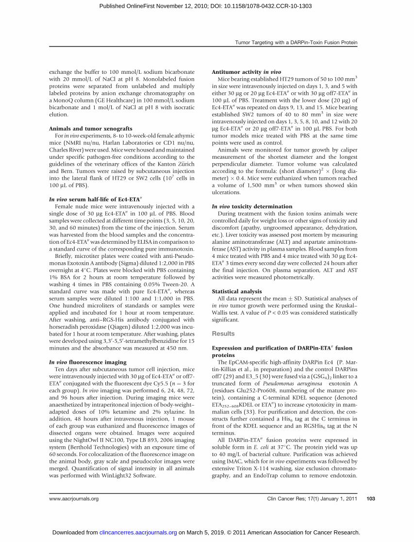
To determine treatment-related unspecific toxicity onEc4-ETA00 and off7-ETA00 administration, mice were mon-itored for weight loss, dehydration, and signs of distress(apathy, hyperalgesia, and ungroomed appearance)throughout the course of the study. Representative for alltherapy experiments, Fig. 5 shows for HT29 tumor-bearingmice that treatments were well tolerated and after reversiblemarginal weight loss after the third injection of Ec4-ETA00
30/3 no further signs of toxicity were observed. Further-more, to assess liver toxicity as a frequent dose-limiting sideeffect of ETA fusion toxin therapy in patients, blood from 4PBS-treated mice and 4mice receiving 3 doses of 30 mg Ec4-ETA00 was collected and analyzed for activity of the livertransaminases AST and ALT. As shown in Table 1, livertoxicity could be excluded for this treatment schedule, asthere was no significant elevation of ALT and AST activity inthe plasma of Ec4-ETA00-treated (ALT: 54 � 22 U/L, andAST: 141� 47 U/L) compared with PBS-treated mice (ALT:57 � 51 U/L, and AST: 99 � 53 U/L).
Discussion
Chemotherapy still has remained the mainstay of cancertherapy. The great majority of approved treatments has no
Figure 4. Antitumor effect of Ec4-ETA00 in athymic mice. A, micebearing subcutaneously growingHT29 tumor xenografts of 50 to100 mm3 in size received tail-veininjections of 3 � 30 mg Ec4-ETA00,3 � 30 mg off7-ETA00 control, 6 �20 mg Ec4-ETA00, or PBS as vehiclecontrol. Tumor growth wasmonitored by calipermeasurement during the course of31 days. Data represent the meantumor volume � SD of 5 to 11mice per group. B, Kaplan–Meiersurvival curves with an endpointdefined as tumor volume of 100mm3. The curves show thepercentage of treatedmice in eachgroup in which tumors did notexceed 100 mm3 in size at thevarious time points after the startof treatment. C, mice bearingsubcutaneously growing SW2tumor xenografts of 40 to 80 mm3
in size received tail-vein injectionsof 6 � 20 mg Ec4-ETA00, 6 � 20 mgoff7-ETA00 control, or PBS asvehicle control. Tumor growth wasmonitored by calipermeasurement during the course of31 days. Data represent the meantumor volume � SD of 5 to 6 miceper group. D, pictures ofrepresentative tumors isolatedfrom mice of the differenttreatment groups.
A B
CD
Tumor Targeting with a DARPin-Toxin Fusion Protein
www.aacrjournals.org Clin Cancer Res; 17(1) January 1, 2011 107
on March 5, 2019. © 2011 American Association for Cancer Research. clincancerres.aacrjournals.org Downloaded from
Published OnlineFirst November 12, 2010; DOI: 10.1158/1078-0432.CCR-10-1303
inherent specificity for tumor cells but attack all dividingcells. More recently many investigations have been carriedout in which a toxic principle was coupled to a recognitionfunction. Immunotoxins based on Pseudomonas aeruginosaexotoxin A (ETA, also termed PE), particularly its N-term-inally truncated variant carrying a C-terminal KDEL peptide(ETA00), have been generated against various cell surfacereceptors and extensively tested in preclinical and earlyclinical studies (5, 34). Most of them use a single-chainfragment (scFv) or a disulfide-stabilized dsFv fragment ofan antibody as targeting moiety. We previously alsoreported on the potent antitumor effect of an EpCAM-specific scFv-ETA00 immunotoxin (11), which is currentlyunder phase II clinical investigation (9).
Commonly used antibody Fv-based formats, however,are difficult to produce in high amounts when comparedwith other proteins and are often aggregation-prone (24,26, 35). ScFv and dsFv fragments used for tumor-targetedfusion proteins must either be expressed in the periplasmor refolded after expression in inclusion bodies. Here, weinvestigated another class of highly advanced bindingmolecules, DARPins, for targeted delivery of ETA00 to tumorcells in vitro and in vivo. DARPins can be selected for highaffinity, which is a major requirement for efficient tumortargeting (36), their inherent robustness allows easy con-jugation with various types of effector molecules, andpharmacology and tumor targeting properties can be easilymodulated, for example, by site-specific PEGylation. There-fore, we are not limited to the simple fusions describedhere, as the molecules are robust enough to allow efficientproduction of more complicated constructs. As shownrecently, the possibility to produce DARPins in functionalform in multigram amounts by fermentation of E. coliand their easy purification can significantly reduce thetime from preclinical development to clinical investi-gation (http://clinicaltrials.gov/ct2/show/NCT01042678,http://clinicaltrials.gov/ct2/show/NCT01086761).
We are expressing the DARPin-ETA00 fusions in the cyto-plasm of E. coli and find that these fusion proteins areproduced in soluble form. Even though the ETA00 partcontains 2 disulfide bonds, which appear to be beneficialin the internalization process (37), we find in the purifiedprotein that they have been formed almost quantitatively,possibly by air oxidation. Thus, we can use the convenientproduction of DARPins in the E. coli cytoplasm also withthe DARPin-ETA00 fusion proteins.
A major challenge for the application of immunotoxins isthe choice of targets that provide sufficient tumor specificityand, at the same time, promote intracellular delivery of thepayload. Several targets have been explored, which serve thispurpose, for example, EGFR, LewisY, and various hemato-poietic markers (2). Most tumor-targeting with fusion toxinshas been studied in hematopoietic malignancies, but eventhere, only 1 (denileukin diftitox, Ontak), a fusion of IL-2with diphtheria toxin, finally received FDA approval (38).
EpCAM is overexpressed in many solid tumors and onthe basolateral cell surfaces of some normal epithelia (15,18) where it is, however, poorly accessible to circulatinganti-EpCAM antibodies (17). Because it also occurs onnormal hepatic stem cells and hepatoblasts, EpCAM-tar-geted immunotherapy may in principle impair liver regen-eration. However, liver toxicity has never been reported inclinical trials with anti-EpCAM antibodies, probablybecause the regenerative capacity of the adult liver issufficiently maintained bymitotic division of mature hepa-tocytes (39). In contrast, the inherent liver toxicity of ETA isbased on inflammatory responses caused by liver macro-phages, independent on specific hepatocyte targeting (40).
Recently, EpCAM was also identified as a marker ofcancer initiating cells in colon (21), breast (22), pancreatic(23), and hepatocellular carcinomas (24). Because cancerstem cells respond poorly to standard therapy and thus arelargely responsible for treatment failure, their eliminationmust be a prime objective for all kinds of innovative cancertherapy. Thus, from a therapeutic point of view, EpCAM isparticularly interesting as a docking site for targetingligands delivering external effector molecules. In fact,EpCAM efficiently mediates internalization of boundligands by receptor-mediated endocytosis and thus per-fectly matches the need of anticancer agents acting onintracellular targets such as protein toxins, chemotherapeu-tic agents, and antisense compounds (11–13).
Recently, we have described for the first time the produc-tion and biochemical characterization of EpCAM-specificDARPins, and a first generation binder was used to success-fully deliver therapeutic siRNA into tumor cells in the formof mono- and multivalent binders fused to highly chargedprotamine (14). Subsequent affinity maturation effortsthen resulted in DARPin Ec4, which displayed affinity toEpCAM in the low nanomolar range and excellent biophy-sical properties (P. Martin-Killias et al., in preparation).
We measured efficient tumor localization of Ec4-ETA00,but not of the control fusion off7-ETA00, by in vivofluorescence imaging upon systemic administration in aHT29 colon carcinoma xenograft model. These data
32
PBS
Off-7-ETA'' 30/3
Ec4-ETA'' 30/3
Ec4-ETA'' 20/6
30
28
26
0 10 20 30
Time after the start of treatment (d)
Wei
ght (
g)
24
22
Figure 5. Average weight of mice on treatment with different doseschedules of Ec4-ETA00, or off-7-ETA00 or with PBS control. Animals wereweighed 3 times per week during the whole experiment to monitortreatment-related toxicity.
Martin-Killias et al.
Clin Cancer Res; 17(1) January 1, 2011 Clinical Cancer Research108
on March 5, 2019. © 2011 American Association for Cancer Research. clincancerres.aacrjournals.org Downloaded from
Published OnlineFirst November 12, 2010; DOI: 10.1158/1078-0432.CCR-10-1303

showed that localization in the tumor was specific anddependent on EpCAM binding with peak tumor accumula-tion 48 to 72 hours after injection.Fluorescence imaging records the distribution of mole-
cules at any particular time point, whereas the previouslyused residualizing label 99mTc(CO)3 (36) gives an integrallocalization since the beginning of injection, because anylabel will accumulate at the site of cellular internalization.This probably accounts for the difference in kidney accu-mulation, which is seen to decay rapidly whenmeasured byfluorescence imaging but not by 99mTc(CO)3 radioactivity.Although immunotoxins (scFv fragments or DARPins fusedto a protein toxin such as ETA) would be expected to notfall in an ideal molecular weight range for maximumaccumulation (36), we see very encouraging enrichmentsand therapeutic effects already with the constructsdescribed here, and there may be even better effects withdifferent molecular formats.In this study, Ec4 was fused with ETA00 to assess for the
first time the potential of a rationally engineered DARPinfor tumor targeting and therapy. The fusion proteincould indeed be well produced in soluble form in E. coliat approximately 80-fold higher yield from shake flasksthan our previously described scFv-ETA00 fusion toxin(4D5MOCB-ETA; ref 11), could be easily purified andwas stable, and resistant to aggregation. This will facilitatesubsequent scaling up of the production process requiredfor clinical trials.We found that the high-affinity binding of Ec4 to intact
cells was fully preserved on fusion with ETA00 and that Ec4-ETA00 showed extremely high and specific in vitro cytotoxicityagainst EpCAM-positive tumor cells of various histotypeswith IC50 values of 0.005 pmol/L. This is remarkably lowcompared with other immunotoxins (41–43) and likelymirrors its high stability, affinity, and efficient internaliza-tion by receptor-mediated endocytosis. These data, com-bined with efficient tumor targeting, indeed translatedinto potent antitumor effects at well-tolerated doses withsomemice showing complete regressionof their tumors. Thehigher overall dose administered as 6 � 20 mg could provemore effective than the lower dose schedule (3 � 30 mg).Probablydue to the shorthalf-lifeof theDARPin-ETA00 fusiontoxin in the circulation, which may limit tumor accumula-tion and also toxicity to normal tissues, the dose required to
achieve tumor responses was higher than for a previouslydescribed scFv-ETA00 immunotoxin (11). The repeatableresponse measured after repeated injections further suggeststhat tumors retained a stable EpCAM expression profile andthat antigen loss did not occur during treatment.
A phase II study with this previously reported EpCAM-specific immunotoxin (11) consisting of ETA00 fused to ascFv antibody is ongoing and will be completed soon (9).On the basis of the findings of this study, it is tempting tospeculate thatDARPins such as Ec4 can replace the antibodyfragment as cell binding ligands also in forthcoming gen-erations of tumor-targeted fusion toxins and other drugdelivery systems.
In summary, we describe for the first time the generationand preclinical evaluationof an EpCAM-specific fusion toxinconsisting of a high-affinity DARPin (Ec4) and ETA00 as acatalytic biotoxin.We provide evidence for its potent activityagainst various tumor cell types in vitro, and its favorabletumor localization and antitumor activity in vivo. The advan-tages of DARPins enabling high yield expression, resistanceagainst aggregation, and stability also in the form of fusiontoxins in conjunction with a tumor-associated target such asEpCAM, opens new avenues for the generation of rationallydesigned protein therapeutics with outstanding efficacy.
Disclosure of Potential Conflicts of Interest
A. Pl€uckthun is a shareholder of Molecular Partners AG, which iscommercializing the DARPin technology. The other authors disclosed nopotential conflicts of interest.
Acknowledgments
We thank Dr. Beat Kunz and Gabriela Nagy-Davidescu for their supportwith animal experiments, Dr. Ykelien Boersma and Manuel Simon forhelpful discussions, Dr. Annemarie Honegger for modeling the structureof Ec4-ETA00, and Dr. Kaspar Binz, Varvara Mitropoulos, and ChristianGehringer for helpful discussions regarding soluble expression and vectors.
Grant support
This work was supported by the Swiss National Science FoundationGrant 310030-119859 (U. Zangemeister-Wittke and A. Pl€uckthun) andKrebsliga of the Kanton Z€urich (U. Zangemeister-Wittke and A.Pl€uckthun).
The costs of publication of this article were defrayed in part by the paymentof page charges. This article must therefore be hereby marked advertisement inaccordance with 18 U.S.C. Section 1734 solely to indicate this fact.
Received May 14, 2010; revised October 29, 2010; accepted November1, 2010; published OnlineFirst November 12, 2010.
Table 1. Liver transaminase activity in the plasma from mice treated with Ec4-ETA00 or PBS
Treatmenta
Ec4-ETA00 PBS
Mouse 1 Mouse 2 Mouse 3 Mouse 4 Mouse 1 Mouse 2 Mouse 3 Mouse 4
ALT U/L 34 84 57 41 134 40 26 29AST U/L 141 200 138 85 177 62 86 70
aMice received 3 � 30 mg of Ec4-ETA00 or PBS intravenously. Activity of the transaminases was measured 24 hours after the finalinjection.
Tumor Targeting with a DARPin-Toxin Fusion Protein
www.aacrjournals.org Clin Cancer Res; 17(1) January 1, 2011 109
on March 5, 2019. © 2011 American Association for Cancer Research. clincancerres.aacrjournals.org Downloaded from
Published OnlineFirst November 12, 2010; DOI: 10.1158/1078-0432.CCR-10-1303
References1. Allen TM. Ligand-targeted therapeutics in anticancer therapy. Nat Rev
Cancer 2002;2:750–63.2. Pastan I, Hassan R, FitzGerald DJ, Kreitman RJ. Immunotoxin treat-
ment of cancer. Annu Rev Med 2007;58:221–37.3. Shimamura T, Husain SR, Puri RK. The IL-4 and IL-13 Pseudomonas
exotoxins: new hope for brain tumor therapy. Neurosurg Focus2006;20:E11.
4. Pastan I, Hassan R, Fitzgerald DJ, Kreitman RJ. Immunotoxin therapyof cancer. Nat Rev Cancer 2006;6:559–65.
5. Wolf P, Elsasser-Beile U. Pseudomonas exotoxin A: from virulencefactor to anti-cancer agent. Int J Med Microbiol 2009;299:161–76.
6. Kreitman RJ, Squires DR, Stetler-StevensonM, Noel P, FitzGerald DJ,Wilson WH, et al. Phase I trial of recombinant immunotoxin RFB4(dsFv)-PE38 (BL22) in patients with B-cell malignancies. J Clin Oncol2005;23:6719–29.
7. Kreitman RJ, Pastan I. Immunotoxins in the treatment of hematologicmalignancies. Curr Drug Targets 2006;7:1301–11.
8. Hassan R, Bullock S, Premkumar A, Kreitman RJ, Kindler H, Will-ingham MC, et al. Phase I study of SS1P, a recombinant anti-mesothelin immunotoxin given as a bolus i.v. infusion to patientswith mesothelin-expressing mesothelioma, ovarian, and pancreaticcancers. Clin Cancer Res 2007;13:5144–9.
9. Biggers K, Scheinfeld N. VB4–845, a conjugated recombinant anti-body and immunotoxin for head and neck cancer and bladder cancer.Curr Opin Mol Ther 2008;10:176–86.
10. Fuchs H, Bachran C. Targeted tumor therapies at a glance. Curr DrugTargets 2009;10:89–93.
11. Di Paolo C, Willuda J, Kubetzko S, Lauffer I, Tschudi D, Waibel R,et al. A recombinant immunotoxin derived from a humanized epithe-lial cell adhesion molecule-specific single-chain antibody fragmenthas potent and selective antitumor activity. Clin Cancer Res 2003;9:2837–48.
12. Hussain S, Pl€uckthun A, Allen TM, Zangemeister-Wittke U. Antitumoractivity of an epithelial cell adhesion molecule targeted nanovesiculardrug delivery system. Mol Cancer Ther 2007;6:3019–27.
13. Hussain S, Pl€uckthun A, Allen TM, Zangemeister-Wittke U. Chemo-sensitization of carcinoma cells using epithelial cell adhesion mole-cule-targeted liposomal antisense against bcl-2/bcl-xL. Mol CancerTher 2006;5:3170–80.
14. Winkler J, Martin-Killias P, Pl€uckthun A, Zangemeister-Wittke U.EpCAM-targeted delivery of nanocomplexed siRNA to tumor cellswith designed ankyrin repeat proteins. Mol Cancer Ther 2009;8:2674–83.
15. Balzar M, Winter MJ, de Boer CJ, Litvinov SV. The biology of the 17–1A antigen (Ep-CAM). J Mol Med 1999;77:699–712.
16. Maetzel D, Denzel S, Mack B, Canis M, Went P, Benk M, et al. Nuclearsignalling by tumour-associated antigen EpCAM. Nat Cell Biol2009;11:162–71.
17. McLaughlin PM, Harmsen MC, Dokter WH, Kroesen BJ, Van DerMolen H, Brinker MG, et al. The epithelial glycoprotein 2 (EGP-2)promoter-driven epithelial-specific expression of EGP-2 in transgenicmice: a new model to study carcinoma-directed immunotherapy.Cancer Res 2001;61:4105–11.
18. Went PT, Lugli A, Meier S, Bundi M, Mirlacher M, Sauter G, et al.Frequent EpCam protein expression in human carcinomas. HumPathol 2004;35:122–8.
19. Gastl G, Spizzo G, Obrist P, Dunser M, Mikuz G. Ep-CAM over-expression in breast cancer as a predictor of survival. Lancet2000;356:1981–2.
20. Spizzo G, Went P, Dirnhofer S, Obrist P, Moch H, Baeuerle PA, et al.Overexpression of epithelial cell adhesion molecule (Ep-CAM) is anindependent prognostic marker for reduced survival of patients withepithelial ovarian cancer. Gynecol Oncol 2006;103:483–8.
21. Boman BM, Huang E. Human colon cancer stem cells: a new para-digm in gastrointestinal oncology. J Clin Oncol 2008;26:2828–38.
22. Fillmore CM, Kuperwasser C. Human breast cancer cell lines con-tain stem-like cells that self-renew, give rise to phenotypicallydiverse progeny and survive chemotherapy. Breast Cancer Res2008;10:R25.
23. Li C, Heidt DG, Dalerba P, Burant CF, Zhang L, Adsay V, et al.Identification of pancreatic cancer stem cells. Cancer Res2007;67:1030–7.
24. Terris B, Cavard C, Perret C. EpCAM, a new marker for cancer stemcells in hepatocellular carcinoma. J Hepatol 2010;52:280–1.
25. Ewert S, Honegger A, Pl€uckthun A. Stability improvement of anti-bodies for extracellular and intracellular applications: CDR grafting tostable frameworks and structure-based framework engineering.Methods 2004;34:184–99.
26. Willuda J, Honegger A, Waibel R, Schubiger PA, Stahel R, Zange-meister-Wittke U, et al. High thermal stability is essential for tumortargeting of antibody fragments: engineering of a humanized anti-epithelial glycoprotein-2 (epithelial cell adhesion molecule) single-chain Fv fragment. Cancer Res 1999;59:5758–67.
27. Binz HK, Amstutz P, Pl€uckthun A. Engineering novel binding proteinsfrom nonimmunoglobulin domains. Nat Biotechnol 2005;23:1257–68.
28. Binz HK, Stumpp MT, Forrer P, Amstutz P, Pl€uckthun A. Designingrepeat proteins: well-expressed, soluble and stable proteins fromcombinatorial libraries of consensus ankyrin repeat proteins. J MolBiol 2003;332:489–503.
29. Binz HK, Amstutz P, Kohl A, Stumpp MT, Briand C, Forrer P, et al.High-affinity binders selected from designed ankyrin repeat proteinlibraries. Nat Biotechnol 2004;22:575–82.
30. Binz HK, Kohl A, Pl€uckthun A, Gütter MG. Crystal structure of aconsensus-designed ankyrin repeat protein: implications for stability.Proteins 2006;65:280–4.
31. Wels W, Harwerth IM, Mueller M, Groner B, Hynes NE. Selectiveinhibition of tumor cell growth by a recombinant single-chain anti-body-toxin specific for the erbB-2 receptor. Cancer Res 1992;52:6310–7.
32. Hansen RE, Ostergaard H, Norgaard P, Winther JR. Quantification ofprotein thiols and dithiols in the picomolar range using sodium bor-ohydride and 4,4’-dithiodipyridine. Anal Biochem 2007;363:77–82.
33. Seetharam S, Chaudhary VK, FitzGerald D, Pastan I. Increased cyto-toxic activity of Pseudomonas exotoxin and two chimeric toxinsending in KDEL. J Biol Chem 1991;266:17376–81.
34. von Minckwitz G, Harder S, Höelmann S, Jäger E, Al-Batran SE, LoiblS, et al. Phase I clinical study of the recombinant antibody toxin scFv(FRP5)-ETA specific for the ErbB2/HER2 receptor in patients withadvanced solid malignomas. Breast Cancer Res 2005;7:R617–26.
35. Wörn A, Pl€uckthun A. Stability engineering of antibody single-chain Fvfragments. J Mol Biol 2001;305:989–1010.
36. Zahnd C, Kawe M, Stumpp MT, de Pasquale C, Tamaskovic R, Nagy-Davidescu G, et al. Efficient tumor targeting with high-affinitydesigned ankyrin repeat proteins: effects of affinity and molecularsize. Cancer Res 2010;70:1595–605.
37. Madshus IH, Collier RJ. Effects of eliminating a disulfide bridge withindomain II of Pseudomonas aeruginosa exotoxin A. Infect Immun1989;57:1873–8.
38. Manoukian G, Hagemeister F. Denileukin diftitox: a novel immuno-toxin. Expert Opin Biol Ther 2009;9:1445–51.
39. Fausto N, Campbell JS, Riehle KJ. Liver regeneration. Hepatology2006;43:S45–53.
40. Mühlen KA, Schümann J, Wittke F, Stenger S, Van Rooijen N, VanKaer L, et al. NK cells, but not NKT cells, are involved in Pseudomonasaeruginosa exotoxin A-induced hepatotoxicity in mice. J Immunol2004;172:3034–41.
41. Wolf P, Gierschner D, Buhler P, Wetterauer U, Elsasser-Beile U. Arecombinant PSMA-specific single-chain immunotoxin has potentand selective toxicity against prostate cancer cells. Cancer ImmunolImmunother 2006;55:1367–73.
42. Mazor Y, Noy R, Wels WS, Benhar I. chFRP5-ZZ-PE38, a large IgG-toxin immunoconjugate outperforms the corresponding smaller FRP5(Fv)-ETA immunotoxin in eradicating ErbB2-expressing tumor xeno-grafts. Cancer Lett 2007;257:124–35.
43. Zielinski R, Lyakhov I, Jacobs A, Chertov O, Kramer-Marek G, Fran-cella N, et al. Affitoxin–a novel recombinant, HER2-specific, antic-ancer agent for targeted therapy of HER2-positive tumors. JImmunother 2009;32:817–25.
Martin-Killias et al.
Clin Cancer Res; 17(1) January 1, 2011 Clinical Cancer Research110
on March 5, 2019. © 2011 American Association for Cancer Research. clincancerres.aacrjournals.org Downloaded from
Published OnlineFirst November 12, 2010; DOI: 10.1158/1078-0432.CCR-10-1303

Correction
Correction: A Novel Fusion Toxin Derived froman EpCAM-Specific Designed Ankyrin RepeatProtein Has Potent Antitumor Activity
In this article (Clin Cancer Res 2011;17:100–10), which was published in theJanuary 1, 2011 issue of Clinical Cancer Research (1), the name of the first authorappeared incorrectly on the online journal. The correct name of the first author isPatricia Martin-Killias. This error has been corrected.
Reference1. Martin-Killias P, Stefan N, Rothschild S, Pl€uckthun A, Zangemeister-Wittke U. A novel fusion toxin
derived from an EpCAM-specific designed ankyrin repeat protein has potent antitumor activity. ClinCancer Res 2011;17:100–10.
Published OnlineFirst May 3, 2011.�2011 American Association for Cancer Research.doi: 10.1158/1078-0432.CCR-11-0905
ClinicalCancer
Research
Clin Cancer Res; 17(11) June 1, 20113850

2011;17:100-110. Published OnlineFirst November 12, 2010.Clin Cancer Res Patricia Martin-Killias, Nikolas Stefan, Sacha Rothschild, et al. Ankyrin Repeat Protein Has Potent Antitumor ActivityA Novel Fusion Toxin Derived from an EpCAM-Specific Designed
Updated version
10.1158/1078-0432.CCR-10-1303doi:
Access the most recent version of this article at:
Material
Supplementary
http://clincancerres.aacrjournals.org/content/suppl/2011/01/03/1078-0432.CCR-10-1303.DC1
Access the most recent supplemental material at:
Cited articles
http://clincancerres.aacrjournals.org/content/17/1/100.full#ref-list-1
This article cites 43 articles, 15 of which you can access for free at:
Citing articles
http://clincancerres.aacrjournals.org/content/17/1/100.full#related-urls
This article has been cited by 5 HighWire-hosted articles. Access the articles at:
E-mail alerts related to this article or journal.Sign up to receive free email-alerts
Subscriptions
Reprints and
To order reprints of this article or to subscribe to the journal, contact the AACR Publications Department at
Permissions
Rightslink site. Click on "Request Permissions" which will take you to the Copyright Clearance Center's (CCC)
.http://clincancerres.aacrjournals.org/content/17/1/100To request permission to re-use all or part of this article, use this link
on March 5, 2019. © 2011 American Association for Cancer Research. clincancerres.aacrjournals.org Downloaded from
Published OnlineFirst November 12, 2010; DOI: 10.1158/1078-0432.CCR-10-1303