A key role for EZH2 and associated genes in mouse and...
7
RESEARCH COMMUNICATION A key role for EZH2 and associated genes in mouse and human adult T-cell acute leukemia Camille Simon, 1 Jalila Chagraoui, 1 Jana Krosl, 1 Patrick Gendron, 1 Brian Wilhelm, 1 Se ´ bastien Lemieux, 1 Genevie ` ve Boucher, 1 Pierre Chagnon, 1 Simon Drouin, 1 Raphae ¨ lle Lambert, 1 Claude Rondeau, 2 Annie Bilodeau, 2 Sylvie Lavalle ´e, 2 Martin Sauvageau, 1 Jose ´e He ´ bert, 1,2,3,4,5 and Guy Sauvageau 1,2,3,4,5 1 The Leucegene Group, Institute for Research in Immunology and Cancer, University of Montreal, Montreal, Quebec H3T 1J4, Canada; 2 Leukemia Cell Bank of Quebec, Maisonneuve-Rosemont Hospital, Montreal, Quebec H1T 2M4, Canada; 3 Division of Hematology-Oncology, Maisonneuve-Rosemont Hospital, Montreal, Quebec H1T 2M4, Canada; 4 Department of Medicine, University of Montreal, Montreal, Quebec H3C 3J7, Canada In this study, we show the high frequency of spontaneous gd T-cell leukemia (T-ALL) occurrence in mice with biallelic deletion of enhancer of zeste homolog 2 (Ezh2). Tumor cells show little residual H3K27 trimethylation marks compared with controls. EZH2 is a component of the PRC2 Polycomb group protein complex, which is associ- ated with DNA methyltransferases. Using next-generation sequencing, we identify alteration in gene expression levels of EZH2 and acquired mutations in PRC2-associated genes (DNMT3A and JARID2) in human adult T-ALL. Together, these studies document that deregulation of EZH2 and as- sociated genes leads to the development of mouse, and likely human, T-ALL. Supplemental material is available for this article. Received December 27, 2011; revised version accepted February 22, 2012. Enhancer of zeste homolog 2 (EZH2) catalyzes di- and trimethylation of Lys 27 on histone H3 (H3K27me2/3) and establishes chromatin marks associated with gene silencing (Surface et al. 2010). The enzymatic activity of EZH2 depends on the formation of a PRC2 complex comprising EZH2, embryonic ectoderm development (EED), and suppressor of zeste 12 homolog (SUZ12) (Pasini et al. 2004; Montgomery et al. 2005). Variations in the expression levels of individual components could therefore affect the activity of the PRC2 holoenzyme. We (Lessard et al. 1999) and others (Richie et al. 2002; Majewski et al. 2008, 2010) have shown that PRC2 restricts the activity of hemopoietic progenitor/stem cells and that heterozy- gosity for mutant Eed alleles accelerates lymphomagene- sis (Sauvageau et al. 2008). EZH2 mutations representing loss-of-function alleles have recently been identified in myelodysplastic syndromes (MDSs) and myeloprolifera- tive neoplasms (MPNs) (Ernst et al. 2010; Nikoloski et al. 2010; Jankowska et al. 2011; Score et al. 2012). Conversely, overexpression of Ezh2 has also been implicated in the progression of various types of human cancers (Sauvageau and Sauvageau 2010; Margueron and Reinberg 2011), and a recurrent EZH2 mutation identified in B-cell lymphomas (Morin et al. 2010) was proposed to act as a dominant, cancer-promoting EZH2 allele (Yap et al. 2011). Genetic data therefore suggest that gene dosage could be determin- istic for the apparently contradictory oncogenic and tumor- suppressing activities of EZH2, but no functional data supporting these possibilities have so far been presented. In this study, we exploit an inducible gene inactivation approach to show that Ezh2, as a PRC2 core component, suppresses T-cell leukemia (T-ALL) development in mice and provide evidence indicating a similar function for this complex in human adult T-ALL. More broadly, we provide key observations linking this disease with alterations in chromatin regulation. Results and Discussion For functional studies presented in this study, we ex- ploited the conditional Ezh2 F allele carrying loxP sites flanking exons 14 and 15, which code for the SET domain (Shen et al. 2008). The Cre-mediated deletion generates a mutated Ezh2 D allele and abrogates production of EZH2 protein (Shen et al. 2008). Ezh2 F/D mice breed normally, are healthy, and have a lifespan comparable with wild-type and Ezh2 F/+ mice. To ablate Ezh2 function in adult bone marrow cells, we crossed the conditional Ezh2 F/F and Ezh2 F/D mice with mice carrying the polyinosine–polycytidine (pIpC)-induc- ible Mx-Cre transgene and induced Cre expression with pIpC in 7- to 8-wk-old animals (Fig. 1A). At 12 d after pIpC induction, a PCR-based genotyping approach (Fig. 1B) demonstrated a partial Ezh2 deletion in the spleens of Ezh2 F/F Mx-Cre + and Ezh2 F/D Mx-Cre mice (hereafter, Ezh2 D/D mice) and nearly complete recombination of the floxed allele in the bone marrow and thymus. In the bone marrow cells of Ezh2 D/D mice, no EZH2 protein could be detected by Western blot analysis (Fig. 1C). Phenotypical analyses revealed a sevenfold to 10-fold decrease in the numbers of Ezh2 D/D thymocytes compared with Ezh2 +/+ Mx-Cre + controls (hereafter, wild-type controls) (Supple- mental Fig. 1A) associated with a loss of Ezh2 D/D CD4 + CD8 + cells and accumulation of the immature CD4 CD8 cell population (Supplemental Fig. 1B), confirming that EZH2 activity is essential for normal thymocyte development (Su et al. 2005; Mochizuki-Kashio et al. 2011). During a 10-mo observation period, all pIpC-treated wild-type and Ezh2 D/+ mice remained healthy, while com- plete inactivation of Ezh2 resulted in the development of spontaneous T-ALL. In the first cohort of Ezh2 D/D mice (three out of four mice), the shortest latency of leukemia development was 152 d (152–281 d) (Fig 1D), and in the second cohort (n = 10), they started to succumb at 122 d [Keywords: T-ALL; EZH2; PRC2; leukemia; JARID2; DNMT3A] 5 Corresponding authors. E-mail [email protected]. E-mail [email protected]. Article published online ahead of print. Article and publication date are online at http://www.genesdev.org/cgi/doi/10.1101/gad.186411.111. GENES & DEVELOPMENT 26:651–656 Ó 2012 by Cold Spring Harbor Laboratory Press ISSN 0890-9369/12; www.genesdev.org 651 Cold Spring Harbor Laboratory Press on April 20, 2020 - Published by genesdev.cshlp.org Downloaded from
Transcript of A key role for EZH2 and associated genes in mouse and...
RESEARCH COMMUNICATION
A key role for EZH2 andassociated genes in mouseand human adult T-cellacute leukemiaCamille Simon,1 Jalila Chagraoui,1 Jana Krosl,1
Patrick Gendron,1 Brian Wilhelm,1
Sebastien Lemieux,1 Genevieve Boucher,1
Pierre Chagnon,1 Simon Drouin,1
Raphaelle Lambert,1 Claude Rondeau,2
Annie Bilodeau,2 Sylvie Lavallee,2
Martin Sauvageau,1 Josee Hebert,1,2,3,4,5
and Guy Sauvageau1,2,3,4,5
1The Leucegene Group, Institute for Research in Immunologyand Cancer, University of Montreal, Montreal, Quebec H3T 1J4,Canada; 2Leukemia Cell Bank of Quebec, Maisonneuve-RosemontHospital, Montreal, Quebec H1T 2M4, Canada; 3Division ofHematology-Oncology, Maisonneuve-Rosemont Hospital,Montreal, Quebec H1T 2M4, Canada; 4Department of Medicine,University of Montreal, Montreal, Quebec H3C 3J7, Canada
In this study, we show the high frequency of spontaneousgd T-cell leukemia (T-ALL) occurrence in mice withbiallelic deletion of enhancer of zeste homolog 2 (Ezh2).Tumor cells show little residual H3K27 trimethylationmarks compared with controls. EZH2 is a component of thePRC2 Polycomb group protein complex, which is associ-ated with DNA methyltransferases. Using next-generationsequencing, we identify alteration in gene expression levelsof EZH2 and acquired mutations in PRC2-associated genes(DNMT3A and JARID2) in human adult T-ALL. Together,these studies document that deregulation of EZH2 and as-sociated genes leads to the development of mouse, andlikely human, T-ALL.
Supplemental material is available for this article.
Received December 27, 2011; revised version acceptedFebruary 22, 2012.
Enhancer of zeste homolog 2 (EZH2) catalyzes di- andtrimethylation of Lys 27 on histone H3 (H3K27me2/3)and establishes chromatin marks associated with genesilencing (Surface et al. 2010). The enzymatic activityof EZH2 depends on the formation of a PRC2 complexcomprising EZH2, embryonic ectoderm development(EED), and suppressor of zeste 12 homolog (SUZ12) (Pasiniet al. 2004; Montgomery et al. 2005). Variations in theexpression levels of individual components could thereforeaffect the activity of the PRC2 holoenzyme. We (Lessard
et al. 1999) and others (Richie et al. 2002; Majewski et al.2008, 2010) have shown that PRC2 restricts the activityof hemopoietic progenitor/stem cells and that heterozy-gosity for mutant Eed alleles accelerates lymphomagene-sis (Sauvageau et al. 2008). EZH2 mutations representingloss-of-function alleles have recently been identified inmyelodysplastic syndromes (MDSs) and myeloprolifera-tive neoplasms (MPNs) (Ernst et al. 2010; Nikoloski et al.2010; Jankowska et al. 2011; Score et al. 2012). Conversely,overexpression of Ezh2 has also been implicated in theprogression of various types of human cancers (Sauvageauand Sauvageau 2010; Margueron and Reinberg 2011), anda recurrent EZH2 mutation identified in B-cell lymphomas(Morin et al. 2010) was proposed to act as a dominant,cancer-promoting EZH2 allele (Yap et al. 2011). Geneticdata therefore suggest that gene dosage could be determin-istic for the apparently contradictory oncogenic and tumor-suppressing activities of EZH2, but no functional datasupporting these possibilities have so far been presented.
In this study, we exploit an inducible gene inactivationapproach to show that Ezh2, as a PRC2 core component,suppresses T-cell leukemia (T-ALL) development in miceand provide evidence indicating a similar function for thiscomplex in human adult T-ALL. More broadly, we providekey observations linking this disease with alterations inchromatin regulation.
Results and Discussion
For functional studies presented in this study, we ex-ploited the conditional Ezh2F allele carrying loxP sitesflanking exons 14 and 15, which code for the SET domain(Shen et al. 2008). The Cre-mediated deletion generatesa mutated Ezh2D allele and abrogates production of EZH2protein (Shen et al. 2008). Ezh2F/D mice breed normally, arehealthy, and have a lifespan comparable with wild-typeand Ezh2F/+ mice.
To ablate Ezh2 function in adult bone marrow cells, wecrossed the conditional Ezh2F/F and Ezh2F/D mice withmice carrying the polyinosine–polycytidine (pIpC)-induc-ible Mx-Cre transgene and induced Cre expression withpIpC in 7- to 8-wk-old animals (Fig. 1A). At 12 d after pIpCinduction, a PCR-based genotyping approach (Fig. 1B)demonstrated a partial Ezh2 deletion in the spleens ofEzh2F/F Mx-Cre+ and Ezh2F/D Mx-Cre mice (hereafter,Ezh2D/D mice) and nearly complete recombination of thefloxed allele in the bone marrow and thymus. In the bonemarrow cells of Ezh2D/D mice, no EZH2 protein could bedetected by Western blot analysis (Fig. 1C). Phenotypicalanalyses revealed a sevenfold to 10-fold decrease in thenumbers of Ezh2D/D thymocytes compared with Ezh2+/+
Mx-Cre+ controls (hereafter, wild-type controls) (Supple-mental Fig. 1A) associated with a loss of Ezh2D/D CD4+CD8+
cells and accumulation of the immature CD4�CD8� cellpopulation (Supplemental Fig. 1B), confirming that EZH2activity is essential for normal thymocyte development (Suet al. 2005; Mochizuki-Kashio et al. 2011).
During a 10-mo observation period, all pIpC-treatedwild-type and Ezh2D/+ mice remained healthy, while com-plete inactivation of Ezh2 resulted in the development ofspontaneous T-ALL. In the first cohort of Ezh2D/D mice(three out of four mice), the shortest latency of leukemiadevelopment was 152 d (152–281 d) (Fig 1D), and in thesecond cohort (n = 10), they started to succumb at 122 d
[Keywords: T-ALL; EZH2; PRC2; leukemia; JARID2; DNMT3A]5Corresponding authors.E-mail [email protected] [email protected] published online ahead of print. Article and publication date areonline at http://www.genesdev.org/cgi/doi/10.1101/gad.186411.111.
GENES & DEVELOPMENT 26:651–656 � 2012 by Cold Spring Harbor Laboratory Press ISSN 0890-9369/12; www.genesdev.org 651
Cold Spring Harbor Laboratory Press on April 20, 2020 - Published by genesdev.cshlp.orgDownloaded from
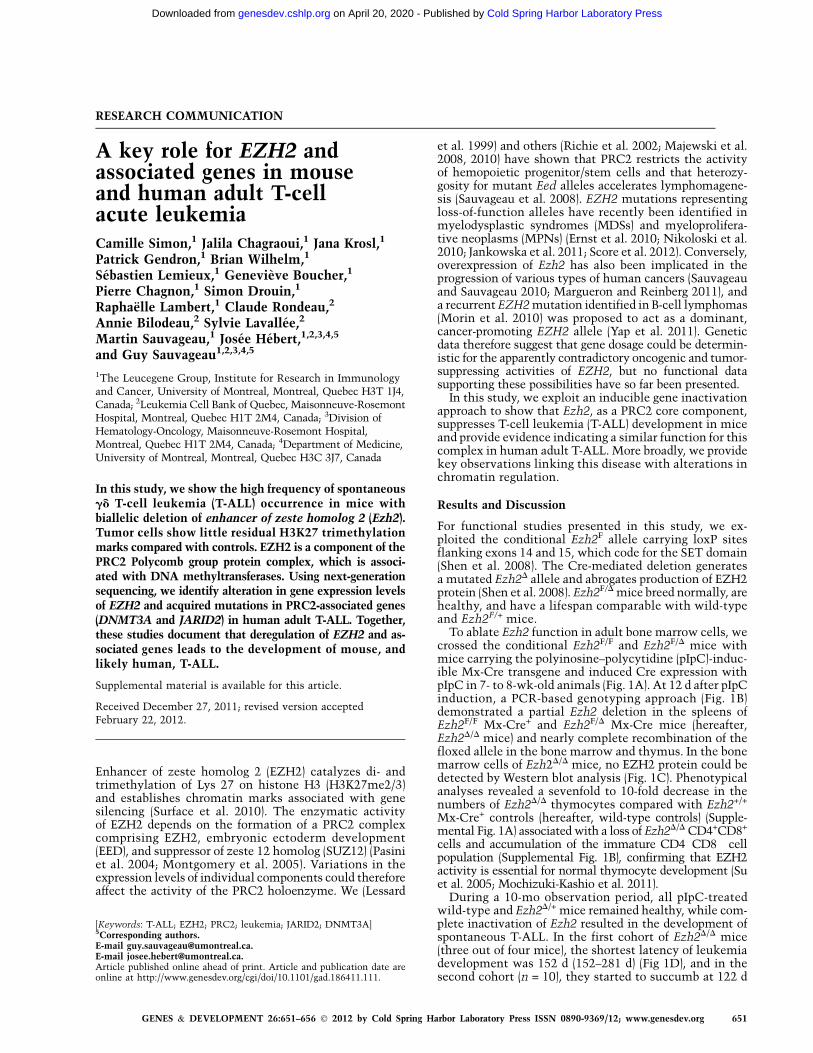
after deletion. Diseased animals presented with grosslyenlarged lymph nodes (Fig. 1E, panels 1,2) and spleens (Fig.1E [panels 2,3], F). Leukemic cells infiltrated the bonemarrow, spleen (Fig. 1G, right panel), liver, and kidney(Supplemental Fig. 2A). Infiltrates comprised a mixture ofrelatively mature lymphoid cells with open chromatin and
cells with blastic appearance andprominent nucleoli (Fig. 1G). Leuke-mias appeared to be phenotypicallyheterogeneous (Fig. 1H, top panel): Inmice #9040 and #12, the majorityof cells were CD4�CD8�; leukemia#7 comprised CD4+, CD8+, andCD4�CD8� cells; and leukemia #11was predominantly CD4+. However,all leukemias expressed CD3 (Fig. 1H,bottom panel) and TCRgd (Supple-mental Fig. 2B, right panel) and werenegative for the cell surface TCRab(Supplemental Fig. 2B, left panel).
The CD4+ and CD8+ cells detected insick Ezh2D/D mice could represent theprogeny of the nondeleted Ezh2F/D pro-genitors. Arguing against this possibil-ity, the PCR-based analyses of Ezh2(Fig. 2A) showed that the nondeletedEzh2F/D cells represented only a minorfraction of leukemic cell populationsand could not contribute to the pro-duction of CD4+ and CD8+ cells as seenin leukemias #7 and #11.
Hemopoietic tissues in wild-typecontrols expressed detectable mRNAlevels of the core PRC2 members Eed,Suz12, Ezh1, and Ezh2 (Fig. 2B, toppanel), and Ezh2 deletion had no majorimpact on the mRNA levels of otherPRC2 genes (Fig. 2B, bottom panel).In Ezh2D/D leukemias, no protein rec-ognized by an antibody directed againstthe region upstream of SET domain ofEZH2 (Fig. 2C) could be detected, whileEZH1 and SUZ12 proteins were pres-ent. The absence of EZH2 correlatedwith a noticeable decrease in the globallevels of H3K27me2/3 (Fig. 2D). To-gether with our previous work show-ing T-cell and B-cell tumor develop-ment in Eed mutant mice (Sauvageauet al. 2008), these observations sug-gested that PRC2 represents a sup-pressor complex with nonredundantfunctions in mouse T-ALL.
Very little is known about the in-tegrity of PRC2 and the associatedgenes in human T-ALL. To addressthis, we analyzed a series of 12 humanadult T-ALL specimens (including onepaired relapsed sample: 03H005 and03H096) (Supplemental Table 2) fornonsynonymous mutations and largeand small insertions and/or deletions(indels) in PRC2 and other associatedgenes. Using the Illumina HiSeq2000platform, we sequenced transcriptomes
and exomes using a strategy described in Figure 3A.Sequencing and mapping statistics are shown in Sup-plemental Table 3. Exome coverage was determinedat >453 (mean, 54.8) with 97.2 6 0.8% of all exonscaptured. The density of exon sequence reads was firstused for in silico reconstitution of chromosomes, which
Figure 1. Leukemia development in Ezh2-deficient mice. (A) Outline of the experimentalapproach. (B) PCR-based analysis of Ezh2 deletion in hemopoietic tissues at 12 d after pIpCtreatment. Primer pairs (sequence available on request) amplify a 314-base-pair (bp) wild-type(WT) Ezh2 fragment, a 280-bp fragment identifying the conditional Ezh2F allele, and a 200-bpfragment of the excised Ezh2D allele. Tail clippings were used as a source of control DNA. (C)Western blot analysis of EZH2 levels in bone marrow cells of wild-type and Ezh2F/D mice 12d after pIpC treatment. a-Tubulin levels are shown as a loading control. (D) Kaplan-Meiersurvival plot of pIpC-treated wild-type, Ezh2F/+, and Ezh2F/D mice. The day of the last pIpCinjection was designated as day 0 of the experiment. (E, panel 1) Photograph of a representativesick Ezh2F/D mouse showing enlarged lymph nodes (red arrows). Note also a bloatedappearance, suggesting the presence of an abdominal mass. An age-matched mouse from thecontrol wild-type cohort is shown for comparison. (Panel 2) Lymph nodes and spleen of anEzh2F/D mouse shown in panel 1. A wild-type spleen is shown for comparison in panel 3. (F)Splenomegaly of the sick Ezh2F/D mice. Result shown represents mean spleen weight 6 SD; n =4. (G) Wright stain-dyed cytospin preparations of bone marrow and spleen cells from a sickEzh2F/D mouse; 40-fold magnification. Mouse identification numbers are shown at the left. (H)Phenotypical characterization of Ezh2F/D leukemias. (Top panels) Proportions of CD4+ and CD8+
cells. (Bottom panels) Proportions of CD3+ cells in the bone marrow and lymph nodes. Mouseidentification numbers are shown at the top, and tissues analyzed are indicated at the left. (BM)Bone marrow; (LN) lymph node; (Thy) thymus.
652 GENES & DEVELOPMENT
Simon et al.
Cold Spring Harbor Laboratory Press on April 20, 2020 - Published by genesdev.cshlp.orgDownloaded from

revealed a good correlation with structural and numer-ical changes determined by G-banding cytogenetic anal-yses (Fig. 3B; Supplemental Table 2). The density ofthe sequence reads for the selected exons also allowedfor a more accurate assessment of deletions in PRC2and other T-ALL-associated genes (n > 30; CDKN2A/CDKN2B, PTEN, etc.) (Fig. 3B). This approach wasvalidated by fluorescent in situ hybridization (FISH)(Supplemental Table 2) and analysis of expression datafrom transcriptomes (Fig. 3C,D). Using this approach, wefound that CDKN2A/CDKN2B are deleted in about halfof our specimens (Fig. 3B). In these cases, CDKN2Aexpression was extremely low to undetectable (see bluein the second row of Fig. 3C). We also found deletionsin STIL, PTEN (Fig. 3B), IKZF1, and ETV6 (Fig. 3D), thelast two being recently associated with T-ALL (VanVlierberghe et al. 2011; Zhang et al. 2012). Using thisapproach, we could not identify deletions in EED or EZH1but found a 1.47-Mb deletion that included SUZ12(chromosome 17: position 29059504–30538257, hg19,patient 05H125) and one patient (03H078) with an extracopy of EZH2 (Fig. 3B; Supplemental Fig. 3). Transcrip-tome analyses revealed that EZH2 expression levels werereduced to 30% of the mean values in this specimen (Fig.3C; Supplemental Fig. 3). Of interest, EZH2 neighbor
genes, also in more than two copies in this leukemia, wereexpressed at levels ;50% higher than those detected inother leukemias (Supplemental Fig. 3B). We also found onespecimen (07H033) that expressed EZH2 at only 14% ofmean values. Interestingly, this leukemia expressed muchhigher levels of the H3K27 demethylase gene KDM6B (Fig.3C). We observed, in other ongoing studies, that humanleukemia with low EZH2 and high KDM6B levels showderegulation of HOXA gene expression (data not shown).Similar findings were observed for this 07H033 leukemiain which 59 HOXA genes are highly expressed (Supplemen-tal Fig. 3C).
We next exploited transcriptome sequencing (average,151 million 6 24 million mapped reads) (SupplementalTable 3) to identify the nonsense and missense mutationsin our T-ALL specimens. More than 47,000 alleles wereidentified, a vast majority of which represented missensemutations of already known variants (single-nucleotidepolymorphisms [SNPs]). Validated indels (not in the con-text of poly[A] or poly[T] sequences and likely under-estimated) comprised only six genes (listed in Fig. 3D asa frameshift mutation [n = 4]; Supplemental Table 5). SNPfiltering, detailed in Figure 3A and the Materials andMethods, reduced the number of SNPs to 564 (see Supple-mental Table 5 for SNP position and patient attribution).Several of the newly identified SNPs occurred in DNA orchromatin-modifying genes that are functionally and/orphysically linked to PRC2, such as DNMT3A (n = 2,including one with a homozygous R882 mutation) (Fig.3D shows only acquired mutations for PRC2-associatedgenes), DNMT1 (n = 1) (Supplemental Fig. 4), JARID2(core PRC2 gene, n = 1) (Fig. 3D), and IDH2 (n = 1) (Fig.3D). DNMT3A, JARID2, and IDH2 mutations wereconfirmed as newly acquired, since they were not pres-ent in patient-matched normal (nonleukemic) DNA(Supplemental Table 8). Mutant IDH2 interfere withactivity of jumonji histone demethylases such as KDM6B(Xu et al. 2011) and thus were included in this functionalcategory.
Western blot analysis indicated that H3K27me1/2/3levels were comparable between our T-ALL samples (Sup-plemental Fig. 5) and were much higher than those detectedin our homozygous mouse Ezh2D/D tumors (cf. results inFig. 2D [mouse] and those in Supplemental Fig. 5B [human]).Given that within a 1-year observation period no heterozy-gous Ezh2 mutant mice developed tumors, these resultsmay suggest that human T cells are more sensitive to PRC2dosage than their mouse counterparts.
Several other mutations, likely not functionally relatedto PRC2, were identified in the previously characterizedT-ALL-associated genes: NOTCH1 (n = 6), PTEN (n = 2),and NRAS (n = 1). This group also included TP53 (n = 2)and recently reported T-ALL mutations or deletions suchas DNM2 (n = 4), JAK3 (n = 1), EP300 (n = 2), IKZF1 (n = 2),and ETV6 (n = 3). A summary of the known and novelT-ALL-associated gene anomalies detected in our patientcohort is provided in the left panel of Figure 3D. In-terestingly, in this patient population, NOTCH1 muta-tions were associated with CDKN2A/B deletions andwere inversely correlated with the pro-T-cell phenotype.PRC2 and associated gene anomalies, on the other hand,were not found in cortical and medullary T-ALL (Fig.3D). Mutations found in the genes shown in Figure 3Dand Supplemental Figure 4 are presented in Supplemen-tal Table 4.
Figure 2. Genetic and biochemical characterizations of Ezh2D/D
leukemias. (A) PCR-based analysis of Ezh2 deletion in leukemic cellpopulations. Mouse identification numbers are shown at the top,and the positions of the conditional (F) and deleted (D) Ezh2 allelesare shown at the right. Controls were as described for Figure 1A. (B)Heat map showing expression of PRC2 transcripts in hemopoietictissues of wild-type (WT) controls. Expression values of genes werenormalized relative to endogenous HPRT (hypoxanthine-guaninephosphoribosyltransferase) controls. (Top panel) Wild-type controls.(Bottom panel) Ezh2D/D leukemias. Transcripts are shown at the top,and mouse identification numbers and tissues analyzed are shown atthe right. Red and blue represent high and low expression levels,respectively. (C) Western blot analysis of PRC2 protein levels inhemopoietic tissues of sick Ezh2D/D mice. a-Tubulin levels are shownas a loading control. Mouse identification numbers and tissues analyzedare shown at the top, the blotting antibodies are identified at the right,and the positions of molecular weight markers are indicated at the left.(D) Western blot analysis of global H3K27me2/3 levels. Histone H3levels are shown as loading controls. Blot labels are as described for C.
EZH2 and acute T-cell leukemia
GENES & DEVELOPMENT 653
Cold Spring Harbor Laboratory Press on April 20, 2020 - Published by genesdev.cshlp.orgDownloaded from

Figure 3. Characterization of genetic anomalies in human adult T-ALL. (A) Flow chart for the identification of genetic alterations in humanT-ALL. Transcriptome and exome sequencing allowed the identification of 47,749 nonsynonymous SNPs, indels, and other anomalies (copynumber variants and deletions). Following filtering (see the Materials and Methods), 564 genes were retained (listed in Supplemental Table 6).Gene annotation enrichment analysis with DAVID Bioinformatics Resources (Huang et al. 2009) identified chromatin regulators (shown in C,D;Supplemental Fig. 4) and the ubl conjugation pathway as significantly perturbed in these specimens (see Supplemental Table 7). All geneticanomalies were validated using exon sequencing, and acquired mutations were validated as described in the Materials and Methods and areshown in Supplemental Table 8. (B) Copy number variants in selected genes using exon capture sequencing (see the Materials and Methods fordetails). All chromosomes were manually inspected: Selected genes that appeared relevant (e.g., EZH2) or redundantly deleted (CDKN2A/B) areshown. The correlation between expression level and copy number variation is presented in Supplemental Figure 3B for EZH2. The color coderefers to change from mean RPKM values (yellow) per exon. Blue and red indicate lower and higher RPKM values, respectively. (C) Expression ofrelevant control (CDKN2A, TLX1, and MYC) and EZH2-associated genes in all T-ALL patients studied herein. Data are expressed in relativeRPKM values, with mean absolute values per gene is shown in the last column. Note high levels of MYC and TLX1 restricted to leukemia withrearrangements in these genes (see Supplemental Table 2) and low EZH2 expression in two leukemias, including one (07H033) with highKDM6B expression. (D) Representation of mutations, indels, and deletions found in T-ALL genes (left panel) and PRC2 and associated genes(right panel). The functional effects of nonsynonymous substitutions (color gradient) were predicted by the PolyPhen-2 program (http://genetics.bwh.harvard.edu/pph/data).
654 GENES & DEVELOPMENT
Cold Spring Harbor Laboratory Press on April 20, 2020 - Published by genesdev.cshlp.orgDownloaded from
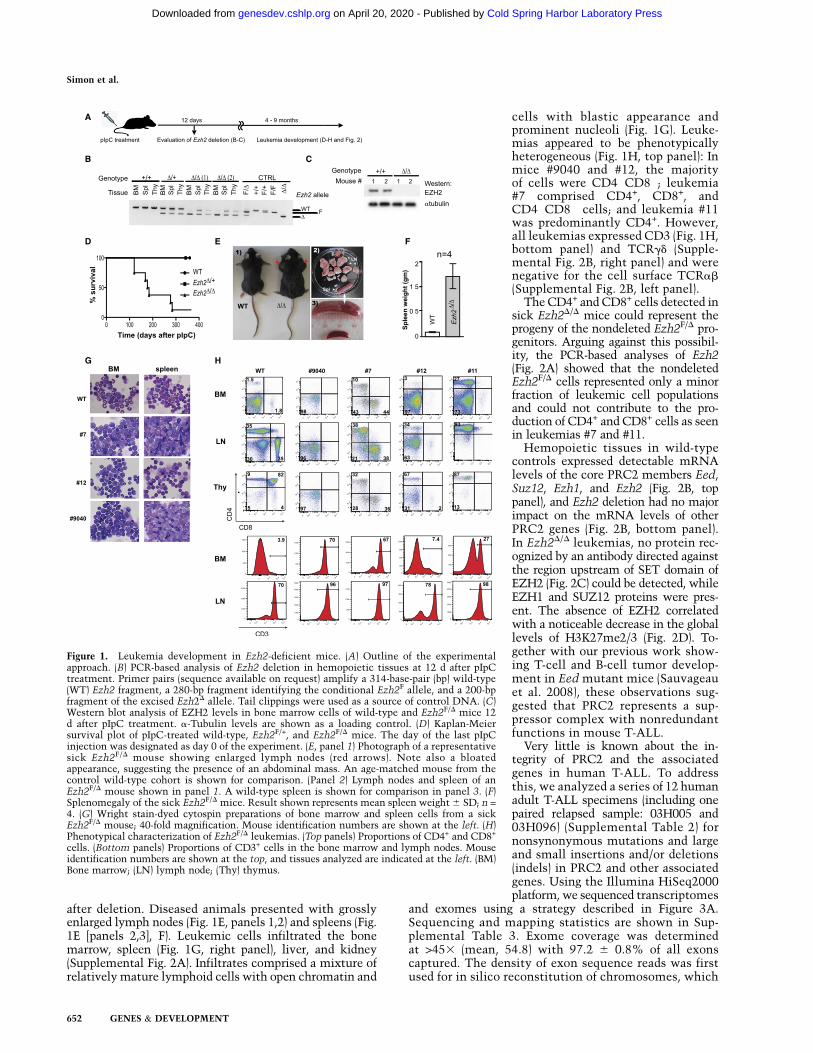
To identify possible genetic networks affected in theseleukemias, we submitted the list of modified alleles(Supplemental Table 6) to the DAVID bioinformatic re-source (Huang et al. 2009; http://david.abcc.ncifcrf.gov/home.jsp) for functional classification. To rule out meth-odological or population biases (e.g., strong founder effectin the Quebec population), we compared functional geneclusters identified in T-ALL with those determined fornormal karyotype acute myeloid leukemia (AML; 19 spec-imens) and found that nine of the top 15 gene clusters aredistinct between the two diseases. Twenty-two functionalgene clusters were found affected in T-ALL (SupplementalTable 7). Of interest, and consistent with PRC2 involve-ment in this disease, chromatin regulation was the categorymost significantly affected in this cohort (Benjamini-corrected �P-value [Huang et al. 2009] at 5 3 10�6). Stringanalysis (Jensen et al. 2009; http://string-db.org) of thisgene cluster suggested that several of the identified genesfunctionally interact with PRC2 (Supplemental Fig. 6).
Three of the other most significant functional clusters(ubl conjugation pathway, ligase, and ubl conjugation) areinvolved in the ubiquitin conjugation pathway. Most in-terestingly, string analysis of these genes revealed TP53 andCDKN2A/CDKN2B, also mutated or deleted in our tumors(Fig. 3D), as common targets for several of the mutatedE2 or E3 ubiquitin ligase genes.
Our studies provide the first in vivo validation of theproposed tumor-suppressive activity of Ezh2 in mice.These results are in line with our previous findings in-dicating that a loss of Eed function sensitizes mice for thedevelopment of T-ALL and B-cell leukemia (B-ALL). More-over, the high incidence of other PRC2 genetic alterationsobserved in human T-ALL, whether in the adult population(mutations: DNMT3A and JARID2; expression changes:EZH2 and KDM6B) (this study) or the childhood population(EZH2, EED, and SUZ12) (Zhang et al. 2012), argues fora more generalized deregulation of this Polycomb proteincomplex and associated genes in T-ALL. More globally, ourresults also suggest that several other regulators of thechromatin state, not necessarily linked with PRC2, maybe involved in the pathogenesis of T-ALL.
Material and methods
Mice and genotyping
C57BL/6 Ezh2F/+ mice were obtained from Stuart H. Orkin (Harvard Stem
Cell Institute, Boston, MA), transgenic B6.Cg-Tg(Mx1-Cre)1Cgn/J were
obtained from The Jackson Laboratory, and MeuCre40 mice [EM:01372,
C57BL/6-Tg(rtetR-tetO-cre) 40Mhz/Orl (MeuCre40)] were obtained from
the European Mouse Mutant Archive. To generate the Ezh2F/D mouse
strain, we crossed Ezh2F/+ mice with transgenic MeuCre40 mice and
backcrossed the Ezh2D/+ progeny with Ezh2F/F mice. Mice were bred and
manipulated in a specific pathogen-free animal facility. Animal handling
followed the guidelines of the Canadian Council on Animal Care, and
the experimental procedures were approved by the University of Mon-
treal Deontology Committee on Animal Experimentation. Primers and
PCR conditions used for genotyping of mice are available on request.
Human normal and leukemic samples
All of the samples used in these studies were collected by the Quebec
Leukemia Cell Bank with an informed consent and approval of the project
by the Research Ethics Board of the Maisonneuve-Rosemont Hospital and
Universite de Montreal. Seventy-eight blood or bone marrow samples
used to assess the mRNA expression levels of the core PRC2 components
(Supplemental Fig. 5A) comprised acute biphenotypic leukemias (n = 10),
B-ALLs (n = 18), T-ALLs (n = 5), plasmocytic leukemias (n = 3), multiple
myeloma (n = 5), and chronic lymphoproliferative disorders of different
morphological types (n = 37). Normal peripheral blood cells were obtained
from healthy volunteers and sorted to isolate the CD4+, CD8+, and CD19+
control cell populations.
Twelve T-ALL samples were analyzed in this study (nine diagnostic and
three relapsed samples, including one paired relapsed sample). Morpho-
logic, immunophenotypic, and cytogenetic characteristics are shown
in Supplemental Table 2. Normal DNA paired with each of these T-ALL
specimens was obtained from buccal swabs or from saliva (sample 11-
H099). DNA was extracted as recommended by the manufacturers
(Isohelix buccal DNA Isolation kit, Cell Projects; Oragene•DNA, DNA
Genotech).
Data analysis
Sequence data were mapped to the reference genome using the Illumina
Casava 1.8.1 package and Elandv2 mapping software. Seventy-five percent
and 76% of the RNA-seq and exon captured sequences, respectively, were
unambiguously mapped to the genome (Supplemental Table 3). On
average, 97.2% of the regions targeted by the TruSeq capture kit were
sequenced, with a mean fold coverage of 54.8. RNA-seq yielding 15 Gb of
mapped reads per sample, with an average exon expression of 15.2 reads
per kilobase per million (RPKM). Variants (SNPs and small indels) were
identified using Casava 1.8.1 and were categorized as homozygous or
heterozygous. We used custom-designed software to categorize each
variant as occurring in introns, exons, or untranslated regions (UTRs)
and identified missense and nonsense mutations (Supplemental Table 5).
Specific regions were further examined across all samples for the presence
of large single-allele deletions or uniparental disomy (UPD) by graphically
assessing the heterozygocity of known SNPs (based on dbSNP 1.3.2).
Exome data were then used to identify copy number variants at these
locations by comparing local coverage between samples. Specifically, we
used RPKM values computed (local scripts available on request) for all
exons as a proxy for assessing local copy numbers at the exon level. To
avoid exon length and sequence composition biases, we normalized these
values by transforming them to Z-scores—subtracting the average and
dividing by the standard deviation of the RPKM measured for all samples.
However, the values obtained cannot be directly interpreted as a copy
number, since they represent deviations from the mean.
SNP filtering
SNP filtering represented the critical step in our analysis, since SNPs were
by far (>99.9%) the most abundant genetic anomalies detected in this
study (Fig. 3A). For this, we focused only on SNPs resulting in amino acid
substitution (nonsynonymous) and then excluded those present in the
dbSNP Human Build 1.3.2 database (http://www.ncbi.nlm.nih.gov/projects/
SNP) unless they occurred in leukemia-associated genes such as DNMT3A.
Next, we focused on SNPs that occurred in expressed genes, defined here
as genes with RPKM values >10 (see Wilhelm et al. 2011 for a description
of expressed genes using Next-Gen Sequencing). We further filtered
this data set by eliminating SNPs in which the allelic ratio was <0.39
(considered not significant; e.g., late occurrence in disease progression or
early occurrence not selected for) and those for which <40 sequences were
available (see mean variant depth ALL in Supplemental Table 5). Finally,
SNPs common to AML (n = 19 specimens, to be reported elsewhere) and
ALL (>75%) were excluded because they most likely represented mis-
aligned sequences (pseudogenes, etc.) or previously unappreciated com-
mon variants. The final list of genetic anomalies (>560) (Supplemental
Table 5) was manually validated in the sequenced exome using the
Integrative Genomics Viewer version 2.0.31 (Robinson et al. 2011).
Suspicious sequences (e.g., mutations following stretch of polyadenine),
were eliminated from the final list. Subsets of SNPs in genes selected for
this study (see Fig. 3D; Supplemental Fig. 4) were verified by genomic
PCR–Sanger sequencing to determine whether the mutation was acquired
in the leukemic cells or was constitutive.
Acknowledgments
We thank Stuart H. Orkin (Harvard Stem Cell Institute, Boston) for
providing us with Ezh2F/+ mice. We also acknowledge Melanie Frechette
and Andrea Evelyn Mejia Alfaro for their assistance with animal care and
EZH2 and acute T-cell leukemia
GENES & DEVELOPMENT 655
Cold Spring Harbor Laboratory Press on April 20, 2020 - Published by genesdev.cshlp.orgDownloaded from
transplantation experiments, Julie Hinsinger from the Histology Core
Facility of IRIC for preparation and staining of tissue sections, and Nadine
Mayotte and Simon Girard for their excellent technical assistance. This
work was supported by grants from Genome Quebec and the Canadian
Cancer Society to G.S., and from the Cancer Research Network of the
FRSQ, the Cole Foundation, and Genome Quebec to J.H. G.S. holds a
Canada Research Chair in the Molecular Genetics of Stem Cells, and J.H.
holds a Research Chair in Leukemia supported by Industrielle-Alliance
(Universite de Montreal).
Note added in proof
While this work was under review, two other studies describing mutations
for EZH2 and other PRC2 genes in T-ALL were published (Ntziachristos et al.
2012; Zhang et al. 2012).
References
Ernst T, Chase AJ, Score J, Hidalgo-Curtis CE, Bryant C, Jones AV, Waghorn
K, Zoi K, Ross FM, Reiter A, et al. 2010. Inactivating mutations of the
histone methyltransferase gene EZH2 in myeloid disorders. Nat Genet
42: 722–726.
Huang DW, Sherman BT, Lempicki RA. 2009. Systematic and integrative
analysis of large gene lists using DAVID bioinformatics resources.
Nat Protoc 4: 44–57.
Jankowska AM, Makishima H, Tiu RV, Szpurka H, Huang Y, Traina F,
Visconte V, Sugimoto Y, Prince C, O’Keefe C, et al. 2011. Mutational
spectrum analysis of chronic myelomonocytic leukemia includes genes
associated with epigenetic regulation: UTX, EZH2, and DNMT3A.
Blood 118: 3932–3941.
Jensen LJ, Kuhn M, Stark M, Chaffron S, Creevey C, Muller J, Doerks T,
Julien P, Roth A, Simonovic M, et al. 2009. STRING 8—a global view
on proteins and their functional interactions in 630 organisms.
Nucleic Acids Res 37: D412–D416. doi: 10.1093/nar/gkn760.
Lessard J, Schumacher A, Thorsteinsdottir U, van Lohuizen M, Magnuson
T, Sauvageau G. 1999. Functional antagonism of the Polycomb-Group
genes eed and Bmi1 in hemopoietic cell proliferation. Genes Dev 13:
2691–2703.
Majewski IJ, Blewitt ME, de Graaf CA, McManus EJ, Bahlo M, Hilton AA,
Hyland CD, Smyth GK, Corbin JE, Metcalf D, et al. 2008. Polycomb
repressive complex 2 (PRC2) restricts hematopoietic stem cell activ-
ity. PLoS Biol 6: e93. doi: 10.1371/journal.pbio.0060093.
Majewski IJ, Ritchie ME, Phipson B, Corbin J, Pakusch M, Ebert A,
Busslinger M, Koseki H, Hu Y, Smyth GK, et al. 2010. Opposing roles
of polycomb repressive complexes in hematopoietic stem and pro-
genitor cells. Blood 116: 731–739.
Margueron R, Reinberg D. 2011. The Polycomb complex PRC2 and its
mark in life. Nature 469: 343–349.
Mochizuki-Kashio M, Mishima Y, Miyagi S, Negishi M, Saraya A,
Konuma T, Shinga J, Koseki H, Iwama A. 2011. Dependency on the
polycomb gene Ezh2 distinguishes fetal from adult hematopoietic
stem cells. Blood 118: 6553–6561.
Montgomery ND, Yee D, Chen A, Kalantry S, Chamberlain SJ, Otte AP,
Magnuson T. 2005. The murine polycomb group protein Eed is
required for global histone H3 lysine-27 methylation. Curr Biol 15:
942–947.
Morin RD, Johnson NA, Severson TM, Mungall AJ, An J, Goya R, Paul JE,
Boyle M, Woolcock BW, Kuchenbauer F, et al. 2010. Somatic mutations
altering EZH2 (Tyr641) in follicular and diffuse large B-cell lymphomas
of germinal-center origin. Nat Genet 42: 181–185.
Nikoloski G, Langemeijer SM, Kuiper RP, Knops R, Massop M, Tonnissen
ER, van der Heijden A, Scheele TN, Vandenberghe P, de Witte T, et al.
2010. Somatic mutations of the histone methyltransferase gene EZH2
in myelodysplastic syndromes. Nat Genet 42: 665–667.
Ntziachristos P, Tsirigos A, Vlierberghe PV, Nedjic J, Trimarchi T, Flaherty
MS, Ferres-Marco D, da Ros V, Tang Z, Siegle J, et al. 2012. Genetic
inactivation of the polycomb repressive complex 2 in T cell acute
lymphoblastic leukemia. Nat Med 18: 298–303.
Pasini D, Bracken AP, Jensen MR, Lazzerini Denchi E, Helin K. 2004.
Suz12 is essential for mouse development and for EZH2 histone
methyltransferase activity. EMBO J 23: 4061–4071.
Richie ER, Schumacher A, Angel JM, Holloway M, Rinchik EM, Magnuson
T. 2002. The Polycomb-group gene eed regulates thymocyte differen-
tiation and suppresses the development of carcinogen-induced T-cell
lymphomas. Oncogene 21: 299–306.
Robinson JT, Thorvaldsdottir H, Winckler W, Guttman M, Lander ES,
Getz G, Mesirov JP. 2011. Integrative genomics viewer. Nat Biotechnol
29: 24–26.
Sauvageau M, Sauvageau G. 2010. Polycomb group proteins: Multi-
faceted regulators of somatic stem cells and cancer. Cell Stem Cell
7: 299–313.
Sauvageau M, Miller M, Lemieux S, Lessard J, Hebert J, Sauvageau G.
2008. Quantitative expression profiling guided by common retroviral
insertion sites reveals novel and cell type specific cancer genes in
leukemia. Blood 111: 790–799.
Score J, Hidalgo-Curtis C, Jones AV, Winkelmann N, Skinner A, Ward D,
Zoi K, Ernst T, Stegelmann F, Dohner K, et al. 2012. Inactivation of
polycomb repressive complex 2 components in myeloproliferative
and myelodysplastic/myeloproliferative neoplasms. Blood 119: 1208–
1213.
Shen X, Liu Y, Hsu YJ, Fujiwara Y, Kim J, Mao X, Yuan GC, Orkin SH.
2008. EZH1 mediates methylation on histone H3 lysine 27 and
complements EZH2 in maintaining stem cell identity and executing
pluripotency. Mol Cell 32: 491–502.
Su IH, Dobenecker MW, Dickinson E, Oser M, Basavaraj A, Marqueron R,
Viale A, Reinberg D, Wulfing C, Tarakhovsky A. 2005. Polycomb
group protein ezh2 controls actin polymerization and cell signaling.
Cell 121: 425–436.
Surface LE, Thornton SR, Boyer LA. 2010. Polycomb group proteins set
the stage for early lineage commitment. Cell Stem Cell 7: 288–298.
Van Vlierberghe P, Ambesi-Impiombato A, Perez-Garcia A, Haydu JE,
Rigo I, Hadler M, Tosello V, Della Gatta G, Paietta E, Racevskis J,
et al. 2011. ETV6 mutations in early immature human T cell
leukemias. J Exp Med 208: 2571–2579.
Wilhelm BT, Briau M, Austin P, Faubert A, Boucher G, Chagnon P, Hope
K, Girard S, Mayotte N, Landry JR, et al. 2011. RNA-seq analysis of 2
closely related leukemia clones that differ in their self-renewal
capacity. Blood 117: e27–e38. doi: 10.1182/blood-2010-07-293332.
Xu W, Yang H, Liu Y, Yang Y, Wang P, Kim SH, Ito S, Yang C, Xiao MT, Liu
LX, et al. 2011. Oncometabolite 2-hydroxyglutarate is a competitive
inhibitor of a-ketoglutarate-dependent dioxygenases. Cancer Cell 19:
17–30.
Yap DB, Chu J, Berg T, Schapira M, Cheng SW, Moradian A, Morin RD,
Mungall AJ, Meissner B, Boyle M, et al. 2011. Somatic mutations at
EZH2 Y641 act dominantly through a mechanism of selectively altered
PRC2 catalytic activity, to increase H3K27 trimethylation. Blood 117:
2451–2459.
Zhang J, Ding L, Holmfeldt L, Wu G, Heatley SL, Payne-Turner D, Easton
J, Chen X, Wang J, Rusch M, et al. 2012. The genetic basis of early
T-cell precursor acute lymphoblastic leukaemia. Nature 481: 157–
163.
Simon et al.
656 GENES & DEVELOPMENT
Cold Spring Harbor Laboratory Press on April 20, 2020 - Published by genesdev.cshlp.orgDownloaded from
10.1101/gad.186411.111Access the most recent version at doi: originally published online March 19, 201226:2012, Genes Dev.
Camille Simon, Jalila Chagraoui, Jana Krosl, et al. T-cell acute leukemia
and associated genes in mouse and human adultEZH2A key role for
Material
Supplemental
http://genesdev.cshlp.org/content/suppl/2012/03/12/gad.186411.111.DC1
Related Content
Genes Dev. April , 2012 26: 751-755
Hanno HockA complex Polycomb issue: the two faces of EZH2 in cancer
References
http://genesdev.cshlp.org/content/26/7/651.full.html#related-urls
Articles cited in:
http://genesdev.cshlp.org/content/26/7/651.full.html#ref-list-1This article cites 27 articles, 10 of which can be accessed free at:
License
ServiceEmail Alerting
click here.right corner of the article or
Receive free email alerts when new articles cite this article - sign up in the box at the top
Copyright © 2012 by Cold Spring Harbor Laboratory Press
Cold Spring Harbor Laboratory Press on April 20, 2020 - Published by genesdev.cshlp.orgDownloaded from