A Highly Arginolytic Streptococcus Species That Potently ... · example, the levels of free...
15
A Highly Arginolytic Streptococcus Species That Potently Antagonizes Streptococcus mutans Xuelian Huang, a Sara R. Palmer, a Sang-Joon Ahn, a Vincent P. Richards, b Matthew L. Williams, a Marcelle M. Nascimento, c Robert A. Burne a Department of Oral Biology, College of Dentistry, University of Florida, Gainesville, Florida, USA a ; Department of Biological Sciences, College of Agriculture, Forestry and Life Sciences, Clemson University, Clemson, South Carolina, USA b ; Department of Restorative Dental Science, Division of Operative Dentistry, College of Dentistry, University of Florida, Gainesville, Florida, USA c The ability of certain oral biofilm bacteria to moderate pH through arginine metabolism by the arginine deiminase system (ADS) is a deterrent to the development of dental caries. Here, we characterize a novel Streptococcus strain, designated strain A12, iso- lated from supragingival dental plaque of a caries-free individual. A12 not only expressed the ADS pathway at high levels under a variety of conditions but also effectively inhibited growth and two intercellular signaling pathways of the dental caries pathogen Streptococcus mutans. A12 produced copious amounts of H 2 O 2 via the pyruvate oxidase enzyme that were sufficient to arrest the growth of S. mutans. A12 also produced a protease similar to challisin (Sgc) of Streptococcus gordonii that was able to block the competence-stimulating peptide (CSP)–ComDE signaling system, which is essential for bacteriocin production by S. mutans. Wild-type A12, but not an sgc mutant derivative, could protect the sensitive indicator strain Streptococcus sanguinis SK150 from killing by the bacteriocins of S. mutans. A12, but not S. gordonii, could also block the XIP (comX-inducing peptide) signaling pathway, which is the proximal regulator of genetic competence in S. mutans, but Sgc was not required for this activity. The complete genome sequence of A12 was determined, and phylogenomic analyses compared A12 to streptococcal reference ge- nomes. A12 was most similar to Streptococcus australis and Streptococcus parasanguinis but sufficiently different that it may represent a new species. A12-like organisms may play crucial roles in the promotion of stable, health-associated oral biofilm communities by moderating plaque pH and interfering with the growth and virulence of caries pathogens. D ental caries remains an enormous health problem worldwide (1–4). The current view of the initiation and progression of dental caries is one of a dynamic process in which the normal balance between the complex environmental factors that control demineralization and remineralization of the tooth is disrupted in a way that favors the loss of tooth mineral. Demineralization is driven primarily by microbial fermentation of carbohydrates to organic acids, which can lower the pH of oral biofilms to the extent that there is significant dissolution of tooth mineral. Alka- lization of oral biofilms occurs during fasting periods and is attrib- utable to salivary clearance of acids, buffering of oral biofilms by host-derived and microbially derived components, and the pro- duction of basic compounds by microbial metabolism of amino acids, polyamines, urea, and other compounds (5–10). Develop- ment or worsening of a carious lesion occurs when acid-mediated enamel demineralization predominates and is associated with changes in the composition and biochemical activities of the as- sociated microflora. Evidence is now accumulating from a variety of sources that supports that alkali generation, particularly in the form of ammonia, plays a major role in pH homeostasis in oral biofilms and inhibits the initiation and progression of dental car- ies (5, 10–15). Arginine is an amino acid found in a wide variety of foods and is relatively abundant in saliva and other oral secretions (16). In the oral cavity, arginine and peptides that contain arginine are primarily catabolized by the arginine deiminase (AD) system (ADS), which is present in several bacterial species that constitute a major proportion of the oral microbiome (5, 17). The ADS con- sists minimally of three enzymes that convert one molecule of arginine to one molecule of ornithine and carbon dioxide and two molecules of ammonia, with the concomitant production of ATP from ADP. The ADS is believed to contribute to the inhibition of dental caries in a variety of ways. The ammonia generated elevates the biofilm pH, which prevents the loss of tooth mineral and pro- motes remineralization. The elevation in pH creates an environ- ment that is less favorable for the outgrowth of cariogenic bacteria and more favorable for the persistence of health-associated taxa. There are also bioenergetic benefits to the bacteria that have this system: the ATP generated by arginine catabolism can be used for growth and maintenance by the organism, while neutralization of the cytoplasm by ammonia enhances the pH component of the proton motive force (5, 11, 18, 19). It is now becoming apparent that arginine may impact oral biofilms and their pathogenic po- tential in other ways (20, 21). Multiple studies have established an association of arginine levels and ADS activity with caries status in human subjects. For example, the levels of free arginine in the parotid saliva of caries- free (CF) adults were significantly higher than those found in age- and sex-matched controls who had a history of dental decay (16). Received 2 December 2015 Accepted 26 January 2016 Accepted manuscript posted online 29 January 2016 Citation Huang X, Palmer SR, Ahn S-J, Richards VP, Williams ML, Nascimento MM, Burne RA. 2016. A highly arginolytic Streptococcus species that potently antagonizes Streptococcus mutans. Appl Environ Microbiol 82:2187–2201. doi:10.1128/AEM.03887-15. Editor: D. W. Schaffner, Rutgers, The State University of New Jersey Address correspondence to Robert A. Burne, [email protected]fl.edu. Supplemental material for this article may be found at http://dx.doi.org/10.1128 /AEM.03887-15. Copyright © 2016, American Society for Microbiology. All Rights Reserved. crossmark April 2016 Volume 82 Number 7 aem.asm.org 2187 Applied and Environmental Microbiology on August 18, 2019 by guest http://aem.asm.org/ Downloaded from
Transcript of A Highly Arginolytic Streptococcus Species That Potently ... · example, the levels of free...

A Highly Arginolytic Streptococcus Species That Potently AntagonizesStreptococcus mutans
Xuelian Huang,a Sara R. Palmer,a Sang-Joon Ahn,a Vincent P. Richards,b Matthew L. Williams,a Marcelle M. Nascimento,c
Robert A. Burnea
Department of Oral Biology, College of Dentistry, University of Florida, Gainesville, Florida, USAa; Department of Biological Sciences, College of Agriculture, Forestry andLife Sciences, Clemson University, Clemson, South Carolina, USAb; Department of Restorative Dental Science, Division of Operative Dentistry, College of Dentistry,University of Florida, Gainesville, Florida, USAc
The ability of certain oral biofilm bacteria to moderate pH through arginine metabolism by the arginine deiminase system (ADS)is a deterrent to the development of dental caries. Here, we characterize a novel Streptococcus strain, designated strain A12, iso-lated from supragingival dental plaque of a caries-free individual. A12 not only expressed the ADS pathway at high levels under avariety of conditions but also effectively inhibited growth and two intercellular signaling pathways of the dental caries pathogenStreptococcus mutans. A12 produced copious amounts of H2O2 via the pyruvate oxidase enzyme that were sufficient to arrest thegrowth of S. mutans. A12 also produced a protease similar to challisin (Sgc) of Streptococcus gordonii that was able to block thecompetence-stimulating peptide (CSP)–ComDE signaling system, which is essential for bacteriocin production by S. mutans.Wild-type A12, but not an sgc mutant derivative, could protect the sensitive indicator strain Streptococcus sanguinis SK150 fromkilling by the bacteriocins of S. mutans. A12, but not S. gordonii, could also block the XIP (comX-inducing peptide) signalingpathway, which is the proximal regulator of genetic competence in S. mutans, but Sgc was not required for this activity. Thecomplete genome sequence of A12 was determined, and phylogenomic analyses compared A12 to streptococcal reference ge-nomes. A12 was most similar to Streptococcus australis and Streptococcus parasanguinis but sufficiently different that it mayrepresent a new species. A12-like organisms may play crucial roles in the promotion of stable, health-associated oral biofilmcommunities by moderating plaque pH and interfering with the growth and virulence of caries pathogens.
Dental caries remains an enormous health problem worldwide(1–4). The current view of the initiation and progression of
dental caries is one of a dynamic process in which the normalbalance between the complex environmental factors that controldemineralization and remineralization of the tooth is disrupted ina way that favors the loss of tooth mineral. Demineralization isdriven primarily by microbial fermentation of carbohydrates toorganic acids, which can lower the pH of oral biofilms to theextent that there is significant dissolution of tooth mineral. Alka-lization of oral biofilms occurs during fasting periods and is attrib-utable to salivary clearance of acids, buffering of oral biofilms byhost-derived and microbially derived components, and the pro-duction of basic compounds by microbial metabolism of aminoacids, polyamines, urea, and other compounds (5–10). Develop-ment or worsening of a carious lesion occurs when acid-mediatedenamel demineralization predominates and is associated withchanges in the composition and biochemical activities of the as-sociated microflora. Evidence is now accumulating from a varietyof sources that supports that alkali generation, particularly in theform of ammonia, plays a major role in pH homeostasis in oralbiofilms and inhibits the initiation and progression of dental car-ies (5, 10–15).
Arginine is an amino acid found in a wide variety of foods andis relatively abundant in saliva and other oral secretions (16). Inthe oral cavity, arginine and peptides that contain arginine areprimarily catabolized by the arginine deiminase (AD) system(ADS), which is present in several bacterial species that constitutea major proportion of the oral microbiome (5, 17). The ADS con-sists minimally of three enzymes that convert one molecule ofarginine to one molecule of ornithine and carbon dioxide and twomolecules of ammonia, with the concomitant production of ATP
from ADP. The ADS is believed to contribute to the inhibition ofdental caries in a variety of ways. The ammonia generated elevatesthe biofilm pH, which prevents the loss of tooth mineral and pro-motes remineralization. The elevation in pH creates an environ-ment that is less favorable for the outgrowth of cariogenic bacteriaand more favorable for the persistence of health-associated taxa.There are also bioenergetic benefits to the bacteria that have thissystem: the ATP generated by arginine catabolism can be used forgrowth and maintenance by the organism, while neutralization ofthe cytoplasm by ammonia enhances the �pH component of theproton motive force (5, 11, 18, 19). It is now becoming apparentthat arginine may impact oral biofilms and their pathogenic po-tential in other ways (20, 21).
Multiple studies have established an association of argininelevels and ADS activity with caries status in human subjects. Forexample, the levels of free arginine in the parotid saliva of caries-free (CF) adults were significantly higher than those found in age-and sex-matched controls who had a history of dental decay (16).
Received 2 December 2015 Accepted 26 January 2016
Accepted manuscript posted online 29 January 2016
Citation Huang X, Palmer SR, Ahn S-J, Richards VP, Williams ML, Nascimento MM,Burne RA. 2016. A highly arginolytic Streptococcus species that potentlyantagonizes Streptococcus mutans. Appl Environ Microbiol 82:2187–2201.doi:10.1128/AEM.03887-15.
Editor: D. W. Schaffner, Rutgers, The State University of New Jersey
Address correspondence to Robert A. Burne, [email protected].
Supplemental material for this article may be found at http://dx.doi.org/10.1128/AEM.03887-15.
Copyright © 2016, American Society for Microbiology. All Rights Reserved.
crossmark
April 2016 Volume 82 Number 7 aem.asm.org 2187Applied and Environmental Microbiology
on August 18, 2019 by guest
http://aem.asm
.org/D
ownloaded from
A number of early studies showed that bacterial metabolism ofarginine, but not other common amino acids, by salivary bacteriacould elicit a rapid rise in pH (12, 14, 18, 22). Studies that com-pared the capacities of plaque and saliva samples from humansubjects to hydrolyze arginine via the ADS found that caries-active(CA) subjects or tooth sites had substantially lower ADS activitythan did similar samples from CF individuals, adding further sup-port showing that arginine metabolism may be a deterrent to den-tal caries (18). Studies with subjects who consumed mints or useddentifrices containing an arginine-bicarbonate formulationshowed that arginine was effective at inhibiting the initiation andprogression of dental caries (13, 18, 23–25). More recently, large-scale randomized clinical trials revealed that dentifrices contain-ing 1.5% arginine plus an insoluble calcium compound and 1,450ppm F provided significantly greater protection against the initi-ation and progression of caries than did dentifrices containing1,450 ppm F alone (23, 25). Furthermore, the use of a fluoride-freetoothpaste containing 1.5% arginine was associated with increasesin the ADS activity of dental plaque from CA individuals andinduced changes in the composition of their plaque microflorasuch that the microbiomes of CA subjects began to more closelyresemble those of CF subjects (18), compared to a control groupusing the same oral hygiene regimen but with a fluoride-contain-ing dentifrice. Thus, a substantial amount of data now supportsthat arginine and the ADS can have a profoundly positive impacton dental health and oral biofilm ecology.
Another major factor shaping the composition and virulenceof dental biofilms is antagonism between commensal and cario-genic bacteria (26). One of the primary ecological advantages forcaries pathogens is acid tolerance (aciduricity). Highly acidogenicand aciduric caries pathogens, such as Streptococcus mutans, Lac-tobacillus spp., Scardovia spp., Bifidobacteria spp., and certainother taxa (6, 27, 28), rapidly ferment dietary carbohydrates to pHvalues that are far lower than those that permit the growth ofhealth-associated commensal bacteria (27, 29–31). While the an-tagonistic effects of acidification of oral biofilms are a major driverof changes in the composition and biochemical activities of dentalbiofilms during caries development, there are more sophisticatedantagonistic strategies used by caries pathogens to gain a compet-itive advantage over health-associated commensals, including theproduction of bacteriocins. Of relevance here, the human dentalcaries pathogen Streptococcus mutans produces a variety of lantibi-otic and nonlantibiotic bacteriocins (often termed “mutacins”)that are active against a spectrum of bacteria, some of which areassociated with oral health. Mutacin production is dominantlycontrolled by the ComDE two-component system, which is or-thologous to BlpRH of Streptococcus pneumoniae (32, 33). CSP(competence-stimulating peptide) (34), an 18-amino-acid (aa)peptide derived from secretion and processing of the ComC pro-tein, induces mutacin expression through ComDE. Notably, CSPcan also be a potent activator of genetic competence (the ability totake up exogenous DNA) under certain growth conditions (35,36). The concurrent activation of mutacins to lyse neighboringspecies and activation of competence to take up exogenous DNAhave been proposed to be a strategy used by S. mutans to increasenutrient availability and enhance its genetic diversity (2, 37).
Activation of competence in S. mutans occurs transiently andrequires the induction of the gene for the alternative sigma factorComX. Notably, the ComDE-CSP circuit does not directly acti-vate comX expression. Instead, the Rgg-like transcriptional regu-
lator ComR is the proximal regulator of comX expression, andComR activity is enhanced by binding to the 7-aa comX-inducingpeptide (XIP). The ComRS regulatory pathway includes process-ing of the 17-aa ComS to XIP, which appears extracellularly, fol-lowed by active transport of XIP into the cell by the Opp oligopep-tide permease and formation of a ComR-XIP complex that bindsnear the comX promoter to activate transcription (35, 38, 39).Higher levels of XIP can also trigger the activation of the bacteri-ocin genes of S. mutans, at least in part because ComX can enhancethe transcription of the comE gene (40, 41). It is also known thatCSP is able to activate comX only in complex medium containingpeptides and only in a subpopulation of cells (bimodally), al-though CSP-dependent activation of mutacin genes occurs uni-modally (across the population) in complex medium or in pep-tide-free chemically defined medium. In contrast, XIP is able toactivate comX only in defined medium, presumably because pep-tides in complex medium compete with XIP for internalizationby Opp.
Commensal bacteria also have various antagonistic strategiesthat they deploy against S. mutans and other cariogenic species. Asnoted above, neutralization of the environment by arginine orurea metabolism (5, 6, 14) diminishes the competitive advantagethat caries pathogens gain at low pH. Thus, alkali generation bythe ADS can be thought of as a nonspecific defense against cariespathogens, which do not compete effectively with many commen-sals at neutral pH values. Streptococcus sanguinis, S. gordonii, andvarious other oral streptococcal species secrete substantialamounts (micromolar) of hydrogen peroxide (H2O2), which is apotent antagonist of the growth of S. mutans and other oral patho-gens. Oxidase enzymes that use various substrates can yield H2O2,but the pyruvate oxidase enzyme (Pox) encoded by spxB has beenshown to be a dominant source of H2O2 for certain commensalstreptococci (42). In addition to antagonism of caries pathogensthrough neutralization of the environment and H2O2 generation,S. gordonii can interfere with peptide signaling by S. mutansthrough the production of a proteinase termed challisin, whichappears to degrade CSP to inhibit bacteriocin production andcompetence development by S. mutans (43).
Recently, in an effort to better understand the basis for thesignificantly higher ADS activities of oral biofilms obtained fromCF subjects than of those obtained from CA subjects, our labora-tory isolated a panel of ADS-positive (ADS�) strains from suprag-ingival plaque (44), including strains of S. sanguinis S. gordonii, S.parasanguinis, S. intermedius, S. australis, and S. cristatus, and con-ducted an analysis of the ability of these isolates to express the ADSunder a variety of conditions known to repress or induce the genesfor the ADS (5, 19, 44–46). Substantial variability in constitutiveand condition-specific arginolytic capacities was noted betweenand within taxa of commensal streptococci. During characteriza-tion of these strains, one strain in particular, a streptococcal straindesignated strain A12, was found to be especially effective at ex-pressing high ADS activity (44), and a preliminary analysis re-vealed a potent capacity to interfere with the growth of S. mutans.The purpose of this study was to begin to explore the microbio-logical, molecular, and genomic basis for the desirable propertiesof Streptococcus clinical isolate A12.
MATERIALS AND METHODSBacterial strains, growth conditions, and reagents. The bacterial strainsand plasmids used in this study are shown in Table 1 (36, 44, 45, 47, 48).
Huang et al.
2188 aem.asm.org April 2016 Volume 82 Number 7Applied and Environmental Microbiology
on August 18, 2019 by guest
http://aem.asm
.org/D
ownloaded from
A12 was isolated from supragingival plaque of a CF subject. Escherichiacoli DH5� was grown in Luria broth (49). A12, S. mutans UA159, andderivatives of these strains were routinely grown in brain heart infusion(BHI) (3.7%) broth, on BHI agar plates (Difco), or in chemically definedFMC medium (50). Multiple medium formulations were developed em-pirically to examine the effects of carbohydrate source and nutrient avail-ability on interspecies interactions: (i) TY (3% tryptone, 0.5% yeast ex-tract) agar plates were supplemented with 25 mM galactose or 25 mMglucose, (ii) a more dilute version of TY (TY-D) (1% tryptone, 0.2% yeastextract) agar plates supplemented with 25 mM galactose, or (iii) half-strength BHI broth (1.85% BHI). For the selection of antibiotic-resistantcolonies after genetic transformation or for enumeration of bacteria frommixed cultures, erythromycin (300 �g ml�1 for E. coli or 12 �g ml�1 for S.mutans), kanamycin (50 �g ml�1 for E. coli or 1 mg ml�1 for S. mutans),or spectinomycin (50 �g ml�1 for E. coli or 1 mg ml�1 for S. mutans) wasadded to the medium. Unless noted otherwise, cultures were grown over-night in BHI medium, with antibiotics when necessary, at 37°C in a 5%CO2 aerobic atmosphere.
Synthetic CSP (sCSP), corresponding to the 21-aa peptide (34, 35),was synthesized by the Interdisciplinary Center for Biotechnology Re-search (ICBR) facility at the University of Florida, and its purity was con-firmed by high-performance liquid chromatography (HPLC). SyntheticXIP (sXIP), corresponding to residues 11 to 17 of ComS (amino acidsequence of GLDWWSL), was synthesized and purified to 96% homoge-neity by NeoBioSci (Cambridge, MA, USA). Lyophilized sXIP was recon-stituted in 99.7% dimethyl sulfoxide (DMSO) to a final concentration of 2mM and stored in 40-�l aliquots at �20°C.
Construction of mutant strains and reporter gene fusions. Mutantstrains of A12 were constructed by double-crossover recombination usinglinear DNA engineered to replace the gene of interest with an antibioticresistance gene. Briefly, using a colony of A12 as the template, 0.5-kbpfragments upstream and downstream of the genes of interest were ampli-fied by PCR using primers (Table 2) based on the genome sequence of A12(see below). Antibiotic resistance markers were amplified by PCR frompJL184 (kanamycin resistance [Kmr]) or pVA838 (erythromycin resistant[Emr]). Each PCR product was gel purified, and 100 ng of each fragmentwas fused by using PCR, as described previously (51). PCR products were
excised from an agarose gel, purified, and used to transform A12 by usinga modification of a method originally developed for Streptococcus australis(52). Briefly, a single colony was inoculated into Todd-Hewitt broth(THB) containing 10% heat-inactivated horse serum and incubated for16 to 18 h. Cells were diluted 1:40 into THB supplemented with 10% horseserum and incubated until the optical density at 600 nm (OD600) reached0.05 to 0.08. PCR products to be used for gene inactivation (0.5 �g) wereadded to the cultures, and incubation was continued for 3 h. PlasmidDNA (pDL278) (48) was used as a positive control to ensure that cellswere competent for transformation. Cultures were plated onto BHI agarplates containing the desired antibiotics. Transformants were verified byPCR and DNA sequencing, including sequencing of the regions used forrecombination to confirm that no unwanted mutations had been intro-duced into flanking genes. Mutant derivatives of S. gordonii DL1 wereconstructed by using similar methods (51).
To construct a strain of S. mutans with a lacZ gene fused to the pro-moter of the cipB bacteriocin gene (PcipB::lacZ; strain SAB249), the pro-moter region of cipB was amplified using primers (Table 2) that incorpo-rated SacI and BamHI sites (53) and cloned into a pMC340B-based lacZreporter vector (54), which carries a Kmr gene. The vector has sequencehomology to the mtlA and phnA genes flanking the cloning sites, allowingfor integration of the PcipB-lacZ construct in a single copy into the S.mutans chromosome through double-crossover homologous recombina-tion. The PcipB-lacZ construct was transformed into the wild-type strain,and the correct integration and sequence of the gene fusion were verifiedby PCR and DNA sequencing. To create a marked strain for cocultureexperiments, S. mutans was transformed with pBGS, a spectinomycin-resistant (Spr) derivative of pBGK in which the Kmr marker was replacedwith a Spr marker. Transformation of S. mutans UA159 with pBGS al-lowed for stable integration of the Spr marker into the gtfA gene (55), anonessential gene that does not adversely affect growth or competitivefitness of S. mutans, to create S. mutans strain UA159::pBGS.
ADS activity. ADS activity was measured by monitoring citrullineproduction from arginine using protocols detailed previously (44). ADSactivity was measured in cells grown as follows: cultures of each straingrown overnight were diluted 1:20 into fresh TY medium containing 25mM galactose, with or without 10 mM arginine, and incubated as de-
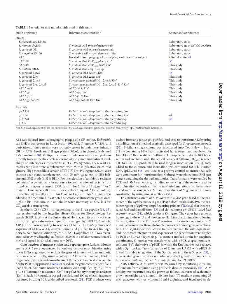
TABLE 1 Bacterial strains and plasmids used in this study
Strain or plasmid Relevant characteristic(s)a Source and/or reference
StrainsEscherichia coli DH5� Laboratory stockS. mutans UA159 S. mutans wild-type reference strain Laboratory stock (ATCC 700610)S. gordonii DL1 S. gordonii wild-type reference strain Laboratory stockS. sanguinis SK150 S. sanguinis wild-type reference strain Laboratory stockA12 Isolated from supragingival dental plaque of caries-free subject Clinical strain, 44SAB358 S. mutans UA159::PcomX-lacZ; Kmr 36SAB249 S. mutans UA159::PcipB-lacZ; Kmr This studyS. mutans pBGS S. mutans UA159::pBGS; Spr This studyS. gordonii �arcR S. gordonii DL1 �arcR; Kmr 45S. gordonii �sgc S. gordonii DL1 �sgc; Emr This studyS. gordonii �spxB Streptococcus gordonii DL1 �spxB; Kmr This studyS. gordonii �sgc �spxB Streptococcus gordonii DL1 �sgc �spxB; Emr Kmr This studyA12 �arcR A12 �arcR; Emr This studyA12 �sgc A12 �sgc; Emr This studyA12 �spxB A12 �spxB; Kmr This studyA12 �sgc �spxB A12 �sgc �spxB; Emr Kmr This study
PlasmidspVA838 Escherichia coli-Streptococcus shuttle vector; Emr
pJL184 Escherichia coli-Streptococcus shuttle vector; Kmr 47pDL278 Escherichia coli-Streptococcus shuttle vector; Spr 48pBGS Escherichia coli-Streptococcus shuttle vector; Spr
a In A12, arcR, sgc, and spxB are the homologs of the arcR, sgc, and spxB genes of S. gordonii, respectively. Spr, spectinomycin resistance.
Novel Beneficial Oral Streptococcus
April 2016 Volume 82 Number 7 aem.asm.org 2189Applied and Environmental Microbiology
on August 18, 2019 by guest
http://aem.asm
.org/D
ownloaded from
scribed above until the OD600 reached 0.5 to 0.6. The cells were thenpermeabilized by using a 1/10 volume of toluene-acetone (1:9) prior todetermination of their ADS activity. The concentration of protein in thepermeabilized cell preparations was measured by using a Pierce (Wal-tham, MA, USA) bicinchoninic acid (BCA) protein assay kit with bovineserum albumin as the standard. ADS activity levels of bacterial strainswere normalized to protein content and defined as nanomoles of citrul-line generated � [minutes � (milligrams protein)]�1. Alternatively, cellswere cultivated in FMC medium containing arginine in concentrationsranging from 0 to 100 mM. Dilutions, incubation, cell preparation, andADS assays for cells grown in FMC medium were the same as those usedfor cells grown in TY medium.
Measurement of H2O2. Cultures of bacterial strains grown overnightwere diluted 1:20 into TY medium plus 25 mM glucose, TY medium plus25 mM galactose, or TY-D medium plus 25 mM galactose and incubatedstatically in a 5% CO2 aerobic atmosphere. When the cultures reached anOD600 of 0.3, they were shaken at 200 rpm for 30 min, and the amount ofH2O2 present in culture supernatants was measured as previously de-scribed (56), with minor modifications: 0.65 ml of the culture supernatantwas added to 0.6 ml of a solution containing 2.5 mM 4-amino-antipyrine(4-amino-2,3-dimethyl-1-phenyl-3-pyrazolin-5-one; Sigma) and 0.17 Mphenol (Fisher). The reaction was allowed to proceed for 4 min at roomtemperature, and horseradish peroxidase (Pierce, Thermo Scientific) in0.2 M potassium phosphate buffer (pH 7.2) was then added to the reac-tion mixture at a final concentration of 13 mU/ml. After a 20-min incu-bation at room temperature, the OD510 was measured in a Beckman DU-640 spectrophotometer. A standard curve was generated for each assaymixture with known concentrations of H2O2 (Fisher Scientific).
Assays for H2O2-generating oxidase enzymes. Cultures grown over-night were centrifuged at 4,000 � g for 10 min and washed twice with 2 mlof phosphate-buffered saline (PBS) (pH 7.3) (137 mM NaCl, 2.7 mM KCl,4.3 mM Na2HPO4·7H2O, 1.4 mM KH2PO4). The pelleted cells were re-suspended in 2 ml PBS. Aliquots of the cell suspension were used fordetermining lactate oxidase activity, and the remaining cell suspensionswere permeabilized for determination of pyruvate oxidase, L-arginine ox-idase, and NADH oxidase activities by mixing the cell suspension with0.02 volumes of toluene-acetone (1:9, vol/vol) and vortexing the mixturefor 2 min. The concentration of protein in the cell preparations was de-termined by using a Pierce (Waltham, MA, USA) BCA protein assay kitwith bovine serum albumin as the standard (57).
Pyruvate oxidase activity was determined by measuring the produc-tion of acetyl phosphate (AcP) (57, 58). Briefly, the reaction mixture con-sisted of 0.45 ml of the permeabilized cell suspension and 0.45 ml of asolution containing 50 mM potassium phosphate buffer (pH 6.0), 10 �MMgCl2, 0.2 �M thiamine pyrophosphate (Sigma), 50 mM potassium py-ruvate, and 12 �M flavin adenine dinucleotide (FAD; Sigma). Reactionmixtures lacking permeabilized cells or pyruvate were included as nega-tive controls. The reaction mixtures were incubated at 37°C for 30 minwith shaking at a speed of 250 rpm, and the amount of acetyl phosphategenerated during the reaction was then measured as follows. An aliquot ofcell suspensions (0.3 ml) was preincubated at 37°C for 1 min in a model111002 heat block (Boekel Scientific, Feasterville, PA, USA). Fifty micro-liters of a 2 M hydroxylamine hydrochloride solution was added, and themixture was placed into a heat block at 60°C and incubated for 5 min.Next, 100 �l of development solution consisting of equal volumes of 0.5M ferric chloride in 5 M HCl and 30% trichloroacetic acid was added, andcolor was allowed to develop for at least 1 min at room temperature. Thesamples were centrifuged, and the OD540 was measured. A standard curvewas generated with known concentrations of lithium potassium acetylphosphate (Sigma).
Lactate oxidase, L-arginine oxidase, and NADH oxidase activities wereassessed by assaying H2O2 production (57). For the lactate oxidase assay,0.45 ml of intact cells was added to 0.45 ml of 0.2 M sodium phosphatebuffer (pH 7.0) containing 20 mM sodium L-lactate; reaction mixtureswithout sodium L-lactate were used as negative controls. The mixtureswere incubated at 37°C with shaking for 30 min at 250 rpm. For L-arginineoxidase assays, 0.45 ml of permeabilized cell suspensions was added to0.45 ml of 0.2 M sodium phosphate buffer (pH 7.0) containing 20 mML-arginine; reaction mixtures without arginine were used as negative con-trols. The mixtures were incubated at 37°C with shaking for 2 h at 250rpm. For NADH oxidase assays, 0.45 ml of permeabilized cell suspensionswas added to 0.45 ml of 0.2 M sodium phosphate buffer (pH 7.0) contain-ing 13 mM NADH (sodium salt); reaction mixtures without NADH wereused as negative controls. The mixtures were incubated at 37°C with shak-ing for 3 h at 250 rpm, and the concentration of H2O2 in the supernatantswas determined.
Competition assays on agar plates. For competition assays between S.mutans UA159 and strains of A12 or S. gordonii DL1, a culture of eachstrain grown overnight was adjusted to an OD600 of 0.5 in BHI medium.An aliquot from each culture (6 �l) was inoculated adjacent to the other
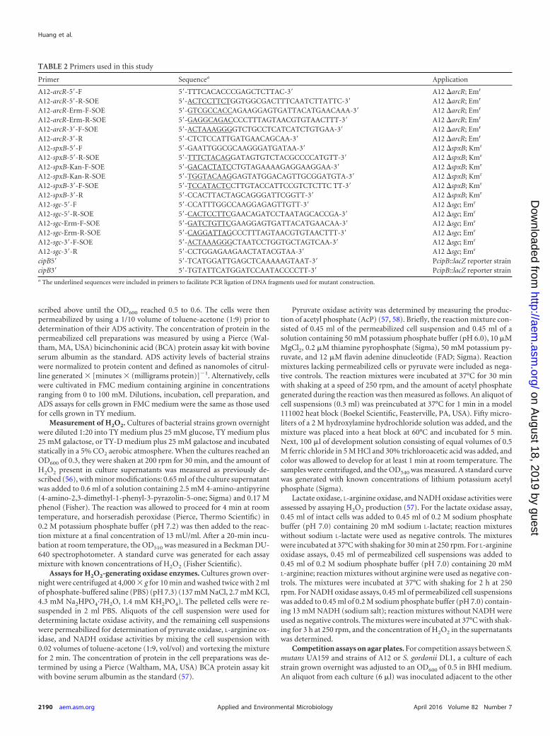
TABLE 2 Primers used in this study
Primer Sequencea Application
A12-arcR-5=-F 5=-TTTCACACCCGAGCTCTTAC-3= A12 �arcR; Emr
A12-arcR-5=-R-SOE 5=-ACTCCTTCTGGTGGCGACTTTCAATCTTATTC-3= A12 �arcR; Emr
A12-arcR-Erm-F-SOE 5=-GTCGCCACCAGAAGGAGTGATTACATGAACAAA-3= A12 �arcR; Emr
A12-arcR-Erm-R-SOE 5=-GAGGCAGACCCCTTTAGTAACGTGTAACTTT-3= A12 �arcR; Emr
A12-arcR-3=-F-SOE 5=-ACTAAAGGGGTCTGCCTCATCATCTGTGAA-3= A12 �arcR; Emr
A12-arcR-3=-R 5=-CTCTCCATTGATGAACAGCAA-3= A12 �arcR; Emr
A12-spxB-5=-F 5=-GAATTGGCGCAAGGGATGATAA-3= A12 �spxB; Kmr
A12-spxB-5=-R-SOE 5=-TTTCTACAGGATAGTGTCTACGCCCCATGTT-3= A12 �spxB; Kmr
A12-spxB-Kan-F-SOE 5=-GACACTATCCTGTAGAAAAGAGGAAGGAA-3= A12 �spxB; Kmr
A12-spxB-Kan-R-SOE 5=-TGGTACAAGGAGTATGGACAGTTGCGGATGTA-3= A12 �spxB; Kmr
A12-spxB-3=-F-SOE 5=-TCCATACTCCTTGTACCATTCCGTCTCTTC TT-3= A12 �spxB; Kmr
A12-spxB-3=-R 5=-CCACTTACTAGCAGGGATTCGGTT-3= A12 �spxB; Kmr
A12-sgc-5=-F 5=-CCATTTGGCCAAGGAGAGTTGTT-3= A12 �sgc; Emr
A12-sgc-5=-R-SOE 5=-CACTCCTTCGAACAGATCCTAATAGCACCGA-3= A12 �sgc; Emr
A12-sgc-Erm-F-SOE 5=-GATCTGTTCGAAGGAGTGATTACATGAACAA-3= A12 �sgc; Emr
A12-sgc-Erm-R-SOE 5=-CAGGATTAGCCCTTTAGTAACGTGTAACTTT-3= A12 �sgc; Emr
A12-sgc-3=-F-SOE 5=-ACTAAAGGGCTAATCCTGGTGCTAGTCAA-3= A12 �sgc; Emr
A12-sgc-3=-R 5=-CCTGGAGAAGAACTATACGTAA-3= A12 �sgc; Emr
cipB5= 5=-TCATGGATTGAGCTCAAAAAGTAAT-3= PcipB::lacZ reporter straincipB3= 5=-TGTATTCATGGATCCAATACCCCTT-3= PcipB::lacZ reporter straina The underlined sequences were included in primers to facilitate PCR ligation of DNA fragments used for mutant construction.
Huang et al.
2190 aem.asm.org April 2016 Volume 82 Number 7Applied and Environmental Microbiology
on August 18, 2019 by guest
http://aem.asm
.org/D
ownloaded from

strain on agar plates as follows: (i) A12 or DL1 was inoculated first, fol-lowed by inoculation of S. mutans 24 h later and an additional 24 h ofincubation; (ii) S. mutans was inoculated first, followed by inoculationwith A12 or DL1 after 24 h plus an additional 24 h of incubation; or (iii)DL1 or A12 and S. mutans were inoculated at the same time, and plateswere incubated for a total of 24 h.
Bacteriocin assays. Cultures of S. mutans UA159::pBGS (55), S. gor-donii DL1, A12, and derivatives of these strains grown overnight werecentrifuged, washed twice with PBS, resuspended in PBS, and adjusted toan OD600 of 0.5. S. mutans was then mixed with other strains at a ratio of1:1. Pure cultures or mixed cultures were spotted onto BHI agar plates andgrown aerobically or anaerobically. After 24 h of incubation at 37°C, 3 mlof a soft agar overlay (1% BHI agar) containing 107 cells of the indicatorstrain Streptococcus sanguinis SK150 was poured evenly onto the plate, theagar was allowed to set, and the plates were incubated for 24 h. Zones ofinhibition were measured and documented with a digital imager. In somecases, the agar plug from mixed cultures was harvested, serially diluted,and plated onto selective agar to enumerate cells of S. mutans, S. gordoniiDL1, strain A12, or derivatives of these strains. CFU were counted afterincubation at 37°C for 48 h.
Monitoring of comX and cipB gene promoter activities. The ability ofsupernatants from A12 and S. gordonii to interfere with CSP or XIP sig-naling by S. mutans was assessed by monitoring the expression of genesthat have been established to be activated by these peptides (36, 37, 59).Specifically, strains derived from S. mutans UA159 that carried lacZ re-porter gene fusions to the promoter regions of cipB, encoding a bacterio-cin (PcipB-lacZ), or to comX, encoding the alternative sigma factor re-quired for the development of genetic competence (PcomX-lacZ), wereused to monitor the ability of S. mutans to respond to sCSP or sXIP,respectively. Supernatants from cultures of strains grown in BHI mediumovernight were obtained after removal of cells by centrifugation, the pHwas adjusted to pH 7.0, and the supernatants were filter sterilized. A subsetof aliquots of supernatants was kept on ice, while others were heat treatedto inactive putative proteases. sCSP was added to supernatants to a finalconcentration of 2 �M, and the mixtures were incubated for 3 h. S. mu-tans UA159::PcipB-lacZ that had been grown to an OD600 of 0.12 was thensuspended in the supernatants. The cultures were then incubated for 2 h,and S. mutans cells were assayed for �-galactosidase activity. For XIPsignaling, which occurs in defined medium, supernatant fluids from cul-tures of A12 and its derivatives, S. gordonii, or S. mutans UA159 that hadbeen grown overnight in FMC medium were adjusted to pH 7.0, supple-mental glucose was added to increase the glucose concentration by 25mM, and the supernatants were filter sterilized. sXIP was added to heat-treated or untreated supernatants as described above and incubated over-night at 37°C. Cultures of S. mutans UA159::PcomX-lacZ that had beengrown in FMC medium to an OD600 of 0.12 were added to supernatants.The cultures were incubated for 2 h, and LacZ assays were performed todetect XIP-dependent activation of the comX promoter. Where noted,A12 was also grown under anaerobic conditions, or the supernatants weretreated with catalase to examine the influence of oxygen and/or hydrogenperoxide on the ability of A12 to inhibit peptide signaling.
LacZ (�-galactosidase) activity was measured by using a modificationof the Miller protocol (35, 60). Briefly, cells were harvested by centrifuga-tion, washed once with Z buffer (Na-phosphate buffer [pH 7.0], 10 mMKCl, 1 mM MgSO4, 5 mM �-mercaptoethanol), and resuspended in 1.2ml of Z buffer. A 400-�l aliquot of the sample was vortexed with 20 �l oftoluene-acetone (1:9) for 2 min, and the mixture was kept at 37°C. Theremainder of the cell suspension was used to measure the OD600. Thereaction was initiated by the addition of 80 �l of an ONPG (o-nitrophe-nyl-�-D-galactopyranoside) solution (4 mg/ml) to the mixture and wasterminated by the addition of 400 �l of 1 M Na2CO3 to the mixture.Samples were centrifuged at 15,250 � g for 1 min, and the OD of thesupernatant fluid was measured at 420 and 550 nm. �-Galactosidase ac-tivity was expressed in Miller units (35, 60).
Whole-genome sequencing and phylogenomic analysis. GenomicDNA was isolated by using Qiagen Genomic-Tip 20/G columns and theQiagen Genomic DNA buffer set according to the supplier’s protocols. A10-kb library was made by the ICBR Core at the University of Florida andsequenced by using PacBio RSII SMRT sequencing technology. TheBioinformatics Core at the University of Florida assembled the genome byusing SMRT analysis software. The genome was annotated by using theRAST server (61). A comprehensive phylogenetic analysis comparing A12to 14 other members of the genus Streptococcus (8 species from the S. mitisgroup, 3 species from the S. sanguinis group, and 3 species from the S.anginosus group) (see Table S1 in the supplemental material) was per-formed as described previously by Richards et al. (62). Briefly, a Markovcluster algorithm was used to determine the core gene set. Core geneclusters were aligned, and those judged to be recombinant were re-moved, leaving 374 putatively nonrecombinant gene alignments. Twophylogenetic approaches were taken. In the first approach, a maximumlikelihood (ML) phylogeny was generated from a concatenation of the374 alignments. In the second approach, 374 separate ML phylogenies(gene trees) were constructed, and a consensus phylogeny was con-structed.
Nucleotide sequence accession number. The DNA sequence forStreptococcus sp. A12 has been deposited with NCBI and assigned Bio-Sample acccession number SAMN04287214.
RESULTSEffects of arginine on ADS induction in A12. The arginine deimi-nase (AD) enzyme activity in A12 was higher than that in thehighly arginolytic reference strain S. gordonii DL1 in cells grownunder conditions that have been shown to be optimal for ADSexpression in vitro (TY medium containing 25 mM galactose and10 mM arginine) (44). Importantly, full induction of AD activityin A12 did not require supplementation of TY medium containinggalactose with arginine, whereas supplementation with argininewas necessary for full expression of the ADS in DL1 (Fig. 1A) andmany other ADS� bacteria (5, 19, 44, 45). ArcR is the transcrip-tional activator of the ADS genes in streptococci, governing ADSgene expression in response to arginine (45). When arcR was in-activated in A12 and S. gordonii DL1, both strains produced muchlower AD activity. The S. gordonii DL1 �arcR strain expressed only8% of the AD activity of the parental strain, whereas the A12�arcR strain retained 25% of the AD activity of wild-type A12. TYmedium contains 3% tryptone, so the concentration of availablearginine would be estimated to be in the 5 to 10 mM range andpresent primarily in the peptide form. To explore the requirementof arginine for ADS expression in more detail, cells were culturedin chemically defined FMC medium with a modified arginine con-tent (Fig. 1B). In FMC medium lacking arginine, both A12 andDL1 were unable to grow (data not shown). As the arginine levelsin FMC were increased, both A12 and DL1 showed increased ADactivity, with A12 producing higher AD levels than DL1, especiallyat lower concentrations of arginine. Both the S. gordonii DL1�arcR and A12 �arcR strains had much lower ADS levels than thewild-type strains, as observed with cells cultured in TY medium.However, the S. gordonii DL1 �arcR strain had almost no AD activ-ity, whereas the A12 �arcR strain retained considerable ADS activityunder the same conditions. Collectively, these data reveal that A12requires less arginine to produce substantial levels of AD activity andthat ArcR-independent ADS gene expression may, at least in part,explain the higher constitutional arginolytic activity of A12.
A12 inhibits growth of S. mutans by pyruvate oxidase-de-pendent H2O2 production. By using a plate-based zone-of-inhi-bition assay, A12 inhibited the growth of S. mutans UA159 under
Novel Beneficial Oral Streptococcus
April 2016 Volume 82 Number 7 aem.asm.org 2191Applied and Environmental Microbiology
on August 18, 2019 by guest
http://aem.asm
.org/D
ownloaded from

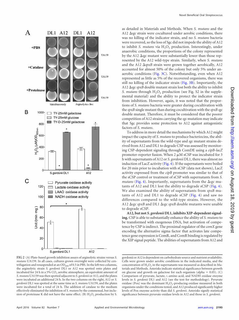
aerobic conditions more effectively than did DL1 when inoculatedprior to or simultaneously with inoculation of S. mutans UA159.H2O2 was clearly a factor in inhibition since the inclusion of cat-alase in the medium effectively eliminated inhibition (Fig. 2A).Both A12 and DL1 produced substantial quantities of H2O2 (Fig.2B) and displayed evidence of carbohydrate catabolite repression(CCR) of the activity responsible for H2O2 generation. More spe-cifically, inhibition of S. mutans by A12 was less efficient on me-dium with glucose than on medium containing galactose, whichelicits little or no evidence of CCR (Table 3). The use of a less-richversion of TY medium (TY-D medium) also resulted in enhancedH2O2 production, further linking the inhibitory activity of A12 tonutrient source and availability. Figure 2C shows that pyruvateoxidase (Pox) activity was the dominant source of H2O2 (Fig. 2C),with lactate oxidase, certain L-amino-acid oxidases (LAAOs), andNADH oxidase producing negligible amounts of H2O2 under theconditions tested. Notably, A12 expressed significantly higher Poxenzyme activity than did S. gordonii DL1 (Fig. 2C).
Modulation of S. mutans bacteriocin production by A12. Asdescribed in Materials and Methods, a standard agar overlay an-tagonism assay was modified to examine mechanisms by whichA12 may interfere with the growth of S. mutans and whether therewere additional beneficial activities associated with A12. In partic-ular, a dual-species cultivation system was created, in which agarplates were coinoculated with A12 and S. mutans and the plateswere subsequently overlaid with the indicator strain S. sanguinisSK150, which is sensitive to the mutacins produced by S. mutansUA159 (see Materials and Methods for more detail). This allowedus to (i) examine whether A12 could modulate the capacity of S.mutans to kill sensitive streptococci and (ii) enumerate, by remov-
ing and dispersing the bacteria at the inoculation site, the propor-tions of A12 and S. mutans bacteria remaining on the plates. First,virtually no S. mutans bacteria could be recovered after coinocu-lation/cocultivation with A12 under aerobic conditions (Fig. 3Aand C), whereas S. mutans accounted for nearly 60% of the recov-ered organisms under anaerobic conditions (Fig. 3B and C), thedifference being apparently attributable to H2O2-mediated killingof S. mutans in aerobic cocultures. In fact, when the predictedhomolog of the spxB gene of A12 encoding pyruvate oxidase wasdeleted, the mutant lost the ability to produce H2O2 during aero-bic growth (see Fig. S1 in the supplemental material) and to in-hibit the growth of S. mutans in the dual-species competition assay(see Fig. S2 in the supplemental material).
The same plate-based assay also revealed that the ability of S.mutans to produce bacteriocins that kill or inhibit the growth of S.sanguinis SK150 could be modulated by A12 and DL1. In partic-ular, when UA159 and A12 were coinoculated, there was no evi-dence of inhibition of the indicator strain (Fig. 3A and B). Whilethe lack of inhibition of SK150 under aerobic conditions was easilyexplained by a failure of S. mutans to grow in the presence of A12or DL1, the lack of inhibition of SK150 was equally as evidentunder anaerobic conditions. It was reported previously that pro-duction of a protease termed challisin, encoded by the sgc gene, byS. gordonii is responsible for degradation of CSP (43), the signalpeptide that induces bacteriocin gene expression through theComDE (32–34) two-component signal transduction system of S.mutans. We were able to identify a gene for a putative challisin-likeprotease in the genome (see below) of A12 that had 60.4% aminoacid sequence identity to S. gordonii challisin. Mutant strains ofA12 and DL1 that lacked the predicted sgc homologs were created,
FIG 1 (A) Arginine deiminase enzyme activity in S. gordonii DL1 and A12 wild-type (WT) and �arcR mutant strains grown in TY medium with or withoutsupplemental arginine. (B) Arginine deiminase activity in wild-type and �arcR mutant strains of S. gordonii grown in chemically defined FMC mediumcontaining the indicated amounts of arginine. A12 and S. gordonii DL1 were not able to grow in FMC medium unless some arginine was added. In panel A,asterisks indicate statistical significance between the wild-type and arcR mutant strains under both growth conditions using an alpha value of 0.05. In panel B,comparisons are between the arcR mutants of A12 and S. gordonii.
Huang et al.
2192 aem.asm.org April 2016 Volume 82 Number 7Applied and Environmental Microbiology
on August 18, 2019 by guest
http://aem.asm
.org/D
ownloaded from
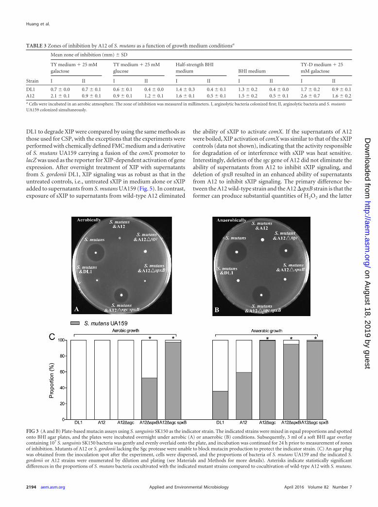
as detailed in Materials and Methods. When S. mutans and theA12 �sgc strain were cocultured under aerobic conditions, therewas no killing of the indicator strain, and no S. mutans bacteriawere recovered, so the loss of Sgc did not impede the ability of A12to inhibit S. mutans via H2O2 production. Interestingly, underanaerobic conditions, the proportions of the colony representedby the A12 �sgc mutant were substantially lower than those rep-resented by the A12 wild-type strain. Similarly, when S. mutansand the A12 �spxB strain were grown together aerobically, A12accounted for almost 50% of the colony but only 5% under an-aerobic conditions (Fig. 3C). Notwithstanding, even when A12represented as little as 5% of the recovered organisms, there wasstill no killing of the indicator strain (Fig. 3B). Importantly, theA12 �sgc spxB double mutant strain lost both the ability to inhibitS. mutans through H2O2 production (see Fig. S2 in the supple-mental material) and the ability to protect the indicator strainfrom inhibition. However, again, it was noted that the propor-tions of S. mutans bacteria were greater during cocultivation withthe spxB single mutant than during cocultivation with the spxB sgcdouble mutant. Therefore, it must be considered that the poorercompetition of A12 strains carrying the sgc mutation may indicatethat Sgc provides some protection to A12 against antagonisticfactors of S. mutans.
To address in more detail the mechanisms by which A12 mightimpact the capacity of S. mutans to produce bacteriocins, the abil-ity of supernatants from the wild-type and sgc mutant strains de-rived from A12 and DL1 to degrade CSP was assessed by monitor-ing CSP-dependent signaling through ComDE using a cipB-lacZpromoter-reporter fusion. When 2 �M sCSP was incubated for 3h with supernatants of A12 or S. gordonii DL1, there was almost noinduction of LacZ activity (Fig. 4). If the supernatants were boiledfor 20 min prior to incubation with sCSP (data not shown), LacZactivity expressed from the cipB promoter was similar to that ofthe sCSP control or treatment of sCSP with supernatants from S.mutans (Fig. 4). Importantly, supernatants from the �sgc mu-tants of A12 and DL1 lost the ability to degrade sCSP (Fig. 4).We also examined the ability of supernatants from spxB mu-tants of A12 and DL1 to degrade sCSP (Fig. 4) and saw nodifferences compared to the wild-type strains. However, theA12 �sgc spxB and DL1 �sgc spxB double mutants were unableto degrade sCSP.
A12, but not S. gordonii DL1, inhibits XIP-dependent signal-ing. CSP is able to substantially enhance the ability of S. mutans tobe transformed with exogenous DNA, but activation of compe-tence by CSP is indirect. The proximal regulator of the comX geneencoding the alternative sigma factor that activates late compe-tence gene expression is ComR, and ComR binding is regulated bythe XIP signal peptide. The abilities of supernatants from A12 and
FIG 2 (A) Plate-based growth inhibition assays of arginolytic strains versus S.mutans UA159. In all cases, cultures grown overnight were collected by cen-trifugation and resuspended at an OD600 of 0.5 in PBS. In the left two columns,the arginolytic strain S. gordonii DL1 or A12 was spotted onto plates andincubated for 24 h in a 5% CO2 aerobic atmosphere, an equivalent amount ofS. mutans UA159 was then spotted adjacent to S. gordonii or A12, and the plateswere incubated an additional 24 h. In the two columns on the right, A12 or S.gordonii DL1 was spotted at the same time as S. mutans UA159, and the plateswere incubated for a total of 24 h. The addition of catalase to the mediumeffectively eliminated the inhibition of S. mutans by the commensal, but inclu-sion of proteinase K did not have the same effect. (B) H2O2 production by S.
gordonii or A12 is dependent on carbohydrate source and nutrient availability.Cells were grown under aerobic conditions in the indicated media, and theconcentration of H2O2 in the supernatants was measured as described in Ma-terials and Methods. Asterisks indicate statistical significance between growthon glucose and growth on galactose for each organism (alpha 0.05). (C)Comparison of pyruvate, lactate, L-amino acid, and NADH oxidase enzymelevels in S. gordonii DL1 and A12 (see the text for methodology). Pyruvateoxidase (Pox) was the dominant H2O2-producing oxidase measured in bothorganisms under the conditions tested, and A12 produced significantly higherlevels of Pox enzyme activity than did S. gordonii. Asterisks signify statisticalsignificance between pyruvate oxidase levels in A12 and those in S. gordonii.
Novel Beneficial Oral Streptococcus
April 2016 Volume 82 Number 7 aem.asm.org 2193Applied and Environmental Microbiology
on August 18, 2019 by guest
http://aem.asm
.org/D
ownloaded from
DL1 to degrade XIP were compared by using the same methods asthose used for CSP, with the exceptions that the experiments wereperformed with chemically defined FMC medium and a derivativeof S. mutans UA159 carrying a fusion of the comX promoter tolacZ was used as the reporter for XIP-dependent activation of geneexpression. After overnight treatment of XIP with supernatantsfrom S. gordonii DL1, XIP signaling was as robust as that in theuntreated controls, i.e., untreated sXIP in medium alone or sXIPadded to supernatants from S. mutans UA159 (Fig. 5). In contrast,exposure of sXIP to supernatants from wild-type A12 eliminated
the ability of sXIP to activate comX. If the supernatants of A12were boiled, XIP activation of comX was similar to that of the sXIPcontrols (data not shown), indicating that the activity responsiblefor degradation of or interference with sXIP was heat sensitive.Interestingly, deletion of the sgc gene of A12 did not eliminate theability of supernatants from A12 to inhibit sXIP signaling, anddeletion of spxB resulted in an enhanced ability of supernatantsfrom A12 to inhibit sXIP signaling. The primary difference be-tween the A12 wild-type strain and the A12 �spxB strain is that theformer can produce substantial quantities of H2O2 and the latter
TABLE 3 Zones of inhibition by A12 of S. mutans as a function of growth medium conditionsa
Strain
Mean zone of inhibition (mm) SD
TY medium � 25 mMgalactose
TY medium � 25 mMglucose
Half-strength BHImedium BHI medium
TY-D medium � 25mM galactose
I II I II I II I II I II
DL1 0.7 0.0 0.7 0.1 0.6 0.1 0.4 0.0 1.4 0.3 0.4 0.1 1.3 0.2 0.4 0.0 1.7 0.2 0.9 0.1A12 2.1 0.1 0.9 0.1 0.9 0.1 1.2 0.1 1.6 0.1 0.5 0.1 1.5 0.2 0.5 0.1 2.6 0.7 1.6 0.2a Cells were incubated in an aerobic atmosphere. The zone of inhibition was measured in millimeters. I, arginolytic bacteria colonized first; II, arginolytic bacteria and S. mutantsUA159 colonized simultaneously.
FIG 3 (A and B) Plate-based mutacin assays using S. sanguinis SK150 as the indicator strain. The indicated strains were mixed in equal proportions and spottedonto BHI agar plates, and the plates were incubated overnight under aerobic (A) or anaerobic (B) conditions. Subsequently, 3 ml of a soft BHI agar overlaycontaining 107 S. sanguinis SK150 bacteria was gently and evenly overlaid onto the plate, and incubation was continued for 24 h prior to measurement of zonesof inhibition. Mutants of A12 or S. gordonii lacking the Sgc protease were unable to block mutacin production to protect the indicator strain. (C) An agar plugwas obtained from the inoculation spot after the experiment, cells were dispersed, and the proportions of bacteria of S. mutans UA159 and the indicated S.gordonii or A12 strains were enumerated by dilution and plating (see Materials and Methods for more details). Asterisks indicate statistically significantdifferences in the proportions of S. mutans bacteria cocultivated with the indicated mutant strains compared to cocultivation of wild-type A12 with S. mutans.
Huang et al.
2194 aem.asm.org April 2016 Volume 82 Number 7Applied and Environmental Microbiology
on August 18, 2019 by guest
http://aem.asm
.org/D
ownloaded from

produces almost no H2O2 under the conditions used for theseparticular experiments. When A12 was grown in either BHI orFMC broth under static conditions in a 5% CO2 aerobic atmo-sphere, no H2O2 was detected in supernatants of cultures grownovernight (data not shown). To be certain that the effects on XIPwere not associated with H2O2, supernatants were pretreated withcatalase prior to the addition of XIP (Fig. 6). As noted for the spxBmutant, elimination of H2O2 resulted in an enhancement of the
capacity of the supernatants from A12 to block sXIP-dependentactivation of comX. Thus, the production or stability of the activitythat interferes with XIP signaling may be enhanced under anaer-obic conditions, perhaps reflecting an adaptation that could en-hance the protection of A12 against S. mutans when A12 is unableto produce H2O2.
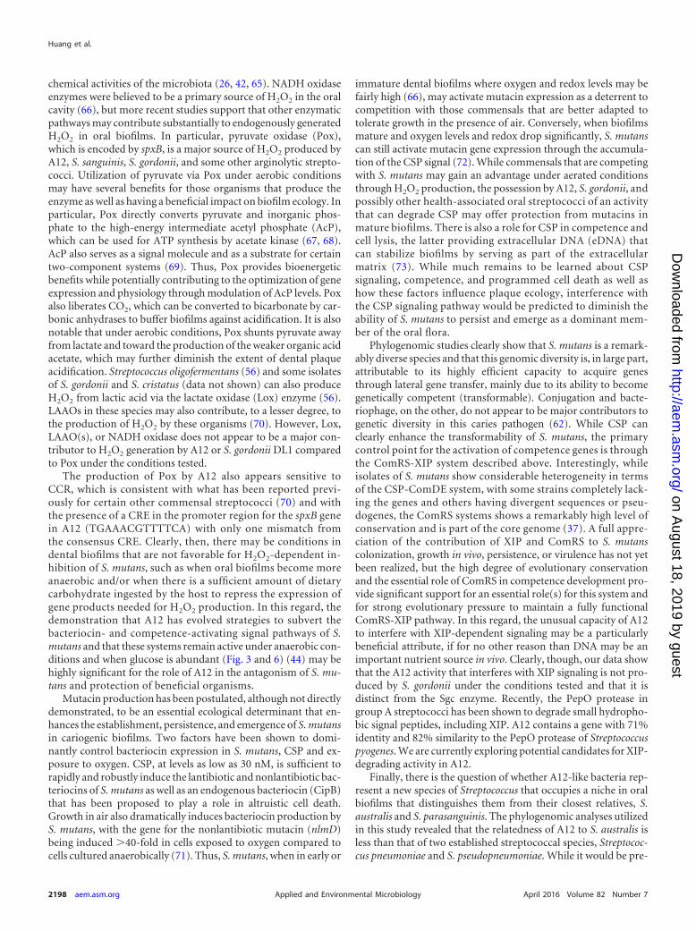
Whole-genome sequencing and phylogenomic analysis ofA12. Comparison of the entire 16S sequence of A12 to the 16S se-quences of other organisms by BLAST revealed that the 16S rRNAgene was most highly conserved with that of the referencestrain for Streptococcus australis (NCBI GenBank accession num-ber NZ_AFUD01000002.1. To more carefully evaluate the relat-edness of A12 to other streptococci, the entire genome sequence ofA12 was determined by using PacBio sequencing technology toattain 60� coverage. A single 1.8-Mb contig was obtained andannotated (BioSample accession number SAMN04287214), and acomprehensive phylogenomic analysis was conducted according tomethodologies detailed previously (62). By using these approaches,it was determined (Fig. 7) that the patristic distance (i.e., diver-gence between lineages) between A12 and its closest relative, S.australis, was 0.058 (5.8% divergence), whereas the patristic dis-tance between Streptococcus pneumoniae and Streptococcus pseu-dopneumoniae, two distinct species, was 0.049 (4.9%). Although itwill be necessary to isolate and sequence additional A12-like bac-teria to propose that A12 represents a new species of Streptococcus,data from phylogenomic analysis suggest that A12 may not beappropriately classified as S. australis or S. parasanguinis. Of note,A12 has a colony morphology distinct from those of other com-monly isolated oral streptococcal species, and comparison of thetranslated sequences of certain genes from A12 with apparent ho-mologs from S. australis or S. parasanguinis revealed substantialdegeneracy, which has proven useful in distinguishing A12-likeorganisms from close relatives in plaque samples. With thisknowledge, we have been able to isolate a small number ofputative A12-like organisms from clinical samples. All of theisolates showed the expected sequence conservation in a geneof interest, with significant differences from S. australis and S.
FIG 4 Effects of treatment of sCSP with supernatants from cultures of theindicated strains grown overnight. A strain of S. mutans containing a lacZfusion to the cipB promoter was grown to an OD600 of 0.12 in BHI medium(optimal responsiveness to sCSP). Supernatants from cultures of the indi-cated strains grown overnight were obtained after centrifugation, adjustedto pH 7.0 (also optimal for sCSP signaling), and filter sterilized. sCSP wasadded to the supernatants, and the mixtures were incubated for 2 h. The S.mutans cipB-lacZ reporter strain was then resuspended in the indicatedsupernatants for 2 h prior to measurement of �-galactosidase (LacZ) ac-tivity. The Sgc protease was necessary for inhibition of sCSP signaling. Theresults shown are from a minimum of three biological replicates, eachperformed in triplicate. Values are averages, and error bars indicate stan-dard deviations. Asterisks indicate statistically significant differences com-pared with wild-type A12.
FIG 5 Effects of treatment of XIP with supernatants from cultures of the indicated strains grown overnight. Supernatant fluids from cultures of A12 and itsderivatives, S. gordonii, or S. mutans UA159 that had been grown in FMC medium overnight were adjusted to pH 7.0, supplemental glucose was added to increasethe glucose concentration by 25 mM, and the supernatants were filter sterilized. sXIP was added to the supernatants, and the mixtures were incubated overnightat 37°C. Cultures of S. mutans UA159::PcomX-lacZ that had been grown in FMC medium to an OD600 of 0.12 were pelleted by centrifugation and resuspendedin the supernatants. The cultures were incubated for 2 h, and LacZ assays were performed to detect XIP-dependent activation of the comX promoter. The resultsshown are from a minimum of three biological replicates, each performed in triplicate. Values are averages, and error bars indicate standard deviations. Data fromtreatment with 400 or 1,000 nM sXIP for DL1 and S. mutans are not shown because neither supernatant influenced sXIP signaling any differently than the positivecontrol. Asterisks indicate statistical significance in comparisons between wild-type A12 with 200 nM XIP and DL1 with 200 nM XIP and between wild-type A12with 1,000 XIP and Spx-deficient A12 with 1,000 nM XIP.
Novel Beneficial Oral Streptococcus
April 2016 Volume 82 Number 7 aem.asm.org 2195Applied and Environmental Microbiology
on August 18, 2019 by guest
http://aem.asm
.org/D
ownloaded from
parasanguinis being notable (data not shown), along with con-stitutively high ADS activity and potent antagonistic capacityagainst S. mutans (X. Huang and R. A. Burne, unpublisheddata).
DISCUSSION
In light of the strong correlation between the absence of dentalcaries and high dental plaque ADS activity (5, 12, 14, 18), oralbacteria with constitutionally high ADS expression levels may
have significant potential for applications in probiotic therapies toprevent and control dental caries. Such strains would be evenmore desirable if they could antagonize the growth of known car-ies pathogens. Here, we characterize a novel Streptococcus isolate,designated A12, with the ability to express high ADS activity underconditions that commonly occur in human dental biofilms (44).A12 was also shown to have a particularly potent inhibitory effecton the growth of S. mutans, mainly through pyruvate oxidase-dependent H2O2 production. Moreover, A12 is able to interfere
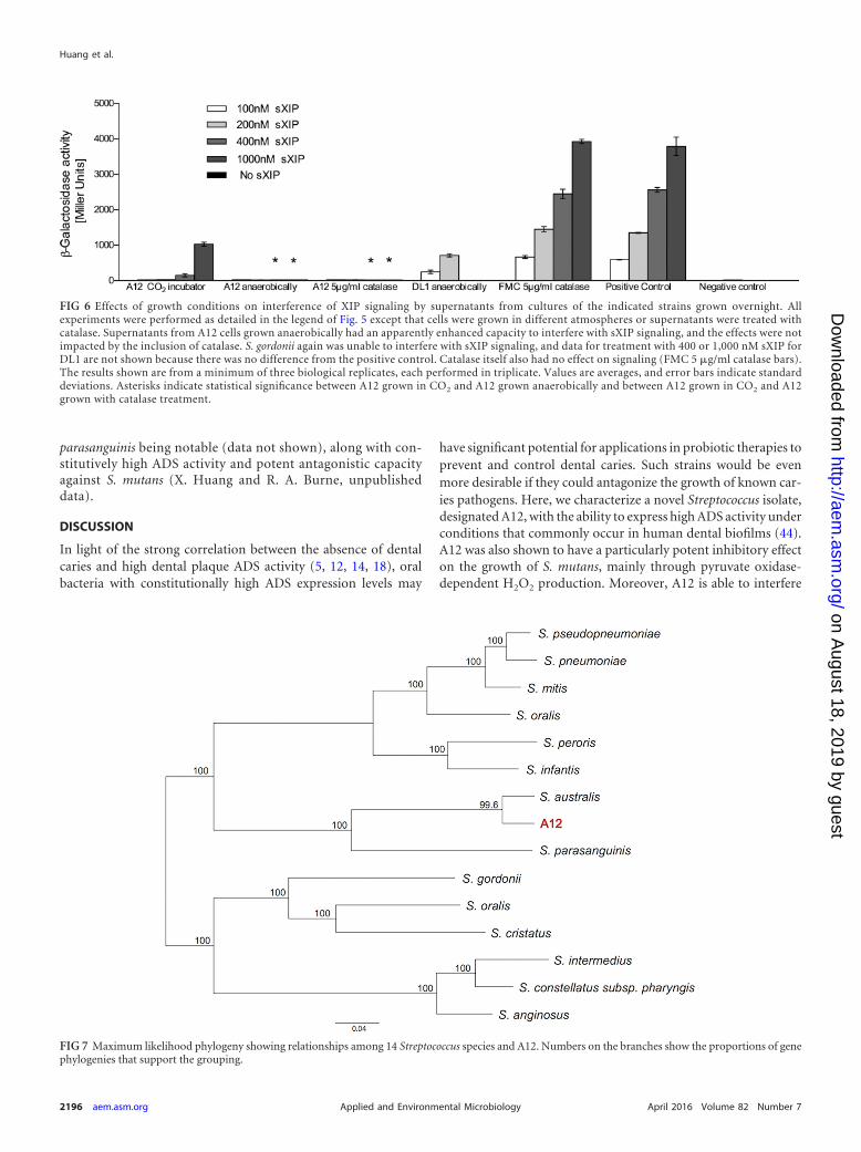
FIG 6 Effects of growth conditions on interference of XIP signaling by supernatants from cultures of the indicated strains grown overnight. Allexperiments were performed as detailed in the legend of Fig. 5 except that cells were grown in different atmospheres or supernatants were treated withcatalase. Supernatants from A12 cells grown anaerobically had an apparently enhanced capacity to interfere with sXIP signaling, and the effects were notimpacted by the inclusion of catalase. S. gordonii again was unable to interfere with sXIP signaling, and data for treatment with 400 or 1,000 nM sXIP forDL1 are not shown because there was no difference from the positive control. Catalase itself also had no effect on signaling (FMC 5 �g/ml catalase bars).The results shown are from a minimum of three biological replicates, each performed in triplicate. Values are averages, and error bars indicate standarddeviations. Asterisks indicate statistical significance between A12 grown in CO2 and A12 grown anaerobically and between A12 grown in CO2 and A12grown with catalase treatment.
FIG 7 Maximum likelihood phylogeny showing relationships among 14 Streptococcus species and A12. Numbers on the branches show the proportions of genephylogenies that support the grouping.
Huang et al.
2196 aem.asm.org April 2016 Volume 82 Number 7Applied and Environmental Microbiology
on August 18, 2019 by guest
http://aem.asm
.org/D
ownloaded from

with the two dominant intercellular peptide-based communica-tion pathways of S. mutans, which has the impact of disabling aprimary antagonistic strategy (bacteriocins) of S. mutans whilerendering S. mutans unable to induce genetic competence, com-petence being associated with nutritional benefits, genome diver-sification, acid tolerance, and biofilm formation. The identifica-tion of an oral isolate with this combination of characteristics,when coupled with our findings on the tremendous heterogeneityof commensal streptococci (44), highlights the profound gap inour understanding of the phenotypic capacity and plasticity of theoral microbiome as these factors relate to the behaviors of partic-ular taxa and populations. The data also reinforce the importanceof complementing current metagenomic approaches that utilize16S rRNA sequences (37, 44, 62) with more in-depth approachesto characterize the oral microbiota if reliable correlations for thecomposition and activities of the microbiome and oral health areto be established.
The ADS is widely distributed among prokaryotes, and theprimary structures of the enzymes in this pathway have been con-served throughout evolution (63). Most microorganisms studiedso far have their ADS genes organized into one cluster. In S. gor-donii, which has the most extensively characterized ADS of anyoral bacterium, the genes for the three enzymes of the pathway,arginine deiminase (AD) (arcA), ornithine carbamoyltransferase(arcB), and carbamate kinase (arcC), are cotranscribed in anoperon with arcD (arginine:ornithine antiporter) and arcT (argi-nine aminopeptidase). Induction of the ADS genes (arcABCDT)by arginine is mediated by a transcriptional activator encoded bythe divergently transcribed arcR gene located immediately down-stream of the arcABCDT operon (45). Notably, arcR of S. gordoniiis the second gene in a two-gene operon that includes the queAgene, which has been shown in other organisms to catalyze thefinal step in queosine modification of tRNAs. QueA-deficient S.gordonii mutants show altered ADS expression. An Fnr-like pro-tein (flp) is encoded immediately upstream of arcA and is requiredfor the activation of expression of the arcA promoter under anaer-obic conditions (46). Catabolite repression of the operon by pre-ferred carbohydrate sources (CCR), such as glucose, is exertedprimarily through CcpA (46) binding to two cis-acting cataboliteresponse elements (CREs) near the arcA promoter (64).
There are some similarities of the ADS operon of A12 withthose of its closest relatives, S. australis and S. parasanguinis, andwith the well-characterized operon of S. gordonii DL1 (see Fig. S3Ain the supplemental material). Most notably, arcABCDT of A12appear to be cotranscribed, as is the case for arcABCDT of S. gor-donii DL1 (45). Also present in A12 are arcR and queA, which areimmediately downstream of arcT and transcribed in the oppositedirection, as is the case for S. gordonii (63). There is also an appar-ent homolog of the flp gene upstream of arcA, with Flp of A12sharing 63% sequence identity (82% similarity) with Flp of S.gordonii (46). The promoter for the S. gordonii arcA gene has beenmapped and is located the proper distance from a fairly well-conserved (TAGAAT) �10 sequence but a weaker �35 sequence(see Fig. S3B in the supplemental material). A near-consensus�10 sequence (TAAAAT) was identified in A12, which is located1 nucleotide (nt) closer to the arcA start codon than the �10element of S. gordonii. However, the predicted �35 sequence ofA12, which is also 1 nt closer to the initiation codon than that of S.gordonii, was more similar to the consensus than the S. gordonii�35 element. Also, similar to S. gordonii and consistent with the
fact that both genes appear to require a transcriptional activator(ArcR) for optimal expression, an ArcR binding site was identifiedupstream of arcA (see Fig. S3B in the supplemental material).
The arc operons of S. gordonii and A12 are sensitive to CCR,although A12 is less so. In particular, in cells grown in the presenceof 25 mM glucose, AD activity in S. gordonii DL1 was only �15%of that expressed in cells grown on 25 mM galactose, whereas A12grown on glucose retained �50% of the AD activity of cells grownon 25 mM galactose (44). Catabolite repression of the ADS operonof S. gordonii is predominantly regulated by CcpA, which is pre-dicted to bind to two conserved CREs located in the arcA pro-moter region. Two sequences in the A12 arcA promoter regionwith conservation of the derived consensus sequence (TQWNANCGNTNWCA) (64) for binding of the catabolite control proteinCcpA were identified at positions �116 to �104 and at positions�34 to �21 in the A12 operon, essentially in the same positions asthe CREs in S. gordonii with respect to the arcA start codon. Theupstream CcpA binding site in A12 at positions �116 to �104(AGAAAGCGGTTCAT) has 4 nucleotides that differ from theconsensus sequence, whereas the binding site at positions �21 to�34 (TGAAAGCGGTACCA) had 1 nucleotide that differed fromthe consensus. Overall, then, adherence of the CREs to the con-sensus sequence was not as high in A12 as that of the CREs in thearcA promoter region of S. gordonii. While the promoter strengthand the effectiveness of the putative cis- and trans-acting factors ininduction and repression of the ADS of A12 remain to be deter-mined, the constitutionally higher levels of A12 ADS activity maybe associated with an inherently stronger promoter and dimin-ished CcpA-dependent CCR.
In a recent communication, we reported that the absolutelevel of ADS activity expressed by multiple commensal strep-tococci under ideal conditions can be highly variable acrossand within species, as can the ability of the system to be re-pressed by carbohydrate or oxygen or induced by arginine. Weposit that there may be an evolutionary basis for this inter- andintraspecies variation that leads to certain isolates, like A12,having higher constitutive ADS gene expression levels and adiminished sensitivity to CCR and other factors. In particular,we propose that A12 and similar isolates may have evolved torely more heavily on the ADS for protection from the detri-mental effects of low pH, whereas low-ADS producers orstrains that are more sensitive to repression by environmentalinputs, such as high levels of carbohydrate, evolved other acidtolerance strategies, for example, higher proton-extruding AT-Pase activity, H�-ATPases with lower optimal pH values, di-minished membrane proton permeability, or other traits asso-ciated with resistance to low pH (5, 29). Should this hypothesisbe proven correct, it can then be extrapolated that the lattergroup of commensals, with lower ADS expression and higherinherent acid tolerance, might tend to be associated with cariesactivity, whereas the former group would tend to be associatedwith health. The difficulty, of course, is that these strains can-not be discriminated by comparing 16S sequences. Isolationand characterization of additional A12-like organisms andgenomic/phenotypic characterization of these isolates and iso-lates of other streptococcal species to test this hypothesis areunder way.
A variety of oral streptococci that are predominant members ofearly and mature biofilms are able to generate H2O2, which isthought to have a profound impact on the composition and bio-
Novel Beneficial Oral Streptococcus
April 2016 Volume 82 Number 7 aem.asm.org 2197Applied and Environmental Microbiology
on August 18, 2019 by guest
http://aem.asm
.org/D
ownloaded from
chemical activities of the microbiota (26, 42, 65). NADH oxidaseenzymes were believed to be a primary source of H2O2 in the oralcavity (66), but more recent studies support that other enzymaticpathways may contribute substantially to endogenously generatedH2O2 in oral biofilms. In particular, pyruvate oxidase (Pox),which is encoded by spxB, is a major source of H2O2 produced byA12, S. sanguinis, S. gordonii, and some other arginolytic strepto-cocci. Utilization of pyruvate via Pox under aerobic conditionsmay have several benefits for those organisms that produce theenzyme as well as having a beneficial impact on biofilm ecology. Inparticular, Pox directly converts pyruvate and inorganic phos-phate to the high-energy intermediate acetyl phosphate (AcP),which can be used for ATP synthesis by acetate kinase (67, 68).AcP also serves as a signal molecule and as a substrate for certaintwo-component systems (69). Thus, Pox provides bioenergeticbenefits while potentially contributing to the optimization of geneexpression and physiology through modulation of AcP levels. Poxalso liberates CO2, which can be converted to bicarbonate by car-bonic anhydrases to buffer biofilms against acidification. It is alsonotable that under aerobic conditions, Pox shunts pyruvate awayfrom lactate and toward the production of the weaker organic acidacetate, which may further diminish the extent of dental plaqueacidification. Streptococcus oligofermentans (56) and some isolatesof S. gordonii and S. cristatus (data not shown) can also produceH2O2 from lactic acid via the lactate oxidase (Lox) enzyme (56).LAAOs in these species may also contribute, to a lesser degree, tothe production of H2O2 by these organisms (70). However, Lox,LAAO(s), or NADH oxidase does not appear to be a major con-tributor to H2O2 generation by A12 or S. gordonii DL1 comparedto Pox under the conditions tested.
The production of Pox by A12 also appears sensitive toCCR, which is consistent with what has been reported previ-ously for certain other commensal streptococci (70) and withthe presence of a CRE in the promoter region for the spxB genein A12 (TGAAACGTTTTCA) with only one mismatch fromthe consensus CRE. Clearly, then, there may be conditions indental biofilms that are not favorable for H2O2-dependent in-hibition of S. mutans, such as when oral biofilms become moreanaerobic and/or when there is a sufficient amount of dietarycarbohydrate ingested by the host to repress the expression ofgene products needed for H2O2 production. In this regard, thedemonstration that A12 has evolved strategies to subvert thebacteriocin- and competence-activating signal pathways of S.mutans and that these systems remain active under anaerobic con-ditions and when glucose is abundant (Fig. 3 and 6) (44) may behighly significant for the role of A12 in the antagonism of S. mu-tans and protection of beneficial organisms.
Mutacin production has been postulated, although not directlydemonstrated, to be an essential ecological determinant that en-hances the establishment, persistence, and emergence of S. mutansin cariogenic biofilms. Two factors have been shown to domi-nantly control bacteriocin expression in S. mutans, CSP and ex-posure to oxygen. CSP, at levels as low as 30 nM, is sufficient torapidly and robustly induce the lantibiotic and nonlantibiotic bac-teriocins of S. mutans as well as an endogenous bacteriocin (CipB)that has been proposed to play a role in altruistic cell death.Growth in air also dramatically induces bacteriocin production byS. mutans, with the gene for the nonlantibiotic mutacin (nlmD)being induced �40-fold in cells exposed to oxygen compared tocells cultured anaerobically (71). Thus, S. mutans, when in early or
immature dental biofilms where oxygen and redox levels may befairly high (66), may activate mutacin expression as a deterrent tocompetition with those commensals that are better adapted totolerate growth in the presence of air. Conversely, when biofilmsmature and oxygen levels and redox drop significantly, S. mutanscan still activate mutacin gene expression through the accumula-tion of the CSP signal (72). While commensals that are competingwith S. mutans may gain an advantage under aerated conditionsthrough H2O2 production, the possession by A12, S. gordonii, andpossibly other health-associated oral streptococci of an activitythat can degrade CSP may offer protection from mutacins inmature biofilms. There is also a role for CSP in competence andcell lysis, the latter providing extracellular DNA (eDNA) thatcan stabilize biofilms by serving as part of the extracellularmatrix (73). While much remains to be learned about CSPsignaling, competence, and programmed cell death as well ashow these factors influence plaque ecology, interference withthe CSP signaling pathway would be predicted to diminish theability of S. mutans to persist and emerge as a dominant mem-ber of the oral flora.
Phylogenomic studies clearly show that S. mutans is a remark-ably diverse species and that this genomic diversity is, in large part,attributable to its highly efficient capacity to acquire genesthrough lateral gene transfer, mainly due to its ability to becomegenetically competent (transformable). Conjugation and bacte-riophage, on the other, do not appear to be major contributors togenetic diversity in this caries pathogen (62). While CSP canclearly enhance the transformability of S. mutans, the primarycontrol point for the activation of competence genes is throughthe ComRS-XIP system described above. Interestingly, whileisolates of S. mutans show considerable heterogeneity in termsof the CSP-ComDE system, with some strains completely lack-ing the genes and others having divergent sequences or pseu-dogenes, the ComRS systems shows a remarkably high level ofconservation and is part of the core genome (37). A full appre-ciation of the contribution of XIP and ComRS to S. mutanscolonization, growth in vivo, persistence, or virulence has not yetbeen realized, but the high degree of evolutionary conservationand the essential role of ComRS in competence development pro-vide significant support for an essential role(s) for this system andfor strong evolutionary pressure to maintain a fully functionalComRS-XIP pathway. In this regard, the unusual capacity of A12to interfere with XIP-dependent signaling may be a particularlybeneficial attribute, if for no other reason than DNA may be animportant nutrient source in vivo. Clearly, though, our data showthat the A12 activity that interferes with XIP signaling is not pro-duced by S. gordonii under the conditions tested and that it isdistinct from the Sgc enzyme. Recently, the PepO protease ingroup A streptococci has been shown to degrade small hydropho-bic signal peptides, including XIP. A12 contains a gene with 71%identity and 82% similarity to the PepO protease of Streptococcuspyogenes. We are currently exploring potential candidates for XIP-degrading activity in A12.
Finally, there is the question of whether A12-like bacteria rep-resent a new species of Streptococcus that occupies a niche in oralbiofilms that distinguishes them from their closest relatives, S.australis and S. parasanguinis. The phylogenomic analyses utilizedin this study revealed that the relatedness of A12 to S. australis isless than that of two established streptococcal species, Streptococ-cus pneumoniae and S. pseudopneumoniae. While it would be pre-
Huang et al.
2198 aem.asm.org April 2016 Volume 82 Number 7Applied and Environmental Microbiology
on August 18, 2019 by guest
http://aem.asm
.org/D
ownloaded from
mature to propose A12 as a new species, the distinct colony mor-phology and ability to find highly conserved genes in what appearto be A12-like organisms that have diverged substantially in se-quence from apparent homologs in S. australis or S. parasanguinisshould allow us to gather sufficient data to establish whether des-ignation as a new species is warranted. Regardless of the outcome,A12 is an isolate that is both highly arginolytic and able to inhibitgrowth and intercellular signaling in S. mutans in a way that maypromote the stability of health-associated biofilm communities.Our recent communication shows that isolates that, by 16S se-quence, are classified as known streptococcal species display aspectrum of arginolytic and antagonistic properties. In contrast-ing the genome sequences of A12-like organisms and isolates withsimilar beneficial properties with poorly arginolytic and poorlyantagonistic members of the same taxa, it should be possible todevelop molecular probes and other tools that can be utilized toascertain the extent to which biofilms of human subjects are pop-ulated with beneficial organisms. Such knowledge could aid thepractice of clinical dentistry by augmenting caries risk assessmentand improving the ability to evaluate the impact of therapeuticapproaches on oral microbiomes.
ACKNOWLEDGMENT
We thank Zezhang (Tom) Wen at the LSU Health Sciences Center forconstructing pBGS.
FUNDING INFORMATIONHHS | NIH | National Institute of Dental and Craniofacial Research(NIDCR) provided funding to Robert A. Burne under grant number R90DE22530. HHS | NIH | National Institute of Dental and CraniofacialResearch (NIDCR) provided funding to Marcelle M. Nascimento undergrant number K23 DE023579. Colgate-Palmolive Company providedfunding to Marcelle M. Nascimento and Robert A. Burne.
REFERENCES1. Selwitz RH, Ismail AI, Pitts NB. 2007. Dental caries. Lancet 369:51–59.
http://dx.doi.org/10.1016/S0140-6736(07)60031-2.2. Lemos JA, Burne RA. 2008. A model of efficiency: stress tolerance by
Streptococcus mutans. Microbiology 154:3247–3255. http://dx.doi.org/10.1099/mic.0.2008/023770-0.
3. Huang X, Cheng L, Exterkate RAM, Liu M, Zhou X, Li J, ten Cate JM.2012. Effect of pH on Galla chinensis extract’s stability and anti-cariesproperties in vitro. Arch Oral Biol 57:1093–1099. http://dx.doi.org/10.1016/j.archoralbio.2012.04.007.
4. Huang X, Guo Q, Ren B, Li Y, Zhou X. 2016. Models in caries research,p 157–173. In Zhou X (ed), Dental caries. Springer, Berlin, Germany.
5. Burne RA, Marquis RE. 2000. Alkali production by oral bacteria andprotection against dental caries. FEMS Microbiol Lett 193:1– 6. http://dx.doi.org/10.1111/j.1574-6968.2000.tb09393.x.
6. Takahashi N, Nyvad B. 2011. The role of bacteria in the caries process:ecological perspectives. J Dent Res 90:294 –303. http://dx.doi.org/10.1177/0022034510379602.
7. Marsh PD. 2003. Are dental diseases examples of ecological catastrophes?Microbiology 149:279 –294. http://dx.doi.org/10.1099/mic.0.26082-0.
8. ten Cate JM. 2006. Biofilms, a new approach to the microbiology of dentalplaque. Odontology 94:1–9. http://dx.doi.org/10.1007/s10266-006-0063-3.
9. Huang X, Liu M, Li J, Zhou X, ten Cate JM. 2012. Chemical composi-tion of Galla chinensis extract and the effect of its main component(s) onthe prevention of enamel demineralization in vitro. Int J Oral Sci 4:146 –151. http://dx.doi.org/10.1038/ijos.2012.44.
10. Burne RA, Zeng L, Ahn SJ, Palmer SR, Liu Y, Lefebure T, Stanhope MJ,Nascimento MM. 2012. Progress dissecting the oral microbiome in cariesand health. Adv Dent Res 24:77– 80. http://dx.doi.org/10.1177/0022034512449462.
11. Huang X, Exterkate RAM, ten Cate JM. 2012. Factors associated with
alkali production from arginine in dental biofilms. J Dent Res 91:1130 –1134. http://dx.doi.org/10.1177/0022034512461652.
12. Nascimento MM, Gordan VV, Garvan CW, Browngardt CM, BurneRA. 2009. Correlations of oral bacterial arginine and urea catabolism withcaries experience. Oral Microbiol Immunol 24:89 –95. http://dx.doi.org/10.1111/j.1399-302X.2008.00477.x.
13. ten Cate JM, Cummins D. 2013. Fluoride toothpaste containing 1.5%arginine and insoluble calcium as a new standard of care in caries preven-tion. J Clin Dent 24:79 – 87.
14. Kleinberg I. 2002. A mixed-bacteria ecological approach to understand-ing the role of the oral bacteria in dental caries causation: an alternative toStreptococcus mutans and the specific-plaque hypothesis. Crit Rev OralBiol Med 13:108 –125. http://dx.doi.org/10.1177/154411130201300202.
15. Margolis HC, Duckworth JH, Moreno EC. 1988. Composition ofpooled resting plaque fluid from caries-free and caries-susceptibleindividuals. J Dent Res 67:1468 –1475. http://dx.doi.org/10.1177/00220345880670120601.
16. Van Wuyckhuyse BC, Perinpanayagam HE, Bevacqua D, RaubertasRF, Billings RJ, Bowen WH, Tabak LA. 1995. Association of freearginine and lysine concentrations in human parotid saliva with cariesexperience. J Dent Res 74:686 – 690. http://dx.doi.org/10.1177/00220345950740021001.
17. Rogers AH, Zilm PS, Gully NJ, Pfennig AL. 1988. Response of a Strep-tococcus sanguis strain to arginine-containing peptides. Infect Immun 56:687– 692.
18. Nascimento MM, Browngardt C, Xiaohui X, Klepac-Ceraj V, Paster BJ,Burne RA. 2014. The effect of arginine on oral biofilm communities. MolOral Microbiol 29:45–54. http://dx.doi.org/10.1111/omi.12044.
19. Liu Y, Nascimento MM, Burne RA. 2012. Progress toward understandingthe contribution of alkali generation in dental biofilms to inhibition of dentalcaries. Int J Oral Sci 4:135–140. http://dx.doi.org/10.1038/ijos.2012.54.
20. Kolderman E, Bettampadi D, Samarian D, Dowd SE, Foxman B,Jakubovics NS, Rickard AH. 2015. L-Arginine destabilizes oral multi-species biofilm communities developed in human saliva. PLoS One 10:e0121835. http://dx.doi.org/10.1371/journal.pone.0121835.
21. Sakanaka A, Kuboniwa M, Takeuchi H, Hashino E, Amano A. 2015.Arginine-ornithine antiporter ArcD controls arginine metabolism and in-terspecies biofilm development of Streptococcus gordonii. J Biol Chem 290:21185–21198. http://dx.doi.org/10.1074/jbc.M115.644401.
22. Nascimento MM, Burne RA. 2014. Caries prevention by arginine metab-olism in oral biofilms: translating science into clinical success. Curr OralHealth Rep 1:79 – 85. http://dx.doi.org/10.1007/s40496-013-0007-2.
23. Kraivaphan P, Amornchat C, Triratana T, Mateo LR, Ellwood R,Cummins D, DeVizio W, Zhang YP. 2013. Two-year caries clinical studyof the efficacy of novel dentifrices containing 1.5% arginine, an insolublecalcium compound and 1,450 ppm fluoride. Caries Res 47:582–590. http://dx.doi.org/10.1159/000353183.
24. Acevedo AM, Montero M, Rojas-Sanchez F, Machado C, Rivera LE,Wolff M, Kleinberg I. 2008. Clinical evaluation of the ability of CaviStatin a mint confection to inhibit the development of dental caries in chil-dren. J Clin Dent 19:1– 8.
25. Yin W, Hu DY, Li X, Fan X, Zhang YP, Pretty IA, Mateo LR,Cummins D, Ellwood RP. 2013. The anticaries efficacy of a dentifricecontaining 1.5% arginine and 1450 ppm fluoride as sodium monofluo-rophosphate assessed using quantitative light-induced fluorescence(QLF). J Dent 41:S22–S28. http://dx.doi.org/10.1016/j.jdent.2010.04.004.
26. Kreth J, Merritt J, Qi F, Dong X, Shi W. 2011. Antagonistic, synergistic,and counteroffensive strategies for streptococcal interspecies interactions,p 331–343. In Kolenbrander P (ed), Oral microbial communities. ASMPress, Washington, DC.
27. van Houte J. 1994. Role of micro-organisms in caries etiology. J Dent Res73:672– 681.
28. Bowden GH. 1990. Microbiology of root surface caries in humans. J DentRes 69:1205–1210. http://dx.doi.org/10.1177/00220345900690051701.
29. Burne RA. 1998. Oral streptococci. Products of their environment. J DentRes 77:445– 452. http://dx.doi.org/10.1177/00220345980770030301.
30. Bradshaw D, Marsh PD. 1998. Analysis of pH-driven disruption of oralmicrobial communities in vitro. Caries Res 32:456 – 462. http://dx.doi.org/10.1159/000016487.
31. Van Ruyven FOJ, Lingström P, Van Houte J, Kent R. 2000. Rela-tionship among mutans streptococci, “low-pH” bacteria, and iodo-philic polysaccharide-producing bacteria in dental plaque and early
Novel Beneficial Oral Streptococcus
April 2016 Volume 82 Number 7 aem.asm.org 2199Applied and Environmental Microbiology
on August 18, 2019 by guest
http://aem.asm
.org/D
ownloaded from

enamel caries in humans. J Dent Res 79:778 –784. http://dx.doi.org/10.1177/00220345000790021201.
32. Martin B, Quentin Y, Fichant G, Claverys JP. 2006. Independent evo-lution of competence regulatory cascades in streptococci? Trends Micro-biol 14:339 –345. http://dx.doi.org/10.1016/j.tim.2006.06.007.
33. Burne RA, Bessen DE, Broadbent JR, Claverys JP. 2007. The Seventh Interna-tional Conference on the Genetics of Streptococci, Lactococci, and Enterococci. JBacteriol 189:1209–1218. http://dx.doi.org/10.1128/JB.01363-06.
34. Hossain MS, Biswas I. 2012. An extracellular protease, SepM, generatesfunctional competence-stimulating peptide in Streptococcus mutans UA159.J Bacteriol 194:5886 –5896. http://dx.doi.org/10.1128/JB.01381-12.
35. Guo Q, Ahn SJ, Kaspar J, Zhou X, Burne RA. 2014. Growth phase and pHinfluence peptide signaling for competence development in Streptococcusmutans. J Bacteriol 196:227–236. http://dx.doi.org/10.1128/JB.00995-13.
36. Son M, Ahn SJ, Guo Q, Burne RA, Hagen SJ. 2012. Microfluidic studyof competence regulation in Streptococcus mutans: environmental inputsmodulate bimodal and unimodal expression of comX. Mol Microbiol 86:258 –272. http://dx.doi.org/10.1111/j.1365-2958.2012.08187.x.
37. Palmer SR, Miller JH, Abranches J, Zeng L, Lefebure T, Richards VP,Lemos JA, Stanhope MJ, Burne RA. 2013. Phenotypic heterogeneity ofgenomically-diverse isolates of Streptococcus mutans. PLoS One 8:e61358.http://dx.doi.org/10.1371/journal.pone.0061358.
38. Kaspar J, Ahn SJ, Palmer SR, Choi SC, Stanhope MJ, Burne RA. 2015.A unique open reading frame within the comX gene of Streptococcus mu-tans regulates genetic competence and oxidative stress tolerance. Mol Mi-crobiol 96:463– 482. http://dx.doi.org/10.1111/mmi.12948.
39. Son M, Ghoreishi D, Ahn SJ, Burne RA, Hagen SJ. 2015. Sharply tunedpH response of genetic competence regulation in Streptococcus mutans: amicrofluidic study of environmental sensitivity of comX. Appl EnvironMicrobiol 81:5622–5631. http://dx.doi.org/10.1128/AEM.01421-15.
40. Reck M, Tomasch J, Wagner-Döbler I. 2015. The alternative sigma factorSigX controls bacteriocin synthesis and competence, the two quorumsensing regulated traits in Streptococcus mutans. PLoS Genet 11:e1005353.http://dx.doi.org/10.1371/journal.pgen.1005353.
41. Son M, Shields RC, Ahn S, Burne RA, Hagen SJ. 10 September 2015.Bidirectional signaling in the competence regulatory pathway of Strepto-coccus mutans. FEMS Microbiol Lett http://dx.doi.org/10.1093/femsle/fnv159.
42. Kreth J, Merritt J, Shi W, Qi F. 2005. Competition and coexistencebetween Streptococcus mutans and Streptococcus sanguinis in the dentalbiofilm. J Bacteriol 187:7193–7203. http://dx.doi.org/10.1128/JB.187.21.7193-7203.2005.
43. Wang B-Y, Kuramitsu HK. 2005. Interactions between oral bacteria:inhibition of Streptococcus mutans bacteriocin production by Streptococcusgordonii. Appl Environ Microbiol 71:354 –362. http://dx.doi.org/10.1128/AEM.71.1.354-362.2005.
44. Huang X, Schulte RM, Burne RA, Nascimento MM. 2015. Character-ization of the arginolytic microflora provides insights into pH homeosta-sis in human oral biofilms. Caries Res 49:165–176. http://dx.doi.org/10.1159/000365296.
45. Dong Y, Chen YYM, Snyder JA, Burne RA. 2002. Isolation and molec-ular analysis of the gene cluster for the arginine deiminase system fromStreptococcus gordonii DL1. Appl Environ Microbiol 68:5549 –5553. http://dx.doi.org/10.1128/AEM.68.11.5549-5553.2002.
46. Dong Y, Chen YYM, Burne RA. 2004. Control of expression of thearginine deiminase operon of Streptococcus gordonii by CcpA and Flp. JBacteriol 186:2511–2514. http://dx.doi.org/10.1128/JB.186.8.2511-2514.2004.
47. Santiago B, MacGilvray M, Faustoferri RC, Quivey RG. 2012. Thebranched-chain amino acid aminotransferase encoded by ilvE is involvedin acid tolerance in Streptococcus mutans. J Bacteriol 194:2010 –2019. http://dx.doi.org/10.1128/JB.06737-11.
48. Ahn SJ, Wen ZT, Burne RA. 2006. Multilevel control of competence devel-opment and stress tolerance in Streptococcus mutans UA159. Infect Immun74:1631–1642. http://dx.doi.org/10.1128/IAI.74.3.1631-1642.2006.
49. Gerhardt P, Murray RGE, Wood WA, Krieg NR. 1994. Methods forgeneral and molecular bacteriology. ASM Press, Washington, DC.
50. Terleckyj B, Willett NP, Shockman GD. 1975. Growth of several cario-genic strains of oral streptococci in a chemically defined medium. InfectImmun 11:649 – 655.
51. Palmer SR, Crowley PJ, Oli MW, Ruelf MA, Michalek SM, Brady LJ.2012. YidC1 and YidC2 are functionally distinct proteins involved in pro-tein secretion, biofilm formation and cariogenicity of Streptococcus mu-
tans. Microbiology 158:1702–1712. http://dx.doi.org/10.1099/mic.0.059139-0.
52. Warburton PJ, Ciric L, Lerner A, Seville LA, Roberts AP, Mullany P,Allan E. 2013. TetAB(46), a predicted heterodimeric ABC transporterconferring tetracycline resistance in Streptococcus australis isolated fromthe oral cavity. J Antimicrob Chemother 68:17–22. http://dx.doi.org/10.1093/jac/dks351.
53. Ahn SJ, Wen ZT, Burne RA. 2007. Effects of oxygen on virulence traits ofStreptococcus mutans. J Bacteriol 189:8519 – 8527. http://dx.doi.org/10.1128/JB.01180-07.
54. Liu Y, Zeng L, Burne RA. 2009. AguR is required for induction of theStreptococcus mutans agmatine deiminase system by low pH and agmatine.Appl Environ Microbiol 75:2629 –2637. http://dx.doi.org/10.1128/AEM.02145-08.
55. Wen ZT, Burne RA. 2001. Construction of a new integration vector foruse in Streptococcus mutans. Plasmid 45:31–36. http://dx.doi.org/10.1006/plas.2000.1498.
56. Tong H, Chen W, Merritt J, Qi F, Shi W, Dong X. 2007. Streptococcusoligofermentans inhibits Streptococcus mutans through conversion of lacticacid into inhibitory H2O2: a possible counteroffensive strategy for inter-species competition. Mol Microbiol 63:872– 880. http://dx.doi.org/10.1111/j.1365-2958.2006.05546.x.
57. Liu L, Tong H, Dong X. 2012. Function of the pyruvate oxidase-lactateoxidase cascade in interspecies competition between Streptococcus oligo-fermentans and Streptococcus mutans. Appl Environ Microbiol 78:2120 –2127. http://dx.doi.org/10.1128/AEM.07539-11.
58. Fowler ML, Ingram-Smith CJ, Smith KS. 2011. Direct detection of theacetate-forming activity of the enzyme acetate kinase. J Vis Exp 58:e3474.http://dx.doi.org/10.3791/3474.
59. Perry J, Jones M, Peterson S, Cvitkovitch D, Levesque C. 2009. Peptidealarmone signalling triggers an auto-active bacteriocin necessary for ge-netic competence. Mol Microbiol 72:905–917. http://dx.doi.org/10.1111/j.1365-2958.2009.06693.x.
60. Zubay G, Morse DE, Schrenk WJ, Miller JHM. 1972. Detection andisolation of the repressor protein for the tryptophan operon of Escherichiacoli. Proc Natl Acad Sci U S A 69:1100 –1103. http://dx.doi.org/10.1073/pnas.69.5.1100.
61. Aziz R, Bartels D, Best A, DeJongh M, Disz T, Edwards R, Formsma K,Gerdes S, Glass E, Kubal M, Meyer F, Olsen G, Olson R, Osterman A,Overbeek R, McNeil L, Paarmann D, Paczian T, Parrello B, Pusch G,Reich C, Stevens R, Vassieva O, Vonstein V, Wilke A, Zagnitko O. 2008.The RAST server: rapid annotations using subsystems technology. BMCGenomics 9:75. http://dx.doi.org/10.1186/1471-2164-9-75.
62. Richards VP, Palmer SR, Pavinski Bitar PD, Qin X, Weinstock GM,Highlander SK, Town CD, Burne RA, Stanhope MJ. 2014. Phylogenom-ics and the dynamic genome evolution of the genus Streptococcus. GenomeBiol Evol 6:741–753. http://dx.doi.org/10.1093/gbe/evu048.
63. Liu Y, Dong Y, Chen YYM, Burne RA. 2008. Environmental and growthphase regulation of the Streptococcus gordonii arginine deiminase genes.Appl Environ Microbiol 74:5023–5030. http://dx.doi.org/10.1128/AEM.00556-08.
64. Zeng L, Dong Y, Burne RA. 2006. Characterization of cis-acting sitescontrolling arginine deiminase gene expression in Streptococcus gordonii. JBacteriol 188:941–949. http://dx.doi.org/10.1128/JB.188.3.941-949.2006.
65. Zheng L, Itzek A, Chen Z, Kreth J. 2011. Environmental influences onStreptococcus gordonii competitive hydrogen peroxide production. ApplEnviron Microbiol 77:4318 – 4328. http://dx.doi.org/10.1128/AEM.00309-11.
66. Marquis R. 1995. Oxygen metabolism, oxidative stress and acid-basephysiology of dental plaque biofilms. J Ind Microbiol 15:198 –207. http://dx.doi.org/10.1007/BF01569826.
67. Kreth J, Zhang Y, Herzberg MC. 2008. Streptococcal antagonism in oralbiofilms: Streptococcus sanguinis and Streptococcus gordonii interferencewith Streptococcus mutans. J Bacteriol 190:4632– 4640. http://dx.doi.org/10.1128/JB.00276-08.
68. Wolfe AJ, Chang DE, Walker JD, Seitz-Partridge JE, Vidaurri MD,Lange CF, Prüß BM, Henk MC, Larkin JC, Conway T. 2003. Evidencethat acetyl phosphate functions as a global signal during biofilm develop-ment. Mol Microbiol 48:977–988. http://dx.doi.org/10.1046/j.1365-2958.2003.03457.x.
69. Stock AM, Robinson VL, Goudreau PN. 2000. Two-component signaltransduction. Annu Rev Biochem 69:183–215. http://dx.doi.org/10.1146/annurev.biochem.69.1.183.
Huang et al.
2200 aem.asm.org April 2016 Volume 82 Number 7Applied and Environmental Microbiology
on August 18, 2019 by guest
http://aem.asm
.org/D
ownloaded from
70. Tong H, Chen W, Shi W, Qi F, Dong X. 2008. SO-LAAO, a novel L-aminoacid oxidase that enables Streptococcus oligofermentans to outcompeteStreptococcus mutans by generating H2O2 from peptone. J Bacteriol 190:4716 – 4721. http://dx.doi.org/10.1128/JB.00363-08.
71. Ahn SJ, Ahn SJ, Browngardt CM, Burne RA. 2009. Changes in biochem-ical and phenotypic properties of Streptococcus mutans during growth withaeration. Appl Environ Microbiol 75:2517–2527. http://dx.doi.org/10.1128/AEM.02367-08.
72. Li YH, Lau PCY, Lee JH, Ellen RP, Cvitkovitch DG. 2001. Natural genetictransformation of Streptococcus mutans growing in biofilms. J Bacteriol 183:897–908. http://dx.doi.org/10.1128/JB.183.3.897-908.2001.
73. Liao S, Klein MI, Heim KP, Fan Y, Bitoun JP, Ahn SJ, Burne RA, Koo H,Brady LJ, Wen ZT. 2014. Streptococcus mutans extracellular DNA is upregu-lated during growth in biofilms, actively released via membrane vesicles, andinfluenced by components of the protein secretion machinery. J Bacteriol196:2355–2366. http://dx.doi.org/10.1128/JB.01493-14.
Novel Beneficial Oral Streptococcus
April 2016 Volume 82 Number 7 aem.asm.org 2201Applied and Environmental Microbiology
on August 18, 2019 by guest
http://aem.asm
.org/D
ownloaded from