


A continuous spectrophotometric assay that distinguishes ... · 1 A continuous spectrophotometric...
35
1 A continuous spectrophotometric assay that distinguishes between phospholipase A 1 and A 2 activities Meddy El Alaoui †,‡ , Laurent Soulère ‡ , Alexandre Noiriel † , Florence Popowycz ‡ , Abdallah Khatib † , Yves Queneau ‡ and Abdelkarim Abousalham †* † Univ Lyon, Université Lyon 1, Institut de Chimie et de Biochimie Molculaires et Supramolculaires, UMR 5246, Mtabolisme, Enzymes et Mcanismes Molculaires (MEM2), 43, Bd du 11 novembre 1918, F-69622 Villeurbanne cedex, France. ‡ Univ Lyon, Université Lyon 1, INSA Lyon, CPE Lyon, Institut de Chimie et de Biochimie Molculaires et Supramolculaires, UMR 5246, Chimie Organique et Bioorganique, Btiment Jules Verne, 20 Avenue Albert Einstein, 69621 Villeurbanne Cedex, France. *To whom correspondence should be addressed: Abdelkarim Abousalham Tel: +33 4 72 44 81 02 E-mail address: [email protected] by guest, on July 6, 2019 www.jlr.org Downloaded from
Transcript of A continuous spectrophotometric assay that distinguishes ... · 1 A continuous spectrophotometric...

1
A continuous spectrophotometric assay that
distinguishes between phospholipase A1 and A2
activities
Meddy El Alaoui†,‡, Laurent Soulère‡, Alexandre Noiriel†, Florence Popowycz‡, Abdallah
Khatib†, Yves Queneau‡ and Abdelkarim Abousalham†*
† Univ Lyon, Université Lyon 1, Institut de Chimie et de Biochimie Moleculaires et
Supramoleculaires, UMR 5246, Metabolisme, Enzymes et Mecanismes Moleculaires (MEM2),
43, Bd du 11 novembre 1918, F-69622 Villeurbanne cedex, France.
‡ Univ Lyon, Université Lyon 1, INSA Lyon, CPE Lyon, Institut de Chimie et de Biochimie
Moleculaires et Supramoleculaires, UMR 5246, Chimie Organique et Bioorganique, Batiment
Jules Verne, 20 Avenue Albert Einstein, 69621 Villeurbanne Cedex, France.
*To whom correspondence should be addressed: Abdelkarim Abousalham
Tel: +33 4 72 44 81 02
E-mail address: [email protected]
by guest, on July 6, 2019w
ww
.jlr.orgD
ownloaded from

2
Abstract
A new spectrophotometric assay was developed to measure continuously, and specifically,
phospholipase A1 (PLA1) or phospholipase A2 (PLA2) activities using synthetic
glycerophosphatidylcholines (PC) containing -eleostearic acid, either at the sn-1 position [1-
α-eleostearoyl-2-octadecyl-rac-glycero-3-phosphocholine (EOPC)] or at the sn-2 position [1-
octadecyl-2-α-eleostearoyl-rac-glycero-3-phosphocholine (OEPC)]. The substrates were
coated onto the wells of microtiter plates. A non-hydrolysable ether bond, with a non UV-
absorbing alkyl chain, was introduced at the other sn position to prevent acyl chain migration
during lipolysis. Upon enzyme action, -eleostearic acid is liberated and then solubilized into
the micellar phase. The PLA1 or PLA2 activity was measured by the increase in absorbance at
272 nm due to the transition of -eleostearic acid from the adsorbed to the soluble state. EOPC
and OEPC differentiate, with excellent accuracy, between PLA1 and PLA2 activity. Lecitase®,
guinea pig pancreatic lipase-related protein 2 (known to be a PLA1 enzyme), bee venom PLA2
and porcine pancreatic PLA2 were all used to validate the assay. Compared to current assays
used for measuring continuously PLA1 or PLA2 activities and/or their inhibitors, the
development of this sensitive enzymatic method, using coated PC substrates analogues to
natural lipids and based on the UV spectroscopic properties of α-eleostearic acid, is a significant
improvement.
Keywords: Phospholipase A1, Phospholipase A2, tung oil, -eleostearic acid, -cyclodextrin,
1--eleostearoyl-2-octadecyl-rac-glycero-3-phosphocholine, 1-octadecyl-2-α-eleostearoyl-
rac-glycero-3-phosphocholine
by guest, on July 6, 2019w
ww
.jlr.orgD
ownloaded from

3
Abbreviations:
BHT, butylated hydroxytoluene; β-CD, β-cyclodextrin; DCC, N,N′-dicyclohexylcarbodiimide;
DMSO, dimethylsulfoxide; EEPC, 1,2-α-eleostearoyl-sn-glycero-3-phosphocholine; EOPC, 1-
-eleostearoyl-2-octadecyl-rac-glycero-3-phosphocholine; GPC, sn-glycero-3-
phosphocholine; GPL-RP2, guinea pig pancreatic lipase-related protein 2; hbPLA2, honey bee
(Apis mellifera) venom PLA2; MI, methyl indoxam; OEPC, 1-octadecyl-2-α-eleostearoyl-rac-
glycero-3-phosphocholine; PC, glycerophosphatidylcholine; ppPLA2, porcine pancreatic
PLA2; PPyr, 4-pyrrolidinopyridine.
by guest, on July 6, 2019w
ww
.jlr.orgD
ownloaded from

4
Introduction
Our knowledge of lipolytic enzymes has been substantially improved with the development of
analytical technologies and the recognition of their various and important functions in cells.
Nevertheless, many mechanisms of lipolytic activity remain unclear and novel technologies are
required for further investigations. Phospholipids are major components in the lipid bilayer
(50% of the cellular lipids is made up of glycerophosphatidylcholine (PC) (1)), together with
plasmatic lipoprotein. Phospholipids can be hydrolyzed by phospholipases A1 (PLA1, EC
3.1.1.32) or phospholipases A2 (PLA2, EC 3.1.1.4) that catalyze hydrolysis of the ester bond of
the acyl group attached to the sn-1 or the sn-2 position of phospholipid, respectively. At present
the class of PLA1 enzymes is not well studied and little structural information is available (2).
Some reports have indicated that PLA1s could be considered as virulence factors (3) or as being
responsible for the production of lysophospholipids that are implicated in many processes, such
as angiogenesis or protein transport (4). The endothelial lipase known to be a PLA1 is involved
in cardiovascular diseases, promoting atherosclerosis, and consequently it is a major therapeutic
target (5). Nevertheless, its mechanism has not been clearly elucidated. Pancreatic lipase-related
protein 2 from guinea pig (GPL-RP2) and human (HPL-RP2) has been reported to have
triglyceride lipase, PLA1 and galactolipase activities (6-8). These enzymes are involved in lipid
digestion and their inhibition could reduce obesity but the physiological involvement of their
PLA1 activity has not been established (7, 9).
PLA2 superfamily comprises a large group of intracellular (calcium-dependent and –
independent) or secreted enzymes which have been extensively studied with numerous crystal
structures (see reference (10) for review). Through the production of bioactive fatty acids (e.g.,
arachidonic acid) and lysophospholipids, PLA2 have been implicated in various physiological
processes (e.g. inflammation, host defense (11)) and diseases (e.g. asthma, rheumatoid arthritis,
and various cancer (12)). The number of PLA2s implicated, and the number of potential
by guest, on July 6, 2019w
ww
.jlr.orgD
ownloaded from

5
inhibitors, is rapidly expanding and this necessitates high throughput specific screening assays
in order to discover new potential treatments.
Most of PLA1 and PLA2 are water soluble lipolytic carboxylester hydrolases capable of
releasing long-chain fatty acids from natural water-insoluble carboxylic esters, leading to an
enzymatic reaction at the lipid-water interface (13). Consequently, these enzymes do not follow
Michaelis-Menten kinetics in which both the enzyme and the substrate must be present in the
same phase (13-15). Furthermore, the catalysis reaction strongly depends on the quality of the
interface, such as an oil-in-water emulsion, liposomal dispersion or a monolayer (13, 14), and
the use of lipidic substrates containing long-chain fatty acids must be taken into account when
accurately assaying the lipolytic activity. All these parameters make it difficult to develop new
reliable phospholipase assay systems. Nevertheless, numerous assays using chromogenic (16,
17), radiolabeled (18), or fluorogenic (19-21) substrates, or indirect measurements (22), have
been developed over the past decade. Some of them screen phospholipase activities with simple
and easy to use molecules (16) and others use substances close to natural lipids (20, 21, 23).
However, these labeled substrates often present a sterically hindered fluorochrome group
(except with radiolabeled probes) and this may interfere with the lipolytic activity.
Phospholipases are lipolytic carboxylester hydrolases capable of releasing long-chain fatty
acids from natural water-insoluble carboxylic esters. The use of lipidic substrates containing
these long-chain fatty acids must be taken into account when accurately assaying the lipolytic
activity.
Crude tung oil (Aleurites fordii seed oil) contains up to 70% -eleostearic acid (9Z, 11E, 13E-
octadecatrienoic acid), a naturally occurring 18-carbon fatty acid esterified mainly at the sn-1
and sn-3 positions of tung oil triglycerides (24). The conjugated triene present in -eleostearic
acid constitutes an intrinsic chromophore, which confers strong ultraviolet (UV) absorption
properties (24) on both the free fatty acid and the triglycerides. This makes a prime probe with
by guest, on July 6, 2019w
ww
.jlr.orgD
ownloaded from

6
no sterically hindered group. Various lipase assays have been developed based on these
properties, using -eleostearic acid esterified into natural (25, 26) and synthetic triglycerides
(27).
With the aim of developing a convenient, specific, sensitive and continuous UV
spectrophotometric assay using a lipidic substrate for monitoring PLA1 or PLA2 activity, we
have recently synthesized a specific PC named 1,2-α-eleostearoyl-sn-glycero-3-
phosphocholine (EEPC), esterified at the sn-1 and sn-2 positions with -eleostearic acid (28).
However, EEPC cannot be used to distinguish between PLA1 and PLA2 activities due, on the
one hand, to the presence of the same fatty acid at the sn-1 and sn-2 position and, on the other
hand, to the migration of the remaining fatty acyl chain yielding lysophosphatidylcholines
(lyso-PCs) during lipolysis (29). In this study, we have synthesized, and then used, new PCs
containing UV absorbing -eleostearic acid at the sn-1 [1-α-eleostearoyl-2-octadecyl-rac-
glycero-3-phosphocholine (EOPC)] or at the sn-2 position [1-octadecyl-2-α-eleostearoyl-rac-
glycero-3-phosphocholine (OEPC)] and a non-absorbing and non-hydrolysable O-ether alkyl
at the other sn position, able to monitor continuously the PLA1 or PLA2 activity, respectively.
The design of these new PCs involves the presence of ether bonds, non-hydrolysable by
phospholipases and, therefore, preventing acyl chain migration during lipolysis which, in turn,
presents a means of discriminating between PLA1 and PLA2 activity.
Materials and Methods
Reagents and materials
β-cyclodextrine (β-CD), butylated hydroxytoluene (BHT), DCC (N,N'-
dicyclohexylcarbodiimide), 4-pyrrolidinopyridine (PPyr), Amberlyst 15 and crude tung oil
were purchased from Sigma-Aldrich-Fluka Chimie. Methyl Indoxam (MI) was kindly provided
by Dr. D. Charmot, and tetrahydrolipstatin (THL), a known digestive lipase inhibitor, was
by guest, on July 6, 2019w
ww
.jlr.orgD
ownloaded from

7
obtained from Hoffmann-La-Roche Ltd. Microtiter plates (Costar® UV-Plate) were purchased
from Corning Inc. Thin-layer chromatography (TLC) aluminum sheets, coated with 0.2 mm
silica gel 60 F254, were purchased from Merck. All other chemicals and solvents of the highest
quality were obtained from local suppliers.
Proteins
Bovine serum albumin (BSA), porcine pancreatic PLA2 (ppPLA2), honey bee (Apis mellifera)
venom PLA2 (hbPLA2) and Lecitase® were all purchased from Sigma-Aldrich-Fluka Chimie.
Guinea pig pancreatic lipase-related protein 2 (GPL-RP2) was kindly provided by Dr F.
Carrière. Candida rugosa lipase AY30 was obtained from Amano Pharmaceuticals Ltd. The
protein concentrations were determined using Bradford's procedure (30), with Bio-Rad Dye
Reagent and BSA as the standard.
Thin-Layer Chromatography
Glycerophospholipids were separated by performing analytical TLC on aluminum sheets
coated with 0.2 mm silica gel 60. The sample migration was first performed with
chloroform/methanol/water (65/35/5, v/v/v), containing 0.001% (w/v) BHT as an antioxidant,
until the solvent front was halfway up the plate. The plate was dried and then placed in a second
chamber containing hexane/diethyl ether/acetic acid (86/14/1, v/v/v), containing 0.001% (w/v)
BHT, until the solvent front reached the top of the plate. The plate was dried again. The various
lipids were revealed with UV light at 254 nm (to reveal -eleostearic-containing species) and
by charring the plate after spraying it with 10% copper sulfate and 10% phosphoric acid in
water (to reveal all the acyl species).
Preparation of Purified α-Eleostearic Acid from Tung Oil
A solution of 20 g of crude tung oil was hydrolyzed with 500 mg of Candida rugosa lipase in
14 mL of water and the reaction was stirred, for 3 h, at 40°C. Total lipids were extracted into a
by guest, on July 6, 2019w
ww
.jlr.orgD
ownloaded from

8
decantation vial with 100 mL of 3 M HCl and 100 mL of diethyl ether containing 0.01% BHT
(w/v). The organic layer was recovered, dried by adding anhydrous MgSO4, filtered, and
concentrated under reduced pressure. The total lipid extract (5 g), containing mainly free fatty
acids, was further purified by recrystallization in 5.5 mL of acetone, at 60°C for 20 min, and
then cooled on ice. The heterogeneous mixture was filtered and the crystalline solid obtained
was treated with dry acetone. The crystals were then collected by filtration and dried in vacuo
(2.2 g, 40% yield from the lipolysis extract).
Synthesis of EOPC and OEPC
See Supporting Information for details.
Coating microtiter plates with synthetic phospholipids.
Microtiter plates were coated with EOPC or OEPC, as described previously (26-28). The
phkedospholipid solution (0.5 mg.mL-1) was prepared in ethanol, containing 0.001% BHT as
an antioxidant, and the wells of the UV-microtiter plate were filled with phospholipids (100
µL/well). The microtiter plate was first partially dried under a fume hood and then left in a
vacuum desiccator until the solvent had completely evaporated (around 30 min). After ethanol
evaporation, the coated EOPC or OEPC plates were found to be stable in the dark for at least 1
week at 4°C.
UV spectrophotometric PLA1 and PLA2 assays using coated synthetic phospholipids
The PLA1 and PLA2 activities were assayed spectrophotometrically by measuring the amount
of α-eleostearic acid continuously released from the phospholipid substrates. The enzyme
activity measurement was performed using 10 mM Tris-HCl buffer (pH 8.0) containing 3 g L-1
β-CD, 150 mM NaCl, 6 mM CaCl2 and 1 mM EDTA. The non-tensioactive β-CD was used in
the reaction buffer in order to solubilize the long-chain lipolytic products. The substrate was
dissolved in ethanol to obtain the desired final concentration and the wells of a 96-well flat-
by guest, on July 6, 2019w
ww
.jlr.orgD
ownloaded from
9
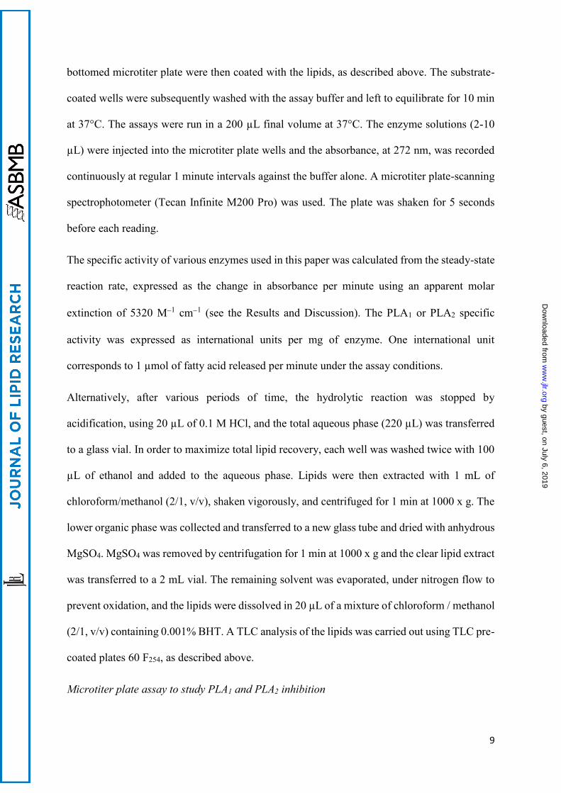
bottomed microtiter plate were then coated with the lipids, as described above. The substrate-
coated wells were subsequently washed with the assay buffer and left to equilibrate for 10 min
at 37°C. The assays were run in a 200 µL final volume at 37°C. The enzyme solutions (2-10
µL) were injected into the microtiter plate wells and the absorbance, at 272 nm, was recorded
continuously at regular 1 minute intervals against the buffer alone. A microtiter plate-scanning
spectrophotometer (Tecan Infinite M200 Pro) was used. The plate was shaken for 5 seconds
before each reading.
The specific activity of various enzymes used in this paper was calculated from the steady-state
reaction rate, expressed as the change in absorbance per minute using an apparent molar
extinction of 5320 M1 cm1 (see the Results and Discussion). The PLA1 or PLA2 specific
activity was expressed as international units per mg of enzyme. One international unit
corresponds to 1 µmol of fatty acid released per minute under the assay conditions.
Alternatively, after various periods of time, the hydrolytic reaction was stopped by
acidification, using 20 µL of 0.1 M HCl, and the total aqueous phase (220 µL) was transferred
to a glass vial. In order to maximize total lipid recovery, each well was washed twice with 100
µL of ethanol and added to the aqueous phase. Lipids were then extracted with 1 mL of
chloroform/methanol (2/1, v/v), shaken vigorously, and centrifuged for 1 min at 1000 x g. The
lower organic phase was collected and transferred to a new glass tube and dried with anhydrous
MgSO4. MgSO4 was removed by centrifugation for 1 min at 1000 x g and the clear lipid extract
was transferred to a 2 mL vial. The remaining solvent was evaporated, under nitrogen flow to
prevent oxidation, and the lipids were dissolved in 20 µL of a mixture of chloroform / methanol
(2/1, v/v) containing 0.001% BHT. A TLC analysis of the lipids was carried out using TLC pre-
coated plates 60 F254, as described above.
Microtiter plate assay to study PLA1 and PLA2 inhibition
by guest, on July 6, 2019w
ww
.jlr.orgD
ownloaded from
10
The enzyme-inhibitor incubation method (31) was used to test, in aqueous medium and in the
absence of the substrate, whether any direct interactions could occur between the enzyme and
the inhibitor. GPL-RP2 (0.5 µg, 69 nM) was incubated, at 25°C, with THL solution (4.8 µM in
dimethylsulfoxide (DMSO)) for 1 h at an enzyme: inhibitor molar ratio of 1:70 (final DMSO
concentration 2.5%). ppPLA2 (0.5 µg, 140 nM) was incubated, at 25°C, with MI solution (14
µM in DMSO) for 1 h at an enzyme: inhibitor molar ratio of 1:100 (final DMSO concentration
2.5%). The residual enzyme activities were then measured using either EOPC or OEPC coated
onto the surface of the microtiter plate wells.
Statistical analysis
Data are expressed as means ± S.D. Statistical significance was determined by the Student’s
unpaired t-test (two-tailed). Samples were considered to be significantly different for P< 0.01
(**).
Results and Discussion
In our previous published work (28), we synthesized a specific PC (EEPC), esterified at both
sn-1 and sn-2 positions with -eleostearic acid and coated onto microtiter plate wells to monitor
continuously the PLA1 or PLA2 activities. However, the fact that the EEPC substrate was
esterified at both sn-positions of the PC with -eleostearic acid made it impossible to
distinguish between the activities of these two enzymes. In the present work, two structural
analogues of PC esterified with -eleostearic acid, either at the sn-1 (EOPC) or at the sn-2
(OEPC) position, and containing a non-UV absorbing and non-hydrolysable ether bond at the
second position (Figure 1A), were synthesized and used as substrates to discriminate between
PLA1 and PLA2 activities. The ether bond was introduced to prevent intramolecular acyl group
migration during lipolysis by PLA1 or PLA2.
Synthesis of EOPC and OEPC, and their spectroscopic properties
by guest, on July 6, 2019w
ww
.jlr.orgD
ownloaded from

11
The α-eleostearic acid was obtained from tung oil enzymatic hydrolysis, and it was
recrystallized from the total lipid extract. The specific UV spectrum obtained showed three
majors peaks located at λ max (ethanol 95%) = 260, 270, and 280 nm (Figure 1B). The synthesis
of EOPC and OEPC (Figure S1) was achieved in eight steps from rac-glycidol, as summarized
in Supporting Information. Because the usual coupling agent DCC with PPyr (32) for the final
acylation (Fig. S1, compounds 8 and 15) gave poor results, another method was tested using
oxalyl chloride (33) to generate the acyl chloride of α-eleostearic acid before the final reaction,
but without success. However, better results were observed with DCC/ PPyr, as used by Borsotti
et al. (34), promoting the acylation of lyso-PC (Fig. S1, compounds 7 and 14) with anhydride
fatty acid derivatives, giving acceptable yields of 48% and 25% for EOPC and OEPC,
respectively. The difference in the yields between the two substrates may be due to a steric
hindrance at position sn-2. Moreover, the rather low yield obtained for this last step could be
due to the specific treatments applied to eliminate any ionic species by exchange ionic
chromatography (similar amphiphilic properties of PCs and PPyr) during the purification
process.
The final yields for the total synthesis of EOPC and OEPC were measured at 12% and 7%,
respectively, over the 8 steps. The 1H NMR spectrum of EOPC (Figure S2 A) and OEPC (Figure
S2 B) showed signals of the vinylic system, between δ 6.4 and 5.6 ppm, as a complex set of
multiplets. A single signal at δ 5.4 ppm is characteristic of the sn-2 single proton. At δ 4.4 to
3.7 ppm, multiplets are referenced to protons close to the glycerol chain group (sn-1 and sn-3
positions and the phosphatidyl group). The methyl groups of the quaternary ammonium gave a
specific signal at δ 3.4 ppm. Saturated protons from the carbonyl chain are located from δ 2.3
ppm (α carbon) to δ 0.9 ppm (ω carbon).
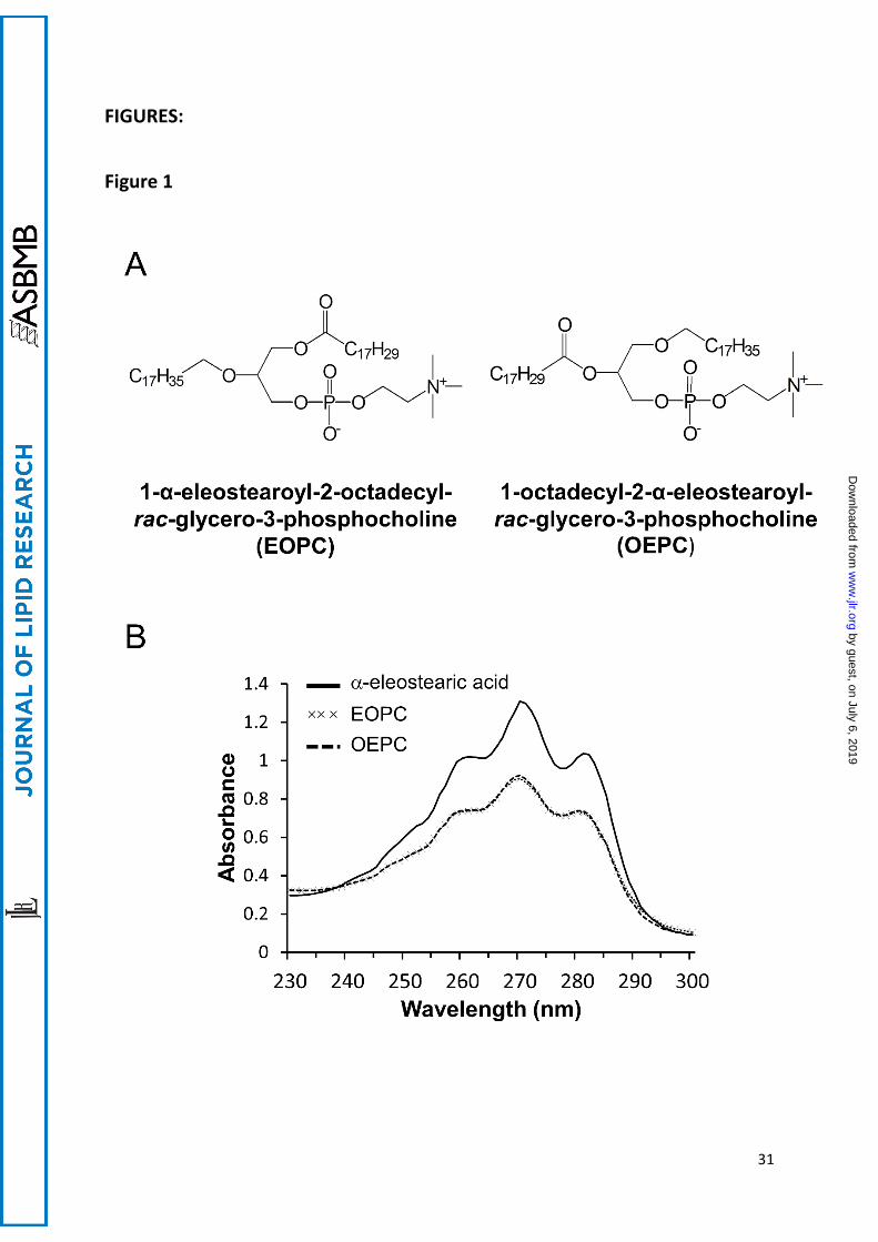
As shown in Figure 1B, the UV absorption spectrum (230-300 nm) of an ethanolic solution of
EOPC and OEPC displayed three major peaks located at 260, 270 and 280 nm. This profile
by guest, on July 6, 2019w
ww
.jlr.orgD
ownloaded from

12
spectrum is similar to that of pure α-eleostearic acid (Figure 1B and (26)), pure tung oil
triglycerides (25), synthetic triglycerides (27) and PC-containing α-eleostearic acid (28). In
aqueous buffer, a solvatochromic effect induced a 2 nm redshift of the UV absorption spectrum
in which the major absorption peak was shifted from 270 nm to 272 nm, as described earlier
(35).
Principle of the PLA1 and PLA2 assay
The principle of the assay using coated PCs has been described in our previous report (28) and
is schematically shown in Figure 2A. PCs (EOPC or OEPC) were first dissolved in ethanol,
containing BHT (0.001%, w/v), and injected into microtiter plate wells. The UV absorption
spectrum obtained was characteristic of pure α-eleostearic acid, with three major peaks located
at 260 nm, 270 nm and 280 nm, respectively. After ethanol evaporation under vacuum, the PCs
remained as a thin film coating the wells. The assay buffer containing the non-tensioactive -
CD was then added and the thin PC films exhibited a UV spectrum with very low absorbance
which cannot be desorbed by any interaction with the aqueous buffer (28). Once injected in the
microtiter plate well, the enzyme (PLA1 or PLA2) in solution (E, enzyme in solution, Figure
2A) can bind to the interface (E*, enzyme at the interface, Figure 2A), where the hydrolysis of
coated PCs is performed by the release of lipolysis products. The long-chain lipolysis products
(α-eleostearic acid and lyso-PC) were solubilized into the aqueous buffer containing -CD. -
CD (seven glucopyranoside units) was reported to form inclusion complexes with free fatty
acids, as well as with monoglycerides (36) and lyso-PC (15, 37). The desorption rate of the
water-insoluble reaction products (α-eleostearic acid and non UV absorbing lyso-PC) probably
involves the complexation of the single acyl chain into the β-CD hydrophobic cavity and the
desorption, into the aqueous phase, of either soluble α-eleostearic acid/β-CD or non UV
absorbing lyso-PC/β-CD complexes. The solubilization of the long-chain lipolysis products
prevents their accumulation at the interface which can lead to a modification of the “interfacial
by guest, on July 6, 2019w
ww
.jlr.orgD
ownloaded from

13
quality” of the lipid structures (14). Moreover, the UV absorbance of α-eleostearic acid was
considerably enhanced due to its transition from the adsorbed state to the soluble state, and the
lipolytic enzyme activity could be followed continuously by recording the variations of the UV
absorption spectra with time. The optimal final concentration of β-CD in the reaction buffer has
been previously optimized and shown to be 3 g.L-1 (28).
Enzyme Kinetic recordings using coated EOPC and OEPC as substrates
To validate the assay method, hydrolysis of coated EOPC and OEPC was performed using
GPL-RP2, Lecitase®, ppPLA2 and hbPLA2. GPL-RP2 belongs to the pancreatic lipase gene
family and in contrast to classical guinea pig pancreatic lipase, it has the ability to hydrolyze
PC at the sn-1 position (9, 38). Lecitase® is a commercially available enzyme obtained from the
fusion of the genes of Thermomyces lanuginosus lipase and Fusarium oxysporum
phospholipase, and known to hydrolyze specifically the sn-1 position of PC (39). As shown in
Figures 2B and 2C, a constant baseline was recorded prior to enzyme injection, indicating that
the various PCs were not desorbed by any interaction with the buffer components. The same
results were obtained when injecting heat-inactivated enzymes, indicating that PCs were also
not desorbed by any interaction with proteins (data not shown).
Following an injection of GPL-RP2 (Figure 2B) or Lecitase® (data not shown) onto coated
EOPC, the absorbance at 272 nm increased rapidly, confirming the high PLA1 activity of these
lipases. However, no hydrolysis was observed when injecting either ppPLA2 (Figure 2B) or
hbPLA2 (data not shown) onto coated EOPC due to the presence of a non-hydrolysable ether
bond at the sn-2 position of the PC substrate.
Following the addition of ppPLA2 (Figure 2C) or hbPLA2 (data not shown) to coated OEPC,
the reaction rate increased in a time-dependent manner, as reflected by the increase of
absorbance measured at 272 nm. Interestingly, the addition of GPL-RP2 (Figure 2C) or
Lecitase® (data not shown) onto coated OEPC showed a weak, but significant, linear increase
by guest, on July 6, 2019w
ww
.jlr.orgD
ownloaded from

14
of absorbance at 272 nm. These findings indicate that GPL-RP2 is able to release free -
eleostearic acid from the sn-1 position of EOPC, and that Lecitase® is able to release it from
the sn-2 position of OEPC. However, the PLA2 activity measured for GPL-RP2 and Lecitase®,
using OEPC, was found to be around 10-fold and 28-fold lower, respectively, than their PLA1
activity using EOPC (see Table 1).
The PLA1 activity previously reported (6-8) for pancreatic lipase-related protein 2 has usually
been measured potentiometrically, and continuously, using egg PC as the substrate. However,
this assay is not specific to the hydrolysis of the ester bond at the sn-1 position of the PC
substrate and, consequently, it does not take into account a possible PLA2 activity. The ability
of pancreatic lipases-RP2 to hydrolyze both the ester bonds at the sn-1 and sn-2 positions of PC
has been previously reported (40) for HPL-RP2, using 1-palmitoyl-2-[1-14C] arachidonyl-sn-
glycero-3-phosphocholine as the substrate. HPL-RP2 was found to hydrolyze both the ester
bounds at the sn-1 and sn-2 positions of the PC substrate and the PLA1 activity was estimated
to be almost 2-times higher than the PLA2 activity of HPL-RP2, as revealed by the radioactivity
measured in the lipolysis product bands from TLC chromatogram (40). The radiometric assay
is, however, tedious and discontinuous and could not be used for high throughput screening.
GPL-RP2 and HPL-RP2 belong to the pancreatic lipase gene family and, compared to classical
lipases, they are reported to be more hydrophilic. Thus, GPL-RP2 and HPL-RP2 are able to
accommodate phospholipids and galactolipids with large polar heads (7).
TLC analysis of the lipolysis products
To further validate the assay method, the lipolysis products were extracted from the wells of
the microtiter plates and analyzed using TLC. The TLC analysis of the lipolysis products of
GPLR-RP2 on EOPC showed a qualitative decrease in EOPC content and the appearance of -
eleostearic acid but not non-absorbing lyso-PC, as revealed with UV light (Figure 3A, UV 254
nm). When a TLC plate from the same experiment with EOPC was analyzed by charring, the
by guest, on July 6, 2019w
ww
.jlr.orgD
ownloaded from

15
appearance of lyso-PC, in addition to -eleostearic acid, was clearly observed (Figure 3A,
charring). In contrast to GPL-RP2, and as expected, ppPLA2 showed no hydrolysis products
using EOPC as the substrate, as revealed with UV 254 nm and charring (Figure 3A).
The TLC analysis of the lipolysis products of a ppPLA2-catalyzed hydrolysis of OEPC showed
clearly a qualitative decrease in the OEPC content and the appearance of UV absorbing -
eleostearic acid (Figure 3B, UV 254 nm) and non-UV absorbing lyso-PC (Figure 3B, charring).
It is worth noting that the production of lyso-PC, due to the action of GPL-RP2 on EOPC (Figure
3A, charring) or ppPLA2 on OEPC (Figure 3B, charring), is only visible by lipid charring
because of the absence of UV-absorbing groups (-eleostearic acid) in octadecyl-PC. The TLC
analysis of the lipolysis products of a GPL-RP2-catalyzed hydrolysis of OEPC showed no
significant decrease in the initial substrate content (Figure 3B). This result is in contrast with
the observed PLA2 activity of GPL-RP2 using OEPC as the substrate, shown in Figure 2C. As
indicated earlier, the PLA2 activity of GPL-RP2 on OEPC was seen to be almost 10-times lower
than its activity on EOPC, under the same experimental conditions (see Table 1). Consequently,
TLC is not sensitive enough to reveal the lipolysis products of a GPL-RP2-catalyzed hydrolysis
of OEPC.
Influence of the initial PC concentration and the amount of enzyme on the steady-state reaction
rates
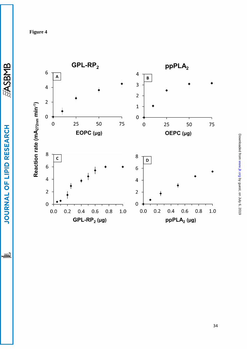
Coated microtiter plates were prepared with variable amounts of OEPC or EOPC ranging from
0 to 75 µg per well and their lipolysis by GPL-RP2 (Figure 4A) or ppPLA2 (Figure 4B) were
performed. The steady state reaction rate (slope of the variations, with time, in the absorbance
at 272 nm) were plotted as a function of the amounts of coated EOPC (Figure 4A) or OEPC
(Figure 4B). The lipolysis reaction rates increased quasi-linearly with the amounts of PCs, up
to 25 µg per well and then decreased slightly with higher PC amounts (Figures 4A and 4B).
Based on these results, a final PC amount of 50 µg per well was selected for further kinetic
by guest, on July 6, 2019w
ww
.jlr.orgD
ownloaded from

16
experiments. The same amount of coated substrate per well was previously used and reported
for the lipolysis of coated tung oil triglycerides (26), as welle as coated synthetic triglycerides
(27) or PC (28) containing α-eleostearic acid.
Using coated EOPC (50 µg per well) as the substrate, the steady-state reaction rate was found
to be linear from 0.2 to 0.6 µg/well for GPL-RP2 (Figure 4C) and from 0 to 0.15 µg/well for
Lecitase® (Figure S3A). At a higher concentration of enzyme, any increase in the reaction rate
was probably due to an excess of enzyme. It was established that as little as 10 ng of GPL-RP2
and 1 ng of Lecitase® can be detected under standard conditions. These measurements
correspond to 3 times the background observed with buffer alone. Similarly, when using coated
OEPC, the increase, with time, of the absorbance at 272 nm was recorded after injecting
variable amounts of ppPLA2 (Figure 4D) or hbPLA2 (Figure S3B). In both cases, the steady-
state reaction rate was found to be linear with time and proportional to the range of 0 to 0.8 µg
of enzyme per well. The detection limit, under standard conditions, was as low as 5 ng of
ppPLA2 and hbPLA2. A similar sensitivity of the assay towards these enzymes has been
previously observed using coated EEPC as the substrate (28). This assay shows a 100-1000
times higher sensitivity of enzyme detection than the pH-stat method (1 µg) which is routinely
used to measure PLA2 activity with PC (41). Moreover, the small amount of PLA2 (5 ng)
detected in our assay system was shown to be in the same range as that reported for PLA2 from
Naja naja (42) (1 ng) and for calcium-dependent secretory PLA2-IIA (43) (0.4 ng), using
synthetic fluorescent substrates. Nevertheless, this method is still less sensitive than the
radiometric or gold standard mass spectrometry end point methods (44, 45).
Estimation of the specific activity
The apparent molar extinction coefficient (εapp) of -eleostearic acid has been previously
determined by recording the absorbance, at 272 nm, of coated EEPC and various amounts of
-eleostearic acid per well and it was found to be 5320 ± 160 M-1 cm-1 (28). Under these
by guest, on July 6, 2019w
ww
.jlr.orgD
ownloaded from

17
conditions, the increase, with time, of the absorbance at 272 nm (A272 nm min-1) was converted
into µmol min-1 mg-1 of enzyme.
Using coated EOPC as the substrate, the specific activity of Lecitase® and GPL-RP2 was
calculated to be 2.2 and 0.3 µmol min-1 mg-1, respectively (Table 1). As expected for coated
EOPC with an ether bond at the sn-2 position, no detectable activity was found for ppPLA2
(Figure 2B and Table 1) or hbPLA2 (Table 1). For the sake of comparison, the specific activity
of pure GPL-RP2 on egg yolk PC, using the pH-technique, was shown to be 45 µmol min-1 mg-
1 (46). It is important to note that the stirring and emulsification conditions differ considerably
depending on whether a microplate or a mechanically stirred pH-stat vessel is used. Using
coated OEPC as the substrate, the specific activities of ppPLA2 and hbPLA2 were found to be
0.25 and 0.78 µmol min-1 mg-1, respectively (Table 1). Using PC analog with a BODIPY-
labeled alkyl ether at the sn-1 position and a N-(DNP)-8-amino-octanoyl group at the sn-2
position as substrate, Hendrickson et al. (20) showed a specific activity of around 18 nmol min-
1 mg-1 for cytosolic (85 kDa) PLA2.
Under the same experimental conditions, the specific activity of Lecitase® and GPL-RP2 was
calculated to be 0.08 and 0.03 µmol min-1 mg-1, respectively (Table 1). The activity of Lecitase®
and GPL-RP2 on EOPC was found to be approximately 28 times and 10 times higher than on
OEPC, respectively (Table 1). Access to the active site of classical pancreatic lipase is
controlled by a surface loop, the lid, which normally only undergoes conformational changes
following the addition of lipids or amphiphiles. A deletion within the lid domain was, however,
observed in GPL-RP2, which is able to accommodate more hydrophilic substrates, such as
phospholipids and galactolipids with large polar head groups (47), than classical pancreatic
lipase. The addition of the classical pancreatic lipase lid to GPL-RP2 has been shown to
decrease, but not abolish, the phospholipase activity suggesting that the lid contributes to
substrate specificity (48). Moreover, GPL-RP2 was shown to be produced at a high level in
by guest, on July 6, 2019w
ww
.jlr.orgD
ownloaded from

18
guinea pig lacking pancreatic PLA2, suggesting a significant role of this enzyme in phospholipid
digestion (49). Our study demonstrates for the first time, to the best of our knowledge, a PLA2
activity of GPL-RP2 and Lecitase® in addition to their triglyceride lipase and PLA1 activities.
Application of the microplate assay for screening potential PLA1 or PLA2 inhibitors
An important feature of this work is the ability to screen new potential PLA1 or PLA2 specific
inhibitors. THL and MI are known to inhibit digestive lipases (7, 50) and PLA2 (51),
respectively.
Using coated EOPC as the substrate, GPL-RP2 was inhibited by about 50% when pre-incubated
for 1 h at an enzyme:THL molar ratio of 1:70 (Figure 5). By contrast, pre-incubating MI with
GPL-RP2 does not affect the catalytic activity of this enzyme measured under the same
experimental conditions (Figure 5). Similarly, pre-incubating THL with Lecitase® for 1 h at a
molar excess of 100 leads to 40% inactivation of the lipolytic activity observed with coated
EOPC (data not shown). THL is known to be a digestive lipase catalytic inhibitor and it has
been shown to inhibit the triglyceride lipase activity of GPL-RP2, even in the absence of an
interface (7). The fact that both the lipase and phospholipase activities of GPL-RP2 and
Lecitase® were inhibited by THL is an indication that the same catalytic environment is
probably involved for triglyceride lipase, PLA1 and PLA2 activities of these lipolytic enzymes.
Using coated OEPC as the substrate, an injection of ppPLA2, pre-incubated (for 1 h) with a 100
molar excess of MI, leads to approximately 50% inactivation of the enzyme (Figure 5). As
expected (52) and under the same experimental conditions, ppPLA2 was not inhibited by THL
(Figure 5). In our previous report (28), we have shown that the pre-incubation of ppPLA2 (1h)
with a 50 or 100 molar excess of MI reduced the enzyme activity by 69% or 79%, respectively.
Conclusions
by guest, on July 6, 2019w
ww
.jlr.orgD
ownloaded from
19
In the method described in this paper, new synthetic PC derivatives with close structural
analogy to natural substrates were designed, esterified with a naturally occurring conjugated
polyene fatty acid with specific optical properties, and substituted with one non UV-absorbing
ether-bonded chain. Since these ether bonds are non-hydrolysable by PLA1 or PLA2, no acyl
chain migration could occur during lipolysis, thus providing the possibility of discriminating
accurately between PLA1 and PLA2 activities. In addition, the lipidic character of these PC
analogues presents a significant qualitative advantage over other methods. Furthermore, this
assay is specific, continuous and sensitive, and it allows screening of new PLA1 and PLA2
activities and/or their inhibitors present in various biological samples. Using this new enzymatic
assay, we have demonstrated, for the first time, to the best of our knowledge, the existence of
the PLA2 activity of GPL-RP2 and Lecitase® in addition to their triglyceride lipase and PLA1
activities.
by guest, on July 6, 2019w
ww
.jlr.orgD
ownloaded from
20
Acknowledgments
We gratefully acknowledge support from the “Region Rhône-Alpes” Programme CIBLE-
2012-1201080801. Meddy El Alaoui was supported by a Ph.D grant from the “Region Rhône-
Alpes” (€32,116/year). English revision by Valerie James is also acknowledged.
by guest, on July 6, 2019w
ww
.jlr.orgD
ownloaded from

21
References
1. van Meer, G. 2005. Cellular lipidomics. EMBO J 24: 3159-3165.
2. Murayama, K., K. Kano, Y. Matsumoto, and D. Sugimori. 2013. Crystal structure of
phospholipase A1 from Streptomyces albidoflavus NA297. J Struct Biol 182: 192-196.
3. Shimuta, K., M. Ohnishi, S. Iyoda, N. Gotoh, N. Koizumi, and H. Watanabe. 2009. The
hemolytic and cytolytic activities of Serratia marcescens phospholipase A (PhlA) depend on
lysophospholipid production by PhlA. BMC Microbiol 9: 261.
4. Hla, T., M. J. Lee, N. Ancellin, J. H. Paik, and M. J. Kluk. 2001. Lysophospholipids--
receptor revelations. Science 294: 1875-1878.
5. Yasuda, T., T. Ishida, and D. J. Rader. 2010. Update on the role of endothelial lipase in
high-density lipoprotein metabolism, reverse cholesterol transport, and atherosclerosis. Circ J
74: 2263-2270.
6. Amara, S., V. Delorme, M. Record, and F. Carriere. 2012. Inhibition of phospholipase
A1, lipase and galactolipase activities of pancreatic lipase-related protein 2 by methyl
arachidonyl fluorophosphonate (MAFP). Biochim Biophys Acta 1821: 1379-1385.
7. Eydoux, C., J. De Caro, F. Ferrato, P. Boullanger, D. Lafont, R. Laugier, F. Carriere,
and A. De Caro. 2007. Further biochemical characterization of human pancreatic lipase-related
protein 2 expressed in yeast cells. J Lipid Res 48: 1539-1549.
8. Xiao, X., L. E. Ross, W. A. Sevilla, Y. Wang, and M. E. Lowe. 2013. Porcine pancreatic
lipase related protein 2 has high triglyceride lipase activity in the absence of colipase. Biochim
Biophys Acta 1831: 1435-1441.
by guest, on July 6, 2019w
ww
.jlr.orgD
ownloaded from

22
9. Withers-Martinez, C., F. Carriere, R. Verger, D. Bourgeois, and C. Cambillau. 1996. A
pancreatic lipase with a phospholipase A1 activity: crystal structure of a chimeric pancreatic
lipase-related protein 2 from guinea pig. Structure 4: 1363-1374.
10. Dennis, E. A., J. Cao, Y. H. Hsu, V. Magrioti, and G. Kokotos. 2011. Phospholipase A2
enzymes: physical structure, biological function, disease implication, chemical inhibition, and
therapeutic intervention. Chem Rev 111: 6130-6185.
11. Murakami, M., Y. Taketomi, Y. Miki, H. Sato, K. Yamamoto, and G. Lambeau. 2014.
Emerging roles of secreted phospholipase A2 enzymes: the 3rd edition. Biochimie 107 Pt A:
105-113.
12. Brglez, V., G. Lambeau, and T. Petan. 2014. Secreted phospholipases A2 in cancer:
diverse mechanisms of action. Biochimie 107 Pt A: 114-123.
13. Schmid, R. D., and R. Verger. 1998. Lipases: interfacial enzymes with attractive
applications. Angew. Chem. Int. Ed. 37: 1608-1633.
14. Panaitov, I., and R. Verger. 2000. Enzymatic reactions at interfaces: Interfacial and
temporal organization of enzymatic lipolysis. In Physical Chemistry of Biological Interfaces.
A. Baszkin and W. Norde, editors. Marcel Dekker, Inc, New York, Basel. 359-400.
15. Ransac, S., M. Ivanova, R. Verger, and I. Panaitov. 1997. Monolayer techniques for
studying lipase kinetics. Methods Enzymol. 286: 263-292.
16. Petrovic, N., C. Grove, P. E. Langton, N. L. Misso, and P. J. Thompson. 2001. A simple
assay for a human serum phospholipase A2 that is associated with high-density lipoproteins. J
Lipid Res 42: 1706-1713.
17. Sharko, O., and M. Kisel. 2011. 1-Acyl-2-[N-(2,4-dinitrophenyl)aminopropionyl]-sn-
glycero-3-phosphocholine as a chromogenic substrate for phospholipase A(2) assay. Anal
Biochem 413: 69-71.
by guest, on July 6, 2019w
ww
.jlr.orgD
ownloaded from

23
18. van den Bosch, H., J. G. de Jong, and A. J. Aarsman. 1991. Phospholipase A2 from rat
liver mitochondria. Methods Enzymol 197: 365-373.
19. Darrow, A. L., M. W. Olson, H. Xin, S. L. Burke, C. Smith, C. Schalk-Hihi, R. Williams,
S. S. Bayoumy, I. C. Deckman, M. J. Todd, B. P. Damiano, and M. A. Connelly. 2011. A novel
fluorogenic substrate for the measurement of endothelial lipase activity. J Lipid Res 52: 374-
382.
20. Hendrickson, H. S., E. K. Hendrickson, I. D. Johnson, and S. A. Farber. 1999.
Intramolecularly quenched BODIPY-labeled phospholipid analogs in phospholipase A(2) and
platelet-activating factor acetylhydrolase assays and in vivo fluorescence imaging. Anal
Biochem 276: 27-35.
21. Mitnaul, L. J., J. Tian, C. Burton, M. H. Lam, Y. Zhu, S. H. Olson, J. E. Schneeweis, P.
Zuck, S. Pandit, M. Anderson, M. M. Maletic, S. T. Waddell, S. D. Wright, C. P. Sparrow, and
E. G. Lund. 2007. Fluorogenic substrates for high-throughput measurements of endothelial
lipase activity. J Lipid Res 48: 472-482.
22. Huang, C., L. Zhou, Y. Liu, and L. Lai. 2006. A continuous fluorescence assay for
phospholipase A2 with nontagged lipid. Anal Biochem 351: 11-17.
23. Basu, D., X. Lei, J. Josekutty, M. M. Hussain, and W. Jin. 2013. Measurement of the
phospholipase activity of endothelial lipase in mouse plasma. J Lipid Res 54: 282-289.
24. Radunz, A., P. He, and G. H. Schmid. 1998. Analysis of the seed lipids of Aleurites
montana. Z. Naturforsch. 53: 305–310.
25. Pencreac'h, G., J. Graille, M. Pina, and R. Verger. 2002. An ultraviolet
spectrophotometric assay for measuring lipase activity using long-chain triacyglycerols from
Aleurites fordii seeds. Anal Biochem 303: 17-24.
by guest, on July 6, 2019w
ww
.jlr.orgD
ownloaded from

24
26. Serveau-Avesque, C., R. Verger, J. A. Rodriguez, and A. Abousalham. 2013.
Development of a high-throughput assay for measuring lipase activity using natural
triacylglycerols coated on microtiter plates. Analyst 138: 5230-5238.
27. Mendoza, L. D., J. A. Rodriguez, J. Leclaire, G. Buono, F. Fotiadu, F. Carriere, and A.
Abousalham. 2012. An ultraviolet spectrophotometric assay for the screening of sn-2-specific
lipases using 1,3-O-dioleoyl-2-O-alpha-eleostearoyl-sn-glycerol as substrate. J Lipid Res 53:
185-194.
28. El Alaoui, M., A. Noiriel, L. Soulere, L. Grand, Y. Queneau, and A. Abousalham. 2014.
Development of a high-throughput assay for measuring phospholipase A activity using
synthetic 1,2-alpha-eleostearoyl-sn-glycero-3-phosphocholine coated on microtiter plates. Anal
Chem 86: 10576-10583.
29. Mu, H. L., J. P. Kurvinen, H. Kallio, X. B. Xu, and C. E. Hoy. 2001. Quantitation of
acyl migration during lipase-catalyzed acidolysis, and of the regioisomers of structured
triacylglycerols formed. J. Am. Oil Chem. Soc. 78: 959-964.
30. Bradford, M. M. 1976. A rapid and sensitive method for quantitation of microgram
quantities of protein utilizing the principle of protein-deye binding. Anal. Biochem. 72: 248-
254.
31. Ransac, S., Y. Gargouri, F. Marguet, G. Buono, C. Beglinger, P. Hildebrand, H.
Lengsfeld, P. Hadvary, and R. Verger. 1997. Covalent inactivation of lipases. Methods
Enzymol. 286: 190-231.
32. Rosseto, R., C. M. Tcacenco, R. Ranganathan, and J. Hajdu. 2008. Synthesis of
Phosphatidylcholine Analogues Derived from Glyceric Acid: a New Class of Biologically
Active Phospholipid Compounds. Tetrahedron Lett 49: 3500-3503.
by guest, on July 6, 2019w
ww
.jlr.orgD
ownloaded from

25
33. Tully, S. E., and B. F. Cravatt. 2010. Activity-based probes that target functional
subclasses of phospholipases in proteomes. J Am Chem Soc 132: 3264-3265.
34. Borsotti, G., G. Guglielmetti, S. Spera, and E. Battistel. 2001. Synthesis of
phosphatidylcholines containing ricinoleic acid. Tetrahedron Lett 57: 10219-10227.
35. Reichardt, C. 1994. Solvatochromic Dyes as Solvent Polarity Indicators. Chem. Rev. 94:
2319–2358.
36. Laurent, S., M. G. Ivanova, D. Pioch, J. Graille, and R. Verger. 1994. Interactions
between b-Cyclodextrin and insoluble glyceride monomolecular films at the argon/water
interface: application to lipase kinetics. Chem. Phys. Lipids 70: 35-42.
37. Ivanova, M., R. Verger, and I. Panaiotov. 1997. Mechanisms underlying the desorption
of long-chain lipolytic products by cyclodextrins: application to lipase kinetics in monolayer.
Colloids Surfaces B: Biointerfaces 10: 1-12.
38. Aloulou, A., Y. B. Ali, S. Bezzine, Y. Gargouri, and M. H. Gelb. 2012. Phospholipases:
an overview. Methods Mol Biol 861: 63-85.
39. Bojsen, K., K. Borch, G. Budolfsen, K. C. Fuglsang, S. S. Glad, A. Petri, A. P.
Shamkant, A. Svendsen, and J. Vind. 2000. Lipolytic enzyme variants. In. N. A/S, editor.
40. Aloulou, A., F. Frikha, A. Noiriel, M. Bou Ali, and A. Abousalham. 2014. Kinetic and
structural characterization of triacylglycerol lipases possessing phospholipase A1 activity.
Biochim Biophys Acta 1841: 581-587.
41. Abousalham, A., and R. Verger. 2000. Egg yolk lipoproteins as substrates for lipases.
Biochim Biophys Acta 1485: 56-62.
by guest, on July 6, 2019w
ww
.jlr.orgD
ownloaded from

26
42. Kinkaid, A. R., and D. C. Wilton. 1993. A continuous fluorescence displacement assay
for phospholipase A2 using albumin and medium chain phospholipid substrates. Anal Biochem
212: 65-70.
43. Tsao, F. H., D. Shanmuganayagam, D. K. Zachman, M. Khosravi, J. D. Folts, and K. C.
Meyer. 2007. A continuous fluorescence assay for the determination of calcium-dependent
secretory phospholipase A2 activity in serum. Clin Chim Acta 379: 119-126.
44. Hsu, Y. H., D. S. Dumlao, J. Cao, and E. A. Dennis. 2013. Assessing phospholipase A2
activity toward cardiolipin by mass spectrometry. PLoS One 8: e59267.
45. Marshall, J., E. Krump, T. Lindsay, G. Downey, D. A. Ford, P. Zhu, P. Walker, and B.
Rubin. 2000. Involvement of cytosolic phospholipase A2 and secretory phospholipase A2 in
arachidonic acid release from human neutrophils. J Immunol 164: 2084-2091.
46. Carriere, F., K. Thirstrup, S. Hjorth, F. Ferrato, P. F. Nielsen, C. Withers-Martinez, C.
Cambillau, E. Boel, L. Thim, and R. Verger. 1997. Pancreatic lipase structure-function
relationships by domain exchange. Biochemistry 36: 239-248.
47. Carrière, F., C. Withers-Martinez, H. van Tilbeurgh, A. Roussel, C. Cambillau, and R.
Verger. 1998. Structural basis for the substrate selectivity of pancreatic lipases and some related
proteins. Biochim Biophys Acta 1376: 417-432.
48. Yang, Y., and M. E. Lowe. 2000. The open lid mediates pancreatic lipase function. J
Lipid Res 41: 48-57.
49. Fauvel, J., M. J. Bonnefis, L. Sarda, H. Chap, J. P. Thouvenot, and L. Douste-Blazy.
1981. Purification of two lipases with high phospholipase A1 activity from guinea-pig pancreas.
Biochim. Biophys. Acta 663: 446-456.
by guest, on July 6, 2019w
ww
.jlr.orgD
ownloaded from

27
50. Ben Ali, Y., H. Chahinian, S. Petry, G. Muller, F. Carriere, R. Verger, and A.
Abousalham. 2004. Might the kinetic behavior of hormone-sensitive lipase reflect the absence
of the lid domain? Biochemistry 43: 9298-9306.
51. Hui, D. Y., M. J. Cope, E. D. Labonte, H. T. Chang, J. Shao, E. Goka, A. Abousalham,
D. Charmot, and J. Buysse. 2009. The phospholipase A(2) inhibitor methyl indoxam suppresses
diet-induced obesity and glucose intolerance in mice. Br J Pharmacol 157: 1263-1269.
52. Hadvary, P., H. Lengsfeld, and H. Wolfer. 1988. Inhibition of pancreatic lipase in vitro
by the covalent inhibitor tetrahydrolipstatin. Biochem J 256: 357-361.
by guest, on July 6, 2019w
ww
.jlr.orgD
ownloaded from

28
Table 1: Estimation of enzyme specific activities using EOPC or OEPC coated on microtiter
plate wells. Comparison of specific activities of Lecitase®, GPL-RP2, ppPLA2 and hbPLA2
using EOPC or OEPC coated on microtiter plate wells. Results are given as means SD for
three independent experiments. * : below the detection limit, estimated as 3 times the
background obtained with buffer alone.
Specific activity (µmol.min-1.mg-1)
Enzyme/Substrate Lecitase® GPL-RP2 ppPLA2 hbPLA2
EOPC 2.20 0.30 0.300 0.019 0* 0*
OEPC 0.08 0.01 0.030 0.003 0.250 0.002 0.780 0.039
EOPC/OEPC ratio 27.5 10 0 0
by guest, on July 6, 2019w
ww
.jlr.orgD
ownloaded from

29
Figure legends
Figure 1: Chemical structure and UV absorption spectra of EOPC and OEPC. (A) Chemical
structure of EOPC and OEPC. (B) UV absorption spectra of -eleostearic acid (62.5 µg mL-1),
EOPC (125 µg mL-1) and OEPC (125 µg mL-1) dissolved in ethanol containing BHT 0.001%.
Figure 2: Principle of the coated PC assay and time course of the enzymatic hydrolysis of
OEPC or EOPC coated on microtiter plate wells. (A) Schematic representation of the assay
reaction showing the hydrolysis of coated EOPC or OEPC in a microwell. E, enzyme in
solution; E*, enzyme at the interface; S, substrate (EOPC or OEPC); P, lipolysis products (-
eleostearic acid and non-UV absorbing lyso-PC). β-CD (3 g.L-1, final concentration) was used
to solubilize the long-chain lipolysis products into the aqueous buffer. (B and C) Kinetic
recordings of coated EOPC (A) and OEPC (B) lipolysis by GPL-RP2 or ppPLA2. Variations,
with time, of the absorbance at 272 nm were recorded for 10 min, for stabilization, and then for
50 min after GPL-RP2 or ppPLA2 (0.5 µg per well). The kinetic recordings shown here are
typical of those obtained in three independent experiments.
Figure 3: Analysis by TLC of coated EOPC (A) and OEPC (B) hydrolysis revealed with UV
light at 254 nm (left panels) and with 10% copper sulfate and 10% phosphoric acid in water,
followed by charring at 150°C for 15 min (right-hand panels). The standards (std) used (10 µg
of each compound) were -eleostearic acid, EOPC and OEPC. Coated PCs were hydrolyzed
by GPL-RP2 or ppPLA2 (0.5 µg/well). The chromatographic solvent system was
chloroform/methanol/water (65/35/4, v/v/v) and hexane/diethylether/acetic acid (84/16/1,
v/v/v) containing 0.001% (w/v) BHT.
Figure 4: Effects of the initial PC concentration (A and B) and the amount of enzyme (C and
D) on the steady-state reaction rates. Variable amounts of coated EOPC (A) or OEPC (B) were
subjected to hydrolysis by GPL-RP2 (0.5 µg, A) or ppPLA2 (0.5 µg, B). Variable amounts of
by guest, on July 6, 2019w
ww
.jlr.orgD
ownloaded from

30
GPL-RP2 (C) or ppPLA2 (D) were injected into the microplate well, containing the coated
EOPC (C) or OEPC (D), in 200 µL of standard buffer. The increase in the absorbance at 272
nm was recorded for 20-40 min after the enzyme injection, and the initial velocity
(mA272nm.min-1) was used for reaction rate determination. Results are given as means SD
for three independent experiments.
Figure 5: Validation of the assay to study PLA1 or PLA2 inhibition. Either GPL-RP2 or ppPLA2
was incubated with THL (enzyme:inhibitor, molar ratio of 1:70) or with MI (enzyme:inhibitor,
molar ratio of 1:100), in the absence of the substrate. The residual activities of GPL-RP2 or
ppPLA2 were then measured, using coated EOPC or OEPC, respectively, as described in the
Methods section, and expressed as a percentage relative to the activity measured in the absence
of inhibitor.Values ± SD are the mean of three independent experiments. ** P<0.01, (vs
DMSO).
by guest, on July 6, 2019w
ww
.jlr.orgD
ownloaded from