
Cutaneous Wegener's granulomatosis - Cleveland Clinic Journal of
J. clin. Path. (1958), 11, 146.
A CASE OF WEGENER'S GRANULOMATOSISBY
A. M. THOMASFrom the Area Laboratory, Musgrove Park Hospital, Taunton
(RECEIVED FOR PUBLICATION MARCH 28, 1957)
In 1931 Klinger described a case of poly-arteritis nodosa with an unusual localization of. thelesions in the nasal septum, lungs, spleen, and
kidneys. Wegener (1937, 1939) reported three moresimilar cases as a new disease, stressing its granu-
lomatous aspect. Since that time, examples of thiscondition have appeared in the literature under a
confusing variety of different names, but recent
authors have preferred the term "Wegener'sgranulomatosis," and the present state of our
knowledge of this subject is summarized byFahey, Leonard, Churg, and Godman (1954) andby Godman and Churg (1954). In two excellentpapers they review the literature, describe seven
new cases and clarify the diagnostic criteria ofthis disease. The present paper describes anothercase of Wegener's granulomatosis and discusses itsrelationship with some cases of Ceelen's haemo-siderosis. The story of the case and its investiga-tions will be more readily understood if it isappreciated that it presented as an intractableanaemia and was investigated and treated as
such. Indeed, apart from haemoptysis, thoughtto originate from an ulcer of the nasal septum,symptoms referable to the respiratory system were
extremely slight.
Case HistoryD. F. M., a butcher aged 40, first went to see his
doctor in January, 1949, on account of " rheumatism."He had no fever but complained of painful, swollenjoints which improved in a few days.On February 28 he was sent to a chest clinic with
a note from the doctor specifying cough, haemoptysis,and night sweats.The clinic notes stated that cough had been pre-
sent for several weeks, sputum, 1-2 oz., blood-stainedfor several days, pain in the left chest on and off.The appetite, bowels, kidneys, and bladder were
normal. The fingers were not clubbed. The patieitdid not suffer from lassitude or loss of weight, butwas in poor general condition. A few moist soundswere heard at the apex of the left lung, and a chestradiograph suggested right basal bronchiectasis.A review of the radiograph showed a thickened
right interlobular pleura and generally increased
bronchovascular shadows with possibly diffuse finefibrosis.On March 23 the patient was sent to the chest
clinic complaining of haemoptysis every morningafter "a cold" at Christmas, 1948, and also ofmultiple septic skin lesions. On examination, ulcerson the nasal septum and right inferior turbinate anda punched ulcer on the palate immediately above theuvula were seen. The Wassermann reaction wasnegative. He was given potassium iodide, and onMarch 30 he had an " iodine" rash and herpeslabialis. The Wassermann reaction was still nega-tive. By April 13 the palate ulcer had healed, butseptal bleeding continued. On May 17 a biopsy ofthe septal ulcer showed chronic inflammatory, non-specific granulation tissue, with no evidence of tuber-culosis or malignancy.On June 5 urticaria was noted. Haemoglobin was
56%.On June 13 he was sent to E.N.T. Department with
letter: "A large slough separated from his rightnostril to-day. During last fortnight he has had aseptic throat. Recurrent herpes, angioneuroticoedema of face and urticaria. His general conditionhas not been too good and he has been in bed forsome of the time."An E.N.T. note on June 15 read: "His trouble
is due to some obscure general condition. There isno enlargement of his liver or spleen." During hisstay in hospital (three weeks) he ran a temperature of99.4° F. on three evenings, otherwise he was prac-tically apyrexial.On October 6 his doctor wrote to the hospital:
"M. had severe herpes around mouth and eyes. Hecame home from hospital about six weeks ago some-what better, but has recently developed a septictoe-? ingrowing toe nail. About ten days ago adiffuse purpuric rash appeared, which is still present.He has had no pyrexia, but complains of rheumaticpains and general weakness."On October 7 he attended hospital as an out-
patient complaining of nasal catarrh and haemoptysis,which has been very slight since his operation. Hedid not feel well and had noticed herpes of the righteye, pains in both shoulders, slight cough, rash onthe arms and legs, chiefly, for one month.On examination, the right eye was seen to be in-
jected. Purpura was everywhere, also septic spots.Nothing abnormal noted in heart, chest, and abdomen
on March 30, 2020 by guest. P
rotected by copyright.http://jcp.bm
j.com/
J Clin P
athol: first published as 10.1136/jcp.11.2.146 on 1 March 1958. D
ownloaded from
A CASE OF WEGENER'S GRANULOMATOSIS
He was admitted to hospital on November 4.Hoarseness was getting worse.On examination, he was rather pale, both eyes were
infected, and he was very hoarse. He had a largeulcer on the left side of the palate, also septic spotsand a purpuric eruption on the arms and legs.Blood pressure was 150/90 mm. Hg. Otherwise noth-ing abnormal was detected.On November 7 R.B.C.s were 3.7 million and Hb
44%, and on November 9 two pinits of blood (packedcells) were given.By January 13, 1950, the ulcer on the palate was
much larger, and also there was a sore on the scrotum.On January 18 two pints of blood (packed cells)
were given.On January 20 he had an ulcer at base of the right
index finger on the back of the hand. Two pints ofblood were given on February 12.The patient's condition declined steadily during the
last month, and at the end, when he was unable toswallow on account of the extensive ulceration of thethroat, his deterioration was rapid. He collapsed andwas in coma for two days before he died on February15, after being ill for 14 months. There was nothingin the clinical picture to suggest massive infarction ofthe lungs.
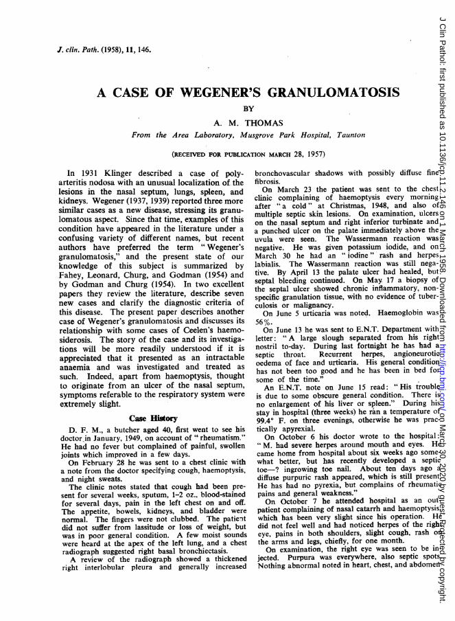
Radiographs taken on February 28 and Novem-ber 7, 1949, showed a progressive condition (Figs. Iand 2). In the first there is throughout both lungs ageneral increase in bronchovascular markings withpatchy congestion, particularly marked at the right
base and in the cardiophrenic angle. In the secondfilm the condition is the same, but the general mark-ings are finer and there are less mottled congestiveareas. In a later film taken on January 10, 1950,there was a general fine striated appearance of bothlungs, giving a ground-glass effect and suggesting ageneral fine fibrosis of both lungs.
Pathological ExaminationsHaematology.-Between June, 1949, and February,
1950, 13 erythrocyte counts and haemoglobin estima-tions were performed. The counts varied between2.9 and 4.2 million per c.mm. and the haemoglobinlevels between 44 and 88% (Haldane), averagingapproximately 50 to 60% with occasional rises aftertransfusion. The colour index was usually about0.7 to 0.8 but rose to 1.0 a month before death.During the above period, 11 leucocyte counts were per-formed and the numbers ranged from 5,000 to 11,000per c.mm. Polymorphs varied between 87 and 57%,eosinophils were noted on five occasions and variedbetween 1 and 2%. Three sternal marrow examina-tions were performed and the first two showedmicronormoblastic erythropoiesis which was lesssevere in the third sample. All three tests showedhyperplastic marrow such as one finds with asecondary anaemia.
Reticulocytes.-5.0, 1.5, and 1.0%/O on December 14and December 29, 1949, and January 10, 1950, re-spectively.
FIG. 2.-Second radiograph of the chest on November 7, 1949.FIG. 1.-First radiograph of the chest on February 28, 1949.
147
on March 30, 2020 by guest. P
rotected by copyright.http://jcp.bm
j.com/
J Clin P
athol: first published as 10.1136/jcp.11.2.146 on 1 March 1958. D
ownloaded from

A. M. THOMAS
Platelets.-510,000, 500,000. and 480.000 per c.mm.on October 7 and November 7. 1949. and January 10,1950. respectively.
Bleedinig Time.-This was 3 minutes 15 seconds onJuly 15, 1949 (normal. 2 to 4 minutes).
Coagutlationi Ti,,ie (Dale and Laidlaw).-This was3 minutes on July 15. 1949 (normal, I to 2 minutes).
The erythrocyte sedimentation rate (Westergren)was 68 mm. in one hour on July 26. 1949.
Ervthrocyte Fragility.- Haemolysis began at 0.48",and was complete at 0.40",, sodium chloride onFebruary 3, 1950.
Serology.-Wassermann and Kahn tests were nega-tive on March 23, March 30, and November 11. 1949.
Biochemistry.-A van den Bergh test was negativeon January 17. 1950. The following resLIlts wereobtained on February 3:Alkaline phosphatase .. 10 units per 100 ml. (normal 3 to 10 units
per 100 mrl.)Thymol turbidity I. unit (normal 0 to 4 units)Serum colloidal gold test NegativeVan den Bergh test NegativeSerum proteins.. 52 g. %
albumin.. .. 2-6 _
globulin 2-6 ,- (A G ratio I: 1).
NecropsyNecropsy was performed on February 15, 1950. 18
hours after death.External Examination.-The body is that of a pale,
thin man of middle age. There are no purpurichaemorrhages, jaundice, or oedema. The dorsum ofthe right hand and the scrotum each show an ulcert4 cm. in diameter. These are deep, penetrating ulcerswith irregular, serpiginous edges, but they appear in-dolent and without surrounding induration. conges-tion, or inflammation.
Respiratory System.-The lungs present a strikingappearance and bulge from the opened thorax. Theyare solid, voluminous, and fail to collapse when re-
moved from the body. To the touch they are rubberywith firmer, ill-defined, rounded nodules scatteredthroughout their substance and measuring up to 2 cm.
in diameter; they give the tactile impression of beingcondensations of fibrous tissue within a generaldiffuse pulmonary fibrosis. The lungs themselves are
of a mottled red colour, varying from rust to darkblood red in places. There are occasional pale areas.
notably at the right apex, but these are very scanty.The cut surface shows many haemorrhages andhaematomata of varving ages, the older in some in-stances appearing to coincide with firm nodules.Ther-e is no evidence of pulmonary neoplasm or
inflammation. Pleltal adhesions and effusions are
abscnt. The hilar lymph nodes are enlarged up to2 cm. in diameter. but are quite soft. The tracheaand bronchi contain blood-staned. frothy fluid. Thepalate is extensively ulcerated and covered with foul.necrotic debris.
Excretory System.-The kidneys are very pale andshow moderately good demarcation of the cortex andmedulla. They are of normal size and their capsuLlesstrip easily.
Cardiovascular System.-The heart is of normalsize and without endocarditis or right ventricularhypertrophy.The organs of the alimentary. reticutlo-endothelial.
and central nervous systems appear normal apartrl-om extreme pallor.
HistologyLungs.-The lungs show virtually no normal
parenchyma and the following changes are found invarying proportions:
(1) Diffuse fibrosis with concentration of oldetcollagen into ill-defined nodular areas (Fig. 3).
(2) There are many diffusely scattered smallhaemorrhages. each consisting of a group of involvedalveoli. The haemorrhages appear to be of differentages, many of them recent. Some of the oldethaemorrhages seem to be related to the fibrousconglomerations.
(3) Heavy infiltration with siderophages. these beingfrequently concentrated into dense aggregations(Fig. 4). The amount of haemosiderin in these lungsis very large indeed.
(4) The minimal changes visible are collagenousthickening and diminished vascularity of the alveolarwalls.
(5) An occasional group of alveoli is seen contain-ing inflammatory exudate.
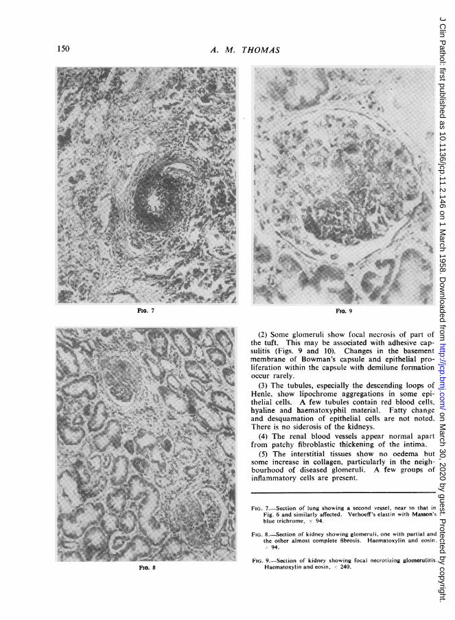
(6) Changes in the lung blood vessels consist of:(a) Recent thrombosis of a few medium-sized
vessels with patchy perivascular inflammation.(b) Calcerosiderosis, distortion, and fragmentation
of the elastica of many small vessels (Fig. 5).(c) Acute arteritis of three small vessels with
narrowing of the lumen, necrosis and fibrin deposi-tion in the wall, destruction of the elastica, and peri-vascular leucocytic infiltration (Figs. 6 and 7). Thereis a zone -of recent haemorrhage surrounding theseve,sels.
(7) The bronchial lymph nodes show numeroussiderophages and carbon particles.
(8) The bronchi show thickening of the walls andsometimes notable cuffing with siderophages and in-fl>;mmatory cells including lymphocytes. histiocytes.and plasma cells. The lumina of the bronchi ma\contain blood, desquamated epithelial cells. and afew siderophages.
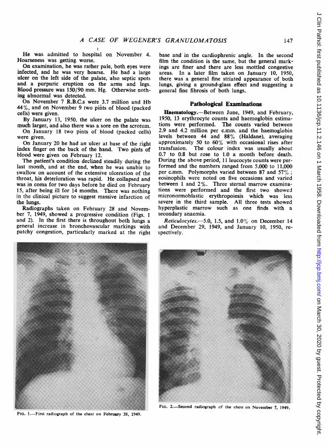
(9) Destruction of elastic fibres in some alveolarwalls and blood vessels.Kidneys.-The kidneys show the following changes:(1) About 200 of the glomeruli are abnormal, and
these appear to be grouped together rather thanscattered evenly throughout the renal parenchyma.The most common lesion is fibrosis of the tufts varv-ing from slight collagenous thickening of the capil-lary membranes to dense scarring of the whole orp.art of the tuft (Fig. 8).
148
on March 30, 2020 by guest. P
rotected by copyright.http://jcp.bm
j.com/
J Clin P
athol: first published as 10.1136/jcp.11.2.146 on 1 March 1958. D
ownloaded from

FIG. 3.-Section of lung showing diffuse fibrosis. Haematoxylin FIG. 4.-Section of lung showing large groups of siderophages.and eosin, x 94. Haematoxylin and eosin, x 94.
FIG. 5-Section of lung showing calcero-siderosis of the elastica withdistortion and fragmentation. Haematoxylin and eosin, > 94.
FIG. 6-Section of lung showing necrotizing arteritis with destructionof elastica. Verhoeff's elastin with Masson's blue trichrome,X 240.
on March 30, 2020 by guest. P
rotected by copyright.http://jcp.bm
j.com/
J Clin P
athol: first published as 10.1136/jcp.11.2.146 on 1 March 1958. D
ownloaded from
A. M. THOMAS
*
R
.# 'RS X
E '.>S . ,
~-.. .410
A.
-..Fto. 9
(2) Some glomeruli show focal necrosis of part ofthe tuft. This may be associated with adhesive cap-sulitis (Figs. 9 and 10). Changes in the basementmembrane of Bowman's capsule and epithelial pro-liferation within the capsule with demilune formationoccur rarely.
(3) The tubules, especially the descending loops ofHenle, show lipochrome aggregations in some epi-thelial cells. A few tubules contain red blood cells.hyaline and haematoxyphil material. Fatty changeand desquamation of epithelial cells are not noted.There is no siderosis of the kidneys.
(4) The renal blood vessels appear normal apartfrom patchy fibroblastic thickening of the intima.
(5) The interstitial tissues show no oedema butsome increase in collagen, particularly in the neigh-bourhood of diseased glomeruli. A few groups ofinflammatory cells are present.
FIG. 7.-Section of lung showing a second vessel, near to that inFig. 6 and similarly affected. Verhoeff's elastin with Masson'sblue trichrome, x 94.
FIG. 8.-Section of kidney showing glomeruli, one with partial andthe other almost complete fibrosis. Haematoxylin and eosin.- 94.
FIG. 9.-Section of kidney showing focal necrotizing glomerulitis.Haematoxylin and eosin, X 240.
FIo. 7
FIG. 8
150
on March 30, 2020 by guest. P
rotected by copyright.http://jcp.bm
j.com/
J Clin P
athol: first published as 10.1136/jcp.11.2.146 on 1 March 1958. D
ownloaded from
A CASE OF WEGENER'S GRANULOMATOSIS
*b, *ffii48. t<'. ;* 2A?' ,."C4. J
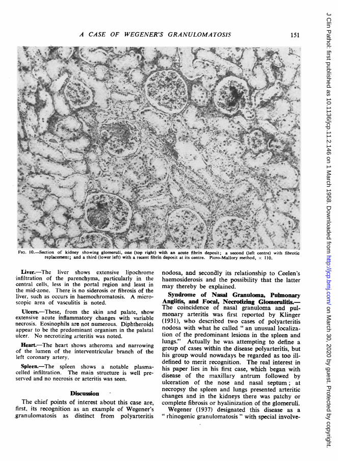
FIG. 10O-ection of kidney showing glomeruli, one (top right) with an acute fibrin deposit; a second (left centre) with fibroticreplacement; and a third (lower left) with a recent fibrin deposit at its centre. Picro-Mallory method, x 110.
Liver.-The liver shows extensive lipochrome-infiltration of the parenchyma, particularly in thecentral cells, less in the portal region and least inthe mid-zone. There is no siderosis or fibrosis of theliver, such as occurs in haemochromatosis. A micro-scopic area of vasculitis is noted.
Ulcers.-These, from the skin and palate, showextensive acute inflammatory changes with variablenecrosis. Eosinophils are not numerous. Diphtheroidsappear to be the predominant organism in the palatalulcer. No necrotizing arteritis was noted.Heart.-The heart shows atheroma and narrowing
of the lumen of the interventricular branch of theleft coronary artery.
Spleen.-The spleen shows a notable plasma-celled infiltration. The main structure is well pre-served and no necrosis or arteritis was seen.
DiscussionThe chief points of interest about this case are,
first, its recognition as an example of Wegener's,granulomatosis as distinct from polyarteritis
nodosa, and secondly its relationship to Ceelen'shaemosiderosis and the possibility that the lattermay thereby be explained.Syndrome of Nasal Granuloma, Pulmonary
Angiitis, and Focal, Necrotizing Glomerulitis.-The coincidence of nasal granuloma and pul-monary arteritis was first reported by Klinger(1931), who described two cases of polyarteritisnodosa with what he called " an unusual localiza-tion of the predominant lesions in the spleen andlungs.4' Actually he was attempting to define agroup of cases within the disease polyarteritis, buthis group would nowadays be regarded as too ill-defined to merit recognition. The real interest inhis paper lies in his first case, which began withdisease of the maxillary antrum followed byulceration of the nose and nasal septum; atnecropsy the spleen and lungs presented arteriticchanges and in the kidneys there was patchy orcomplete fibrosis or hyalinization of the glomeruli.Wegener (1937) designated this disease as a
"rhinogenic granulomatosis" with special involve-
151
on March 30, 2020 by guest. P
rotected by copyright.http://jcp.bm
j.com/
J Clin P
athol: first published as 10.1136/jcp.11.2.146 on 1 March 1958. D
ownloaded from
A. M. THOMAS
ment of the arterial system and of the kidneys.This definition was elaborated by Godman andChurg (1954), who state that Wegener's granulo-matosis is always characterized by the three fol-lowing features: " (1) Necrotizing granulomatouslesions in the upper air passages or in the lowerrespiratory tract or both; (2) generalized focalnecrotizing vasculitis, involving both arteries andveins, almost always in the lungs and more or lesswidely disseminated in other sites; (3) glomeru-litis characterized by necrosis (and thrombosis) ofloops or lobes of the capillary tuft, capsularadhesion and evolution as a glomerular lesion."The chief difference between the diagnostic
criteria of Godman and Churg (1954) and those ofWegener (1937, 1939) lies in the localization of theinitial granuloma, and recent examples justify theextension of the definition to include all casesstarting with a respiratory tract granuloma insteadof restricting it to those of nasal origin. Thisdisease, like other types of polyarteritis nodosa,presents so many variable features that only con-fusion results from regarding any one or two ofthese features as diagnostic. Wegener's originaldefinition includes all that is constant and can beimproved only by expanding the term " rhino-genous " to " respiratory " granulomatosis. Otherlesions, which occur frequently but are not diag-nostic, include pulmonary, renal, and splenicgranulomatosis, which may or may not be asso-ciated with necrotizing arteritis. Fever, jointpains, and cutaneous lesions are often found, andthe clinical picture is usually governed by the siteof the initial granuloma. Leucocytosis, elevationof the erythrocyte sedimentation rate, and anaemiaare the principal haematological findings. Widelydiffering degrees of severity may be encounteredand most recorded examples are far more acuteand severe than the present case. Its chronicityno doubt accounts for many of its unusualfeatures including the lack of visceral granulo-mata, pyrexia, and leucocytosis and for the pre-sence of a low-grade, intractable anaemia and theintense pulmonary haemosiderosis.The main object in defining Wegener's granu-
lomatosis is to differentiate a particular variety ofpolyarteritis nodosa distinguished by the presenceof an initial respiratory tract granuloma, theinvolvement of only small vessels (veins as well asarteries), and the granulomatous evolution of thedisease. The present case satisfies all thesecriteria, although the preponderance of haemo-siderotic changes in the lungs and the cutaneousposition of the secondary granulomata areatypical features. The type of vascular lesion in
the viscera, including the lungs, and the character-istic focal lesions in the glomeruli, clearly resembledrug-induced vascular disease and the other hyper-ergic affections of the blood vessels (" hyper-sensitivity angiitis" of Zeek, Smith, and Weeter(1948), " hyperergic fibrinous vasculosis " ofLendrum (1956)) far more than classical poly-arteritis nodosa. So close is the similarity that itseems reasonable to regard the upper respiratorytract granuloma as the source of an allergen towhich all the other manifestations can be attri-buted.A tentative diagnosis of Wegener's granulo-
matosis may often be made on the clinical historyof nasal or paranasal ulceration followed by signsand symptoms of polyarteritis nodosa, but in theend the proof is histological, depending on thedemonstration of pulmonary, renal, and possiblyvisceral vasculitis. It must be realized that theselesions are essentiafly focal and may be veryscanty. This is probably responsible for much ofthe confusion in the literature concerning casessuch as that of Alexander (1954) where an arter-itic lesion in the viscera can only be presumedfrom the gross description.
In the past some writers have adopted a quitedifferent pathological pattern to characterizethese cases, and, coupled with the use of over adozen different titles, this has led to indescribablechaos in the literature. The two terms causingmost confusion have been " giant-cell granuloma "
and " malignant granuloma" of the nose. Theformer was used by Howells and Friedmann(1950) and by Seidelin and Willcox (1954) todescribe undoubted cases of Wegener's granulo-matosis. The epithet " giant-cell" is particularlyundesirable, because the presence or absence ofthese cells in the lesions has little diagnostic value.Their presence may represent a reactive idio-syncrasy of the patient, similar to the capriciouseosinophilia of some cases of polyarteritis nodosa,rather than a diagnostic characteristic of thedisease. The journals of otology and laryngologycontain a number of these cases under a varietyof titles, the commonest of which are " malignantgranuloma " (Woods, 1921) and " lethal granu-loma" (Stewart, 1933). Their relationship toWegener's granulomatosis must remain obscure,because the early workers were so preoccupiedwith the unfruitful study of the initial respiratorytract granuloma that no search appears to havebeen made for visceral arteritic lesions, even inthose cases subject to necropsy. Some cases oflethal granuloma, such as that of Alexander(1954), may well be examples of Wegener's granu-
152
on March 30, 2020 by guest. P
rotected by copyright.http://jcp.bm
j.com/
J Clin P
athol: first published as 10.1136/jcp.11.2.146 on 1 March 1958. D
ownloaded from

A CASE OF WEGENER'S GRANULOMATOSIS
lomatosis; others almost certainly are not, andthese include the case of McBride (1897) and thefirst two cases presented by Williams (1949), whomade an extensive but unsuccessful search for vis-ceral arteritic lesions. Moreover, Stratton, Price,and Skelton (1953) failed to find vasculitis innecropsy material from four cases of malignantgranuloma, and WQodburn and Harris (1951)found evidence of polyarteritis nodosa in only one
of six cases of lethal ulceration of the nose andface.The number of cases collected from the litera-
ture by Fahey et al. (1954) and Godman andChurg (1954) is 22, to which they add a furtherseven of their own. Details of 11 more examplesof Wegener's granulomatosis, together with thepresent case, are given in Table I, and this now
brings the total up to 41. In addition mentionmay be made of a further intriguing case pre-
sented at a clinico-pathological conference (1955)and pertinent to both sections of this discussion.The patient, a girl of 16 years of age, died after an
illness lasting eight months and starting with a
bloody discharge from the left nostril. Thenecropsy findings included polyarteritis of thepancreas, healed focal embolic nephritis, anddiffuse haemosiderosis of the lungs. Although thedetails are incomplete, it is interesting to note thecomment that the necropsy findings in the lungsare best interpreted as " an incidental early case
of haemosiderosis."Some of the cases in the literature are incon-
clusive; for example no glomerulitis was demon-strated in Fienberg's case (1953) or in the firstcase of Godman and Churg (1954), and the renallesions described by Klinger (1931) are notcharacteristic.Pulmonary Arteritis and Ceelen's Haemo-
siderosis.-The condition of pulmonary haemo-
siderosis has been recognized by Lendrum (1950)as occurring in three forms, one associated withmitral stenosis, one with prolonged left ventricularfailure, and the third, the type described byCeelen (1931), which is a disease mainly of youngpeople and quite unrelated to cardiac disease.This latter type under the title " idiopathic pul-monary haemosiderosis (essential brown indura-tion of the lung)" was reviewed by Wyllie,Sheldon, Bodian, and Barlow (1948), who addseven new cases including a man of 38 years ofage. Much of their description of the histo-pathology of the lungs could be applied exactlyto the present case, but they attribute the diseaseto "an increase of reticulin, collagen and muscleand a decrease of elastic fibres in the pulmonaryinterstitial tissue, leading to a lack of distensibilityof the lungs with consequent peripheral stasis inthe capillary bed. This is followed by haemor-rhage by diapedesis and deposition of haemo-siderin." The histological changes described byWyllie et al. (1948) are regarded by Lendrum,Scott, and Park (1950) as identical with those theythemselves saw not only in one example ofCeelen's disease (since reported by Hutchison,1954) but also in numerous examples of cardiachaemosiderosis; they considered that the incrus-tation and fractures of the elastic tissue were allsecondary to the damaging effect of a locally highconcentration of iron. These same authors havebroadened the concept of Ceelen's disease by theinclusion of the cases described by Kockel (1899),by Sylla (1929), and by Engel and Newns (1939).The more recent reports on Ceelen's disease
include one by Hirrle (1952) with a brief review;he believes that a congenital hypoplasia of theelastic tissue is the most likely cause of the pul-monary haemorrhages. Defective vasomotor con-
trol of the lung capillaries followed by diapedesis
TABLE ISUMMARY OF FINDINGS IN CASES STARTING WITH RESPIRATORY TRACT GRANULOM\A FOLLOWED BY
POLYARTERITIS
DurationSieoDate Author Sex Age of Illness Site of Organs Showing Arteritis
(Mnh) Initial Granuloma Ogn hwn reii
I 1936 Rossle M 44 Not stated Middle ear and nose Lungs, spleen, kidneys, liver2 1949 Williams M 21 9 Nose, antrum, and ears .,3 1951 Woodburn and Harris M 31 9 Nose , kidneys, not spleen4 1953 Ahlstrom et al. M 70 2 Lung , spleen, and kidneys5 1953 , ,,., .. F 41 14 Maxillary antrum ,, and kidneys, ? spleen*6 1954 McCallum ..M ! 31 2 Nasal septum kidneys, spleen7 1954 Seidelin and Willcox M 55 14 ,, ,, Kidneys, spleen, lungs not described8 1954 ,,, " M 54 18 ,, ,, Lungs, kidneys, spleen9 1954 M- .. F 55 8 Middle ear spleen, glomeruli abnormal10 195'~ Milner F 41 2 Mouth, nose Kidneys, spleen, pancreas, hea'rt, not lungsII 1955 Walton and Leggat F 42 10 Nasal septum ? lung Kidneys and spleen, lungs equivocal12 1956 Thomas M 40 14 Nose and palate Lungs, kidneys
* Yellow areas were noted on the capsule of the spleen, but no histological details of these are given.
153
on March 30, 2020 by guest. P
rotected by copyright.http://jcp.bm
j.com/
J Clin P
athol: first published as 10.1136/jcp.11.2.146 on 1 March 1958. D
ownloaded from
A. M. THOMAS
of the erythrocytes is held by McLetchie andColpitts (1949) to be the primary abnormality, butHutchison (1954) regards the underlying cause ofthe haemorrhages as still unknown. Quite adifferent view is taken by Steiner (1954), whomaintains that essential pulmonary haemosiderosisis an allergic disease, in which the pulmonaryalveoli represent the shock tissue, and that thelung capillaries react by dilatation and increasedpermeability in an attack. The fact that the con-dition was relieved by splenectomy in his case ispossibly a confirmation of his view, but the natureof the primary sensitizing agent was not dis-covered. It may be added here that in the presentcase the diagnosis of hypersplenism had been sug-gested and splenectomy was to have been under-taken but for the patient's terminal collapse.The present case, although obviously not one
of Ceelen's haemosiderosis, suggests a possiblemechanism for that disease, and one which hasapparently not as yet been suggested. On the wholeit would seem that the source of the pulmonaryhaemorrhages in Ceelen's disease has not yet beenestablished, and it is possible that the source tendsto be hidden in the mass of blood, fibrin, sidero-phores, and reactive fibrosis. In the present casethe lung in large part presented this same con-fusing picture, but in it were observed definiteexamples of arteritis. Indeed, even if theselesions had not been found, their existence couldreasonably have been postulated on the basis of thesimilarity of this case with the reported cases ofWegener's granulomatosis. The hypothesis nowoffered that hyperergic arteritis may underly thecondition of Ceelen's haemosiderosis receivessome confirmation from the fact that in one of thereported cases of this diseAse pulmonary arteritiswas actually observed; this was in a girl aged 7(Anspach, 1939) with a three-year history ofattacks of cough, haemoptysis, dyspnoea, pallor,and fever up to 1000 F. Radiologically there wasdiffuse clouding of 'both hilar areas, and atnecropsy the lungs showed haemorrhages, haemo-siderosis, fibrosis, and necrotizing arteritis. Thekidneys were not examined. In two of the casesof Wyllie et al. (1948) the kidneys were found tobe normal, as they were also in Hutchison's case(1954). There is thus no real evidence to suggest
that Ceelen's haemosiderosis is an incompleteform of Wegener's granulomatosis, but the presentstudy of this latter condition does appear to offerincidentally a clue to the mechanism of Ceelen'shaemosiderosis.
SummaryA case of Wegener's granulomatosis is described
with clinical, pathological, and necropsy findings.The discussion is concerned with the recognitionof this entity as a distinct variety of polyarteritisnodosa and its relationship to Ceelen's haemo-siderosis.
I should like to thank Dr. Joseph Benn for per-mission to publish this case and Professor A. C.Lendrum for his kind and helpful criticism as wellas for the photograph, Fig. 10.
REFERENCESAhistrom, C. G., Liedholm, K., and Truedsson, E. (1953). Acta
med. scand., 144, 323.Alexander, F. W. (1954). Ann. Otol. (St. Louis), 63, 171.Anspach, W. E. (1939). Amer. J. Roentgenol., 41, 592.Ceelen, W. (1931). In Handbuch der speziellen pathologischen
Anatomie und Histologie, vol. 3, pt. 3, p. 20, ed. F. Henke and0. Lubarsch. Springer, Berlin.
Clinico-pathologic Conference (1955). Amer. J. Med., 18, 335.Engel, S., and Newns, G. H. (1939). J. Path. Bact., 49, 381.Fahey, J. L., Leonard, L., Churg, J., and Godman, G. (1954). Amer.
J. Med., 17, 168.Fienberg, R. (1953). Amer. J. clin. Path., 23, 413.Godman, G. C., and Churg, J. (1954). A.M.A. Arch. Path., 58, 533Hirrle, W. (1952). Frankfurt Z. Path., 63, 329.Howells, G. H., and Friedmann, I. (1950). J. clin. Path., 3, 220.Hutchison, H. E. (1954). Glasg. med. J., n.s. 35, 217.Klinger, H. (1931). Frankfurt Z. Path., 42, 455.Kockel, R. (1899). Dtsch. Arch. klin. Med., 64, 332.Lendrum, A. C. (1950). J. Path. Bact., 62, 555.
(1956). Pulmonary Circulation and Respiratory Function. ASymposium held at Queen's College, Dundee. Livingstone,Edinburgh.
- Scott, L. D. W., and Park, S. D. S. (1950). Quart. J. Med.,n.s. 19, 249.
McBride, P. (1897). J. Laryng., 12, 64.McCallum, A. G. (1954). Ibid., 68, 560.McLetchie, N. G. B., and Colpitts, G. (1949). Canad. med. Ass. J.,
61, 129.Milner, P. F. (1955). Brit. med. J., 2, 1597.Rossle, R. (1936). Arch. Ohr.-, Nas.-, u. KehIkHeilk., 142, 193.Seidelin, R., and Willcox, A. (1954). Arch. Middx Hosp., n.s. 4, 171.Steiner, B. (1954). Arch. Dis. Childh., 29, 391.Stewart, J. P. (1933). J. Laryng., 48, 657.Stratton, H. J. M., Price, T. M. L., and Skelton, M. 0. (1953).
Brit. med. J., 1, 127.Sylla, A. (1929). Dtsch. Arch. klin. Med., 163, 309.Walton, E. W., and Leggat, P. 0. (1956). J. clin. Path., 9, 31.Wegener, F. (1937). Verh. dtsch. path. Ges., 1936, 29, 202.
(1939). Beitr. path. Anat., 102, 36.Williams, H. L. (1949). Ann. Otol. (St. Louis), 58, 1013.Woodburn, C. C., and Harris, H. E. (1951). Cleveland Clin. Quart.,
18, 165.Woods, R. (1921). Brit. med. J., 2, 65.Wyllie, W. G., Sheldon, W., Bodian, M., and Barlow, A. (1948).
Quart. J. Med., n.s. 17, 25.Zeek, P. M., Smith, C. C., and Weeter. J. C. (1948). Amer. J. Path.,
24, 889.
154
on March 30, 2020 by guest. P
rotected by copyright.http://jcp.bm
j.com/
J Clin P
athol: first published as 10.1136/jcp.11.2.146 on 1 March 1958. D
ownloaded from


