A Bacillus subtilis Secreted Protein with a Role in Endospore...
12
JOURNAL OF BACTERIOLOGY, 0021-9193/99/$04.0010 June 1999, p. 3632–3643 Vol. 181, No. 12 Copyright © 1999, American Society for Microbiology. All Rights Reserved. A Bacillus subtilis Secreted Protein with a Role in Endospore Coat Assembly and Function MO ´ NICA SERRANO, 1,2 RITA ZILHA ˜ O, 2 EZIO RICCA, 3 AMANDA J. OZIN, 4 CHARLES P. MORAN, JR., 4 * AND ADRIANO O. HENRIQUES 1,4 Instituto de Tecnologia Quı ´mica e Biolo ´gica, 2780 Oeiras Codex, 1 and Centro de Gene ´tica e Biologia Molecular, Universidade de Lisboa, Campo Grande C2, 1700 Lisbon, 2 Portugal; Department of General and Environmental Physiology, Federico II University, 80134 Naples, Italy 3 ; and Department of Microbiology and Immunology, Emory University School of Medicine, Atlanta, Georgia 30322 4 Received 8 February 1999/Accepted 9 April 1999 Bacterial endospores are encased in a complex protein coat, which confers protection against noxious chemicals and influences the germination response. In Bacillus subtilis, over 20 polypeptides are organized into an amorphous undercoat, a lamellar lightly staining inner structure, and an electron-dense outer coat. Here we report on the identification of a polypeptide of about 30 kDa required for proper coat assembly, which was extracted from spores of a gerE mutant. The N-terminal sequence of this polypeptide matched the deduced product of the tasA gene, after removal of a putative 27-residue signal peptide, and TasA was immunologically detected in material extracted from purified spores. Remarkably, deletion of tasA results in the production of asymmetric spores that accumulate misassembled material in one pole and have a greatly expanded undercoat and an altered outer coat structure. Moreover, we found that tasA and gerE mutations act synergistically to decrease the efficiency of spore germination. We show that tasA is the most distal member of a three-gene operon, which also encodes the type I signal peptidase SipW. Expression of the tasA operon is enhanced 2 h after the onset of sporulation, under the control of s H . When tasA transcription is uncoupled from sipW expression, a presumptive TasA precursor accumulates, suggesting that its maturation depends on SipW. Mature TasA is found in supernatants of sporulating cultures and intracellularly from 2 h of sporulation onward. We suggest that, at an early stage of sporulation, TasA is secreted to the septal compartment. Later, after engulfment of the prespore by the mother cell, TasA acts from the septal-proximal pole of the spore membranes to nucleate the organization of the undercoat region. TasA is the first example of a polypeptide involved in coat assembly whose production is not mother cell specific but rather precedes its formation. Our results implicate secretion as a mechanism to target individual proteins to specific cellular locations during the assembly of the bacterial endospore coat. Bacterial endospores are encased within a complex multi- layered protein structure known as the coat, which serves two main roles. First, it confers protection against bactericidal en- zymes and chemicals, such as lysozyme and chloroform, thus contributing to the spore’s resistance properties and viability. Second, the coat influences the spore’s ability to monitor its environment and to germinate within minutes of exposure to appropriate germinants (1, 15). In Bacillus subtilis, the coat is composed of a heterogeneous group of over 20 polypeptides, ranging in size from about 6 to 69 kDa, which are arranged in three main structural layers: a diffuse undercoat, a laminated lightly staining inner layer, and a thick and electron-dense outer coat. The process of coat assembly spans a long devel- opmental period, during which the sporangium undergoes pro- found cytological modifications (15, 43). Early in the process of sporulation, the rod-shaped cell is asymmetrically divided into a smaller prespore and a larger mother cell compartment. The septal membranes, which define a septal compartment, then migrate around the forespore, eventually engulfing it. The en- gulfed prespore is then enveloped by the cortex peptidoglycan, which fills the septal compartment, and later by the coat layers, which are assembled on the forespore outer membrane (15). A large number of genes have been directly implicated in coat assembly. These include genes for 18 coat structural proteins (cot genes), as well as genes encoding morphogenetic proteins that act by guiding the assembly of the structural components but need not be part of the final structure. Expression of all the coat genes is governed by a cascade of four transcription fac- tors which appear in the mother cell compartment in the se- quence s E , SpoIIID, s K , and GerE (23, 26, 43, 51). Thus, the process of coat assembly is normally seen as an exclusive func- tion of the mother cell. The initial stages in coat assembly occur soon after septation and involve functional interactions among at least three mor- phogenetic proteins, all of which are made under s E control (4, 35, 41, 50, 52). First, the SpoIVA protein localizes at the outer forespore membrane. Second, SpoIVA directs the assembly of CotE in a ring-like structure that surrounds the forespore at a distance of about 75 nm from it (12). The gap defined by the localization of SpoIVA and CotE is thought to become the site of assembly of the inner coat components. Within this region, the undercoat may correspond to the more internal sector, adjacent to the SpoIVA protein. In contrast, the outer coat proteins are assembled on the outside of the CotE structure (12). Most of the coat components are made after engulfment of the forespore by the mother cell, when s K is activated (2, 7, 16, 19, 27, 36, 44, 48, 50). Certain components such as CotT and CotS are targeted to the inner coat (6, 44), while others such as CotB, CotC, and CotG are directed to the outer coat (36, 52). Only one protein, CotJC, has been proposed to asso- ciate with the undercoat (39). A variety of posttranslational * Corresponding author. Mailing address: Department of Microbi- ology and Immunology, Emory University School of Medicine, At- lanta, GA 30322. Phone: (404) 727-5969. Fax: (404) 727-3659. E-mail: [email protected]. 3632 on November 9, 2018 by guest http://jb.asm.org/ Downloaded from
Transcript of A Bacillus subtilis Secreted Protein with a Role in Endospore...
JOURNAL OF BACTERIOLOGY,0021-9193/99/$04.0010
June 1999, p. 3632–3643 Vol. 181, No. 12
Copyright © 1999, American Society for Microbiology. All Rights Reserved.
A Bacillus subtilis Secreted Protein with a Role in EndosporeCoat Assembly and Function
MONICA SERRANO,1,2 RITA ZILHAO,2 EZIO RICCA,3 AMANDA J. OZIN,4
CHARLES P. MORAN, JR.,4* AND ADRIANO O. HENRIQUES1,4
Instituto de Tecnologia Quımica e Biologica, 2780 Oeiras Codex,1 and Centro de Genetica e Biologia Molecular,Universidade de Lisboa, Campo Grande C2, 1700 Lisbon,2 Portugal; Department of General and Environmental
Physiology, Federico II University, 80134 Naples, Italy3; and Department of Microbiology and Immunology,Emory University School of Medicine, Atlanta, Georgia 303224
Received 8 February 1999/Accepted 9 April 1999
Bacterial endospores are encased in a complex protein coat, which confers protection against noxiouschemicals and influences the germination response. In Bacillus subtilis, over 20 polypeptides are organized intoan amorphous undercoat, a lamellar lightly staining inner structure, and an electron-dense outer coat. Herewe report on the identification of a polypeptide of about 30 kDa required for proper coat assembly, which wasextracted from spores of a gerE mutant. The N-terminal sequence of this polypeptide matched the deducedproduct of the tasA gene, after removal of a putative 27-residue signal peptide, and TasA was immunologicallydetected in material extracted from purified spores. Remarkably, deletion of tasA results in the production ofasymmetric spores that accumulate misassembled material in one pole and have a greatly expanded undercoatand an altered outer coat structure. Moreover, we found that tasA and gerE mutations act synergistically todecrease the efficiency of spore germination. We show that tasA is the most distal member of a three-geneoperon, which also encodes the type I signal peptidase SipW. Expression of the tasA operon is enhanced 2 hafter the onset of sporulation, under the control of sH. When tasA transcription is uncoupled from sipWexpression, a presumptive TasA precursor accumulates, suggesting that its maturation depends on SipW.Mature TasA is found in supernatants of sporulating cultures and intracellularly from 2 h of sporulationonward. We suggest that, at an early stage of sporulation, TasA is secreted to the septal compartment. Later,after engulfment of the prespore by the mother cell, TasA acts from the septal-proximal pole of the sporemembranes to nucleate the organization of the undercoat region. TasA is the first example of a polypeptideinvolved in coat assembly whose production is not mother cell specific but rather precedes its formation. Ourresults implicate secretion as a mechanism to target individual proteins to specific cellular locations during theassembly of the bacterial endospore coat.
Bacterial endospores are encased within a complex multi-layered protein structure known as the coat, which serves twomain roles. First, it confers protection against bactericidal en-zymes and chemicals, such as lysozyme and chloroform, thuscontributing to the spore’s resistance properties and viability.Second, the coat influences the spore’s ability to monitor itsenvironment and to germinate within minutes of exposure toappropriate germinants (1, 15). In Bacillus subtilis, the coat iscomposed of a heterogeneous group of over 20 polypeptides,ranging in size from about 6 to 69 kDa, which are arranged inthree main structural layers: a diffuse undercoat, a laminatedlightly staining inner layer, and a thick and electron-denseouter coat. The process of coat assembly spans a long devel-opmental period, during which the sporangium undergoes pro-found cytological modifications (15, 43). Early in the process ofsporulation, the rod-shaped cell is asymmetrically divided intoa smaller prespore and a larger mother cell compartment. Theseptal membranes, which define a septal compartment, thenmigrate around the forespore, eventually engulfing it. The en-gulfed prespore is then enveloped by the cortex peptidoglycan,which fills the septal compartment, and later by the coat layers,which are assembled on the forespore outer membrane (15). Alarge number of genes have been directly implicated in coat
assembly. These include genes for 18 coat structural proteins(cot genes), as well as genes encoding morphogenetic proteinsthat act by guiding the assembly of the structural componentsbut need not be part of the final structure. Expression of all thecoat genes is governed by a cascade of four transcription fac-tors which appear in the mother cell compartment in the se-quence sE, SpoIIID, sK, and GerE (23, 26, 43, 51). Thus, theprocess of coat assembly is normally seen as an exclusive func-tion of the mother cell.
The initial stages in coat assembly occur soon after septationand involve functional interactions among at least three mor-phogenetic proteins, all of which are made under sE control (4,35, 41, 50, 52). First, the SpoIVA protein localizes at the outerforespore membrane. Second, SpoIVA directs the assembly ofCotE in a ring-like structure that surrounds the forespore at adistance of about 75 nm from it (12). The gap defined by thelocalization of SpoIVA and CotE is thought to become the siteof assembly of the inner coat components. Within this region,the undercoat may correspond to the more internal sector,adjacent to the SpoIVA protein. In contrast, the outer coatproteins are assembled on the outside of the CotE structure(12). Most of the coat components are made after engulfmentof the forespore by the mother cell, when sK is activated (2, 7,16, 19, 27, 36, 44, 48, 50). Certain components such as CotTand CotS are targeted to the inner coat (6, 44), while otherssuch as CotB, CotC, and CotG are directed to the outer coat(36, 52). Only one protein, CotJC, has been proposed to asso-ciate with the undercoat (39). A variety of posttranslational
* Corresponding author. Mailing address: Department of Microbi-ology and Immunology, Emory University School of Medicine, At-lanta, GA 30322. Phone: (404) 727-5969. Fax: (404) 727-3659. E-mail:[email protected].
3632
on Novem
ber 9, 2018 by guesthttp://jb.asm
.org/D
ownloaded from

modifications, including protein-protein cross-linking and en-doproteolytic processing, may in part be designed to reinforcethe correct interactions among individual components or theirtargeting to specific layers within the maturing coat (15).
Our study focuses on a 30-kDa polypeptide which appears asthe predominant product solubilized by NaOH treatment fromspores of a gerE mutant. The N-terminal amino acid sequenceanalysis revealed that the 30-kDa polypeptide is the matureform of a protein encoded by the tasA gene (Fig. 1). We showthat tasA is preceded by the yqxM and sipW genes in an operonwhose expression is under sH control and that the maturationof the TasA precursor is coupled to the expression of the sipWgene, encoding a type I signal peptidase (SPase) (46). TasA isdetected in sporulating cells and in culture supernatants from2 h of sporulation onward. TasA is also found in extractsenriched for coat material from gerE mutant spores or from
wild-type spores purified 48 h after the onset of sporulation.Remarkably, disruption of tasA results in the production ofasymmetric spores that accumulate misassembled material inthe undercoat region. Inactivation of tasA also causes abnor-mal assembly of the outer coat layer. Our results implicateprotein secretion as a mechanism in assembly of the spore coat.Moreover, our results suggest that coat assembly is initiated atseptation by the synthesis of proteins that are secreted to theseptal compartment. It is hypothesized that these componentseither are translocated across the forespore outer membraneor act from this position to nucleate undercoat formation.
MATERIALS AND METHODS
Bacterial strains and media. The B. subtilis strains used in this work are listedin Table 1. The wild-type strain MB24 (trpC2 metC3), and its congenic derivativeswere used for the isolation of spore coat proteins (by the sodium dodecyl
FIG. 1. Chromosomal organization and structure of tasA. (A) Partial restriction map and genetic organization of the tasA region. The boxes below the restrictionmap indicate the extent and direction of transcription of the different cistrons in the region, as deduced from the analysis of the B. subtilis genome sequence (22). Thestem-and-loop structure downstream of tasA indicates the position of a possible transcription terminator. The lines below the restriction map represent DNA fragmentscloned into the indicated plasmids. The plus or minus sign to the right of a plasmid denotes the Lac phenotype of a B. subtilis strain carrying a transcriptional fusionof the corresponding fragment to the lacZ gene inserted in single copy at the amyE locus or at the tasA region, respectively (see text). pRSZ04 (integrating at the tasAregion) and pRSZ05 (amyE integrational) are shown in separate lines, since the inserts in each plasmid differ slightly. “NT” indicates that no transformants of pRSZ07were obtained. (B) Deduced primary structure of TasA. The thick line above the sequence denotes the N-terminal amino acid sequence determined by the Edmanreaction of the coat-associated TasA polypeptide. The first 23 residues of TasA are thought to be a signal peptide, and the processing site is indicated by a vertical arrow.The internal segments underlined were determined by MALDI mass spectrometry analysis, after cleavage of a six-His-S.Tag-TasA fusion protein with Lys-C protease(see Materials and Methods). The sequences in boldface were determined by the Edman reaction. (C) Regions of sequence similarity between TasA and the indicatedproteins. The numbers above the horizontal lines indicate the residues that delimit those regions in both proteins. The numbers above the vertical lines indicate thepercentages of sequence identity for the indicated segments. The ActA, USO1, FcrA, and Toll-like protein sequences have the following database accession numbers:S20887, Z74106, S35760, and U88879, respectively. recep., receptor.
VOL. 181, 1999 ROLE OF SECRETION IN COAT ASSEMBLY 3633
on Novem
ber 9, 2018 by guesthttp://jb.asm
.org/D
ownloaded from

sulfate-dithiothreitol [SDS-DTT] method; see below), as well as the analysis ofTasA production, processing, and assembly. Strain PY79 (prototrophic) andcongenic derivatives bearing different developmental mutations were used forthe analysis of b-galactosidase production driven by various lacZ fusions, forspore heat and lysozyme resistance tests, for germination assays, and for theextraction of coat proteins by treatment with NaOH. Cloning experiments werecarried out with Escherichia coli DH5a. Strain BL21(DE3)pLysS (Novagen) wasused for the production of a six-His–S.Tag-TasA fusion protein (see below).Yeast extract-tryptone (YT; 23) medium was used for routine growth of E. coliand B. subtilis. Sporulation of B. subtilis was induced by exhaustion in Difcosporulation medium (DSM) (30). Antibiotics, 5-bromo-4-chloro-3-indolyl-b-D-galactopyranoside (X-Gal), and isopropyl-b-D-thiogalactopyranoside (IPTG)were used as previously described (14, 27).
Construction of tasA insertional mutants. An 813-bp DNA fragment, internalto the tasA coding region, was amplified by PCR with oligonucleotides N1(59-CGATCAGCAGCGCCATTA-39) and N4 (59-TCGAATGAGAATTGAAGC-39). The amplified product was purified and doubly digested with EcoRI andHindIII, and a 516-bp fragment was cloned between the EcoRI and HindIII sitesof the integrational plasmids pER19 (34) and pUS19 (5), to yield plasmidspRSZ01 and pMS14, respectively (Fig. 1). Competent cells of strains PY17 andMB24 were transformed with pRSZ01, with selection for Cmr cells. Thesecrosses produced the tasA insertion mutants AZ402 and AH1822, which wereshown by PCR analysis to result from the integration of pRSZ01 into the tasAregion of the chromosome by a single reciprocal crossover (a Campbell-typerecombination event). Plasmid pRSZ01 was similarly transferred to the chromo-somes of AH94 (gerE36) and AH1823 (DcotE::erm) to yield the double mutantsAH1827 (gerE36 tasA::cat) and AH1824 (DcotE::erm tasA::cat). Mutant AH1825(gerE36 DcotE::erm) was constructed by transforming AH94 with chromosomalDNA from AH1823 (absence of congression to Ger1 strain was verified with anSPb cotC-lacZ-transducing phage). Finally, the triple mutant AH1826 (gerE36DcotE::erm tasA::sp) was obtained by transformation of AH1825 with pMS14,
with selection for spectinomycin resistance. Chromosomal DNA from AZ402was also used to transform KS450 (gerE36) to Cmr, generating AZ403 (gerE36tasA::cat) (Table 1).
Construction of a Pspac-tasA fusion. A 495-bp DNA fragment carrying part ofthe tasA 59 region and part of its coding sequence was amplified via PCR with thehigh-fidelity Pfu polymerase (Stratagene) and oligonucleotides N15 (59-CTACTTAAGCTTCAGTTGTAAACCTGGC-39) and N16 (59-ACATCAAATACAGATCTTTAAGGTTCGC-39). The 495-bp fragment was digested with BglII andHindIII and inserted into pDH88 that had been cut with the same enzymes (13).This produced pMS3, in which the tasA fragment is just downstream of theIPTG-inducible Pspac promoter (Fig. 1). Integration of pMS3 into the chromo-some of wild-type host MB24 by Campbell-type recombination created the Cmr
Pspac-tasA conditional mutant AH1802 (Table 1).Transcriptional fusions of yqxM, sipW, and tasA to the lacZ gene. The 59 region
of tasA was cloned by a chromosome walking step. First, we prepared chromo-somal DNA from strain AZ402 and digested it with NdeI. Next, the digestedDNA was religated in the presence of phage T4 DNA ligase, under conditionsknown to promote recircularization of the DNA. Finally, the ligated DNA wasutilized to transform E. coli, with selection for Apr. The transformants wereanalyzed, and the majority were found to carry a plasmid, named pRSZ02, witha 3.4-kb genomic insert. A 1.3-kb EcoRI fragment from pRSZ02 was cloned intothe same site of transcriptional fusion vector pJM783 (31), generating plasmidpRSZ03. In this plasmid, a 0.9-kb EcoRI-to-NdeI fragment found downstream ofthe tasA gene was artificially fused to the 59 region of tasA. Thus, a 2.5-kb NaeI(adjacent to NdeI)-to-SstI (on the lacZ gene of pJM783) fragment was isolatedfrom pRSZ03 and recloned into the 5.5-kb-long backbone of pJM783 that hadbeen digested with SmaI and SstI. This step created the integrational plasmidpRSZ04, which carries 0.4 kb of DNA from the tasA 59 region (as well as the first51 codons of the gene) fused to the lacZ gene (Fig. 1A). To create a tasA-lacZtranscriptional fusion in an amyE integrational vector, oligomers N12 (59-GTTCGCGCGCAATACAC-39) and N13 (59-CAAGCGTACCTGATGC-39) were
TABLE 1. B. subtilis strains
Strain Genotype/phenotype Origin
AZ393 spo0H::sp/Spr Spo2 Laboratory stockBK556 spoIVCB23/Spo2 Laboratory stock (S. Cutting)BK395 spoIIID83/Spo2 Laboratory stock (S. Cutting)KS450 gerE36/Ger2 Laboratory stock (S. Cutting)SC500 spoIIIGD1/Spo2 Laboratory stock (S. Cutting)SC1159 spoIIAC1/Spo2 Laboratory stock (S. Cutting)SC1163 spoIIGB55/Spo2 Laboratory stock (S. Cutting)PY17 trpC2 SPb Laboratory stockPY79 Prototrophic Laboratory stockMB24 trpC2 metC3 Laboratory stockAH17 trpC2 spo0H81/Spo2 Laboratory stockAH94 trpC2 gerE36 Laboratory stockAH131 trpC2 metC3 DamyE::erm Laboratory stockAH679 trpC2 metC3 Pspac-spo0H/Cmr Spb spoVE-lacZ/Cmr MLSra A. O. Henriques (13a)AH1700 trpC2 metC3 tasA::pRSZ01/Cmr This workAH1802 trpC2 metC3 Pspac-tasA/Cmr This workAH1822 trpC2 metC3 tasA::cat This workAH1823 trpC2 metC3 DcotE::erm This workAH1824 trpC2 metC3 DcotE::erm tasA::cat This workAH1825 trpC2 gerE36 DcotE::erm This workAH1826 trpC2 gerE36 DcotE::erm tasA::sp This workAH1827 trpC2 gerE36 tasA::cat This workAZ402 tasA::pRSZ01(tasA-lacZ9 bla cat)/Cmr This workAZ403 gerE36 tasA::pRSZ01(tasA-lacZ9 bla cat)/Cmr This workAZ404 tasA::pRSZ04(tasA9-lacZ bla cat)/Cmr This workAZ405 DamyE::tasA9-lacZ/Cmr This workAZ406 DamyE::sipW9-9lacZ/Cmr This workAZ408 DamyE::yqxM-9lacZ/Cmr This workAZ409 yqxM::pRSZ07(yqxM9-lacZ bla cat)/Cmr This workAZ410 tasA-lacZ spc This workAZ411 Pspac-spo0H tasA::tasA9-lacZ/Cmr Spr This workAZ412 gerE36 tasA::tasA9-lacZ/Cmr This workAZ413 spo0H::sp tasA::tasA9-lacZ/Cmr This workAZ414 spoIIID83 tasA::tasA9-lacZ/Cmr This workAZ415 spoIVCB23 tasA::tasA9-lacZ/Cmr This workAZ416 spoIIIGD1 tasA::tasA9-lacZ/Cmr This workAZ417 spoIIAC1 tasA::tasA9-lacZ/Cmr This workAZ418 spoIIGB55 tasA::tasA9-lacZ/Cmr This work
a MLSr, macrolide-lincosamide-streptogramin B resistant.
3634 SERRANO ET AL. J. BACTERIOL.
on Novem
ber 9, 2018 by guesthttp://jb.asm
.org/D
ownloaded from

used to generate by high-fidelity PCR a 400-bp fragment encompassing the 59region and the first 42 codons of tasA. Digestion of the PCR product with EcoRIand RsaI produced a DNA fragment that could be cloned between the EcoRIand SmaI sites of the amyE integrational vector pSN32 (provided by I. Sa-Nogueira), thereby creating the tasA-lacZ transcriptional fusion plasmid pRSZ05(Fig. 1A).
Oligonucleotides P5 (59-GCACGAATTCCAAACCCGGCATTTATGC-39)and P3 (59-GCGTGGATCCTCTCCCCCGGATGAACGT-39) generated byhigh-fidelity PCR a DNA fragment containing 377 bp of DNA upstream of thesipW start codon, as well as its initial 32 codons. The PCR product was purified,digested with EcoRI and BamHI, and cloned between the same sites of plasmidpAC5 (24), creating the amyE integrational plasmid pRSZ06, which carries asipW-lacZ fusion. A 2.5-kb EcoRI-to-SstI (on lacZ) fragment isolated frompRSZ06 was cloned into EcoRI- and SstI-cut pJM783, originating the integra-tional plasmid pRSZ07 (Fig. 1A).
Lastly, fusions of yqxM to lacZ were generated as follows. First, a 1,543-bpPCR fragment encompassing the yqxM regulatory region was amplified witholigonucleotides OM127 (59-TAAGAGTGTCGACGGATTCGGGAACAG-39), and OM128 (59-CGCATTTTGCTAGCCTCATAGGCTCCG-39). The PCRfragment, flanked by engineered SalI and NheI sites, was cloned between the SalIand SpeI sites of pMLK83 (18), creating pOZ5. Second, a 1,084-bp EcoRIfragment was obtained from pOZ5, which contained 520 bp of DNA upstream ofyqxM and the first 188 codons of its coding sequence. The fragment encompass-ing the 59 region of yqxM was inserted at the EcoRI site of both pSN32 andpJM783, thereby creating the yqhM-lacZ plasmids pRSZ08 and pRSZ09, respec-tively (the first of which can be used to transfer the fusion to the amyE locus)(Fig. 1A).
Construction of strains carrying tasA-, sipW-, and yqxM-lacZ fusions at thetasA region or ectopically inserted at the amyE locus. Plasmids pRSZ04 andpRSZ09 were used to transfer the tasA- and yqxM-lacZ fusions to the corre-sponding regions of homology in the chromosome of the wild-type strain PY79.The corresponding strains, AZ404 and AZ409, were the result of Campbell-typerecombination at the corresponding region of homology with the chromosome asverified by PCR analysis of total genomic DNA. No transformants were everobtained in similar crosses involving pRSZ07, a possible explanation for which isgiven in the Results section. Chromosomal DNA from strain AZ404 (tasA-lacZ)was prepared and used to transfer the fusion by transformation to the sporula-tion mutants KS450 (gerE36), AZ393 (spo0H::sp), BK395 (spoIIID83), BK556(spoIVCB23), SC500 (spoIIIGD1), SC1159 (spoIIAC1), and SC1163 (spoIIGB55),generating the Cmr strains AZ412, AZ413, AZ414, AZ415, AZ416, AZ417, andAZ418 (Table 1). The antibiotic resistance marker in strain AZ404 was changedfrom Cmr to Specr by transformation with pCm::Sp (40). Strain AZ410 (Cms
Specr) was then transformed with chromosomal DNA from strain AH679 (Cmr
[Table 1]), yielding the Pspac-spo0H tasA-lacZ strain AZ411 (Table 1).Plasmids pRSZ05, pRSZ06, and pRSZ08 (see above) were incubated with
ScaI, and the linearized DNA was used to transform the Emr strain AH131(DamyE::erm) to Cmr (Table 1). Transformants were the results of a doublecrossover (marker replacement) recombination event that introduced a singlecopy of the tasA-, sipW-, or yqhM-lacZ fusions at the amyE locus. The resultingstrains were named AZ405, AZ406, and AZ408, respectively (Table 1). Thesewere also shown to be deficient in a-amylase production, as assayed by growth on1% starch plates followed by staining of the agar with Gram’s iodine stain (8).
Isolation of spore coat proteins and N-terminal sequence analysis. Sporeswere harvested by centrifugation of DSM cultures 24 and 48 h after the onset ofsporulation. The spore suspension was washed, and the spores were purified ona step gradient of Renocal-76 (Squibb Diagnostics), as described elsewhere (14,16). Coat proteins were extracted from about 2 U of optical density at 580 nm(OD580) of purified spores by boiling the suspension for 8 min in the presence of125 mM Tris–4% SDS–10% (vol/vol) 2-mercaptoethanol–1 mM DTT–0.05%bromophenol blue–10% glycerol at pH 6.8 (14, 16). Alternatively, an alkalitreatment was employed. The spores were incubated in the presence of 0.1 Msodium hydroxide at 0°C for 30 min. After centrifugation, the supernatants withthe solubilized coat proteins were subjected to electrophoretic fractionation in 10or 12.5% polyacrylamide gels containing SDS (SDS-polyacrylamide gel electro-phoresis [PAGE]). For the N-terminal sequence analysis, the electrophoreticallyresolved proteins were electrotransferred to polyvinylidene difluoride (PVDF)membranes and subjected to several cycles of the Edman degradation reaction.
Purification of TasA and generation of a polyclonal antiserum. The portion onthe tasA gene encoding the mature form of the TasA protein was amplified byPCR with Pfu polymerase and oligonucleotides OM252 (59-GGAGGACCATGGGGGCAGCATTTAACGACA-39) and OM253 (59-GCTGTTAAATATTTTTATCCTCGCTATGC-39). The 726-bp-long PCR product was cut with NcoI andinserted between the NcoI and EcoRV sites in plasmid pET-30a(1) (Novagen).This created pMS2, consisting of an in-frame fusion (verified by sequence anal-ysis) between a six-His–S.Tag-encoding sequence and tasA. Plasmid pMS2 wasintroduced into the E. coli strain BL21(DE3)pLysS, generating a strain in whichthe TasA fusion protein could be produced under the control of the T7lacpromoter. The fusion protein, found predominantly in the soluble fraction, waspurified over a HisTrap column (Pharmacia Biotech), as described by the man-ufacturer. The purified fusion protein was transferred to a PVDF membrane andsubjected to on-membrane cleavage with Lys-C protease. Several of the resultingpeptides were purified by high-pressure liquid chromatography, and their masses
were determined by MALDI mass spectrometry at the Emory MicrochemicalFacility and compared to the TasA-deduced peptide map. Two peptides weresequenced by Edman degradation, yielding sequences DFQFENNGSLAIK andANGGSNTSPEDFLSQFEVTLLTVGK (Fig. 1B). The results of this analysissuggested that the complete TasA protein had been produced in E. coli. Gel-purified TasA antigen was then sent to Eurogentec (Seraing, Belgium) for theimmunization of rabbits.
Immunoblotting. Samples (15 ml) of DSM cultures of various strains werecollected at 1-h intervals throughout growth and sporulation. The cells wereharvested by centrifugation and resuspended in 1 ml of lysis buffer, and thesuspension was passed twice through a French pressure cell at 19,000 lb/in2 (39).Proteins in 30-ml samples of culture supernatants were precipitated with anequal volume of ice-cold 10% trichloroacetic acid, incubated on ice for 20 min.The precipitated proteins were recovered by centrifugation, and the pellet waswashed with ice-cold ethanol and resuspended in 1/100 of the original buffer inSDS loading dye (125 mM Tris, 4% SDS, 10% [vol/vol] 2-mercaptoethanol, 1mM DTT, 0.05% bromophenol blue, 10% glycerol at pH 6.8). Samples of 30 mgof total protein were electrophoretically resolved on SDS–12.5% polyacrylamidegels (SDS-PAGE), and the resolved proteins were electrotransferred to nitro-cellulose membranes. The membranes were incubated overnight in phosphate-buffered saline–Tween (8 mM sodium phosphate [pH 7.5], 150 mM NaCl, 0.1%Tween 20), containing 5% low-fat milk. The membranes were then incubated for1 h at room temperature with an anti-TasA antiserum (at a dilution of 1:1,000),in phosphate-buffered saline–Tween 20 containing 0.5% milk. Incubation with asecondary antibody conjugated to horseradish peroxidase (Amersham Biotech)was for 20 min at a 1:3,000 dilution. The immunoblots were washed and devel-oped with enhanced chemiluminescence reagents, as described by the manufac-turer (Amersham Biotech).
Electron microscopy. Spores were purified as described above from DSMcultures of strain MB24 (wild type) and of its congenic derivative AH1700 (tasA),approximately 24 h after the onset of sporulation. The spores were fixed andembedded essentially as described before (17). Electron microscopy analysis andphotography were conducted on a Philips EM301 microscope, operated at 80keV.
Germination efficiency and spore resistance properties. Purified spores wereheat activated and diluted in 10 mM Tris-HCl (pH 8.0) buffer containing 1 mMglucose, 1 mM fructose, and 10 mM KCl (GFK). After 15 min at 37°C, germi-nation was induced by addition of 10 mM L-alanine or 10 mM L-asparagine.Germination was monitored at 5-min intervals, by monitoring the decrease in theOD580 of the suspension, until a constant reading was reached (27). Sporeviability was assessed as CFU per milliliter on 23 YT plates before and after heatand lysozyme treatment (30).
b-Galactosidase assays. b-Galactosidase activity was measured during sporu-lation and exponential growth. Samples (1 ml) were taken at appropriate times,and the specific activity of b-galactosidase was determined with the substrateo-nitrophenol-b-D-galactoside as described previously (27, 36).
RESULTS
TasA is the predominant polypeptide extracted from gerEmutant spores. We have been determining the N-terminalsequences of several of the proteins that are extractable fromthe coats of wild-type spores of B. subtilis, with the goal ofidentifying the complete collection of polypeptides that com-pose it. However, there are two main problems with this ap-proach. One results from the complexity of the protein samplethat can be solubilized from wild-type coats (15). Anotherproblem results because of extensive cross-linking; about 30%of the total coat protein in wild-type spores is refractory to theextraction procedures normally employed and therefore notamenable to electrophoretic analysis (17, 49). The gerE mutantof B. subtilis is pleiotropically impaired in the transcription ofseveral of the cot genes (36, 44, 48) and forms spores withaltered coat layers which lack several of the proteins that canbe extracted from wild-type coats (25). Most bands found inSDS-DTT extracts from gerE mutant spores are also found inextracts made from wild-type spores (see, for example, refer-ence 17), and thus it is unlikely that these bands representproteins that artifactually bind to gerE mutant coats. We rea-soned that by using gerE mutant spores certain proteins wouldbe more readily extractable. We purified gerE mutant spores24 h after the onset of sporulation and analyzed their coatcomposition by extracting the coat proteins, either by NaOH orby SDS-DTT treatments (see Materials and Methods). Thesolubilized proteins were then fractionated on 12.5% poly-
VOL. 181, 1999 ROLE OF SECRETION IN COAT ASSEMBLY 3635
on Novem
ber 9, 2018 by guesthttp://jb.asm
.org/D
ownloaded from

acrylamide gels containing SDS (Fig. 2). The results in Fig. 2Bindicate that a polypeptide of about 30 kDa was the predom-inant species extracted from gerE mutant spores by treatmentwith NaOH (Fig. 2B, lane 3). The 30-kDa polypeptide couldalso be extracted from gerE mutant spores by treatment withSDS-DTT (Fig. 2A, lane 5). A prominent band of about 66kDa was also found in extracts from gerE mutant spores. Thiscould correspond to CotA, which has the same apparent mo-bility (11) and is encoded by the GerE-repressed gene (38).The 30-kDa species was not noticeably extracted from similarlyaged wild-type spores (Fig. 2, lanes 1). Increased extractabilityof the polypeptide directly correlated with the presence of thegerE mutation. In contrast, extractability was not significantlyenhanced by the cotE allele (Fig. 2A, lanes 4 and 8), known toprevent outer coat assembly (52). To determine the identity ofthe 30-kDa polypeptide, the electrophoretically resolved coatproteins were transferred to PVDF membranes and the N-terminal amino acid sequence of the polypeptide was deter-mined by the Edman degradation reaction. The sequence ob-tained (AFNDIKSKDATFA [Fig. 1B]) revealed that the 30-kDa component corresponded to the deduced product of agene in the comGG-sinR intergenic region (22), after removalof a 27-residue amino-terminal extension resembling a signalpeptide (see below). In a different study (42), the same genehad been named tasA, for translocation-dependent antimicro-bial spore component. This designation, which emphasizes thegene’s multiple functions, was herein retained. We note thatTasA shows sequence similarity, albeit weak, to the C-terminalhalf (residues 353 to 569) of the ActA protein of Listeriamonocytogenes (Fig. 1B), involved in the reorganization of thehost’s cell actin cytoskeleton (10, 21, 32). In addition, we notethat TasA has regions of sequence identity with the Saccharo-myces cerevisiae USO1 protein, a cytoskeletal component re-quired for intracellular protein transport (29). Finally, TasAalso has regions similar to parts of the Streptococcus pyogenesFcrA protein (accession no. S35760), and to a human toll-likereceptor (accession no. U88879). These regions of the TasAprotein, as well as the percentages of identity to the ActA,USO1, FcrA, and toll-like sequences are indicated in Fig. 1C.
Properties of a tasA insertional mutant. To investigate apossible role of the tasA locus in coat assembly, we generateda tasA insertional mutant. Competent cells of a wild-type
strain, MB24, were transformed with plasmid pRSZ01, whichcarries a 516-bp EcoRI-HindIII DNA fragment internal to thetasA coding region (Fig. 1). A Cmr integrant, the result of asingle Campbell-like recombination event between the plasmidand the region of homology in the host genome, was namedAZ402 and chosen for further study. Plasmid pRSZ01 was alsointroduced in a series of congenic strains bearing mutations ineither the gerE or the cotE locus or both. We then used treat-ments for the extraction of coat proteins (see Materials andMethods), to release proteins from purified spores of differentstrains prepared at 24 h of sporulation. The extracted proteinswere then resolved by SDS-PAGE. As shown in Fig. 2, disrup-tion of tasA strongly reduced the amount of the 30-kDa com-ponent extracted from gerE or gerE cotE mutant spores by theSDS-DTT treatment (Fig. 2A, lanes 6 and 8). Moreover, thetasA mutation completely prevented the alkali extraction of theprominent 30-kDa component from spores carrying the gerEmutation (Fig. 2B, lanes 3 and 4). The material seen in the30-kDa region of the gel after SDS-DTT extraction of gerEtasA or gerE cotE tasA mutant spores is likely to correspond toa tasA-independent component with the same mobility asTasA. The component of about 30 kDa that can be releasedfrom wild-type coats by treatment with SDS-DTT (Fig. 2A,lane 1) is not changed by the tasA mutation. Thus, its solubi-lization is tasA independent. This contaminating component isevidently not solubilized by the alkali treatment from either thewild type or a gerE tasA double mutant (Fig. 2B, lanes 1 and 4).Inactivation of tasA did not noticeably alter the pattern ofproteins released from gerE or cotE mutant spores (Fig. 2A,lanes 4 and 6, and 2B, lane 4). We conclude that extraction ofthe 30-kDa component from the coats of gerE mutant sporesrequires a functional tasA locus. TasA either is a minor com-ponent or is refractory to solubilization in wild-type sporesprepared at 24 h of sporulation (see also below).
Spores from the congenic strains PY17 (wild type), AZ402(tasA), KS450 (gerE36), and AZ403 (gerE36 tasA) were pre-pared and used to measure lysozyme resistance and the effi-ciency of germination. These are spore properties known to bedetermined in part by the coat layers (25, 52). We also mea-sured heat resistance of the spores, a property that is deter-mined by the status of the cortex layer (see, for example,reference 33). The tasA mutation did not alter the heat resis-tance properties of wild-type or gerE mutant spores (data notshown). In addition, the tasA mutation did not confer lysozymesensitivity upon wild-type spores, nor did it accentuate thelysozyme-sensitive phenotype of gerE mutant spores (data notshown). We also found that disruption of tasA did not impairgermination in response to L-alanine (Fig. 3) or to L-asparagine(data not shown) in GFK. However, the efficiency of sporegermination (the percentage of spores that germinate) in re-sponse to L-alanine in GFK appeared reduced in spores doublymutant for tasA and gerE, compared to gerE single-mutantspores (Fig. 3). Thus, gerE and tasA act synergistically in ger-mination. Because disruption of tasA did not affect heat resis-tance, the mutation is unlikely to cause any gross alteration ofthe cortex layer. Rather, TasA either is involved in the re-sponse to germinants or is needed for accurate coat formation,which, in turn, is required for efficient germination.
Disruption of tasA renders the spores asymmetric. Becauseour results implicated tasA in both the assembly and the func-tion of the coat structure (see above), we wanted to examinethe impact of the same tasA insertional mutation on the nor-mal morphological pattern of the coat layers. Cultures of awild-type strain and a congenic tasA derivative (see Materialsand Methods) were incubated until 48 h after the onset ofsporulation in DSM. At that time, the spores were collected,
FIG. 2. Extraction of TasA from gerE mutant spores. Spores of a wild-typestrain and various mutant strains were purified, and the coat proteins wereextracted by treatment with a buffer containing SDS-DTT (A) or with alkali (B).The extracted proteins were resolved in 12.5% polyacrylamide gels containingSDS, and the gels were stained with Coomassie brilliant blue. The spores usedfor the extraction of the coat proteins were as follows: (A) lane 1, wild type; lane2, tasA mutant; lane 3, cotE mutant; lane 4, cotE tasA mutant; lane 5, gerEmutant; lane 6, gerE tasA mutant; lane 7, cotE gerE mutant; lane 8, gerE cotE tasAmutant; (B) lane 1, wild type; 2, tasA mutant; lane 3, gerE mutant; lane 4, gerEtasA mutant. The arrowheads indicate the position of the 30-kDa TasA polypep-tide which is readily extracted from gerE mutant spores. Also indicated are thepositions of the molecular mass markers (in kilodaltons).
3636 SERRANO ET AL. J. BACTERIOL.
on Novem
ber 9, 2018 by guesthttp://jb.asm
.org/D
ownloaded from

washed, purified by equilibrium sedimentation in step gradi-ents of Renocal-76, and processed for electron microscopyanalysis (17). In wild-type spores, the coat layers consist of adiffuse or amorphous undercoat, no more than 10 to 20 nmwide, which separates the electron-translucent cortex from theinner coat (3, 15). The undercoat adheres tightly to the interiorsurface of the inner coat (see smaller arrowheads in Fig. 4Aand E). The inner coat consists of three to five lightly staininglaminae and is typically 20 to 40 nm wide (Fig. 4A and E).Closely apposed to the inner coat is a wider (40 to 90 nm) outercoat, which typically consists of three to five electron-densestriations (Fig. 4, larger arrowheads in panels A and E) (15).
Examination of the tasA mutant revealed several distinctivefeatures, the most striking of which is that the mutant sporestend to accumulate electron-dense material (between the twosmall arrowheads in Fig. 4B, C, D, and G) on one pole of thespore. This asymmetry was found for all the spore longitudinalsections showing abnormal accumulation of electron-densematerial (about 20% of the total number of longitudinal sec-tions examined). This material accumulates in the region be-tween the cortex and the inner coat and in some cases is inclose proximity to the inner coat (Fig. 4D and G). In othercases, the electron-dense material accumulates on the outsideof a thin layer (indicated by the two smaller arrowheads) thatseems to define the outer boundary of the cortex region andmay correspond to the forespore outer membrane (see longi-tudinal sections in Fig. 4B and C; see also reference 37). In thiscase, there is virtually no accumulation of electron-dense un-dercoat material apposed to the interior surface of the innercoat (see in particular Fig. 4B and C), as occurs in the wild type(Fig. 4A and E). Instead, the space between the presumptiveforespore outer membrane and the inner coat is greatly ex-panded, whether accumulation of electron-dense material oc-curs at the cortex-undercoat boundary (for example, Fig. 4F).Note that the relative volume of the cortex region remainsessentially unchanged (compare the cortex in the wild-typespore in Fig. 4A with the cortex in the mutant in Fig. 4B, C, orD, or the wild-type spore in Fig. 4E with the mutant in Fig.4G). The expanded undercoat region in the tasA mutant isfilled with a lightly staining material that presumably fails toaccumulate against the inner coat layer (for example, Fig. 4B,
C, and F). However, this region may have essentially the samepolypeptide composition as that in wild-type spores (Fig. 2).We also note that both coat layers are reduced and misas-sembled in the mutant (Fig. 4B, D, and F) and that the outercoat has a diffuse appearance (longer arrowhead in Fig. 4G).Sporadically, we also noted a pattern of indentations on theoutermost layer of the outer coat (longer arrowhead in Fig. 4F)that is reminiscent of a similar feature of a mutant thought tohave a specific deficiency in outer coat formation (16).
The observation of a morphological phenotype mainly asso-ciated with the coat layers is in agreement with our inferencethat TasA is required for proper coat assembly and sporegermination (see above). Moreover, the asymmetric phenotypeof the tasA mutant suggests that, at an early stage of sporula-tion, the TasA protein localizes to the polar septum and thatafter engulfment TasA may influence coat assembly from anasymmetric localization. In addition, TasA appears to be re-quired for correct formation of the outer coat.
Assembly of TasA into wild-type and mutant spores. Toinvestigate whether TasA could associate with the spore coats,we raised a polyclonal antibody against the TasA protein pro-duced in E. coli as an N-terminal His-tag fusion (see Materialsand Methods). We used the anti-TasA antiserum in immuno-blots of material extracted from wild-type and various mutantspores. Spores of 24- and 48-h cultures were purified by cen-trifugation through gradients of Renocal and subjected to theSDS-DTT procedure described in Materials and Methods,known to solubilize most of the coat proteins. The TasA pro-tein was detected in material extracted from gerE mutantspores prepared 24 h after the onset of sporulation, suggestingits association with the coat components (Fig. 5A, lane 3). TheTasA antigen was not detected in material extracted fromspores doubly mutant for gerE and tasA (Fig. 5A, lane 6). TasAantigen was also detectable in material purified from cotEmutant spores of the same age (Fig. 5A, lane 2). In both cases,the component detected was TasA dependent, as it was elim-inated by the insertional inactivation of tasA (lanes 4, 5, and 6).In confirmation of earlier results (Fig. 2), we failed to detectTasA among the sample of proteins released from wild-typespores prepared 24 h after the onset of sporulation (Fig. 5A,lane 1). Surprisingly, immunoblots of material prepared fromwild-type spores purified 48 h after the commencement ofsporulation revealed that TasA was present among thepolypeptides solubilized by the SDS-DTT treatment (Fig. 5B,lane 1). In contrast, the level of TasA extracted from gerEmutant spores prepared at 48 h of sporulation was reduced(Fig. 5B, lane 3), compared to wild-type spores of the same ageor to gerE mutant spores of 24 h (Fig. 5B, lane 1, and 5A, lane3). To determine whether TasA was loosely associated with old(48-h) wild-type spores, the coat proteins were extracted be-fore or after washing of the spores with a 1 M KCl solution(45). The results from these experiments are shown in Fig. 5C.Even though in this particular experiment the amount of an-tigen released was lower than that in the experiment in Fig. 5B,treatment with KCl did not change the amount of TasA anti-gen released from the coats of wild-type spores of 48 h (lanes1 and 2, respectively). In comparison, the same treatment com-pletely washed TasA off gerE mutant spores (Fig. 5C, lanes 4and 5). In all cases, proteins were extracted from an equalnumber of spores (see Materials and Methods).
One possibility is that TasA is present (but refractory toextraction) in wild-type spores prepared at 24 h of sporulation.The gerE mutation greatly increases TasA extractability, pre-sumably because TasA has a diminished capacity to interactwith or to be retained by the altered coat layers. As the result,as gerE mutant spores age, TasA is lost from the coat layers
FIG. 3. Germination efficiency of wild-type and mutant spores. Spores pro-duced by a wild type (wt), as well as by tasA, gerE, and gerE tasA mutants, werepurified, and the kinetics of germination were examined. Germination was in-duced by L-alanine in GFK, as previously described (27), and monitored by thedecrease in OD600 of the spore suspension. The efficiency of germination isdefined as the ratio between the optical density of the culture at a given timeafter exposure to the germinant mixture and the original optical density of thesuspension.
VOL. 181, 1999 ROLE OF SECRETION IN COAT ASSEMBLY 3637
on Novem
ber 9, 2018 by guesthttp://jb.asm
.org/D
ownloaded from

(Fig. 5B, lane 3). In contrast, over time TasA becomes extract-able from wild-type spores by the SDS-DTT treatment, al-though it resists solubilization by a high-salt solution (Fig. 5C,lane 1).
sH-dependent tasA transcription is enhanced in the predi-visional cell at the onset of sporulation. The results in thesection above indicated that TasA is required for the normal
assembly of the spore coat. The expression of all genes directlyinvolved in coat assembly occurs in the mother cell compart-ment of the sporangium (2, 7, 14, 16, 20, 27, 36, 44, 48, 50).However, no cot mutations were known to result in asymmetryof the coat layers. Moreover, the morphology of spores mutantfor tasA suggested that the locus acted in part at septation,which is earlier than all other cot genes (Fig. 4). We therefore
FIG. 4. Electron microscopy of wild-type and tasA mutant spores. Spores were collected from DSM cultures of a wild type (MB24 [A and E]) and the tasA mutant(AH1700 [B to D, F, and G]), 48 h after the initiation of sporulation. The spores were purified by centrifugation through Renocal gradients and processed for electronmicroscopy analysis as described in Materials and Methods. The large arrowheads point to the outer coat structure. The smaller arrowheads indicate the boundarybetween the cortex and the undercoat, which is probably defined by the forespore outer membrane. Darkly staining material (between the two small arrowheads)accumulates in the undercoat region of the tasA mutant but not in wild-type spores. Scale bar, 0.2 mm.
3638 SERRANO ET AL. J. BACTERIOL.
on Novem
ber 9, 2018 by guesthttp://jb.asm
.org/D
ownloaded from

decided to examine its transcriptional regulation. PlasmidpRSZ04 (Fig. 1A) carries an NaeI-EcoRI segment encompass-ing the upstream region and the first 51 codons of the tasAcoding region joined to the E. coli lacZ gene. The plasmid wastransferred to a wild-type recipient by Campbell-type recom-bination, involving cloned DNA and homologous sequences inthe genome. This cross created strain AZ404, which carries atasA-lacZ fusion integrated at the tasA locus. Production ofb-galactosidase was then monitored throughout growth andsporulation of AZ404 in DSM. Transcription of tasA-lacZ,which was detected during growth, was enhanced early insporulation, reaching maximum levels some 2 h after the onsetof the process (Fig. 6A). Expression of tasA was also detectedduring stationary phase in 23 YT medium (Fig. 6B). Consis-tent with the results of Fig. 6A, indicating an early expressionof tasA during sporulation, tasA-directed b-galactosidase pro-duction was not diminished in strains mutated in the spoIIAC,spoIIGB, spoIIID, spoIIIG, spoIVCB, or gerE gene (data notshown). These loci encode transcriptional factors that controlintermediate to late gene expression during sporulation andinclude the regulators responsible for the transcription of all
cot genes so far characterized (15). In contrast, tasA-directedb-galactosidase production was strongly impaired in DSM(Fig. 6A) or 23 YT medium (data not shown), by a mutationin the spo0H gene, encoding sH. To confirm that tasA expres-sion was under sH control, we isolated a strain containing atasA-lacZ fusion and the spo0H gene under the control of theIPTG-inducible Pspac promoter (47). To do this, strain AZ410(tasA-lacZ spc) was transformed with chromosomal DNA iso-lated from strain AH679 (Pspac-spo0H), with selection for Cmr
Specr cells. The results in Fig. 6C show that addition of IPTGto a culture of the resulting strain (AZ411) in 23 YT mediumtriggers synthesis of b-galactosidase, confirming that tasA isunder sH control. However, the requirement for sH for ex-pression of tasA may be indirect. sH is required at the onset ofsporulation for the activation of the transcription factorSpo0A, which then acts at several promoters utilized by bothsA- and sH-containing RNA polymerase (26, 43). In any case,other genes regulated by either sA or sH during the earlystages of sporulation have the same temporal pattern of ex-pression of tasA and are transcribed prior to septation (43).Their products are thought to partition between both compart-ments issued from the asymmetric division of sporulation (43).The finding that tasA is expressed during vegetative growth andearly sporulation in a sH-dependent manner suggests thatTasA is present in both cell chambers of the sporangium.
In support of our suggestion (based on the ultrastructuralanalysis of tasA spores; see above) that tasA acts early insporulation, the results in this section indicate that tasA rep-resents a new, very early class of coat genes, transcribed priorto creation of the mother cell compartment.
tasA is the third gene in an operon. The tasA gene maps at218° on the B. subtilis chromosome in the comGG-sinR inter-genic region and is preceded by two open reading frames, yqxMand sipW (22, 42, 46) (Fig. 1A). A region of dyad symmetry,possibly a factor-independent transcription terminator, sepa-rates tasA from sinR (Fig. 1A). Because the tasA-lacZ fusion inplasmid pRSZ04 was Campbell integrated at the tasA locus,the possibility existed that in strain AZ404 (see above) expres-sion of the transcriptional fusion was driven by a promoterlocated upstream of the locus. We checked for the presence ofa promoter in the tasA region by fusion of 400 bp of DNAderived from the region just upstream of tasA to lacZ in an
FIG. 5. Detection of TasA in wild-type and mutant spores. Immunoblotanalysis of material extracted from purified spores produced by a wild-type strainand various mutant strains. (A) Spores of a wild-type strain (MB24, lane 1) or ofthe following mutant strains were purified 24 h after the onset of sporulation:cotE mutant (AH1823, lane 2), gerE mutant (AH94, lane 3), tasA mutant(AH1822, lane 4), cotE tasA mutant (AH1824, lane 5), and gerE tasA mutant(AH1827, lane 6). (B) Spores used were purified 48 h after the onset of sporu-lation from cultures of a wild-type strain (MB24, lane 1), a tasA mutant (AH1822,lane 2), a gerE mutant (AH94, lane 3), or a gerE tasA double mutant (AH1827,lane 4). (C) Coat proteins were extracted from purified spores before or afterwashing of the spore suspension with 1 M KCl (45). Lane 1, wild type, washed;lane 2, wild type, before washing; lane 3, tasA mutant, not washed; lane 4, gerEmutant, washed; lane 5, gerE mutant, before washing. Proteins were resolved onSDS-containing 12.5% polyacrylamide gels and transferred to nitrocellulosemembranes. The membranes were then probed with an anti-TasA antiserum.The arrowheads indicate the positions of TasA antigen. The positions of molec-ular mass markers (in kilodaltons) are also indicated.
FIG. 6. tasA is controlled by sH. The figure illustrates the time course ofb-galactosidase production by various strains bearing a tasA-lacZ transcriptionalfusion integrated at the tasA locus. Different growth conditions were used. (A)Strains were induced to sporulate in DSM, and T0 defines the onset of sporula-tion. (B) YT medium (23) was used (T0 corresponds to an OD600 of about 0.3,whereas maximum enzyme activity was reached about 3 to 4 h later). Enzymeproduction was measured in strain AZ404 (tasA-lacZ) (dark triangles) and itscongenic sigH mutant (AZ393, dark squares). (C) A Pspac-spo0H strain carryingthe tasA-lacZ fusion was grown in 23 YT medium to a low OD600 value (about0.2), at which point the culture was divided in half. IPTG was added to one flask(open circles) but not to the other (closed circles). Samples were collected at theindicated times, and the specific activity of b-galactosidase was determined withthe substrate o-nitrophenol-b-D-galactopyranoside (ONPG). Background levelsof b-galactosidase synthesis were estimated for the wild-type strain PY79 (opentriangles in panels A and B).
VOL. 181, 1999 ROLE OF SECRETION IN COAT ASSEMBLY 3639
on Novem
ber 9, 2018 by guesthttp://jb.asm
.org/D
ownloaded from
amyE integrational vector. The resulting plasmid, pRSZ05, wasused to replace the DamyE::erm marker in strain AH131 (Ta-ble 1) with the tasA-lacZ fusion linked to a Cmr marker. Theresulting strain was named AZ405 (Table 1). In contrast to thecorresponding Campbell-integrated tasA-lacZ fusion (strainAZ404; see above), no b-galactosidase activity was detected inAZ405 (data not shown). Moreover, TasA was no longer seenamong the proteins extracted from spores of a gerE mutantstrain containing the Campbell-integrated tasA-lacZ fusion(strain AZ412 [data not shown]). These results indicate thatthe region 59 of tasA cloned in pRSZ05 (Fig. 1) does not carrysequences able to promote transcription initiation and stronglysuggest that tasA is part of a larger transcriptional unit.
To define the boundaries of the tasA-containing operon, wetried to inactivate the yqxM and sipW genes, which precedetasA on the chromosome (Fig. 1A). However, several attemptsto inactivate these genes, by either a single or a double recom-bination event, were unsuccessful. These observations sug-gested that YqxM and/or SipW (but not TasA) could be es-sential for growth and viability of B. subtilis, at least in thebackground of our reference strain, MB24 (Table 1). We notethat other investigators were able to insertionally inactivate thesipW gene (42, 46). As an alternative strategy, we createdfusions of yqxM and sipW to lacZ in amyE integrational plas-mids (pRSZ08 and pRSZ06), as well as equivalent fusions forCampbell-type integration at the corresponding chromosomalloci (pRSZ09 and pRSZ07). The results of these studies aresummarized in Fig. 1A. No promoter activity was detectedupstream of sipW in strain AZ406 (sipW-lacZ at the amyElocus), but several attempts to integrate the sipW-lacZ fusion-bearing plasmid pRSZ07 at the sipW locus were never success-ful. In keeping with the notion that sipW may be essential, thisresult suggested that the promoter for sipW was upstream ofthe region cloned in the plasmid (Fig. 1A). In contrast to thesituation with sipW, pRSZ09 (carrying a yqxM-lacZ fusion)could be easily integrated by a Campbell-type mechanism atthe yqxM locus, generating strain AZ409. We infer that theyqxM region cloned in plasmid pRSZ09 carries all the 59 se-quences required for efficient expression of the gene. In sup-port of this interpretation, the pattern of b-galactosidase pro-duction in strain AZ409 was similar to that of AZ408 (data notshown), which carries an equivalent yqxM-lacZ fusion ectopi-cally integrated at the amyE locus (Fig. 1A). Moreover, thepattern of b-galactosidase formation and the genetic depen-dency on spo0H in strain AZ409 or AZ408 (data not shown)were indistinguishable from those observed for strain AZ404
(tasA-lacZ at the tasA locus [see above]). We conclude thatyqxM is the first, sipW is the middle, and tasA is the third andlast gene of an operon controlled by sH.
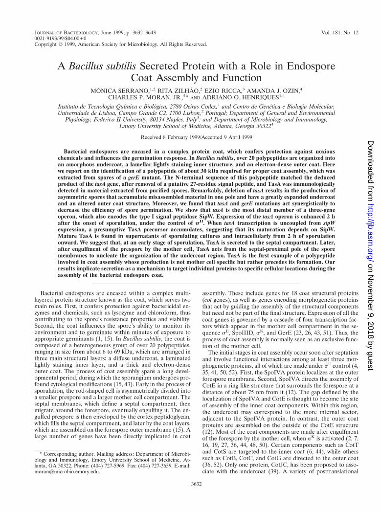
TasA is a secreted protein. The primary structure of TasA,deduced from the B. subtilis genome sequence (22), includes 27amino-terminal residues that were not found when the 30-kDacoat-associated polypeptide was subjected to Edman degrada-tion (Fig. 1B) (see above). The N-terminal extension of TasAresembles signal peptides from B. subtilis (28): it has threepositively charged residues (KKK) near its N terminus, fol-lowed by a central hydrophobic region flanked by a G residue,five positions prior to the deduced cleavage site, between twoalanines (vertical arrow in Fig. 1B). The coat-associated TasAthus results from the removal of a signal peptide-like sequencefrom the preprotein. The presence of a signal peptide and thecotranscription of tasA with sipW, which encodes a type I SPase(46), strongly suggested that maturation of pre-TasA couldinvolve SipW (9, 46). We used the anti-TasA antibody to in-vestigate the pattern of TasA accumulation throughout growthand sporulation of a gerE mutant (Fig. 7A). We found that theantiserum identified a cross-reactive component of about 30kDa, which in agreement with the transcriptional analysis (Fig.6A) was first detected at 2 h of sporulation and persisted inwhole-cell extracts until at least 6 h of sporulation (Fig. 7A,lanes 1 to 5). TasA was not detected in whole-cell extractsprepared from a sH mutant or from a gerE tasA double mutantat 2 h of sporulation (Fig. 7A, lanes 7 and 8). We infer that theantiserum raised against purified TasA recognizes the TasAprotein in whole-cell extracts, as it does in purified coat mate-rial. In addition, we found that a component with the sameelectrophoretic mobility as the species detected in whole-cellextracts and purified coat material could be found in culturesupernatants from the tasA1 strain MB24 (but not from a tasAmutant), at 2 h of sporulation (Fig. 7B). TasA antigen wasmore difficult to detect in the supernatants of gerE mutantcultures, raising the possibility that gerE controls the level ofTasA secretion (data not shown). Together with the result ofthe N-terminal sequence analysis, these observations suggestthat the 30-kDa antigen is the mature form of TasA (see alsobelow). Thus, TasA is found extracellularly concomitantly withits accumulation in sporulating cells.
SipW is involved in pre-TasA processing at the onset ofsporulation. Because the tasA operon was transcribed at lowlevels during the vegetative phase of growth (Fig. 6), we rea-soned that induction of tasA expression during growth couldlead to accumulation of the precursor protein. Plasmid pMS3,
FIG. 7. TasA production and pre-TasA processing during growth and sporulation. (A) Samples of DSM cultures of strain AH94 (gerE36) were harvested duringthe logarithmic phase of growth (lane 1), at T0 (lane 2), T2 (lane 3), T4 (lane 4), and T6 (lane 5), and whole-cell extracts were prepared. The cultures were allowed tosporulate, and coat material was then isolated from spores purified 24 h after T0 (lane 6). DSM cultures of a sH mutant (strain AH17; lane 7) or of a gerE tasA insertionalmutant (strain AH1827, lane 8) were also collected at T2. (B) Samples of supernatants of a culture of a tasA1 strain (AH94, lane 1) or of a tasA mutant (AH1700, lane2) were collected at T2, and the proteins were concentrated as described in Materials and Methods. (C) The inducer IPTG was added to a DSM culture of the Pspac-tasAstrain AH1802 at T0. Samples were collected at 30 (lane 1), 60 (lane 2), 90 (lane 3), and 120 min (lane 4) after the addition of IPTG, and whole-cell lysates wereprepared. Samples of DSM cultures of a tasA mutant (lane 5) and of a tasA1 strain (lane 6) at T2 were also analyzed. Proteins in all samples were resolved on 12.5%polyacrylamide gels containing SDS and transferred to nitrocellulose membranes. The membranes were probed with an anti-TasA antiserum. The arrows indicate thepositions of the TasA antigen. Molecular mass markers (in kilodaltons) are also indicated.
3640 SERRANO ET AL. J. BACTERIOL.
on Novem
ber 9, 2018 by guesthttp://jb.asm
.org/D
ownloaded from

carrying a Pspac-tasA fusion, was integrated into the chromo-some by a single crossover (Fig. 1A). In the resulting strainAH1802, tasA is separated from the first cistrons in the operon,whose expression remains under the control of the native pro-moter. In addition, as the result of integration of pMS3, ex-pression of a full-length copy of tasA was placed under thecontrol of the IPTG-inducible Pspac promoter. However, afteraddition of IPTG to mid-log-phase cultures of AH1802, wefailed to detect TasA in whole-cell extracts, a fact that couldindicate instability of TasA or its efficient secretion to thegrowth medium (data not shown). When IPTG was added atthe onset of sporulation, a species with an apparent molecularmass of about 35 kDa formed about 70% of the TasA antigendetected 30 min after induction (Fig. 7C, lane 1). No otherspecies is detected by the antibody in the 30- to 46-kDa range(for example, Fig. 7A). Therefore, the 35-kDa band is likely tobe a TasA precursor. One hour after induction, as expressionof the truncated yqxM sipW operon started to peak (Fig. 6A),the 35-kDa precursor was completely converted to the pro-cessed 30-kDa form (Fig. 7C, lanes 2 to 4). We suggest that, atleast at the onset of sporulation, processing of pre-TasA re-quires sipW expression and that SipW uses pre-TasA as asubstrate. We further suggest that the imbalance created be-tween TasA production and processing at the initiation ofsporulation was possible because, at the onset of sporulation,SipW may be the only SPase capable of processing pre-TasA.These results do not exclude, however, the possibility thatduring the exponential phase of growth other type I SPases cancontribute to its maturation.
The processed form of TasA that is detected in whole-cellextracts prepared from sporulating cells may be translocatedacross the cell membrane while remaining associated with thecell envelope or, as suggested by the ultrastructural analysis(see above), may be secreted to a membrane-delimited cellularcompartment, such as that formed by the asymmetric sporula-tion septum.
DISCUSSION
The 30-kDa product of the tasA gene (22, 42) is one of theproteins that can be more efficiently extracted from gerE mu-tant spores by treatment with NaOH. The TasA polypeptidecan also be extracted from wild-type spores prepared 48 h afterthe onset of sporulation by an SDS-DTT treatment. Becauseboth processes are known to solubilize spore coat polypeptides(11, 14, 16, 27, 36), we investigated the role of TasA in sporecoat assembly. Our results suggest that TasA is involved in theassembly of the coat layers. Unlike all other genes involved incoat assembly, the expression of which is confined to themother cell compartment of the sporangium (15), tasA is tran-scribed in the predivisional cell under sH control. No otherregulators of the sporulation process known to act in either theforespore or the mother cell were found to influence tasAexpression. Second, TasA is made as a secretory preprotein,and it is the processed form that is found in mature spores,implicating protein secretion in coat assembly.
tasA is the third cistron of an operon that also encodes thetype I SPase SipW (46). From the time of septation onward,the mature form of TasA is found in culture supernatants, aswell as in whole-cell extracts, and pre-TasA processing appearsto be entirely dependent upon sipW expression. Mature TasAis detected in whole-cell extracts prepared during sporulationof a tasA1 strain, presumably because the protein is secreted toand accumulates in the cellular compartment defined by thesporulation septum. In support of this model, the ultrastruc-tural analysis of the tasA mutant revealed that the spores
formed were asymmetric, accumulating misassembled materialat one spore pole. Moreover, this observation suggests that,after engulfment, TasA retains an asymmetric position. Pre-sumably, this location corresponds to the spore pole proximalto the initial site of septation. Translocation of TasA to theseptum depends upon SipW function, but SipW is unlikely tolocalize exclusively in the septal membranes, since TasA is alsodetected in the supernatant of sporulating cultures. SipW be-longs to the endoplasmic reticulum (ER)-type subfamily oftype I SPases, making B. subtilis the only known eubacteriumthat contains SPases of both the prokaryotic and ER types (9,46). We propose that SipW has properties that allow it toefficiently translocate proteins into the septal compartmentformed during sporulation, which in that respect may be ananalog of the eukaryotic ER organelle. However, SipW con-tributes to protein secretion during the vegetative growthphase (reference 46 and this work), and the tasA sipW operonis also induced postexponentially in 23 YT medium, althoughthe physiological significance of this, if any, is uncertain (thiswork).
One important inference from the present work is that in-trasporangial protein secretion is used as a mechanism to di-rect components to specific cellular locations during sporedifferentiation and that this process is somehow required forproper coat assembly and normal germination properties.TasA has sequence similarities with proteins known to pro-mote interactions between cytoskeletal elements (USO1 andActA), to promote adhesion between cells (FcrA), or to act aspattern recognition receptors (the extracytoplasmic domain ofthe human TLR) (10, 21, 29, 32). In spores of a tasA mutant,the region comprising the undercoat, defined by the foresporeouter membrane (see also Fig. 1 in reference 37) and the innercoat, is considerably expanded. In wild-type spores, this regionappears compacted and completely apposed to the inner coatstructure. Accumulation of misassembled coat-like material inthe tasA mutant seems to occur on the outside of the foresporeouter membrane. In light of its similarity to proteins implicatedin adhesion, cytoskeletal organization, or pattern recognition,it is tempting to speculate that TasA acts from the septalcompartment to promote adhesion between the cortex andundercoat structures (see also below). Albeit weak, the se-quence similarity of TasA to the C-terminal half of the ActAprotein of L. monocytogenes, a bacterial surface protein in-volved in the reorganization of the cytoskeletal elements of thehost cell (10, 21, 32), is particularly interesting. Like TasA,ActA is also made as a secretory preprotein, but it associateswith the cell via a C-terminal membrane anchor which is ab-sent in TasA. When the actA gene is expressed in eukaryoticcells, the ActA polypeptide is targeted to the mitochondrialmembrane via its C-terminal membrane anchor. The ActAprotein is then able to induce actin accumulation (as well asother cytoskeletal elements) around mitochondria, mimickingits function in the bacterial cells (32). Thus, in a parallel withour model for TasA function, ActA can act as a nucleator ofthe polymerization of actin filaments around the subcellularstructure on which it localizes (32). In tasA mutant spores, themisassembled material that accumulates at the cortex-under-coat boundary (sometimes in close contact with the inner coatstructure) appears to be asymmetrically located. WhetherTasA acts from the septal compartment (or cortical region),this observation suggests that TasA may function from onlyone pole of the spore, to nucleate the organization of theundercoat, which then can proceed all around the spore with-out further TasA participation (Fig. 8). Thus, the material thataccumulates in the mutant could correspond to a scar resultingfrom the absence of TasA function during the initial stages of
VOL. 181, 1999 ROLE OF SECRETION IN COAT ASSEMBLY 3641
on Novem
ber 9, 2018 by guesthttp://jb.asm
.org/D
ownloaded from

the process. In contrast, the rest of the undercoat region,although lacking the compaction seen in the wild type, is notinvolved in the initial nucleation stages and therefore does notdisplay the same phenotype.
Our results indicate that TasA has a role in coat assembly.Our results do not imply an association of TasA with the coatlayers, nor do they exclude an association of TasA with otherspore structures. Because the localization of TasA within thesporangium is presently unknown, we cannot decide with cer-tainty whether TasA is a true coat component. At least at anearly stage in spore maturation, most of the mature form ofTasA in sporulating cells is presumably confined to the septallumen. Thus, TasA could in principle become associated withthe cortex, which is formed in this region, or even with moreinternal spore structures (42). Its involvement in coat assemblydoes not requires TasA to be a coat component. The effectsherein described on the assembly of the coat layers can beexplained by assuming that TasA somehow (perhaps via otheras yet unknown proteins) acts from the cortical region. In thelight of the available evidence, this explanation is favored.Thus, TasA may be not a coat component but rather a proteinrequired for the normal assembly of the coat structure.
Whether or not TasA is a true coat protein, it may becross-linked in a GerE-dependent manner. The gerE locus hasalready been implicated in the cross-linking of the CotJC pro-tein, a component of the inner coat layers (14, 39), and gerE isrequired for the production of a coat-associated transglutami-nase (19, 20). The weak association of TasA with gerE mutantspores is not likely to be caused by a profound defect in thecortex structure (gerE mutant spores are heat resistant andappear to have a normal cortex) but may indicate absence ofTasA cross-linking. In contrast, our failure to extract TasAfrom wild-type spores within 24 h suggests that in these sporesTasA may be in a form that resists extraction. In support of thisidea, we note that most of the CotJC antigen can be releasedfrom wild-type spores (39) (see also above). tasA mutantspores also show alterations of the outer coat structure. Be-cause TasA can be detected in wild-type spores from 48-h
cultures, one possibility is that TasA in the culture medium canassociate with the coat layers (Fig. 8). In contrast to certainextracellular proteins that can loosely associate with the spores(45), TasA may adhere tightly to wild-type coats. Alternatively,spore-associated TasA could become more extractable as wild-type spores age (Fig. 8). In either case, coat maturation ap-pears to continue long after lysis of the mother cell and con-comitant release of the spore.
ACKNOWLEDGMENTS
We are grateful to Richard Losick for the gift of several stains andfor critical reading of the manuscript. We are also grateful to CarlaCaruso (Universita della Tuscia, Viterbo, Italy) and J. Pohl (EmoryMicrochemical Facility) for the N-terminal sequence analysis, to AdamDriks for sharing unpublished information, and to Paulo Tavares forhelpful discussions.
This work was supported by grants CNR (Progetti Finalizzati “Bio-tecnologie”) to E. Ricca, Praxis XXI/PCNA/C/BIA/129/96 to R. Zil-hao, Convenio de Cooperacao Cientıfica JNICT/CNR (Proc. 423/CNR/SCIAE) to R. Zilhao and E. Ricca, and by GM54395 from theNational Institutes of Health to C. P. Moran, Jr. M.S. and A.O.H. werethe recipients of pre- and postdoctoral fellowships from Junta Nacio-nal de Investigacao Cientıfica e Tecnologica (J.N.I.C.T.), respectively.
REFERENCES
1. Aronson, A. I., and P. Fitz-James. 1976. Structure and morphogenesis of thebacterial spore coat. Bacteriol. Rev. 40:360–402.
2. Aronson, A. I., H.-Y. Song, and N. Bourne. 1988. Gene structure and pre-cursor processing of a novel Bacillus subtilis spore coat protein. Mol. Micro-biol. 3:437–444.
3. Aronson, A. I., L. Ekanayake, and P. C. Fitz-James. 1992. Protein filamentsmay initiate the assembly of the Bacillus subtilis spore coat. Biochimie 74:661–667.
4. Beall, B., A. Driks, R. Losick, and C. P. Moran, Jr. 1993. Cloning andcharacterization of a gene required for assembly of the Bacillus subtilis sporecoat. J. Bacteriol. 175:1705–1716.
5. Benson, A. K., and W. G. Haldenwang. 1993. Regulation of sB levels andactivity in Bacillus subtilis. J. Bacteriol. 175:2347–2356.
6. Bourne, N., P. C. Fitz-James, and A. I. Aronson. 1991. Structural and ger-mination defects of Bacillus subtilis spores with altered contents of a sporecoat protein. J. Bacteriol. 173:6618–6625.
7. Cutting, S., L. Zheng, and R. Losick. 1991. Gene encoding two alkali-soluble
FIG. 8. Model for TasA function in coat assembly. The figure illustrates the various morphological stages of development in relation to TasA production. TasAproduction is initiated under sH command early in sporulation, before the asymmetric division that marks morphological stage II. In this early period, TasA is secretedto the culture medium, where its mature form accumulates. Processing is thought to rely on the SipW type I signal peptidase. After formation of the sporulation septum,the mature form of TasA accumulates in the space defined by the two septal membranes. After engulfment, TasA may localize preferentially on the septum-proximalside of the spore. From this position, TasA may nucleate the organization of the undercoat material, which then proceeds around the entire forespore. In the absenceof TasA, a scar is left at the nucleation position. Finally, after release of the spore upon lysis of the mother cell, TasA in the culture medium can associate with thespore coats. Alternatively (not represented), in old spores, the spore-associated TasA becomes more extractable.
3642 SERRANO ET AL. J. BACTERIOL.
on Novem
ber 9, 2018 by guesthttp://jb.asm
.org/D
ownloaded from

components of the spore coat from Bacillus subtilis. J. Bacteriol. 173:2915–2919.
8. Cutting, S. M., and P. B. Vander Horn. 1990. Genetic analysis, p. 27–74. InC. R. Harwood and S. M. Cutting (ed.), Molecular biology methods forBacillus. John Wiley and Sons, Ltd., Chichester, United Kingdom.
9. Dalbey, R. E., M. O. Lively, S. Bron, and J. M. van Dijl. 1997. The chemistryand enzymology of the type I signal peptidases. Protein Sci. 6:1129–1138.
10. Domann, E., J. Wehland, M. Rohde, S. Pistor, M. Hartl, W. Goebel, M.Leimeister-Wachter, M. Wuenscher, and T. Chakraborty. 1992. A novelbacterial virulence gene in Listeria monocytogenes required for host cellmicrofilament interaction with homology to the proline-rich region of vin-culin. EMBO J. 11:1981–1990.
11. Donovan, W., L. Zheng, K. Sandman, and R. Losick. 1987. Genes encodingspore coat polypeptides from Bacillus subtilis. J. Mol. Biol. 196:1–10.
12. Driks, A., S. Roels, B. Beall, C. P. Moran, Jr., and R. Losick. 1994. Subcel-lular localization of proteins involved in the assembly of the spore coat ofBacillus subtilis. Genes Dev. 8:234–244.
13. Henner, D. J. 1990. Inducible expression of regulatory genes in Bacillussubtilis. Methods Enzymol. 185:223–228.
13a.Henriques, A. O. Unpublished data.14. Henriques, A. O., B. W. Beall, K. Roland, and C. P. Moran, Jr. 1995.
Characterization of cotJ, a sE-controlled operon affecting the polypeptidecomposition of the coat of Bacillus subtilis spores. J. Bacteriol. 177:3394–3406.
15. Henriques, A. O., and C. P. Moran, Jr. Structure and assembly of thebacterial endospore coat. Submitted for publication.
16. Henriques, A. O., B. W. Beall, K. Roland, and C. P. Moran, Jr. 1997. CotMof Bacillus subtilis, a member of the a-crystallin family of stress proteins, isinduced during development and participates in spore outer coat formation.J. Bacteriol. 179:1887–1897.
17. Henriques, A. O., L. R. Melsen, and C. P. Moran, Jr. 1998. Involvement ofsuperoxide dismutase in spore coat assembly in Bacillus subtilis. J. Bacteriol.180:2285–2291.
18. Karow, M. L., and P. J. Piggot. 1995. Construction of gusA transcriptionalfusion vectors for Bacillus subtilis and their utilization for studies of sporeformation. Gene 163:69–74.
19. Kobayashi, K., S. Suzuki, K. Hashiguchi, and S. Yamanaka. 1997. Trans-glutaminase expressed in sporulating cells of Bacillus subtilis, abstr. 39. In 9thInternational Conference on Bacilli, Lausanne, Switzerland, 15 to 19 July1997.
20. Kobayashi, K., Y. Kumazawa, K. Miwa, and S. Yamanaka. 1996. ε-(g-Glutamyl)lysine cross-links of spore coat proteins and transglutaminase ac-tivity in Bacillus subtilis. FEMS Microbiol. Lett. 144:157–160.
21. Kochs, C., E. Gouin, M. Tabouret, P. Berche, H. Ohayon, and P. Cossart.1992. Listeria monocytogenes-induced actin assembly requires the actA geneproduct, a surface protein. Cell 68:521–531.
22. Kunst, F., et al. 1997. The complete genome sequence of the Gram-positivebacterium Bacillus subtilis. Nature 390:249–256.
23. Losick, R., and P. Stragier. 1992. Crisscross regulation of cell-type-specificgene expression during development in B. subtilis. Nature 355:601–604.
24. Martin-Verstraete, I., M. Debarbouille, A. Klier, and G. Rapoport. 1992.Mutagenesis of the Bacillus subtilis “-12, -24” promoter of the levanaseoperon and evidence of an upstream activating sequence. J. Mol. Biol.226:85–99.
25. Moir, A. 1981. Germination properties of a spore coat-defective mutant ofBacillus subtilis. J. Bacteriol. 146:1106–1116.
26. Moran, C. P., Jr. 1993. RNA polymerase and transcription factors, p. 653–667. In A. L. Sonenshein, J. A. Hoch, and R. Losick (ed.), Bacillus subtilisand other gram-positive bacteria: biochemistry, physiology, and moleculargenetics. American Society for Microbiology, Washington, D.C.
27. Naclerio, G., L. Baccigalupi, R. Zilhao, M. de Felice, and E. Ricca. 1996.Bacillus subtilis spore coat assembly requires cotH gene expression. J. Bac-teriol. 178:4375–4380.
28. Nagarajan, V. 1993. Protein secretion, p. 713–726. In A. L. Sonenshein, J. A.Hoch, and R. Losick (ed.), Bacillus subtilis and other gram-positive bacteria:biochemistry, physiology, and molecular genetics. American Society for Mi-crobiology, Washington, D.C.
29. Nakajima, H., A. Hirata, Y. Ogawa, T. Yonehara, K. Yoda, and M. Ya-masaki. 1991. A cytoskeleton-related gene, USO1, is required for intracel-lular protein transport in Saccharomyces cerevisiae. J. Cell Biol. 113:245–260.
30. Nicholson, W. L., and P. Setlow. 1990. Sporulation, germination and out-growth, p. 391–450. In C. R. Harwood and S. M. Cutting (ed.), Molecular
biology methods for Bacillus. John Wiley and Sons, Ltd., Chichester, UnitedKingdom.
31. Perego, M., and J. A. Hoch. 1987. Isolation and sequence of the spo0E gene:its role in initiation of sporulation in Bacillus subtilis. Mol. Microbiol. 1:125–132.
32. Pistor, S., T. Chakraborty, K. Niebuhr, E. Domann, and J. Wehland. 1994.The ActA protein of Listeria monocytogenes acts as a nucleator inducingreorganization of the actin cytoskeleton. EMBO J. 13:758–763.
33. Popham, D. L., B. Illades-Aguiar, and P. Setlow. 1995. The Bacillus subtilisdacB gene, encoding penicillin-binding protein 5*, is part of a three-geneoperon required for proper spore cortex synthesis and spore core dehydra-tion. J. Bacteriol. 177:4721–4729.
34. Ricca, E., S. Cutting, and R. Losick. 1992. Characterization of bofA, a geneinvolved in intercompartmental regulation of pro-sK processing duringsporulation in Bacillus subtilis. J. Bacteriol. 174:3177–3184.
35. Roels, S., A. Driks, and R. Losick. 1992. Characterization of spoIVA, asporulation gene involved in coat morphogenesis in Bacillus subtilis. J. Bac-teriol. 174:575–585.
36. Sacco, M., E. Ricca, R. Losick, and S. Cutting. 1995. An additional GerE-controlled gene encoding an abundant spore coat protein from Bacillussubtilis. J. Bacteriol. 177:372–377.
37. Sakae, Y., Y. Yasuda, and K. Tochikubo. 1995. Immunoelectron microscopiclocalization of one of the spore germination proteins, GerAB, in Bacillussubtilis spores. J. Bacteriol. 177:6294–6296.
38. Sandman, K., L. Kroos, S. Cutting, P. Youngman, and R. Losick. 1988.Identification of a promoter for a spore coat protein gene in Bacillus subtilisand studies on the regulation of its induction at a late stage of sporulation.J. Mol. Biol. 200:461–473.
39. Seyler, R., A. O. Henriques, A. Ozin, and C. P. Moran, Jr. 1997. Interactionsand assembly of cotJ-encoded products, constituents of the inner layers ofthe Bacillus subtilis spore coat. Mol. Microbiol. 25:955–966.
40. Steinmetz, M., and R. Richter. 1994. Plasmids designed to alter the antibioticresistance expressed by insertion mutations in Bacillus subtilis, through invivo recombination. Gene 142:79–83.
41. Stevens, C. M., R. Daniel, N. Illing, and J. Errington. 1992. Characterizationof a sporulation gene, spoIVA, involved in spore coat morphogenesis inBacillus subtilis. J. Bacteriol. 174:586–594.
42. Stover, A. G., and A. Driks. 1999. Secretion, localization, and antibacterialactivity of TasA, a Bacillus subtilis spore-associated protein. J. Bacteriol.181:1664–1672.
43. Stragier, P., and R. Losick. 1996. Molecular genetics of sporulation in Ba-cillus subtilis. Annu. Rev. Genet. 30:297–341.
44. Takamatsu, H., Y. Chikahiro, T. Kodama, H. Koide, S. Kozuka, K.Tochikubo, and K. Watabe. 1998. A spore coat protein, CotS, of Bacillussubtilis is synthesized under the regulation of sK and GerE during develop-ment and is located in the inner coat layer of spores. J. Bacteriol. 180:2968–2974.
45. Tesone, C., and A. Torriani. 1975. Protease associated with spores of Bacilluscereus. J. Bacteriol. 124:593–594.
46. Tjalsma, H., A. Bolhuis, M. L. van Roosmalen, T. Wiegert, W. Schumann,C. P. Broekhuizen, W. J. Quax, G. Venema, S. Bron, and J. M. van Dijl. 1998.Functional analysis of the secretory precursor machinery of Bacillus subtilis:identification of a eubacterial homolog of archael and eukaryotic signalpeptidases. Genes Dev. 12:2318–2331.
47. Yansura, D. G., and D. J. Henner. 1984. Use of the Escherichia coli lacrepressor and operator to control gene expression in Bacillus subtilis. Proc.Natl. Acad. Sci. USA 81:439–443.
48. Zhang, J., H. Ichikawa, R. Halberg, L. Kroos, and A. I. Aronson. 1994.Regulation of the transcription of a cluster of Bacillus subtilis spore coatgenes. J. Mol. Biol. 240:405–415.
49. Zhang, J., P. C. Fitz-James, and A. I. Aronson. 1993. Cloning and charac-terization of a cluster of genes encoding polypeptides present in the insolublefraction of the spore coat of Bacillus subtilis. J. Bacteriol. 175:3757–3766.
50. Zheng, L., and R. Losick. 1990. Cascade regulation of spore coat geneexpression in Bacillus subtilis. J. Mol. Biol. 212:645–660.
51. Zheng, L., R. Halberg, S. Roels, H. Ichikawa, L. Kroos, and R. Losick. 1992.Sporulation regulatory protein GerE from Bacillus subtilis binds to and canactivate or repress transcription from promoters for mother-cell-specificgenes. J. Mol. Biol. 226:1037–1050.
52. Zheng, L., W. Donovan, P. C. Fitz-James, and R. Losick. 1988. Gene encod-ing a morphogenic protein required in the assembly of the outer coat of theBacillus subtilis endospore. Genes Dev. 2:1047–1054.
VOL. 181, 1999 ROLE OF SECRETION IN COAT ASSEMBLY 3643
on Novem
ber 9, 2018 by guesthttp://jb.asm
.org/D
ownloaded from
![Characterization of AprE176, a Fibrinolytic Enzyme from ... · Nattokinase secreted by Bacillus subtilis natto is the most well-known example [32]. Similar fibrinolytic enzymes are](https://static.fdocuments.us/doc/165x107/60041d225ce539424f66bbc2/characterization-of-apre176-a-fibrinolytic-enzyme-from-nattokinase-secreted.jpg)