8th - University of Manitoba
120
Transcript of 8th - University of Manitoba


8th SRCA symposium, May 24-26 2017, Winnipeg
1

8th SRCA symposium, May 24-26 2017, Winnipeg
2
Welcome
Dear colleagues, friends and attendees,
On behalf of organizing committee for the Society for Research on the Cerebellum
and Ataxia (SRCA) Annual meeting, I am pleased to welcome you to the “8th International
Symposium on the Cerebellum: from Development to Disease”, held in the Rady Faculty
of Health Sciences, University of Manitoba, Winnipeg, May 24-26, 2017.
The SRCA is an international society of scientists and researchers interested in
research on the cerebellum and its associated disorders. In recent years, there has been
tremendous growth in research on cerebellar motor and non-motor functions. The
cerebellum has been shown to play a critical role in diseases ranging from ataxias, autism
spectrum disorders and cognitive operations. The SRCA offers an essential link to
improve, share and intensify this knowledge, by supporting and promoting both basic and
clinical research on the cerebellum. The society’s vision is to promote research and
education, and this symposium provides an excellent platform to fulfill this vision.
I hope you will have three very productive days of interesting and stimulating
discussions. I sincerely wish that this symposium will be a great success not only in
providing an opportunity to share knowledge and expertise in cerebellum research, but
also as the beginning of a long and fruitful cooperation and friendship among fellow
researchers, new investigators and trainees, who will shape our future.
I hope that all of you will enjoy your stay in Winnipeg, one of the “cultural cradles
of Canada,” and Manitoba’s cosmopolitan capital city.
Yours sincerely,
Hassan Marzban
Chair
8th SRCA symposium, May 24-26 2017, Winnipeg
3
Dear colleagues, friends and attendees,
As the president of the Society for Research on the Cerebellum and Ataxia (SRCA)
and on behalf of the Symposium committee members, I am very pleased to welcome you
to the «8th International Symposium on the Cerebellum: from Development to
Disease”, held in Winnipeg, Manitoba, May 24-26, 2017.
This is the 8th meeting of the of the Society for Research on Cerebellum (SRC)
since the inaugural meeting in 2008. It is also the first held under the new name of our
society the Society for Research on Cerebellum and Ataxias (SRCA). In recent years,
there has been tremendous growth in research on cerebellar motor and non-motor
functions, and the cerebellum has been shown to play a critical role in diseases ranging
from ataxias, autism spectrum disorder and cognitive operations. The new title SRCA
reflects the crucial need of interactions between basic scientists and clinicians to increase
our knowledge of cerebellar functions and interactions with other brain regions. This
meeting was organized by Drs. Hassan Marzban and Mario Manto, who along with the
scientific committee, have built a broad and exciting program. Meanwhile, the local
organizing committee has worked to provide the very best conditions for researchers and
trainees to interact, discuss their results and share their hypothesis.
The symposium will be held in the beautiful Basic Medical Sciences Building, on
the Bannatyne campus of the University in Winnipeg. This city offers many attractive
features, from Human Right Museum to Assiniboine Zoo and the famous Winnipeg Ballet,
allowing you to enjoy both excellent science and a pleasant stay.
Once again I welcome you and wish you a wonderful meeting and a nice stay in
Winnipeg
Jean Mariani, President of the SRCA

8th SRCA symposium, May 24-26 2017, Winnipeg
4
8th SRCA symposium, May 24-26 2017, Winnipeg
5

8th SRCA symposium, May 24-26 2017, Winnipeg
6
Symposium Sponsors
Health Sciences Centre Foundation
Tourism Winnipeg
City of Winnipeg
Department of Human Anatomy and Cell Science
Research Manitoba
University of Manitoba
Max Rady College of Medicine
Rady Faculty of Health Sciences
The Children’s Hospital Foundation University of Manitoba (CHRIM)
Faculty of Graduate Studies
Peter A. Cattini, Henry G. Friesen Chair in Endocrine & Metabolic Disorders

8th SRCA symposium, May 24-26 2017, Winnipeg
7
Department of Biochemistry & Medical Genetics
Department of Pharmacology and Therapeutics
St. Boniface Research Centre
Springer
The Cerebellum
8th SRCA symposium, May 24-26 2017, Winnipeg
8
SFN Cerebellum Social 2017
Tuesday, November 14th, 6:45-8:45pm, Washington D.C.
Chair: Roy V. Sillitoe, PhD, Contact: [email protected]

8th SRCA symposium, May 24-26 2017, Winnipeg
9
PROGRAM:
May 24, 2017
14:00 - 16:00 Registration and Check-in- Brodie Attrium
16:00 - 18:20 Opening Ceremony- Theatre A
16:00 - 17:00 Opening Buffet and Reception- Second Floor Concourse
17:00 - 17:10 Opening: Dr. Hassan Marzban; Chair
17:10 - 17:20 Welcoming Remarks: Dr. Brian Postl; Dean of Medicine
17:20 - 18:20 Session 1: Opening and Plenary Speaker- Theatre A
Session Chair: Dr. Dan Goldowitz
17:20 - 18:20 Dr. Richard Hawkes (CA) -- The Ferdinando Rossi Memorial Lecture: Zones and Stripes - Pattern Formation in the Cerebellar Cortex
18:20 - 21:00 Gala Evening- Brodie Attrium
May 25, 2017
08:45 - 10:25 Session 2: Neuro- and –Morphogenesis- Theatre A
Session Chair: Dr. Michisuke Yuzaki and Dr. Giacomo Consalez
08:45 - 09:10 Dr. Mikio Hoshino (JP) -- Multiple functions of Myeloid Ectopic viral Integration Site 1 homolog in cerebellar granule cell development.
09:10 - 09:35 Dr. Richard Wingate (UK) -- The development of Cerebellar Output
09:35 - 10:00 Dr. Alexandra Joyner (US) -- Cellular interactions underlying proportional scaling of cell types during cerebellar development and repair
10:00 - 10:25 Dr. Joanna Yeung (CA) -- Rhombic Lip Development, Molecular Determinant of Patterning and Cell Specification
10:25 - 10:40 Tea/Coffee- Second Floor Concourse
10:40 - 12:20 Session 3: Normal and Abnormal Differentiation- Theatre A
Session Chair: Dr. Kathleen Millen and Dr. Nori, Koibuchi
10:40 - 11:05 Dr. Azad Bonni (US) -- Epigenetic Regulation of Cerebellar Circuit Assembly and Function
11:05 - 11:30 Dr. James Li (US) -- Bergmann Glia Development, Genesis and Differentiation
11:30 - 11:55 Dr. David Solecki (US) -- Granule Cell Migration -Polarity, Link to Medulloblastoma
11:55 - 12:20 Dr. Martine Roussel (US) -- Epigenetics Drivers in Pediatric Medulloblastoma
12:20 - 13:30 Lunch- Second Floor Concourse

8th SRCA symposium, May 24-26 2017, Winnipeg
10
13:30 - 15:25 Session 4: Circuitry & Functional Development- Theatre A
Session Chair: Dr. Tim Ebner, Dr. Ray Turner and Dr. Marco Molinari
13:30 - 13:55 Dr. Izumi Sugihara (JP) -- The ansiform lobule (crus I in the rodent cerebellum) is unique in its conformation, axonal connection, striped pattern, evolution and development in the mammalian cerebellum
13:55 - 14:20 Dr. Alanna Watt (CA) -- Transient Developmental Purkinje Cell Axonal Torpedoes in Healthy and Ataxic Mouse Cerebellum
14:20 - 14:45 Dr. Karl Schilling (DE) -- Developmental Migration Of Cerebellar Basket and Stellate Cells: Do Synapses Point the Way?
14:45 - 15:10 Dr. Keiko Muguruma (JP) -- Disease modeling with patient-derived iPS cells
15:10 - 15:25 Tea/Coffee- Second Floor Concourse
15:25 - 17:05 Session 5: Aberrations of Cerebellar Development and Function: Genetics and Imaging- Theatre A
Session Chair: Dr. Jeremy D. Schmahmann and Dr. Esther Becker
15:25 - 15:50 Dr. Michael Salman (CA) -- Epidemiology of Cerebellar Diseases and Therapeutic Approaches
15:50 - 16:15 Dr. Bill Dobyns (US) -- Canary in the coal mine: the cerebellum as a sentinel for developmental brain disorders
16:15 - 16:40 Dr. Catherine Limperopoulos (US) -- Harnessing the power of advanced MRI to understand the role of early-life cerebellar injury on impaired cerebral-cerebellar function
16:40 - 17:05 Dr. Christopher Gomez (US) -- Therapeutic Interventions (SCA)
16:00 – 21:00 Students Networking-TBA
May 26, 2017
08:45 - 10:15 Session 6: Making Connections/Synaptogenesis- Theatre A
Session Chair: Dr. Masanobu Kano and Dr. Rachel Sherrard
08:45 - 09:10 Dr. Keiji Ibata (JP) -- Time-lapse Imaging of Cbln1 Release from Granule Cell Axons and its Accumulation on Purkinje Cell Dendrites
09:10 - 09:35 Dr. Naofumi Uesaka (JP) -- Roles of retrograde signaling in climbing fiber to Purkinje cell synapse elimination during postnatal cerebellar development
09:35 - 10:00 Dr. Fabrice Ango (FR) -- Synaptogenesis: Guidance Molecules in GABA to Pc synapses
10:00 - 10:15 Dr. Laurence Cathala (FR) -- Cellular mechanism of interneuron synaptic integration in developing cerebellum
10:15 - 12:15 Session 7: Posters- Brodie Attrium plus Coffee
Session Chair: Dr. Egidio D'Angelo and Dr. Ying Shen
12:15 - 13:30 plus lunch with experts- Second Floor Concourse
8th SRCA symposium, May 24-26 2017, Winnipeg
11
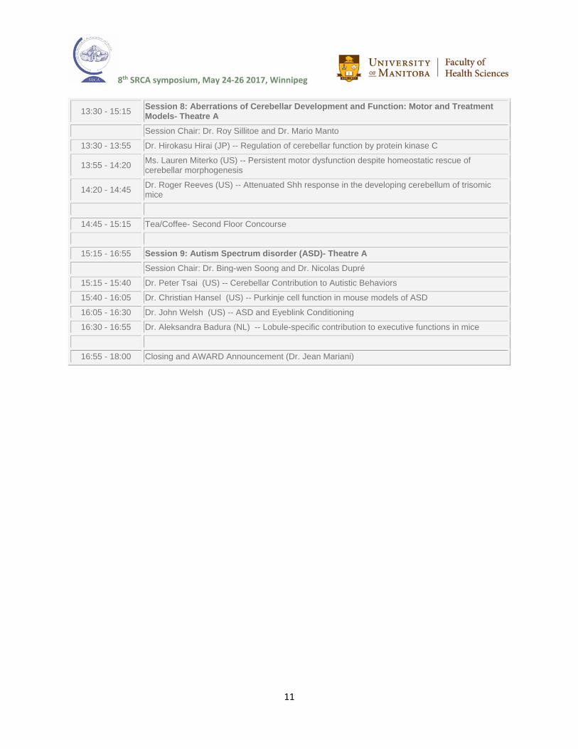
13:30 - 15:15 Session 8: Aberrations of Cerebellar Development and Function: Motor and Treatment Models- Theatre A
Session Chair: Dr. Roy Sillitoe and Dr. Mario Manto
13:30 - 13:55 Dr. Hirokasu Hirai (JP) -- Regulation of cerebellar function by protein kinase C
13:55 - 14:20 Ms. Lauren Miterko (US) -- Persistent motor dysfunction despite homeostatic rescue of cerebellar morphogenesis
14:20 - 14:45 Dr. Roger Reeves (US) -- Attenuated Shh response in the developing cerebellum of trisomic mice
14:45 - 15:15 Tea/Coffee- Second Floor Concourse
15:15 - 16:55 Session 9: Autism Spectrum disorder (ASD)- Theatre A
Session Chair: Dr. Bing-wen Soong and Dr. Nicolas Dupré
15:15 - 15:40 Dr. Peter Tsai (US) -- Cerebellar Contribution to Autistic Behaviors
15:40 - 16:05 Dr. Christian Hansel (US) -- Purkinje cell function in mouse models of ASD
16:05 - 16:30 Dr. John Welsh (US) -- ASD and Eyeblink Conditioning
16:30 - 16:55 Dr. Aleksandra Badura (NL) -- Lobule-specific contribution to executive functions in mice
16:55 - 18:00 Closing and AWARD Announcement (Dr. Jean Mariani)

8th SRCA symposium, May 24-26 2017, Winnipeg
12
Speakers
Dr. Richard Hawkes (CA)
The Ferdinando Rossi Memorial Lecture: Zones and Stripes - Pattern Formation in the Cerebellar Cortex. Richard Hawkes, Department of Cell Biology and Anatomy and Hotchkiss Brain Institute, University of Calgary, Calgary, Alberta, Canada.
The cerebellum has a complex architecture – highly reproducible and conserved through evolution. The lecture will first review the molecular patterning of the adult cerebellar cortex and then survey the processes that lead to pattern formation during embryonic development.
Cerebellar architecture is organized around the Purkinje cell. Purkinje cells in the
mouse cerebellum come in many different subtypes, identifiable by expression markers,
sensitivity to mutation etc. These are organized first into four or five “transverse zones”,
each of which is further subdivided into hundreds of reproducible “stripes”. This
arrangement serves as the scaffolding to organize afferent topography and restrict the
distribution of excitatory and inhibitory interneurons. The lecture will first review the
molecular patterning of the adult cerebellar cortex and its conservation through evolution.
Secondly, the lecture will briefly survey some of the mechanisms that lead to
pattern formation during cerebellar development. Pattern formation in the cerebellar
cortex is a multistage process that begins early in development with the generation of the
various Purkinje cell subtypes, and matures through the dispersal of Purkinje cell clusters
into stripes. Two developmental processes will be discussed in particular: the
mechanisms that lead to Purkinje cell subtype specification (i.e., how do we make
different kinds of Purkinje cells?), and the role played by Purkinje cell migration in pattern
formation (i.e., how do the Purkinje cells end up in a reproducible array of stripes?).
8th SRCA symposium, May 24-26 2017, Winnipeg
13
Dr. Mikio Hoshino (JP)
Multiple functions of Myeloid Ectopic viral Integration Site 1 homolog in cerebellar
granule cell development.
Mikio Hoshino, Department of Biochemistry and Cellular Biology, National Institute of
Neuroscience, National Center of Neurology and Psychiatry NCNP, Kodaira, Tokyo,
Japan
Myeloid Ectopic viral Integration Site 1 homolog (Meis1) is a transcription factor of
the TALE (Three Amino acid Loop Extension) protein family. Meis1 has been reported to
maintain the undifferentiated state of progenitor cells, including retinal progenitor cells,
olfactory epithelial cells, and hematopoietic stem cells etc. Although Meis1 expression in
the cerebellum, especially in the EGL (Morales and Hatten, 2006), the function of Meis1
in the granule cell (GC) development has not been clarified.
We reveal that Meis1 is required for proper cerebellar structure formation and for
Pax6 transcription in granule cell precursors (GCPs) and GCs. Meis1-Pax6 pathway
upregulates BMP signaling to induce Atoh1 degradation. However, in the outer EGL,
Meis1 binds to Atoh1 to prohibit its degradation; Meis1-Atoh1 complex upregulates Atoh1
transcription in an autoregulatory fashion. Opposing effects of Meis1 on Atoh1 expression
seem to be attributed to the Atoh1 phosphorylation status. This work reveals multiple
functions of Meis1 to coordinate GC differentiation and gives insights into understanding
neuronal development.

8th SRCA symposium, May 24-26 2017, Winnipeg
14
Dr. Richard Wingate (UK)
The development of cerebellar output
Richard Wingate, MRC Centre - Developmental Neurobiology, King's College London,
United Kingdom
A prominent feature of many cerebellar circuits are the nuclei which receive mossy
and climbing fibre inputs as well as being the target of the majority of Purkinje cell
inhibition. Nuclei send long-range connections to other brain regions that allow the
cerebellum to participate in a variety of central nervous system functions. My group has
examined the origins of nuclear projection neurons in both avian and murine models to
try to understand the factors that regulate their specification. Glutamatergic projection
neurons are derivatives of the Atoh1 positive precursors of the rhombic lip and specified
as part of a sequence of migratory populations. Each population is characterised by a
distinct set of axonal targets. By contrast, inhibitory output neurons, which project only to
the inferior olive, arise from a Sox14 positive pool of precursors that are likely derivatives
of the ventricular zone.

8th SRCA symposium, May 24-26 2017, Winnipeg
15
Dr. Alexandra Joyner (US)
Cellular interactions underlying proportional scaling of cell types during cerebellar
development and repair
Joyner, A.L.1, Wojcinski, A.1, Willet, R.1, Bayin, N.S.1 1Developmental Biology Program, Sloan Kettering Institute, New York, NY, USA
A fascinating question is how during development the number of each cell type in
the cerebellum is generated in the correct proportions (scaled) in order to produce robust
functional circuits. The Purkinje cells have been found to be a critical cell type in the
cerebellar cortex that regulates the expansion of the number of granule cell precursors,
as well as progenitors for interneurons, astrocytes and Bergmann glia produced after
birth. Sonic hedgehog (SHH) secreted by Purkinje cells is the key ligand that regulates
proliferation of the various cortical progenitors after birth. We are exploring the
interactions that occur between the deep cerebellar nuclei, which are the first neurons
born in the cerebellum, and the Purkinje cells that project to the cerebellar nuclei during
embryonic development, by analyzing the phenotypes of mouse engrailed gene (En1/2)
condition mutants. We are characterizing the defects in cerebellar growth resulting from
deletion of En1/2 only in the cerebellar nuclei projection neurons or in the granule cell
precursors, compared to in both cell types generated by the rhombic lip. In another set of
studies, we are testing the degree to which the developing cerebellum can recover from
loss of granule cells soon after birth. The signaling pathways driving normal growth and
recovery after injury have implications for normal development and disease states, as
well as for therapeutic approaches, especially given that the cerebellum is prone to injury
in premature babies, and this is a high risk factor for autism.
8th SRCA symposium, May 24-26 2017, Winnipeg
16
Dr. Joanna Yeung (CA)
Rhombic Lip Development, Molecular Determinant of Patterning and Cell
Specification
Joanna Yeung, Medical Genetics, University of British Columbia, Vancouver, BC, Canada
The cerebellar rhombic lip (RL) generates all glutamatergic neurons in the
cerebellum. We characterized a novel RL marker, Wntless (Wls), relative to its interaction
with other RL markers (Atoh1, Pax6 and Lmx1a). Using the Wls marker, four distinct
molecular compartments were identified in the developing RL. Our study of Pax6
indicates that Wls is regulated by Pax6 in the RL. Wls is aberrantly expressed in the Pax6
mutant. We also find that the cerebellar nuclear neurons and unipolar brush cells are
missing in the Pax6-null cerebellum, which indicates a novel and crucial role of Pax6 in
the development of all glutamatergic cerebellar neurons. Conversely, the examination of
Wls conditional knockout revealed that Wls regulates Pax6 expression in the RL. Wls
impacts the placement of neurons that leads to an array of ectopic neurons in the Wls-
null cerebellum. Our work demonstrates a novel molecule engaged in cerebellar
development that points to a highly dynamic molecular regulation in the RL during
development.

8th SRCA symposium, May 24-26 2017, Winnipeg
17
Dr. Azad Bonni (US)
Epigenetic Regulation of Cerebellar Circuit Assembly and Function
Azad Bonni, Department of Neuroscience, McDonnell Center for Cellular and Molecular
Neurobiology, Washington University School of Medicine, St. Louis, MO, USA
The chief goal of research in our laboratory is to identify the principles and
mechanisms that govern the assembly and function of neural circuits in the cerebellum
and determine how these mechanisms are deregulated in autism spectrum disorders. We
have discovered fundamental epigenetic, transcriptional, and ubiquitin-signaling networks
that orchestrate distinct aspects of neuronal connectivity in the mammalian cerebellar
cortex. In recent studies, we have identified crucial roles for the nucleosome remodeling
and deacetylase (NuRD) complex in the control of granule neuron parallel fiber
presynaptic differentiation as well as granule neuron dendrite pruning in the mouse
cerebellar cortex in vivo. The NuRD complex triggers long-term silencing of
developmental genes through alterations of histone tail modifications to promote parallel
fiber presynaptic differentiation. By contrast, the NuRD complex dynamically shuts off
activity-dependent gene expression via deposition of the histone variant H2A.z at
promoters of activity-dependent genes to promote granule neuron dendrite pruning and
sparse encoding to sensorimotor stimuli. These findings suggest that the NuRD complex
employs distinct mechanisms to control key aspects of neuronal connectivity in the
cerebellar cortex.

8th SRCA symposium, May 24-26 2017, Winnipeg
18
Dr. James Li (US)
Bergmann Glia Development, Genesis and Differentiation
James Li ,Department of Genetics and Genome Sciences, UConn Health, CT, USA
Folding of the cortex and persistence of radial glia-like cells called Bergmann glia
(BG) are hallmarks of the mammalian cerebellum. Similar to basal radial glia in the
primate neocortex, BG maintain basal processes and molecular features of neural
progenitors. The generation of BG and their role in cerebellar foliation are not well
understood. We have performed mouse genetic experiments, RNA-sequencing, and co-
expression network analyses to study the developmental programs underlying BG
formation. We found that heightened FGF-ERK signaling activity was linked to the timely
transition of radial glia in the cerebellar ventricular zone to BG. Inhibition of FGF-ERK
signaling by deleting Shp2 blocked generation of BG, as well as cerebellar foliation.
Restoring ERK or Etv5 function rescued BG formation in the absence of Shp2. Our results
demonstrate that an FGF-ERK-ETV axis is crucial to BG induction. Furthermore, we
reveal a crucial function of BG in organizing cerebellar foliation.

8th SRCA symposium, May 24-26 2017, Winnipeg
19
Dr. David Solecki (US)
Granule Cell Migration -Polarity, Link to Medulloblastoma
David Solecki, Developmental Neurobiology Department, St. Jude Children's Research
Hospital, TN, USA
Cell polarity is a driving force that coordinates the choreography of neural
development. How polarity signaling organizes the behavior of immature neurons and
how polarity signaling cascades are regulated remain key questions facing the field of
developmental neurobiology. These questions are critical to understand the pathology of
neurodevelopmental diseases, where the production of neurons or their subsequent
migration is defective. Studies combining necessity-sufficiency testing and cutting edge
imaging technology in the developing cerebellum show that a conserved polarity-signaling
module, called the Pard complex, is essential for neuronal progenitor germinal zone exit
by regulating cytoskeletal dynamics and cell-cell interactions needed for neuronal
migration. I will present our progress identifying an upstream regulator of the Pard
complex: an E3 ubiquitin ligase, Seven in Absentia, which mediates proteosomal
degradation of Pard3; to control a shift from tangential to radial migration when cerebellar
granule neurons leave their mitogenic niche, and drebrin; to control microtubule-actin
crosslinking during CGN differentiation.
8th SRCA symposium, May 24-26 2017, Winnipeg
20
Dr. Martine Roussel (US)
Epigenetic Drivers in Pediatric Medulloblastoma
Martine Roussel, Department of Molecular Sciences, St. Jude Children's Research
Hospital, TN, USA
-Medulloblastoma (MB), an embryonal cerebellar tumor, comprises four distinct
subgroups: Sonic Hedgehog (SHH), Wingless (WNT), Group3 (G3) and Group4 (G4).
MBs have a paucity of mutations with genetic alterations in oncogenes and tumor
suppressors (β-CATENIN, PATCHED, SUFU, GLI2, MYC and MYCN) account for
only 20-30% of cases. Next generation sequencing revealed somatic altered genes,
many of which involved in epigenetic regulators and chromatin modification.
Studies by the Washington University/ St. Jude Pediatric Cancer Genome Project
showed that mouse and human G3 MBs express increased levels of EZH2, the
catalytic partner of the polycomb repressor complex PRC2, and of histone 3 lysine
27 trimethylation (H3K27me3). Deletion of EZH2 or SUZ12 in G3 MB via TALEN and
CRISPR-Cas9 gene editing approaches revealed that the PRC2 complex has tumor
suppressive functions in G3 MB, via in part the suppression of GFI1, a transcriptional
repressor overexpressed in a subset of G3 MBs.

8th SRCA symposium, May 24-26 2017, Winnipeg
21
Dr. Izumi Sugihara (JP)
The ansiform lobule (crus I in the rodent cerebellum) is unique in its conformation,
axonal connection, striped pattern, evolution and development in the mammalian
cerebellum
Izumi Sugihara, Dept. Systems Neurophysiol Tokyo Medical and Dental Univ., Tokyo,
Japan
In the human cerebellum, crus I and crus II lobules (or ansiform lobule), which are
implicated in cognitive and visuomotor functions, are significantly expanded compared to
anterior and posterior lobules, which are involved in somatosensorimotor function. In
applying rodent models, it is essential to identify the lobules that are homologous to
human crus I and crus II. Observation of the lobular structure in human, macaque,
marmoset, rat, and mouse indicated that the human crus I/II were homologous to
crus I in rodents (referred to as “ansiform area, AA”). Our lobular definition was
supported by lobule-based mapping of the olivocerebellar climbing fiber and Purkinje cell
(PC) projection patterns in rodents; Crus II and simple lobule was innervated by the
mediocaudal part of each inferior olive subnucleus and project to the dorsal part of
the cerebellar nuclei, while crus I (or the AA) was innervated by the rostrolateral part
of each inferior olive subnucleus and project to the ventral part of the cerebellar nuclei.
A gap in the cortical structure was observed in the paravermal area of the AA in both
rodents and primates. Concerning zebrin stripes, the central lobules (lobules VI-VII, and
AA or crus I in rodents) show a laterally-expanded arrangement solely of positive stripes.
Our analysis showed that this arrangement of zebrin-positive stripes in the AA originated
from their developmental process. Between E14.5 and E17.5, lateral protrusion and shift
were observed in the domains of protocadherin 10-positive PC subsets (which would
become zebrin-positive later) in the central area of the immature cerebellum that would
eventually become lobules VI-VII and AA or crus I. The results indicate that the AA (or
crus I in rodents) is characterized by distinct connectivity from neighboring lobules and a
massive expansion in skillful primates.
8th SRCA symposium, May 24-26 2017, Winnipeg
22
Dr. Alanna Watt (CA)
Transient Developmental Purkinje Cell Axonal Torpedoes in Healthy and Ataxic
Mouse Cerebellum
Alanna Watt, Department of Biology, McGill University, Montreal, Quebec, CA
Information is carried out of the cerebellar cortical microcircuit via action potentials
propagated along Purkinje cell axons. In several human neurodegenerative diseases,
focal axonal swellings on Purkinje cells – known as torpedoes – have been associated
with Purkinje cell loss. Interestingly, torpedoes are also reported to appear transiently
during development in rat cerebellum. The function of Purkinje cell axonal torpedoes in
health as well as in disease is poorly understood. We are investigating the properties of
developmental torpedoes in the postnatal mouse cerebellum of wild-type and transgenic
mice. Our findings to date suggest that the transient emergence of Purkinje cell axonal
torpedoes during the second postnatal week in mice represents a normal morphological
feature in the developing cerebellar microcircuit.
8th SRCA symposium, May 24-26 2017, Winnipeg
23
Dr. Karl Schilling (DE)
Developmental migration of cerebellar basket and stellate cells: do synapses
point the way?
Karl Schilling Anatomisches Institut, Anatomie & Zellbiologie Universität Bonn, Bonn,
Germany. [email protected]
In the developing cerebellum, molecular layer inhibitory interneurons are among
the last nerve cells to reach their adult positions and to differentiate. They originate from
the ventricular epithelium lining the fourth ventricle, and their distribution to and within the
cerebellar cortex requires extensive migration through dynamically changing cellular
environments, from the nascent white matter through the immature granule cell layer, and
finally within the emerging molecular layer. The mode of migration of these cells, the cues
they use to navigate, and the mechanisms to interpret such cues remain largely elusive.
I will summarize some recent data that allows us to describe and to quantify how
molecular layer interneurons navigate through the nascent cerebellar cortex. Further, I
will present data that show that these cells are synaptically innervated much earlier than
hitherto thought, and in fact while still in transit. This developmental vesicular transmitter
release is part of the machinery that ensures proper migration and navigation of these
cells. Beyond the implications for our understanding of cerebellar histogenesis, these
findings suggest a novel mechanism how functional activity of early maturing nerve cells
may tune the cellular composition and functional properties of emergent nerve cell
networks. These findings also define a hitherto unknown role for early synapses as
activity-tunable guideposts for neural migration.
8th SRCA symposium, May 24-26 2017, Winnipeg
24
Dr. Keiko Muguruma (JP)
Disease modeling with patient-derived iPS cells
Keiko Muguruma, RIKEN, Center for Developmental Biology (CDB), Laboratory for Cell
Asymmetry, 2-2-3 Minatojima-minamimachi, Chuo, Kobe, Japan
Recent advances in the techniques that differentiate induced pluripotent stem cells
(iPSCs) into specific types of cells enabled us to establish in vitro disease models from
the patient-derived iPSCs. The advantage of the model utilizing disease-specific iPSC
is that it is able to generate a large number of cells required for high-throughput
screening. The patient-derived iPSCs are expected to recapitulate the disease-specific
pathogenesis and physiology in vitro. We developed 3D culture systems using human
pluripotent stem cells, which would promote the research on the construction of complex
brain regions. Recently we have succeeded in generation of spinocerebellar ataxia
(SCA), SCA6 and SCA42, patient-derived Purkinje cells by combining the iPSC
technology and the self-organizing stem cell 3D culture technology. We have constructed
an in vitro disease model recapitulating both ontogenesis and pathogenesis for SCA.
Here we will talk about approaches for intractable diseases utilizing patient-specific
iPSCs.
8th SRCA symposium, May 24-26 2017, Winnipeg
25
Dr. Michael Salman (CA)
Epidemiology of cerebellar diseases and therapeutic approaches
Michael Salman, Department of Pediatrics and Child Health, University of Manitoba,
Winnipeg, MB, Canada
Cerebellar diseases occur relatively commonly in children and adults around the
globe. Many factors influence their epidemiology including geography, ethnicity,
consanguinity, and the methodology used to ascertain patients. In addition, reliable
epidemiological data relies heavily on accurate disease classification. The continuous
advances in genetics and neuroimaging modalities have resulted in improved
understanding of cerebellar diseases and have led to several revisions in their
classification. Recent global epidemiological studies on ataxia reported an estimated
overall prevalence rate of 26/ 100,000 in children, a prevalence rate of dominant
hereditary cerebellar ataxia of 2.7/ 100,000, and a prevalence rate of recessive hereditary
cerebellar ataxia of 3.3/ 100,000. The management of cerebellar diseases is
multidisciplinary and multimodal. General supportive and symptomatic therapies should
be initiated. Genetic counselling should be offered, where appropriate. Few drugs,
specific motor rehabilitation programs, and non-invasive cerebellar stimulation for the
treatment of ataxia have been developed and seem to show early promise but more
studies are needed to replicate and fine-tune their benefits further. Some disease-specific
treatments are available. For example, acetazolamide or 4-aminopyridine for patients with
episodic ataxia type 2 and vitamin E for patients with vitamin E deficiency.

8th SRCA symposium, May 24-26 2017, Winnipeg
26
Dr. Bill Dobyns (US)
Canary in the coal mine: the cerebellum as a sentinel for developmental brain
disorders
Dobyns WB1,2; 1Center for Integrative Brain Research, Seattle Children’s Research
Institute, Seattle, WA, USA, 2Department of Pediatrics, University of Washington, Seattle,
WA, USA
The cerebellum is often overlooked in assessing fetuses and children with
developmental brain disorders because of multiple patterns of malformation that are
inconsistently defined, lack of experience in recognizing these patterns, variable severity
including non-penetrance, occasional co-occurrence of atrophy, and limited
understanding of the underlying causes. After excluding two well-known groups of
autosomal recessive disorders with recognizable patterns of malformation (Joubert
syndrome and pontocerebellar hypoplasia), cerebellar malformations have been
consistently observed with only two copy number variants (deletion 3q24 or 6p25.3) and
a few genes (CASK, OPHN1 and FOXC1). Recent experience has shown that prenatal
events such as late 2nd-early 3rd trimester posterior fossa and cerebellar bleeds and
(less often) cerebellar ischemia, can cause cerebellar injuries that mimic cerebellar
malformations. Accordingly, genetic studies have shown a lower rate of abnormalities
than other developmental disorders such as agenesis of the corpus callosum, intellectual
disability and autism.
We have performed SNP microarrays in ~250 children and whole exome
sequencing data in ~100 children (and parents) with cerebellar malformations. Our
analysis suggests that cerebellar hypoplasia is a variable feature in many genetic
developmental brain disorders, that prenatal injuries to the cerebellum are common and
can often be recognized based on the pattern of abnormality, and that these two
processes may co-occur.

8th SRCA symposium, May 24-26 2017, Winnipeg
27
Dr. Catherine Limperopoulos (US)
Harnessing the power of advanced MRI to understand the role of early-life
cerebellar injury on impaired cerebral-cerebellar function.
Catherine Limperopoulos, PhD
Director, the Developing Brain Research Program Vice Chief of Research, Division of
Diagnostic Imaging and Radiology
Co-Director of Research, Division of Neonatology Children’s National Health System
Associate Professor of Neurology, Radiology, and Pediatrics George Washington
University School of Medicine and Health Sciences
Cerebellar development follows a highly orchestrated and complex program of
critical developmental processes. Consequently, this vulnerable developmental period
of the cerebellum can be derailed by a host of potential insults. Recently, we have applied
quantitative MRI (qMRI) tools to study the developmental trajectory of the human
cerebellum in utero. Using qMRI we have demonstrated that the cerebellum undergoes
its most rapid growth that is unmatched by any other cerebral structure over the third
trimester. However, this accelerated growth is impeded by premature birth where many
of these complex cerebellar development events take place within the hazards of a hostile
extrauterine environment. We will review the role of both direct and indirect cerebellar
injury on cerebral development and relate these disturbances in cerebellar development
to a prevalent and distinct profile of cognitive, language and social-behavioral dysfunction
including autism spectrum disorders. Finally, we will explore the emerging functional
topography of the immature cerebellum and its relationship to long-term
neurodevelopmental disabilities.

8th SRCA symposium, May 24-26 2017, Winnipeg
28
Dr. Christopher Gomez (US)
Therapeutic Interventions (SCA)
Department of Neurology, The University of Chicago, Chicago IL, USA
We have discovered that the P/Q-type voltage-gated Ca2+ channel (VGCC) gene,
CACNA1A, is a bicistronic cellular gene, i.e, encodes two structurally unrelated proteins,
with distinct functions, that are separately encoded within the same mRNA. CACNA1A
encodes both the α1A (Cav2.1) subunit and a newly recognized transcription factor,
α1ACT, within an overlapping open reading frame (ORF) within the same mRNA
transcript. This is achieved by the presence of a novel internal ribosomal entry site (IRES)
upstream of a second ORF encoding α1ACT, which, when mutated, mediates the
disease, spinocerebellar ataxia type 6 (SCA6). The IRES controlling α1ACT is an
excellent drugable target and we have used this strategy to suppress an acute form of
SCA6. However, this approach suppresses both the normal and expanded α1ACT.
Therefore, we have used a doxycycline suppressible wild type α1ACT transgene to
demonstrate its critical role in early Purkinje cell maturation, whose elimination in adults
does not have adverse effects on cerebellar cortex function.

8th SRCA symposium, May 24-26 2017, Winnipeg
29
Dr. Keiji Ibata (JP)
Time-lapse imaging of Cbln1 release from granule cell axons and its accumulation
on Purkinje cell dendrites
Ibata K1, Yuzaki M1; 1Department of Physiology, Keio University School of Medicine
Cbln1 belongs to the C1q/TNF superfamily, whose members are involved in
various intercellular signaling in multiple systems. Cbln1 is released from cerebellar
granule cells and plays an essential role in formation and maintenance of synapses
between parallel fibers and Purkinje cells (Matsuda et al, Science 2010). Cbln1 forms a
tripartite complex across the synapse by binding to its pre- and postsynaptic receptors,
neurexin (Nrx) and the delta2 glutamate receptor (GluD2). Nevertheless, a fundamental
question how and where Cbln1 is released from granule cells has remained unclear. To
perform time-lapse imaging of Cbln1 release, we expressed Cbln1 tagged with pH-
sensitive GFP (super-ecliptic-pHluorin; SEP) in cultured granule cells. Electrical field
stimulation rapidly increased SEP-Cbln1 fluorescence signals from mostly non-synaptic
sites, which were negative for presynaptic markers. Upon encountering with HEK293 cells
expressing GluD2, surface SEP-Cbln1 signals along granule cell axons started to
accumulate at the contact site in the absence of electrical field stimulation. From these
results, we propose that Cbln1, secreted from extrasynaptic sites in an activity-dependent
manner, accumulates on the region where axons make contact with GluD2 on dendritic
spines of Purkinje cells in an activity-independent manner.

8th SRCA symposium, May 24-26 2017, Winnipeg
30
Dr. Naofumi Uesaka (JP)
Roles of retrograde signaling in climbing fiber to Purkinje cell synapse
elimination during postnatal cerebellar development
Uesaka N1, Abe M2, Yamazaki M2, Konno K3, Mikuni T1, Watanabe M3, Sakimura
K2and Kano M1 1The University of Tokyo, Tokyo, Japan 2Niigata University, Niigata, Japan 3Hokkaido University, Sapporo, Japan
Presenting author’s e-mail address: [email protected]
Purkinje cells (PCs) in the neonatal cerebellum are innervated by multiple climbing
fibers (CFs). During postnatal development, a single CF is selectively strengthened in
each PC and becomes a ‘winner’ CF that is presumed to remain into adulthood, whereas
the other ‘loser’ CFs are eliminated. Our recent studies have uncovered molecular
mechanisms by which postsynaptic PCs regulate CF synapse elimination. We have
demonstrated that Semaphorin7A and Semaphorin3A mediate retrograde signals from
postsynaptic PCs to presynaptic CFs and regulate elimination and maintenance
respectively of CF synapses. We further screened candidate molecules that may mediate
retrograde signaling for strengthening or weakening of CF synapses. We found that PC-
specific deletion of progranulin, a growth factor implicated in the pathogenesis of
frontotemporal dementia, and knockdown of Sort1, a receptor of progranulin, in CFs
accelerated elimination of redundant CFs and reduced the amplitude of synaptic inputs
from winner CFs. These results suggest that progranulin derived from PCs retrogradely
acts on Sort1 in CFs, strengthens/maintains CF synapses.
8th SRCA symposium, May 24-26 2017, Winnipeg
31
Dr. Fabrice Ango (FR)
Synaptogenesis: Guidance Molecules in GABA to Pc synapses
Fabrice Ango, IGF/Neurobiology, Université de Montpellier, France
One of the remarkable features of neuronal circuits is the specificity and precision
of the synaptic connections during development. These highly specific patterns of
connections between different populations of neurons are crucial for brain function, and
require an intricate coordination of various developmental events. However, little is known
about how growing axons select correct post-synaptic partner at the cellular and
subcellular level within multiple heterogeneous targets they encounter. This is well
exemplified by GABAergic interneurons, which innervated specific cell types at precise
subcellular location (i.e. dendrites, cell soma or axon initial segment (AIS)). Our recent
studies suggest that the axon guidance receptor Neuropilin-1 expressed by GABAergic
interneurons orchestrated both guidance and subcellular synapse targeting through
timely interactions with local cues. Coordination of both guidance and recognition by the
same molecular cue might alleviate some of the coding power for synapse specificity.

8th SRCA symposium, May 24-26 2017, Winnipeg
32
Dr. Laurence Cathala (FR)
Cellular mechanism of interneuron synaptic integration in developing cerebellum
Laurence Cathala; Adaptation Biologique et Vieillissement, CNRS UMR 8256, Case 14,
Université Pierre et Marie Curie – P6, Sorbonne Universités, 9 Quai St Bernard, 75005
Paris, France. [email protected]
Interneurons are critical for neuronal circuit function, but how their dendritic
morphologies and membrane properties influence information flow within neuronal
circuits is not well understood, let alone how these properties change with development
and allow networks to acquire and refine their functional properties. We have addressed
this question by studying synaptic integration of excitatory inputs onto stellate cells,
molecular layer interneurons, in the immature cerebellum. With a multidisciplinary
approach combining electrophysiological recording in brain slices, morphological analysis
and neuronal simulation, we found that information processing from immature stellate
cells differs from what we previously described in the adult stellate cell (Abrahamsson et
al., 2012). This developmental change in the integration of excitatory synaptic inputs
results predominantly from a difference in synapse location and quantal size. These
alterations are likely to lead to a shift in the subthreshold input-output transformations
from more linear relationship in young SCs to a more sublinear relationship in older SCs.

8th SRCA symposium, May 24-26 2017, Winnipeg
33
Dr. Hirokazu Hirai (JP)
Regulation of cerebellar function by protein kinase C
Hirokazu Hirai, Department of Neurophysiology & Neural Repair, Gunma University
Graduate School of Medicine
Prof. Yasutomi Nishizuka discovered protein kinase C (PKC), a family of serine-
and threonine-specific protein kinases, which regulates myriad of physiological functions.
I learned biochemistry including PKC from Prof. Nishizuka in Kobe University just around
the time he and his lab members were actively studying the mechanism of PKC activation
following membrane lipid hydrolysis. The PKC that Prof. Nishizuka originally discovered
is categorized in classical (conventional) PKCs. Classical PKCs (PKCα, PKCβI/βII and
PKCγ) are activated by calcium and second messenger diacylglycerol, in which PKCγ is
expressed exclusively in neurons of the brain and spinal cord. Cerebellar Purkinje cell
(PC) expresses PKCα and PKCγ. PKCα is indispensable for the expression of cerebellar
long-term depression (LTD) at parallel fiber to PC synapses because PKCα can bind to
PICK1 via the PDZ domain-binding motif, accesses to and phosphorylates the C-terminal
domain of GluA2, eventually leading to clathrin-mediated endocytosis of postsynaptic
AMPA receptors. Systemic PKCγ-knockout mice have been shown to exhibit deficient
pruning of climbing fibers (CFs) from developing PCs and mild motor coordination deficit,
suggesting a critical role in elimination of CF synapses from PCs during development.
There are currently, at least, 2 open questions. First one is why PKCα cannot substitute
for PKCγ to eliminate surplus CFs from developing PKCγ-null PCs. Second one is what
role PKCγ plays in after maturation of the cerebellum, which has remained totally
unknown. We challenged to resolve these 2 questions.
8th SRCA symposium, May 24-26 2017, Winnipeg
34
Ms. Lauren Miterko (US)
Persistent motor dysfunction despite homeostatic rescue of cerebellar
morphogenesis
Miterko LN and Sillitoe RV; Pathology & Immunology, Neuroscience, Developmental
Biology, Baylor College of Medicine, Duncan Neurological Research Institute, Houston
Texas 77030, USA
Purkinje cells play a central role in establishing the cerebellar circuit. It is well
known that disrupting Purkinje cell development impairs cerebellar morphogenesis and
motor function. Surprisingly, in the Car8wdl mouse model of hereditary cerebellar ataxia,
severe motor deficits arise despite the cerebellum developing to its correct size and
morphology. We revealed that loss of the Purkinje cell protein called CAR8 (Carbonic
anhydrase 8), a regulator of IP3R1 Ca2+ signaling, delays cerebellar morphogenesis by
transiently restricting growth. The mechanism involved a reduction of granule cell
proliferation as observed in postnatal day (P) 5 mutants, although by P15 proliferation
was maintained at a higher level compared to controls. The prolonged period of granule
cell proliferation was accompanied by a restructured assembly of Purkinje cell and
Bergmann glia architecture, which both coordinate granule cell migration. We next used
in vivo electrophysiology, EMG, and behavior to show that the onset of motor dysfunction
occurs by P20. Together, our findings indicate that CAR8 mediates Purkinje cell-granule
cell communication during cerebellar growth, and its loss triggers an inter-cellular
compensatory response to rescue structure, but not motor function. These data raised
the possibility of using the Car8wdl model to test whether therapeutic targeting of an adult
circuit can be used to correct a developmentally derived behavioral deficit. Towards this,
we devised a cerebellar nuclei deep brain stimulation (DBS) approach to correct
movement in mice. After four days of DBS treatment, Car8wdl mutant mice showed
sustained increases in motor performance and learning. Given these data, we propose
that cerebellar DBS could be a promising therapy for non-degenerative cerebellar ataxias
and in addition Car8wdl mice may provide a critical new opportunity to finally solve the
cellular mechanism(s) for how DBS works in vivo.
8th SRCA symposium, May 24-26 2017, Winnipeg
35
Dr. Roger Reeves (US)
Attenuated Shh response in the developing cerebellum of trisomic mice
Roger H. Reeves, Johns Hopkins Univ. Schl. Med., Baltimore, MD USA
Trisomy 21 results in Down syndrome which presents as a constellation of features
including a significantly reduced volume and cellularity of the cerebellum. This reduction
also occurs in mouse models of Down syndrome such as Ts65Dn, which is trisomic for
orthologs of more than half of the genes conserved with human chromosome 21 (Hsa21).
We traced the basis of the defect to the proliferation of granule cell neuron precursors
(gcp) in the days after birth. This reduction could be attributed substantially to an
attenuated gcp response to the mitogenic effects of Shh growth factor. A single injection
on the day of birth of the Shh pathway agonist, SAG, normalized cerebellar structure
through life and produced a surprising improvement in hippocampal function in behavior
tests and a normalization of LTP, an electrophysiological correlate of hippocampal
learning. We are pursuing three basic lines of research, one defining the brain regions
where acute Shh pathway stimulation around birth can improve synaptic function in
hippocampus throughout life, a second characterizing anatomical changes in tracts from
deep cerebellar nuclei throughout the brain, and a third seeking to identify the trisomic
gene or genes that are responsible for the attenuated response to Shh.
8th SRCA symposium, May 24-26 2017, Winnipeg
36
Dr. Peter Tsai (US)
Cerebellar Contribution to Autistic Behaviors
Peter Tsai, Department of Neurology and Neurotherapeutics, Neuroscience, Pediatrics,
Psychiatry, Center for Autism and Developmental Disabilities, University of Texas
Southwestern, TX, USA
The cerebellum has been implicated in the pathogenesis of multiple
neuropsychiatric disorders. In particular, cerebellar abnormalities and pathology has been
identified in many studies of autism while cerebellar disorders predispose to significantly
increased rates of autism. We have recently demonstrated that cerebellar dysfunction is
sufficient to generate autistic behaviors. We will discuss our recent studies investigating
sensitive periods of treatment and mechanisms by which the cerebellum regulates these
behaviors.
8th SRCA symposium, May 24-26 2017, Winnipeg
37
Dr. Christian Hansel (US)
Purkinje cell function in mouse models of ASD
Christian Hansel (University of Chicago), USA
Autism Spectrum Disorder (ASD) is characterized by deficits in social interaction
and by repetitive behaviors, and is often accompanied by motor impairment. In a mouse
model for the human 15q11-13 duplication (Dup15q syndrome), which often presents
itself with autism, delayed motor milestones and seizures, we have found abnormal
synaptic function and plasticity at cerebellar synapses as well as impaired eyeblink
conditioning (EBC), a form of cerebellum-dependent motor learning that is also affected
in ASD patients. We currently perform studies to determine the cause of motor deficits in
Dup15q syndrome / ASD, and to characterize synaptic and behavioral phenotypes in
CYFIP1 overexpressing mice to assess whether increased dosage of this candidate gene
causes ASD-typical motor and non-motor phenotypes / symptoms in Dup15q syndrome.
8th SRCA symposium, May 24-26 2017, Winnipeg
38
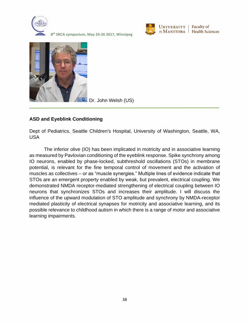
Dr. John Welsh (US)
ASD and Eyeblink Conditioning
Dept of Pediatrics, Seattle Children's Hospital, University of Washington, Seattle, WA,
USA
The inferior olive (IO) has been implicated in motricity and in associative learning
as measured by Pavlovian conditioning of the eyeblink response. Spike synchrony among
IO neurons, enabled by phase-locked, subthreshold oscillations (STOs) in membrane
potential, is relevant for the fine temporal control of movement and the activation of
muscles as collectives – or as “muscle synergies.” Multiple lines of evidence indicate that
STOs are an emergent property enabled by weak, but prevalent, electrical coupling. We
demonstrated NMDA receptor-mediated strengthening of electrical coupling between IO
neurons that synchronizes STOs and increases their amplitude. I will discuss the
influence of the upward modulation of STO amplitude and synchrony by NMDA-receptor
mediated plasticity of electrical synapses for motricity and associative learning, and its
possible relevance to childhood autism in which there is a range of motor and associative
learning impairments.
8th SRCA symposium, May 24-26 2017, Winnipeg
39
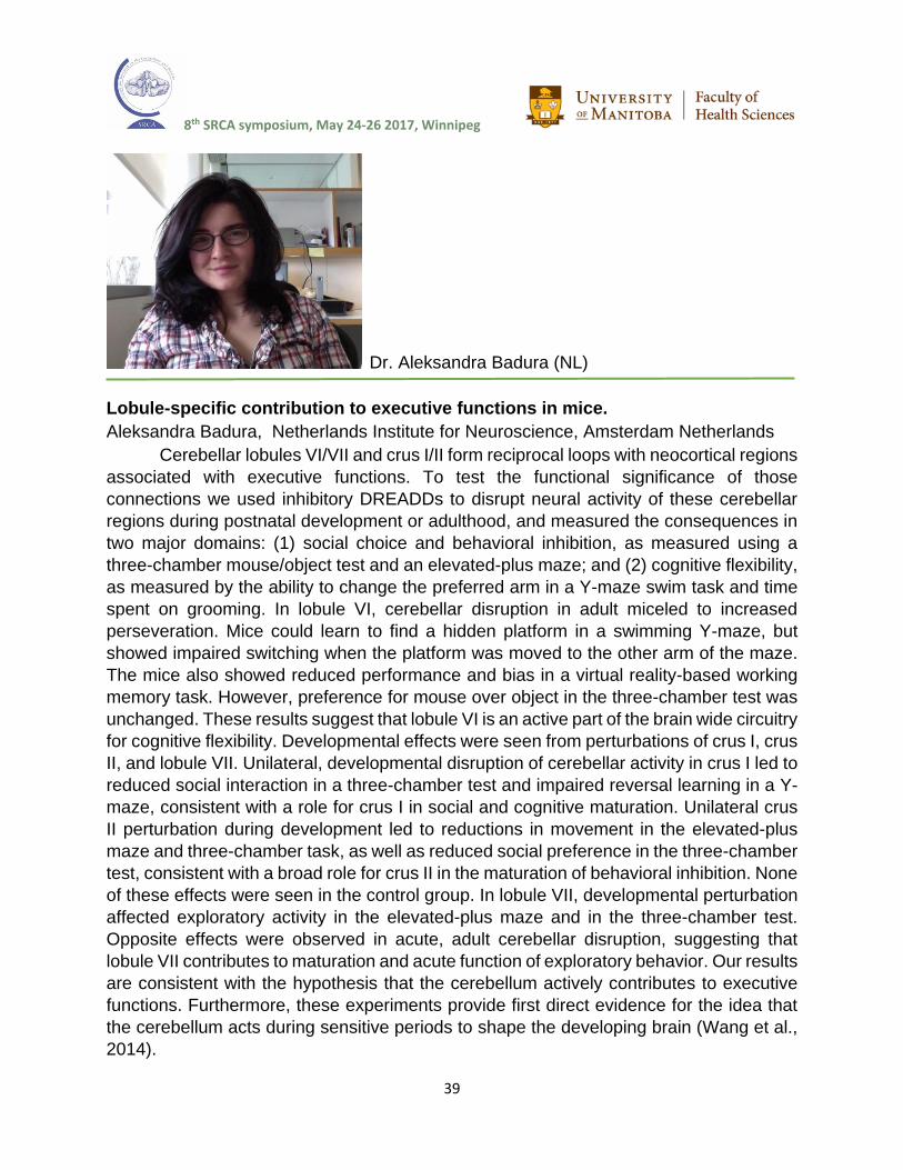
Dr. Aleksandra Badura (NL)
Lobule-specific contribution to executive functions in mice.
Aleksandra Badura, Netherlands Institute for Neuroscience, Amsterdam Netherlands
Cerebellar lobules VI/VII and crus I/II form reciprocal loops with neocortical regions
associated with executive functions. To test the functional significance of those
connections we used inhibitory DREADDs to disrupt neural activity of these cerebellar
regions during postnatal development or adulthood, and measured the consequences in
two major domains: (1) social choice and behavioral inhibition, as measured using a
three-chamber mouse/object test and an elevated-plus maze; and (2) cognitive flexibility,
as measured by the ability to change the preferred arm in a Y-maze swim task and time
spent on grooming. In lobule VI, cerebellar disruption in adult miceled to increased
perseveration. Mice could learn to find a hidden platform in a swimming Y-maze, but
showed impaired switching when the platform was moved to the other arm of the maze.
The mice also showed reduced performance and bias in a virtual reality-based working
memory task. However, preference for mouse over object in the three-chamber test was
unchanged. These results suggest that lobule VI is an active part of the brain wide circuitry
for cognitive flexibility. Developmental effects were seen from perturbations of crus I, crus
II, and lobule VII. Unilateral, developmental disruption of cerebellar activity in crus I led to
reduced social interaction in a three-chamber test and impaired reversal learning in a Y-
maze, consistent with a role for crus I in social and cognitive maturation. Unilateral crus
II perturbation during development led to reductions in movement in the elevated-plus
maze and three-chamber task, as well as reduced social preference in the three-chamber
test, consistent with a broad role for crus II in the maturation of behavioral inhibition. None
of these effects were seen in the control group. In lobule VII, developmental perturbation
affected exploratory activity in the elevated-plus maze and in the three-chamber test.
Opposite effects were observed in acute, adult cerebellar disruption, suggesting that
lobule VII contributes to maturation and acute function of exploratory behavior. Our results
are consistent with the hypothesis that the cerebellum actively contributes to executive
functions. Furthermore, these experiments provide first direct evidence for the idea that
the cerebellum acts during sensitive periods to shape the developing brain (Wang et al.,
2014).
8th SRCA symposium, May 24-26 2017, Winnipeg
40
Poster Number/Presenter/Title
Theme 1- Cerebellar Neuro- and Morphogenesis.
P1 / Mrs. Maryam Rahimi-Balaei/ The Role of a Novel Subset of Mesencephalic Neural Crest
Derived Cells in Cerebellar Nuclei Development in Mice
P2 / Miss. Tsz Ching Ma/ Canonical BMP signaling is required to maintain neural stem cells at
cerebellar ventricular zone.
P3 / Prof. Annalisa Buffo/ Multiple origins and spatiotemporal emergence of cerebellar astrocyte
heterogeneity.
P4 / Dr. Filippo Casoni/ Zfp423, a Joubert syndrome gene, is a domain-specific regulator of cell
cycle progression, DNA damage response and Purkinje cell development in the cerebellar
primordium.
P5 / Prof. Im Joo Rhyu/ Stereological analysis of Purkinje cell synapse in the molecular layer of
the rat cerebellum according to its phylogenic lobules.
P6 / Dr. Daniel Turnbull/ In Vivo 4D MRI of the Developing Mouse Cerebellum.
P7 / Dr. Parthiv Haldipur/ Disrupted rhombic lip development caused by aberrant mesenchymal
signaling likely represents a unifying developmental mechanism for human Dandy-Walker
malformation.
P8 / Dr. Thomas J. Ha/ Identification of key regulators for cerebellar development using
FANTOM5 time-course CAGE data.
8th SRCA symposium, May 24-26 2017, Winnipeg
41
P1
The Role of a Novel Subset of Mesencephalic Neural Crest Derived Cells in Cerebellar
Nuclei Development in Mice
Rahimi Balaei M 1, Ashtari N 1, Jiao X 1, Hassan Marzban 1
1Department of Human Anatomy and Cell Science, The Children’s Hospital Research
Institute of Manitoba (CHRIM), Max Rady College of Medicine, Rady Faculty of Health
science, University of Manitoba, Winnipeg, MB, R3E 0J9, Canada,
Introduction: During cerebellar development, cerebellar nuclei (CN) neurons and Purkinje
cells are the earliest born among the different neuronal subtypes. Purkinje cells are the
sole output of the cerebellar cortex that project to the CN. The CN represents the main
output of the cerebellum, which is generated from the rhombic lip. In this study, we
investigated new origin for subet of the CN neurons during early cerebellar development.
Methods: We used whole mount/section immunohistochemistry, cerebellar culture,
Western blot, and embryonic cultures to examine the origin of a new subset of CN
neurons from the mesencephalon during early cerebellar development.
Results: Our results show that a subset of CN neurons, which are immunopositive for α-
synuclein (SNCA) and orthodenticle homeobox 2 (Otx2), originate from the
mesencephalon and cross the isthmus toward the rostral end of the nuclear transitory
zone. Interestingly, double immunostaining of the SNCA with Otx2 or p75 neurotrophin
receptor (p75ntr) suggests that these cells are probably derived from neural crest cells.
We also showed that this population of neurons with nerve fibers terminates at the subpial
surface of putative lobules VI/VII. The SNCA+/Otx2+/p75+ cells, which divide the
cerebellar primordium into rosterodorsal and caudoventral compartments, show
increased cleaved caspase-3 (CC3+) activation.
Conclusion: These results suggest that early CN neurons originate from the
mesencephalic neural crest population; contrary to popular opinion that Otx2 has been
shown to be involved in prosencephalon and mesencephalon establishment, but not the
rhombencephlon. The p75 immunopositive cells which show activation of caspase-3
during embryonic stage suggest their role in proliferation, differentiation, survival and
axonal guidance. The presence of migratory mesencephalic derived neural crest cells in
the nuclear transitory zone suggests that these neurons/fibers have a regulatory role as
a signaling center that may play as an intrinsic organizer during early cerebellar
development.

8th SRCA symposium, May 24-26 2017, Winnipeg
42
P2
Canonical BMP signaling is required to maintain neural stem cells at cerebellar
ventricular zone
Ma, T.C.1, Vong, K.I.1, Kwan, K.M.1, 2, 3
1 School of Life Sciences, 2 Centre for Cell and Developmental Biology, 3 State Key
Laboratory of Agrobiotechnology (CUHK), The Chinese University of Hong Kong, Hong
Kong, China.
The anterior rhombic lip (ARL) and ventricular zone (VZ) are functionally distinct neural
stem cell pools in embryonic cerebellum. While ARL generates glutamatergic neurons,
cerebellar VZ is responsible for the production of GABAergic neurons and glial cells.
Canonical BMP signaling is essential to cell specification at ARL, while its role at
cerebellar VZ remains largely unknown. In view of the expression of phosphorylated
Smad1/5 in VZ at embryonic day (E) 11.5 of mouse, we hypothesize canonical BMP
signaling regulates neural stem cell maintenance and/or neurogenesis at cerebellar VZ.
We found that conditional knockout of Smad1/5 via Engrailed 1 (En1) promoter-driven
Cre resulted in drastic reduction in cell proliferation at cerebellar VZ. To assess the
depletion rate of neural stem cells, we examined the expression pattern of Sox2, a neural
stem cell marker, by immunohistochemistry. Our results revealed a quicker depletion of
neural stem cells at the VZ in mutant cerebella. Loss of Smad1/5 promoted specification
of neural stem cells at cerebellar VZ and this led to increased neurogenesis. On the other
hand, radial glial cells at cerebellar VZ give rise to Bergmann glia and astrocytes from
around E14. Therefore, we analyzed the expression pattern of radial glial cells/Bergmann
glia marker BLBP and astrocyte marker GFAP. Our results suggested generation of
Bergmann glia was also impaired in Smad1/5 mutants. Taken together, canonical BMP
signaling plays a crucial role in neural stem cell maintenance at cerebellar VZ. Smad1/5
is required to prevent premature neurogenesis and enables proper development of
Bergmann glia.
8th SRCA symposium, May 24-26 2017, Winnipeg
43
P3
Multiple origins and spatiotemporal emergence of cerebellar astrocyte heterogeneity
Cerrato V. 1, Parmigiani E.1, Figueres Oñate M.2, de’Sperati C.4, Lopez-Mascaraque
L.2, Buffo A.1
University of Turin
Despite astrocytes are viewed as a homogeneous population, a growing body of evidence
indicates a high degree of morphological, molecular and functional astroglial
heterogeneity. Yet, the developmental processes that lead to this heterogeneity are still
unclear. The cerebellum, with its variety of morphologically distinct astroglial phenotypes
allocated in different layers, is an excellent model to address this issue. To this aim, we
performed in vivo clonal analysis of embryonic ventricular progenitors using Star Track
plasmids. Clone dispersion revealed that astrocyte generation follows the spatiotemporal
pattern of birth of Purkinje neurons, with early and late-generated clones being located in
the most lateral or medial parts of the cerebellum, respectively. Further analyses
disclosed the existence of four major ventricular progenitor types producing either
granular layer (GL) or white matter (WM) astrocytes, or mixed progenies including
Bergmann glia (BG) and GL astrocytes or all types of cerebellar astrocytes. Notably,
postnatally radial progenitors in the Purkinje cell layer that divide in situ to generate both
BG and GL. Moreover, the frequency of mixed progenies declines with time together with
clone size and spatial dispersion, indicating a time-regulated decrease in fate potential.
Interestingly, triple clones showed a constant architecture, with astrocytes in the cortical
layers outnumbering those in the WM. This suggests that layer-specific dynamics regulate
the amplification of sister cells, as proved by proliferation analyses. Finally, in search for
intrinsic regulators of astroglial types, we found that the abrogation of the transcription
factor Sox2 appears to specifically impact on BG differentiation. In conclusion, this study
demonstrates that cerebellar astrogliogenesis occurs according to a well-defined
spatiotemporal pattern from distinct embryonic and postnatal progenitors, whose fate
potential undergoes a progressive restriction.

8th SRCA symposium, May 24-26 2017, Winnipeg
44
P4
Zfp423, a Joubert syndrome gene, is a domain-specific regulator of cell cycle
progression, DNA damage response and Purkinje cell development in the cerebellar
primordium
Casoni F. 1,2, Croci L. 1, D’Ambrosio R. 1, Bosone C. 1,2, Sarna J. R5, Warming S. 4.§,
Hawkes R. 5, Consalez G. G. 1,2
The Zfp423 gene encodes a 30-Zn-finger transcription factor (TF) involved in some
regulatory cascades of relevance in cerebellar development. While Zfp423 null mutants
show a significant decrease in the total number of cerebellar Purkinje cells (PCs), the
underlying mechanism remains unclear. Mutations of the human ortholog ZNF423 have
been identified in patients carrying cerebellar vermis hypoplasia or Joubert Syndrome
(JS), associated with other classical ciliopathy signs. ZNF423 also plays a role in the DNA
damage response (DDR). To further characterize the role of ZFP423 in PC development,
we have analyzed two mouse lines carrying allelic deletions of ZFP423. One of them
lacks Zn-finger domain 9-20 (Δ9-20), which mediates functional interactions with BMP
and Notch signaling pathways, and with the DNA repair cofactor PARP1. The other
mutant lacks a C-terminal domain (Δ28-30), which binds to EBF TFs, involved in neuronal
differentiation. In both lines the cerebellar ventricular zone (VZ) features a delay in
progenitor cell cycle progression and an increase in the number of phosphorylated H2A
histone family member X (γH2AX)-positive progenitors, revealing an excess of DNA
breaks in cerebellar VZ progenitors. However, other defects are allele specific. Zfp423
Δ9-20/Δ9-20 mutants exhibit a premature decline of the OLIG2+ PC progenitor pool in
the VZ. In these mutants, M-phase progenitors of the cerebellar VZ display changes in
spindle orientation indicative of a precocious switch from symmetric to asymmetric cell
divisions. Conversely, the Zfp423 Δ28-30/Δ28-30 primordium features a sharp decrease
in the expression of PC differentiation markers, including CORL2. Our in vivo evidence
sheds light on the global and domain-specific roles played by ZFP423 in different aspects
of PC progenitor development, and at the same time supports the emerging notion that
an impairment of the DNA damage response may be a key factor in the pathogenesis of
JS and other ciliopathies.
8th SRCA symposium, May 24-26 2017, Winnipeg
45
P5
Stereological analysis of Purkinje cell synapse in the molecular layer of the rat
cerebellum according to its phylogenic lobules
Seung Hak Oh, Hyun Wook Kim, Im Joo Rhyu
The cerebellum is a region of the brain that plays an important role in motor control. It is
classified phylogenetically into archicerebellum, paledcerebellum and neocerebellum.
The Purkinje cell is one of the key cells lined in a row, called Purkinje cell layer and it
have a unique dendritic branches with numerous spines.
The previous study reported that there is a difference of synapse density according to the
lobules based on large two-dimensional data. But, recent preliminary data showed there
was no difference in dendritic spine density of the Purkinje cell according to its phylogenic
lobule classification. We designed a stereological analysis of the Purkinje cells synapse
in the molecular layer according to their phylogenic location as a first step to understand
this question.
The 6 weeks old Sparague Dawley rats were perfused and cerebella were dissected and
embedded in resin. We analyzed soma size of the Purkinje cells, their density in the
lobules of II, VI and X with stereological modules such as nucleator and physical
fractionator. The synaptic density was estimated by double disector based on Purkinje
cell density and Purkinje cell synapse density in the molecular layer of the each cerebellar
lobule.
The results showed that there are significant difference in the density of Purkinje cells
and number of synapse per Purkinje cell according to their phylogenic lobules. The
number of Purkinje cell in a given volume was larger in the archicerebellum, but density
of synapse per a Purkinje cell was higher in the neocerebellum.
This data suggest that cellular and synaptic organization of the Purkinje cell is different
according to their phylogenic classification. Further detailed analyses of the dendritic tree
would be important to explain this differential organization.

8th SRCA symposium, May 24-26 2017, Winnipeg
46
P6
In Vivo 4D MRI of the Developing Mouse Cerebellum
Turnbull DH1, Holmes H1, Rallapalli H1, Suero-Abreu G1, Szulc K1, Tan I2, Joyner AL2
1.Skirball Institute of Biomolecular Medicine, NYU School of Medicine, New York, USA 2.Developmental Biology Program, Sloan Kettering Institute, New York, USA
The early postnatal mouse cerebellum poses a unique challenge for in vivo
developmental imaging studies, with rapidly changing cellular and morphological features
that are difficult to detect and characterize with conventional approaches. High field (≥ 7
Tesla) magnetic resonance imaging (MRI) can be utilized effectively for adult mouse
neuroimaging, but conventional MRI contrast depends on differences in tissue properties
that are largely absent in the developing brain. We have developed 4D (3D + time)
Manganese (Mn)-Enhanced MRI (MEMRI) for in vivo longitudinal analysis of the
developing mouse brain, from fetal stages through the critical neonatal stages of
cerebellar growth and foliation. Non-toxic levels of paramagnetic Mn2+ ions are
introduced by maternal intraperitoneal (IP) injection, and delivered to the pups
noninvasively via lactation. Recent ultra-high resolution images demonstrate that Mn-
uptake and contrast enhancement in the cerebellum is localized to the Purkinje cell layer
and the cerebellar nuclei (CN), allowing exquisite visualization and volumetric analyses
of the developing lobules, and an effective in vivo phenotyping approach for mouse
mutants with defects in CN morphology and cerebellar foliation. The ability to visualize
motor nuclei has also led to applications of MEMRI for in vivo mapping of functional
cerebellar circuits. In addition to imaging cerebellum foliation and nuclei, MEMRI also
provides a sensitive method to detect early pre-neoplastic lesions and to quantify tumor
formation and progression in mouse models of medulloblastoma. These in vivo imaging
methods are providing a quantitative framework for understanding the morphogenesis of
the normal mouse cerebellum, and for analyzing mutant phenotypes and disease in a
wide range of mouse models of cerebellar disorders.

8th SRCA symposium, May 24-26 2017, Winnipeg
47
P7
Disrupted rhombic lip development caused by aberrant mesenchymal signaling likely
represents a unifying developmental mechanism for human Dandy-Walker malformation
Haldipur P. 1, Dang D. 1, Aldinger KS. 1, Guimiot F. 2, Adle-Biasette H. 2, Bernardo S. 3,
Manganaro L. 3, Silvestri E. 4, Kidron D. 5, Dobyns WB. 1, 6, and Millen KJ. 1, 6
1Center for Integrative Brain Research, Seattle Children's Research Institute, Seattle,
United States 2Hôpital Robert-Debré, INSERM UMR 1141, Paris, France 3Department of Radiological, Oncological and Pathological Sciences, Sapienza
University of Rome Policlinico Umberto I Hospital, Rome, Italy. 4Surgical Pathology Unit, San Camillo Forlanini Hospital, Rome, Italy. 5The Sackler School of Medicine, Tel Aviv University, Tel Aviv, Israel. 6Department of
Pediatrics, Genetics Division, University of Washington, Seattle, United States
Human cerebellar malformations are recognized with relative ease through brain imaging
studies. However, the molecular and cellular mechanisms contributing to cerebellar birth
defects are poorly understood and their developmental pathology is largely undescribed.
We have reported that in rare patients, FOXC1 loss contributes to a posterior fossa
phenotypic spectrum that includes Dandy-Walker malformation (DWM), a common
human cerebellar malformation. We now demonstrate that the null and hypomorphic
Foxc1 mutant mice have early granule and Purkinje cell (PC) abnormalities and
subsequent disruptions in cerebellar foliation and lamination. Particularly striking is the
presence of a partially formed unpaired posterior vermis lobule which echoes the
posterior vermis DW “tail sign” observed in human imaging studies. Lineage tracing
experiments in both the null and hypomorphic Foxc1 mouse mutants indicate that the
main cause of this feature is the aberrant migration of granule cell progenitors from the
rhombic lip that are destined to form the posterior-most lobule. This phenotype is due to
loss of required signaling molecules including SDF1 from the mesenchyme surrounding
the developing cerebellum. Analyses of rare human DW fetal cerebella with chr 6p25
(FOXC1) heterozygous deletions demonstrate extensive phenotypic overlap with our
Foxc1 mutant mouse models, validating our DWM models and demonstrating that many
key mechanisms controlling cerebellar development are conserved between mouse and
human. Ongoing analysis of additional DWM fetal samples of unknown genotypes
demonstrates remarkably similar features, suggesting that we have identified a unifying
developmental mechanism for DWM.

8th SRCA symposium, May 24-26 2017, Winnipeg
48
P8
Identification of key regulators for cerebellar development using FANTOM5 time-course
CAGE data
Thomas J. Ha1*, Anthony Mathelier1, 2, 3*, Peter Zhang1*, Remi Robert1, Tyler Funnel4,5,
The FANTOM Consortium, Wyeth W. Wasserman1, Daniel Goldowitz1**
1 Centre for Molecular Medicine and Therapeutics at the Child and Family Research
Institute, Department of Medical Genetics, University of British Columbia, Vancouver,
BC, Canada, 2Centre for Molecular Medicine Norway (NCMM), Nordic EMBL
Partnership, University of Oslo, 0318 Oslo, Norway, 3Department of Cancer Genetics,
Institute for Cancer Research, Oslo University Hospital Radiumhospitalet, 0372 Oslo,
Norway, 4Department of Molecular Oncology, BC Cancer Agency, 675 W10th Avenue,
Vancouver, BC, V5Z 1L3, Canada, 5Bioinformatics Graduate Program, University of
British Columbia, Vancouver V5Z 1L3, BC, Canada *These authors contributed equally to this work ** Corresponding Author: [email protected]
The work of the FANTOM5 Consortium has brought forth a new level of understanding
of the promoter regulation and cellular processes involved in creating diversity of cell
types. In this study, we extended the analysis of the FANTOM5 Cap Analysis of Gene
Expression (CAGE) transcriptome data to focus on understanding the genetic regulatory
mechanisms involved in mouse cerebellar development. We performed
HeliScopeCAGE library sequencing on cerebellar samples over 8 embryonic and 4
early postnatal times. This study showcases temporal expression pattern changes
during cerebellar development. We have completed a bioinformatics analysis that
focused on the transcription factors, their promoters and binding sites which identifies
genes that appear as strong candidates for involvement in cerebellar development. We
selected several candidate cerebellar gene regulators for validation experiments
including qRT-PCR, immunocytochemistry and shRNA transcript knockdown. We
observed severe developmental defect in Atf4, Rfx3 and Scrt2 knockdown embryos,
which indicate these three genes as key regulatory genes in cerebellar development.
More importantly, the successful identification of these novel cerebellar gene regulator
demonstrated that the FANTOM5 cerebellum time series is an accessible, high-quality
transcriptome database for functional investigation of gene regulatory networks in
cerebellar development.
8th SRCA symposium, May 24-26 2017, Winnipeg
49
Poster Number/Presenter/Title
Theme 2- Cerebellar Normal and Abnormal Differentiation.
P9 / Miss Xiaodan Jiao/ The sonic hedgehog signaling pathway in development of cerebellar
granule cells
P10 / Miss Niloufar Ashtari/ Cerebellar corticogenesis in the lysosomal acid phosphatase (Acp2)
mutant mouse: Purkinje cell migration disorder
P11 / Miss. Margaret Stromecki/ OTX2 controls an axon guidance gene expression network to
regulate medulloblastoma self-renewal.
P12 / Miss. Lisa Liang/ CD271 (p75 Neurotrophin Receptor) as a novel diagnostic marker and
therapeutic target in sonic hedgehog medulloblastoma.
P13 /Prof. Jerry Vriend/ Differential expression of genes for proteasome subunits and ubiquitin
ligases in medulloblastoma subtypes.

8th SRCA symposium, May 24-26 2017, Winnipeg
50
P9
The sonic hedgehog signaling pathway in development of cerebellar granule cells
Jiao X, Ashtari N, Rahimi Balaei M, and Marzban H
Department of Human Anatomy and Cell Science, The Children’s Hospital Research
Institute of Manitoba (CHRIM), Max Rady College of Medicine, Rady Faculty of Health
science, University of Manitoba, Winnipeg, MB, R3E 0J9, Canada,
Introduction: During development, cerebellar granule cell precursors arise from the
rhombic lip and form the external germinal zone. The granule cell precursors proliferate
and then migrate to the granule cell layer and differentiate into mature granule cells. In
this process, the sonic hedgehog (Shh) and N-myc pathways are important promoters for
granule cell precursor proliferation. The lysosomal acid phosphatase 2 (Acp2) mutant
mouse (naked-ataxia, nax) shows a significant reduction in granule cells in the
cerebellum. We hypothesize that the decrease N-myc expression interrupts the Shh
pathway in nax cerebellar granule cells development.
Methods: We used nax mice and wild-type siblings as controls in this study. In vivo and
in vitro immunohistochemistry and Western-blotting were used to detect molecular
expression.
Results: In the nax mouse, there is an 80% reduction in cerebellar granular cells
compared with wild-type sibling mice during postnatal development. The Shh expression
changed with a delay in the nax cerebellum compared with wild-type. Our data showed
strikingly reduced N-Myc expression in the nax cerebellum, which was accompanied by
an increase in proteasome activity.
Conclusion: This study suggests that Shh and the N-myc pathway are impaired in the nax
mouse and granule cells proliferation is prevented during cerebellar development. The
significant reduction in proliferation and probably differentiation of granule cells in the nax
mouse reveals that Acp2 mutation affects proteasome activity and dysregulates N-myc
expression in the Shh pathway.

8th SRCA symposium, May 24-26 2017, Winnipeg
51
P10
Cerebellar corticogenesis in the lysosomal acid phosphatase (Acp2) mutant mouse:
Purkinje cell migration disorder
Ashtari N, Jiao X, Rahimi Balaei M, Ghavami S, and Marzban H
Department of Human Anatomy and Cell Science, The Children’s Hospital Research
Institute of Manitoba (CHRIM), Max Rady College of Medicine, Rady Faculty of Health
science, University of Manitoba, Winnipeg, MB, R3E 0J9, Canada,
Introduction: A mouse mutant called nax (naked-ataxia), resulting from a spontaneous
mutation in the lysosomal acid phosphatase (Acp2) gene, shows severe cerebellar
defects and neuronal developmental disorders in its cerebellum. In the Acp2 mutant
mouse, three layers of the cortex were significantly impaired and monolayer Purkinje cells
(Pcs) turn into multi-layered Pcs that ectopically invade the molecular layer, indicating
that they have no stop signal. We investigated the Reelin-VLDLr-Dab1 signaling pathway,
which is important in Pcs migration and monolayer formation in the cerebellar cortex.
ERK1/2, which is a member of the mitogen activated kinase family and downstream of
Reelin signaling, was examined. We hypothesize that the establishment of mono-layered
Pcs is dependent on Reelin through the ERK1/2 signaling pathway.
Methods: Acp2 mutant mice were used for this study and molecular expression and
distribution were assessed by immunohistochemistry, Western blotting, and cell culture.
Results: The cerebellar cortex of Acp2 mutant mice revealed the presence of Pcs in a
randomized and dispersed manner spanning the entire molecular and Pc layers, rather
than a monolayer in the cerebellar cortex. The pattern of Reelin expression showed a
down-regulation in both wild type and nax mice, while less protein is detected in the nax
mutant (significantly at P4, P7 that is around Pc layer formation) compared with the wild
type during postnatal development. QPCR examination showed that other molecules
downstream of Reelin including VLDLr and Dab1 have a higher transcript expression level
in nax. In addition, the ERK1/2 expression level is increased in the nax mutant mouse.
Conclusion: These results advance our understanding of the mechanism of Pcs
migration. These observations suggest that less Reelin expression may lead to increased
VLDLr and Dab1 levels in the nax mutant mouse cerebellum. Because Reelin can
modulate expression of the ERK1/2 pathway, our data suggest that overexpression of
ERK1/2 in nax mice may affect the Pcs stop signal to establish their proper position and
form a multilayer of Pcs during cerebellar development.

8th SRCA symposium, May 24-26 2017, Winnipeg
52
P11
OTX2 controls an axon guidance gene expression network to regulate medulloblastoma
self-renewal
Stromecki M. 1, Tatari N. 1, Morrison L. 1, Kaur R. 1, Palidwor G. 2, Porter C. 2, Skowron
P. 4 , Wölfl M. 3, Taylor M. 4, Werbowetski-Ogilvie T. 1
1 University of Manitoba, Department of Biochemistry and Medical Genetics, Winnipeg,
Canada; 2 Ottawa Hospital Research Institute, Ottawa, Canada; 3 University of Wuerzburg, Würzburg, Germany; 4 The Hospital for Sick Children, Arthur and Sonia Labatt Brain Tumour Research
Centre and Program in Developmental and Stem Cell Biology, Toronto, Canada.
Medulloblastoma (MB) is a highly heterogeneous primary pediatric brain cancer that is
frequently accompanied by metastatic dissemination and poor long-term prognosis. The
most aggressive tumors are refractory to conventional chemotherapy and radiation. Our
goal is to identify new signaling pathways that regulate the treatment-resistant MB cellular
phenotypes, namely the stem cells. We previously discovered that the OTX2 gene, which
is amplified or overexpressed in the majority of aggressive MBs, is a central regulator of
stem cell function or self-renewal in these tumors. However, the molecular mechanisms
by which OTX2 regulates these functions are still unknown. Here, we employed
complementary bioinformatics approaches to characterize the OTX2 regulatory network
and identified a novel relationship between OTX2 and genes associated with axon
guidance signaling in Group 3 and Group 4 MB stem/progenitor cells. ChIP-sequencing
identified putative OTX2 DNA binding sites and revealed statistically significant
associations between OTX2 and these axon guidance pathway genes. Group 3 and 4
MB patient samples were evaluated for correlations between expression of axon
guidance pathway genes, OTX2 and survival. Semaphorin signaling was the most
overrepresented pathway across all datasets with expression of all pathway genes being
upregulated following OTX2 KD. Moreover, SEMA4D expression was identified as a novel
prognostic biomarker. Functional validation studies demonstrated that increased levels of
semaphorin pathway genes are associated with a decrease in self-renewal underscoring
a potential tumor suppressive role in MB. Downstream pathways known to mediate the
effects of semaphorin signaling including, RHOA and MAPK were differentially expressed
following OTX2 KD. Our results offer critical insights into the molecular drivers of the most
aggressive MB tumors and provide an informed framework to pursue novel targeted
therapies aimed at differentiating MB tumors cells.
8th SRCA symposium, May 24-26 2017, Winnipeg
53
P12
CD271 (p75 Neurotrophin Receptor) as a novel diagnostic marker and therapeutic
target in sonic hedgehog medulloblastoma
Liang, L 1, Coudiere-Morrison, L 1, Tatari, N 1, Ramaswamy, V 2, Ryken, T 4, Del Bigio,
D 5, Taylor, M 2, Hawkins, C 2, Chan, J 6, Werbowetski-Ogilvie, T 1
1 Regenerative Medicine Program, Department of Biochemistry and Medical Genetics,
University of Manitoba, Winnipeg, Manitoba, Canada 2 Arthur and Sonia Labatt Brain
Tumour Research Centre and Program in Developmental and Stem Cell Biology, The
Hospital for Sick Children, Toronto, Ontario, Canada 3 Cancer Care Manitoba (CCMB),
Winnipeg, Manitoba, Canada 4 Department of Neurosurgery, University of Kansas,
Kansas City, Kansas 5 Department of Pathology, University of Manitoba and Manitoba
Institute of Child Health, Winnipeg, Manitoba, Canada 6 Department of Pathology and
Laboratory Medicine, University of Calgary, Calgary, Alberta, Canada
Medulloblastoma (MB) is the most common malignant primary brain tumor and is
currently classified into 5 distinct molecular subtypes. This extensive heterogeneity has
revealed a critical need for subtype-specific, functionally validated biomarkers and
therapeutic strategies. Using a high throughput flow cytometry screen and gain/loss of
function studies, we previously identified CD271/p75NTR as a candidate stem cell
marker, specifically in SHH MB. Here, we show that CD271+ and CD271- subpopulations
do not exhibit a hierarchal organization; but are rather two co-existing cellular subsets.
FACS sorting followed by re-culturing of CD271- and CD271+ cells from low passage
primary cultures demonstrated that both cell populations can recapitulate the parental
phenotype. However, global gene expression profiling by RNA sequencing revealed that
genes/pathways associated with cell survival, proliferation and motility are downregulated
in CD271- vs. CD271+ subpopulations. Cell cycle analysis supported these findings and
demonstrated that CD271- cells exhibited a decrease in proliferation compared to
CD271+ cells. Moreover, PI3K/AKT, JAK/STAT, and NFkB, are downregulated in CD271-
vs. CD271+ cells, while pathways such as RAS/MYC are upregulated in CD271- vs.
CD271+ cells. IHC analysis of CD271 levels across the MB subtypes demonstrated that
nearly all SHH tumors express CD271, while Group 3 tumors exhibit moderate staining.
In contrast, Group 4 MB and WNT do not express CD271. Importantly SHH MB exhibit a
nodular CD271 staining pattern, further underscoring the heterogeneity within these
tumors and supporting our findings that CD271- and CD271+ cells are distinct, yet
clinically relevant, subpopulations. Our results suggest that CD271, in combination with
other markers, could be effectively utilized as a diagnostic tool for SHH MB and that
therapeutic strategies concomitantly targeting both CD271+ and CD271- subpopulations
would be the most effective in SHH MB treatment.
8th SRCA symposium, May 24-26 2017, Winnipeg
54
P13
Differential expression of genes for proteasome subunits and ubiquitin ligases in
medulloblastoma subtypes
Jerry Vriend
Department of Human Anatomy and Cell Science, University of Manitoba
Introduction; A study of gene expression in a group of 46 medulloblastomas (MB) reported
differential gene expression of more than 2600 genes among five different subtypes of
MB in 2006 (The Thompson dataset). The Thompson dataset later contributed to a
consensus classification in which there are four MB molecular subtypes (Wnt, SHH,
Group 3 and Group 4). Investigators of the Thompson report made available data in
which they tabulated the expression of many genes not discussed, including those coding
for proteasome subunits and ubiquitin ligases. Several additional available datasets
supporting the consensus classification also included data on expression of genes for
proteasome subunits and for ubiquitin ligases.
Methods and Objectives; Using the Ubiquitin and Ubiquitin-like Conjugation Database
(UUCA) we screened the various datasets for the purpose of determining whether
expression of genes of the ubiquitin proteasome pathway were differentially expressed
among the subtypes of MB identified in these studies. We then compared gene
expression in the Thompson dataset to that of other publically datasets of
medulloblastoma subtypes.
Results and Conclusions; Genes with significant differential expression among MB
subtypes included genes for core proteasome subunits, genes for ubiquitin conjugating
enzymes, genes for numerous ubiquitin E3 ligases and genes for several deubiquitinases.
The Thompson dataset thus provides evidence that the expression of genes for core
proteasome subunits, as well as the genes for numerous ubiquitin ligases, contribute to
the classification of medulloblastomas into subtypes. Much of the Thompson data was
confirmed by datasets of Kool, Gilbertson, Pfister and Northcott. The data show that
genes for components of the ubiquitin proteasome system contribute to the molecular
classification of MBs. They should be investigated further as therapeutic targets.
8th SRCA symposium, May 24-26 2017, Winnipeg
55
Poster Number/Presenter/Title
Theme 3- Cerebellum Circuitry and Functional development.
P14 / Dr. James M. Bower/ The ascending branch of the granule cell axon: Implications for
development and function.
P15 / Miss. Julie Marocha/ Clustered protocadherin diversity in Purkinje cell dendrite
development.
P16 / Prof. Kin Ming Kwan/ Lhx1/5 is critical in controlling dendritogenesis and spine
morphogenesis of Purkinje cells via regulation of Espin.
P17 / Miss. Wendy Wang/ Regulation of Cerebellar Interneuron Morphology and Integration by
the Clustered Protocadherins.
P18 / Dr. Mehdi Mehdizadeh/ Dopamine Receptors Expression in Lysosomal Acid Phosphatase
(Acp2) Mutant Mice Cerebellar Cortex
P19 / Mr. Zachary Nurcombe/ The expression of the small heat shock protein Hsp25 in the
developing rodent cerebellum.
P20 / Miss. Jennifer Flood/ The expression of the small heat shock protein Hsp25 in the
embryonic chick cerebellum.
8th SRCA symposium, May 24-26 2017, Winnipeg
56
P14
The ascending branch of the granule cell axon: Implications for development and
function.
Bower, James M. 1
1Virtual Worlds IP Inc.
Almost 40 years ago, as a first year graduate student newly joining the laboratory of Dr.
Wally Welker at the University of Wisconsin, I had an idea for an experiment. Using high-
density micro-electrode mapping techniques, Dr. Welker and his colleagues had recently
discovered an unusual ‘fractured’ pattern of tactile afferent projections to the granule cell
layers of the lateral hemispheres of the rat cerebellum. The maps were of a finer grain
than any previously recorded and immediately suggested that overlying Purkinje cells
should respond, via the parallel fibers, to tactile inputs originating from many locations on
the body surface. Accordingly, I proposed to explore parallel fiber driven Purkinje cell
activity in response to the multiple different types of tactile inputs projecting to the granule
cell layer. Surprisingly, however, the recorded Purkinje cell responses suggested that
parallel fibers did not directly drive Purkinje cell output. Those results and 40 years of
subsequent experimental and modeling results suggest instead that the granule cells
provide two distinctly different types of synaptic influences on Purkinje cells with the
primary influence on somatic spiking provided by synapses associated with the ascending
segment of the granule cell layer, modulated by a much more subtle influence of the
parallel fibers. These results in turn suggest that the cerebellar cortex has a very different
functional organization than assumed by most theories of its function. This poster will
first provide evidence for this dual granule cell influence and then consider the
implications for both the development of the cerebellar cortex as well as its function and
dysfunction.

8th SRCA symposium, May 24-26 2017, Winnipeg
57
P15
Clustered protocadherin diversity in Purkinje cell dendrite development
Marocha J. 1, 2, Lefebvre J.L. 1, 2
1 The Hospital for Sick Children, Program for Neuroscience and Mental Health, Toronto,
Canada 2 University of Toronto, Department of Molecular Genetics, Toronto, Canada
Dendrites of individual neurons develop non-overlapping arbors through a mechanism
called neurite self-avoidance. By evenly spacing out their dendrites, neurons maximize
their receptive fields for effective sampling of inputs and minimize the likelihood of making
self-connections. In some cases, neurons that exhibit self-avoidance also interact
extensively with dendrites of neighbouring neurons of the same type suggesting that
neurons can discriminate 'self' dendrites from 'non-self'. The clustered Protocadherins
(Pcdhs), a large group of cell surface molecules, have been implicated in these
processes, as they provide enormous molecular diversity and single neuron identity. The
Pcdh locus is divided into the alpha (α), beta (β), and gamma (γ) gene clusters and
encodes 58 isoforms. Pcdh isoforms exhibit homophilic recognition properties and have
been shown to be randomly and combinatorially expressed among individual neurons.
We have previously shown that γ-Pcdhs mediate dendrite self-avoidance in cerebellar
Purkinje cells (PCs) (Lefebvre et al, Nature 2015). The first objective of this study was to
determine whether α-Pcdhs also signal in PC dendrite self-avoidance. The second
objective of this study was to investigate the functional interactions between members of
multiple Pcdh clusters. Behavioural outcomes of deleting Pcdhs from cerebellar cells in
mice will be discussed. I will also present ongoing work elucidating the cellular and
homotypic mechanisms underlying dendrite self-avoidance. One major finding in my work
was that α-Pcdhs are also required for dendrite self-avoidance. I used an adenovirus
expressing GFP to selectively label and analyze individual PCs. This study will contribute
to our understanding of the molecular mechanisms of PC dendrite development and the
impact of dendritic patterning on cerebellar-related functions.
8th SRCA symposium, May 24-26 2017, Winnipeg
58
P16
Lhx1/5 is critical in controlling dendritogenesis and spine morphogenesis of Purkinje
cells via regulation of Espin
Kwan K.M. 1, 2, 3, Lui N.C. 1, Tam W.Y. 1, Gao C. 1, Wang C.C. 4, 5, 6, Jiang L. 1, 2, 3, Yung
W.H. 5, 7, Huang J. 8
1 School of Life Sciences, 2 Center for Cell & Developmental Biology, 3 Partner State
Key Laboratory of Agrobiotechnology (CUHK), 4 Department of Obstetrics and
Gynecology, 5 School of Biomedical Sciences, 6 Li Ka Shing Institute of Health
Sciences, 7 Gerald Choa Neuroscience Centre, The Chinese University of Hong Kong,
Shatin, Hong Kong, China. 8 Department of Biochemistry, University of Hong Kong,
Pokfulam, Hong Kong, China.
Purkinje cells (PCs), the only output neurons in cerebellar cortex, have extensively
branched dendrites to receive signals from different cerebellar neurons and serve as an
integration centre in cerebellar cortex. Defects in the dendritic development of PCs thus
disrupt cerebellar circuitry and cause ataxia. However, the molecular mechanism of
dendritic development remains unclear. Our group found that the specific inactivation of
both Lhx1 and Lhx5 in postnatal PCs resulted in ataxic mutant mice with abnormal PC
dendritic development. The PCs of Lhx1/5 mutants had reduced expression of Espin, a
novel F-actin cytoskeleton regulator. We later identified that Espin expression was
transcriptionally activated by Lhx1/5. Downregulation of Espin in the PCs caused F-actin
mislocalization and impaired dendritogenesis and spine morphogenesis. The mutant PCs
could not properly innervate with the pre-synaptic inputs, leading to aberrant
electrophysiological properties. By overexpressing Espin in the mutant PCs, we were able
to rescue the F-actin localization defects and the dendritic defects in the mutant PCs. Our
findings give evidences for a novel pathway controlling dendritic development in which
Lhx1/5, through regulating Espin expression, govern dendritogenesis and spine
morphogenesis in postnatal PCs.

8th SRCA symposium, May 24-26 2017, Winnipeg
59
P17
Regulation of Cerebellar Interneuron Morphology and Integration by the Clustered
Protocadherins
Wang W. 1, 2, Lefebvre J.L. 1, 2
1 Program for Neuroscience and Mental Health, the Hospital for Sick Children, Toronto,
Canada 2 Department of Molecular Genetics, University of Toronto, Toronto, Canada
The large diversity of neurons can be subdivided into morphological classes with distinct
connectivity patterns, suggesting that cell-type specific morphologies are critical for
function. The cerebellar cortex, with its protracted timeline of development and
stereotyped cellular architecture, is an ideal model to study mechanisms that shape
neuronal arborization and circuit assembly. Improper development or maintenance of this
circuitry is implicated in several neurodevelopmental and neurological disorders, such as
autism and spinocerebellar ataxias. Proper development of the cerebellar circuitry
requires coordinated and cell-type specific actions of recognition proteins, but molecules
and mechanisms that contribute to precise wiring are poorly understood. Here, I present
my work on the roles of the clustered Protocadherin (Pcdh) family of cell-surface
receptors in neurite arborization of the Molecular Layer Interneurons (MLIs). With a
remarkable potential for molecular diversity and wiring specificity, Pcdhs are expressed
in multiple cell-types in the cerebellum. We have previously shown that Pcdhs regulate
Purkinje cell dendrite arborization. To investigate how Pcdhs pattern other cerebellar cell-
types, and whether they regulate circuit assembly through cell-cell interactions, I
interrogated the consequences of Pcdh deletion in MLIs at various stages of integration.
Using conditional mutant mouse alleles and techniques to sparsely label MLIs, I show
that loss of the gamma-Pcdh isoforms (Pcdh-γ) lead to decreased MLI dendritic
branching, confirming cerebellum-wide roles in dendrite arborization. In addition, MLIs in
Pcdh-γ mutants have decreased axonal innervations, suggesting novel roles in axonal
patterning. These findings implicate the Pcdhs in regulating neurite patterning of
cerebellar MLIs. My future work aims to delineate the roles of Pcdh-dependent isoform
diversity and cell-cell interactions in proper establishment of MLIs within the cerebellar
circuitry.
8th SRCA symposium, May 24-26 2017, Winnipeg
60
P18
Dopamine Receptors Expression in Lysosomal Acid Phosphatase (Acp2) Mutant Mice
Cerebellar Cortex
Mehdizadeh M.1, Ashtari N.2, Rahimi Balaei M.2, Jiao X.2, and Marzban H.2
1Department of Human Anatomy, Iran University of Medical Sciences, Tehran, Iran 2Department of Human Anatomy and Cell Science, The Children’s Hospital Research
Institute of Manitoba (CHRIM), Max Rady College of Medicine, Rady Faculty of Health
science, University of Manitoba, Winnipeg, MB, R3E 0J9, Canada.
Introduction: In mice, a spontaneous mutation in Lysosomal Acid Phosphatase (ACP2)
enzyme results in severe cerebellar defects. These include a reduced size, abnormal
lobulation, and neuronal migration disorders with an absent or hypoplastic anterior
vermis. Dopamine receptors are involved in cerebellar development and functions. In
order to better understand the specific interactions of dopaminergic system in developing
cerebellum, we studied the dopamine receptors expression in the cerebellum of the nax
mutant mouse.
Methods: Anti-dopamine receptors antibodies (D1, D2, D3, D4, and D5) were used to
perform immunohistochemistry from P1 to P18 in nax mutant and wild type sibling
cerebellar tissues.
Results: Immunohistochemistry revealed D1 and D2 receptor expression in Purkinje cells
and cerebellar nuclei neurons and was increased in nax cerebellum in comparison to the
wild type sibling. There were no differences in D3 receptor expression and were mainly
expressed in Purkinje cells, cerebellar nuclei neurons and molecular layer interneurons.
The D4 receptor expression was patterned in Purkinje cell and molecular layers, while D5
receptor expression was mostly localized in nerve fibers with slight differences between
nax and wild type sibling cerebellum.
Conclusion: This study contributes to our understanding of the distribution of the
dopaminergic system in cerebellum. Our result shows alteration in the expression pattern
of dopamine receptors in nax mutant cerebellum in comparison to the wild type sibling.
These observations highlight the role of dopaminergic innervation in the cerebellum and
suggest the role of Acp2 in regulating the dopaminergic system.

8th SRCA symposium, May 24-26 2017, Winnipeg
61
P19
The expression of the small heat shock protein Hsp25 in the developing rodent
cerebellum.
Nurcombe Z 1, Gorrie J 2, Flood JM 1, Armstrong CL 1, 3
1Mt Royal University, Calgary AB CANADA 2University of Guelph, Guelph ON CANADA 3University of Calgary, Calgary AB CANADA
The small heat shock protein Hsp25 is expressed in stripes of Purkinje cells in the central
and nodular zones of the adult mouse cerebellum. This expression is constant in
numerous strains of mice (e.g. CD1, BalbC, C3H, C57Bl6, etc) and not altered by
perturbation (e.g heat shock, afferent fiber ablation etc). In contrast, Hsp25 is not seen
in the adult cerebellum of the KJR/MsKist mouse line (originally derived from Korean wild
mice) or in the adult rat cerebellum. In an attempt to understand this variability and the
role of Hsp25 in cerebellar Purkinje cells, we used western blot analysis and
immunohistochemistry to look at the developmental expression of Hsp25 in the postnatal
KJR and rat cerebella and compare it to that seen in the developing wildtype mouse
cerebellum.
Hsp25 expression in the developing wildtype mouse cerebellum is dynamic in that Hsp25
is initially turned on in a few clusters of Purkinje cells shortly after birth, and then widely
expressed in the majority of Purkinje cells, and then refined into the stripe pattern seen in
the adult.
Preliminary results of this study show that despite the absence of Hsp25 in the KJR adult
cerebellum, there is a similar pattern of expression seen during KJR development albeit
in a compressed time frame. In the developing KJR cerebellum, Hsp25 was widely
expressed in Purkinje cells at postnatal day (P) 4, refined into stripes by P8 and then gone
by P12. In contrast, aside from a few scattered Hsp25-immuopositive Purkinje cells seen
in the anterior zone of the neonatal rat cerebellum, Hsp25 was not seen in either the
developing or adult rat cerebellum.
8th SRCA symposium, May 24-26 2017, Winnipeg
62
P20
The expression of the small heat shock protein Hsp25 in the embryonic chick
cerebellum.
Flood JM 1. Lim D 2, Gilbert EA 2, Kameka A 3, Armstrong CL, 1, 3
1Mt Royal University, Calgary AB CANADA 2University of Guelph, Guelph ON CANADA 3University of Calgary, Calgary AB CANADA
The embryonic chick is an ideal model for studying pattern formation in the cerebellum
due to the easily accessible embryo at all stages of development. The newly-hatched
chick is precocial - able to stand, walk and run within hours. Despite a similar length
gestation, mouse pups are altricial (immature, immobile) at birth and locomotor
development takes weeks: it is plausible that this difference in coordinated motor
movement may be reflected in the timing of protein expression patterns in the cerebellum.
Previous findings in our lab have demonstrated that Zebrin II, a ‘patterning’ antigen
expressed in a specific subset of Purkinje cells is expressed in posterior folia as early as
embryonic day 12 (compared to ~postnatal day 5 in the mouse cerebellum). ZII
expression is robust by E14-E16 and ZII-immunopositive and -negative stripes of Purkinje
cells emerge at E18-20 (Gilbert et al., 2012).
In the present study, we used western blot analysis and immunohistochemistry to
determine the expression of an additional Purkinje cell ‘patterning’ antigen - the small
heat shock protein Hsp25. Preliminary results suggest that, like ZII, Hsp25 is initially
expressed between E12-E14. From E16 through to hatch, Hsp25 is widely expressed in
Purkinje cells throughout folia of embryonic chick cerebellum. This embryonic expression
precedes that seen in neonatal mice by nearly a week. The earlier/accelerated timing of
these antigens in the embryonic chick cerebellum as compared to the mouse may reflect
the precocial character (e.g. motor co-ordination, mobility) of chicks at hatch. This study
will broaden our understanding of the specific patterning and organization of the
developing avian cerebellum and provide the foundation for further studies designed to
investigate the function of Hsp25 in cerebellar Purkinje cells.

8th SRCA symposium, May 24-26 2017, Winnipeg
63
Poster Number/Presenter/Title
Theme 4- Aberrations of Cerebellar Development and Function: Genetics and Imaging
P21 / Miss. Suteera Vibulyaseck/ Rearrangement of domains of protocadherin 10-positive
Purkinje cell subsets in the mouse embryonic cerebellum.
P22 / Dr. Ryan Willett/ The cerebellar nuclei dictate the size of the cerebellum by regulating
Purkinje cell number.
P23 /Dr. Xavier Guell Paradis/ Functional topography of the human cerebellum: An fMRI study
using the Human Connectome Project dataset.
P24 / Dr. Kimberly Aldinger/ Insights into the genetic landscape of cerebellar malformations.
P25 / Dr. Shervin Pejhan/ Methyl-CPG binding protein 2 isoform-specific regulatory network in
human cerebellum.
P26 / Dr. Jan Cendelin/ Embryonic cerebellar graft development in the cerebellum of normal and
cerebellar mutant mice.
8th SRCA symposium, May 24-26 2017, Winnipeg
64
P21
Rearrangement of domains of protocadherin 10-positive Purkinje cell subsets in the
mouse embryonic cerebellum
Vibulyaseck S1, Fujita H1, 3, Sugihara I1, 2
1 Tokyo Medical and Dental University, department of Systems Neurophysiology 2 Center for Brain Integration Research, Tokyo, Japan 3 Johns Hopkins University, department of Otolaryngology-HNS, Baltimore, U.S.A.
The mammalian cerebellum is subdivided into several compartments that consist of
transversely-oriented lobules and longitudinally-arranged Purkinje cell (PC) stripes. The
anterior and posterior lobules are associated with somatosensorimotor function while the
central lobules are involved in non-motor function. The PC monolayer reveals
heterogeneous compartments that have different molecular compositions; among these
is Aldolase C (Aldoc) which is a late-onset marker. Therefore, we looked at
Protocadherin10 (Pcdh10) which is expressed before birth, and hence can help us
understand this compartmental pattern. In the late embryonic day (E) 17.5, we have
previously found that clusters are formed already as some 50 Purkinje cell clusters.
However, the relationship between embryonic cluster formation before E17.5 and adult
stripes is not much clarified yet. In this present study, first, PC subsets are reconstructed
at E14.5 to see their spatial distribution in the entire mouse cerebellar cortex. Then the
expression of Pcdh10-positive subareas, visualized in OL-KO knock-in mouse strain, are
shown in some particular domains as defined by medial, dorsal, central, and mid-lateral
groups from E14.5 to E17.5. We tracked the change of these domains in shape and
position by reconstructing them in the three-dimensional space from serial sections. We
found that these domains of PC subsets separated and shifted into particular lobules with
immature fissures separating them. While the longitudinally-shifting domains settled in
the anterior and posterior lobules, transversely-shifting domains settled in the central
lobules. The results indicate that the process of formation of multiple stripes in adult
cerebellar cortex occur during the early stages by the separation and shifting of domains
of PC subsets. Support: KAKENHI 16K070025. No conflict of interest.
8th SRCA symposium, May 24-26 2017, Winnipeg
65
P22
The cerebellar nuclei dictate the size of the cerebellum by regulating Purkinje cell
number
Willett R.T., Roberts K.J., Tournaire G, and Joyner A.L.
Memorial Sloan Kettering Cancer Center, Developmental Biology Program, New York,
USA
The cerebellar nuclei (CN) are specialized neuronal assemblies - collectively comprised
of the fastigial, interpositus and dentate nuclei - that reside in the confluence of white
matter in the cerebellar interior. The CN operate as signal integration centers and relay
posts between the cerebellar cortex, cerebellar afferents and the targets for their efferents
elsewhere in the brain. With the exception of the posterior vermis, Purkinje cells (PCs)
project to the CN. PCs function as a physiological hub, integrating information from
cerebellar afferents and activity within the cerebellar cortex, as well as a developmental
hub secreting Sonic Hedgehog (SHH) that stimulates proliferation of progenitors of
interneurons and astrocytes, as well as granule cell precursors (GCPs). Cerebellar growth
is largely driven by the SHH-driven expansion of GCP numbers and their concomitant
differentiation. Surprisingly, we found that conditional knockout of the homeobox genes
En1 and En2 (En1/2) in only the CN projection neurons (CN-En1/2) results in greatly
reduced cerebellar growth with preferential disruption of particular lobules. In contrast,
loss of En1/2 in GCPs does not cause hypoplasia. We demonstrate that in CN-En1/2
mutants, CN development proceeds normally, but as the projection neurons descend into
the cerebellum, the medial Tbr1+ nuclei cells become disordered and then some cells are
lost between E15.5-E16.5. This CN cell loss is accompanied by a cell non-autonomous
decrease in the number Purkinje cells (PCs), and a delay in their maturation and reduced
Shh expression. Strikingly, throughout development, the vermis hypoplasia phenotype of
CN-En1/2 mutants is accompanied by a normal cerebellar cortical cytoarchitecture, as
the arrangement and proportions of cell types are preserved. Thus, the number of
neurons in the cerebellar cortex scales to match their earlier born output neurons by fine-
tuning proliferation and cell viability, and thus ensuring proper circuit design.
8th SRCA symposium, May 24-26 2017, Winnipeg
66
P23
Functional topography of the human cerebellum: An fMRI study using the Human
Connectome Project dataset.
Guell X. 1, 2, Gabrieli J.D.E. 1, Schmahmann J.D. 2
1 Massachusetts Institute of Technology, Cambridge, USA; 2 Massachusettes General Hospital and Harvard Medical School, Boston, USA.
Evidence suggests that the cerebellum is engaged in cognition and affect as well as motor
control; however, its contribution to such functions is incompletely understood. One
central question is whether functional subregions exist in the cerebellum and, if so, how
they are organized. The conclusion that the cerebellum is organized into multiple motor
and nonmotor functional subregions is derived from a previous meta-analysis and
prospective study of nine subjects by our group (Stoodley and Schmahmann, 2009;
Stoodley et al., 2010, 2012), and from the meta-analysis of E et al (2014).
We used data of the Human Connectome Project (HCP; n=787) to analyze cerebellar
fMRI task activity (motor, working memory, language, social and emotion processing) and
resting-state functional connectivity. The latter was calculated using cerebral cortical
seeds placed at the peak Cohen’s d of each task contrast.
Our results replicate and extend previous findings of cerebellar functional topography.
We observed motor task activation in lobules IV/V/VI and VIII. In nonmotor tasks,
activation was situated in lobules VI and VII. We did not replicate previous observations
of activation in lobules IV/V and VIII by nonmotor tasks, further supporting the selective
engagement of these lobules in sensorimotor control. Our analysis also revealed
nonmotor activation in lobules IX/X, consistent with some previous observations.
Further, we compared topographical patterns of motor and nonmotor activity as well as
resting-state functional connectivity from the HCP with classical studies using
electrophysiology, tract-tracing, and grey matter morphometry. This was inspired by
current trends in neuroscience suggesting a close relationship between anatomical
connections, task activity, resting-state functional connectivity and pathological grey
matter decrease. Using this approach, we were able to investigate potential overarching
principles of cerebellar organization across multiple topographical domains.

8th SRCA symposium, May 24-26 2017, Winnipeg
67
P24
Insights into the genetic landscape of cerebellar malformations
Aldinger KA1 Timms AE2 Chung V1 Dobyns WB1, 3
1 Center for Integrative Brain Research, Seattle Children’s Research Institute, Seattle,
USA 2 Center for Developmental Biology and Regenerative Medicine, Seattle Children’s
Research Institute, Seattle, USA 3 Department of Pediatrics, University of Washington, Seattle, USA
Cerebellar malformations are common, heterogeneous birth defects that are associated
with significant developmental disabilities, including coordination problems, epilepsy, and
intellectual and psychiatric impairments. Both genetic and non-genetic prenatal factors
have been implicated in the cause of cerebellar malformations. However, few patients
receive a genetic diagnosis and, for most patients, no underlying cause is ever identified.
Objective: To address the genetic basis of cerebellar malformations, we performed
exome sequencing in 100 families with one or more children diagnosed with Dandy-
Walker malformation (DWM), cerebellar vermis hypoplasia (CBVH), or diffuse cerebellar
hypoplasia (CBLH).
Results: We identified an underlying genetic cause in 40 of 100 families. Among the 49
variants identified, 55% (27/49) were de novo, 70% (19/35) were missense (19/35), and
24% (12/49) were already known to be pathogenic. Our results include 27 genes
previously associated with neurodevelopmental disorders, including 6 genes with
mutations found in more than one patient, and 5 novel genes. Overall, we obtained a
genetic diagnosis for 25% of children diagnosed with classic DWM and for 52% of children
diagnosed with other cerebellar malformations.
Conclusions: Our data show that exome sequencing is a powerful tool for establishing
molecular diagnoses for children diagnosed with common cerebellar malformations. We
also newly implicate several genes in cerebellar development and further show that
cerebellar malformations occur as one component of a broader developmental brain
disorder.
8th SRCA symposium, May 24-26 2017, Winnipeg
68
P25
Methyl-CPG binding protein 2 isoform-specific regulatory network in human cerebellum
Pejhan S.1, Olson C.O. 1, Del Bigio M. 2, Siu V. 3, Ang L.C. 4, Rastegar M. 1
1 Regenerative Medicine Program, and Department of Biochemistry and Medical
Genetics, College of Medicine, Faculty of Health Sciences, University of Manitoba, MB,
Canada; 2 Department of Pathology, College of Medicine, Faculty of Health Sciences,
University of Manitoba, MB, Canada, 3 Department of Biochemistry, Schulich School of
Medicine and Dentistry, Western University, ON, Canada; 4 Department of Pathology,
Schulich School of Medicine and Dentistry, Western University, ON, Canada
Rett syndrome (RTT) is a severe neurodevelopmental disorder that affects mainly
females who are apparently normal at birth, but develop delay and regression in their
developmental milestones after 6 to 18 months. Mutation in the Methyl-CPG binding
protein 2 gene (MECP2) is the principal cause of RTT, and motor deficits such as loss of
motor coordination, ataxia, and gait apraxia are among the common phenotypes of this
syndrome. Impairments in modulatory neurotransmitter systems have been shown in
small size cerebellum of RTT animal models, which correspond well with the motor
phenotype observed in these animals. MeCP2 level in rat brain has been shown to be
regulated by Brain Derived Neurotrophic Factor (BDNF), and miR132, which is a
neuronal-specific microRNA. Our lab has shown region- specific distribution of MeCP2
isoforms in mice brain, which is in concordance with not fully redundant function of the
isoforms suggested by different studies. Considering the conservation of miR132 from
rodents to humans, and similarities of BDNF structure among mammals, our project
investigates the regulatory association between MeCP2 isoforms, BDNF, and miR132 in
human cerebellum. This study clarifies how region-specific impairments in MeCP2
regulatory systems may be involved in the pathogenesis of RTT. In order to study MeCP2
homeostasis regulation in human cerebellum, we examine post-mortem cerebellar tissue
of RTT patients with mutations in three different functional units of MeCP2, in comparison
with sex and age-matched controls. The fresh frozen samples are studied for transcription
level of MeCP2 isoforms, BDNF, and miR132. The protein levels are assessed in frozen
and fixed samples. The regulatory mechanisms will be studied in human neurons derived
from neural stem cells.
The outcome of our research provides further insight into MeCP2 isoforms regulatory
network in human cerebellum, and how its deregulation may correlate with the motor
deficits in RTT patients.
8th SRCA symposium, May 24-26 2017, Winnipeg
69
P26
Embryonic cerebellar graft development in the cerebellum of normal and cerebellar
mutant mice
Cendelin, J. 1, 2, Purkartova. Z.1, Kubik, J.1, Ulbricht, E.1, Kolinko, Y.3, 4
1 Department of Pathological Physiology, Faculty of Medicine in Pilsen, Charles
University, Pilsen, Czech Republic; 2 Laboratory of Neurodegenerative Disorders, Biomedical Center, Faculty of Medicine in
Pilsen, Charles University, Pilsen, Czech Republic; 3 Laboratory of Quantitative Histology, Biomedical Center, Faculty of Medicine in Pilsen,
Charles University, Pilsen, Czech Republic; 4 Department of Histology and Embryology, Faculty of Medicine in Pilsen, Charles
University, Pilsen, Czech Republic.
Neurotransplantation therapy is investigated as a potential tool to treat cerebellar
degenerations and their consequences. For this purpose, various cerebellar mutant mice
are used as models of these diseases. The aim of this work was to study the effect of two
different mouse cerebellar degenerations and of the background mouse strain on survival
and morphology of embryonic cerebellar grafts.
Mouse embryonic (E12) cerebellar cell suspension was injected bilaterally into the
cerebellum of adult Lurcher mice of the B6CBA and C3H strains, Purkinje cell
degeneration (pcd) mice of the B6.BR strain and wild type mice of the same strains. The
mice were sacrificed 2 months (to compare Lurcher and pcd mice) or 6 months (to
compare B6CBA and C3H Lurcher mice) after the surgery and their brains were examined
histologically for the presence, morphology and volume of the graft.
The graft survived in the vast majority of mice of all types. In both B6CBA and C3H
Lurcher mice only few graft-derived cells were found outside the main graft mass and the
fibers sprouting from the graft were rare. On the other hand, in wild type mice and in pcd
mutants colonization of host’s molecular layer adjacent to the graft site by graft derived
Purkinje cell-shaped cells and fiber sprouting have been observed.
This study was supported by the Charles University Research Fund (project number Q39)
and by the National Sustainability Program I (NPU I) Nr. LO1503 provided by the Ministry
of Education Youth and Sports of the Czech Republic.

8th SRCA symposium, May 24-26 2017, Winnipeg
70
Poster Number/Presenter/Title
Theme 5- Making Connections/Synaptogenesis in Cerebellum.
P27 / Prof. Naofumi Uesaka / Roles of retrograde signaling in climbing fiber to Purkinje cell
synapse elimination during postnatal cerebellar development.
P28 / Mr. Gideon Sarpong/ Compartmental organization in the mouse cerebellar nuclei and
inferior olive based on molecular expression patterns, and topography of Purkinje cell and
climbing fiber axonal projections.
P29 / Dr. Hirofumi Fujita/ Molecularly and anatomically distinct types of projection neurons in
the medial cerebellar nucleus mediate diverse outputs of the cerebellar vermis.
P30 / Dr. Aasef Shaikh/ Impaired motor learning in a disorder of the inferior olive: the
cerebellum learns a bad habit.
8th SRCA symposium, May 24-26 2017, Winnipeg
71
P27
Roles of retrograde signaling in climbing fiber to Purkinje cell synapse elimination during
postnatal cerebellar development
Uesaka N. 1, Abe M. 2, Yamazaki M. 2, Konno K. 3, Mikuni T. 1, Watanabe M. 3,
Sakimura K. 2 and Kano M. 1
Elimination of early-formed redundant synapses during postnatal development is
essential for functional neural circuit formation. Purkinje cells (PCs) in the neonatal
cerebellum are innervated by multiple climbing fibers (CFs). During postnatal
development, a single CF is selectively strengthened in each PC and becomes a ‘winner’
CF that is presumed to remain into adulthood, whereas the other ‘loser’ CFs are
eliminated. These developmental changes are dependent on postsynaptic activity in PCs.
Our recent studies have uncovered molecular mechanisms by which postsynaptic PCs
regulate CF synapse elimination. We have demonstrated that Semaphorin7A and
Semaphorin3A mediate retrograde signals from postsynaptic PCs to presynaptic CFs and
regulate elimination and maintenance respectively of CF synapses (Uesaka et al.,
Science, 2014). We further screened candidate retrograde signaling molecules for
strengthening or eliminating of CF synapses. We found that PC-specific knockout or
knockdown of progranulin, a growth factor implicated in the pathogenesis of
frontotemporal dementia, accelerated elimination of redundant CFs and reduced the
amplitude of synaptic inputs from winner CFs from postnatal day 11 (P11) to 16. These
results suggest that, similarly to Semaphorin3A, progranulin retrogradely
strengthens/maintains CF synapses and counteracts their elimination. Knockdown of
Semaphorin3A in progranulin-deficient PCs induced larger changes than those by
progranulin deletion alone, suggesting that progranulin and Semaphorin3A function
independently and may cooperate to maintain CF synapses. Furthermore, we found that
knockdown of Sort1, which is known to bind to progranulin, in CFs resulted in the similar
phenotypes to those caused by the progranulin deletion in PCs. These results suggest
that progranulin derived from PCs retrogradely acts on Sort1 in CFs,
strengthens/maintains CF synapses and thus counteracts CF synapse elimination from
P11 to P16.

8th SRCA symposium, May 24-26 2017, Winnipeg
72
P28
Compartmental organization in the mouse cerebellar nuclei and inferior olive based on
molecular expression patterns, and topography of Purkinje cell and climbing fiber axonal
projections
Sarpong G.A. 1, Fujita H. 1, 2, Vibulyaseck S. 1, Sugihara I. 1
1 Department of Systems Neurophysiology, Tokyo Medical and Dental University
Graduate School, Bunkyo-ku, Tokyo, Japan; 2 Department of Otolaryngology-Head and Neck Surgery, Johns Hopkins University
School of Medicine, Baltimore, Maryland, U.S.A.
The adult cerebellum is organized into longitudinal compartments that are revealed by
specific axonal projections and thus are presumably involved in different functions.
Heterogeneous Purkinje cell (PC) subsets with different molecular compositions are
distributed into transverse zones (combination of lobules) and parasagittal stripes in the
cerebellar cortex to form multiple compartments. In this study, we compared the
differences in expression pattern of some heterogeneously-expressed molecules,
including Protocadherin 10 (Pcdh10), with aldolase C (=zebrin II), which has been the
most extensively used marker for compartmentalizing the cerebellum, to identify
parasagittal stripes of PC subsets finer than those identified solely by aldolase C.
The Pcdh10 expression pattern was visualized by the reporter molecule β-galactosidase
using heterozygote samples of the knock-in mouse strain OL-KO, and analyzed in the
cerebellar cortex, nuclei (CN) and inferior olive (IO). In addition, we investigated PC and
climbing fiber axonal projections of cortical subpopulations using tracer injection.
Results of β-gal histochemistry revealed that Pcdh10 can selectively label PCs within the
cerebellar cortex by identifying distinct compartments and sub-stripes in other areas
particularly the paraflocculus where aldolase C does not show clear patterning. Pcdh10
was, in addition expressed in topographically matching subareas in the cerebellar nuclei
and IO. Each injection of tracer in the cortex labeled neurons or axonal terminals in a
single sub-nucleus of the IO or CN, indicating presence of fine compartmentalization. We
identified subareas that are topographically connected to different stripes in the
paraflocculus (distinguished by Pcdh10 expression), in the ventral part of the cerebellar
nucleus and ventrolateral part of the inferior olive. This heterogeneous expression of
Pcdh10 in PC subsets may be linked with topographic axonal projections and formation
of neuronal circuits.
8th SRCA symposium, May 24-26 2017, Winnipeg
73
P29
Molecularly and anatomically distinct types of projection neurons in the medial
cerebellar nucleus mediate diverse outputs of the cerebellar vermis
Fujita H. 1, Sima RJ. 1, Kodama T. 1, du Lac S. 1.
1Johns Hopkins University, Baltimore, USA.
The cerebellar vermis of the ‘spinocerebellum’ has been suggested to play critical roles
not only in motor control but also in limbic and cognitive functions. The neural substrates
for the diverse vermal functions, however, have not been fully understood due to the lack
of molecular markers for cell-type specific manipulation and the divergent projections from
its output nucleus, the medial cerebellar (fastigial) nucleus (FN). We hypothesized that
the fastigial neurons projecting to distinct downstream targets belong to molecularly
distinct neuronal types. To establish fastigial neuronal types, we performed single-cell
gene expression profiling with acutely dissociated fastigial neurons, with a focus on ion
channels and cell type marker candidates. The distribution of identified FN cell type was
subsequently examined by immunostaining for identified marker molecules. Projection
targets of each cell type were identified with anterograde and retrograde tracing combined
with immunostaining for marker molecules. Overall we discovered four major excitatory
cell types in the FN, that were distinguished by molecular expression, distribution within
the FN, and projection targets: 1) large Spp1+ neurons in the rostral FN and dorsolateral
protuberance projecting to vestibular nuclei, medullarly reticular formation, and spinal
cord, 2) small Calb2+ neurons in the ventrolateral FN projecting to vestibular nuclei, 3)
small to medium-sized Spp1+ and Snca+ neurons in the caudal FN projecting to VL
thalamus, superior colliculus, mesencephalic/pontine/medullary reticular formation,
vestibular nuclei, and spinal cord, and 4) small Snca+ neurons in the caudal FN projecting
to intralaminar and VM thalamus, hypothalamus, and brainstem neuromodulatory nuclei.
These results revealed that the cerebellar vermis broadcasts its output to a variety of
brain regions through at least four FN cell types, each of which may play a specific role
in the diverse functions of the cerebellar vermis.
8th SRCA symposium, May 24-26 2017, Winnipeg
74
P30
Impaired motor learning in a disorder of the inferior olive: the cerebellum learns a bad
habit
Aasef G. Shaikh 1, Aaron Wong 2, Lance M. Optican 3, David S. Zee 2
1 Case Western Reserve University, Cleveland, USA 2 Johns Hopkins University, Baltimore, USA 3 NIH, Bethesda, USA
Objective: An attractive hypothesis about how the brain learns to keep its motor
commands accurate is centered on the idea that the cerebellar cortex associates error
signals carried by climbing fibers with simultaneous activity in parallel fibers. Motor
learning can be impaired if the error signals are not transmitted, are incorrect, or are
misinterpreted by the cerebellar cortex. Learning might also be impaired if the brain is
overwhelmed with a sustained barrage of meaningless information unrelated to
simultaneously appearing error signals about incorrect performance. We demonstrate
that spontaneous, synchronous, and meaningless output from the inferior olive interferes
with the motor learning by disrupting or masking appropriate errors signals destined for
the cerebellum.
Methods: We test this concept in patients with syndrome of oculopalatal tremor (OPT), a
rare disease with spontaneous and irregular oscillations of the eyes. Such oscillations are
thought to reflect an abnormal, synchronous, spontaneous discharge to the cerebellum
from the degenerating neurons in the inferior olive.
Results and Conclusions: We examined motor learning during a short-term saccade
adaptation paradigm in patients with OPT and found a unique pattern of disturbed
adaptation, very different from the abnormal learning from direct involvement of the
cerebellum. In the OPT patients both fast (seconds) and slow (minutes) timescales of
learning were impaired. We suggest that the spontaneous, continuous, synchronous
output from the inferior olive prevents the cerebellum from receiving the error signals it
needs for appropriate motor learning. The important message from this study is that the
dysmetria due to impairment in motor learning is not exclusive feature of cerebellar
disorders, but it also highlights disorders of the inferior olive and its connections to the
cerebellum.

8th SRCA symposium, May 24-26 2017, Winnipeg
75
Poster Number/Presenter/Title
Theme 6- Aberrations of cerebellar Development and Function: Motor and Treatment
models.
P31 / Dr. Jens Claassen/ Cerebellar tDCS effects on the adaptation of arm reaching movements
to force-field and visuomotor perturbations.
P32 / Mrs. Marie Beaudin/ Systematic review of autosomal recessive ataxias and proposal for a
classification.
P33 / Dr. Lauren Watson/ Using induced pluripotent stem cells to investigate the disease
mechanisms of spinocerebellar ataxia.
P34 / Miss. Kim van Dun/ Targeting the cerebellum by noninvasive neurostimulation: a review.
P35 / Dr. Chiara Ferrari/ Visual motion discrimination and the vermis: A pilot study with slow-
frequency TMS.
P36 / Dr. Thais Monte/ Validation of the Neurological Examination Score for the assessment of
Spinocerebellar Ataxias (NESSCA) and responsiveness of several rating scales in
Spinocerebellar Ataxia type 2
P37 / Dr. Thais Monte/ Neurological phenotypes in spinocerebellar ataxia type 2: role of
mitochondrial polymorphism A10398G and other risk factors.
P38 / Prof. Laura Jardim/ The progression rate of neurological deterioration in spinocerebellar
ataxia type 2 changes according to stage of disease.
P39 / Prof. Laura Jardim/ Prediction of the age at onset in Spinocerebellar ataxia type 3 varies
according to population of origin.
P40 / Prof. Carlo Casali/ Gait abnormalities in Patients with Degenerative Cerebellar Ataxias.
P41 / Prof. Hong Jiang/ Alteration of methylation status in the ATXN3 gene promoter region is
linked to the SCA3/MJD.
P42 / Prof. Hong Jiang/ Association study between CAG repeats of PolyQ-related genes and
SCA3/MJD.
P43 / Prof. Hong Jiang/ Ubiquitin-related network underlain by (CAG)n loci modulate age at
onset in Machado-Joseph disease.
P44 / Prof. Hong Jiang/ ATXN2 polymorphism modulates age at onset in spinocerebellar ataxia
3/Machado-Joseph disease.
P45 /Dr. Basma Yacoubi/ SCA6 with stereotypical time control: evidence for impaired
functional capacity and differential neural activation.

8th SRCA symposium, May 24-26 2017, Winnipeg
76
P46 /Dr. David Szmulewicz/ A novel oculomotor biomarker in Friedreich’s Ataxia.
P47 /Dr. David Szmulewicz/ Toward objective clinical diagnosis of cerebellar ataxia.
P48 /Dr. David Szmulewicz/ Developing a clinically meaningful instrumented measure of upper
limb function in Friedreich ataxia.

8th SRCA symposium, May 24-26 2017, Winnipeg
77
P31
Cerebellar tDCS effects on the adaptation of arm reaching movements to force-field and
visuomotor perturbations
Claassen J 1, Mamlins A1, Hulst T1, Donchin O 2, Timmann D 1
1University Duisburg-Essen, Department of Neurology, Essen, Germany; 2Ben-Gurion University of the Negev, Department of Biomedical Engineering and
Zlotowski Center for Neuroscience, Beer Sheva, Israel.
Introduction: Anodal transcranial direct current stimulation (tDCS) of cerebellar
hemispheres leads to faster adaptation of arm reaching movements to visuomotor
rotation and force field perturbations (Galea et al., 2011; Herzfeld et al., 2014). The aim
of the present study was to replicate these findings and, secondly, to investigate the
influence of the onset of tDCS stimulation.
Patients and Methods: 120 healthy and right-handed subjects (60 females, aged 18-31
years, mean 23.2 ± 2.7) had to moved a cursor with a two-joined manipulandum to one
of eight targets presented on a screen. There were three baseline blocks, one adaptation
block and three washout blocks. 60 subjects performed a force field task (velocity-
dependent clockwise perpendicular force), 60 subjects a visuomotor adaptation task (30°
clockwise rotation). Equal numbers of subjects received anodal, cathodal or sham
stimulation over the right cerebellum beginning in the third baseline or the adaptation
block. The maximum error, final error and perpendicular velocity were assessed.
Results: During force field and visuomotor adaptation all subjects showed significant
effects of learning. They improved significantly regarding maximum error, final error and
perpendicular velocity (p < 0.001). There were no significant effects of stimulation, no
significant effects of onset of stimulation and no significant interactions. The same was
true for the washout blocks (all p-values > 0.05).
Conclusion: We were unable to replicate previous findings of modulatory cerebellar tDCS
effects. Furthermore, no differential effects of the onset of stimulation were observed. Our
results show that prior possible clinical application, future experiments are needed to
determine which cerebellar tDCS and task parameters lead to robust tDCS effects.
Funded by ELAN and DFG TI 239/16-1

8th SRCA symposium, May 24-26 2017, Winnipeg
78
P32
Systematic review of autosomal recessive ataxias and proposal for a classification
Beaudin M. 1, Klein C.J. 2, Rouleau G.A. 3, Dupré N. 1, 4
1 Faculty of Medicine, Université Laval, Quebec city, Canada 2 Department of Neurology, Mayo Clinic, Rochester, USA 3 Department of Neurology and Neurosurgery, McGill University, Montreal, Canada 4 Department of Neurological Sciences, CHU de Quebec - Université Laval, Quebec
city, Canada
BACKGROUND The classification of autosomal recessive ataxias represents a
significant challenge because of high genetic heterogeneity and complex phenotypes.
We conducted a comprehensive systematic review of the literature to examine all
recessive ataxias in order to propose a new classification and properly circumscribe this
field as new technologies are emerging for comprehensive targeted gene testing.
METHODS: We searched Pubmed and Embase to identify original articles on recessive
forms of ataxia in humans for which a causative gene had been identified. Reference lists
and public databases, including OMIM and GeneReviews, were also reviewed. We
evaluated the clinical descriptions to determine if ataxia was a core feature of the
phenotype and assessed the available evidence on the genotype-phenotype association.
Included disorders were classified as primary recessive ataxias, as other complex
movement or multisystem disorders with prominent ataxia, or as disorders that may
occasionally present with ataxia.
RESULTS After removal of duplicates, 2354 references were reviewed and assessed for
inclusion. A total of 130 articles were completely reviewed and included in this qualitative
analysis. The proposed new list of autosomal recessive ataxias includes 45 gene-defined
disorders for which ataxia is a core presenting feature. We propose a clinical algorithm
based on the associated symptoms.
CONCLUSION: We present a new classification for autosomal recessive ataxias that
brings awareness to their complex phenotypes while providing a unified categorization of
this group of disorders. This review should assist in the development of a consensus
nomenclature useful in both clinical and research applications.

8th SRCA symposium, May 24-26 2017, Winnipeg
79
P33
Using induced pluripotent stem cells to investigate the disease mechanisms of
spinocerebellar ataxia
Watson L.M. 1, Wong M.M.K. 1, Cowley S.A. 2, Vowles J. 2, Pribadi M. 3, Coppola G. 3,
Fogel B.L. 3, Becker E.B.E. 1
1 Department of Physiology, Anatomy and Genetics, University of Oxford, Oxford,
United Kingdom 2 Sir William Dunn School of Pathology, University of Oxford, Oxford, United Kingdom 3 Department of Neurology, University of California at Los Angeles, Los Angeles,
California, USA
The spinocerebellar ataxias (SCAs) are a heterogeneous group of incurable brain
diseases, defined by a loss of motor coordination. The vast number of SCA-causing
genes identified to date presents a challenge to understanding disease mechanisms and
developing therapies. However, increasing evidence points towards common pathogenic
pathways shared across multiple SCA subtypes, which could provide novel therapeutic
targets. Through our work, Transient Receptor Potential Cation Channel, Subfamily C,
Member 3 (TRPC3), a non-selective cation channel, has emerged as a central player in
cerebellar ataxia. TRPC3 is required for metabotropic glutamate receptor subtype 1
(mGluR1)-dependent synaptic transmission in cerebellar Purkinje cells. Defects in
TRPC3 and mGluR1 signalling are linked to several genetically distinct forms of ataxia,
including SCA1, 3, 5 and 14, making TRPC3-mediated signalling a strong candidate for
a common ataxia pathway. We have also recently identified the first human ataxia patient
harbouring a mutation (R762H) in the TRPC3 gene, causing a novel disorder now termed
SCA41 (OMIM #616410). Overexpression of mutant TRPC3 in Neuro-2A cells resulted in
increased cell death and enhanced calcium signalling, consistent with increased channel
activity, similar to the mouse Mwk Trpc3 mutation that causes ataxia via a toxic gain-of-
function mechanism. To investigate the role of TRPC3 in a more physiologically relevant
model of human ataxia, we have developed a novel method for the differentiation of
cerebellar neurons from induced pluripotent stem cells (iPSCs). Using this approach, we
have generated the first SCA41 patient Purkinje cells in vitro, which show characteristic
dendritic branching and stain positive for Calbindin-D28k. Current investigations are
focused on the evaluation of gene expression patterns, survival, morphology and
electrophysiology in these cells, in order to identify pathogenic mechanisms which may
be relevant to multiple subtypes of SCA.
8th SRCA symposium, May 24-26 2017, Winnipeg
80
P34
Targeting the cerebellum by noninvasive neurostimulation: a review
Van Dun K 1, Mariën P 1, 2, Manto M 3, 4
1 Clinical and Experimental Neurolinguistics, Vrije Universiteit Brussel, Brussels,
Belgium 2 Department of Neurology and Memory Clinic, ZNA Middelheim General Hospital,
Antwerp, Belgium 3 Unité d’Etude du Mouvement, Laboratoire de Neurologie Expérimentale, Université
libre de Bruxelles (ULB), Brussels, Belgium 4 Service des Neurosciences, Université de Mons, Mons, Belgium
Improving brain functions by modulating neuronal excitability with noninvasive techniques
such as tDCS and TMS is an exciting new research domain. Since the cerebellum is
intrinsically connected with the cortical regions subserving motor, cognitive and affective
functions, it might be an interesting target. We conducted a literature search to determine
the validity of cerebellar stimulation in each of these domains.
The cerebellum is involved in motor functioning and is responsible for monitoring ongoing
movements, predicting future states, and detecting and correcting errors (state
estimations). Cerebellar TMS and tDCS seem to have a different impact on motor
functioning depending on complexity, task, and strategy. Cerebellar stimulation usually
interferes with cognitive processing in a subtle but definite manner. Therefore, specific
methods to measure potential effects and timing are crucial. A lot of parallels may be
drawn with the motor literature, with a differential impact depending on complexity, task,
and strategy, and a role for the cerebellum in perception/processing, error correction,
learning, and accuracy. Only a handful of studies have investigated the impact of
cerebellar stimulation on affective processing but there is evidence that cerebellar
stimulation might modulate affective and somatosensory processing.
In clinical populations, cerebellar TMS and tDCS might become powerful substituting or
adjuvant therapeutic tools. Restoration of the cerebello-cerebral functional connectivity
by means of cerebellar stimulation might not only improve motor but also nonmotor
cognitive and affective deficits following cerebellar pathology. Several studies have
shown that repeated sessions of cerebellar stimulation may exert long-lasting positive
effects. However, in order to use TMS and tDCS as standard (clinical) practice
techniques, it is crucial to learn more about the working mechanisms and impact of the
different stimulation protocols.

8th SRCA symposium, May 24-26 2017, Winnipeg
81
P35
Visual motion discrimination and the vermis: A pilot study with slow-frequency TMS
Ferrari C. 1, Vecchi T. 2, 3, Oldrati V. 2, 3, Reverberi C. 1, & Cattaneo Z. 1, 3
1 Department of Psychology, University of Milano-Bicocca, Milan 20126, Italy. 2 Department of Brain and Behavioral Sciences, University of Pavia, Pavia 27100, Italy. 3 Brain Connectivity Center, National Neurological Institute C. Mondino, Pavia 27100,
Italy.
A new hypothesis suggesting a possible role of the cerebellum in coordinating sensory
data in the motor domain and in the cognitive domain has been proposed. However,
inconsistent evidence exists relatively to the topographic contribution of the cerebellum
to this function, with some lesion and neuroimaging studies pointing to the midline
structures but not to cerebellar hemispheres and others pointing also to the lateral parts
of the cerebellum as crucial in underpinning motion discrimination. A recent study showed
participants' impaired performance in identifying the direction of moving dots while
transcranial magnetic stimulation (TMS) was delivered over the vermis but not while TMS
was delivered over the hemispheres. The aim of the present study is to extend prior
evidence relative to the contribution of the vermis to motion perception processes, by
employing more complex stimuli, including dots moving on the horizontal axis (leftward
and rightward) and dots moving on the vertical axis (downward and upward). We asked
participants to perform a motion discrimination task before and after receiving TMS over
the vermis, V1 and a control condition (vertex). We found that TMS applied over the
vermis significantly impaired participants’ performance, similarly to TMS over V1,
corroborating previous findings on the causal role of the vermis in motion perception.
Moreover, the detrimental effects of TMS over the cerebellum were comparable when
dots moved on the horizontal axis and on the vertical axis. Results are discussed in light
of sensory integration mechanisms possibly contributing to this phenomenon. Finally, our
findings support the possible employment of TMS as a tool to promote changes in the
brain activity paralleled by behavioral modifications able to overcome the exact time of
the stimulation.
8th SRCA symposium, May 24-26 2017, Winnipeg
82
P36
Validation of the Neurological Examination Score for the assessment of Spinocerebellar
Ataxias (NESSCA) and responsiveness of several rating scales in Spinocerebellar
Ataxia type 2
Monte TL 1,7 , Reckziegel ER 9, Augustin MC 9, Silva ASP 9, Locks-Coelho LD 9,
Barsottini OP 10, Pedroso JL 10, Vargas FR 11,12, Saraiva-Pereira ML 2, 3, 6, Leotti VB 4,8,
Jardim LB 2, 3, 5, 7, 8, 13, on behalf of Rede Neurogenética.
1 Serviço de Neurologia, Hospital de Clínicas de Porto Alegre, Rio Grande do Sul,
Brazil; 2 Serviço de Genética Médica, Hospital de Clínicas de Porto Alegre, Rio Grande
do Sul, Brazil; 3 Laboratório de Identificação Genética, Hospital de Clínicas de Porto
Alegre, Rio Grande do Sul, Brazil; 4 Departamento de Matemática e Estatística,
Universidade Federal do Rio Grande do Sul, Brazil; 5 Departamento de Medicina
Interna, Universidade Federal do Rio Grande do Sul, Brazil; 6 Departamento de
Bioquímica, Universidade Federal do Rio Grande do Sul, Brazil;7 Programa de Pós-
Graduação em Ciências Médicas, Universidade Federal do Rio Grande do Sul, Brazil; 8
Programa de Pós-Graduação em Epidemiologia, Universidade Federal do Rio Grande
do Sul, Brazil; 9 Faculdade de Medicina, Universidade Federal do Rio Grande do Sul,
Brasil; 10 Setor de Neurologia Geral e Ataxias. Disciplina de Neurologia Clínica da
UNIFESP - Escola Paulista de Medicina, Universidade Federal de São Paulo, Brazil;11
Laboratório de Epidemiologia de Malformações Congênitas, Fundação Oswaldo Cruz,
Rio de Janeiro, Brazil;12 Departamento de Genética e Biologia Molecular, Universidade
Federal do Estado do Rio de Janeiro, Brazil;13 Instituto Nacional de Genética Médica
Populacional, Brazil.
Background: Spinocerebellar ataxia type 2 (SCA2), caused by a CAG expansion
(CAGexp) at ATXN2, has a complex clinical picture. While validated ataxia scales are
available, comprehensive instruments to measure all SCA2 neurological manifestations
are required. Aim: to validate the Neurological Examination Score for the assessment of
Spinocerebellar Ataxias (NESSCA) to be used in SCA2 and to compare its
responsiveness to those obtained with other instruments.
Methods: NESSCA, SARA, SCAFI, and CCFS scales were applied in symptomatic SCA2
patients. Correlations were done with age at onset, disease duration, CAGexp, and
between scales. Responsiveness was estimated by comparing deltas of stable to worse
patients after 12 months, according to Patient Global Impression of change, and the area
under the curve (AUC) of the Receiver Operating Characteristics curve of scores range.
Results: Eighty-eight evaluations (49 patients) were obtained. NESSCA had an even
distribution, and correlated with disease duration (r=0.55), SARA (r=0.63), and CAGexp
(rho=0.32): both explained 44% of NESSCA variance. Deltas (CI 95%) after one year in
8th SRCA symposium, May 24-26 2017, Winnipeg
83
stable and worse patients were only significantly different for SARA. NESSCA, SARA,
SCAFI, and CCFS AUC were 0.63, 0.81, 0.49, and 0.48, respectively.
Discussion: NESSCA is valid to be used in SCA2. However, the only instrument that
presented good responsiveness to change in one year was SARA. We suggest that
NESSCA can be used as a secondary outcome in future trials in SCA2, due to the burden
of neurological disabilities related to disease progression.
P37
Neurological phenotypes in spinocerebellar ataxia type 2: role of mitochondrial
polymorphism A10398G and other risk factors
Monte TL 1,6 , Pereira FS 8, Reckziegel ER 3, Augustin MC 3, Silva ASP 3, Locks-Coelho
LD 3, Pedroso JL 10, Barsottini OP 10, Vargas FR 11, Saraiva-Pereira ML 2,4,7,8,9, Jardim
LB 1, 2, 3, 5, 7, 8, 9,12, on behalf of Rede Neurogenética.
1 Programa de Pós-Graduação em Ciências Médicas, Universidade Federal do Rio
Grande do Sul, Brazil; 2 Programa de Pós-Graduação em Genética e Biologia
Molecular, Universidade Federal do Rio Grande do Sul, Brazil; 3 Faculdade de
Medicina, Universidade Federal do Rio Grande do Sul, Brazil; 4 Departamento de
Bioquímica, Universidade Federal do Rio Grande do Sul, Brazil; 5 Departamento de
Medicina Interna, Universidade Federal do Rio Grande do Sul, Brazil; 6 Serviço de
Neurologia, Hospital de Clínicas de Porto Alegre, Rio Grande do Sul, Brazil; 7 Serviço
de Genética Médica, Hospital de Clínicas de Porto Alegre, Rio Grande do Sul, Brazil;8
Laboratório de Identificação Genética, Hospital de Clínicas de Porto Alegre, Rio Grande
do Sul, Brazil;9 Rede Neurogenética, Hospital de Clínicas de Porto Alegre, Rio Grande
do Sul, Brazil; 10 Setor de Neurologia Geral e Ataxias. Disciplina de Neurologia Clínica
da UNIFESP - Escola Paulista de Medicina, Universidade Federal de São Paulo,
Brazil;11 Departamento de Genética e Biologia Molecular, Universidade Federal do
Estado do Rio de Janeiro, Brazil;12 Instituto Nacional de Genética Médica Populacional,
Brazil.
Background: Spinocerebelar ataxia type 2 (SCA2) is due to a CAG expansion (CAGexp)
at ATXN2. Alongside characteristic ataxia with saccadic slowness, SCA2 presents great
clinical variability. Aims: to study parkinsonism, dementia, dystonia, and amyotrophy, as
subphenotypes of SCA2, and to explore the effect of CAG repeats at several loci and of
mitochondrial polymorphism A10398G as modifiers of phenotype.
Methods: Symptomatic subjects were classified by presence/absence of neurological
signs mentioned above; SARA and NESSCA scores were obtained. CAG repeats at

8th SRCA symposium, May 24-26 2017, Winnipeg
84
ATXN1, ATXN2, ATXN3, CACNA1A, ATXN7 and RAI1, and polymorphism A10398G at
mtDNA were established. Group characteristics were compared, with a p < 0.05.
Results: Forty-eight SCA2 individuals were included. Age at onset, CAGexp, and disease
duration explained 53% and 43% of SARA and NESSCA variations. CAGexp of subjects
with and without parkinsonism were different (medians of 42 and 39 repeats) as well as
of subjects with and without dystonia (44 and 40 repeats). Amyotrophy was not
significantly related to any variable under study. Concerning polymorphism A10398G,
83% of subjects with and 34% of those without cognitive decline carried 10398G at
(p=0.003).
Discussion: Treating the four phenotypical subgroups as oucomes was a valid strategy
to identify modifiers of disease. Among the correlations found, some confirmed previous
reports, such as those between dystonia and CAGexp. Of note was the association
between cognitive decline and the variant G at mitochondrial polymorphism A10398G, a
variant formerly related to earlier ages at onset in SCA2.
P38
The progression rate of neurological deterioration in spinocerebellar ataxia type 2
changes according to stage of disease
Monte TL1, 7 , Reckziegel ER 10, Augustin MC 10, Locks-Coelho LD 10, Silva ASP 10,
Barsottini OP 11, Pedroso JL 11, Vargas FR 12, 13, Saraiva-Pereira ML 2, 6, 8 , Camey SA 4
,9, Leotti VB 4, 9, Jardim LB 2, 3, 5, 7, 8, 10, 14, on behalf of Rede Neurogenética.
1 Serviço de Neurologia, Hospital de Clínicas de Porto Alegre, Rio Grande do Sul,
Brazil; 2 Serviço de Genética Médica, Hospital de Clínicas de Porto Alegre, Rio Grande
do Sul, Brazil; 3 Laboratório de Identificação Genética, Hospital de Clínicas de Porto
Alegre, Rio Grande do Sul, Brazil; 4 Departamento de Estatística, Universidade Federal
do Rio Grande do Sul, Brazil; 5 Departamento de Medicina Interna, Universidade
Federal do Rio Grande do Sul, Brazil; 6 Departamento de Bioquímica, Universidade
Federal do Rio Grande do Sul, Brazil;7 Programa de Pós-Graduação em Ciências
Médicas, Universidade Federal do Rio Grande do Sul, Brazil; 8 Programa de Pós-
Graduação em Genética e Biologia Molecular , Universidade Federal do Rio Grande do
Sul, Brazil; 9 Programa de Pós-Graduação em Epidemiologia, Universidade Federal do
Rio Grande do Sul, Brazil; 10 Faculdade de Medicina, Universidade Federal do Rio
Grande do Sul, Brasil; 11 Setor de Neurologia Geral e Ataxias. Disciplina de Neurologia
Clínica da UNIFESP - Escola Paulista de Medicina, Universidade Federal de São
Paulo, Brazil;12 Laboratório de Epidemiologia de Malformações Congênitas, Fundação
Oswaldo Cruz, Rio de Janeiro, Brazil;13 Departamento de Genética e Biologia

8th SRCA symposium, May 24-26 2017, Winnipeg
85
Molecular, Universidade Federal do Estado do Rio de Janeiro, Brazil; 14 Instituto
Nacional de Genética Médica Populacional, Brazil.
Background: Spinocerebellar ataxia type 2 (SCA2) has heterogeneous symptoms.
Previous studies showed progression of ataxic manifestations only, and all used the study
entry as the start of the measurements. Aims: to describe the progression of Scale for the
Assessment and Rating of Ataxia (SARA), SCA Functional-Index (SCAFI), Composite-
Cerebellar-Functional-Score (CCFS), and the Neurological Examination Score for
Spinocerebellar Ataxias (NESSCA) in SCA2; to explore whether progression is linear
during all the disease duration; and to look for potential modifiers.
Methods: 49 subjects were examined. Age at onset and disease duration, CAGexp, and
amyotrophy, parkinsonism, dystonia, and cognitive losses at baseline were used as
independent variables. Linear growth curve models were adjusted to model relationships
between outcomes and time in two ways: a study duration model (baseline and follow up
observations) versus a disease duration model (disease onset according to patient,
baseline, and follow up observations).
Results: SARA progressed 1.75 versus 0.79 points/year in the study duration and disease
duration models. NESSCA progressed 1.45 versus 0.41 points/year in the study duration
and disease duration models. Therefore, NESSCA and SARA progression rates were not
constant during disease duration. Individuals with less and more than 10 years of disease
duration progressed 0.35 and 2.45 points/year in SARA scores (p = 0.013) in the study
duration model.
Discussion: Early phases of disease were associated with slower SARA and NESSCA
progressions. Modelling of future studies should take those parameters into account. Our
database was made available online in order to help future meta-analyses intended to
clarify SCA2 progression.
P39
Prediction of the age at onset in spinocerebellar ataxia type 3 varies according to
population of origin
Leotti VB 1,8, Mattos EP 2,7, Souza GN 3, Furtado GV 2,7, Saraiva-Pereira ML 2,4,6,7 ,
Saute JAM 6, Camey SA 1,8, Jardim LB 2,3,5,6
1 Departamento de Estatística, Universidade Federal do Rio Grande do Sul, Brazil.2
Programa de Pós-Graduação em Genética e Biologia Molecular, Universidade Federal
do Rio Grande do Sul, Brazil.3 Programa de Pós-Graduação em Ciências Médicas,
Universidade Federal do Rio Grande do Sul, Brazil.4 Departamento de Bioquímica,
Universidade Federal do Rio Grande do Sul, Brazil.5 Departamento de Medicina
8th SRCA symposium, May 24-26 2017, Winnipeg
86
Interna, Universidade Federal do Rio Grande do Sul, Brazil.6 Serviço de Genética
Médica, Hospital de Clínicas de Porto Alegre, Brazil.7 Laboratório de Identificação
Genética, Hospital de Clínicas de Porto Alegre, Brazil.8 Programa de Pós-Graduação
em Epidemiologia, Universidade Federal do Rio Grande do Sul, Brazil.
Background: The CAG repeat expansion (CAGexp) correlates with age at onset (AO) in
spinocebellar ataxia type 3/Machado-Joseph disease (SCA3/MJD). Recently, a specific
formula was proposed to predict AO based on CAGexp, based on an European
population (Tezenas du Montcel et al, 2014). Aim: to test whether the formula proposed
by the European study can predict accurately AO of Brazilian SCA3/MJD patients and to
build a new one, addressed to this population.
Methods: data of all SCA3/MJD carriers, living in our region, were registered and kept
confidential. A survey was performed among affected individuals to confirm information
on AO of gait ataxia (AOga). The predicted median AOga from birth were calculated for
symptomatic individuals from the survey and for anonymous data about asymptomatic
individuals, based on his/her CAGexp value with a critical range (CR) from 5th to 95th
percentiles. Equations and estimates presented by Tezenas du Montcel et al (2014) were
implemented.
Results: 100 symptomatic and 47 asymptomatic SCA3/MJD carriers were studied.
Predicted AOga underestimated the actual AOga by 10.41 years (CI 9.01 - 11.80).
Underestimations were present in all individuals who received a predicted AOga of 25
years or more. Thirty one (66%) of the 47 asymptomatic carriers were still asymptomatic
in an age older than the predicted AOga, based on their CAGexp. A formula addressed
to our population was developed.
Discussion: The data obtained from the European cohort was not sufficient to propose a
model to predict age at onset for SCA3/MJD in general. These differences pointed to a
populational stratification effect operating in SCA3/MJD. Prediction of the AOga should
be modelled for each specific population of origin.

8th SRCA symposium, May 24-26 2017, Winnipeg
87
P40
Gait abnormalities in Patients with Degenerative Cerebellar Ataxias
Carlo Casali, Giorgia Chini, Carmela Conte, Martina Rinaldi, Alberto Ranavolo,
Christian Marcotull, Luca Leonardi, Gaia Fragiotta, Fabiano Bini, Gianluca Coppola,
Francesco Pierelli, Mariano Serrao
Objectives: In the present study, the progression of gait impairment in a group of patients
with primary degenerative cerebellar ataxias was observed over a period of 4 years.
Materials and Methods: A total of 30 patients underwent an initial gait analysis study, and
thereafter only 12 were evaluated because they completed the 2- and 4-year follow-up
evaluations. Time-distance parameters, trunk and joint range of motion (RoM), and
variability parameters (e.g., coefficients of variation) were measured at the baseline and
at each follow-up evaluation. The scale for the assessment and rating of ataxia (SARA)
was used to evaluate disease severity.
Results and conclusions: We found a significant increase in the SARA score at both the
2- and 4-year follow-up evaluations. Almost all the gait variables changed significantly
only at the 4-year follow-up. Particularly, we found a significant decrease in the step length
and in the hip, knee, and ankle joint RoM values and noted a significant increase in the
trunk rotation RoM and stride-to-stride and step length variability. Furthermore, a
significant difference in ankle joint RoM was found between spinocerebellar ataxia and
sporadic adult-onset ataxia patients, with the value being lower in the former group of
patients. Our findings suggest that patients with degenerative cerebellar ataxias exhibit
gait decline after 4 years from the baseline. Moreover, patients try to maintain an effective
gait by adopting different compensatory mechanisms during the course of the disease in
spite of disease progression.

8th SRCA symposium, May 24-26 2017, Winnipeg
88
P41
Alteration of methylation status in the ATXN3 gene promoter region is linked to the
SCA3/MJD
Wang C. 1, Peng H. 1, Li J. 2, Jiang H. 1,2,3
1 Department of Neurology,Xiangya Hospital, Central South University, Changsha,
Hunan, 410008, P. R. China; 2 State Key Laboratory of Medical Genetics, Central South
University, Changsha, Hunan, 410078, P. R. China; 3 Key Laboratory of Hunan Province
in Neurodegenerative Disorders, Central South University, Changsha, Hunan, 410008,
P. R. China
Background: DNA methylation can regulate the repeats instability of disease-causing
repeats and modify the neurodegenerative diseases progression. In this study, blood
samples collected from SCA3/MJD patients vs. control were used to explore the potential
link of DNA methylation levels at ATXN3 gene promoter to the pathogenesis of
SCA3/MJD.
Methods: 141 SCA3/MJD patients from 71 families and 112 health controls were enrolled
in our study. Density gradient centrifugation was used to isolate PBMCs of all subjects.
The DNA methylation levels of ATXN3 promoter were detected by BSP. Recombinant
DNA technology with T-vector cloning followed by direct DNA sequencing was applied to
evaluate the CAG repeats size of ATXN3. To further understand the regulation of ATXN3
promoter activity by DNA methylation, cell based luciferase assays were carried out.
Results: Hypermethylation status was observed in the first CpG island of ATXN3
promoter. Predominantly non-methylated status was observed in the second CpG island
of ATXN3 promoter. Statistically significant difference was found when comparing the first
CpG island methylation levels between the SCA3/MJD patients and controls (82.61±3.97
vs 78.97±2.22, p=0.00). Higher DNA methylation levels were observed in the patients
with earlier age at onset than the patients with later age at onset who shared the same
CAG repeats size in the same family. Higher levels of DNA methylation were observed in
the parents from SCA3/MJD kindreds with an intergenerational CAG repeats instability
than those without intergenerational CAG repeats instability (86.04±2.87 vs 80.00±2.81,
p=0.00). In addition, the first CpG island of the ATXN3 promoter served as the main
regulation region of DNA methylation.
Conclusions: This is the first study of DNA methylation levels of ATXN3 promoter using
BSP technology. We found potential links between epigenetics and the CAG repeats
stability at ATXN3 as well as AAO of SCA3/MJD patients. These findings suggested that
an epigenetic change may contribute to the pathogenesis of the SCA3/MJD and provide
potential therapeutic targets for CAG repeats based diseases.

8th SRCA symposium, May 24-26 2017, Winnipeg
89
P42
Association study between CAG repeats of PolyQ-related genes and SCA3/MJD
Chen Z. 1, Zheng C. 1, Long Z1, Tang B. 1, 2, 3 Jiang H.1, 2, 3
1 Department of Neurology, Xiangya Hospital, Central South University, Changsha,
Hunan, P.R. China;2 Key Laboratory of Hunan Province in Neurodegenerative
Disorders, Central South University, Changsha, Hunan, P.R. China;3 State Key
Laboratory of Medical Genetics, Central South University, Changsha, Hunan, P.R.
China.
Objective: To investigate other factors involved in the variability of age at onset (AO) for
Chinese Han SCA3/MJD patients.
Background: Machado-Joseph disease (MJD), also known as spinocerebellar ataxia type
3 (SCA3), is the most prevalent subtype of SCAs in China. Studies showed that AO is
negatively correlated with the number of CAG repeats, but only 50-70% of the variation
in AO is explained by CAG repeat length in ATXN3. We speculate that some CAG repeats
of PolyQ-related genes may modulate the AO for SCA3/MJD.
Methods: A total of 802 SCA3/MJD patients from mainland China were recruited. All
subjects had genetically determined CAG repeat expansion in ATXN3 by PCR
amplification and capillary electrophoresis. The participating subjects were genotyped for
9 other polymorphic (CAG)n-containing genes (including ATXN1, ATXN2, CACNA1A,
ATXN7, TBP, ATN1, IT15, KCNN3, RAI1). Analysis of the CAG repeats related to the AO
of SCA3/MJD were performed using ANCOVA and multiple regressive.
Results: The AO did not differ according to the gender in the subjects or their transmitting
parents. The longer allele of ATXN3 contributed to 56.9% variation of AO for SCA3/MJD.
The shorter allele of ATXN3 with CAG repeats <19 or ranging from 26 to 40, contributed
to 1.3% and 1.9% variation of AO respectively.The longer allele of ATXN3 interacting with
the shorter allele of ATXN1, the difference of IT15 alleles and the difference of KCNN3
alleles contributed to 0.4%, 0.7% and 0.8% variation of AO respectively. Subjects with an
intermediate ATXN2 allele ranging from 27 to 32, which contributed to 23.2% variation of
AO, had an earlier AO (about 2.48±1.58 years).The intermediate CACNA1A homozygous
alleles ranging from 9 to 17 in SCA3/MJD patients contributed to 4.8% variation of AO.
The shorter allele of ATXN7 with CAG repeats <10 or the longer allele ranging from 10 to
15 in SCA3/MJD patients contributed to 2.5% and 1.6% variation of AO respectively. The
longer allele of ATXN7 with CAG repeatsranging from 12 to 17 and the shorter allele (>10)
in SCA3/MJD individuals contributed to 3.8% variation. The shorter RAI1 allele with CAG
repeats12 or the longer allele ranging from 13 to 14contributed to 0.7% and 5.2% variation
of AO respectively.The longer allele of RAI1 with CAG repeatsranging from 13 to 14 and
the shorter allele (>11) in SCA3/MJD individuals contributed to 7.3% variation.
8th SRCA symposium, May 24-26 2017, Winnipeg
90
Conclusion: In Chinese Han population the CAG repeats in the longer allele of ATXN3
contribute to 56.9% variation of AO in SCA3/MJD. ATXN1, ATXN2, KCNN3, CACNA1A,
ATXN7 and RAI1 gene may modulate the AO of SCA3/MJD.
P43
Ubiquitin-related network underlain by (CAG)n loci modulate age at onset in Machado-
Joseph disease
Chen Z. 1, Wang C. 1, Tang b.1,2,3, Jiang H. 1,2,3
1 Department of Neurology, Xiangya Hospital, Central South University, Changsha,
Hunan, P.R. China;2 Key Laboratory of Hunan Province in Neurodegenerative
Disorders, Central South University, Changsha, Hunan, P.R. China;3 State Key
Laboratory of Medical Genetics, Central South University, Changsha, Hunan, P.R.
China.
Objective: To investigate networkbased genetic factors involved in the variability of age
at onset (AO) for Chinese Han SCA3/MJD patients.
Background: Machado-Joseph disease (MJD), also known as spinocerebellar ataxia type
3 (SCA3), is the most prevalent subtype of SCAs in China. Studies showed that AO is
negatively correlated with the number of CAG repeats, but only 46-76%of the variation in
AO is explained by CAG repeat length in ATXN3. We speculate that on a network of
(CAG)n loci may modulate the AO for SCA3/MJD.
Methods: A total of 825 SCA3/MJD patients from mainland China were recruited. All
subjects had genetically determined CAG repeat expansion in ATXN3 by PCR
amplification and capillary electrophoresis. The participating subjects were genotyped for
7 other polymorphic (CAG)n-containing genes (including ATXN1, ATXN2, CACNA1A,
ATXN7, TBP, ATN1, HTT). Analysis of ubiquitin-related network related to the AO of
SCA3/MJD were performed using interacting network and statistical analysis.
Results: Analysis of polyQ-relatedinteracting network between ATXN3 and other polyQ-
disease geneswas performed to generate fourubiquitin C (UBC)-related genetic modules:
I) ATXN3,ATXN1andATXN2;II) ATXN3, ATXN1, ATXN2, HTT; III) ATXN3, ATXN1,
ATXN2 and ATXN7; and IV)ATXN3, ATXN1, ATXN2, HTT, ATXN7, CACNA1A, TBP and
ATN1.Theoverall effect of the fourmodules on AO wasobserved for module I (p=0.005)
and module II (p=0.01), suggesting a more robust association among ATXN3, ATXN1,
ATXN2 andHTT,than with ATXN6, ATXN7, TBP and ATN1. A “module impact score” was
constructedby summing the differences between the actual and average size of
(CAG)nfor each allele at each locus, and weighted that with their effect in our

8th SRCA symposium, May 24-26 2017, Winnipeg
91
cohort,suggesting a positive correlation of AO with each quartile of this module impact
score andaccounting for 1.8% of the variance of residual AO.
Conclusion: We propose genetic networks contributing to variation of AO in MJD in an
ubiquitin-dependent pathway, offering a new perspective for candidate gene analysis.
P44
ATXN2 polymorphism modulates age at onset in spinocerebellar ataxia 3/Machado-
Joseph disease
Ding D. 1, Li K. 1, Wang C. 1, Tang B. 1, 2, 3, Jiang H. 1,2,3,4
1 Department of Neurology, Xiangya Hospital, Central South University, Changsha,
Hunan, P.R. China; 2 Key Laboratory of Hunan Province in Neurodegenerative
Disorders, Central South University, Changsha, Hunan, P.R. China; 3 State Key
Laboratory of Medical Genetics, Central South University, Changsha, Hunan, P.R.
China; 4 Xinjiang Medical University, Xinjiang, 830011, P.R. China.
Objective: Spinocerebellar ataxia type 3/Machado-Joseph disease is caused by the
abnormal expansion of CAG repeats within exon 10 of the ATXN3. The length of
expanded CAG repeats in ATXN3 is negatively related with age at onset (AAO), but it
could only explain 50-70% of its variability, emphasizing other potential factors may
contribute to the variability. ATXN2 has been implicated in neurodegeneration and the
length of longer CAG alleles in ATXN2 was recently proved associating with age at onset.
In this study, we tried to explore a functional polymorphism (rs7969300) in ATXN2 in
Chinese population to determine whether and how much it will explain the variability of
AAO in SCA3/MJD.
Methods: The CAG repeats in ATXN3 and ATXN2 were determined by capillary
electrophoresis and DNA sequencing with T-vector cloning.Genotypes of the SNP were
examined by matrix-assisted laser desorption/ionization-time-of-flight mass spectrometry
via MassARRAY system. Linear regression analysis was performed to explore the
association between rs12957023 and AAO of SCA3/MJD patients.
Results: A significant association between rs7969300 and AAO was observed.The AAO
of patients who carrying the rare GG genotype was almost 3 years earlier than AG and
AA genotypes after adjusting for the size of abnormal expanded CAG repeats in ATXN3
and excluding the effect of CAG repeats in ATXN2.
Conclusion: This study firstly demonstrated that age at onset could be moderated by
rs7969300, which may also indicate that other than ever-known CAG alleles, SNPs could
also be taken into consideration as a new potential component of genetic factors when
exploring the variability of age at onset in the future.
8th SRCA symposium, May 24-26 2017, Winnipeg
92
P45
SCA6 with stereotypical time control: evidence for impaired functional capacity and
differential neural activation
Yacoubi B.1, Casamento-Moran A.1, Subramony S.H.2, Vaillancourt D.E.1, 2, and
Christou E.A.1
1 Department of Applied Physiology and Kinesiology, University of Florida 2 Department of Neurology, University of Florida
Spinocerebellar ataxia type 6 (SCA6) is a genetic disease causing cerebellar
degeneration. Our purpose was to determine the effect of motor output variability on the
functional capacity of SCA6 individuals. Seventeen individuals diagnosed with SCA6
(60.7 ± 9.6 yrs., 10 F) and 12 healthy controls (60.7 ± 9.6 yrs., 10 F) performed 50 goal
directed contractions with ankle dorsiflexion. The target force was 15% maximum and the
target time was 180 ms. We provided visual feedback of the force trajectory relative to
the target 3 s after each trial. We recorded EMG activity of the agonist muscle (Tibialis
Anterior; TA). We quantified the following: 1) SCA6 functional capacity using clinical
assessments scales; the International Cooperative Ataxia Rating Scale (ICARS) and the
Scale for the assessment and Rating of Ataxia (SARA); 2) Force and time dysmetria; 3)
Force and time endpoint variability; 4) TA EMG burst; 5) TA EMG burst variability. We
identified two groups of SCA6 patients distinguished by the degree of variability in time
endpoint (stereotypy). The first group (n=8) exhibited low variability in time (12-19%;
stereotypical). The second group (n=9) exhibited a degree of variability comparable to
healthy controls (21-40%; normal variability). The stereotypical group exhibited impaired
functional capacity relative to the normal variability group as assessed by ICARS (37.4 ±
5.45 vs. 22.9 ± 5.02; P<0.05) and SARA (14.7 ± 2.17 vs 9.1 ± 1.78; P<0.05). It also
exhibited greater force dysmetria (81.6 ± 17 vs. 42.3 ± 8.7; P<0.05). The stereotypy in
time control was associated with higher fMRI activity in cerebellar lobules IV and V
(R2=0.26; P<0.05) and more variable TA EMG burst duration (R2=0.26; P<0.05). Our
findings provide novel evidence that SCA6 with stereotypical time control exhibit impaired
functional capacity and force control as well as differential activation of the cerebellum
and muscle.

8th SRCA symposium, May 24-26 2017, Winnipeg
93
P46
A novel oculomotor biomarker in Friedreich’s Ataxia
Szmulewicz DJ. 1, 2, 3, MacDougall HG. 4, Storey GM. 5, Halmagyi GM. 6, Cremer P. 7,
Corben L. 8,9,10, Delatycki M. 8, 9, 10, 11
1 Balance Disorders and Ataxia Service, Royal Victorian Eye and Ear Hospital,
Melbourne, Australia, 2 Cerebellar Ataxia Clinic, Neuroscience Department,
AlfredHealth, Melbourne, Australia, 3 Florey Institute of Neuroscience and Mental
Health, Melbourne, Australia, 4 Faculty of Science, University of Sydney, Sydney,
Australia, 5 Medicine, Nursing and Health Sciences Department, Monash University, 6
Department of Neurology, Royal Prince Alfred Hospital, New South Wales, Australia, 7
Department of Neurology, Royal North Shore Hospital, New South Wales, Australia, 8
Bruce Lefroy Centre for Genetic Health Research, Murdoch Childrens Research
Institute, Parkville, Vic., Australia, 9 School of Psychological Sciences, Monash
University, Clayton, Australia, 10 Department of Paediatrics, University of Melbourne,
Parkville, Australia, 11 Victorian Clinical Genetics Service, Parkville, Vic., Australia.
Objective: To investigate the vestibulo-cerebellar interaction in Friedreich's ataxia (FA) to
further elucidate the neuro-otological manifestations of this disease and elucidate a
possible bio-marker for FA clinical treatment trials.
Background: Friedreich's ataxia (FA) is the most commonly occurring inherited ataxia,
and involves widespread neurodegenerative sequelae. Whilst oculomotor, vestibular and
cerebellar affects have been documented, little is understood about the clinical
consequences of pathology affecting these interacting systems. Impairment of the visually
enhanced vestibulo-ocular reflex (VVOR; also called the “doll's head”, “doll's eye” or
oculo-cephalic reflex) reveals a compound deficit in the three compensatory reflexes
involved in eye movement, namely the vestibulo-ocular reflex, smooth pursuit, and the
optokinetic reflex.
Materials and methods: A prospective observational study.
Results: We report 20 patients with genetically confirmed FA and uniformly reduced
VVOR gain on rapid video-oculography, that is, eye velocity which failed to match head
velocity, resulting in gaze position errors, which were corrected with bursts of saccades
and perceptible as the clinical sign of an impaired VVOR.
Conclusions: This study further elucidates the pathophysiology of the neuro-otological
manifestations of FA. Given the robust and uniform nature of these results, the VVOR is
a planned biomarker for implementation in FA treatment trials.
8th SRCA symposium, May 24-26 2017, Winnipeg
94
P47
Toward objective clinical diagnosis of cerebellar ataxia
Szmulewicz DJ. 1, 2, 3, Pathirana P.4, Power L.3, Horne M3
1.Balance Disorders and Ataxia Service, Royal Victorian Eye and Ear Hospital,
Melbourne, Australia 2.Cerebellar Ataxia Clinic, Neuroscience Department, AlfredHealth, Melbourne, Australia 3.Florey Institute of Neuroscience and Mental Health, Melbourne, Australia 4.Faculty of Science Engineering and Built Environment, Deakin University, Melbourne,
Australia
Background: One of the most common and medically concerning manifestations of ataxia
(or incoordination) is gait imbalance. Imbalance represents one of the most prevalent
medical complaints globally and in the developed world is as common a presentation as
back pain or headache. With an overall incidence of 5-10%, imbalance effects 40% of
people older than 40 years and the incidence of falls is 25% in those aged 65 and over.
Diagnosis of dizziness or balance disorders is very often challenging, with no single cause
accounting for more than 5–10% of cases. There has also been a lack of ‘tools’ for readily
describing and measuring dysfunction in these systems.
Objectives: This current program of work aims to instrument key aspects of the clinical
examination that are utilized in the assessment of the imbalanced patient.
Materials and methods: Customised inertial measurement units, speech recognition and
visual-kinematic systems have been applied to a set of functional cerebellar domains.
Results: We present objective, stratified data on clinical cerebellar examination metrics.
8th SRCA symposium, May 24-26 2017, Winnipeg
95
P48
Developing a clinically meaningful instrumented measure of upper limb function in
Friedreich ataxia.
Corben LA1, 2, 3, Tai G1, Szmulewicz D4,5,6, Horne MK6, Pathirana PN7, Delatycki MB1, 2,
3,8 1 Bruce Lefroy Centre for Genetic Health Research, Murdoch Childrens Research
Institute, Parkville, Vic., Australia, 2 School of Psychological Sciences, Monash
University, Clayton, Vic., Australia, 3 Department of Paediatrics, University of
Melbourne, Parkville, Vic., Australia, 4 Balance Disorders and Ataxia Service, Royal
Victorian Eye and Ear Hospital, Melbourne, Australia, 5 Cerebellar Ataxia Clinic,
Neuroscience Department, AlfredHealth, Melbourne, Australia, 6 Florey Institute of
Neuroscience and Mental Health, Melbourne, Australia, 7 School of Engineering, Deakin
University, Geelong, Vic., Australia, 8 Victorian Clinical Genetics Service, Parkville, Vic.,
Australia.
Objectives: Friedreich ataxia (FRDA) has a significant effect on upper limb function which
in turn, compromises independence and quality of life. The most common measure of
upper limb function in FRDA is the Nine Hole Peg Test (9HPT). Increasingly, regulatory
bodies are calling for outcome measures to reflect changes in functional status however
the capacity for the 9HPT to reflect functional capacity is uncertain. The aims of this study
were twofold: 1) to identify the functional upper limb tasks that individuals with FRDA
found most challenging and 2) and use these results, to develop and pilot an instrumented
measure of upper limb function that captures burden of disease and potentially clinically
meaningful change.
Material and methods: We analysed the upper limb component of the Friedreich Ataxia
Impact Scale (FAIS) in 120 individuals with FRDA. In addition, we examined performance
on the Jebsen Taylor Hand Function Test (JHFT) and 9HPT in 73 individuals with FRDA
correlating both measures with clinical parameters of FRDA. Based on this analysis we
developed an instrumented motion capture functional upper limb measure for FRDA.
Results: Intricate tasks such as taking a spoon to the mouth proved to be most
problematic in 88% of participants, significantly correlating with age at disease onset (r=-
0.229, p<0.05), disease duration (r=0.53, p<0.00), the dominant 9HPT (r=0.37, p<0.00)
and all items in the upper limb section of the Friedreich Ataxia Rating Scale (FARS).
Simulated feeding with the dominant hand on the JHFT significantly correlated with
disease duration (rho=0.40, p<0.00) and the 9HPT (rho=0.58, p<0.00).
Conclusion: We have systematically identified a functional task that has provided the
genesis for development of a true measure of upper limb function. This novel
instrumented measure aims to accurately reflect upper limb function in individuals with
FRDA and as such will be of significant utility in future clinical trials.
8th SRCA symposium, May 24-26 2017, Winnipeg
96
Poster Number/Presenter/Title
Theme 7- Autism Spectrum Disorder.
P49 / Miss. Chie Morimoto/ Contribution of temporal processing instability with millisecond
accuracy to motor impairments in ASD: an analysis of a synchronized finger-tapping task.
P50 / Dr. Lisa Mapelli/ Autism spectrum disorders and cerebellum: new clues from the IB2 KO
mouse model.
P51 / Dr. Shen Ying/ Ablation of TFR1 in Purkinje cells inhibits mGlu1 trafficking and impairs
motor coordination but not autistic-like behavior.
P52 / Mrs. Mehnosh Toback/ The effects of electromagnetic field (EMF) exposure on cerebellum
development especially regarding autism spectrum disorder (ASD) (Review)
P53 / Mr. Carl O. Olson/ Abnormal molecular properties of cerebellum in Rett Syndrome
patients.

8th SRCA symposium, May 24-26 2017, Winnipeg
97
P49
Contribution of temporal processing instability with millisecond accuracy to motor
impairments in ASD: an analysis of a synchronized finger-tapping task
Morimoto C. 1, Okamura H. 1
1 Hiroshima University, department of Psychosocial Rehabilitation Institute of
Biomedical & Health Sciences, Hiroshima, Japan
Autism Spectrum Disorder (ASD) is a neurodevelopmental disorder, but its underlying
pathogenesis remains largely unknown. Motor impairments are the most common feature
in ASD and some part of motor impairments is assumed to be a result of dysfunctions of
the cerebellum. Therefore, analysis of motor dysfunctions relating to the cerebellum
would be indispensable for evaluation of severity and developmental changes of ASD.
More recent studies suggest that the cerebellum contributes to critically temporal
processing with accuracy down to a millisecond. In the present study, we focused on time
processing with millisecond accuracy to find characteristic marker for motor impairments
of ASD. A synchronized finger-tapping task was employed to evaluate two types of
temporal processing information: (1) phase information and (2) periodic information. The
means and standard deviations (SD) were compared statistically between individuals-
matched along age- sex-with ASD (n=51) and Typical Development (TD) (n=58). To
discriminate age-related change from disease-related change, we compared ASD with
TD across two age groups: 10-14 and 15-19. The averages of temporal processing
parameters showed no significant differences between TD and ASD in both age groups.
However, the SDs of both temporal processing parameters was significantly greater in
ASD than TD for both age groups, with greater variability observed in the younger group.
In addition, receiver operating characteristic (ROC) analyses indicated that altered
temporal processing parameters can play a role in discriminating individuals with ASD
from those without it. These results suggest that altered temporal processing with
millisecond accuracy is a cardinal feature in motor impairments of ASD and useful as the
diagnostic marker for ASD.
8th SRCA symposium, May 24-26 2017, Winnipeg
98
P50
Autism spectrum disorders and cerebellum: new clues from the IB2 KO mouse model
Mapelli L1, Soda T1,2, Locatelli F1, Botta L3, Goldfarb M4, Prestori F1, D'Angelo E1,5
1 Dept of Brain and Behavioral Sciences, University of Pavia, Pavia, Italy 2 Museo Storico della Fisica e Centro Studi e Ricerche Enrico Fermi, Rome, Italy 3 Dept of Biology and Biotechnology "L. Spallanzani", University of Pavia, Pavia, Italy 4 Dept of Biological Sciences, Hunter College, New York, USA 5 Brain Connectivity Center, C. Mondino National Neurological Institute, Pavia, Italy
Autism spectrum disorders (ASD) are pervasive neurodevelopmental conditions including
familial syndromes with known genetic background. Recent research, despite
heterogeneity in ASD landmarks, in many cases revealed a remarkable involvement of
the cerebellum (Allen, 2006; Hampson and Blatt, 2015). In order to address the role of
cerebellar alterations in ASD pathophysiology, single-neuron and microcircuit properties
were investigated in the IB2 knock-out (KO) mouse model of ASD (Giza et al., 2010),
which corresponds to the Phelan-McDermid syndrome in humans. By exploiting patch-
clamp electrophysiology and voltage-sensitive dye imaging (VSDi), we observed severe
alterations of information processing in the granular layer of IB2 KO mice. The granule
cells showed up to 3.3-times larger NMDA receptor-mediated currents, enhanced intrinsic
excitability, altered excitatory/inhibitory ratio, enhanced long-term potentiation magnitude
and extension. Given these alterations, special attention was devoted to the spatial
organization of granular layer activity in response to inputs, which normally assumes a
center/surround structure. In IB2 KO mice, we found larger centers of excitation with
smaller inhibitory surrounds, revealing a shift from a "Mexican hat" to a "stovepipe hat"
profile (Casanova, 2003, 2006). In aggregate, our results confirm the validity of the IB2
KO mouse as an ASD model and strongly support the cerebellar hypothesis of ASD.
Consistent with observations on neocortical areas, the present data indicate that the
hyper-excitation hypothesis of ASD can be extended to the cerebellum. In detail, the
cerebellar hypothesis predicts that enhanced NMDA receptor functions would cause
hyper-plasticity and derangement of the spatially organized microcircuit activity. The
repercussion of cerebellar alterations inside the whole cerebello-cortical loops (especially
with the prefrontal cortex) opens a new scenario in which multiple circuits concur to ASD
dysfunctions.

8th SRCA symposium, May 24-26 2017, Winnipeg
99
P51
Ablation of TFR1 in Purkinje cells inhibits mGlu1 trafficking and impairs motor
coordination but not autistic-like behavior
Zhou J.H. 1, Zhou L. 1, Shen Y. 1
1 Department of Neurobiology, Zhejiang University School of Medicine, Hangzhou, P. R.
China.
Group 1 mGlu receptors (mGlu1/5) is critical to synapse formation during development
and participates in long-term potentiation (LTP) and long-term depression (LTD) in the
hippocampus and the cerebellum. mGlu1/5 signaling alterations have been documented
in cognitive impairment, neurodegenerative disorders, and psychiatric diseases.
However, therapeutic strategies for developing mGlu1/5-related agents remain
challenging and the difficult lies in the complex mGlu1/5 signaling. Particularly, the
mechanisms controlling the trafficking of mGlu1/5 are less understood. A recent work
shows that global knockout of Nlgn3 causes an increase in mGlu1 expression in PCs.
Thus, an interesting hypothesis is whether the alteration of mGlu1 signaling in PCs results
in autistic-like behaviors. In the present work, we created a mutant mouse, in which
transferrin receptor 1 (TFR1) was deleted specifically in PCs. The deletion of TFR1 does
not affect the cyto-architecture of PCs, but significantly reduces synaptic expression of
mGlu1 in PCs and inhibits the expression of parallel fiber-LTD. We further found that
TFR1 modulates the trafficking of mGlu1 through Rab proteins. Finally, we demonstrated
that PC ablation of TFR1 impairs motor coordination but does not affect social behaviors.
Together, these results suggest that mGlu1-dependent parallel fiber-PC LTD is
associated with motor learning but not autistic-like behaviors.

8th SRCA symposium, May 24-26 2017, Winnipeg
100
P52
The effects of electromagnetic field (EMF) exposure on cerebellum development
especially regarding autism spectrum disorder (ASD) (Review)
Toback M.1, Zangeneh K, Marzban A, Akbari M.2, Bergen H.3, Marzban H.3
1Foothills Hospital, 1403, 29 Street N.W., Calgary, Alberta, T2N 2T9, Canada 2Laboratory for Innovations in Microengineering (LiME), Department of Mechanical
Engineering, Center for Biomedical Research, University of Victoria, Victoria, British
Columbia, , V8P 2C5, Canada 3Department of Human Anatomy and Cell Science, Faculty of Health Sciences,
University of Manitoba, Winnipeg, Manitoba, R3E 0J9, Canada,
Because of the increased number of autism spectrum disorder (ASD) patients and
significant increase in electromagnetic field (EMF) exposures over the past few decades,
we hypothesized that there is a link between biological effects of exposure to EMF and
ASD.
ASD is a neurodevelopmental disorder with cerebellum involvement. The cerebellum is
known as a center for motor function and motor skills learning. The cerebellum contains
most of the neurons and synapses in the brain, and therefore it plays a critical role in
emotion and cognitive functions such as language, cognitive processing, and affective
regulation. Deficiencies in cerebellar functions are most likely responsible for ASD
symptoms, patients with abnormalities related to social-cognitive and executive functions
deficits, atypical use of language, and speech difficulties.
Recent studies indicate that exposure to EMF in prenatal life impedes the establishment
and differentiation of neural stem cells and possibly inhibits their transformation into the
neurons during embryonic development. Additionally, EMF induces neural cell death, and
also a major loss of pyramidal cells in the hippocampus of juvenile rats and a substantial
loss of granule cells in the postnatal period. Considerable cerebellar underdevelopment
and a reduced overall number of Purkinje cells, which are significant in ASD, are reported
in response to prenatal exposure to EMF.
Conclusion: This review supports the positive association between ASD and exposure to
EMF, while the increasing number of ASD patients necessitates further research to
explore the adverse effects of EMF during cerebellar development.

8th SRCA symposium, May 24-26 2017, Winnipeg
101
P53
Abnormal molecular properties of cerebellum in Rett Syndrome patients
Carl Olson. 1, Marc Del Bigio. 2, Victoria Siu. 3, Lee Cyn Ang. 4, Mojgan Rastegar. 1
1 Regenerative Medicine Program, and Department of Biochemistry & Medical Genetics,
Max Rady College of Medicine, Rady Faculty of Health Sciences, University of
Manitoba, MB, Canada, 2 Department of Pathology, Max Rady College of Medicine,
Rady Faculty of Health Sciences, University of Manitoba, MB, Canada, 3 Department of
Biochemistry, Schulich School of Medicine and Dentistry, Western University, ON,
Canada, 4 Department of Pathology, Schulich School of Medicine and Dentistry,
Western University, ON, Canada
Rett Syndrome (RTT) is a severe neurodevelopmental disorder with a frequency of
1:10,000. RTT is associated with neurological symptoms including seizers, anxiety,
breathing problems, and autism. RTT is caused by genetic mutations in an epigenetic
factor that binds to methylated DNA and is called “MeCP2”. Multiple brain regions are
involved in RTT pathobiology, but many animal model studies have focused on
cerebellum. Currently, a gap in knowledge exists on understanding the molecular
abnormalities of human cerebellum in RTT patients.
Objectives: The aim of this study is to uncover the associated molecular interruption of
human cerebellum resulting from common MECP2 mutations. While hundreds of RTT-
associated MECP2 mutations exist, genetic mutations in the 2 functional domains of
MeCP2 (DNA binding domain and transcriptional repression domain) are the cause of
two thirds of RTT cases.
Material and Methods: Human RTT brain tissues are received through donations by family
members from Ontario, Canada, or from NIH NeuroBiobank at the University of Maryland
Brain and Tissue Bank. MeCP2 knockout mice Mecp2tm1.1Bird y/− were obtained from
Jackson labs. Methods of the study include Western blot and immunohistochemical
analysis.
Results and Conclusions: By comprehensive analysis of human RTT cerebellum, we
have identified abnormal characteristics of human cerebellum in the neurons of different
cellular layers of cerebellum. Parallel studies in murine Mecp2-deficient cerebellum
indicate that some of these abnormalities might be specific to human patients. For a better
understanding of the pathobiology of the disease, examination of human RTT brain in
parallel to animal models of RTT is critically important.
This research is supported by International Rett Syndrome Foundation Award#3212,
Ontario Rett Syndrome Association, and Children’s Research Institute of Manitoba. We
are grateful for access to the human brain samples from University of Maryland Brain and
Tissue Bank.
8th SRCA symposium, May 24-26 2017, Winnipeg
102
Topic 8- Other
P54 / Dr. Takeru Honda/ Novel clinical indexes for evaluating cerebellar internal models
revealed by prism adaptation of human hand-reaching behavior.
P55 / Dr. Franziska Hoche / The Cerebellar Cognitive Affective (CCAS) / Schmahmann
Syndrome Scale
P56 / Dr. Heather Titley/ Clustered Complex Spike Activity Rescues Long-term Depression in
Cerebellar Slices Under Near-Physiological Conditions.
P57 / Dr. Hossein Nahangi/ Effects of adenosine A2a receptor on cerebellar nuclear induced
MDMA toxicity.
P58 / Miss. Kim van Dun/ Polyglot aphasia after left cerebellar stroke.
P59 / Dr. Tadashi Yamazaki/ Large-scale simulation of a cat-scale cerebellar model with one
billion neurons.
P60 /Mr. Shayan Amiri/ Early life stress-induced behavioral abnormalities and mitochondrial
dysfunction in the cerebellum of adult male rats; Effects of pharmacological and non-
pharmacological treatments.
P61 /Prof. Shahram Ejtemaei-Mehr/ The Role of NMDAR/NO Pathway in the Neuroprotective
Effects of Lithium on Cerebellum.
P62 / Mr. Filip Tichanek/ Forced activity subtly mitigates motor and behavioural deficits in
Lurcher mutant mice.
P63 / Dr. Katharina Marie Steiner/ Context-dependency of extinction of conditioned eyeblink
responses in a renewal paradigm in patients with cerebellar lesions.
P64 / Yi-Mei Yang Phosphorylation-specific interaction of potassium channels with Fragile X
mental retardation protein tunes inhibitory neurotransmission
8th SRCA symposium, May 24-26 2017, Winnipeg
103
P54
Novel clinical indexes for evaluating cerebellar internal models revealed by prism
adaptation of human hand-reaching behavior
Honda T. 1, 3, Matsumura K. 2, Hashimoto Y. 2, Ishikawa K. 2, Mizusawa H. 2, Nagao, S.3
1 Tokyo Metropolitan Institute of Medical Science, Tokyo, Japan 2 Tokyo Med Dent Univ, Tokyo, Japan 3 RIKEN Brain Science Institute, Saitama, Japan
In daily life, we can take any object without viewing the object and our hand attentively
after confirming the object in a short time before the movement. The cerebellar internal
model learning has been assumed to underlie such a feedforward movement control.
Two different internal models have been proposed for cerebellar feedforward movement
control; the inverse model which learns how to move the hand, and the forward model
which learns to move it to where. Which of these two models dose the cerebellum
actually learn has been debated for more than 40 years.
In our study, we proposed theoretically the hybrid cerebellar learning of forward and
inverse models, implementing the long-term depression (LTD) of parallel fiber-Purkinje
cell synapses for learning rule. We applied our model to behavioral data obtained during
prism adaptation from both healthy subjects and patients with cerebellar diseases, and
conducted computer simulations. Our simulations successfully fitted the behaviors of both
healthy subjects and cerebellar patients. We proposed two novel clinical indexes for
explaining the behaviors during prism adaptation: the forward model index (FMI) and the
inverse model index (IMI). Our simulations showed that FMI and IMI represent the abilities
of updating the forward and inverse models, respectively. We compared FMI and IMI
among 10 healthy subjects and 30 (9 MSA-C, 3 MJD, 7 SCA6, 6 SCA31, 4 CCA, and one
SCRA10) cerebellar patients. FMI and IMI distributed in three patterns: ① high FMI and
high IMI in 10 healthy subjects, ② high FMI and low IMI in 21 patients, ③ low FMI and
low IMI in another 9 patients. These observations suggest that the precise motor controls
may be achieved by both the forward and inverse models stored in the cerebellum. The
two novel indexes will help to prepare the protocols of rehabilitations for cerebellar
patients.

8th SRCA symposium, May 24-26 2017, Winnipeg
104
P55 The Cerebellar Cognitive Affective (CCAS) / Schmahmann Syndrome Scale Franziska Hoche1, Xavier Guell1,2, Mark G. Vangel3, Janet C. Sherman4, Jeremy D. Schmahmann1 1 Ataxia Unit, Cognitive Behavioral Neurology Unit, Laboratory for Neuroanatomy and Cerebellar Neurobiology, Department of Neurology, Massachusetts General Hospital and Harvard Medical School, Boston, MA, USA., 2 Department of Brain and Cognitive Sciences, Massachusetts Institute of Technology, Cambridge, MA, USA., 3 Martinos Center for Biomedical Imaging, Massachusetts General Hospital and Harvard Medical School, Boston, MA, USA., 4 Psychology Assessment Center, Department of Neurology, Massachusetts General Hospital and Harvard Medical School, Boston, MA, USA Objectives: The cerebellar cognitive affective syndrome (CCAS) is characterized by deficits in executive function, linguistic processing, spatial cognition, and affect regulation (Schmahmann and Sherman, 1998; Schmahmann’s Syndrome - Manto and Mariën, 2015). Diagnosis relies on detailed neuropsychological testing. There is an urgent need for a bedside / office screening test for detection of the CCAS in patients with cerebellar disease. We aimed to: 1) develop a cerebellar cognitive screen to identify CCAS in cerebellar patients, 2) determine whether available brief tests of mental function (Mini Mental State Examination, MMSE; Montreal Cognitive Assessment, MoCA) detect cognitive impairment in cerebellar patients, 3) compare cognitive performance in patients with isolated cerebellar versus those with complex cerebrocerebellar pathology, and 4) detect cognitive deficits that raise red flags about extra-cerebellar pathology. Methods: We tested 77 patients with cerebellar disease – 36 isolated cerebellar degeneration or injury, 41 with complex cerebrocerebellar pathology, and healthy controls on standard and experimental neuropsychological measures. Tests that differentiated patients from controls were used to develop a screening instrument that detects the cardinal elements of the CCAS. This new scale was validated in a separate cohort of 26 cerebellar patients and controls. Results: MMSE and MoCA were not sensitive to cerebellar cognitive impairments. Standard and experimental neuropsychological tests confirmed the defining features of the CCAS, which were present in isolated as well as complex cerebrocerebellar patients. Memory loss was a red flag for non-cerebellar disease. From these results, we derived a 10-item scale that includes semantic fluency, phonemic fluency, category switching, episodic memory, forward and reverse digit span, similarities, cube draw and copy, go/no-go task, and a subjective assessment of affective state. The Scale provides total raw score, cut-offs for each test, and pass-fail criteria that determine Possible, Probable, and Definite CCAS with high sensitivity and selectivity. Conclusion: The Cerebellar Cognitive Affective / Schmahmann Syndrome Scale is useful for the expedited clinical assessment of the CCAS in patients with cerebellar disorders.

8th SRCA symposium, May 24-26 2017, Winnipeg
105
P56
Clustered Complex Spike Activity Rescues Long-term Depression in Cerebellar Slices
under Near-Physiological Conditions
Titley HK. 1, Kislin M. 2, Wang SS-H. 2, Hansel C. 1
1 University of Chicago, Chicago, Illinois, USA 2 Princeton University, Princeton, New Jersey, USA
Synaptic plasticity at the parallel fiber (PF) to Purkinje cell synapse has been widely
regarded as a correlate for motor learning. In cerebellar slices, long-term potentiation can
be induced by PF stimulation, while long-term depression (LTD) is induced by the
consistent pairing of a complex spike (CS). In previous slice studies, the time interval
required between the PF and climbing fiber (CF) stimulation to obtain LTD has been
suggested to match the time intervals needed between conditioned and unconditioned
stimuli in associative learning tasks (CF delay: 0-150ms; e.g. Wang et al., Nat. Neurosci.
3, 2000). However, recent studies claim that CS-US intervals ≤50ms are inefficient to
drive associate learning (Wetmore et al., J. Neurosci. 34, 2014), and that LTD time
intervals may differ between cerebellar areas (Sruvathan et al., Neuron 92, 2016). To
assess LTD induction parameters under realistic, near-physiological conditions, we
performed whole-cell patch-clamp recordings in mouse slices with GABAergic inhibition
intact, at elevated temperatures (32°C) and using physiological Mg2+ and Ca2+ ASCF
concentrations (1 and 1.2mM, respectively). Under these conditions, a CF pulse paired
with PF stimulation resulted in potentiation at 0, 70, 100, and 200ms PF-CF timing
intervals (total n=13 cells), and no change at 150ms intervals (n=5). In independent in
vivo patch-clamp recordings from Crus I/II Purkinje cells in anesthetized rats (n=7), we
found that complex spikes occurred at a mean firing rate of 1.24 ± 0.08Hz (n=420 CS). In
addition, we found that CS firing can have a clustered pattern with 23% of CSs showing
an inter-CS interval of less than 250ms. In our LTD experiments, we observed that LTD
is rescued (PF-CF interval =150ms) when two complex spikes are evoked with CS-CS
intervals of 150-250ms; n=6). It remains to be determined whether CS cluster stimulation
also rescues LTD at PF-CF timing intervals <150ms.

8th SRCA symposium, May 24-26 2017, Winnipeg
106
P57
Effects of adenosine A2a receptor on cerebellar nuclear induced MDMA toxicity.
Hossein Nahangi1, Fatemeh Kermanian 2, Mehdi Mehdizadeh3
1Department of Biology & Anatomical Sciences, Shahid Sadoughi University of Medical
Sciences, Yazd, Iran.
BACKGROUND: Adenosine is an endogenous purine nucleoside that has a
neuromodulatory role in the central nervous system. The amphetamine derivative (±)-3,4
methylenedioxymethamphetamine (MDMA or ecstasy) is a synthetic amphetamine
analogue used recreationally to obtain an enhanced affiliated emotional response. MDMA
is a potent monoaminergic neurotoxin with the potential of damage to brain neurons. The
NF-kB family of proteins are ubiquitously expressed and are inducible transcription factors
that regulate the expression of genes involved in disparate processes such as immunity
and ingrowth, development and cell-death regulation. In this study we investigated the
effects of the A2a adenosine receptor (A2a-R) agonist (CGS) and antagonist (SCH) on
NF-kB expression after MDMA administration.
METHODS: Sixty three male Sprague-Dawley rats were injected to MDMA (10 and
20mg/kg) followed by intraperitoneal CGS (0.03 mg/kg) or SCH (0.03mg/kg) injection.
The cerebellum were then removed for cresylviolet staining, Western blot and RT- PCR
analyses. MDMA significantly elevated NF-kB expression. Our results showed that
MDMA increased the number of cerebellar dark neurons.
RESULTS: We observed that administration of CGS following MDMA, significantly
elevated the NF-kB expression both at mRNA and protein levels. By contrast,
administration of the A2a-R antagonist SCH resulted in a decrease in the NF-kB levels.
CONCLUSION: These results indicated that, co-administration of A2a agonist (CGS) can
protect against MDMA neurotoxic effects by increasing NF-kB expression levels;
suggesting a potential application for protection against the neurotoxic effects observed
in MDMA users.

8th SRCA symposium, May 24-26 2017, Winnipeg
107
P58
Polyglot aphasia after left cerebellar stroke
Van Dun K1, Manto M2, 3, Mariën P1, 4
1 Clinical and Experimental Neurolinguistics, Vrije Universiteit Brussel, Brussels,
Belgium 2 Unité d’Etude du Mouvement, Laboratoire de Neurologie Expérimentale, Université
libre de Bruxelles (ULB), Brussels, Belgium 3 Service des Neurosciences, Université de Mons, Mons, Belgium 4 Department of Neurology and Memory Clinic, ZNA Middelheim General Hospital,
Antwerp, Belgium
We report clinical and fMRI findings in a 72-year-old right-handed man who was submitted
to the hospital after an acute episode of balance problems, vertigo, and vomiting. He
could only speak his maternal language, English, but none of the six languages he
acquired as a late polyglot. MRI revealed an infarction in the vascular territory of the left
posterior inferior cerebellar artery.
In-depth neuropsychological investigations were performed in English one week after
stroke, which revealed no abnormalities except for a deficient score on the Stroop Color
Word test. Formal investigations of language were performed in both English and Dutch.
English was unaffected, but Dutch appeared to be impaired at both the receptive and
expressive level. To investigate lexical-semantic representation, an fMRI naming
paradigm was constructed in English, Dutch, and French, his three best-preserved
languages. fMRI revealed that L1 is lateralized in the language dominant left hemisphere
with a cluster in the left postcentral gyrus and a bilaterally distributed dorsolateral
prefrontal activation (right > left), typically found in multilinguals (response
selection/inhibition). As expected, a larger bilateral network was recruited in the non-
native languages, with stronger activations in the left and right frontotemporal areas, and
cerebellar activations.
From an anatomoclinical point of view, we believe that due to the functional and
anatomical connections between the cerebellum and the prefrontal areas, damage to the
left cerebellar hemisphere could have affected the ability to inhibit the stronger L1,
causing a temporary loss of all non-native languages.
8th SRCA symposium, May 24-26 2017, Winnipeg
108
P59
Large-scale simulation of a cat-scale cerebellar model with one billion neurons
Yamazaki T. 1, 2, Furusho W. 1
1 The University of Electro-Communications, Graduate School of Informatics and
Engineering, Tokyo, Japan; 2 RIKEN Brain Science Institute, Neuroinformatics Japan Center, Saitama, Japan.
Objectives: Cerebellar corticonuclear microcomplex is a functional unit of the cerebellum,
and the entire cerebellum is thought to be built by copy-and-paste of the microcomplexes.
Previous Theoretical/computational models of the cerebellum have taken only one
microcomplex or multiple independent microcomplexes that do not communicate from
each other into account. However, nearby microcomplexes could share the same parallel
fiber inputs. To understand how multiple microcomplexes communicate and work
together, it is important to build a large-scale model composed of mutually connected
microcomplexes via parallel fibers.
Materials and Methods: We built a large-scale network model of the cerebellum
composed of one billion spiking neurons on a supercomputer, where the network size is
comparable to the whole cerebellum of a cat. We employed several high-performance
computing techniques, such as parallel reduction of calculation of synaptic inputs, and
overlap of calculation and communication over multiple computational nodes. We also
implemented synaptic plasticity including long-term depression and potentiation at
parallel fiber-Purkinje cell synapses. To test the learning capability, we carried out a
computer simulation of gain adaptation of optokinetic response eye movements (OKR),
which is a simple reflex learning task.
Results and Conclusions: In OKR adaptation simulation, our cerebellar model
successfully reproduced the decrease of Purkinje cells' activity due to the long-term
depression of parallel fiber-Purkinje cell synapses caused by climbing fiber inputs, and
increase of the vestibular nuclear neurons' activity resulting in the increase of the
simulated eye movement gain. Furthermore, the computer simulation is performed in
realtime. These results suggest that our cerebellar model would simulate the activity of
the entire cerebellum of a cat during a task, where multiple microcomplexes
communicate each other and work synergistically.

8th SRCA symposium, May 24-26 2017, Winnipeg
109
P60
Early life stress-induced behavioral abnormalities and mitochondrial dysfunction in the
cerebellum of adult male rats; Effects of pharmacological and non-pharmacological
treatments
Amiri S. 1, 2*, Hosseini MJ. 3, Peeri M. 4*
1 current address; Regenerative Medicine Program, Department of Biochemistry and
Medical Genetics, Max Rady College of Medicine, Rady Faculty of Health Sciences,
University of Manitoba, Winnipeg, Manitoba, Canada 2 Department of Pharmacology, School of Medicine, Tehran University of Medical
Sciences, Tehran, Iran 3 Department of Pharmacology and Toxicology, Faculty of Pharmacy, Zanjan University
of Medical Sciences, Zanjan, Tehran 4 Department of Exercise Physiology, Central Tehran Branch, Islamic Azad University,
Tehran, Iran
Increasing lines of evidence indicate that cerebellum is involved in the cognition and
pathophysiology of psychiatric disorders such as depression. It has been well-evident that
experiencing early life adversities is able to negatively affect the brain and behavior in
later life through a variety of mechanisms including mitochondrial dysfunction. Abnormal
mitochondrial function is associated with impairment in energy hemostasis, massive
production of reactive oxygen species, initiation of immune-inflammatory and apoptotic
pathways, and consequently brain damage and dysfunction. Applying maternal
separation stress (MS) to male rats on postnatal day (PND) 2-14, we showed that MS is
able to induce depressive-like behaviors (using forced swimming test, sucrose preference
test and splash test) and cerebellar mitochondrial dysfunction (Increased ROS
production, Decreased ATP and GSH) in adult male rats (PND 60). We also showed that
treating animals with chronic fluoxetine (5 mg/kg/day, i.p), selegiline (1 mg/kg/day, i.p)
and voluntary running wheel exercise during adolescence (PND 30 to PND 60) effectively
attenuated the behavioral deficits and cerebellar mitochondrial dysfunction in adult male
rats. These results suggest that cerebellar mitochondrial dysfunction at least partly is
involved in the pathophysiology of psychiatric disorders such as depression. Also, we
highlighted the importance of adolescence as a period in which treating subjects with
(non) pharmacological therapies is able to mitigate the negative effects of early life stress
on brain and behavior in adulthood.
*Corresponding Authors

8th SRCA symposium, May 24-26 2017, Winnipeg
110
P61
The Role of NMDAR/NO Pathway in the Neuroprotective Effects of Lithium on
Cerebellum
Shahram Ejtemaei Mehr1, 2, Razieh Mohammad Jafari1, 2, Mohammad Hossein
Ghahremani3, Ahmad Reza Dehpour1, 2*
1 Experimental Medicine Research Center, Tehran University of Medical Sciences,
Tehran, Iran, 2 Department of Pharmacology, School of Medicine, Tehran University of
Medical Sciences, Tehran, 3 Department of Toxicology and Pharmacology, Faculty of
Pharmacy, Tehran University of Medical Sciences, Tehran, Iran
Introduction: Cerebellum is a complex structure in the brain, and its abnormal activity is
observed in a variety of brain disorders such as epilepsy. In this regard, cerebellum has
been suggested as the potential target for the treatment of seizure disorders. For
decades, lithium has been used as the main pharmacological agent for the treatment of
bipolar disorder. Evidence suggests that lithium recruits N-Methyl-D-aspartate (NMDA)
receptor/ nitric oxide (NO) pathway to exert its anticonvulsant effects in animal models of
seizures. Since cerebellum is involved in the pathophysiology of seizure disorders, and it
contains high levels of NMDARs, we decided to evaluate whether therapeutic effects of
lithium are associated with modulating of NMDAR/NO pathway in cerebellar granular
cells.
Method: Both in vitro and in vivo studies were conducted. For both model systems, we
used a series of pharmacological treatments with NMDAR antagonist (MK-801), neuronal
NO synthase inhibitor (7-NI), Lithium chloride and the combination of mentioned drugs.
To assess the seizure threshold in mice, pentylentetrazole (PTZ) was administered
intravenously after the 1) chronic treatment of animals with the different doses of lithium
(10, 20 and 30 mg/kg/day, i.p.), 2) lithium + MK-801, 3) lithium + 7-NI and 4) lithium +MK-
801+ 7-NI. For in vitro studies, we used cerebellar granule neurons culture for evaluating
lithium protective effect on cell death in vitro model.
Results: Lithium showed anticonvulsant effects in animals exposed to PTZ. Chronic
lithium treatment before the induction of the clonic seizure significantly increased the
seizure threshold. The serum level of lithium in treated mice were 0.4 mEq/L.
Administration of 7-NI (30 mg/kg, i.p.) had no effect on seizure threshold, but co-
administration of 7-NI before the sub-effective dose of lithium (10 mg/kg, i.p.) decreased
the anticonvulsant effect of lithium significantly. Furthermore, acute injection of MK-801
significantly augmented the anticonvulsant effect of lithium (10 mg/kg, i.p). Also, MK-801
had no significant effect on seizure threshold. Co-administration of low doses of MK-801
and 7-NI with chronic doses of lithium chloride significantly increased the anticonvulsant
effect of lithium.
8th SRCA symposium, May 24-26 2017, Winnipeg
111
Further, by using MTT assay and morphologic examinations, chronic lithium treatment
showed protective effects against glutamate toxicity in cerebellar granule neurons.
Discussion: Our results suggest that NMDAR/NO pathway is involved in anticonvulsant
effects of lithium against PTZ-induced seizures in animals. Also, we showed that lithium
has protective effects against glutamate toxicity in cerebellar granule neurons. Also, more
experiments in this study are in process to show the involvement of NMDAR/NO pathway
in the effects of lithium on cerebellar neurons.
8th SRCA symposium, May 24-26 2017, Winnipeg
112
P62
Forced activity subtly mitigates motor and behavioural deficits in Lurcher mutant mice
Tichanek F. 1, 2, Salomova M. 1, Jelinkova D. 1, 2, Cendelin J. 1, 2
1 Department of Pathological Physiology, Faculty of Medicine in Pilsen, Charles
University, Pilsen, Czech Republic 2 Biomedical Center, Faculty of Medicine in Pilsen, Charles University, Pilsen, Czech
Republic
Physiotherapy represents basic treatment for cerebellar ataxia in humans, modestly
supporting motor skills and thus self-sufficiency of ataxic patients. Analogously to
physiotherapy in humans, forced activity (FA) improves motor deficits in animal models
of cerebellar degeneration via boosting the neuroplasticity. This study aimed to examine
the efficiency of the forced activity as a therapeutic tool for mitigating both the motor and
behavioural dysfunctions in Lurcher mutant mice, a mouse model of cerebellar
degeneration. Once a month, Lurcher mutant mice and their healthy counterparts (starting
at two months of age) were exposed to five day-sessions of either FA training in mowing
treadmill (trained) or to stable treadmill (control). Every month the mice were tested on a
rotarod and the muscle power was measured. After completing four training cycles their
gait was analyzed using CatWalk and DigiGait systems and the behavior was examined
using the open field, elevated plus maze, forced swimming test, novel object recognition,
prepulse inhibition and startle response tests. Only parameters that had been shown to
represent highly sensitive indicator of cerebellar degeneration (i.e. those that were highly
different between Lurcher and Wild-type mice; p<0.001) were used to evaluate effect of
FA. The training significantly improved results in forced swimming test and mitigated
deficits in some gait parameters in Lurcher mice. Although trained Lurcher mice did not
perform better in the rotarod test, compared to controls, they displayed better timecourse
over training months. In conclusion, our preliminary results suggest that FA could partially
mitigate some motor and behavioral deficits in cerebellar mutant mice.
This study was supported by the National Sustainability Program I (NPU I) Nr. LO1503
provided by the Ministry of Education Youth and Sports of the Czech Republic and by the
Charles University Research Fund (project number Q39).
8th SRCA symposium, May 24-26 2017, Winnipeg
113
P63
Context-dependency of extinction of conditioned eyeblink responses in a renewal
paradigm in patients with cerebellar lesions
Steiner K. M. 1, Gisbertz Y. 1, Chang D.-I. 1, Koch B. 1, Uslar E. 1, Claassen J. 1,
Wondzinski E. 2, Goricke S. 3, Siebler M. 2, Timmann D. 1
The cerebellum is known to be involved in both the acquisition and extinction of classical
conditioned eyeblink responses (CRs). Whereas CRs acquired in a certain setting usually
generalize across contexts, CR extinction has been shown to be context-specific, which
is demonstrated by the renewal effect. When CR acquisition takes place in context A and
is unlearned in context B, renewal refers to the reemerging of the CR in context A (ABA-
paradigm). In the present study acquisition, extinction and renewal of classical
conditioned eyeblink responses were tested in an ABA paradigm in patients with focal
cerebellar lesions compared to age-matched healthy controls.
19 patients with subacute cerebellar stroke (12 male, 7 female, mean age 53.1 years,
range 24 to 79 years) and 19 age- and sex-matched healthy controls took part. Standard
delay eyeblink conditioning was performed with a total 140 trials, divided into three
phases (acquisition phase: trial 1-80 in context A, extinction phase: trial 81-110 in context
B, renewal phase: trial 111-140 in context A). Moreover all patients received a 1.5T
structural brain MRI scans that were used for lesion-symptom mapping (LSM).
CR acquisition was not significantly different between cerebellar patients and controls
allowing to draw conclusions on extinction. CR extinction was significantly less in patients
compared to controls. LSM revealed that impaired CR extinction was more likely in
patients with lesions in the lateral lobule VI and Crus I. Controls showed a significant
renewal effect. A significant renewal effect was not present in patients. The present data
provide further evidence that the cerebellum contributes to extinction of conditioned
eyeblink responses in humans. Based on the known cerebello-cerebral connections, Crus
I may contribute to context-related processes in extinction.
Supported by DFG TI 239/10-2.
8th SRCA symposium, May 24-26 2017, Winnipeg
114
P64
Phosphorylation-specific interaction of potassium channels with Fragile X mental
retardation protein tunes inhibitory neurotransmission
Yi-Mei Yang, Jason Arsenault, Alaji Bah, Mickael Krzeminski, Adam Fekete, Laura K
Pacey, Alex Wang, Julie Forman-Kay, David R Hampson and Lu-Yang Wang
Principal neurons encode and project information by varying their firing rates and
patterns that are precisely defined by inhibition from GABAergic interneurons. However,
the molecular basis underlying inhibitory control remains elusive. We find that excessive
presynaptic GABA release from interneurons dramatically attenuates the firing rate of
Purkinje neurons in the cerebellum of Fragile X mental retardation protein (FMRP)
knockout mice. This inhibitory overtone is resulted from increased excitability of the
interneuron terminals where Kv1.2 potassium channels are downregulated in the
absence of FMRP. We further reveal that the N-terminus of FMRP directly binds the C-
terminus of Kv1.2, only when specific serine residues in Kv1.2 are phosphorylated. This
interaction promotes the function of Kv1.2 at the nerve terminal, providing a key
mechanism for dynamic tuning of inhibition; and further understanding of the novel
molecular locus will help develop genetic and pharmacological therapies to rectify
excitation/inhibition imbalance in neuropsychiatric disorders such as Fragile X syndrome
and autism.
8th SRCA symposium, May 24-26 2017, Winnipeg
115
Organizers
Dr. Hassan Marzban (Associate Professor University of Manitoba, Canada) Dr. Mario Manto (Professor, ULB-Erasme, Brussels, Belgium) Program Committee:
Dr. Dan Goldowitz, PC Chair, (Professor, Centre for Molecular Medicine and Therapeutics, Canada) Dr. Timothy Ebner (Professor, University of Minnesota, USA) Dr. Masanobu Kano (Professor, University of Tokyo, Japan) Dr. Kathleen Millen (Professor, Seattle Children's Hospital, USA) Dr. Rachel Sherrard (Professor, Pierre and Marie Curie University, France) Dr. Bing-wen Soong (Professor, National Yang Ming University, Taiwan) Dr. Michisuke Yuzaki (Professor, Keio University School of Medicine, Japan) Dr. Marc Del Bigio (Professor, University of Manitoba, Canada) Dr. Hassan Marzban (Associate Professor University of Manitoba, Canada) Dr. Egidio D'Angelo (Professor, Università di Pavia, Italy) SRCA Executive Committee:
President: Dr. Jean Mariani (Professor, Pierre & Marie Curie University, Paris, France) Vice-President: Marco Molinari (Professor, Translational Research at Fondazione Santa Lucia IRCCS, Rome, Italy) Scientific Chair: Timothy Ebner (Professor, Department of Neuroscience at the University of Minnesota, USA) Secretary: Nori Koibuchi (Professor, Gunma University, Japan) Adjunct Secretary: Esther Becker (Professor, University of Oxford) Treasurer: Jeremy D. Schmahmann (Professor, Massachusetts General Hospital) Editor of The Cerebellum: Mario Manto (Professor, ULB-Erasme, Brussels, Belgium) Local Organizing Committee:
Dr. Mojgan Rastegar, LOC Chair, (Associate Professor, Department of Biochemistry and Medical Genetics) Dr. Michael F. Jackson, LOC Co-Chair, (Assistant Professor, Department of Pharmacology & Therapeutics) Dr. Chris Anderson (Professor and Director, Neuroscience Research Program) Ms. Maryam Rahimi Balaei (PhD student, Department of Human Anatomy and Cell Science) Dr. Hugo Bergen (Associate Professor, Department of Human Anatomy and Cell Science) Dr. Eftekhar Eftekharpour (Assistant Professor, Department of Physiology) Dr. Saeid Ghavami (Assistant Professor, Department of Human Anatomy and Cell Science) Dr. Jean-Eric Ghia (Associate Professor, Departments of Immunology and Internal Medicine section of Gastroenterology) Dr. Ji Hyun Ko (Assistant Professor, Department of Human Anatomy and Cell Science) Dr. Jiming Kong (Professor, Department of Human Anatomy and Cell Science) Mr. Wenyan Li (PhD student, Department of Human Anatomy and Cell Science) Dr. Marc Del Bigio (Professor, University of Manitoba, Canada) Dr. Hassan Marzban (Associate Professor University of Manitoba, Canada)

8th SRCA symposium, May 24-26 2017, Winnipeg
116

8th SRCA symposium, May 24-26 2017, Winnipeg
117

8th SRCA symposium, May 24-26 2017, Winnipeg
118
Winnipeg Map

8th SRCA symposium, May 24-26 2017, Winnipeg
119
Symposium Sponsors
Department of Human Anatomy and Cell Science
Faculty of Graduate Studies
Peter A. Cattini, Henry G. Friesen Chair in Endocrine & Metabolic Disorders
Department of Biochemistry & Medical Genetics
Department of Pharmacology and Therapeutics