3 Proportional Assist Ventilation - Home - Springer Proportional Assist Ventilation 43 maximal. With...
35
3 Proportional Assist Ventilation M. Younes Supported by the Respiratory Health Network of Centres of Excellence (INSPIRAPLEX) and by the Medical Research Council of Canada Proportional Assist Ventilation and PAVare trademarks of the Uni versity of Manitoba 3.1 Definition Proportional assist ventilation (PAV) is a form of synchronized partial ventilato ry support in which the ventilator generates pressure in proportion to the patient's instantaneous effort, and this proportionality applies from breath to breath as well as continuously throughout each inspiration [1]. In effect, patient effort is amplified, as if the patient has acquired additional inspiratory muscles that remain under the control of the patient's own respiratory control system (Fig. 3.1). Unlike other modes of partial support, there is no target flow, tidal volume, ventilation or airway pressure. Rather, the objective of PAY is to allow the patient to comfortably attain whatever ventilation and breathing pattern his or Fig. 3.1. Idealized representation of ventilator response during PAV. Ven tilator output (Paw) tracks patient's inspiratory muscle output {P mu ,}. What is adjusted is proportionality between Paw and P mus' Two examples are represented where the prop or tionalities are 1:1 and 2:1. Clearly, other ratios are possible J. Mancebo et al. (eds.), Mechanical Ventilation and Weaning © Springer-Verlag Berlin Heidelberg 2003
Transcript of 3 Proportional Assist Ventilation - Home - Springer Proportional Assist Ventilation 43 maximal. With...

3 Proportional Assist Ventilation
M. Younes
Supported by the Respiratory Health Network of Centres of Excellence (INSPIRAPLEX) and by the Medical Research Council of Canada Proportional Assist Ventilation and PAVare trademarks of the University of Manitoba
3.1 Definition
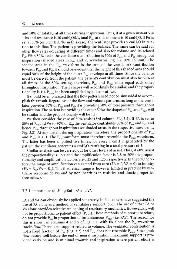
Proportional assist ventilation (PAV) is a form of synchronized partial ventilatory support in which the ventilator generates pressure in proportion to the patient's instantaneous effort, and this proportionality applies from breath to breath as well as continuously throughout each inspiration [1]. In effect, patient effort is amplified, as if the patient has acquired additional inspiratory muscles that remain under the control of the patient's own respiratory control system (Fig. 3.1).
Unlike other modes of partial support, there is no target flow, tidal volume, ventilation or airway pressure. Rather, the objective of PAY is to allow the patient to comfortably attain whatever ventilation and breathing pattern his or
Fig. 3.1. Idealized representation of ventilator response during PAV. Ventilator output (Paw) tracks patient's inspiratory muscle output {Pmu,}.
What is adjusted is proportionality between Paw and P mus' Two examples are represented where the prop ortionalities are 1:1 and 2:1. Clearly, other ratios are possible
J. Mancebo et al. (eds.), Mechanical Ventilation and Weaning© Springer-Verlag Berlin Heidelberg 2003

40 M. Younes
her control system sees fit. The responsibility for determining level and pattern of breathing is shifted entirely from the caregiver to the patient.
3.2 How to Get the Ventilator to Respond to the Patient's Instantaneous Inspiratory Effort
The objectives of PAV can, theoretically, be accomplished by recording the activity from a respiratory muscle (EMG) or nerve (ENG) and using the signal to drive the ventilator. Systems of this kind have been developed and used to produce a "normal" breathing pattern in experimental animals subjected to thoracotomy or paralysis [2,3].
Alternatively, the pressure output of an inspiratory muscle, for example the diaphragm (P dJ, may be used as the command signal to the ventilator. Although this would theoretically accomplish the same objectives, there are practical difficulties, including the need for invasive monitoring (esophageal and gastric catheters) and the substantial artifacts produced in this signal by the heart beat and by swallowing.*
The method proposed to accomplish the objectives of PAV [1], and which is suitable for clinical application, is to provide pressure assist in proportion to ongoing inspiratory flow (flow assist, FA) and volume (volume assist, VA). For FA and VA to result in airway pressure (Paw) being proportional to instantaneous effort (i.e. PAV) the following two conditions must be met: 1. Both FA and VA need to be used. 2. FA (which is expressed in cmH20/l/s) and VA (expressed in cmH20/l) must be
less than the patient's resistance (Rr) and elastance (ErJ, respectively, and the fractions (i.e. FA/Rrs and VAIE rs should, ideally, be similar.
Figure 3.2 illustrates how applying these two principles can result in proportionality between Paw and patient's inspiratory effort (P muJ. Assume a flow pattern of the form shown in the first column (nearly sinusoidal, as is most often the case during normal breathing). Volume, which is the integral of flow, rises in a completely different pattern and reaches its peak at the end of inspiration. The total pressure used to generate flow (Pre') is a function of V and resistance (Rr,). If Rrs is constant (i.e. not flow dependent) the time course of Pres will resemble the time course of flow (Pres= V· Rrs ), as shown by the outer envelope of the Pres waveform. The total pressure used to overcome elastic recoil (Pel) is a function of volume above FRe and respiratory system elastance (Ers' Ers = l/compliance). In the case where Ers is constant in the V T range, the time course of Pel resembles the time
* Pleural (esophageal) pressure (P pi) alone is not a suitable signal. Apart from being subject to the same difficulties of P pi (invasiveness and artifacts), P pi during mechanical ventilation does not bear a fixed relation to the pressure generated by respiratory muscles (P mus)' since P pi represents Pmus after subtracting the pressure dissipated in moving the chest wall [4]. In fact, Ppl
can be increasing (i.e. becoming more positive) in the course of inspiration even as inspiratory P mus is increasing (which should reduce Ppl)' (See [4] for details).

PAV 50% PAV 80%
Flow
Proportional Assist Ventilation 41
FA only 80%
VA only 80%
Fig. 3.2. Schematic representation of how using both flow assist (FA) and volume assist (VA) in the same fraction of resistance (R) and elastance (E) results in ventilator pressure (Paw) becoming a function of respiratory muscle pressure (P mus)' Hypothetical flow and volume waveforms are used, but the results are simpar regardless of waveform shape. Pm is the pressure used to overcome resistance and equals V R' Pel is the pressure used to overcome elastic recoil and equals V E' Ptotal is the sum of P 'es and Pel' In this example elastic and resistive losses are approximately equal. The shaded area represents the pressure provided by the ventilator. At 50% assist the ventilator provides half Pel and Pees at all times. Note that total ventilator output (shaded area, PtoUd) is, necessarily, half Ptotal at all times. Pmus is patient's contribution and is the difference between Ptotal and Paw' Note that Paw and Pmus resemble each other, and the proportionality is l:1.A similar analysis, but with FA and VA representing 80% of Rand E, results in Paw again resembling P mus' but the proportionality is 4:1. When FA is given alone, the shapes of Paw and Pmus differ substantially and there is minimal assist near the end of inspiration where inspiratory effort is maximal. With VA, the assist lags behind P mus' There is little assist at the beginning of inspiration. Furthermore, at the end of neural inspiration Paw is rising as the patient reduces his effort in order to terminate flow
course of volume (Pel = V . ErJ, as shown in the outer envelope of the Pel waveform. During mechanical ventilation the pressure used to generate flow and to offset elastic recoil is provided jointly by the ventilator (Paw) and the patient (P muJ. At any instant during inspiration the combined pressure (Ptot ) is used to offset resistive and elastic losses. The time course of Ptot is therefore the sum of the Pres and Pel outer envelopes, as shown by the outer envelope of the P tot waveform (Fig. 3.2).
Consider the case where the ventilator is set to deliver FA (in cmH20/l/s) that is 50% of the patient's resistance and VA (in cmH20/l) that is 50% of the patient's elastance (2nd column, Fig. 3.2). The ventilator will thus deliver 50% of total Pres
42 M. Younes
and 50% of total Pel at all times during inspiration. Thus, if at a given instant V =
Ills and resistance is 10 cmH20/lIs, total Pres at this moment is 10 cmH20. If FA is set at 50% (or 5 cmH20/l/s in this case), the ventilator provides 5 cmH20 in relation to this flow. The patient is providing the balance. The same can be said for other flow rates occurring at different times and also for volume and its related Pel' With 50% assist the ventilator's contribution is 50% of Pres and Pel throughout inspiration (shaded areas in Pres and Pel waveforms, Fig. 3.2, 50% column). The shaded area in the Ptot waveform is the sum of the ventilator's contribution towards Pres and Pel' It should be evident that the height of this shaded area should equal 50% of the height of the outer Ptot envelope at all times. Since the balance must be derived from the patient, the patient's contribution must also be 50% at all times. At the 50% setting, therefore, Paw and P mus must equal each other throughout inspiration. Their shapes will accordingly be similar, and the proportionality is 1: l. P mus has been amplified by a factor of two.
It should be emphasized that the flow pattern need not be sinusoidal to accomplish this result. Regardless of the flow and volume patterns, as long as the ventilator provides 50% of P res and Pel' it is providing 50% of total pressure throughout inspiration. The patient is providing the other 50%; the shapes of Paw and P mus will be similar and the proportionality will be 1: 1.
We then consider the case of 80% assist (3rd column, Fig. 3.2). If FA is set to 80% of Rrs and VA to 80% of Ers' the ventilator contributes 80% of Pres and Pel' and hence Ptot' throughout inspiration (see shaded areas in the respective waveforms, Fig. 3.2). At any instant during inspiration, therefore, the proportionality of Paw and P mus is 4: 1. The Paw waveform must therefore resemble the P mus waveform. The latter has been amplified five times; for every 1 cmH20 generated by the patient the ventilator generates 4 cmH20, resulting in a total pressure of 5.
Similar analysis can be carried out for other levels of assist. Thus, at 60% assist the proportionality is 1.5: 1 and the amplification factor is 2.5. At 20% the proportionality and amplification factors are 0.25 and l.25, respectively. In theory, therefore, the range of amplification can extend from zero (FA = 0, VA = 0) to infinity (FA = Rrs ' VA = Er,). This theoretical range is, however, limited in practice by ventilator response delays and by nonlinearities in resistive and elastic properties (see below).
3.2.1 Importance of Using Both FA and VA
FA and VA can obviously be applied separately. In fact, others have suggested the use of FA alone as a method of ventilatory support [5,6]. The use of either FA or VA alone provides selective unloading of respiratory mechanics. However, Paw will not be proportional to patient effort (P mu,)' These methods of support, therefore, do not provide Paw in proportion to instantaneous P mus (i.e. PAV). The reason for this is shown in columns 4 and 5 of Fig. 3.2. With FA alone the Paw waveform tracks flow. There is no support related to volume. The ventilator contribution is not a fixed fraction of Ptot (Fig. 3.2) and Paw does not resemble P mus' Since peak flow occurs well before the end of neural inspiration, maximum support is provided early on and is minimal towards end-inspiration where patient effort is

3 Proportional Assist Ventilation 43
maximal. With VA alone (last column, Fig. 3.2) the opposite occurs. The support lags behind patient effort, and there is little support at the beginning of inspiration. Furthermore, Paw continues to increase for a while after P mus has started to decrease at the end of neural inspiration (Fig. 3.2, last column).
Why is it useful to provide support in proportion to P mus as opposed to selective unloading? Selective unloading might be useful in cases where ventilator dependence is related to pure selective mechanical abnormality (i.e. high resistance OR high elastance) with no muscle weakness. This situation is almost never encountered in ventilator-dependent patients. First, it is only the rare ventilator-dependent patient who can generate a maximum inspiratory pressure (MIP) that is >50 cmH20, and in many MIP is <40 cmH20. Given normal MIP values of 70-100 cmH20 [7], inspiratory muscle weakness is an important contributor to ventilator dependence in virtually all patients, with the reasons being related to hyperinflation (in COPD and asthma [8]), sepsis [9], chronic heart disease [10], malnutrition [11], drugs [12] or simply disuse secondary to lengthy ventilator use. By amplifying muscle effort, PAV compensates for muscle weakness. Selective unloading reduces a specific load. However, the weak muscles may have difficulty coping with a normal load.
Second, pure resistive or elastic abnormalities are uncommon. Where the disease introduces primarily a pure elastic load (i.e. increased lung stiffness), the addition of an ET tube presents an added resistive load. Patients with obstructive diseases often face an additional elastic load related to dynamic hyperinflation [13].
The rationale behind PAV is that if the muscles are made (artificially) stronger they can cope with the increased load whether it is resistive or elastic. It is evident that through the application of PAY, where the disease is associated with predominantly high resistance the assist will necessarily include a relatively higher FA, and vice versa. Thus, preferential unloading of resistance or elastance continues to be included in PAY, but the presence of muscle weakness is also automatically dealt with.
3.2.2 Importance of FA and VA Being Less Than Patient's Resistance and Elastance
As long as FA <Rrs and VA <Ers the patient must contribute a fraction of the total applied pressure (Fig. 3.2 and related text). Because with PAV Paw is proportional to P mus' any changes in P mus result in qualitatively similar changes in Paw. Thus when P mus progressively decreases at the end of inspiration Paw must also decrease. A point is reached where the sum of P mus and Paw becomes lower than Pel' which is highest near end-inspiration. At this point flow stops and the ventilator cycle is terminated.
When the gains of the FA and/or VA are greater than Rrs and/or Ers, respectively, the ventilator-delivered pressure (Paw) will exceed total pressure (Plot) at some point in the inspiratory phase. With flow overassist, this point will occur early in inspiration whereas with volume overassist Paw may not exceed P tot until late in inspiration. Once Paw exceeds P tot' the requirement that the patient contribute to total pressure no longer exists and the ventilator acquires a life of its own. As the

44 M.Younes
extent of overassist increases, the control of ventilator output (flow and volume) shifts progressively from patient to ventilator. With substantial overassist, the patient has little control over V T and flow, and these become determined by the FA and VA settings and the volume and pressure limits on the ventilator.
The patterns observed during overassist vary considerably depending on which component (FA or VA) is excessive, the extent to which this component exceeds patient's resistance or elastance, and the extent of assist on the other component. At one extreme, there is the pattern associated with a large flow overassist (e.g. FA>3 Rr.). Here (Fig. 3.3), as soon as the ventilator is triggered flow increases in a rapidly accelerating manner. Paw increases also rapidly as a result. Both increase until the cycle is terminated by a volume or Paw limit.
Figure 3.4 shows the case at the other extreme, VA slightly above Ers and FA = o. Here, when the patient stops the inspiratory effort, flow fails to decrease to zero (since Paw is >Pel), as would normally occur if VA is <Ers (see above). Because inspiratory flow continues, volume continues to rise during neural expiration. Volume and, hence, Paw continue to rise in an accelerating manner until: (a) the patient recruits expiratory muscles to force termination of inspiratory flow, (b)
25.0
-25.0
2.5
-2,5
Fig. 3.3. Effect of increasing flow assist (FA) above patient's resistance. In the first two breaths FA and volume assist were at 50% of Rrs and Ers. FA was increased to 15 cm H20/lis (four times Rrs) between the second and third breaths. Note that flow and Paw increase in a rapidly accelerating manner as soon as inspiratory flow begins. The cycle is terminated by a pressure limit on the ventilator (25 cmH20 in this case). Note that flow pattern is entirely different from the normal pattern observed earlier and T; is much shorter

<Ii
§::::::: 0.5 -~ 0 g.
s: _ -0.5 o~ 0
;:;: -:::::. 0.5
3 Proportional Assist Ventilation 45
,....,.., o 1 2 s
Fig. 3.4. Volume "runaway". In this sleeping patient volume assist (VA) was increased in small steps with flow assist being zero. The numbers above the airway pressure tracing are the values of VA. Note that at the transition between VA of 13 and 14 there is a pronounced change in pattern. Flow (inspiration down) fails to return to zero at the usual Ti (preceding breaths) and begins to increase again. This results in a characteristic saddle-shaped flow pattern, a long Ti and larger volume. From the response shown it can be surmised that the patient's Ers is between 13 and 14 cmH20/1
volume rises to a point where Ers is no longer lower than VA on account of the nonlinear pressure-volume relation of the respiratory system [14J,or (c) a volume or pressure limit is reached which aborts the cycle. The transition from a normallooking breathing pattern to the characteristic pattern of saddle-shaped flow and a long Ti as a result of a minor increase in VA (i.e. as in Fig. 3.4) signifies that the VA setting is equal to the patient's Ers This feature can be used to advantage to determine the patient's Ers' With FA = 0, VA is increased in small steps until the characteristic transition is seen (i.e. Fig. 3.4). The VA setting at this point is Ers '
Furthermore, this characteristic pattern, representing minor volume overassist, may at times appear spontaneously and sporadically in occasional breaths during an otherwise normal-looking pattern. This again signifies that VA is very close to Ers• The occasional "runaway" pattern occurs as a result of minor breath-bybreath changes in Ers produced, for example, as a result of breath-by-breath changes in end-expiratory volume or in V T in a patient where the P-V relation is nonlinear in the operating V T range. When VA is very near average Ers, some breaths with a below-average Ers will be slightly overassisted.
As VA is further increased above Ers and/or as FA is further added and increased, the pattern of overassist shifts progressively from that of Fig. 3.4 to that of Fig. 3.3. Examples are shown in Fig. 3.5. With all these patterns of overassist, the end of the ventilator cycle is no longer linked to the end of the patient's inspiratory effort and the cycle is terminated by machine-set limits or by the patient fighting the ventilator. These overassist patterns can be recognized by the fact that majority of the cycles are pressure or volume limited and, depending on which limiting mechanism is activated, peak pressure and/or V T is monotonous as opposed to the variable Paw or V T observed when the patient controls ventilator output with properly adjusted PAV (i.e. FA<Rrs and VA<Ers )'

46 M. Younes
a b c
6' 25J.l
N :r: E ~ 0 2 " VI V>
E: 0.
-25.0 2.5
'" -...
:;: 0 0 w:
-25
Fig. 3.5. Different patterns of "runaway". In all panels the first two breaths were supported at FA=500/0 Rrs and VA=500/0 Ers. A normal breathing pattern is seen. In the third breath FA and VA were altered. In the left panel (third breath) VA=Ers and FA=500/0 Rrs. Note that the pattern is similar to Fig. 3.4 but is somewhat more aggressive. In the middle panel VA remained at Ers and FA was 2 Rrs. In the right panel FA was increased further to 3 Rrs. In the last two examples the cycle was terminated by a pressure limit. Note that flow pattern is not similar to normal pattern in any of the panels. Flow and Tj can be adjusted at will by manipulating FA and VA in the range above the patient's Rrs and Ers
It should be pointed out that VA and/or FA have been proposed earlier as means to attain a target ventilator pressure [15], to attain a target VT [16, 17], or to control PaC02 [18]. With all such applications the gains of FA and VA cannot be constrained to be below the patient's Rrs and Ers, and the patient, as a result and by definition, has no control over ventilatory output. These other uses of FA and VA represent variants of pressure-cycled and volume-cycled ventilation and should not be confused with PAY [19], where no ventilatory or pressure targets are set.
3.3 Differences from Other Modes of Assisted Ventilation
A detailed discussion of the clinical and technical advantages and disadvantages of PAY relative to PSV and A/C ventilation is beyond the scope of this review. These will be mentioned only briefly. The interested reader is referred to several recent publications for further discussion [1,4,14,20-22].
The main clinical/physiological advantages of PAY include: (a) assured synchrony between end of ventilator cycle and end of patient's inspiratory effort; (b) lesser likelihood of failed triggering efforts; (c) automatic adjustment of level of assist as ventilatory needs change as a result of changes in metabolic rate, temperature, acid-base status, sleep/wake cycle and behavioral factors; (d) a generally lower peak and mean airway pressure; (e) a generally lower VT and hence the

3 Proportional Assist Ventilation 47
potential for less barotrauma; (f) lesser likelihood of hyperventilation; and (g) the ability to determine the patient's natural (desired) breathing pattern in the absence of distress, which may facilitate weaning (see below).
The main technical advantages of PAV include: (a) Ease of adjustment in the majority of patients, as there is only one variable (% assist) to be decided upon [14), and (b) greater tolerance of artifacts that may cause false triggering, thereby permitting the continued use of a sensitive triggering mechanism in the face of such noise (e.g. cardiac artifacts, hiccups).
The main clinical/physiological disadvantages include the fact that there is no guaranteed minimum ventilation (cf. A/C ventilation) or V T (cf. A/C and PSV); PaCOz is likely to rise more in the presence of respiratory depression. Also, in some patients tidal volume can be quite small despite absence of distress (see below). Whether this may have adverse effects, in the long term, on gas exchange and respiratory mechanics is unknown.
The main technical disadvantages of PAV include: (a) sensitivity to leaks [14); (b) need to know respiratory mechanics for proper adjustment; (c) difficulties in adjustment when pressure-volume and pressure-flow relations are grossly nonlinear in the operating range (see below); (d) potential for overassist, which necessitates careful attention to pressure and volume limits [14); (e) excessive sensitivity of degree of assist to uncompensated dynamic hyperinflation (see below).
PAV represents a major departure from conventional methods in the extent to which the caregiver can exercise control over ventilator output. The caregiver can determine only how hard or how little the patient is working. However, helshe cannot reliably influence the level of ventilation or breathing pattern. As with normal subjects [23) breathing pattern varies greatly among patients on PAV even with near-maximal assist ([24), see below). If the caregiver is unhappy with the tidal volume (too high or too low) or with VE or frequency and attempts to increase or decrease the assist to bring these variables to what helshe is accustomed to, or feels appropriate, the patient will usually undertake an opposite action to maintain the same breathing pattern and VE (see below). The adjustment to this "loss of control" is one of the major challenges facing caregivers when first using PAV.
3.4 Clinical Experience to Date
Several clinical studies have been completed. Some have been published, others are at different stages of the publication process and have been reported only in abstract form [4, 24-43). The following is a summary of the findings most relevant to clinical application in the critical care setting.
3.4.1 Response to Different Levels of PAV
The response to different levels of PAV has been examined in a group of stable ventilator-dependent patients with assorted pathology [24), in patients with COPD where the response to different levels ofPSV was also included [30), and in

48 M. Younes
normal subjects while awake [36] and asleep [28]. In all cases PAY (% assist) was varied from the maximum (near 100% of Ers and Rrs) to the minimum tolerable (i.e. with no clinical distress) level (0% in normals, 30%-60% assist in ventilatordependent patients). The results in ventilator-dependent patients and in normals during sleep, were quite consistent. On average, tidal volume increased only marginally between minimum and maximum assist (Figs. 3.6 and 3.7). Respiratory rate changed little. Thus, under these conditions, increases in degree of assist result in down-regulation of P mus so that V T and VE increase only marginally. This down-regulation of P mus is almost certainly related to a small reduction in PaCOz between minimum and maximum assist.
The results were somewhat different in alert, awake subjects [36]. Although in the majority of subjects P di and P mus were down-regulated to a very low level as assist increased, resulting in only small increases in V T and VE, in some subjects the pressure output of inspiratory muscles remained high at all levels of assist, despite marked hypocapnia. These subjects, then, had a substantial non-C02-
dependent source of respiratory drive (likely the consciousness factor [44]). In these subjects the amplification produced by PAY in the face of persistently high P mus resulted in marked increases in V T and VE with subsequent hypocapnia similar to PSV and Ale.
The conclusion that emerges from these studies is that when respiratory drive is dominated by COz feedback (as during sleep, obtundation) VT and VE become fairly independent of level of assist, since VE cannot increase by more that what is required to lower PaCOz by a few mmHg. In sleep [45] and under anesthesia [44] reductions in PaCOz by only a few mmHg cause down-regulation to zero (apnea). Where significant non-COz sources to respiratory drive exist, down-regulation would be markedly attenuated, and V T and VE then become a function of level of
~
q; E :> -0 >
-;;; "0 i=
1.0
0.9
0.8
0.7
0.6
0.5
0.4
0.3
0.2
0.1
10 20 30 40 50 60 70 80 90 100
% assist
Fig. 3.6. Effect of progressively increasing level of PAY support on tidal volume. x-axis, Gain of volume assist as percentage of patient's elastance; thin lines, individual responses with lowest x value pertaining to lowest tolerable level of support; thick line, average response. From [24]

1.0
~ 0.8 Q;
E ::> '0 > 0.6 iO "tl t=
0.4
0 2 3
min Level of assist
4 max
E 0.. .0
35
30
-; 25 ~
10
3 Proportional Assist Ventilation 49
2 3 4 min max
Level of assist
Fig. 3.7. Average response of tidal volume and ventilator rate with different levels of PAY and PSV in 11 ventilator-dependent patients with COPD. Minimum level of assist is that just above clinical distress in either mode. Maximum level was 90% assist in PAY and the level needed to achieve 10 mllkg tidal volume in the case of PSV. Note that VT increases progressively with PSV (thick line) but changes relatively much less with PAY (dotted line). The progressive decrease in rate with PSV was largely related to increasing number of efforts that failed to trigger the ventilator. From [30]
assist. Failure of V T and VE to increase substantially in the two patient groups studied [24,30] therefore signifies that in these patients drive was predominantly CO2 mediated. These patients were stable and, like most ICU patients, somewhat obtunded, albeit awake.
It may, accordingly, be expected that under other circumstances, where significant non-C02 sources of respiratory drive may exist, VT and VE may increase substantially with level of assist. Such situations may include patients with high metabolic rate, drive-promoting reflexes from the lung (? j receptors), severe metabolic acidosis, or patients who are very alert and/or agitated. In fact, the extent to which V T and VE increase with increasing assist may be a suitable test to determine the extent to which respiratory drive is related to non-C02 sources.
An important finding from the two ICU studies [24,30] is that there was a very wide variability in desired V T among patients (Fig. 3.6); V T in the assist-insensitive range varied from 200 to 850 ml (3.4-14.1 mllkg). All these patients were quite comfortable at their respective V TS and were able to increase their V T to much higher levels on command. Thus, when V T was small, this was not due to mechanical constraints; the V T was that spontaneously selected by the patient's respiratory control.
A further relevant finding was that in some patients respiratory rate was quite high (up to 34 min-I) despite high assist, comfort, and insensitivity to level of assist. This, again, indicates that high respiratory rates need not indicate distress. Since making this observation, we have successfully weaned several

50 M. Younes
patients who were deemed weaning failures because every time they underwent a weaning trial from PSV or AIC their rate was found to be too high. When these patients were placed on high-level PAY their rate remained high, indicating that the high rate in these particular patients was not a marker of distress but represented the spontaneously selected pattern of breathing. Once this conclusion was reached, the patients were observed on zero assist for a few hours and, no change or distress being noted, were extubated. This successful outcome clearly applies to only a minority of patients with weaning difficulty, since in most cases a high rate during a weaning trial signifies distress [46]. PAY can be used to distinguish the two groups.
It must be pointed out that in the ventilator-dependent patients studied [24, 30] insensitivity of respiratory rate to level of assist applied only above a certain level of assist. In each patient, when assist was terminated or reduced below this point, rate increased along with appearance of clinical signs of distress. It follows that in every ventilator-dependent patient there is a range (range 1) over which rate decreases progressively with level of assist and a higher range (range 2) where it becomes fairly insensitive. We believe that respiratory rate in range 2 is the rate desired by the patient's control system, while range 1 signifies the patient's inability to attain the desired V T' with the patient then compensating with rate. It is likely that range 1 is unsustainable. Until evidence to the contrary is produced, it is prudent to consider the patient as ventilator dependent until range 1 disappears (i.e. rate changes little as assist is gradually decreased to 0).
Finally, Fig. 3.7 shows the difference in response of COPD patients to varying levels of PAY and PSV [30]. Unlike the case with PAY, with PSV VT increased, and respiratory rate decreased, progressively as a function of PSV level. The differences in respiratory rate response were largely artifactual and related to progressively more efforts failing to trigger the ventilator as PSV level increased; pressure continued well into neural expiration, thereby encroaching on time available for emptying before the next inspiratory effort. In fact, when patient's rate was counted considering the missed ventilator cycles (identified from transient dips in expiratory flow) there was no difference between PSV and PAY in response of respiratory rate to different levels of assist.
3.4.2 Peak Airway Pressure
One of the most consistent findings is that upon switching from a volume-cycled mode (SIMV, AIC) to PAY, peak Paw decreases substantially even with very high levels of PAY assist [4,31] (Fig. 3.8). Peak Paw with high-level PAY assist (80%-90% of Ers and RrS> rarely exceeds 30 cmH20 and is commonly less than 20 cmH20 even with 5-10 cmH20 of PEEP. This is true even in patients with moderately severe ARDS (Ers ",,40 cmH20). The reasons for the lower peak Paw have been discussed in detail elsewhere [14] and include a usually smaller V T' coincidence of end of machine cycle with maximum inspiratory pressure generated by the patient, lack of expiratory activity during the inflation phase, and occurrence of peak flow earlier in the inflation cycle.

Fig. 3.8. Response of peak airway pressure (top) and cardiac output (bottom) to alternation between volume-cycled assist/control (A/C) ventilation and proportional-assist ventilation (PAV).From [31]
45
0 -£ 40
~ 35 l!!
a '" !!! 30 !l<
~ 25 ',;; .x
20 '" <li a
15
9.5
9.0 'E' 'E 8.5 "'-e '5 8.0 13-::l ¢ 7,5
'" .1!l 'E 7.0 '" u
6S
M
3.4.3 Noninvasive Use of PAV
3 Proportional Assist Ventilation 51
Ale PAY Ale PAY Ale
Ale PAV AlC PAY AlC Mode of ventilation
The low peak Paw requirement with PAY made it possible to attempt noninvasive support in patients with de novo acute respiratory failure (i.e. not acute or chronic) who were deemed to require urgent intubation and in whom the precipitating cause was felt to be correctable within 1-2 days (acute pulmonary edema, status asthmaticus, sepsis with ARDS, pneumonia). Eight of 11 patients were successfully supported and were spared intubation [25]. Noninvasive PAY is currently in routine use in our emergency room and medical ICU.
3.4.4 Hemodynamics in Septic Shock
Switching from AIC to PAY in eight patients with septic shock, on vasopressors, consistently resulted in an increase in cardiac output (Fig. 3.8). This was due to a greater stroke volume; heart rate did not change [31]. The increase in stroke volume is likely related to the slightly lower mean intrathoracic pressure (estimated to be only -1.7 cmH20 despite the marked reduction in peak Paw' Fig. 3.8), with a

52 M. Younes
consequent increase in intrathoracic blood volume and, hence, ventricular preload. It should be pointed out that the reduction in mean intrathoracic pressure with PAY was due largely to greater inspiratory muscle pressure relative to A/C. The increase in mean inspiratory muscle pressure (PI) was estimated to be 1.5 cmH20 (range 0.7-5.0 cmH20). Although such an increase in p[ is insignificant in a person with normal respiratory muscles and represents an increase in tension time index of ",2%, it may represent a significant stress, at least in some patients, given the abnormal respiratory muscle function in sepsis [9].
3.4.5 Effect on VD/VT and Oxygenation
Switching from AIC to PAY in a group of ventilator-dependent patients with assorted pathology had no effect, in the short term, on V D/V T or PaO/F[02 ratio despite a significantly lower VT [32]. The increase in VDiVT. expected from the lower V T did not materialize. This was likely related to a reduction in zone 1 perfusion (due to lower transpulmonary pressure and greater pulmonary artery-topleural pressure gradient), which offset the expected increase related to a nearly fixed anatomic dead space becoming a larger fraction of VT' The long-term impact of the smaller V T on gas exchange is not yet known.
3.4.6 Importance of VA and FA Together
In a recent study, Navalesi et al. [26] confirmed several of the theoretical predictions and experimental findings described above in a group of ventilator-dependent patients with assorted pathology. They demonstrated that the use of VA alone (i.e. without FA), increases the resistive work of breathing, thereby confirming the importance of using both FA and VA (i.e. PAY), and that the patient's effort (P di' PmuJ is down-regulated in a continuous fashion as the level of assist increases. In agreement with the study of Marantz et al. [24], V T and V E changed little as assist increased from 20% to 80% as a result of this down-regulation. Thus, V T increased only by 15% at between 20% and 80% assist, whereas without down-regulation it would have more than doubled.
3.4.7 PAV Versus PSV
Ranieri et al. compared the response to an added dead space, which simulates an increase in ventilatory demand, when ventilator-dependent patients were on either PAVor PSV [27]. When on PAY, addition of dead space caused, as expected, an increase in assist, and the breathing pattern response consisted of an increase in V T with little change in rate, a response that is similar to the response of normal subjects to increased CO2 [47]. On PSV the response consisted of tachypnea with little change in VT [27].

3 Proportional Assist Ventilation 53
3.4.8 Long-term PAV Application In Ventilator-Dependent Patients
With the confidence gained in the locally constructed ventilators, several units were assigned to the intensive care areas and are currently operated by the regular respiratory therapists. For suitable patients (see below), and with the consent of the attending physician and patient, PAY is instituted and maintained throughout the duration of ventilatory support unless an exit criterion is met. Several dozen patients have been managed so far on PAY for extended periods (up to 10 days). The experience gained from these patients is incorporated into the practical approach outlined below.
3.5 Approach to the Clinical Use of PAV in Intubated, Critically III, Ventilator-Dependent Patients
3.5.1 Eligible Patients
Because spontaneous efforts are essential with PAY, central apnea and severe central depression are absolute contraindications. Severe central depression is recognized from lack of respiratory distress (e.g. accessory muscle use or tachypnea) in the face of respiratory or metabolic acidosis (e.g. pH <7 .30).
Furthermore, until controlled studies are carried out to evaluate the safety of PAY (versus other modes) in cardiogenic shock and in the presence of serious arrhythmias, it is not advisable to use PAY when serious arrhythmias have occurred in the recent past or the patient has been in cardiogenic shock and is currently well controlled with conventional ventilation.
With the preceding exceptions, all patients can be placed on PAY, provided that the ventilator meets the requirements and personnel are readily available who are familiar with this mode and with the equipment.
3.5.2 The Ventilator
The ventilator should conform to certain minimum requirements with respect to mechanical performance, alarm and monitoring features and the presence of suitable back-up systems. These minimum requirements have been outlined elsewhere [14]. Using a ventilator with bare minimum requirements (The Winnipeg Ventilator) we have identified several technical problems that unnecessarily complicate the implementation of PAY. These will be discussed here. Hopefully, commercial PAY ventilators will incorporate features that obviate some or all of these difficulties, thereby greatly facilitating implementation.
Ventilator Response Characteristics When the PAY assist is set at a given fraction of the patient's elastance and resistance the expectation is that the ventilator will assume this fraction of total pressure output, with the patient assuming the balance. This expectation can be met only if the ventilator faithfully follows the pressure waveform dictated by

54 M.Younes
the settings. Thus, if SO% assist is dialed, Paw should, ideally, track the following function at all times during inspiration:
Paw = .S V· Rrs+·S V· Ers
These expectations are unrealistic, since all mechanical systems display response delays. These delays dictate that the assist received is less than the level intended. Clearly, the greater the response delay, the greater the discrepancy between actual and intended assist. Furthermore, for a given response delay, the shorter the inspiratory time the greater the discrepancy (Fig. 3.9). The degree of discrepancy between actual and intended assist is a function of the relative areas under the actual and intended pressure-time waveforms (Fig. 3.9). Thus, if the area under the actual waveform is 50% of the area under the intended waveform, the patient is receiving 50% of the intended assist (40% in the case of an intended SO%, etc.). Clearly, the better the response of the ventilator, the less the discrepancy. At times, respiratory distress or excessive patient work is present despite high levels of assist settings. Slow ventilator response can be one of the reasons. A procedure has been described earlier for measuring ventilator responsiveness [14]. This, however, requires removal of the ventilator from the patient for the sake of testing. Preferably, the ventilator itself should display the fraction of intended area that is actually being delivered. This would facilitate the identification of this problem, if present, as a cause of the patient's excessive work.
Poor Trigger Sensitivity Delays in triggering cause the assist to begin after a finite fraction of the patient's inspiratory effort has elapsed. Triggering delays have an impact similar to that of poor ventilator response; the patient receives less assist than intended. Such delays may have three sources: 1. Maximum trigger sensitivity is still too insensitive: With PAY, the assist should,
ideally, begin at the very onset of inspiratory effort. Because the assist will automatically terminate at the end of inspiratory effort, any delay in onset of assist reduces the fraction of neural Tj that is being assisted [14]. Apart from
Fig. 3.9. Effect of delay in ventilator response on the degree of assist. Solid lines indicate desired pressure output, dashed lines indicate actual pressure output. A flxed delay of 200 ms was assumed. The patient receives less assist than intended, and the extent of the discrepancy increases as duration of inspiration decreases (note that the area between the solid and dashed lines becomes a bigger fraction of total area). From [14]

3 Proportional Assist Ventilation 55
the impact of delays in onset of assist, the level of effort required to trigger the ventilator is not assisted throughout the breath. Thus, if the patient is required to generate 2 cmH20 or to generate a flow of 1511min to trigger the assist, the assist will be applied to only a fraction of the patient's effort (i.e. actual effort minus trigger level) even after triggering starts [14]. Mitigating this vulnerability to poor trigger sensitivity is the fact that in the PAY mode trigger sensitivity can be increased substantially relative to other modes. This is because brief artifacts will not trigger a full breath [14]. For this reason, the range of trigger sensitivity on the ventilator should be wider to allow triggering to occur with minimal effort (e.g. 0.2 cmH20 or flow of 211min).
2. Leaks during expiration: In the presence of leaks during expiration, Paw may decrease below the PEEP level, causing false triggering. This is usually countered by decreasing trigger sensitivity to the point where false triggering does not occur. With PAY, expiratory time (Te) can be quite variable. Accordingly, the extent to which Paw decreases below PEEP prior to onset of inspiratory effort, as a result of a leak, may vary depending on variability in T e and magnitude of leak. If trigger sensitivity is reduced to offset the leak with the longest Te. trigger sensitivity will be good (i.e. high) in breaths following a long Te but may be poor in breaths following a short Te. On average, therefore, trigger sensitivity may be suboptimal. This problem can be obviated by abandoning the use of fixed pressure or flow as the trigger level and moving to more intelligent methods which utilize the change in Paw or flow trajectory during expiration as the marker of inspiratory onset. Furthermore, modern ventilators are capable of measuring leaks. A display of the prevailing leak level can alert the user to this potential problem.
3. Dynamic hyperinflation (DH): The presence of DH can further delay triggering because the patient must first decrease expiratory flow to zero, using inspiratory muscles, before he/she can begin to lower pressure below PEEP, or generate inspiratory flow, to trigger the ventilator [48]. This problem can, again, be obviated by using intelligent algorithms for detection of onset of inspiratory effort.
Nonlinearity of the Pressure-Flow (P-V) Relation When the P- V relation is nonlinear in the operating range, resistance is not a constant but increases with flow rate. There is therefore no fixed Rrs to rely on for the sake of adjusting the level of the flow-assist component of PAY. Because the patient's own resistance (Rpat) is quite independent of flow (i.e. Rpat nearly constant), even with severe obstructive disease (e.g. Fig. 5 of ref. [49]), practically important nonlinearities are related to the ET tube and occur mostly when the ET tube is small (e.g. ::;;7) and/or when flow rates are high [46,50].
The practical difficulties imposed by a substantially nonlinear P-V relation, in terms of setting the FA level, are as follows: Assume that total resistances at flow rates of 0.5,1.0, and 1.5 lis are 10, 17, and 24 cmH20/lIs, respectively. There is, in this case, no unique resistance value which can be used to set the flow-assist (FA) component of PAY. If one sets the FA gain based on the value at low flow, the assist may be too little if the patient wishes to generate a high flow (where actual resistance is much higher). On the other hand, if one chooses the resistance at a high

56 M. Younes
flow as the guide to FA gain, flow overassist will result at low flow rates. Particularly when the volume assist is a large fraction of the patient's elastance, the flow overassist will promote flow "runaway" until a flow is reached where actual resistance equals the FA setting. Thus, if one sets the FA gain in the preceding example to 17 cmH20/l/s, a "runaway" situation will exist as long as flow is less than 1.0 lis. Unless the patient actively opposes it, flow will increase until it reaches 1.0 lis, even if the patient stops his or her own inspiratory effort. The ventilator then takes on a life of its own, helping to dictate flow rate, and this defeats the purpose ofPAV,
The implementation difficulties related to nonlinear P-V relation can be obviated in one of three ways: 1. Using tracheal pressure, as opposed to external airway pressure, as the con
trolled pressure in the PAY algorithm. In this fashion ET tube resistance, the main source of nonlinearity, is automatically compensated for. The variable FA level of PAY can then be set taking only Rpat into consideration.
2. The FA component of PAY may be made up of a tube-specific nonlinear component and a patient-related constant component. This solution does not take into account differences between in vitro and in vivo ET tube resistance [50].
3. Automatic determination, by the ventilator, of the pressure-flow relation in the operating flow range. The FA term in the PAY equation would then be a function of flow as determined by the established nonlinear P-V relation under operating conditions. This approach has the advantage of adjusting the assist level as resistive properties change (e.g. ET tube secretions, changes in bronchomotor tone, etc.).
Nonlinearity in the Elastic Pressure-VT Relation The relation between respiratory system volume and elastic recoil is sigmoid, both in health and in disease [51]. In the mid 50%-60% of vital capacity the pressure-volume relation is linear (i.e. Ers nearly constant) and the elastance is least (compliance is highest). The system becomes stiffer at both ends. The majority of patients breathe in the linear midrange and Ers is nearly constant within the operating V T range. In others, the P-V relation is not linear within V T' This creates some difficulties, since there is no fixed Ers value to use for setting the volumeassist component of PAY. The difficulties depend on whether the VT falls within the stiff upper range (high-end nonlinearity) or stiff lower range (low-end nonlinearity) of the sigmoid P-V relation.
High-end Nonlinearity. This occurs when end-inspiratory volume approaches the physiological ceiling of the respiratory system (total lung capacity, TLC). The clinical situations where this happens include: (a) severe dynamic hyperinflation, (b) excessive external PEEP, and (c) severe restrictive disease (e.g. severe ARDS), where the vital capacity is so reduced that the inspiratory capacity (difference between TLC and FRC), normally 2-3 I, is reduced to values within the usual V T
range (004-0.8 1). Fig. 3.10, left, illustrates the case where the nonlinear upper range occurs within 0.6 I from FRC. As can be seen from the right panel, elastance (P elV T) is constant only over the lower V T range and increases sharply in the higher range. The dashed line in the left panel represents volume assist (VA) of

0.8
0.7
0.6
"'" v 0.5 E :;, '0 0.4 > ~ 0.3 "0 i=
0.2
0.1
10 20 30 40
Pel {em H20)
50 0
3 Proportional Assist Ventilation 57
20 40 60 80
Elastanee (em H20)
Fig. 3.10. Confounding effect of high-end nonlinearity in the pressure-volume relation on adjustment of volume assist (VA). The P-V curve (left) describes the case in a patient with severe restrictive disease. The P-V relation is linear over a small volume range (0.31); thereafter elastic recoil increases disproportionately. Calculated elastance (PeINT ) is constant at 25 cmH20/1 up to a volume of 0.3 I. Thereafter it increases progressively as a function of volume. Setting VA as a percent of the elastance (e.g. 20 cmH20/1 or 80% of the fIxed E in the low volume range) results in underassist if the patient wishes higher volumes (note that width of the shaded area, representing patient's contribution, becomes a greater fraction of total elastic recoil pressure). To provide adequate assistance in a higher volume range by using a VA at 80% of elastance at V T of 0.6 results in volume overassist at lower volumes (note assist line is to the right of the P-V relation). VT will automatically runaway to the point of intersection of the VA and P-V lines unless the patient fIghts the ventilator. Under these conditions, V T tends to become constant and not very responsive to changes in patient's efforts
80% of the patient's elastance in the low volume range. Because VA is constant while the P-V curve is not, the distance between the two lines (i.e. patient's contribution) increases disproportionally as a function of V T' As long as V T is less than 0.3 1, the patient is receiving, as intended, 80% assist. If the patient desires a larger V T and makes more effort, the % assist decreases (width of shaded area/total recoil). The patient is thus less able to increase V T beyond the range where Ers is constant, and respiratory distress may result. If one responds by increasing VA, to take account of the higher elastance in the higher V T range (dotted line), there will be volume overassist (ventilator providing more pressure than elastic recoil) through most of the V T (note dotted line to the right of the P-V curve). Under these conditions VT is dictated to rise to the point of intersection of the dotted and solid lines, unless the patient fights the ventilator or a pressure limit is reached. The patient is no longer comfortably in control of V T' In effect, in doing so, one has switched from patient-controlled VT (i.e. PAY) to ventilator-controlled VT•
Low-end Nonlinearity: Here the system is stiffest (least compliant) in the low range of V T and becomes progressively less stiff as volume increases. This may occur under two broad circumstances: (a) Dynamic hyperinflation where elastic

58 M.Younes
recoil pressure at end-expiration (i.e. onset of inspiration) is not zero but a finite positive value (Fig. 3.11, top, left, x intercept). Even if the P-V relation within V Tis linear the overall P-V relation (end-inspiratory PelVT) is nonlinear; elastance decreases as volume increases (top right panel). (b) When end-expiratory volume occurs close to the natural lower end of vital capacity (residual volume). This is encountered with obesity and abdominal distension and in some cases of severe obstructive disease. In the latter case, the increase in elastance at low volume and its decrease at higher volume are related to reversible, progressive, volume-dependent airway closure: at low volumes there are fewer communicating alveoli, and vice versa.
The impact of these two abnormalities on PAY setting is the same. Clearly, if the patient breathes with a near constant V T' one can determine elastance at this V T (see below) and adjust VA to the desired % assist. With PAY, however, there is often substantial breath-by-breath variability, particularly in alert patients. This presents some problems in deciding on an appropriate VA. Assume that Ers with the average V T (004) was 28 cmH20 (Fig. 3.11). VA is set at 90%, or 2S cmH20/i. At
0.8
6: 0.6 <IJ E :::> g 0.4
"iii -0 i= 0.2
0
0.8
::::- 0.6 <IJ E :l
0.4 g iii '0 i= 0.2
Elastanee (em H20)
Fig. 3.11. Confounding effect of low-end nonlinearity in the P-V curve produced by dynamic hyperinflation (top, elastic recoil at end-expiration = 5 cmH20) or by breathing near the stiff lower range of vital capacity. Note that in either case elastance (PellV T) decreases progressively as a function of V!, Setting VA based on the high E at low volumes results in runaway (assist greater than elastic recoil) if patient takes a somewhat large breath. Setting VA based on the lower E at higher V T avoids runaway but leaves smaller tidal volumes with little assist. Shaded area is patient contribution to elastic recoil. Note that patient receives considerably less assist early in the breath, when volume is low. See text for additional details

3 Proportional Assist Ventilation 59
a VT of 0.4, the patient is receiving 90% assist. However, the assist is less at volumes below 0.4 l. Furthermore, should the patient decide to take a deeper breath, the assist may exceed elastic recoil (dashed line crossing to the right of the solid line, Fig. 3.11, left) and a runaway develops which is terminated by the patient's active expiratory effort or by a machine pressure or volume limit. Reducing the assist to avert these occasional "runaway" breaths causes further underassist in the average and below-average V r. A choice therefore has to be made between decreasing the level of assist or putting up with frequent beeping sounds as the ventilator limits the occasional "runaway" breath.
The technical implementation difficulties posed by nonlinear P-V relation can be obviated by suitable additions to ventilators. Ideally, the ventilator should be capable of determining the relation between V T and elastic recoil over the operating volume range and providing VA as a % of this relation, including nonlinear relations.
3.5.3 Procedure
The procedure for placing a patient on PAY will clearly be affected by the features available on the ventilator. At the time of this writing it is not clear which of the facilitating features described in "The Ventilator" section (see before) will be available. Best- and worst-case scenarios will be described.
Best-Case Scenario The best case is where the ventilator is equipped with automatic detection of onset of inspiration, automatic adjustment of trigger sensitivity, and automatic determination of the P-V and P-V relations in the operating range. In this case, one would simply set F,02 and PEEP according to the usual criteria. Initially, pressure-limiting and volume-limiting levels are set at 40 cmH20 and 15 mllkg, respectively. All that remains is to set a % assist and default values for E and R to be used until the ventilator determines the P-V and P-V relations. A high assist value of 80% is recommended initially. This would be adjusted later (see Subsequent Management, below). The initial default values for E and R may be values determined earlier in the volume-cycled (see Worst-Case Scenario, below) mode, or empiric values. A suitable empiric value for E is 12 cmH20/l. At 80% assist the patient would receive a VA of 9.6 cmHzO, which is about the average normal respiratory system elastance [51]. Alternatively, the default E value may be size dependent since compliance is size dependent (normal compliance = 0.02 predicted vital capacity/cmHzO [51]). Thus, a reasonable default E value would be l/predicted normal compliance. Since the patient's elastance will almost certainly be higher than normal, there is little likelihood of overassist, and this setting would provide a reasonable amount of support pending determination of the real P-V relations. Once the patient is switched to PAY a quick elastance can be determined using the end-expiratory occlusion (see below), and the default value is adjusted accordingly.
For the default R value, one can use the resistance of the ET tube at 0.5 lis plus an assumed value of 3 cmHzO/lIs for the patient's own resistance. Again, this

60 M. Younes
would provide reasonable support with little likelihood of flow overassist (since 3.0 is the normal respiratory resistance [52]). If it is strongly suspected that the patient suffers from obstructive disease, a higher default value (e. g. ET tube + 8) may be used until the actual P-V relation is determined.
It should be emphasized that the choice of these default values is not terribly critical. A minor underestimation (e.g. 20%) of the patient's mechanics would have little consequence. A gross underestimation (e.g. 50%-70%) would result in underestimation of assist between onset of PAY and determination of actual P-V and P-V relations. Depending on how borderline the patient is, this may result in some clinical evidence of distress, at which point it would become obvious that the default values need to be increased. With the above-suggested default values the likelihood of substantial overassist is virtually nonexistent and, should it happen, the breath will be terminated by the relatively low pressure and/or volume limits.
Once the actual P-V and P-V relations are determined the desired % assist will be applied to these relations. The pressure and volume limits and the alarm settings are then readjusted according to prevailing peak Paw and V T' respectively (see steps 12 and 13, below).
Worst-Case Scenario This procedure for the worst-case scenario is based on the one we used with the Winnipeg Ventilator and assumes that the ventilator has none of the automatic features described in "The Ventilator" section. Trigger sensitivity is adjusted manually, and VA and FA are dialed directly with no provision for nonlinear compensation. In this account it will be assumed that the patient was placed initially on a conventional mode and is being switched to PAY. Although institution of PAY immediately after intubation is not contraindicated, commonly the patient will not meet the eligibility criteria on account of excessive sedation or paralysis given prior to intubation or to control agitation following intubation (i.e. central depression).
3.6 What to Do
1. Determine the patient'S elastance and resistance. This is done using conventional techniques. It is recommended, however, that measurements be carried out during a brief period of controlled mechanical ventilation (CMV). The patient is switched to assist-control ventilation, if not already on it, with a square flow pattern, and the backup rate is increased until no visible efforts are observed. This ensures relaxation and hence more reliable measurements. Calculate Ers from (P plateau - PEEP)/V T' P plateau should be measured early (e.g. 0.2 s) after the onset of the inspiratory hold since the relevant elastance for setting PAY is the dynamic elastance, determined as close as possible to the point of occurrence of zero flow. Calculate resistance (Rrs) from (P peak -
P plateau)/V: Again, P plateau should be measured early after the onset of inspiratory hold. It would be useful to determine Rrs at different flow rates (e.g. 0.5,1.0, 1.5 lis). This would facilitate setting the appropriate FA after the spontaneous

3 Proportional Assist Ventilation 61
flow demand of the patient on PAY is known (see step 11 below). For this purpose, flow rate, in the CMV modes, is altered for 1-2 breaths and a O.5-s inspiratory hold is obtained. Rrs is computed at the respective flow rates.
2. Set the PAV controls. Set the volume gain to approximately 80% of the patient's elastance [(P plateau - PEEP)/V T' in the CMV mode J. Set the flow assist to 80% of Rrs at 0.5 lis. Use of a conservative flow of 0.5 lis to initially estimate operating resistance avoids the development of flow "runaway", thereby making it possible to determine the flow desired by the patient (this would not be possible in the presence of flow "runaway". The estimated resistance can be adjusted subsequently once actual flow in PAY is estimated.*
3. PEEP. The use of PEEP with PAY is no different from its use with other modes and is guided by the need to maintain oxygenation and to counteract dynamic hyperinflation. The level selected prior to PAY, based on these indications, would then be retained. It should be remembered, however, that the use of external PEEP to counteract dynamic hyperinflation is effective only where dynamic hyperinflation is the result of flow limitation in the patient's airway and not due to high resistance in the ET tube or exhalation tubing (including the exhalation valve). Thus, in the presence of dynamic hyperinflation, every effort must be made to reduce the latter sources of resistance.
4. Adjust the peak pressure limit initially to 40 cmH20 and the maximum tidal volume to l.21. These can be readjusted later as needed (see below). A TI limit of 3.0 s should be selected if an external control is available.
5. Switch to PAV. 6. Adjust trigger sensitivity to the level just necessary to eliminate repetitive trig
gering during expiration (automatic cycling). 7. Look for evidence of volume overassist. There should be no "runaway" if the
patient's elastance on PAY is similar to that measured earlier on CMV. At times, however, elastance is lower on PAY, so 80% of the elastance determined under CMV may be higher than actual elastance on PAV. The reasons for these differences vary but may be related to a smaller V T on PAY in the face of the nonlinear pressure-volume relation near FRC, to less auto-PEEP on PAY, or to the presence of expiratory activity during the inspiratory hold in the pre-PAY measurements (thereby causing overestimation of elastance values). In the alert patient, volume overassist is identified from the visible activation of the expiratory muscles prior to cycling off. The patient also may, upon questioning, indicate that the pressure delivered is too much for his or her liking (even though actual pressure may be low by usual criteria). In this case, the gain of the volume assist is reduced to a level deemed comfortable by the patient. In the obtunded patient, volume overassist should be suspected when the inspiratory phase is terminated by one of the limiting variables (peak pressure, V T'
T1; see step 4 above). Here the problem may be either volume overassist or high ventilatory demand by the patient. The volume-assist level should be decreased
* Actual flow rate on PAY can be determined from the direct flow display, when available, or from the displayed values of VE and T/TTOT' Thus average inspiratory flow (Yr) is given by VE
(Tr/TTOT)' For example, ifVE is I2l!min and T/TTOT is 0.4, Vr, is 30 l!min or 0.5 lis.

62 M.Younes
gradually (Le., over 1-2 min). In the former case (overassist), there will be a step change in breathing pattern (large reduction in VT and/or T1) as volume assist is reduced below a certain level, with the new levels remaining fairly stable as the assist is reduced further. This is the reverse of the "runaway" procedure illustrated in Fig. 3.4. In the case of high ventilatory demand, this will not occur.
8. Measure elastance on PAY. The reason for repeating this measurement has been outlined above. Two methods can be used. In the inspiratory hold method (Fig. 3.12) the expiratory line is occluded during an inspiration, and the occlusion is maintained through part of the ensuing expiration. A well-defined pressure plateau is seen, particularly in obtunded patients. Elastance is determined from (P plateau - PEEP)/V T' where V T is the specific tidal volume whose expiration was occluded (note that with PAY, tidal volume is quite variable). Where there is appreciable breath-by-breath variability in V T' one should attempt to obtain data from small and large breaths. This could identify nonlinearities in the P-V relation which would help to explain situations in which excessive patient effort or respiratory distress is present despite a seemingly high level of support (see below). As shown in Fig. 3.12 (the fourth inspiration), and unlike the case with CMV or AIC, plateau pressure is higher than peak pressure. This occurs because plateau pressure reflects total elastic recoil, whereas the ventilator provides only a fraction of elastic recoil during the inspiratory phase. The higher the level of volume assist, the closer peak and plateau pressures are to each other.
25.0
-25.0
2.5
;: 0 o u:
-2.5
Fig. 3.12. Tracings showing inspiratory-hold method of determining elastance on PAV. The expiratory line was occluded during the fourth inspiration, and occlusion was maintained for approximately 1 s into expiration. Note that the plateau pressure is greater than the peak pressure

3 Proportional Assist Ventilation 63
The only circumstance where peak pressure may exceed plateau pressure is where the flow-assist level is high relative to the elastic assist (e.g. in a patient with very high resistance and low elastance). In this case, peak pressure is dominated by the flow assist. In the "runaway" method, with FA at zero, the level of volume assist is slowly increased until a level is reached where the ventilator fails to reset at the usual inspiratory time (i.e. that observed at lower levels of support) (Fig. 3.4). The rationale for this approach has been explained earlier (see Fig. 3.4 and related text). The advantage of this approach over the inspiratory-hold technique is that no computation is required. The volume-assist level at the point of the "runaway" is essentially the patient's elastance. The results of the two methods are very similar; however, this method is suitable only for obtunded patients; alert patients tend to activate expiratory muscles and terminate the cycle soon after the onset of the hold, making it difficult to recognize the characteristic pattern of minimum overassist (e.g. Fig. 3.4).
9. Adjust volume assist to 80% of the elastance measured on PAY. 10. Observe the patient's ventilation, breathing pattern, peak flow and airway pres
sure. 11. Adjust flow assist to 80% of Rrs at the prevailing average flow rate (see foot
note 2). 12. Reset the pressure and volume limits to be somewhat above the prevailing peak
pressure and Vr. Note that with PAY there can be substantial breath-by-breath differences in these variables. Set the limits to permit the patient the full range of his or her desired V r, as indicated by the spontaneous variability.
13. Set the alarms based on the prevailing breathing pattern. For example, the lowpressure and low-V r alarms can be set at 50% of average peak pressure and average Vr.
3.7 What to Expect
3.7.1 Usual Response
When eligibility criteria are adhered to, the ventilator is technically satisfactory, and the preceding procedure for initiating PAY is followed, the majority of patients experience no difficulty following the transition. Some inspiratory effort is evident, but there is no distress. Breathing pattern varies considerably among patients, with some electing slow, deep breathing and others breathing in a rapid, shallow pattern (see above). In the absence of respiratory distress, shallow breathing represents the preferred pattern by the patient's own control system. The treating physician may decide, however, that this pattern is not in the best interest of the patient. Until studies have been done to assess this issue, switching back to conventional ventilation or continued observation under PAY, with monitoring of hemodynamics and oxygenation, and return to conventional ventilation in the event of deterioration are both reasonable approaches.
Except in obtunded patients, there is usually substantial breath-by-breath variability in V T> respiratory rate, and flow rate, and spontaneous sighing is common

64 M. Younes
(Fig. 3.l3). Airway pressure reflects these changes (Fig. 3.13). This variability is normal [53,54]. Prevailing peak airway pressure will almost invariably be lower, and usually much lower, than with AMV if a standard VT (8-12 ml!kg) was used previously. Whether or not it will be lower than PSV depends on what PSV level was used previously.
When F,02 is matched, Pa02 will usually be within a few mmHg (in either direction) relative to the pre-PAY level. PaC02 usually will increase immediately upon switching to PAY, but pH remains normal (>7.35). The extent of increase in PaC02 depends on how much the patient was overventilated relative to his or her target PaC02 prior to PAY. With PAY, the patient selects his or her PaC02, whereas with other methods, PaC02 is determined largely by ventilator settings. Provided that pH is normal, the patient is comfortable, and the increase in PaC02 is not progressive, some increase in PaC02 is acceptable and indicates that the patient was overventilated previously. In the usual case, blood pressure and heart rate change little upon switching to PAY.
3.7.2 Respiratory Distress
In a minority of patients evidence of excessive inspiratory effort (accessory muscle use, intercostal indrawing) with or without clinical distress continues or appears (if patient is switching from another mode) despite seemingly adequate PAY support (e.g. >80% of E and R). There are several possible reasons for this: 1. Poor mechanical response of the ventilator 2. Poor trigger sensitivity related to inappropriate setting of this function, to
leaks, or to dynamic hyperinflation 3. Nonlinearities in the P-V and/or P-V relation 4. The presence of a high, non-C02-related source of respiratory drive: From a
large number of observations made during studies with normals and patients under a variety of circumstances, we are convinced that down-regulation of respiratory muscle output, and hence relief of distress by mechanical ventilation, is mediated largely via reduction in PaC02• Unloading per se, without concomitant reduction in PaC02, does not materially reduce respiratory effort. It follows that when the source of excessive respiratory drive is not CO2 relat-
o ~ "'" 05 ~ -- .
1.0
o ,,~ 10 :\;x
0. E 20 .s 30
r----r---1
o 5 10 s
Fig. 3.13. Example of volume and airway pressure tracings during PAY (constant gain throughout). Note the considerable breath-by-breath variability, including a "sigh" with a brief post -sigh apnea. From [14]

3 Proportional Assist Ventilation 65
ed, reducing PaCOz by increasing ventilator support will not be very effective in relieving the distress. Patients who display high non-COz-related respiratory drive include agitated patients and patients with systemic sepsis, severe metabolic acidosis, high metabolic rate, and shock of any cause.
Approach to Respiratory Distress on PAY 1. The presence of high, non-COz-related respiratory drive is easily recognized
when V E is high (e.g. >15 lImin) despite normal or low PaCOz. Often the mechanics are not very abnormal. These patients will continue to work hard no matter which mode they are ventilated with. Until the primary abnormality responsible for the increased drive is corrected, sedation with or without paralysis is the most effective way to reduce ventilatory demand. Although judicious administration of sedatives with continued use of PAY, along with careful monitoring of pH and hemodynamics, is a feasible option, it would be prudent under these conditions to switch the patient to another mode with which the physician is more familiar. Because of the exquisite sensitivity of pH to PaCOz in the presence of severe metabolic acidosis, if the use of sedative plus PAY is elected in the presence of severe metabolic acidosis a low ventilation alarm should be set at the ventilation level consistent with a tolerable pH. Should ventilation decrease below this level due to excessive sedation, the patient is switched to another mode in which VE is not so vulnerable to patient effort (e.g.A/C).
2. Look for evidence of delayed triggering. Note the extent of delay between the first visible inspiratory effort on the patient's thorax and the sound signifying onset of inspiratory flow from the ventilator. When triggering is excellent, the delay between the two signals is so small as to be imperceptible. A trained observer can identify undesirable delays using this simple technique. Alternatively, the airway pressure signal or expiratory flow signal (if available) should be inspected to see the time lag between the point at which there is a clear inflection in the tracing in an inspiratory direction (regardless of whether pressure is still above PEEP or flow is still expiratory) and the point of triggering (Fig. 3.14). Should delays exist, recheck to make sure that trigger sensitivity is just below the level that results in auto cycling. If delays still exist, increase external PEEP in small steps. Should this reduce the delay, increase PEEP further. Should increasing PEEP result in no further reduction in delay and the delay is still excessive (e.g. >0.3 s), it will be concluded the dynamic hyperinflation cannot be offset by external PEEP (Le. main resistance is in tubing). If patient's effort continues to be high, consider switching to a mode that is not so vulnerable to uncompensated dynamic hyperinflation. It should be pointed out that this difficulty can be obviated by suitable additional features on the ventilator.
3. In the absence of significant triggering delay or excessive drive, the problem is either a poor ventilator response or nonlinearities in the P-V and P-V relation. An indication of nonlinearities in the P-V relation may already exist from inspiratory hold manoeuvres at different V T (see step 8 above). Suction ET tube to make sure that secretions are not increasing its resistance. Increase FA level gradually until evidence of flow runaway appears (high flow acceleration at the

66 M. Younes
2
0 N
:I: E H ~
0
"" VI
'" !!! 0..
Fig. 3.14. Airway pressure and flow tracings at transition between expiration and inspiration. Pressure scale greatly amplified (±2 cmH20). Onset of inspiratory effort is identified from sharp change in trajectory of Paw or flow with the change being in an inspiratory direction (first arrowhead). Note the substantial delay between onset of inspiratory effort and triggering (second arrowhead). The delay between first arrowhead and zero flow crossing is related to intrinsic PEEP. The delay between zero flow crossing and triggering is related to poor ventilator trigger sensitivity
beginning of inspiration with a very early peak, Figs. 3.3, 3.5}. Decrease FA to just below this level. At this point flow should not rise precipitously to an early peak. If excessive respiratory effort continues, increase VA gradually until volume runaway occurs. This will first appear in the occasional breath (breath terminated by pressure or volume limit along with a beeping sound). Further increases cause runaway more frequently. VA should be set to the level just below the runaway point. If effort continues to be high, it must be concluded that the extent of nonlinearities is such that high level unloading cannot be offset by linear FA and VA algorithms or that there is a ventilator malfunction in the PAV mode. Switch to another mode.
3.7.3 Progressive Respiratory Acidosis
The development of progressive hypercapnia and acidosis despite seemingly adequate PAV support and in the absence of respiratory distress is unequivocal evidence of central depression. Although such patients are usually identifiable prior to the institution of PAV and do not meet eligibility criteria, in some cases one

3 Proportional Assist Ventilation 67
cannot be sure about the adequacy of the patient's ventilatory control system in advance. PAY can be instituted, with close monitoring of ventilation and blood gas tensions, as a test to assess the patient's respiratory control. If there is no obvious reason for central depression (e.g. sedatives, CNS pathology), consider the possibility that the patient was previously (i.e. prior to the current episode which led to intubation) in chronic respiratory failure and that the PaC02 was artificially driven down earlier during the use of another mode (CMV, A/C, PSV). Such patients tend to retain their high CO2 set point for a long time, even after artificially normalizing PaC02• When weaning is attempted, or upon switching to PAY, respiratory output is very low until PaC02 approximates their high CO2 set point. If this scenario is suspected, and particularly if it becomes known for sure that the patient was in chronic respiratory failure earlier, it would be reasonable to let PaC02 rise, accepting a low pH (within limits), as long as the patient shows no complications related to acute respiratory acidosis (drowsiness, arrhythmias) and there is no respiratory distress. Often PaC02 and pH will stabilize at a new level (e.g. PaC02 70-80, pH 7.25-7.30), the pH corrects later, and the patient is found to be weanable soon after.
3.7.4 Hemodynamic Deterioration
In my experience with over 200 patients, I encountered only one patient who developed angina upon switching from AMV to PAY. This resolved promptly upon reinstitution of AMY. We have not observed clinically significant hypotension to develop after switching to PAY in any patient. In fact, in septic shock we have observed that BP improves (see earlier). The lack of significant hemodynamic deterioration in our experience may be related to our practice, so far, of not instituting PAY in any patient with recent myocardial infarction or who is in cardiogenic shock, or in patients with recent significant arrhythmias. In these patients it is theoretically possible that the higher distending pressures associated with conventional ventilation may facilitate left ventricular function and that a switch to PAY may result in decreased cardiac output with attendant hypotension, arrhythmias, or angina. The development of any of these complications upon a switch from conventional ventilation to PAY should contraindicate the use of PAY and cause one to consider the presence of very marginal left ventricular function, if that has not already been established.
3.8 Subsequent Management
One of the major advantages of PAY is that ventilator output faithfully reflects many important patient variables. The ventilator thus serves as an accurate monitor of the patient's ventilation and tidal volume demands, and its rate is an accurate reflection of the patient's own respiratory rate. This feature should be used to advantage in the clinical management of the patient. Thus a high level of ventilation reflects a high ventilatory demand and is not due to too much tidal volume setting (as in AMV) or pressure setting (as in PSV). Such an occurrence should

68 M. Younes
prompt an investigation into the causes of a high respiratory drive, without the need to first experiment with reducing the level of assist, as is the case with other modes. A change in machine rate signifies a corresponding change in the patient's rate. This cannot be taken for granted with other modes, where synchrony between the end of the machine cycle and the end of a patient's inspiration is less assured. Thus an increase in machine rate from an average of2D-3D/min with PAY reflects a substantial change in respiratory drive and possibly imminent, if not existent, distress. The same change with PSV (or AMV) may simply reflect improved synchrony between the patient and the ventilator due, for example, to a somewhat larger effort per breath [21]. With properly adjusted PAY, changes in ventilatory demand are both obvious (from monitored VE) and largely compensated for (since machine assist increases with increasing effort). The development of respiratory distress on previously satisfactory PAY settings indicates with greater certainty that something significant has occurred to respiratory mechanics (including tubing) or ventilator performance. Distress developing with other modes may simply be due to an increase in demand which rendered a previously satisfactory setting unsatisfactory.
In the usual case, ventilation, V T' and Paw are expected to vary from time to time with demand. This complicates the process of setting up the alarms. Some experience with PAY and with the specific patient is required to set the alarms such that safety is ensured while the alarms are not frequently and unnecessarily beeping.
By their very nature, patients in intensive care units on mechanical ventilation are susceptible to many respiratory, cardiovascular, metabolic, and psychiatric complications in the course of their stay. These complications may occur with any mode of ventilation, and the patient on PAY is clearly not immune to them. The purpose of the next section is to alert the reader to developments that are unique to the mode (e.g. no. 1 below) and to provide a guide, based on limited information available so far, or advice about when to abandon PAY in favour of a more conventional mode. Unless so stated, recommendation to switch to another mode need not signify that PAY is deleterious under the circumstance. Rather, it simply means that we do not know whether it is beneficial, harmful, or indifferent relative to other modes, and prudence dictates that under these conditions the patient should be switched. These recommendations may thus change as clinical experience expands. Decision-making with respect to some of the following occurrences is expected also to be facilitated in the future with availability of ventilators that automatically monitor leaks, ventilator performance, and changes in respiratory mechanics. 1. The alarms reflecting breath termination by one of the machine's set limits
(P aw' V T' T1) may sound very frequently or with every breath. This indicates either a substantial increase in ventilatory demand or a "runaway" situation. The latter may be due to a new leak or to the assist becoming greater than the patient's elastance or resistance. Should this occur, end-inspiratory occlusion is performed. Leaks are identified from difficulty in maintaining pressure during the occlusion. If a satisfactory plateau can be obtained, elastance is recomputed. If E is lower than the gain of volume assist, then a "runaway" exists (volume-assist gain was increased or patient's elastance decreased rel-

3 Proportional Assist Ventilation 69
ative to previous levels). If measured E is higher than the volume-assist gain, then the increase in peak pressure and VT (and hence the activation of machine limits) is due to increased ventilatory demand. Readjust the machine limits and alarms.
2. Respiratory distress develops. This should indicate a machine malfunction, a change in respiratory mechanics due to medical (e.g., pneumothorax, pulmonary edema, etc.) or technical causes (including partial blockage of tubing, ET tube sliding into a main-stem bronchus, etc.), a change in level of autoPEEP, anxiety, or other medical problems frequently associated with distress (shock, embolus, etc.). Although such complications are not related to PAY, it would be prudent, for the time being, to switch the patient to a conventional mode while the cause is investigated. It is also advisable to monitor respiratory mechanics at intervals in order to detect changes in mechanics before they have progressed to the point of causing distress. The frequency of such measurements should depend on the likelihood of such changes taking place.
3. Deterioration occurs in gas exchange (i.e. Pa02 at a given FP2). In the patient whose spontaneously selected V Ton PAY is within the currently accepted range (e.g. >8 mllkg) , the deterioration is clearly not related to PAY per se, and management should be along the usual lines. In patients who insist on breathing with a small V T on PAY the situation is somewhat different. Although the deterioration may be unrelated to PAY, a contribution of the small V T cannot be excluded. Until the results of more definitive studies to assess the net impact of the small V T on morbidity become available, it is prudent to switch such patients to a conventional mode.
4. Hypoventilation and respiratory acidosis occur. The de novo appearance of respiratory acidosis without distress indicates the development of central depression. The most likely cause is excessive sedation. If hypoventilation is significant (e.g. pH <7.30), the patient may be placed temporarily on a conventional mode until the effect of the sedative abates. The previously given dose should be noted and avoided in the future. Should undistressed respiratory acidosis develop in the absence of sedative intake, a medical reason for central depression should be sought, and PAY is discontinued until this is corrected.
5. Hemodynamic instability may occur. The de novo appearance of shock, arrhythmias, or angina in a patient already on PAY for hours or days cannot be attributed to PAY. Nonetheless, the use of PAY beyond this point should be reconsidered. Where the problem is likely to be cardiac in origin, PAY may not be an appropriate mode, although direct information in this regard is lacking. PAY has been shown to be superior, from the hemodynamic standpoint, in septic shock. The occurrence of this complication while the patient is on PAY should not call for a switch to conventional ventilation unless, of course, heavy sedation and/or paralysis is indicated to combat agitation or to reduce ventilatory demand, in which case PAY would have to be discontinued. The situation in hypovolemic shock is likely similar to that in septic shock, although this needs to be confirmed experimentally.
6. Agitation: Sedatives are not contraindicated when PAY is used. However, particularly when the response of a given patient to sedatives is not known, the

70 M. Younes
dose should be titrated initially until the amount required to attain the desired effect without severe depression is learned. Since backup systems must always be available on PAY delivery systems, the inadvertent use of an excessive amount should not be hazardous.
The frequency with which the preceding complications are encountered clearly depends on the type of patient managed with PAY. Patients with naturally unstable medical conditions are more likely to sustain one or more of these complications. It is prudent for medical personnel who contemplate using PAY to begin with fairly stable patients until they have acquired sufficient understanding of, and experience with, this unconventional mode, where the patient, and not the care giver, determines ventilation and pattern. Only then should they proceed to use it on unstable patients.
3.9 Weaning
A high level of support (e.g. 70%-90% of E and R) is recommended throughout the acute phase of the illness unless the patient indicates that he or she is receiving too much pressure. Although the amount of patient work at this level of support is small, it is almost certainly above the level required to prevent disuse. Once the underlying medical condition is brought under control, the level of support is decreased gradually. Some reserve should be allowed, however, to meet changing metabolic needs and moderate changes in mechanics. Thus it would be appropriate to maintain volume and flow assist somewhat higher (e.g. 20% of E and R) than the minimum level tolerated by the patient. This minimum level can be tested at intervals, as the patient recovers, by gradually reducing the level of assist in steps, each lasting 10-20 min.
3.10 References
1. Younes M (1992) Proportional assist ventilation, a new approach to ventilatory support: theory. Am Rev Respir Dis 145: 114-120
2. Remmers JE, Gautier H (1976) Servorespirator constructed from a positive pressure ventilator. J AppI PhysioI41:252-256
3. Daubenspeck JA, Pichon D, Knuth KV, Bartlett D jr, St John WM (1988) An inexpensive servorespirator based upon regulation of a shunt resistance. Respir Physiol 73:87-96
4. Younes M, Puddy A, Roberts D, Light RB, Quesada A, Taylor K, Oppenheimer L, Cramp H (1992) Proportional assist ventilation: results of an initial clinical trial. Am Rev Respir Dis 145:121-129
5. Poon CS, Ward SA (1986) A device to provide respiratory mechanical unloading. IEEE Trans Biomed Eng 33:361-365
6. Schulze A, Schaller P, Gehrhardt B, Madler H -J, Gmyrek D (1990) An infant ventilator technique for resistive unloading during spontaneous breathing. Results in a rabbit model of airway obstruction. Pediatr Res 28:79-82
7. Black LF, Hyatt RE (1969) Maximal respiratory pressures: normal values and relationship to age and sex. Am Rev Respir Dis 99:696-702
8. Rochester DF, Braun NMT (1985) Determinants of maximal inspiratory pressure in chronic obstructive pulmonary disease. Am Rev Respir Dis 132:42-47

3 Proportional Assist Ventilation 71
9. Boczkowski J, Aubier M (1995) The respiratory muscles in sepsis. In: Roussos C (ed) The thorax. Lung biology in health and disease, vol 85, chap 51. Dekker, New York, pp 1483-1513
10. McParland C, Krishnan B, Wang Y, Gallagher CG (1992) Inspiratory muscle weakness and dyspnea in chronic heart failure. Am Rev Respir Dis 146:467-472
11. Lewis MT, Belman MJ (1988) Nutrition and the respiratory muscles. Clin Chest Med 9:337-348
12. Aldrich TK, Prezant DJ (1990) Adverse effect of drugs on the respiratory muscles. Clin Chest Med 11: 177-189
13. Younes M (1990) Load responses, dyspnea and respiratory failure. "State of the art." Chest 97:595-685
14. Younes M (1994) Proportional assist ventilation. In: Tobin M (ed) Principles and practice of mechanical ventilation. McGraw-Hill, New York, pp 349-370
15. Harries JR, Tyler JM (1964) Mechanical assistance to respiration in emphysema. Am J Med 36:68-78
16. Stawitcke FA, Mordan WJ, Jimison HB, Piziali R, Ream AK (May 15, 1984) Medical ventilator device parametrically controlled for patient ventilation. U.S. Patent 4,448,192
17. Etherington TJ, Greene JG, Poon CS, Storsved M (1988) A new means of ventilation. The Negative Impedance Respirator. Biomed Sci Instrum 24:209-215
18. Huang KA(1985) A microprocessor-based servo-respirator with synchronized airflow pattern. MSc Thesis, North Dakota State University of Agriculture and Applied Science, Fargo, ND
19. Younes M (1992) Proportional assist and negative impedance ventilation (letter to the editor). Am Rev Respir Dis 146:1642-1643
20. Younes M (1991) Proportional assist ventilation and pressure support ventilation: similarities and differences. In: Update in Intensive Care and Emergency Medicine, vol 15. Springer, Berlin Heidelberg New York, pp 361-380
21. Younes M (1993) Patient-ventilator interaction with pressure-assist€!d modalities of venti latory support. Semin Respir Med 14:299-322
22. Younes M (1995) Interactions between patients and ventilators. In: Roussos C (ed) The thorax. Lung biology in health and disease, vol 85, chap 81. Dekker, New York, pp 2367-2420
23. Jammes Y, Auran Y, Gouvermet 1, Delpierre S, Grimaud C (1979) The ventilatory pattern of conscious man according to age and morphology. Bull Eur Physiopathol Respir 15:527-540
24. Marantz S, Patrick W, Webster K, Roberts D, Oppenheimer L, Younes M (1996) Response of ventilator-dependent patients to different levels of proportional assist (PAV). J Appl Physiol 80:397-403
25. Patrick W, Webster K, Ludwig L, Roberts D, Wiebe P, Younes M (1996) Noninvasive positivepressure ventilation in acute respiratory distress without prior chronic respiratory failure. Am J Respir Crit Care Med 153:1005-1011
26. Navalesi P, Hernandez P, Wongsa A, Laporta D, Goldberg P, Gottfried SB (1996) Proportional assist ventilation in acute respiratory failure: effects on breathing pattern and inspiratory effort. Am J Respir Crit Care Med 154:1330-1338
27. Ranieri VM, Giuliani R, Mascia L, Grasso S, Petruzzelli V, Puntillo N, Perchiazzi G, Fiore T, Brienza A (1996) Patient ventilator interaction during acute hypercapnia: pressure support vs proportional assist ventilation. J Appl Physiol 81 :426-436
28. Meza S, Giannouli E, Younes M (1995) Ventilatory response to inspiratory muscle unloading with PAY (proportional assist) during sleep. Am J Respir Crit Care Med 151:A639
29. Giannouli E, Webster K, Patrick W, Roberts D, Younes M (1994) Determination of dynamic hyperinflation in patients on synchronized (assist) methods of ventilatory support. Am J Respir Crit Care Med 149:A72
30. Giannouli E, Webster K, Roberts D, Younes M (J 999) Response of ventilator-dependent patients to different levels of pressure support and proportional assist. Am J Respir Crit Care Med 159:1716-1725

72 M. Younes
31. Patrick W, Webster K, Wiebe P, Roberts D, Light B, Oppenheimer L, Kassum D, Younes M (1993) Effect of proportional assist ventilation on the hemodynamics of patients in septic shock. Am J Respir Crit Care Med 147:A611
32. Mettias M, Mendoza M, Wiebe P, Younes M, Light B (1993) Effect of proportional assist ventilation (PAV) vs controlled mandatory and pressure support ventilation (CMV, PSV) on gas exchange in patients with hypoxemic respiratory failure. Clin Invest Med 16:B23
33. Georgopoulos D, Webster K. Mitrouska I, Bshouty Z, Younes M (1995) Effect of inspiratory muscle unloading on the ventilatory response to CO2 Am J Respir Crit Care Med 151:A639
34. Rimmer KP, Rose MS, Hecker Js, Whitelaw WA (1995) Effect of proportional assist ventilation on ventilatory response to CO2 Am J Respir Crit Care Med 151:A639
35. Chrusch C, Bauerle 0, Younes M (1996) The effect of proportional assist ventilation (PAV) on exercise endurance time in COPD.Am J Respir Crit Care Med 153:A171
36. Hernandez P, Maltais F, Gursahaney A, LeBlanc P, Navalesi P, Gottfried SB (1996) Proportional assist ventilation (PAV) improves exercise performance in severe COPD. Am J Respir Crit Care Med 153: Al72
37. Appendini L, Purro A, Patessio A, Zanaboni S, Polese G, Donner CF, Rossi A (1996) CPAP improves ventilatory assistance with proportional assist ventilation (PAV) in COPD patients. Am J Respir Crit Care Med 153:A380
38. Mendez M, Fernandez R, Younes M (1996) Changes in respiratory motor output with different levels of volume-cycled, pressure support and proportional assist ventilation in normal subjects. Am J Respir Crit Care Med 153:A375
39. Puntillo F, Vitale N, Grasso S, Mascia L, Giuliani R, Tunzi P, Cicale P, DeLuca Tupputi L, Fiore T, Ranieri M (1996) Compensation for increase in elastic load during pressure assisted modes (part I). Am J Respir Crit Care Med 153:A373
40. Grasso S, Puntillo F,Mascia L,Ancona G, Fiore T, Bruno F, Slutsky AS, Ranieri M (2000) Compensation for increase in respiratory workload during mechanical ventilation. Pressure support versus proportional-assist ventilation. Am J Respir Crit Care Med 161:819-826
41. Gursahaney A, Mirkovic T, Hernandez P, Navalesi P, Laporta D, Goldberg, P, Gottfried 5B (1996) Comparison of airway opening and tracheal pressure regulated forms of proportional assist ventilation (PAV). Am J Respir Crit Care Med 153:A372
42. Gursahaney A, Mirkovic T, Hernandez P, Laporta D, Goldberg P, Gottfried 5B (1996) Proportional assist ventilation (PAV) improves breathing pattern and inspiratory effort in acute respiratory failure (ARF). Am J Respir Crit Care Med 153:A372
43. Appendini L, Purro A, Zanaboni S, Patessio A, Spada E, Carone M, Polese G, Ganassini A, Donner CF, Rossi A (1996) Effects of endotracheal tube (ET) on patient-ventilator interactions during proportional assist ventilation (PAV) in COPD patients. Am J Respir Crit Care Med 153:A373
44. Fink BR, Hanks EC, Ngal EH, Papper EM (1963) Central regulation of respiration during anesthesia and wakefulness. Ann NY Acad Sci 190:892-899
45. Skatrud Sf, Dempsey JA (1983) Interaction of sleep state and chemical stimuli in sustaining rhythmic ventilation. J Appl Physiol 55:813-22
46. Tobin M (1990) Weaning from mechanical ventilation. Curr Pulmonology 11 :47 -105 47. Hey EN, Lloyd BB, Cunningham DJC, Jukes MGM, Bolton DPG (1966) Effects of various
respiratory stimuli on the depth and frequency of breathing in man. Respir Physiol 1:193-205
48. Marini JJ (1990) Lung mechanics determination at the bedside: instrumentation and clinical application. Respir Care 35:669-696
49. Guerin C Coussa ML, Eissa NT, Corbeil C, Chasse M, Braidy J, Matar N, Milic-Emili J (1993) Lung and chest wall mechanics in mechanically ventilated COPD patients. J Appl Physiol 74: 1570-1 580
50. Wright PE, Marini JJ, Bernard GR (1989) In vitro versus in vivo comparison of endotracheal tube airflow resistance. Am Rev Respir Dis 140: 1 0-16
51. Agostoni E, Mead J (1964) Statics of the respiratory system. In: Handbook of physiology. Respiration, sect 3, vol 1, chap 13. Am Physiol Soc., Washington DC, pp 387-409

3 Proportional Assist Ventilation 73
52. Mead J, Agostoni E (1964) Dynamics of breathing. In: Handbook of physiology. Respiration, sect 3, vol 1, chap 14. Am Physiol Soc., Washington DC, pp 411-427
53. Newsom-Davis J, Stag. D (1975) Interrelationships of the volume and time components of individual breaths in resting man. J Physiol (Lond) 245:481-498
54. Tobin MJ, Mador MJ, Guenther SM, et al (1988) Variability of resting respiratory drive and timing in healthy subjects. J Appl PhysioI65:309-317