ESC Guidelines for the Diagnosis and Treatment of Chronic Heart Failure

ESC GUIDELINES
2016 ESC Guidelines for the diagnosis andtreatment of acute and chronic heart failureThe Task Force for the diagnosis and treatment of acute and chronicheart failure of the European Society of Cardiology (ESC)
Developed with the special contribution of the Heart FailureAssociation (HFA) of the ESC
Authors/Task Force Members: Piotr Ponikowski* (Chairperson) (Poland),Adriaan A. Voors* (Co-Chairperson) (The Netherlands), Stefan D. Anker (Germany),Hector Bueno (Spain), John G. F. Cleland (UK), Andrew J. S. Coats (UK),Volkmar Falk (Germany), Jose Ramon Gonzalez-Juanatey (Spain), Veli-Pekka Harjola(Finland), Ewa A. Jankowska (Poland), Mariell Jessup (USA), Cecilia Linde (Sweden),Petros Nihoyannopoulos (UK), John T. Parissis (Greece), Burkert Pieske (Germany),Jillian P. Riley (UK), Giuseppe M. C. Rosano (UK/Italy), Luis M. Ruilope (Spain),Frank Ruschitzka (Switzerland), Frans H. Rutten (The Netherlands),Peter van der Meer (The Netherlands)
Document Reviewers: Gerasimos Filippatos (CPG Review Coordinator) (Greece), John J. V. McMurray (CPG ReviewCoordinator) (UK), Victor Aboyans (France), Stephan Achenbach (Germany), Stefan Agewall (Norway),Nawwar Al-Attar (UK), John James Atherton (Australia), Johann Bauersachs (Germany), A. John Camm (UK),Scipione Carerj (Italy), Claudio Ceconi (Italy), Antonio Coca (Spain), Perry Elliott (UK), Çetin Erol (Turkey),Justin Ezekowitz (Canada), Covadonga Fernandez-Golfın (Spain), Donna Fitzsimons (UK), Marco Guazzi (Italy),
* Corresponding authors: Piotr Ponikowski, Department of Heart Diseases, Wroclaw Medical University, Centre for Heart Diseases, Military Hospital, ul. Weigla 5, 50-981 Wroclaw,Poland, Tel: +48 261 660 279, Tel/Fax: +48 261 660 237, E-mail: [email protected].
Adriaan Voors, Cardiology, University of Groningen, University Medical Center Groningen, Hanzeplein 1, PO Box 30.001, 9700 RB Groningen, The Netherlands, Tel: +31 50 3612355,Fax: +31 50 3614391, E-mail: [email protected].
ESC Committee for Practice Guidelines (CPG) and National Cardiac Societies document reviewers: listed in the Appendix.
ESC entities having participated in the development of this document:
Associations: Acute Cardiovascular Care Association (ACCA), European Association for Cardiovascular Prevention and Rehabilitation (EACPR), European Association ofCardiovascular Imaging (EACVI), European Heart Rhythm Association (EHRA), Heart Failure Association (HFA).
Councils: Council on Cardiovascular Nursing and Allied Professions, Council for Cardiology Practice, Council on Cardiovascular Primary Care, Council on Hypertension.
Working Groups: Cardiovascular Pharmacotherapy, Cardiovascular Surgery, Myocardial and Pericardial Diseases, Myocardial Function, Pulmonary Circulation and Right VentricularFunction, Valvular Heart Disease.
The content of these European Society of Cardiology (ESC) Guidelines has been published for personal and educational use only. No commercial use is authorized. No part of the ESCGuidelines may be translated or reproduced in any form without written permission from the ESC. Permission can be obtained upon submission of a written request to OxfordUniversity Press, the publisher of the European Heart Journal and the party authorized to handle such permissions on behalf of the ESC ([email protected]).
Disclaimer. The ESC Guidelines represent the views of the ESC and were produced after careful consideration of the scientific and medical knowledge and the evidence available atthe time of their publication. The ESC is not responsible in the event of any contradiction, discrepancy and/or ambiguity between the ESC Guidelines and any other official recom-mendations or guidelines issued by the relevant public health authorities, in particular in relation to good use of healthcare or therapeutic strategies. Health professionals are encour-aged to take the ESC Guidelines fully into account when exercising their clinical judgment, as well as in the determination and the implementation of preventive, diagnostic ortherapeutic medical strategies; however, the ESC Guidelines do not override, in any way whatsoever, the individual responsibility of health professionals to make appropriate andaccurate decisions in consideration of each patient’s health condition and in consultation with that patient and, where appropriate and/or necessary, the patient’s caregiver. Nordo the ESC Guidelines exempt health professionals from taking into full and careful consideration the relevant official updated recommendations or guidelines issued by the competentpublic health authorities, in order to manage each patient’s case in light of the scientifically accepted data pursuant to their respective ethical and professional obligations. It is also thehealth professional’s responsibility to verify the applicable rules and regulations relating to drugs and medical devices at the time of prescription.
The article has been co-published with permission in European Heart Journal and European Journal of Heart Failure. All rights reserved in respect of European Heart Journal.& European Society of Cardiology 2016. All rights reserved. For permissions please email: [email protected].
European Heart Journaldoi:10.1093/eurheartj/ehw128
European Heart Journal Advance Access published May 20, 2016 by guest on June 2, 2016
http://eurheartj.oxfordjournals.org/D
ownloaded from

Maxime Guenoun (France), Gerd Hasenfuss (Germany), Gerhard Hindricks (Germany), Arno W. Hoes(The Netherlands), Bernard Iung (France), Tiny Jaarsma (Sweden), Paulus Kirchhof (UK/Germany), Juhani Knuuti(Finland), Philippe Kolh (Belgium), Stavros Konstantinides (Germany/Greece), Mitja Lainscak (Slovenia),Patrizio Lancellotti (Belgium), Gregory Y. H. Lip (UK), Francesco Maisano (Switzerland), Christian Mueller(Switzerland), Mark C. Petrie (UK), Massimo F. Piepoli (Italy), Silvia G. Priori (Italy), Adam Torbicki (Poland),Hiroyuki Tsutsui (Japan), Dirk J. van Veldhuisen (The Netherlands), Stephan Windecker (Switzerland), Clyde Yancy(USA), Jose Luis Zamorano (Spain)
The disclosure forms of all experts involved in the development of these guidelines are available on the ESC websitehttp://www.escardio.org/guidelines.
- - - - - - - - - - - - - - - - - - - - - - - - - - - - - - - - - - - - - - - - - - - - - - - - - - - - - - - - - - - - - - - - - - - - - - - - - - -- - - - - - - - - - - - - - - - - - - - - - - - - - - - - - - - - - - - - - - - - - - - - - - - - - - - - - - - - - - - - - - - - - - - - - - - - - -Keywords Guidelines † Heart failure † Natriuretic peptides † Ejection fraction † Diagnosis † Pharmacotherapy †
Neuro-hormonal antagonists † Cardiac resynchronization therapy † Mechanical circulatory support †
Transplantation † Arrhythmias † Co-morbidities † Hospitalization † Multidisciplinary management
Table of ContentsAbbreviations and acronyms . . . . . . . . . . . . . . . . . . . . . . . . 3
1. Preamble . . . . . . . . . . . . . . . . . . . . . . . . . . . . . . . . . . . 7
2. Introduction . . . . . . . . . . . . . . . . . . . . . . . . . . . . . . . . . 8
3. Definition, epidemiology and prognosis . . . . . . . . . . . . . . . 8
3.1 Definition of heart failure . . . . . . . . . . . . . . . . . . . . . 8
3.2 Terminology . . . . . . . . . . . . . . . . . . . . . . . . . . . . . 9
3.2.1 Heart failure with preserved, mid-range and reduced
ejection fraction . . . . . . . . . . . . . . . . . . . . . . . . . . . . 9
3.2.2 Terminology related to the time course of heart
failure . . . . . . . . . . . . . . . . . . . . . . . . . . . . . . . . . . 9
3.2.3 Terminology related to the symptomatic severity
of heart failure . . . . . . . . . . . . . . . . . . . . . . . . . . . . . 10
3.3 Epidemiology, aetiology and natural history of heart failure 10
3.4 Prognosis . . . . . . . . . . . . . . . . . . . . . . . . . . . . . . . 10
4. Diagnosis . . . . . . . . . . . . . . . . . . . . . . . . . . . . . . . . . . . 10
4.1 Symptoms and signs . . . . . . . . . . . . . . . . . . . . . . . . 10
4.2 Essential initial investigations: natriuretic peptides,
electrocardiogram, and echocardiography . . . . . . . . . . . . . 11
4.3 Algorithm for the diagnosis of heart failure . . . . . . . . . 12
4.3.1 Algorithm for the diagnosis of heart failure in the
non-acute setting . . . . . . . . . . . . . . . . . . . . . . . . . . . 12
4.3.2 Diagnosis of heart failure with preserved ejection
fraction . . . . . . . . . . . . . . . . . . . . . . . . . . . . . . . . . 12
5. Cardiac imaging and other diagnostic tests . . . . . . . . . . . . . 14
5.1 Chest X-ray . . . . . . . . . . . . . . . . . . . . . . . . . . . . . 14
5.2 Transthoracic echocardiography . . . . . . . . . . . . . . . . 14
5.2.1 Assessment of left ventricular systolic function . . . . 14
5.2.2 Assessment of left ventricular diastolic function . . . 15
5.2.3 Assessment of right ventricular function and
pulmonary arterial pressure . . . . . . . . . . . . . . . . . . . . 15
5.3 Transoesophageal echocardiography . . . . . . . . . . . . . 15
5.4 Stress echocardiography . . . . . . . . . . . . . . . . . . . . . 15
5.5 Cardiac magnetic resonance . . . . . . . . . . . . . . . . . . . 15
5.6 Single-photon emission computed tomography and
radionuclide ventriculography . . . . . . . . . . . . . . . . . . . . . 15
5.7 Positron emission tomography . . . . . . . . . . . . . . . . . 15
5.8 Coronary angiography . . . . . . . . . . . . . . . . . . . . . . . 16
5.9 Cardiac computed tomography . . . . . . . . . . . . . . . . . 16
5.10 Other diagnostic tests . . . . . . . . . . . . . . . . . . . . . . 17
5.10.1 Genetic testing in heart failure . . . . . . . . . . . . . . 17
6. Delaying or preventing the development of overt heart
failure or preventing death before the onset of symptoms . . . . . 18
7. Pharmacological treatment of heart failure with reduced
ejection fraction . . . . . . . . . . . . . . . . . . . . . . . . . . . . . . . . 19
7.1 Objectives in the management of heart failure . . . . . . . 19
7.2 Treatments recommended in all symptomatic patients
with heart failure with reduced ejection fraction . . . . . . . . . 20
7.2.1 Angiotensin-converting enzyme inhibitors . . . . . . . 20
7.2.2 Beta-blockers . . . . . . . . . . . . . . . . . . . . . . . . . . 20
7.2.3 Mineralocorticoid/aldosterone receptor antagonists . 20
7.3 Other treatments recommended in selected symptomatic
patients with heart failure with reduced ejection fraction . . . 20
7.3.1 Diuretics . . . . . . . . . . . . . . . . . . . . . . . . . . . . . 20
7.3.2 Angiotensin receptor neprilysin inhibitor . . . . . . . . 23
7.3.3 If - channel inhibitor . . . . . . . . . . . . . . . . . . . . . . 24
7.3.4 Angiotensin II type I receptor blockers . . . . . . . . . 24
7.3.5 Combination of hydralazine and isosorbide
dinitrate . . . . . . . . . . . . . . . . . . . . . . . . . . . . . . . . . 24
7.4 Other treatments with less certain benefits in
symptomatic patients with heart failure with reduced ejection
fraction . . . . . . . . . . . . . . . . . . . . . . . . . . . . . . . . . . . 24
7.4.1 Digoxin and other digitalis glycosides . . . . . . . . . . 24
7.4.2 n-3 polyunsaturated fatty acids . . . . . . . . . . . . . . 25
7.5 Treatments not recommended (unproven benefit) in
symptomatic patients with heart failure with reduced ejection
fraction . . . . . . . . . . . . . . . . . . . . . . . . . . . . . . . . . . . 25
7.5.1 3-Hydroxy-3-methylglutaryl-coenzyme A reductase
inhibitors (‘statins’) . . . . . . . . . . . . . . . . . . . . . . . . . . 25
7.5.2 Oral anticoagulants and antiplatelet therapy . . . . . . 25
7.5.3 Renin inhibitors . . . . . . . . . . . . . . . . . . . . . . . . 25
7.6 Treatments not recommended (believed to cause harm)
in symptomatic patients with heart failure with reduced
ejection fraction . . . . . . . . . . . . . . . . . . . . . . . . . . . . . . 26
7.6.1 Calcium-channel blockers . . . . . . . . . . . . . . . . . . 26
ESC GuidelinesPage 2 of 85
by guest on June 2, 2016http://eurheartj.oxfordjournals.org/
Dow
nloaded from

8. Non-surgical device treatment of heart failure with reduced
ejection fraction . . . . . . . . . . . . . . . . . . . . . . . . . . . . . . . . 26
8.1 Implantable cardioverter-defibrillator . . . . . . . . . . . . . 26
8.1.1 Secondary prevention of sudden cardiac death . . . . 26
8.1.2 Primary prevention of sudden cardiac death . . . . . . 27
8.2 Cardiac resynchronization therapy . . . . . . . . . . . . . . . 28
8.3 Other implantable electrical devices . . . . . . . . . . . . . . 29
9. Treatment of heart failure with preserved ejection fraction . . 29
9.1 Effect of treatment on symptoms in heart failure with
preserved ejection fraction . . . . . . . . . . . . . . . . . . . . . . . 30
9.2 Effect of treatment on hospitalization for heart failure in
heart failure with preserved ejection fraction . . . . . . . . . . . 30
9.3 Effect of treatment on mortality in heart failure with
preserved ejection fraction . . . . . . . . . . . . . . . . . . . . . . . 30
9.4 Other considerations . . . . . . . . . . . . . . . . . . . . . . . 30
10. Arrhythmias and conductance disturbances . . . . . . . . . . . . 30
10.1 Atrial fibrillation . . . . . . . . . . . . . . . . . . . . . . . . . . 31
10.1.1 Prevention of atrial fibrillation in patients with heart
failure . . . . . . . . . . . . . . . . . . . . . . . . . . . . . . . . . . 31
10.1.2 Management of new-onset, rapid atrial fibrillation in
patients with heart failure . . . . . . . . . . . . . . . . . . . . . . 31
10.1.3 Rate control . . . . . . . . . . . . . . . . . . . . . . . . . 31
10.1.4 Rhythm control . . . . . . . . . . . . . . . . . . . . . . . 32
10.1.5 Thromboembolism prophylaxis . . . . . . . . . . . . . 33
10.2 Ventricular arrhythmias . . . . . . . . . . . . . . . . . . . . . 33
10.3 Symptomatic bradycardia, pauses and
atrio-ventricular block . . . . . . . . . . . . . . . . . . . . . . . . . . 34
11. Co-morbidities . . . . . . . . . . . . . . . . . . . . . . . . . . . . . . 35
11.1 Heart failure and co-morbidities . . . . . . . . . . . . . . . 35
11.2 Angina and coronary artery disease . . . . . . . . . . . . . 35
11.2.1 Pharmacological management . . . . . . . . . . . . . . 35
11.2.2 Myocardial revascularization . . . . . . . . . . . . . . . 35
11.3 Cachexia and sarcopenia (for frailty, please refer to
Section 14) . . . . . . . . . . . . . . . . . . . . . . . . . . . . . . . . . 36
11.4 Cancer . . . . . . . . . . . . . . . . . . . . . . . . . . . . . . . . 36
11.5 Central nervous system (including depression, stroke and
autonomic dysfunction) . . . . . . . . . . . . . . . . . . . . . . . . . 37
11.6 Diabetes . . . . . . . . . . . . . . . . . . . . . . . . . . . . . . . 37
11.7 Erectile dysfunction . . . . . . . . . . . . . . . . . . . . . . . . 38
11.8 Gout and arthritis . . . . . . . . . . . . . . . . . . . . . . . . . 38
11.9 Hypokalaemia and hyperkalaemia . . . . . . . . . . . . . . . 38
11.10 Hyperlipidaemia . . . . . . . . . . . . . . . . . . . . . . . . . 38
11.11 Hypertension . . . . . . . . . . . . . . . . . . . . . . . . . . . 38
11.12 Iron deficiency and anaemia . . . . . . . . . . . . . . . . . . 39
11.13 Kidney dysfunction (including chronic kidney disease,
acute kidney injury, cardio-renal syndrome, and prostatic
obstruction) . . . . . . . . . . . . . . . . . . . . . . . . . . . . . . . . 40
11.14 Lung disease (including asthma and chronic obstructive
pulmonary disease) . . . . . . . . . . . . . . . . . . . . . . . . . . . . 41
11.15 Obesity . . . . . . . . . . . . . . . . . . . . . . . . . . . . . . . 41
11.16 Sleep disturbance and sleep-disordered
breathing . . . . . . . . . . . . . . . . . . . . . . . . . . . . . . . . . 41
11.17 Valvular heart disease . . . . . . . . . . . . . . . . . . . . . . 42
11.17.1 Aortic stenosis . . . . . . . . . . . . . . . . . . . . . . . 42
11.17.2 Aortic regurgitation . . . . . . . . . . . . . . . . . . . . 42
11.17.3 Mitral regurgitation . . . . . . . . . . . . . . . . . . . . 42
11.17.4 Tricuspid regurgitation . . . . . . . . . . . . . . . . . . 42
12. Acute heart failure . . . . . . . . . . . . . . . . . . . . . . . . . . . . 43
12.1 Definition and classification . . . . . . . . . . . . . . . . . . . 43
12.2 Diagnosis and initial prognostic evaluation . . . . . . . . . 44
12.3 Management . . . . . . . . . . . . . . . . . . . . . . . . . . . . 48
12.3.1 Identification of precipitants/causes leading to
decompensation that needs urgent management . . . . . . . 48
12.3.2 Criteria for hospitalization in ward vs intensive
care/coronary care unit . . . . . . . . . . . . . . . . . . . . . . . 49
12.3.3 Management of the early phase . . . . . . . . . . . . . 49
12.3.4 Management of patients with cardiogenic shock . . 54
12.4 Management of evidence-based oral therapies . . . . . . 54
12.5 Monitoring of clinical status of patients hospitalized due
to acute heart failure . . . . . . . . . . . . . . . . . . . . . . . . . . 55
12.6 Criteria for discharge from hospital and follow-up in
high-risk period . . . . . . . . . . . . . . . . . . . . . . . . . . . . . . 55
12.7 Goals of treatment during the different stages of
management of acute heart failure . . . . . . . . . . . . . . . . . . 55
13. Mechanical circulatory support and heart transplantation . . . 56
13.1 Mechanical circulatory support . . . . . . . . . . . . . . . . 56
13.1.1 Mechanical circulatory support in acute heart
failure . . . . . . . . . . . . . . . . . . . . . . . . . . . . . . . . . . 56
13.1.2 Mechanical circulatory support in end-stage chronic
heart failure . . . . . . . . . . . . . . . . . . . . . . . . . . . . . . . 56
13.2 Heart transplantation . . . . . . . . . . . . . . . . . . . . . . . 58
14. Multidisciplinary team management . . . . . . . . . . . . . . . . . 59
14.1 Organization of care . . . . . . . . . . . . . . . . . . . . . . . 59
14.2 Discharge planning . . . . . . . . . . . . . . . . . . . . . . . . 61
14.3 Lifestyle advice . . . . . . . . . . . . . . . . . . . . . . . . . . . 61
14.4 Exercise training . . . . . . . . . . . . . . . . . . . . . . . . . . 61
14.5 Follow-up and monitoring . . . . . . . . . . . . . . . . . . . . 61
14.6 The older adult, frailty and cognitive impairment . . . . . 62
14.7 Palliative and end-of-life care . . . . . . . . . . . . . . . . . . 62
15. Gaps in evidence . . . . . . . . . . . . . . . . . . . . . . . . . . . . . 63
16. To do and not to messages from the Guidelines . . . . . . . . . 64
17. Web Addenda . . . . . . . . . . . . . . . . . . . . . . . . . . . . . . 65
18. Appendix . . . . . . . . . . . . . . . . . . . . . . . . . . . . . . . . . . 66
19. References . . . . . . . . . . . . . . . . . . . . . . . . . . . . . . . . . 66
Abbreviations and acronyms
ACC/AHA American College of Cardiology/AmericanHeart Association
ACCF/AHA American College of Cardiology Foundation/American Heart Association
ACE angiotensin-converting enzymeACEI angiotensin-converting enzyme inhibitorACS acute coronary syndromeAF atrial fibrillationAHF acute heart failureAHI apnoea/hypopnoea indexAIDS acquired immunodeficiency syndromeAKI acute kidney injuryAldo-DHF aldosterone receptor blockade in diastolic
heart failureAL amyloid light chainALT alanine aminotransferase
ESC Guidelines Page 3 of 85
by guest on June 2, 2016http://eurheartj.oxfordjournals.org/
Dow
nloaded from

of Cardiology Working Group on Myocardial and Pericardial Dis-eases.94 In most patients with a definite clinical diagnosis of HF, thereis no confirmatory role for routine genetic testing to establish thediagnosis. Genetic counselling is recommended in patients withHCM, idiopathic DCM and ARVC. Restrictive cardiomyopathyand isolated non-compaction cardiomyopathies are of a possiblegenetic origin and should also be considered for genetic testing.
HCM is mostly inherited as an autosomal dominant disease withvariable expressivity and age-related penetrance. Currently, morethan 20 genes and 1400 mutations have been identified, most of whichare located in the sarcomere genes encoding cardiac b-myosin heavychain (MYH7) and cardiac myosin binding protein C (MYBPC3).88,122
DCM is idiopathic in 50% of cases, about one-third of which are her-editary. There are already more than 50 genes identified that are asso-ciated with DCM. Many genes are related to the cytoskeleton. The mostfrequent ones are titin (TTN), lamin (LMNA) and desmin (DES).88,123
ARVC is hereditary in most cases and is caused by gene mutationsthat encode elements of the desmosome. Desmosomal gene muta-tions explain 50% of cases and 10 genes are currently associatedwith the disease.124
Counselling should be performed by someone with sufficientknowledge of the specific psychological, social and medical implica-tions of a diagnosis. Determination of the genotype is important,since some forms [e.g. mutations in LMNA and phospholamban(PLN)] are related to a poorer prognosis. DNA analysis could alsobe of help to establish the diagnosis of rare forms, such as mitochon-drial cardiomyopathies. Screening of first-degree relatives for earlydetection is recommended from early adolescence onwards, al-though earlier screening may be considered depending on the ageof disease onset in other family members.
Recently, the MOGE(S) classification of inherited cardiomyopathieshas been proposed, which includes the morphofunctional phenotype(M), organ(s) involvement (O), genetic inheritance pattern (G), aetio-logical annotation (E), including genetic defect or underlying disease/substrate, and the functional status (S) of the disease.125
6. Delaying or preventing thedevelopment of overt heart failureor preventing death before theonset of symptomsThere is considerable evidence that the onset of HF may be delayedor prevented through interventions aimed at modifying risk factorsfor HF or treating asymptomatic LV systolic dysfunction (see recom-mendations table). Many trials show that control of hypertensionwill delay the onset of HF and some also show that it will prolonglife.126 – 129 Different antihypertensive drugs [diuretics, ACEIs, angio-tensin receptor blockers (ARBs), beta-blockers] have been shownto be effective, especially in older people, both in patients withand without a history of myocardial infarction.126 – 128 Along withthe ongoing discussion on optimal target blood pressure values inhypertensive non-diabetic subjects, the recent SPRINT study hasalready demonstrated that treating hypertension to a lower goal[systolic blood pressure (SBP) ,120 mmHg vs. ,140 mmHg] inolder hypertensive subjects (≥75 years of age) or high-risk
hypertensive patients reduces the risk of cardiovascular disease,death and hospitalization for HF.129
Recently, empaglifozin (an inhibitor of sodium-glucose cotran-sporter 2), has been shown to improve outcomes (including the re-duction of mortality and HF hospitalizations) in patients with type 2diabetes.130 Other hypoglycaemic agents have not been shown con-vincingly to reduce the risk of cardiovascular events and may in-crease the risk of HF. Intensification of hypoglycaemic therapy todrive down glycated haemoglobin (HbA1c) with agents other thanempagliflozin does not reduce the risk of developing HF (for detailssee Section 11.6 on diabetes).
Although smoking cessation has not been shown to reduce therisk of developing HF, the epidemiological associations with the de-velopment of cardiovascular disease131 suggest that such advice, iffollowed, would be beneficial.
The association between alcohol intake and the risk of developingde novo HF is U-shaped, with the lowest risk with modest alcoholconsumption (up to 7 drinks/week).132 –134 Greater alcohol intakemay trigger the development of toxic cardiomyopathy, and whenpresent, complete abstention from alcohol is recommended.
An inverse relationship between physical activity and the risk ofHF has been reported. A recent meta-analysis found that dosesof physical activity in excess of the guideline recommendedminimal levels may be required for more substantial reductions inHF risk.135
It has been shown that among subjects ≥40 years of age with ei-ther cardiovascular risk factors or cardiovascular disease (but nei-ther asymptomatic LV dysfunction nor overt HF), BNP-drivencollaborative care between the primary care physician and the spe-cialist cardiovascular centre may reduce the combined rates of LVsystolic dysfunction and overt HF.136
Statins reduce the rate of cardiovascular events and mortality;there is also reasonable evidence that they prevent or delay the on-set of HF.137 – 140 Neither aspirin nor other antiplatelet agents, norrevascularization, have been shown to reduce the risk of developingHF or mortality in patients with stable CAD. Obesity is also a riskfactor for HF,141 but the impact of treatments of obesity on the de-velopment of HF is unknown.
In patients with CAD, without LV systolic dysfunction or HF, ACEIsprevent or delay the onset of HF and reduce cardiovascular and all-cause mortality, although the benefit may be small in thecontemporary setting, especially in patients receiving aspirin.142
Up-titration of renin–angiotensin system antagonists and beta-blockersto maximum tolerated dosages may improve outcomes, including HF, inpatients with increased plasma concentrations of NPs.136,143
A primary percutaneous coronary intervention (PCI) at the earli-est phase of an ST segment elevation myocardial infarction (STEMI)to reduce infarct size decreases the risk of developing a substantialreduction in LVEF and subsequent development of HFrEF.112 Initi-ation of an ACEI, a beta-blocker and an MRA immediately after amyocardial infarction, especially when it is associated with LVsystolic dysfunction, reduces the rate of hospitalization for HF andmortality,144 – 148 as do statins.137 – 139
In asymptomatic patients with chronically reduced LVEF, regard-less of its aetiology, an ACEI can reduce the risk of HF requiring hos-pitalization.5,144,145 This has not yet been shown for beta-blockersor MRAs.
ESC GuidelinesPage 18 of 85
by guest on June 2, 2016http://eurheartj.oxfordjournals.org/
Dow
nloaded from

In patients with asymptomatic LV systolic dysfunction (LVEF,30%) of ischaemic origin who are ≥40 days after an AMI, an im-
plantable cardioverter-defibrillator (ICD) is recommended toprolong life.149
7. Pharmacological treatment ofheart failure with reduced ejectionfraction
7.1 Objectives in the management ofheart failureThe goals of treatment in patients with HF are to improve their clin-ical status, functional capacity and quality of life, prevent hospital ad-mission and reduce mortality. The fact that several drugs for HFhave shown detrimental effects on long-term outcomes, despiteshowing beneficial effects on shorter-term surrogate markers, hasled regulatory bodies and clinical practice guidelines to seek mortal-ity/morbidity data for approving/recommending therapeutic inter-ventions for HF. However, it is now recognized that preventingHF hospitalization and improving functional capacity are importantbenefits to be considered if a mortality excess is ruled out.159 –161
Figure 7.1 shows a treatment strategy for the use of drugs (and de-vices) in patients with HFrEF. The recommendations for each treat-ment are summarized below.
Neuro-hormonal antagonists (ACEIs, MRAs and beta-blockers)have been shown to improve survival in patients with HFrEF andare recommended for the treatment of every patient with HFrEF,unless contraindicated or not tolerated. A new compound(LCZ696) that combines the moieties of an ARB (valsartan) and aneprilysin (NEP) inhibitor (sacubitril) has recently been shown tobe superior to an ACEI (enalapril) in reducing the risk of deathand of hospitalization for HF in a single trial with strict inclusion/ex-clusion criteria.162 Sacubitril/valsartan is therefore recommended toreplace ACEIs in ambulatory HFrEF patients who remain symptom-atic despite optimal therapy and who fit these trial criteria. ARBshave not been consistently proven to reduce mortality in patientswith HFrEF and their use should be restricted to patients intolerantof an ACEI or those who take an ACEI but are unable to tolerate an
Recommendations to prevent or delay the development of overt heart failure or prevent death before the onset ofsymptoms
Recommendations Class a Level b Ref c
Treatment of hypertension is recommended to prevent or delay the onset of HF and prolong life. I A126, 129, 150, 151
Treatment with statins is recommended in patients with or at high-risk of CAD whether or not they have LV systolic dysfunction, in order to prevent or delay the onset of HF and prolong life.
I A137–140,
152
Counselling and treatment for smoking cessation and alcohol intake reduction is recommended for people who smoke or who consume excess alcohol in order to prevent or delay the onset of HF.
I C 131–134
Treating other risk factors of HF (e.g. obesity, dysglycaemia) should be considered in order to prevent or delay the onset of HF. IIa C130, 141, 153–155
IIa B 130
ACE-I is recommended in patients with asymptomatic LV systolic dysfunction and a history of myocardial infarction in order to prevent or delay the onset of HF and prolong life.
I A5, 144, 145
ACE-I is recommended in patients with asymptomatic LV systolic dysfunction without a history of myocardial infarction, in order to prevent or delay the onset of HF.
I B 5
ACE-I should be considered in patients with stable CAD even if they do not have LV systolic dysfunction, in order to prevent or delay the onset of HF.
IIa A 142
Beta-blocker is recommended in patients with asymptomatic LV systolic dysfunction and a history of myocardial infarction, in order to prevent or delay the onset of HF or prolong life.
I B 146
ICD is recommended in patients:a) with asymptomatic LV systolic dysfunction (LVEF ≤30%) of ischaemic origin, who are at least 40 days after acute myocardial infarction,b) with asymptomatic non-ischaemic dilated cardiomyopathy (LVEF ≤30%), who receive OMT therapy,
in order to prevent sudden death and prolong life.
I B149,
156–158
ACEI ¼ angiotensin-converting enzyme inhibitor; CAD ¼ coronary artery disease; HF ¼ heart failure; ICD ¼ implantable cardioverter-defibrillator; LV ¼ left ventricular;LVEF ¼ left ventricular ejection fraction; OMT ¼ optimal medical therapyaClass of recommendation.bLevel of evidence.cReference(s) supporting recommendations.
ESC Guidelines Page 19 of 85
by guest on June 2, 2016http://eurheartj.oxfordjournals.org/
Dow
nloaded from
MRA. Ivabradine reduces the elevated heart rate often seen inHFrEF and has also been shown to improve outcomes, and shouldbe considered when appropriate.
The above medications should be used in conjunction with diure-tics in patients with symptoms and/or signs of congestion. The use ofdiuretics should be modulated according to the patient’s clinicalstatus.
The key evidence supporting the recommendations in thissection is given in Web Table 7.1. The recommended doses of thesedisease-modifying medications are given in Table 7.2. Therecommendations given in Sections 7.5 and 7.6 summarize drugsthat should be avoided or used with caution in patients with HFrEF.
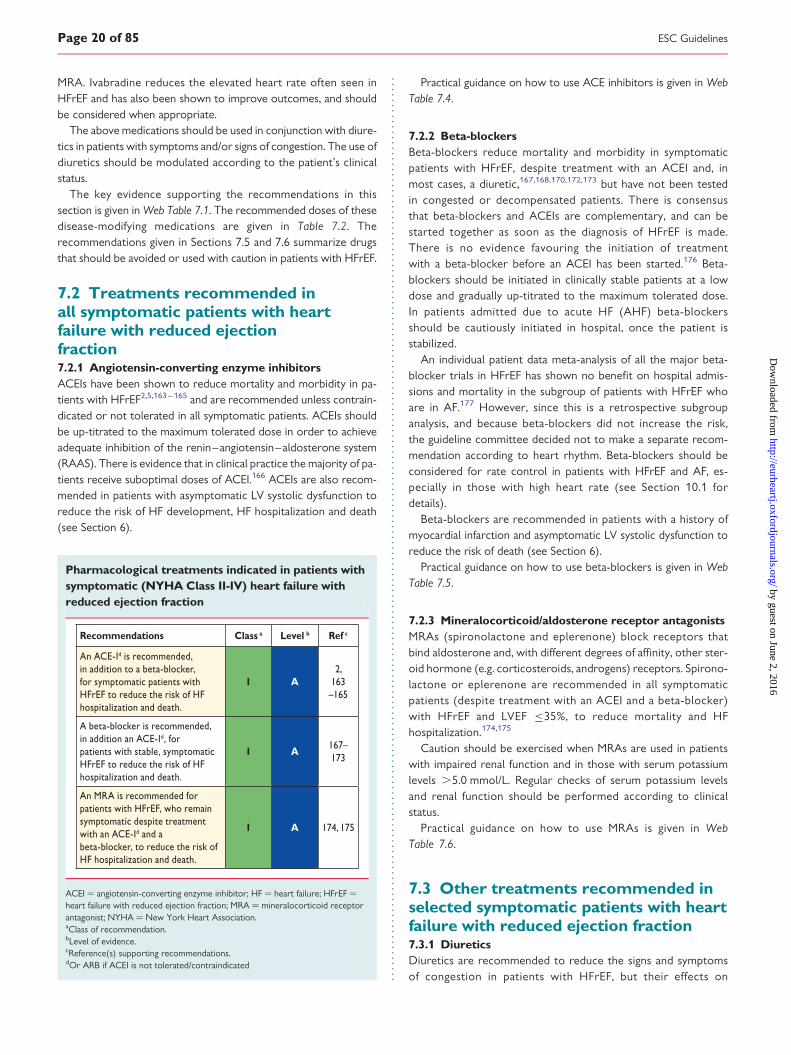
7.2 Treatments recommended inall symptomatic patients with heartfailure with reduced ejectionfraction7.2.1 Angiotensin-converting enzyme inhibitorsACEIs have been shown to reduce mortality and morbidity in pa-tients with HFrEF2,5,163 –165 and are recommended unless contrain-dicated or not tolerated in all symptomatic patients. ACEIs shouldbe up-titrated to the maximum tolerated dose in order to achieveadequate inhibition of the renin–angiotensin–aldosterone system(RAAS). There is evidence that in clinical practice the majority of pa-tients receive suboptimal doses of ACEI.166 ACEIs are also recom-mended in patients with asymptomatic LV systolic dysfunction toreduce the risk of HF development, HF hospitalization and death(see Section 6).
Pharmacological treatments indicated in patients withsymptomatic (NYHA Class II-IV) heart failure withreduced ejection fraction
Recommendations Class a Level b Ref c
An ACE-Id is recommended, in addition to a beta-blocker, for symptomatic patients with HFrEF to reduce the risk of HF hospitalization and death.
I A2,
163 –165
A beta-blocker is recommended, in addition an ACE-Id, for patients with stable, symptomatic HFrEF to reduce the risk of HF hospitalization and death.
I A167–173
An MRA is recommended for patients with HFrEF, who remain symptomatic despite treatment with an ACE-Id and a beta-blocker, to reduce the risk of HF hospitalization and death.
I A 174, 175
ACEI ¼ angiotensin-converting enzyme inhibitor; HF ¼ heart failure; HFrEF ¼heart failure with reduced ejection fraction; MRA ¼ mineralocorticoid receptorantagonist; NYHA ¼ New York Heart Association.aClass of recommendation.bLevel of evidence.cReference(s) supporting recommendations.dOr ARB if ACEI is not tolerated/contraindicated
Practical guidance on how to use ACE inhibitors is given in WebTable 7.4.
7.2.2 Beta-blockersBeta-blockers reduce mortality and morbidity in symptomaticpatients with HFrEF, despite treatment with an ACEI and, inmost cases, a diuretic,167,168,170,172,173 but have not been testedin congested or decompensated patients. There is consensusthat beta-blockers and ACEIs are complementary, and can bestarted together as soon as the diagnosis of HFrEF is made.There is no evidence favouring the initiation of treatmentwith a beta-blocker before an ACEI has been started.176 Beta-blockers should be initiated in clinically stable patients at a lowdose and gradually up-titrated to the maximum tolerated dose.In patients admitted due to acute HF (AHF) beta-blockersshould be cautiously initiated in hospital, once the patient isstabilized.
An individual patient data meta-analysis of all the major beta-blocker trials in HFrEF has shown no benefit on hospital admis-sions and mortality in the subgroup of patients with HFrEF whoare in AF.177 However, since this is a retrospective subgroupanalysis, and because beta-blockers did not increase the risk,the guideline committee decided not to make a separate recom-mendation according to heart rhythm. Beta-blockers should beconsidered for rate control in patients with HFrEF and AF, es-pecially in those with high heart rate (see Section 10.1 fordetails).
Beta-blockers are recommended in patients with a history ofmyocardial infarction and asymptomatic LV systolic dysfunction toreduce the risk of death (see Section 6).
Practical guidance on how to use beta-blockers is given in WebTable 7.5.
7.2.3 Mineralocorticoid/aldosterone receptor antagonistsMRAs (spironolactone and eplerenone) block receptors thatbind aldosterone and, with different degrees of affinity, other ster-oid hormone (e.g. corticosteroids, androgens) receptors. Spirono-lactone or eplerenone are recommended in all symptomaticpatients (despite treatment with an ACEI and a beta-blocker)with HFrEF and LVEF ≤35%, to reduce mortality and HFhospitalization.174,175
Caution should be exercised when MRAs are used in patientswith impaired renal function and in those with serum potassiumlevels .5.0 mmol/L. Regular checks of serum potassium levelsand renal function should be performed according to clinicalstatus.
Practical guidance on how to use MRAs is given in WebTable 7.6.
7.3 Other treatments recommended inselected symptomatic patients with heartfailure with reduced ejection fraction7.3.1 DiureticsDiuretics are recommended to reduce the signs and symptomsof congestion in patients with HFrEF, but their effects on
ESC GuidelinesPage 20 of 85
by guest on June 2, 2016http://eurheartj.oxfordjournals.org/
Dow
nloaded from
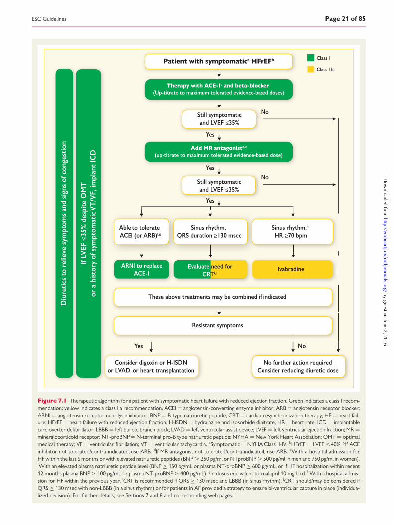
Figure 7.1 Therapeutic algorithm for a patient with symptomatic heart failure with reduced ejection fraction. Green indicates a class I recom-mendation; yellow indicates a class IIa recommendation. ACEI ¼ angiotensin-converting enzyme inhibitor; ARB ¼ angiotensin receptor blocker;ARNI ¼ angiotensin receptor neprilysin inhibitor; BNP ¼ B-type natriuretic peptide; CRT ¼ cardiac resynchronization therapy; HF ¼ heart fail-ure; HFrEF ¼ heart failure with reduced ejection fraction; H-ISDN ¼ hydralazine and isosorbide dinitrate; HR ¼ heart rate; ICD ¼ implantablecardioverter defibrillator; LBBB ¼ left bundle branch block; LVAD ¼ left ventricular assist device; LVEF ¼ left ventricular ejection fraction; MR ¼mineralocorticoid receptor; NT-proBNP ¼ N-terminal pro-B type natriuretic peptide; NYHA ¼ New York Heart Association; OMT ¼ optimalmedical therapy; VF ¼ ventricular fibrillation; VT ¼ ventricular tachycardia. aSymptomatic ¼ NYHA Class II-IV. bHFrEF ¼ LVEF ,40%. cIf ACEinhibitor not tolerated/contra-indicated, use ARB. dIf MR antagonist not tolerated/contra-indicated, use ARB. eWith a hospital admission forHF within the last 6 months or with elevated natriuretic peptides (BNP . 250 pg/ml or NTproBNP . 500 pg/ml in men and 750 pg/ml in women).fWith an elevated plasma natriuretic peptide level (BNP ≥ 150 pg/mL or plasma NT-proBNP ≥ 600 pg/mL, or if HF hospitalization within recent12 months plasma BNP ≥ 100 pg/mL or plasma NT-proBNP ≥ 400 pg/mL). gIn doses equivalent to enalapril 10 mg b.i.d. hWith a hospital admis-sion for HF within the previous year. iCRT is recommended if QRS ≥ 130 msec and LBBB (in sinus rhythm). jCRT should/may be considered ifQRS ≥ 130 msec with non-LBBB (in a sinus rhythm) or for patients in AF provided a strategy to ensure bi-ventricular capture in place (individua-lized decision). For further details, see Sections 7 and 8 and corresponding web pages.
ESC Guidelines Page 21 of 85
by guest on June 2, 2016http://eurheartj.oxfordjournals.org/
Dow
nloaded from

mortality and morbidity have not been studied in RCTs. A Co-chrane meta-analysis has shown that in patients with chronic HF,loop and thiazide diuretics appear to reduce the risk of deathand worsening HF compared with placebo, and comparedwith an active control, diuretics appear to improve exercisecapacity.178,179
Loop diuretics produce a more intense and shorter diuresisthan thiazides, although they act synergistically and the combin-ation may be used to treat resistant oedema. However, adverseeffects are more likely and these combinations should only beused with care. The aim of diuretic therapy is to achieve and main-tain euvolaemia with the lowest achievable dose. The dose of thediuretic must be adjusted according to the individual needs overtime. In selected asymptomatic euvolaemic/hypovolaemic patients,the use of a diuretic drug might be (temporarily) discontinued. Pa-tients can be trained to self-adjust their diuretic dose based onmonitoring of symptoms/signs of congestion and daily weightmeasurements.
Doses of diuretics commonly used to treat HF are provided inTable 7.3. Practical guidance on how to use diuretics is given inWeb Table 7.7.
Table 7.3 Doses of diuretics commonly used inpatients with heart failure
Diuretics Initial dose (mg) Usual daily dose (mg)
Loop diuretics a
Furosemide 20–40 40–240
Bumetanide 0.5–1.0 1–5
Torasemide 5–10 10–20
Thiazidesb
2.5 2.5–10
Hydrochlorothiazide 25 12.5–100
Metolazone 2.5 2.5–10
lndapamidec 2.5 2.5–5
Potassium-sparing diureticsd
+ACE-I/ ARB
-ACE-I/ ARB
+ACE-I/ ARB
-ACE-I/ ARB
Spironolactone/ eplerenone
12.5–25 50 50 100–200
Amiloride 2.5 5 5–10 10–20
Triamterene 25 50 100 200
ACE-I ¼ angiontensin-converting enzyme inhibitor, ARB ¼ angiotensin receptorblocker.aOral or intravenous; dose might need to be adjusted according to volume status/weight; excessive doses may cause renal impairment and ototoxicity.bDo not use thiazides if estimated glomerular filtration rate ,30 mL/min/1.73 m2 ,except when prescribed synergistically with loop diuretics.clndapamide is a non-thiazide sulfonamide.dA mineralocorticoid antagonist (MRA) i.e. spironolactone/eplerenone is alwayspreferred. Amiloride and triamterene should not be combined with an MRA.
Table 7.2 Evidence-based doses of disease-modifyingdrugs in key randomized trials in heart failure withreduced ejection fraction (or after myocardialinfarction)
Starting dose (mg) Target dose (mg)
ACE-I
Captoprila 6.25 t.i.d. 50 t.i.d.
Enalapril 2.5 b.i.d. 20 b.i.d.
Lisinoprilb 2.5–5.0 o.d. 20–35 o.d.
Ramipril 2.5 o.d. 10 o.d.
Trandolaprila 0.5 o.d. 4 o.d.
Beta-blockers
Bisoprolol 1.25 o.d. 10 o.d.
Carvedilol 3.125 b.i.d. 25 b.i.d.d
Metoprolol succinate (CR/XL) 12.5–25 o.d. 200 o.d.
Nebivololc 1.25 o.d. 10 o.d.
ARBs
Candesartan 4–8 o.d. 32 o.d.
Valsartan 40 b.i.d. 160 b.i.d.
Losartanb,c 50 o.d. 150 o.d.
MRAs
Eplerenone 25 o.d. 50 o.d.
Spironolactone 25 o.d. 50 o.d.
ARNI
Sacubitril/valsartan 49/51 b.i.d. 97/103 b.i.d.
If -channel blocker
Ivabradine 5 b.i.d. 7.5 b.i.d.
ACE ¼ angiotensin-converting enzyme; ARB ¼ angiotensin receptor blocker;ARNI ¼ angiotensin receptor neprilysin inhibitor; b.i.d. ¼ bis in die (twice daily);MRA ¼ mineralocorticoid receptor antagonist; o.d. ¼ omne in die (once daily);t.i.d. ¼ ter in die (three times a day).aIndicates an ACE-I where the dosing target is derived from post-myocardialinfarction trials.bIndicates drugs where a higher dose has been shown to reduce morbidity/mortality compared with a lower dose of the same drug, but there is no substantiverandomized, placebo-controlled trial and the optimum dose is uncertain.cIndicates a treatment not shown to reduce cardiovascular or all-cause mortality inpatients with heart failure (or shown to be non-inferior to a treatment that does).dA maximum dose of 50 mg twice daily can be administered to patients weighingover 85 kg.
ESC GuidelinesPage 22 of 85
by guest on June 2, 2016http://eurheartj.oxfordjournals.org/
Dow
nloaded from

7.3.2 Angiotensin receptor neprilysin inhibitorA new therapeutic class of agents acting on the RAAS and the neu-tral endopeptidase system has been developed [angiotensin recep-tor neprilysin inhibitor (ARNI)]. The first in class is LCZ696, which isa molecule that combines the moieties of valsartan and sacubitril(neprilysin inhibitor) in a single substance. By inhibiting neprilysin,the degradation of NPs, bradykinin and other peptides is slowed.High circulating A-type natriuretic peptide (ANP) and BNP exert
physiologic effects through binding to NP receptors and the aug-mented generation of cGMP, thereby enhancing diuresis, natriuresisand myocardial relaxation and anti-remodelling. ANP and BNP alsoinhibit renin and aldosterone secretion. Selective AT1-receptorblockade reduces vasoconstriction, sodium and water retentionand myocardial hypertrophy.187,188
A recent trial investigated the long-term effects of sacubi-tril/valsartan compared with an ACEI (enalapril) on morbidity
Other pharmacological treatments recommended in selected patients with symptomatic (NYHA Class II-IV) heartfailure with reduced ejection fraction
Recommendations Class a Level b Ref c
Diuretics
Diuretics are recommended in order to improve symptoms and exercise capacity in patients with signs and/or symptoms of congestion. I B 178, 179
Diuretics should be considered to reduce the risk of HF hospitalization in patients with signs and/or symptoms of congestion. IIa B 178, 179
Angiotensin receptor neprilysin inhibitor
Sacubitril/valsartan is recommended as a replacement for an ACE-I to further reduce the risk of HF hospitalization and death in ambulatory patients with HFrEF who remain symptomatic despite optimal treatment with an ACE-I, a beta-blocker and an MRAd
I B 162
If -channel inhibitor
Ivabradine should be considered to reduce the risk of HF hospitalization and cardiovascular death in symptomatic patients with LVEF ≤35%, in sinus rhythm and a resting heart rate ≥70 bpm despite treatment with an evidence-based dose of beta-blocker (or maximum tolerated dose below that), ACE-I (or ARB), and an MRA (or ARB).
IIa B 180
Ivabradine should be considered to reduce the risk of HF hospitalization and cardiovascular death in symptomatic patients with LVEF ≤35%, in sinus rhythm and a resting heart rate ≥70 bpm who are unable to tolerate or have contra-indications for a beta-blocker. Patients should also receive an ACE-I (or ARB) and an MRA (or ARB).
IIa C 181
ARB
An ARB is recommended to reduce the risk of HF hospitalization and cardiovascular death in symptomatic patients unable to tolerate an ACE-I (patients should also receive a beta-blocker and an MRA).
I B 182
An ARB may be considered to reduce the risk of HF hospitalization and death in patients who are symptomatic despite treatment with a beta-blocker who are unable to tolerate an MRA.
IIb C -
Hydralazine and isosorbide dinitrate
≤35% or with anLVEF <45% combined with a dilated LV in NYHA Class III–IV despite treatment with an ACE-I a beta-blocker and an MRAto reduce the risk of HF hospitalization and death.
IIa B 183
Hydralazine and isosorbide dinitrate may be considered in symptomatic patients with HFrEF who can tolerate neither an ACE-I nor an ARB (or they are contra-indicated) to reduce the risk of death.
IIb B 184
Digoxin
Digoxin may be considered in symptomatic patients in sinus rhythm despite treatment with an ACE-I (or ARB), a beta-blocker and an MRA, to reduce the risk of hospitalization (both all-cause and HF-hospitalizations).
IIb B 185
N-3 PUFA
An n-3 PUFAe preparation may be considered in symptomatic HF patients to reduce the risk of cardiovascular hospitalization and cardiovascular death.
IIb B 186
ACEI ¼ angiotensin-converting enzyme inhibitor; ARB ¼ angiotensin receptor blocker; BNP ¼ B-type natriuretic peptide; bpm ¼ beats per minute; HF ¼ heart failure; HFrEF ¼heart failure with reduced ejection fraction; LVEF ¼ left ventricular ejection fraction; MRA ¼ mineralocorticoid receptor antagonist; NT-proBNP ¼ N-terminal pro-B typenatriuretic peptide; NYHA ¼ New York Heart Association; PUFA ¼ polyunsaturated fatty acid. OMT ¼ optimal medical therapy (for HFrEF this mostly comprises an ACEI orsacubitril/valsartan, a beta-blocker and an MRA).aClass of recommendation.bLevel of evidence.cReference(s) supporting recommendations.dPatient should have elevated natriuretic peptides (plasma BNP ≥150 pg/mL or plasma NT-proBNP ≥600 pg/mL, or if HF hospitalization within the last 12 months, plasma BNP≥100 pg/mL or plasma NT-proBNP ≥400 pg/mL) and able to tolerate enalapril 10 mg b.i.d.eApplies only to preparation studied in cited trial.
ESC Guidelines Page 23 of 85
by guest on June 2, 2016http://eurheartj.oxfordjournals.org/
Dow
nloaded from

and mortality in patients with ambulatory, symptomatic HFrEFwith LVEF ≤40% (this was changed to ≤35% during thestudy), elevated plasma NP levels (BNP ≥150 pg/mL orNT-proBNP ≥600 pg/mL or, if they had been hospitalizedfor HF within the previous 12 months, BNP ≥100 pg/mL orNT-proBNP ≥400 pg/mL), and an estimated GFR (eGFR)≥30 mL/min/1.73 m2 of body surface area, who were ableto tolerate separate treatments periods with enalapril(10 mg b.i.d.) and sacubitril/valsartan (97/103 mg b.i.d.) duringa run-in period.162 In this population, sacubitril/valsartan (97/103 mg b.i.d.) was superior to ACEI (enalapril 10 mg b.i.d.) inreducing hospitalizations for worsening HF, cardiovascularmortality and overall mortality.162 Sacubitril/valsartan is there-fore recommended in patients with HFrEF who fit this profile.
Despite the superiority of sacubitril/valsartan over enalapril inthe PARADIGM-HF trial, some relevant safety issues remainwhen initiating therapy with this drug in clinical practice. Symp-tomatic hypotension was more often present in the sacubitril/valsartan group (in those ≥75 years of age, it affected 18% inthe sacubitril/valsartan group vs. 12% in the enalapril group), al-though there was no increase in the rate of discontinuation.162
The risk of angioedema in the trial was reduced by recruitingonly those who tolerated therapy with enalapril 10 mg b.i.d.and an sacubitril/valsartan during an active run-in phase of 5–9weeks (it resulted in a 0.4% rate of angioedema in sacubitril/val-sartan group vs. 0.2% in an enalapril group). Also, the number ofAfrican American patients, who are at a higher risk of angioede-ma, was relatively small in this study. To minimize the risk of an-gioedema caused by overlapping ACE and neprilysin inhibition,the ACEI should be withheld for at least 36 h before initiatingsacubitril/valsartan. Combined treatment with an ACEI (orARB) and sacubitril/valsartan is contraindicated. There are add-itional concerns about its effects on the degradation ofbeta-amyloid peptide in the brain, which could theoretically ac-celerate amyloid deposition.189 – 191 However, a recent small14-day study with healthy subjects showed elevation of thebeta-amyloid protein in the soluble rather than the aggregableform, which if confirmed over longer time periods in patientswith HFrEF may indicate the cerebral safety of sacubitril/valsar-tan.192 Long-term safety needs to be addressed.
7.3.3 If-channel inhibitorIvabradine slows the heart rate through inhibition of the Ifchannel in the sinus node and therefore should only be usedfor patients in sinus rhythm. Ivabradine reduced the combinedendpoint of mortality and hospitalization for HF in patientswith symptomatic HFrEF and LVEF ≤35%, in sinus rhythmand with a heart rate ≥70 beats per minute (bpm) who hadbeen hospitalized for HF within the previous 12 months, re-ceiving treatment with an evidence-based dose of beta-blocker(or maximum tolerated dose), an ACEI (or ARB) and anMRA.180 The European Medicines Agency (EMA) approvedivabradine for use in Europe in patients with HFrEF withLVEF ≤35% and in sinus rhythm with a resting heart rate≥75 bpm, because in this group ivabradine conferred a survival
benefit193 based on a retrospective subgroup analysis re-quested by the EMA.
Practical guidance on how to use ivabradine is given in WebTable 7.8.
7.3.4 Angiotensin II type I receptor blockersARBs are recommended only as an alternative in patients intolerantof an ACEI.182 Candesartan has been shown to reduce cardiovascu-lar mortality.182 Valsartan showed an effect on hospitalization for HF(but not on all-cause hospitalizations) in patients with HFrEF receiv-ing background ACEIs.194
The combination of ACEI/ARB for HFrEF was reviewed by theEMA, which suggested that benefits are thought to outweigh risksonly in a select group of patients with HFrEF in whom other treat-ments are unsuitable. Therefore, ARBs are indicated for the treat-ment of HFrEF only in patients who cannot tolerate an ACEIbecause of serious side effects. The combination of ACEI/ARBshould be restricted to symptomatic HFrEF patients receiving abeta-blocker who are unable to tolerate an MRA, and must beused under strict supervision.
7.3.5 Combination of hydralazine and isosorbide dinitrateThere is no clear evidence to suggest the use of this fixed-dosecombination therapy in all patients with HFrEF. Evidence on theclinical utility of this combination is scanty and comes from onerelatively small RCT conducted exclusively in men and beforeACEIs or beta-blockers were used to treat HF.184 A subsequentRCT conducted in self-identified black patients (defined as beingof African descent) showed that addition of the combination of hy-dralazine and isosorbide dinitrate to conventional therapy (ACEI,beta-blocker and MRA) reduced mortality and HF hospitalizationsin patients with HFrEF and NYHA Classes III– IV.183 The results ofthis study are difficult to translate to patients of other racial or eth-nic origins.
Additionally, a combination of hydralazine and isosorbide dini-trate may be considered in symptomatic patients with HFrEF whocan tolerate neither ACEI nor ARB (or they are contraindicated)to reduce mortality. However, this recommendation is based onthe results of the Veterans Administration Cooperative Study,which recruited symptomatic HFrEF patients who received only di-goxin and diuretics.184
7.4 Other treatments with less certainbenefits in symptomatic patients withheart failure with reduced ejectionfractionThis section describes treatments that have shown benefits interms of symptomatic improvement, reduction in HF hospitaliza-tions or both, and are useful additional treatments in patientswith HFrEF.
7.4.1 Digoxin and other digitalis glycosidesDigoxin may be considered in patients in sinus rhythm with symp-tomatic HFrEF to reduce the risk of hospitalization (both all-causeand HF hospitalizations),185 although its effect on top of beta-blockers has never been tested. The effects of digoxin in patients
ESC GuidelinesPage 24 of 85
by guest on June 2, 2016http://eurheartj.oxfordjournals.org/
Dow
nloaded from

with HFrEF and AF have not been studied in RCTs, and recent stud-ies have suggested potentially higher risk of events (mortality and HFhospitalization) in patients with AF receiving digoxin.195,196 How-ever, this remains controversial, as another recent meta-analysisconcluded on the basis of non-RCTs that digoxin has no deleteriouseffect on mortality in patients with AF and concomitant HF, most ofwhom had HFrEF.197
In patients with symptomatic HF and AF, digoxin may be use-ful to slow a rapid ventricular rate, but it is only recommendedfor the treatment of patients with HFrEF and AF with rapid ven-tricular rate when other therapeutic options cannot be pur-sued.196,198 – 201 Of note, the optimal ventricular rate forpatients with HF and AF has not been well established, butthe prevailing evidence suggests that strict rate control mightbe deleterious. A resting ventricular rate in the range of 70–90 bpm is recommended based on current opinion, althoughone trial suggested that a resting ventricular rate of up to 110bpm might still be acceptable.202 This should be tested and re-fined by further research.
Digitalis should always be prescribed under specialist supervi-sion. Given its distribution and clearance, caution should be ex-erted in females, in the elderly and in patients with reducedrenal function. In the latter patients, digitoxin should bepreferred.
7.4.2 n-3 polyunsaturated fatty acidsn-3 polyunsaturated fatty acids (n-3 PUFAs) have shown a smalltreatment effect in a large RCT.186 n-3 PUFA preparations dif-fer in composition and dose. Only preparations with eicosa-pentaenoic acid (EPA) and docosahexaenoic acid (DHA) asethyl esters of at least 85% (850 mg/g) have shown an effecton the cumulative endpoint of cardiovascular death and hospi-talization. No effect of n-3 PUFA preparations containing,850 mg/g has been shown in either HFrEF or post-myocardialinfarction.203 n-3 PUFA preparations containing 850–882 mg ofEPA and DHA as ethyl esters in the average ratio of 1 : 1.2 maybe considered as an adjunctive therapy in patients with symp-tomatic HFrEF who are already receiving optimized recom-mended therapy with an ACEI (or ARB), a beta-blocker andan MRA.
7.5 Treatments not recommended(unproven benefit) in symptomaticpatients with heart failure with reducedejection fraction7.5.1 3-Hydroxy-3-methylglutaryl-coenzyme A reductaseinhibitors (‘statins’)Although statins reduce mortality and morbidity in patients withatherosclerotic disease, statins are not effective in improving theprognosis in patients with HFrEF. Most statin trials excluded pa-tients with HF (because it was uncertain that they would bene-fit).204 The two major trials that studied the effect of statintreatment in patients with chronic HF did not demonstrate anyevidence of benefit.205 Therefore, evidence does not supportthe initiation of statins in most patients with chronic HF.
However, in patients who already receive a statin because ofunderlying CAD or/and hyperlipidaemia, a continuation of thistherapy should be considered.
7.5.2 Oral anticoagulants and antiplatelet therapyOther than in patients with AF (both HFrEF and HFpEF), there is noevidence that an oral anticoagulant reduces mortality/morbiditycompared with placebo or aspirin.206,207 Studies testing the non-vitamin K antagonist oral anticoagulants (NOACs) in patients withHFrEF are currently ongoing. Patients with HFrEF receiving oral an-ticoagulation because of concurrent AF or risk of venous thrombo-embolism should continue anticoagulation. Detailed information isprovided in Section 10.1.
Similarly, there is no evidence on the benefits of antiplateletdrugs (including acetylsalicylic acid) in patients with HF without ac-companying CAD, whereas there is a substantial risk of gastro-intestinal bleeding, particularly in elderly subjects, related withthis treatment.
7.5.3 Renin inhibitorsAliskiren (direct renin inhibitor) failed to improve outcomes for pa-tients hospitalized for HF at 6 months or 12 months in one study208
and is not presently recommended as an alternative to an ACEI orARB.
Treatments (or combinations of treatments) that maycause harm in patients with symptomatic (NYHA ClassII–IV) heart failure with reduced ejection fraction
Recommendations Class a Level b Ref c
Thiazolidinediones (glitazones) are not recommended in patients with HF, as they increase the risk of HF worsening and HF hospitalization.
III A 209, 210
NSAIDs or COX-2 inhibitors are not recommended in patients with HF, as they increase the risk of HF worsening and HF hospitalization.
III B211–213
Diltiazem or verapamil are not recommended in patients with HFrEF, as they increase the risk of HF worsening and HF hospitalization.
III C 214
The addition of an ARB (or renin inhibitor) to the combination of an ACE-I and an MRA is not recommended in patients with HF, because of the increased risk of renal dysfunction and hyperkalaemia.
III C
ACEI ¼ angiotensin-converting enzyme inhibitor; ARB ¼ angiotensin receptorblocker; COX-2 inhibitor ¼ cyclooxygenase-2 inhibitor; HF ¼ heart failure;HFrEF ¼ heart failure with reduced ejection fraction; MRA ¼ mineralocorticoidreceptor antagonist; NSAIDs ¼ non-steroidal anti-inflammatory drugs.aClass of recommendation.bLevel of evidence.cReference(s) supporting recommendations
ESC Guidelines Page 25 of 85
by guest on June 2, 2016http://eurheartj.oxfordjournals.org/
Dow
nloaded from

7.6 Treatments not recommended(believed to cause harm) in symptomaticpatients with heart failure with reducedejection fraction7.6.1 Calcium-channel blockersNon-dihydropyridine calcium-channel blockers (CCBs) are not in-dicated for the treatment of patients with HFrEF. Diltiazem and ver-apamil have been shown to be unsafe in patients with HFrEF.214
There is a variety of dihydropyridine CCBs; some are known toincrease sympathetic tone and they may have a negative safety pro-file in HFrEF. There is only evidence on safety for amlodipine215 andfelodipine216 in patients with HFrEF, and they can be used only ifthere is a compelling indication in patients with HFrEF.
8. Non-surgical device treatmentof heart failure with reducedejection fractionThis section provides recommendations on the use of ICDs andCRT. Currently, the evidence is considered insufficient to support
specific guideline recommendations for other therapeutic technolo-gies, including baroreflex activation therapy,217 vagal stimulation,218
diaphragmatic pacing219,220 and cardiac contractility modula-tion;221,222 further research is required. Implantable devices tomonitor arrhythmias or haemodynamics are discussed elsewherein these guidelines.
8.1 Implantable cardioverter-defibrillatorA high proportion of deaths among patients with HF, especiallythose with milder symptoms, occur suddenly and unexpectedly.Many of these are due to electrical disturbances, including ven-tricular arrhythmias, bradycardia and asystole, although some aredue to coronary, cerebral or aortic vascular events. Treatmentsthat improve or delay the progression of cardiovascular diseasewill reduce the annual rate of sudden death, but they may have lit-tle effect on lifetime risk and will not treat arrhythmic events whenthey occur. ICDs are effective in preventing bradycardia and cor-recting potentially lethal ventricular arrhythmias. Some antiar-rhythmic drugs might reduce the rate of tachyarrhythmias andsudden death, but they do not reduce overall mortality and mayincrease it.
8.1.1 Secondary prevention of sudden cardiac deathCompared with amiodarone treatment, ICDs reduce mortalityin survivors of cardiac arrest and in patients who have experi-enced sustained symptomatic ventricular arrhythmias. An ICDis recommended in such patients when the intent is to increase
survival; the decision to implant should take into account thepatient’s view and their quality of life, the LVEF (survival bene-fit is uncertain when the LVEF is .35%) and the absence ofother diseases likely to cause death within the followingyear.223 – 225
Recommendations for implantable cardioverter-defibrillator in patients with heart failure
Recommendations Class a Level b Ref c
Secondary preventionAn ICD is recommended to reduce the risk of sudden death and all-cause mortality in patients who have recovered from a ventricular arrhythmia causing haemodynamic instability, and who are expected to survive for >1 year with good functional status.
I A 223–226
Primary preventionAn ICD is recommended to reduce the risk of sudden death and all-cause mortality in patients with symptomatic HF (NYHA Class II–III), and an LVEF ≤35% despite ≥3 months of OMT, provided they are expected to survive substantially longer than one year with good functional status, and they have:
• IHD (unless they have had an MI in the prior 40 days – see below). I A149, 156,
227
• DCM. I B156, 157,
227
ICD implantation is not recommended within 40 days of an MI as implantation at this time does not improve prognosis. III A 158, 228
ICD therapy is not recommended in patients in NYHA Class IV with severe symptoms refractory to pharmacological therapy unless they are candidates for CRT, a ventricular assist device, or cardiac transplantation.
III C 229–233
Patients should be carefully evaluated by an experienced cardiologist before generator replacement, because management goals and the patient’s needs and clinical status may have changed.
IIa B 234–238
A wearable ICD may be considered for patients with HF who are at risk of sudden cardiac death for a limited period or as a bridge to an implanted device.
IIb C 239–241
CAD ¼ coronary artery disease; CRT ¼ cardiac resynchronization therapy; DCM ¼ dilated cardiomyopathy; HF ¼ heart failure; ICD ¼ implantable cardioverter-defibrillator;IHD ¼ ischaemic heart disease; LVEF ¼ left ventricular ejection fraction; MI ¼ myocardial infarction; NYHA ¼ New York Heart Association, OMT ¼ optimal medical therapy.aClass of recommendation.bLevel of evidence.cReference(s) supporting recommendations.
ESC GuidelinesPage 26 of 85
by guest on June 2, 2016http://eurheartj.oxfordjournals.org/
Dow
nloaded from

8.1.2 Primary prevention of sudden cardiac deathAlthough amiodarone may have reduced mortality in older trials ofHF,242,243 contemporary studies conducted since the widespreadintroduction of beta-blockers suggest that it does not reduce mor-tality in patients with HFrEF.227,244,245 Dronedarone246,247 and classI antiarrhythmic agents246,248 should not be used for prevention ofarrhythmias in this population.
Some guideline-recommended therapies, including beta-blockers, MRAs, sacubitril/valsartan and pacemakers with CRT(CRT-Ps), reduce the risk of sudden death (see Section 7).
An ICD reduces the rate of sudden arrhythmic death in patientswith HFrEF.249,250 In patients with moderate or severe HF, a reduc-tion in sudden death may be partially or wholly offset by an increasein death due to worsening HF.227 In patients with mild HF (NYHA II),an ICD will prevent about two deaths per year for every 100 devicesimplanted.227 On average, patients with IHD are at greater risk ofsudden death than patients with DCM and therefore, although therelative benefits are similar, the absolute benefit is greater in pa-tients with IHD.249 Patients with longer QRS durations may also re-ceive greater benefit from an ICD, but these patients should oftenreceive a CRT device.227,251
Two RCTs showed no benefit in patients who had an ICD im-planted within 40 days after a myocardial infarction.158,228 Al-though sudden arrhythmic deaths were reduced, this wasbalanced by an increase in non-arrhythmic deaths. Accordingly,an ICD is contraindicated in this time period. A wearable defibril-lator may be considered if the patient is deemed to be at high riskof ventricular fibrillation, although evidence from randomizedtrials is lacking.239 – 241
ICD implantation is recommended only after a sufficient trial(minimum 3 months) of optimal medical therapy (OMT) has failedto increase the LVEF to .35%. However, one of the two landmarkpapers on which these recommendations are based included pa-tients with an LVEF .30%. Fewer than 400 patients with an LVEFof 30–35% were included in the landmark studies, and althoughthere was no statistical interaction between treatment effect andLVEF, the evidence of benefit is less robust in this group of patients.
Conservative programming with long delays252 between detec-tion and the ICD delivering therapy dramatically reduces the riskof both inappropriate (due to artefacts or AF) and appropriatebut unnecessary [due to self-terminating ventricular tachycardia(VT)] shocks.252 – 254
Patients with a QRS duration ≥130 ms should be considered fora defibrillator with CRT (CRT-D) rather than ICD. See the guidelineon CRT for further details (Section 8.2).
ICD therapy is not recommended in patients in NYHA Class IVwith severe symptoms refractory to pharmacological therapy whoare not candidates for CRT, a ventricular assist device or cardiactransplantation, because such patients have a very limited life ex-pectancy and are likely to die from pump failure.
Patients with serious co-morbidities who are unlikely to survivesubstantially more than 1 year are unlikely to obtain substantialbenefit from an ICD.229 – 233
Patients should be counselled as to the purpose of an ICD, com-plications related to implantation and device activation (predomin-antly inappropriate shocks) and under what circumstances it mightbe deactivated (terminal disease) or explanted (infection, recoveryof LV function).255
If HF deteriorates, deactivation of a patient’s ICD may be consid-ered after appropriate discussion with the patient and caregiver(s).
If the ICD generator reaches its end of life or requires explant-ation, it should not automatically be replaced.234 –238 Patients shouldbe carefully evaluated by an experienced cardiologist before gener-ator replacement. Treatment goals may have changed and the risk offatal arrhythmia may be lower or the risk of non-arrhythmic deathhigher. It is a matter of some controversy whether patients whoseLVEF has greatly improved and who have not required device ther-apy during the lifetime of the ICD should have another device im-planted.234– 238
Subcutaneous defibrillators may be as effective as conventionalICDs with a lower risk from the implantation procedure.256,257
They may be the preferred option for patients with difficult accessor who require ICD explantation due to infection. Patients must becarefully selected, as they have limited capacity to treat serious bra-dyarrhythmia and can deliver neither antitachycardia pacing norCRT. Substantial RCTs with these devices and more data on safetyand efficacy are awaited.258,259
A wearable ICD (an external defibrillator with leads and elec-trode pads attached to a wearable vest) that is able to recognizeand interrupt VT/ventricular fibrillation may be considered for a lim-ited period of time in selected patients with HF who are at high riskfor sudden death but otherwise are not suitable for ICD implant-ation (e.g. those with poor LVEF after acute myocardial damage untilLV function recovers, patients scheduled for heart transplant-ation).239 –241,260 However, no prospective RCTs evaluating this de-vice have been reported.
For detailed recommendations on the use/indications of ICD werefer the reader to the ESC/European Heart Rhythm Association(EHRA) guidelines on ventricular tachyarrhythmias and sudden car-diac death.260
ESC Guidelines Page 27 of 85
by guest on June 2, 2016http://eurheartj.oxfordjournals.org/
Dow
nloaded from

8.2 Cardiac resynchronization therapy
CRT improves cardiac performance in appropriately selected pa-tients and improves symptoms286 and well-being286 and reducesmorbidity and mortality.266 Of the improvement in quality-adjustedlife-years (QALYs) with CRT among patients with moderate to se-vere HF, two-thirds may be attributed to improved quality of life andone-third to increased longevity.287
Only the COMPANION265 and CARE-HF trials262,263 comparedthe effect of CRT to guideline-advised medical therapy. Most othertrials have compared CRT-D to ICD, and a few have comparedCRT-P to backup pacing. The prevention of lethal bradycardia mightbe an important mechanism of benefit shared by all pacing devices.In CARE-HF, at baseline, 25% of patients had a resting heart rate of≤60 bpm.262 – 264 If prevention of bradycardia is important, the ef-fect of CRT will appear greater in trials where there is no devicein the control group.
Most studies of CRT have specified that the LVEF should be ,35%,but RAFT267 and MADIT-CRT268,269 specified an LVEF ,30%, whileREVERSE270 –272 specified ,40% and BLOCK-HF274 ,50%. Rela-tively few patients with an LVEF of 35–40% have been randomized,but an individual participant data (IPD) meta-analysis suggests nodiminution of the effect of CRT in this group.266
Not all patients respond favourably to CRT.286 Several character-istics predict improvement in morbidity and mortality, and the ex-tent of reverse remodelling is one of the most importantmechanisms of action of CRT. Patients with ischaemic aetiologywill have less improvement in LV function due to myocardial scar tis-sue, which is less likely to undergo favourable remodelling.288 Con-versely, women may be more likely to respond than men, possiblydue to smaller body and heart size.273,285,289 QRS width predicts
CRT response and was the inclusion criterion in all randomizedtrials. But QRS morphology has also been related to a beneficial re-sponse to CRT. Several studies have shown that patients with leftbundle branch block (LBBB) morphology are more likely to respondfavourably to CRT, whereas there is less certainty about patientswith non-LBBB morphology. However, patients with LBBB morph-ology often have wider QRS duration, and there is a current debateabout whether QRS duration or QRS morphology is the main pre-dictor of a beneficial response to CRT. Evidence from two IPDmeta-analyses indicates that after accounting for QRS duration,there is little evidence to suggest that QRS morphology or aetiologyof disease influence the effect of CRT on morbidity or mortal-ity.266,273 In addition, none of the landmark trials selected patientsfor inclusion according to QRS morphology, sex or ischaemic aeti-ology, nor were they powered for subgroup analyses.
The Echo-CRT283,284 trial and an IPD meta-analysis266 suggestpossible harm from CRT when QRS duration is ,130 ms, thus im-plantation of CRT is not recommended if QRS duration is ,130ms.266,283,284
If a patient is scheduled to receive an ICD and is in sinus rhythmwith a QRS duration ≥130 ms, CRT-D should be considered ifQRS is between 130 and 149 ms and is recommended if QRS is≥150 ms. However, if the primary reason for implanting a CRTis for the relief of symptoms, then the clinician should chooseCRT-P or CRT-D, whichever they consider appropriate. Clinicalpractice varies widely among countries. The only randomized trialto compare CRT-P and CRT-D265 failed to demonstrate a differ-ence in morbidity or mortality between these technologies.288 Ifthe primary reason for implanting CRT is to improve prognosis,
Recommendations for cardiac resynchronization therapy implantation in patients with heart failure
Recommendations Class a Level b Ref c
CRT is recommended for symptomatic patients with HF in sinus rhythm with a QRS duration ≥150 msec and LBBB QRS morphology and with LVEF ≤35% despite OMT in order to improve symptoms and reduce morbidity and mortality.
I A 261–272
CRT should be considered for symptomatic patients with HF in sinus rhythm with a QRS duration ≥150 msec and non-LBBB QRS morphology and with LVEF ≤35% despite OMT in order to improve symptoms and reduce morbidity and mortality.
IIa B 261–272
CRT is recommended for symptomatic patients with HF in sinus rhythm with a QRS duration of 130–149 msec and LBBB QRS morphology and with LVEF ≤35% despite OMT in order to improve symptoms and reduce morbidity and mortality.
I B 266, 273
CRT may be considered for symptomatic patients with HF in sinus rhythm with a QRS duration of 130–149 msec and non-LBBB QRS morphology and with LVEF ≤35% despite OMT in order to improve symptoms and reduce morbidity and mortality.
IIb B 266, 273
CRT rather than RV pacing is recommended for patients with HFrEF regardless of NYHA class who have an indication for ventricular pacing and high degree AV block in order to reduce morbidity. This includes patients with AF (see Section 10.1).
I A 274–277
CRT should be considered for patients with LVEF ≤35% in NYHA Class III–IVd despite OMT in order to improve symptoms and reduce morbidity and mortality, if they are in AF and have a QRS duration ≥130 msec provided a strategy to ensure bi-ventricular capture is in place or the patient is expected to return to sinus rhythm.
IIa B275,
278–281
Patients with HFrEF who have received a conventional pacemaker or an ICD and subsequently develop worsening HF despite OMT and who have a high proportion of RV pacing may be considered for upgrade to CRT. This does not apply to patients with stable HF.
IIb B 282
CRT is contra-indicated in patients with a QRS duration < 130 msec. III A266,
283–285
AF ¼ atrial fibrillation; AV ¼ atrio-ventricular; CRT ¼ cardiac resynchronization therapy; HF ¼ heart failure; HFrEF ¼ heart failure with reduced ejection fraction; ICD ¼implantable cardioverter-defibrillator; LBBB ¼ left bundle branch block; LVEF ¼ left ventricular ejection fraction; NYHA ¼ New York Heart Association; OMT ¼ optimal medicaltherapy; QRS ¼ Q, R and S waves (combination of three of the graphical deflections); RV ¼ right ventricular.aClass of recommendation.bLevel of evidence.cReference(s) supporting recommendations.dUse judgement for patients with end-stage HF who might be managed conservatively rather than with treatments to improve symptoms or prognosis.
ESC GuidelinesPage 28 of 85
by guest on June 2, 2016http://eurheartj.oxfordjournals.org/
Dow
nloaded from

then the majority of evidence lies with CRT-D for patients inNYHA Class II and with CRT-P for patients in NYHA ClassesIII – IV. It is unclear whether CRT reduces the need for an ICD(by reducing the arrhythmia burden) or increases the benefitfrom an ICD (by reducing mortality rates from worsening HF, lead-ing to longer exposure to the risk of arrhythmia).
When LVEF is reduced, RV pacing may exacerbate cardiac dyssyn-chrony. This can be prevented by CRT, which might improve patientoutcomes.274,275,277,290 However, a difference in outcome was notobserved between CRT and RV pacing in a subgroup analysis ofRAFT267 or in patients without HFrEF in BioPACE.291 On balance,CRT rather than RV pacing is recommended for patients with HFrEFregardless of NYHA class who have an indication for ventricular pa-cing in order to reduce morbidity, although no clear effect on mor-tality was observed. Patients with HFrEF who have received aconventional pacemaker or an ICD and subsequently develop wor-sening HF with a high proportion of RV pacing, despite OMT, shouldbe considered for upgrading to CRT.
Only two small trials have compared pharmacological therapyalone vs. CRT in patients with AF, with conflicting results. Severalstudies have indicated that CRT is superior to RV pacing in patientsundergoing atrio-ventricular (AV) node ablation.275,277,290 How-ever, CRT is not an indication to carry out AV node ablation exceptin rare cases when ventricular rate remains persistently high (.110bpm) despite attempts at pharmacological rate control. A subgroupanalysis of patients with AF from the RAFT study found no benefitfrom CRT-D compared with ICD, although less than half of patientshad .90% biventricular capture.276 Observational studies reportthat when biventricular capture is ,98%, the prognosis of patientswith CRT declines.277 Whether this association reflects a loss of re-synchronization (which might be remedied by device programming),poor placing of the LV lead (which might be avoided at implantation)or greater difficulty in pacing severely diseased myocardium (whichmight not be amenable to the above) is uncertain. This observationhas not been confirmed in a randomized trial.
Imaging tests for dyssynchrony have not yet been shown to be ofvalue in selecting patients for CRT.292 Patients with extensive myo-cardial scar will have less improvement in LV function with CRT, butthis is true of any treatment for HFrEF and does not reliably predictless clinical benefit.293 Pacing thresholds are higher in scarred myo-cardium and, if possible, lead placement should avoid such re-gions.294,295 Although patients with extensive scarring have anintrinsically worse prognosis, there is little evidence that they obtainless prognostic benefit from CRT.266
The reader is directed to guidelines on pacing and CRT for re-commendations on device implantation procedures. The value oftrying to optimize AV or VV intervals after implantation usingecho- or electrocardiographic criteria or blood pressure responseis uncertain, but may be considered for patients who have had a dis-appointing response to CRT.296,297
8.3 Other implantable electrical devicesForpatientswithHFrEFwhoremainsymptomaticdespiteOMTanddonot have an indication for CRT, new device therapies have been pro-posed and in some cases are approved for clinical use in several Euro-pean Union (EU) countries but remain under trial evaluation.
Cardiac contractility modulation (CCM) is similar in its mode ofinsertion to CRT, but it involves non-excitatory electrical stimula-tion of the ventricle during the absolute refractory period to
enhance contractile performance without activating extra systoliccontractions. CCM has been evaluated in patients with HFrEF inNYHA Classes II– III with normal QRS duration (,120 ms).221,222
An individual patient data meta-analysis demonstrated an improve-ment in exercise tolerance (peak VO2) and quality of life (MinnesotaLiving with Heart Failure questionnaire). Thus CCM may be consid-ered in selected patients with HF. The effect of CCM on HF morbid-ity and mortality remains to be established.
Most other devices under evaluation involve some modificationof the activity of the autonomic nervous system (ANS) by targetedelectrical stimulation.298,299 These include vagal nerve stimulation,spinal cord stimulation, carotid body ablation and renal denervation,but so far none of the devices has improved symptoms or outcomesin RCTs.
Devices for remote monitoring are discussed in Section 14.
9. Treatment of heart failure withpreserved ejection fractionWhile there is clear agreement that the diagnosis of HFrEF requiresan LVEF ,40%, the exact definition of HFpEF is less clear. Accord-ing to the definition provided in this document (see Section 3), thediagnosis of HFpEF requires an LVEF ≥50%, whereas patients withLVEF between 40 and 49% are considered to have HFmrEF (for de-tails, please refer to Section 3). Patients with HFmrEF have generallybeen included in trials of HFpEF. Accordingly, the guidance in thissection applies to patients with both HFmrEF and HFpEF. As newdata and analyses become available, it might be possible to make re-commendations for each phenotype separately.
In clinical practice and clinical trials, compared with HFrEF patients,only slightly fewer patients with HFpEF and HFmrEF currently appearto receive diuretics, beta-blockers, MRAs and ACEIs or ARBs.166,300–
302 This may reflect treatment of cardiovascular co-morbidities, suchas hypertension, CAD and AF, or extrapolation of results from trialsconducted for these conditions showing a reduction in new-onsetHF,127 or failure to distinguish between guideline recommendationsfor HFrEF and HFmrEF/HFpEF or a belief that existing clinical trialsprovide some evidence of benefit with these agents.
A summary of phase II and III clinical trials of patients with HFpEFand HFmrEF is presented in Web Table 9.1.
The pathophysiology underlying HFpEF and HFmrEF is heteroge-neous, and they are associated with different phenotypes includingdiverse concomitant cardiovascular diseases (e.g. AF, arterial hyper-tension, CAD, pulmonary hypertension) and non-cardiovasculardiseases [diabetes, chronic kidney disease (CKD), anaemia, iron de-ficiency, COPD and obesity].303,304 Compared with HFrEF patients,hospitalizations and deaths in patients with HFmrEF/HFpEF aremore likely to be non-cardiovascular.305,306 Therefore patientsshould be screened for cardiovascular and non-cardiovascular co-morbidities, which if present should be managed with interventionsthat have been shown to improve symptoms, well-being or out-come related to that co-morbidity and not to exacerbate HF (seeSection 11).
No treatment has yet been shown, convincingly, to reduce mor-bidity or mortality in patients with HFpEF or HFmrEF. However,since these patients are often elderly and highly symptomatic, andoften have a poor quality of life,307 an important aim of therapymay be to alleviate symptoms and improve well-being.308
ESC Guidelines Page 29 of 85
by guest on June 2, 2016http://eurheartj.oxfordjournals.org/
Dow
nloaded from

9.1 Effect of treatment on symptomsin heart failure with preserved ejectionfractionDiuretics will usually improve congestion, if present, thereby im-proving symptoms and signs of HF. The evidence that diureticsimprove symptoms is similar across the spectrum of LVEF.178,179
Evidence that beta-blockers and MRAs improve symptoms inthese patients is lacking. There is inconsistent evidence for an im-provement in symptoms in those treated with ARBs (only for can-desartan was there an improvement in NYHA class)309,310 andACEIs.311
9.2 Effect of treatment on hospitalizationfor heart failure in heart failure withpreserved ejection fractionFor patients in sinus rhythm, there is some evidence that nebivo-lol,173,312,313 digoxin,314 spironolactone301 and candesartan310
might reduce HF hospitalizations. For patients in AF, beta-blockersdo not appear to be effective and digoxin has not been studied. Theevidence in support of either ARBs315 or ACEIs311 is inconclusive.
9.3 Effect of treatment on mortality inheart failure with preserved ejectionfractionTrials of ACEIs, ARBs, beta-blockers and MRAs have all failed to re-duce mortality in patients with HFpEF or HFmrEF. However, in old-er patients with HFrEF, HFpEF or HFmrEF, nebivolol reduced thecombined endpoint of death or cardiovascular hospitalization,173,312
with no significant interaction between treatment effect and base-line LVEF.313
9.4 Other considerationsPatients in AF should receive an anticoagulant to reduce the risk ofthromboembolic events (for details, see the ESC guidelines ofAF316]. Antiplatelet agents are ineffective for this purpose. Renaldysfunction, which is common in this population, may contraindicateor increase the risk of haemorrhage with NOACs.
The optimal ventricular rate in patients with HFmrEF/HFpEF andAF is uncertain, and aggressive rate control might be deleterious.Whether digoxin, beta-blockers or rate-limiting CCBs, or a combin-ation of these, should be preferred is unknown. Verapamil or diltia-zem should not be combined with a beta-blocker. There areinsufficient data to recommend ablation strategies (either pulmon-ary venous or AV node) for HFpEF and HFmrEF.
Circumstantial evidence suggests that treating hypertension, of-ten predominantly systolic, is important in HFmrEF/HFpEF.127,317
Diuretics, ACEIs, ARBs and MRAs all appear appropriate agents,but beta-blockers may be less effective in reducing SBP. A recentstudy suggests that patients with hypertension and HFpEF orHFmrEF should not receive an ARB (olmesartan) if they are receiv-ing ACEIs and beta-blockers.318
The first-line oral hypoglycaemic drug for patients with HFpEFand HFmrEF should be metformin319 (see also Section 11.6). Re-cently, a trial of empagliflozin showed a reduction in blood pressureand body weight, probably by inducing glycosuria and osmotic diur-esis. Its use was associated with a reduction in hospitalization for HF
and in cardiovascular mortality.130 However, aggressive manage-ment of dysglycaemia may be harmful.153,320
Myocardial ischaemia may contribute to symptoms, morbidityand mortality and should be considered when assessing patients.However, there is only anecdotal evidence that revascularizationimproves symptoms or outcome. Patients with angina should followthe same management route as patients with HFrEF.112
Patients with HFpEF and HFmrEF have impaired exercise toler-ance, commonly accompanied by an augmented blood pressure re-sponse to exercise and chronotropic incompetence. Combinedendurance/resistance training appears safe for patients with HFpEFand HFmrEF and improves exercise capacity (as reflected by an in-crease in peak oxygen consumption), physical functioning score anddiastolic function.307,321
Recommendations for treatment of patients with heartfailure with preserved ejection fraction and heart failurewith mid-range ejection fraction
Recommendations Class a Level b Ref c
it is recommended to screen patients with HFpEF or HFmrEF for both cardiovascular and non-cardiovascular comorbidities, which, if present, should be treated provided safe and effective interventions exist to improve symptoms, well-being and/or prognosis.
I C
Diuretics are recommended in congested patients with HFpEF or HFmrEF in order to alleviate symptoms and signs.
I B 178, 179
HFmrEF ¼ heart failure with mid-range ejection fraction; HFpEF ¼ heart failurewith preserved ejection fraction.aClass of recommendation.bLevel of evidence.cReference(s) supporting recommendations.
10. Arrhythmias and conductancedisturbancesAmbulatory electrocardiographic monitoring can be used to investi-gate symptoms that may be due to arrhythmias,322–324 but evidenceis lacking to support routine, systematic monitoring for all patientswith HF to identify tachy- and bradyarrhythmias. There is no evidencethat clinical decisions based on routine ambulatory electrocardio-graphic monitoring improve outcomes for patients with HF.
Ambulatory electrocardiographic recording detects prematureventricular complexes in virtually all patients with HF. Episodes ofasymptomatic, non-sustained VT are common, increasing in fre-quency with the severity of HF and ventricular dysfunction and indi-cating a poor prognosis in patients with HF, but provide littlediscrimination between sudden death or death due to progressiveHF.316,325 Bradycardia and pauses are also commonly observed, es-pecially at night when sympathetic activity is often lower and para-sympathetic activity higher; sleep apnoea may be a trigger.326 – 328
Pauses are associated with a poor prognosis in patients with CADand left ventricular dysfunction.329 Bradyarrhythmias may make animportant contribution to sudden death in HF.330
ESC GuidelinesPage 30 of 85
by guest on June 2, 2016http://eurheartj.oxfordjournals.org/
Dow
nloaded from

10.1 Atrial fibrillationAF is the most common arrhythmia in HF irrespective of concomi-tant LVEF; it increases the risk of thromboembolic complications(particularly stroke) and may impair cardiac function, leading toworsening symptoms of HF.316 Incident HF precipitated by AF is as-sociated with a more benign prognosis,331 but new-onset AF in a pa-tient with established HF is associated with a worse outcome,probably because it is both a marker of a sicker patient and becauseit impairs cardiac function.332,333 Patients with chronic HF and per-manent AF have a worse outcome than those in sinus rhythm, al-though this is largely explained by more advanced age and HFseverity.332,333 Persistent ventricular rates .150 bpm may causeHFrEF that resolves with rate control or rhythm correction (‘tachy-cardiomyopathy’).334,335 AF should be classified and managed ac-cording to the current AF guidelines (i.e. first diagnosed episode,paroxysmal, persistent, long-standing persistent or permanent), rec-ognizing the uncertainty about the actual duration of the episodeand about previous undetected episodes.316
The following issues need to be considered in patients with HFpresenting with AF, irrespective of LVEF, especially with a first diag-nosed episode of AF or paroxysmal AF:316
† identification of potentially correctable causes (e.g. hypothyroid-ism or hyperthyroidism, electrolyte disorders, uncontrolledhypertension, mitral valve disease) and precipitating factors(e.g. recent surgery, chest infection or exacerbation of COPD/asthma, acute myocardial ischaemia, alcohol binge), as this maydetermine management strategy;
† assessment of stroke risk and need for anticoagulation;† assessment of ventricular rate and need for rate control;† evaluation of symptoms of HF and AF.
For details, the reader should refer to the 2016 ESC guidelines onAF.316
10.1.1 Prevention of atrial fibrillation in patients with heartfailureMany treatments for HF, including ACEIs,336 ARBs,337 beta-blockers177,338 and MRAs,339,340 will reduce the incidence of AF,but ivabradine may increase it.341 CRT has little effect on the inci-dence of AF.342
Amiodarone will reduce the incidence of AF, induce pharmaco-logical cardioversion, maintain more patients in sinus rhythm aftercardioversion and may be used to control symptoms in patientswith paroxysmal AF if beta-blockers fail to do so.343 –346 Amiodar-one should generally be restricted to short-term (,6 months) usein patients with paroxysmal or persistent AF to help attain sinusrhythm and to reduce the high rate of recurrent AF immediatelyafter cardioversion. Dronedarone is contraindicated in patientswith HF and AF.246,247,347
10.1.2 Management of new-onset, rapid atrial fibrillation inpatients with heart failureIf the patient has no distressing symptoms of HF, then treatmentwith oral beta-blockers may be initiated to provide ventricularrate control. For patients with marked congestion who nonethelesshave few symptoms at rest, initial treatment with oral or intravenous(i.v.) digoxin is preferred. For patients in haemodynamic instability,an i.v. bolus of digoxin or amiodarone348,349 should be administeredinto a peripheral vein with extreme care to avoid extravasation intotissues; where uncertainty exists about venous access, amiodarone
must not be given. Longer-term infusion of amiodarone should begiven only by central or long-line venous access to avoid peripheralvein phlebitis. In patients with haemodynamic collapse, emergencyelectrical cardioversion is recommended (see also Section 12).
Recommendations for initial management of arapid ventricular rate in patients with heart failure andatrial fibrillation in the acute or chronic setting
Recommendations Class a Level b Ref c
Urgent electrical cardioversion is recommended if AF is thought to be contributing to the patient’s haemodynamic compromise in order to improve the patient clinical condition.
I C
For patients in NYHA Class IV, in addition to treatment for AHF, an intravenous bolus of amiodarone or, in digoxin-naïve patients, an intravenous bolus of digoxin should be considered to reduce the ventricular rate.
IIa B 348, 349
For patients in NYHA Class I–III, a beta-blocker, usually given orally, is safe and therefore is recommended
ventricular rate, provided the patient is euvolaemic.
I A 177
For patients in NYHA Class I–III, digoxin, should be considered when ventricular rate remains highd despite beta-blockers or when beta-blockers are not tolerated or contra-indicated.
IIa B 197
AV node catheter ablation may be considered to control heart rate and relieve symptoms in patients unresponsive or intolerant to intensive pharmacological rate and rhythm control therapy, accepting that these patients will become pacemaker dependent.
IIb B 290
Treatment with dronedarone to improve ventricular rate control is not recommended due to safety concerns.
III A 347
AF ¼ atrial fibrillation; AHF ¼ acute heart failure; AV ¼ atrio-ventricular; bpm ¼beats per minute; HF ¼ heart failure; NYHA ¼ New York Heart Association.aClass of recommendation.bLevel of evidence.cReference(s) supporting recommendations.dThe optimal ventricular rate for patients with HF and AF has not been established,but the prevailing evidence suggests that strict rate control might be deleterious. Aresting ventricular rate in the range of 60–100 bpm may be considered based onthe current opinion of this Task Force,350,351 although one trial suggested that aresting ventricular rate of up to 110 bpm might still be acceptable, and this iscurrently recommended by the ESC guidelines on AF.198,316 This should be testedand refined by further research.
10.1.3 Rate controlAssessment of ventricular rate control from the radial pulse is notideal, especially in patients with HF, as ventricular activation maynot always generate a palpable pulse. Rate control should be docu-mented electrocardiographically. A wearable device enables
ESC Guidelines Page 31 of 85
by guest on June 2, 2016http://eurheartj.oxfordjournals.org/
Dow
nloaded from

ventricular rate to be assessed during rest, exercise and sleep, butthe value of routine monitoring has not yet been established. Im-planted devices such as pacemakers, CRT or ICDs can also beused to measure ventricular rate.
The optimal resting ventricular rate in patients with AF and HF isuncertain but may be between 60–100 bpm.350,352 – 354 One trialsuggested that a resting ventricular rate of up to 110 bpm might stillbe acceptable,198,202 and 2016 ESC AF guidelines recommend thisthreshold as the target for rate control therapy.316 However, thisTask Force believes that a lower rate for patients with HF may bepreferable (60–100 bpm). Ventricular rates ,70 bpm are asso-ciated with a worse outcome.351 This may explain why beta-blockers titrated to guideline-target doses failed to reduce morbid-ity or mortality in patients with HFrEF and AF,177 and might also ex-plain the association between digoxin and adverse outcomesreported in some observational studies of AF.355 – 357 The optimalventricular rate during exercise is also uncertain, but may be,110 bpm during light exercise.354 Beta-blockers, digoxin and theircombination may be used to control ventricular rate.358 It is uncer-tain which approach is optimal, but beta-blockers appear safe as thefirst-line agent even if it is not clear that they reduce morbidity andmortality in patients with AF. Beta-blockers reduce ventricular rateduring periods of activity, while digoxin exerts a greater effect atnight.358 Persistently high ventricular rates may indicate thyrotoxi-cosis or excessive sympathetic activity due to congestion, whichmight respond to diuresis. Although amiodarone and non-dihydropyridine CCBs can reduce ventricular rate, they havemore adverse effects and should generally be avoided in patientswith HFrEF and, with less certainty, in patients with HFpEF andHFmrEF. Rarely, ventricular rate cannot be reduced below 100–110 bpm by pharmacological means alone and AV node ablationwith ventricular pacing may be considered; in this situation, for pa-tients with HFrEF, CRT should be considered instead of convention-al RV pacing. There is little evidence, other than from registries, tosupport a strategy of AV node ablation and CRT compared withpharmacological therapy alone in patients with AF and a resting ven-tricular rate ,100–110 bpm (see Section 8.2).281 However, in pa-tients with a fast ventricular rate and intractable symptoms, AV nodeablation may be considered. Also, if the patient is indicated for anICD, AV node ablation with implantation of CRT-D may be a pre-ferred option, especially if the patient has moderate to severesymptoms.
10.1.4 Rhythm controlIn patients with chronic HF, a rhythm control strategy (includingpharmacological or electrical cardioversion) has not been shown tobe superior to a rate control strategy in reducing mortality or morbid-ity.359 Urgent cardioversion is indicated only if the AF is life threaten-ing, otherwise both HF and ventricular rate should be controlledprior to cardioversion. A rhythm control strategy is probably best re-served for patients with a reversible secondary cause of AF (e.g.hyperthyroidism) or an obvious precipitant (e.g. recent pneumonia)and in patients with troublesome symptoms due to AF after optimiza-tion of rate control and HF therapy. The use of class I antiarrhythmicagents and dronedarone increases morbidity and mortality in patientswith HF and AF and should be avoided.246,247,347 Amiodarone willcause some patients with chronic AF to revert to sinus rhythm,may reduce symptomatic paroxysms of AF and will help maintainpatients in sinus rhythm after spontaneous or electrical
cardioversion.343–346 When used, the need for continued administra-tion of amiodarone should be regularly reviewed and justified.
The safety and efficacy of catheter ablation in the atria and pul-monary veins (PV) as a rhythm control strategy in HF is at presentuncertain except for tachycardia induced cardiomyopathy.316 Onesmall study suggested that AF ablation was superior to AV node ab-lation and CRT.360 Another study, including 203 patients with per-sistent AF, HF and an ICD or CRT device, showed that AF ablationwas superior to amiodarone in correcting AF, and this was asso-ciated with fewer hospitalizations for HF and lower mortality.Two small studies of AF ablation compared with rate control metwith mixed success in terms of procedural complications and suc-cess in improving symptoms.278,279 The most recent evidencefrom a meta-analysis that included 914 patients suggests an encour-aging success rate of PV ablation of AF in patients with LV dysfunc-tion, with improvements in LVEF and functional capacity.361 Theseresults need to be confirmed in ongoing RCTs such as CASTLEAF,362 AMICA and CABANA.
Recommendations for a rhythm control managementstrategy in patients with atrial fibrillation, symptomaticheart failure (NYHA Class II–IV) and left ventricularsystolic dysfunction and no evidence of acutedecompensation
Recommendations Class a Level b Ref c
Electrical cardioversion or pharmacological cardioversion with amiodarone may be considered in patients with persisting symptoms and/or signs of HF, despite OMT and adequate control of ventricular rate, to improve clinical/symptomatic status.
IIb B 344
AF ablation may be considered in order to restore sinus rhythm to improve symptoms in patients with persisting symptoms and/or signs of HF, despite OMT and adequate control of ventricular rate, to improve clinical/symptomatic status.
IIb B 279, 363
Amiodarone may be considered prior to (and following) successful electrical cardioversion to maintain sinus rhythm.
IIb B 342, 360
Dronedarone is not recommended because of an increased risk of hospital admissions for cardiovascular causes and an increased risk of premature death in NYHA Class III–IV patients.
III A 247, 347
Class I antiarrhythmic agents are not recommended because of an increased risk of premature death.
III A248, 364,
365
AF ¼ atrial fibrillation; HF ¼ heart failure; NYHA ¼ New York Heart Association,OMT ¼ optimal medical therapy.Patients should generally be anticoagulated for 6 weeks prior to electricalcardioversion.aClass of recommendation.bLevel of evidence.cReference(s) supporting recommendations.
ESC GuidelinesPage 32 of 85
by guest on June 2, 2016http://eurheartj.oxfordjournals.org/
Dow
nloaded from
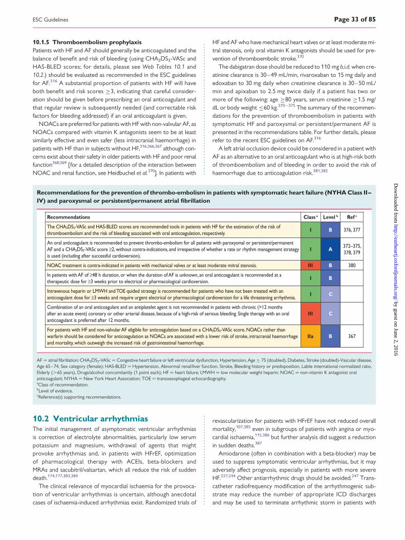
10.1.5 Thromboembolism prophylaxisPatients with HF and AF should generally be anticoagulated and thebalance of benefit and risk of bleeding (using CHA2DS2-VASc andHAS-BLED scores; for details, please see Web Tables 10.1 and10.2.) should be evaluated as recommended in the ESC guidelinesfor AF.316 A substantial proportion of patients with HF will haveboth benefit and risk scores ≥3, indicating that careful consider-ation should be given before prescribing an oral anticoagulant andthat regular review is subsequently needed (and correctable riskfactors for bleeding addressed) if an oral anticoagulant is given.
NOACs are preferred for patients with HF with non-valvular AF, asNOACs compared with vitamin K antagonists seem to be at leastsimilarly effective and even safer (less intracranial haemorrhage) inpatients with HF than in subjects without HF,316,366,367 although con-cerns exist about their safety in older patients with HF and poor renalfunction368,369 [for a detailed description of the interaction betweenNOAC and renal function, see Heidbuchel et al.370]. In patients with
HF and AF who have mechanical heart valves or at least moderate mi-tral stenosis, only oral vitamin K antagonists should be used for pre-vention of thromboembolic stroke.370
The dabigatran dose should be reduced to 110 mg b.i.d. when cre-atinine clearance is 30–49 mL/min, rivaroxaban to 15 mg daily andedoxaban to 30 mg daily when creatinine clearance is 30–50 mL/min and apixaban to 2.5 mg twice daily if a patient has two ormore of the following: age ≥80 years, serum creatinine ≥1.5 mg/dL or body weight ≤60 kg.370 –375 The summary of the recommen-dations for the prevention of thromboembolism in patients withsymptomatic HF and paroxysmal or persistent/permanent AF ispresented in the recommendations table. For further details, pleaserefer to the recent ESC guidelines on AF.316
A left atrial occlusion device could be considered in a patient withAF as an alternative to an oral anticoagulant who is at high-risk bothof thromboembolism and of bleeding in order to avoid the risk ofhaemorrhage due to anticoagulation risk.381,382
10.2 Ventricular arrhythmiasThe initial management of asymptomatic ventricular arrhythmiasis correction of electrolyte abnormalities, particularly low serumpotassium and magnesium, withdrawal of agents that mightprovoke arrhythmias and, in patients with HFrEF, optimizationof pharmacological therapy with ACEIs, beta-blockers andMRAs and sacubitril/valsartan, which all reduce the risk of suddendeath.174,177,383,384
The clinical relevance of myocardial ischaemia for the provoca-tion of ventricular arrhythmias is uncertain, although anecdotalcases of ischaemia-induced arrhythmias exist. Randomized trials of
revascularization for patients with HFrEF have not reduced overallmortality,107,385 even in subgroups of patients with angina or myo-cardial ischaemia,115,386 but further analysis did suggest a reductionin sudden deaths.387
Amiodarone (often in combination with a beta-blocker) may beused to suppress symptomatic ventricular arrhythmias, but it mayadversely affect prognosis, especially in patients with more severeHF.227,244 Other antiarrhythmic drugs should be avoided.247 Trans-catheter radiofrequency modification of the arrhythmogenic sub-strate may reduce the number of appropriate ICD dischargesand may be used to terminate arrhythmic storm in patients with
Recommendations for the prevention of thrombo-embolism in patients with symptomatic heart failure (NYHA Class II–IV) and paroxysmal or persistent/permanent atrial fibrillation
Recommendations Class a Level b Ref c
The CHA2DS2-VASc and HAS-BLED scores are recommended tools in patients with HF for the estimation of the risk of thromboembolism and the risk of bleeding associated with oral anticoagulation, respectively.
I B 376, 377
An oral anticoagulant is recommended to prevent thrombo-embolism for all patients with paroxysmal or persistent/permanent AF and a CHA2DS2-VASc score ≥2, without contra-indications, and irrespective of whether a rate or rhythm management strategy is used (including after successful cardioversion).
I A372–375, 378, 379
NOAC treatment is contra-indicated in patients with mechanical valves or at least moderate mitral stenosis. III B 380
In patients with AF of ≥48 h duration, or when the duration of AF is unknown, an oral anticoagulant is recommended at a therapeutic dose for ≥3 weeks prior to electrical or pharmacological cardioversion.
I B
Intravenous heparin or LMWH and TOE quided strategy is recommended for patients who have not been treated with an anticoagulant dose for ≥3 weeks and require urgent electrical or pharmacological cardioversion for a life threatening arrhythmia.
I C
Combination of an oral anticoagulant and an antiplatelet agent is not recommended in patients with chronic (>12 months after an acute event) coronary or other arterial disease, because of a high-risk of serious bleeding. Single therapy with an oral anticoagulant is preferred after 12 months.
III C
For patients with HF and non-valvular AF eligible for anticoagulation based on a CHA2DS2-VASc score, NOACs rather than warfarin should be considered for anticoagulation as NOACs are associated with a lower risk of stroke, intracranial haemorrhage and mortality, which outweigh the increased risk of gastrointestinal haemorrhage.
IIa B 367
AF ¼ atrial fibrillation; CHA2DS2-VASc ¼ Congestive heart failure or left ventricular dysfunction, Hypertension, Age ≥ 75 (doubled), Diabetes, Stroke (doubled)-Vascular disease,Age 65–74, Sex category (female); HAS-BLED ¼ Hypertension, Abnormal renal/liver function, Stroke, Bleeding history or predisposition, Labile international normalized ratio,Elderly (.65 years), Drugs/alcohol concomitantly (1 point each); HF ¼ heart failure; LMWH ¼ low molecular weight heparin; NOAC ¼ non-vitamin K antagonist oralanticoagulant; NYHA ¼ New York Heart Association; TOE ¼ transoesophageal echocardiography.aClass of recommendation.bLevel of evidence.cReference(s) supporting recommendations.
ESC Guidelines Page 33 of 85
by guest on June 2, 2016http://eurheartj.oxfordjournals.org/
Dow
nloaded from

HF and frequent, recurrent ventricular tachyarrhythmias andtherefore should be considered in such patients. Seeking theadvice of the members of the HF Team with expertise in electro-physiology is recommended in patients with recalcitrant ventricu-lar arrhythmias. For further details we refer the reader to the ESC/EHRA guidelines on ventricular arrhythmias and sudden cardiacdeath.260
Recommendations for the management of ventriculartachyarrhythmias in heart failure
Recommendations Class a Level b Ref c
Potential aggravating/precipitating factors (e.g. low serum potassium/magnesium, ongoing ischaemia) should be sought and corrected in patients with ventricular arrhythmias.
IIa C
Treatment with beta-blocker, MRA and sacubitril/valsartan reduces the risk of sudden death and is recommended for patients with HFrEF and ventricular arrhythmias (as for other patients)(see Section 7).
I A162,
170–175
Implantation of an ICD or CRT-D device is recommended for selected patients with HFrEF (see Section 8).
I A223–226,
388
Several strategies should be considered to reducerecurrent symptomatic arrhythmias in patients with an ICD(or in those who are not eligible for ICD), including attention to risk factors and optimal pharmacological treatment of HF, amiodarone, catheter ablation and CRT.
IIa C
Routine use of antiarrhythmic agents is not recommended in patients with HF and asymptomatic ventricular arrhythmias because of safety concerns (worsening HF, proarrhythmia, and death).
III A247, 248, 364, 365
ACEI ¼ angiotensin-converting enzyme inhibitor; ARB ¼ angiotensin receptorblocker; CRT ¼ cardiac resynchronization therapy; CRT-D ¼ defibrillator withcardiac resynchronization therapy; HF ¼ heart failure; HFrEF ¼ heart failure withreduced ejection fraction; ICD ¼ implantable cardioverter defibrillator; MRA ¼mineralocorticoid receptor antagonist.aClass of recommendation.bLevel of evidence.cReference(s) supporting recommendations.
10.3 Symptomatic bradycardia, pausesand atrio-ventricular blockThe ESC Guidelines on Pacing and CRT recommended interventionwhen pauses exceed 6 s, even when this is not associated with symp-toms.389 However, these recommendations were generated mainlyfor patients without obvious myocardial dysfunction, and shorterpauses might require intervention in patients with HFrEF.329 Ifpauses .3 s are identified on electrocardiographic monitoring,medications should be reviewed and the following agents stoppedor reduced in dose, starting with rate-limiting CCBs then
amiodarone, digoxin and ivabradine. For patients in AF, a reductionin the dose of beta-blockers allowing the daytime resting ventricularrate to rise to 70–90 bpm may be considered, since evidence thatbeta-blockers improve outcome in patients with AF is lacking.177 Forpatients with pauses but in sinus rhythm, a reduction in the dose ofbeta-blockers should be avoided unless the pauses are symptomatic,prolonged or frequent, in which case the relative merits of dose re-duction, beta-blocker withdrawal and (biventricular) pacing may beconsidered. However, evidence is lacking to support a strategy ofpacing solely to permit initiation or titration of beta-blocker therapyin the absence of a conventional pacing indication; this strategy is notrecommended. For patients with HFrEF and high-degree AV block,CRT is preferred over RV pacing (Section 8.2). When the cause ofbradycardia or pauses is sinus node disease with intact AV conduc-tion, then therapeutic strategies that avoid inducing ventricular dys-synchrony are preferred, although clinical trial evidence to supportthis expert opinion for patients with HF is sparse. For other pacingindications, please consult the ESC Guidelines on Pacing andCRT.389
Recommendations for the management ofbradyarrhythmias in heart failure
Recommendations Class a Level b Ref c
When pauses >3 seconds are
bradycardia is symptomatic and the resting ventricular rate is <50 bpm in sinus rhythm or <60 bpm in AF, it should be considered whether there is need for any rate limiting medications prescribed; for patients in sinus rhythm beta-blockers should be reduced in dose or withdrawn only as a last resort.
IIa C
For patients with symptomatic, prolonged or frequent pauses despite adjustment of rate limiting medication, either beta-blocker withdrawal or pacing may be considered as the next step.
IIb C
Pacing solely to permit initiation or titration of beta-blocker therapy in the absence of a conventional pacing indication is not recommended.
III C
In patients with HFrEF who require pacing and who have high degree AV block, CRT rather than RV pacing is recommended.
I A274, 275,
290
In patients with HFrEF who require pacing who do not have high degree AV block, pacing modes that avoid inducing or exacerbating ventricular dyssynchrony should be considered.
IIa C
AF ¼ atrial fibrillation; AV ¼ atrio-ventricular; bpm ¼ beats per minute; CRT ¼cardiac resynchronization therapy; ECG ¼ electrocardiogram; HFrEF ¼ heartfailure with reduced ejection fraction; RV ¼ right ventricular.aClass of recommendation.bLevel of evidence.cReference(s) supporting recommendations.
ESC GuidelinesPage 34 of 85
by guest on June 2, 2016http://eurheartj.oxfordjournals.org/
Dow
nloaded from

11. Co-morbidities
11.1. Heart failure and co-morbiditiesCo-morbidities are of great importance in HF (Table 11.1) and mayaffect the use of treatments for HF (e.g. it may not be possible to userenin–angiotensin system inhibitors is some patients with severe re-nal dysfunction) (see Section 7). The drugs used to treat co-morbidities may cause worsening of HF (e.g. NSAIDs given for arth-ritis, some anti-cancer drugs) (see Section 7). Management of co-morbidities is a key component of the holistic care of patientswith HF (see Section 14). Many co-morbidities are actively managedby specialists in the field of the co-morbidity, and these physicianswill follow their own specialist guidelines. The current guidelineswill identify where the presence of HF should change the way a co-morbidity would normally be treated. This may be because eithersafety or efficacy may be different in the presence of HF (or may sim-ply be unknown) or because of evidence of particular effects in anHF population, either beneficial or detrimental. HFpEF has an evenhigher prevalence of co-morbidities compared with HFrEF, andmany of these may be instrumental in the progression of thissyndrome.398
11.2 Angina and coronary artery disease11.2.1 Pharmacological managementBeta-blockers, and in selected patients ivabradine,180 are effectiveagents for angina control, as well as an essential component ofHFrEF therapy. In HFpEF patients, they may also be used for anginarelief, although this has never been formally tested. In the SIGNIFYtrial in patients with activity-limiting angina without HF, ivabradineincreased the risk of death from cardiovascular causes or non-fatalmyocardial infarction and therefore is not recommended in thissetting.399
Trimetazidine has been shown to exert some beneficial effect asan add-on to beta-blockers in patients with HF and angina.400 –406
There are data suggesting that it may improve NYHA functionalcapacity, exercise duration and LV function in patients withHFrEF.402 – 406 Certain other effective anti-anginal drugs havebeen studied in sizeable numbers of HFrEF/LV dysfunction patientsand shown to be safe [e.g. amlodipine,215,407 nicorandil408 and ni-trates183,184,409]. The safety of other anti-anginal agents in HFrEF,such as ranolazine, is uncertain, while other drugs, specifically dil-tiazem and verapamil, are thought to be unsafe in patients withHFrEF (although they may be used in HFpEF).214 DihydropyridineCCBs may all increase sympathetic tone, and their safety inHFrEF [except amlodipine215 and felodipine216] and HFpEF isuncertain.
11.2.2 Myocardial revascularizationFor indications for invasive coronary angiography in patients withHF, please refer to Section 5.8.
Percutaneous and surgical revascularization are complementaryapproaches for symptomatic relief of angina in HFpEF, but whetherthese interventions improve outcomes is not entirely clear. RecentESC guidelines on myocardial revascularization recommendedcoronary artery bypass grafting (CABG) for patients with
significant left main stenosis and left main equivalent (proximalstenosis of both the left anterior descending and left circumflex ar-teries) to improve prognosis.112,113 However, one needs to beaware of a lack of studies including patients who have well-definedHF, therefore this recommendation is solely based on expert opin-ion. On the basis of the results of the STICH trial [which excludedpatients with left main disease and Canadian Cardiovascular Soci-ety (CCS) angina classes III– IV], CABG is also recommended in pa-tients with HFrEF, significant CAD (left anterior descending arteryor multivessel disease) and LVEF ≤35% to reduce death and hos-pitalization for cardiovascular causes.385 Patients with .10% dys-functional but viable LV myocardium may be more likely to benefitfrom myocardial revascularization (and those with ≤10% are lesslikely to benefit), although this approach to patient selection forrevascularization is unproven. In the STICH trial, neither the pres-ence of viability nor the severity of LV remodelling identified thosewho benefited from CABG in terms of a reduction in mortality.118
For the assessment of techniques to assess myocardial viability,please refer to Section 5. Post hoc analyses from the STICH trialrevealed that the presence of inducible myocardial ischaemia (ei-ther on radionuclide stress test or dobutamine stress echocardio-gram) or angina does not identify those with worse prognosis andgreater benefit from CABG over OMT.115,386 However, CABGdoes improve angina to a greater extent than medical therapyalone.
The choice between CABG and PCI should be made by the HeartTeam after careful evaluation of the patient’s clinical status and cor-onary anatomy, expected completeness of revascularization, coex-isting valvular disease and co-morbidities.
Table 11.1 Importance of co-morbidities in patientswith heart failure
1. interfere with the diagnostic process of HF (e.g. COPD as a potentially confounding cause of dyspnoea).390, 391
2. aggravate HF symptoms and further impair quality of life.391, 392
3. contribute to the burden of hospitalizations and mortality,393 as the main cause of readmissions at 1 and 3 months.394
4. may affect the use of treatments for HF (e.g. renin–angiotensin system inhibitors contra-indicated in some patients with severe renal dysfunction or beta-blockers relatively contra-indicated in asthma).395, 396
5. evidence base for HF treatment is more limited as co-morbidities were
is therefore often lacking in the presence of co-morbidities.
6. drugs used to treat co-morbidities may cause worsening HF (e.g. NSAIDs given for arthritis, some anti-cancer drugs).397
7. interaction between drugs used to treat HF and those used to treat
occurrence of side effects (e.g. beta-blockers for HFrEF and beta-agonists for COPD and asthma).391, 395, 396
HF ¼ heart failure; COPD ¼ chronic obstructive pulmonary disease; HFrEF ¼heart failure with reduced ejection fraction; NSAIDs ¼ non-steroidalanti-inflammatory drugs.
ESC Guidelines Page 35 of 85
by guest on June 2, 2016http://eurheartj.oxfordjournals.org/
Dow
nloaded from
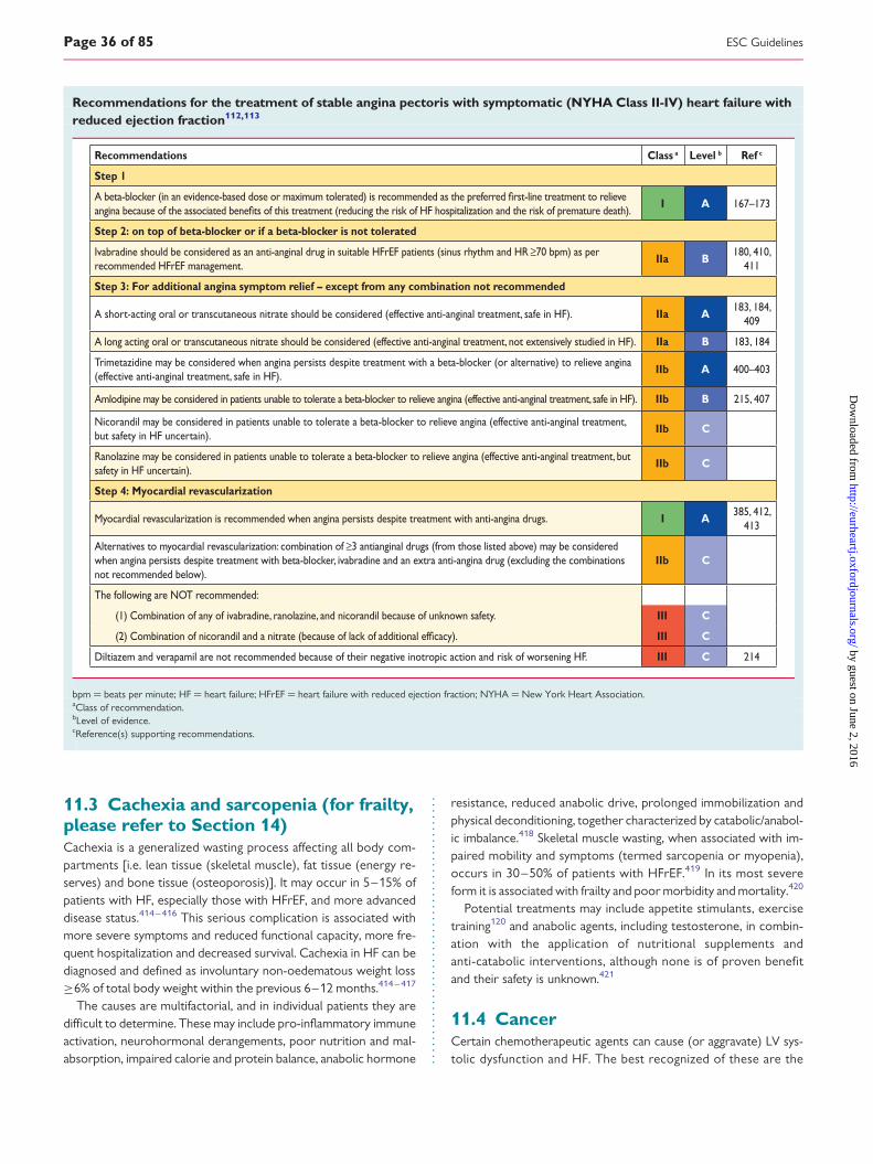
11.3 Cachexia and sarcopenia (for frailty,please refer to Section 14)Cachexia is a generalized wasting process affecting all body com-partments [i.e. lean tissue (skeletal muscle), fat tissue (energy re-serves) and bone tissue (osteoporosis)]. It may occur in 5–15% ofpatients with HF, especially those with HFrEF, and more advanceddisease status.414 – 416 This serious complication is associated withmore severe symptoms and reduced functional capacity, more fre-quent hospitalization and decreased survival. Cachexia in HF can bediagnosed and defined as involuntary non-oedematous weight loss≥6% of total body weight within the previous 6–12 months.414 – 417
The causes are multifactorial, and in individual patients they aredifficult to determine. These may include pro-inflammatory immuneactivation, neurohormonal derangements, poor nutrition and mal-absorption, impaired calorie and protein balance, anabolic hormone
resistance, reduced anabolic drive, prolonged immobilization andphysical deconditioning, together characterized by catabolic/anabol-ic imbalance.418 Skeletal muscle wasting, when associated with im-paired mobility and symptoms (termed sarcopenia or myopenia),occurs in 30–50% of patients with HFrEF.419 In its most severeform it is associated with frailty and poor morbidity and mortality.420
Potential treatments may include appetite stimulants, exercisetraining120 and anabolic agents, including testosterone, in combin-ation with the application of nutritional supplements andanti-catabolic interventions, although none is of proven benefitand their safety is unknown.421
11.4 CancerCertain chemotherapeutic agents can cause (or aggravate) LV sys-tolic dysfunction and HF. The best recognized of these are the
Recommendations for the treatment of stable angina pectoris with symptomatic (NYHA Class II-IV) heart failure withreduced ejection fraction112,113
Recommendations Class a Level b Ref c
Step 1
I A 167 –173
Step 2: on top of beta-blocker or if a beta-blocker is not tolerated
Ivabradine should be considered as an anti-anginal drug in suitable HFrEF patients (sinus rhythm and HR ≥70 bpm) as per recommended HFrEF management.
IIa B180, 410,
411
Step 3: For additional angina symptom relief – except from any combination not recommended
A short-acting oral or transcutaneous nitrate should be considered (effective anti-anginal treatment, safe in HF). IIa A183, 184,
409
A long acting oral or transcutaneous nitrate should be considered (effective anti-anginal treatment, not extensively studied in HF). IIa B 183, 184
Trimetazidine may be considered when angina persists despite treatment with a beta-blocker (or alternative) to relieve angina (effective anti-anginal treatment, safe in HF).
IIb A 400–403
Amlodipine may be considered in patients unable to tolerate a beta-blocker to relieve angina (effective anti-anginal treatment, safe in HF). IIb B 215, 407
Nicorandil may be considered in patients unable to tolerate a beta-blocker to relieve angina (effective anti-anginal treatment, but safety in HF uncertain).
IIb C
Ranolazine may be considered in patients unable to tolerate a beta-blocker to relieve angina (effective anti-anginal treatment, but safety in HF uncertain).
IIb C
Step 4: Myocardial revascularization
Myocardial revascularization is recommended when angina persists despite treatment with anti-angina drugs. I A385, 412,
413
Alternatives to myocardial revascularization: combination of ≥3 antianginal drugs (from those listed above) may be considered when angina persists despite treatment with beta-blocker, ivabradine and an extra anti-angina drug (excluding the combinations not recommended below).
IIb C
The following are NOT recommended:
(1) Combination of any of ivabradine, ranolazine, and nicorandil because of unknown safety. III C
(2) Combination of nicorandil and a nitrate (because of lack of III C
Diltiazem and verapamil are not recommended because of their negative inotropic action and risk of worsening HF. III C 214
bpm ¼ beats per minute; HF ¼ heart failure; HFrEF ¼ heart failure with reduced ejection fraction; NYHA ¼ New York Heart Association.aClass of recommendation.bLevel of evidence.cReference(s) supporting recommendations.
ESC GuidelinesPage 36 of 85
by guest on June 2, 2016http://eurheartj.oxfordjournals.org/
Dow
nloaded from

anthracyclines (e.g. doxorubicin), trastuzumab and tyrosine kinaseinhibitors.397,422 A recent Cochrane review found that dexrazoxanemay confer some cardioprotection in patients receiving anthracy-clines.423 Pre- and post-evaluation of LVEF, if available with myocar-dial strain imaging, is essential in patients receiving cardiotoxicchemotherapy, as detailed elsewhere.397,422 A risk score for identi-fying women with breast cancer at risk of developing HF during tras-tuzumab therapy has been developed based on age, chemotherapydetails, baseline cardiovascular status and other co-morbidities, andmay be helpful.424 Chemotherapy should be discontinued andHFrEF therapy commenced in patients developing moderate to se-vere LV systolic dysfunction. If LV function improves, the risks andbenefits of further chemotherapy need to be reconsid-ered.397,425,426 Mediastinal irradiation can also lead to a variety oflong-term cardiac complications. Cardiac biomarkers (NPs and tro-ponins) can be used to identify patients at higher risk of cardiotoxi-city and may be helpful in monitoring the use and dosing ofcardiotoxic cytotoxics.397,425,426
11.5 Central nervous system (includingdepression, stroke and autonomicdysfunction)Stroke and HF commonly coexist because of an overlap of sharedrisk factors. Both contribute to a worse prognosis. Stroke maymake self-care more difficult for the HF patient. Management ofhigh-risk stroke patients may require balancing the risk of anticoagu-lant and antiplatelet therapies.
Autonomic dysfunction is common in HFrEF, especially when se-vere.427 Combined with low blood pressure, it can make fainting andinjuries more likely and can interfere with optimal dosing of beta-blockers, ACEIs, ARBs and MRAs. Diuretic dosage may be reducedto reduce the severity of postural hypotension.
Depression is common and is associated with worse clinical sta-tus and a poor prognosis in HF.428 – 430 It may also contribute topoor adherence and social isolation. A high index of suspicion isneeded to make the diagnosis, especially in the elderly. Routinescreening using a validated questionnaire is good practice. Untilnow, the Beck Depression Inventory (BDI) and Cardiac DepressionScale have been formally validated as reliable tools for the assess-ment of depressive mood in patients with HF,431,432 but other ques-tionnaires have been broadly used in this group of patients (e.g.Geriatric Depression Scale, Hamilton Depression Scale, HospitalAnxiety and Depression Scale).
Psychosocial intervention and pharmacological treatment arehelpful, as well as exercise training, in patients with HFrEF anddepression.433 Cognitive behavioural therapy delivered in pa-tients with HF and major depression beyond standard careand a structured education programme were able to reducedepression severity, anxiety and fatigue symptoms, as well asimprove social functioning and mental and HF-related qualityof life.434
Selective serotonin reuptake inhibitors are thought to be safe, al-though the Sertraline Antidepressant Heart Attack RandomizedTrial did not confirm that sertraline provides a greater reduction
in depressive symptoms or improvement in cardiovascular statuscompared with placebo in HFrEF patients, but this trial was notpowered enough to prove the latter.435 Similarly, escitalopramhad no effect on either depression or clinical outcomes during the24-month follow-up as compared with placebo in patients withHFrEF and depression. Importantly, tricyclic antidepressants shouldbe avoided, because they may cause hypotension, worsening HF andarrhythmias.429,435
11.6 DiabetesDysglycaemia and diabetes are very common in HF, and diabetes isassociated with poorer functional status and worse prognosis. In pa-tients with HFrEF, interventions that reduce morbidity and mortalityconfer similar benefit in the presence or absence of diabetes.320 Forinstance, beta-blockers improve outcome similarly, whether or notthe patient has diabetes, although different beta-blockers may varyin their effects on glycaemic indices.436
Whether strict glycaemic control alters the risk of cardiovascularevents in patients with HF is uncertain.437 Among patients with HFwho have not been treated for diabetes, higher HbA1c is associatedwith greater risk of cardiovascular events,438,439 but this may not bethe case once treatment for diabetes has been commenced.439
In patients with diabetes and HF, glycaemic control should be im-plemented gradually and moderately, giving preference to thosedrugs, such as metformin, that have been shown to be safe and ef-fective. In contrast to what was previously believed, metformin issafe to use in patients with HFrEF, and it should be the treatmentof choice in patients with HF440,441 but is contraindicated in patientswith severe renal or hepatic impairment, because of the risk of lacticacidosis.
Insulin is required for patients with type 1 diabetes and to treatsymptomatic hyperglycaemia in patients with type 2 diabetes andpancreatic islet b cell exhaustion. However, insulin is a powerfulsodium-retaining hormone, and when combined with a reductionin glycosuria, may exacerbate fluid retention, leading to HF wor-sening. Sulphonylurea derivatives have also been associated withan increased risk of worsening HF and should be used withcaution.
Thiazolidinediones (glitazones) cause sodium and waterretention and increased risk of worsening HF and hospitalizationand are not recommended in patients with HF.209,210
Dipeptidylpeptidase-4 inhibitors (DPP4is; gliptins), which increaseincretin secretion, thereby stimulating insulin release, and long-acting glucagon-like peptide 1 (GLP-1) receptor agonists, whichact as incretin mimetics, improve glycaemic indices but do not re-duce and may increase the risk of cardiovascular events and wor-sening HF.320,442,443 Importantly, there are no data on the safety ofgliptins and GLP-1 analogues in patients with HF.
Recently, empagliflozin, an inhibitor of sodium-glucose co-transporter 2, reduced hospitalization for HF and mortality, butnot myocardial infarction or stroke, in patients with diabetes athigh cardiovascular risk, some of whom had HF.130 In the absenceof other studies with drugs from this group, the results obtainedwith empaglifozin cannot be considered as a proof of a classeffect.
ESC Guidelines Page 37 of 85
by guest on June 2, 2016http://eurheartj.oxfordjournals.org/
Dow
nloaded from

As glycaemic derangement progresses, the judgement on gly-caemic control should be made according to cardiac conditions,and if the new anti-diabetic drugs are to be prescribed, they haveto be closely monitored by an HF team.
11.7 Erectile dysfunctionErectile dysfunction is a common and important component of qual-ity of life in men with HF.444,445 Its treatment should include optimaltherapies for underlying cardiovascular diseases and other interfer-ing co-morbidities (e.g. diabetes) and amelioration of anxiety anddepressive symptoms. Some drugs applied for HF therapy (e.g. thia-zide diuretics, spironolactone and beta-blockers) may augmenterectile dysfunction.444,445 Phosphodiesterase type 5 inhibitors(PDE5Is) have been shown to have favourable haemodynamic andanti-remodelling effects and to improve exercise capacity and qual-ity of life in patients with HFrEF,446,447 but they are contraindicatedin patients taking nitrates.
11.8 Gout and arthritisHyperuricaemia and gout are common in HF and may be caused oraggravated by diuretic treatment. Hyperuricaemia is associated witha worse prognosis in HFrEF.448 The current European LeagueAgainst Rheumatism (EULAR) guideline for the management ofgout recommends that urate-lowering therapy (ULT) is indicatedin patients with recurrent acute flares, arthropathy, tophi or radio-graphic changes of gout, aiming to maintain a serum urate level be-low the saturation point for monosodium urate [,357 mmol/L(,6 mg/dL)].449
Xanthine oxidase inhibitors (allopurinol, oxypurinol) may be usedto prevent gout, although their safety in HFrEF is uncertain.450 Goutattacks are better treated with colchicine rather than with NSAIDs(although colchicine should not be used in patients with very severerenal dysfunction and may cause diarrhoea). Intra-articular corticos-teroids are an alternative for monoarticular gout, but systemic corti-costeroids cause sodium and water retention.
Arthritis is a common co-morbidity and is a common cause ofboth self-taken and prescribed drugs that can worsen renal functionand HF, especially NSAIDs. Rheumatoid arthritis is associated withan increased risk of HFpEF. The safety of disease-modifying drugscommonly given to patients with rheumatoid arthritis has notbeen established in HF.
11.9 Hypokalaemia and hyperkalaemiaBoth hypokalaemia and hyperkalaemia are associated with HF andwith many drugs used for HF treatment.451 Both can aggravate ven-tricular arrhythmias.
Loop and thiazide diuretics reduce serum potassium, whileACEIs, ARBs and MRAs can all increase serum potassium. Amilorideand triamterene are sometimes used as adjunct diuretics in resistantoedema and to assist in preventing hypokalaemia. The treatment ofhypokalaemia can involve recommending high potassium foods orprescribing potassium supplements.
The management of acute hyperkalaemia (.6.0 mmol/L) may re-quire a short-term cessation of potassium-retaining agents andRAAS inhibitors, but this should be minimized and RAAS inhibitorsshould be carefully reintroduced as soon as possible while monitor-ing potassium levels. A Cochrane review452 found no trial evidenceof major outcome benefits for any emergency therapy regimen forhyperkalaemia. Two new potassium binders (patiromer and sodiumzirconium cyclosilicate) are currently under consideration for regu-latory approval.453,454 Initial results from patients with HF are avail-able and confirm the efficacy of these therapies in reducing serumpotassium455 and preventing recurrent hyperkalaemia in patientswith HF and CKD in the context of treatment with RAASinhibitors.456
11.10 HyperlipidaemiaElevated low-density lipoprotein cholesterol is uncommon inHFrEF; patients with advanced HFrEF often have low concentrationsof low-density lipoprotein, which is associated with a worse progno-sis. Rosuvastatin did not reduce the primary composite mortality/morbidity endpoints in two large RCTs in patients with HF withor without IHD, but it also did not increase risk, and may have re-duced, hospitalizations.205,457 Therefore there is no evidence to rec-ommend the initiation of statins in most patients with HF. However,in patients who are already receiving a statin for CAD, a continu-ation of this therapy may be considered.
11.11 HypertensionHypertension is associated with an increased risk of developing HF;antihypertensive therapy markedly reduces the incidence of HF(with an exception of a-adrenoceptor blockers, which are less ef-fective than other antihypertensives in preventing HF).458 A recentprospective cohort study documented that in a population with in-cident HF, higher baseline systolic, diastolic and pulse pressure levelswere associated with a higher rate of adverse events, which furthersupports the importance for optimized blood pressure control inthis population.459 Blood pressure control is an element of the hol-istic management of patients with HF.
Negatively inotropic CCBs (i.e. diltiazem and verapamil) shouldnot be used to treat hypertension in patients with HFrEF (but arebelieved to be safe in HFpEF), and moxonidine should also beavoided in patients with HFrEF, as it increased mortality in patientsin one RCT.460 If blood pressure is not controlled with an ACEI (oran ARB), a beta-blocker, an MRA and a diuretic, then hydralazineand amlodipine215 [or felodipine216] are additional blood pressurelowering agents that have been shown to be safe in systolic HF. Theblood pressure targets recommended in hypertension guide-lines317 are applicable to HF. Uncontrolled hypertension inpatients with HFrEF is very rare, provided they are optimally trea-ted for HF. In contrast, treatment of hypertension is an importantissue in patients with HFpEF. In patients with AHF, i.v. nitrates (orsodium nitroprusside) are recommended to lower blood pressure(see Section 12).
ESC GuidelinesPage 38 of 85
by guest on June 2, 2016http://eurheartj.oxfordjournals.org/
Dow
nloaded from

11.12 Iron deficiency and anaemiaIron deficiency is common in HF, as it is with other chronic ill-nesses, and it can lead to anaemia and/or skeletal muscle dysfunc-tion without anaemia.466 Within an HF population, iron deficiencyis associated with a worse prognosis.467,468 Intravenous iron hasbeen specifically studied in two RCTs in patients with HF andiron deficiency (serum ferritin ,100 mg/L or ferritin between100 and 299 mg/L and transferrin saturation ,20%)469,470 bothwith and without anaemia. Intravenous ferric carboxymaltose(FCM) has been shown to improve self-reported patient global as-sessment, quality of life and NYHA class (over 6 months) in theFAIR-HF trial469 both in anaemic and non-anaemic patients withHF,471 and in the CONFIRM-HF trial470, exercise capacity im-proved over 24 weeks. In the analysis of secondary endpoints inthe CONFIRM-HF trial, i.v. iron reduced the risk of HF hospitaliza-tions in iron-deficient patients with HFrEF.470 A meta-analysis of
i.v. iron therapy in HFrEF patients with iron deficiency over upto 52 weeks showed reduced hospitalization rates and improvedHF symptoms, exercise capacity and quality of life.472 Treatmentwith FCM may therefore result in sustainable improvement in func-tional capacity, symptoms and quality of life. Treatment was alsoassociated with a significant reduction in hospitalizations for wor-sening HF. The number of deaths and the incidence of adverseevents were similar. Neither i.v. iron trial was powered to testfor an effect on major outcomes or to evaluate separately the ef-fects in anaemic and non-anaemic patients. The effect of treatingiron deficiency in HFpEF/HFmrEF and the long-term safety ofiron therapy in either HFrEF, HFmrEF or HFpEF is unknown. Thesafety of i.v. iron is unknown in patients with HF and haemoglobin.15 g/dL.469,470 Patients with iron deficiency need to be screenedfor any potentially treatable/reversible causes (e.g. gastrointestinalsources of bleeding).
Recommendations for the treatment of hypertension in patients with symptomatic (NYHA Class II-IV) heart failure withreduced ejection fraction
Recommendations Class a Level b Ref c
Step 1
They are also safe in HFpEF.I A
2, 164, 165, 167,
168, 171–174,
182, 461–463
Step 2
A thiazide diuretic (or if the patient is being treated with a thiazide diuretic, switching to a loop diuretic) is recommended to reduce blood pressure when hypertension persists despite treatment with a combination of an ACE-I (or alternatively ARB but NOT together withan ACE-I), a beta-blocker and an MRA.
I C
Step 3
Amlodipine or hydralazine is recommended to reduce blood pressure when hypertension persists despite treatment with a combination of an ACE-I (or alternatively ARB but NOT together withan ACE-I), a beta-blocker, an MRA and a diuretic.
I A183, 184, 215, 409
Felodipine should be considered to reduce blood pressure when hypertension persists despite treatment with a combination of an ACE-I (or alternatively ARB but NOT together withan ACE-I), a beta-blocker, an MRA and a diuretic.
IIa B 216
Moxonidine is not recommended to reduce blood pressure because of safety concerns in HFrEF patients (increased mortality). III B 460
Alpha-adrenoceptor antagonists are not recommended to reduce blood pressure because of safety concerns in HFrEF patients III A
458, 464, 465
Diltiazem and verapamil are not recommended to reduce blood pressure in patients with HFrEF because of their negative inotropic action and risk of worsening HF.
III C 214
ACE ¼ angiotensin-converting enzyme; ARB ¼ angiotensin receptor blocker; HF ¼ heart failure; HFmrEF ¼ heart failure with mid-range ejection fraction; HFpEF ¼ heart failurewith preserved ejection fraction; HFrEF ¼ heart failure with reduced ejection fraction; MRA ¼ mineralocorticoid receptor antagonist; NYHA ¼ New York Heart Association.aClass of recommendation.bLevel of evidence.cReference(s) supporting recommendations.
ESC Guidelines Page 39 of 85
by guest on June 2, 2016http://eurheartj.oxfordjournals.org/
Dow
nloaded from

Recommendations for the treatment of otherco-morbidities in patients with heart failure
Recommendations Class a Level b Ref c
Intravenous FCM should be considered in symptomatic patients
(serum ferritin <100 µg/L, or ferritin between 100–299 µg/L and transferrin saturation <20%) in order to alleviate HF symptoms, and improve exercise capacity and quality of life.
IIa A 469, 470
Diabetes
Metformin should be considered as
control in patients with diabetes and HF, unless contra-indicated.
IIa C 440 ,441
FCM ¼ ferric carboxymaltose; HF ¼ heart failure; HFrEF ¼ heart failure withreduced ejection fraction.aClass of recommendation.bLevel of evidence.cReference(s) supporting recommendations.Treatments not recommended for other co-morbidities in patients with heart failure
Treatments not recommended of other co-morbiditiesin patients with heart failure
Recommendations Class a Level b Ref c
Sleep apnoea
Adaptive servo-ventilation is not recommended in patients with HFrEF and a predominant central sleep apnoea because of an increased all-cause and cardiovascular mortality.
III B 473
Diabetes
Thiazolidinediones (glitazones) are not recommended in patients with HF, as they increase the risk of HF worsening and HF hospitalization.
III A 209, 210
Arthritis
NSAIDs or COX-2 inhibitors are not recommended in patients with HF, as they increase the risk of HF worsening and HF hospitalization.
III B 211–213
COX-2 ¼ cyclooxygenase 2; HF ¼ heart failure; HFrEF ¼ heart failure withreduced ejection fraction; NSAID ¼ non-steroidal anti-inflammatory drug.aClass of recommendation.bLevel of evidence.cReference(s) supporting recommendations.
Anaemia (defined as a haemoglobin concentration ,13.0 g/dL inmen and ,12.0 g/dL in women) is common in HF, particularly inhospitalized patients. It is more common in women, the elderlyand in patients with renal impairment and is associated with ad-vanced myocardial remodelling, inflammation and volume over-load.474 Anaemia is associated with advanced symptoms, worse
functional status, greater risk of HF hospitalization and reduced sur-vival. A diagnostic workup to seek a cause for any finding of anaemiais indicated (e.g. occult blood loss, iron deficiency, B12/folate defi-ciency, blood dyscrasias), although in many patients no specificcause is found. The erythropoietin-stimulating agent darbepoetinalfa did not improve clinical outcomes in HFrEF patients with mildto moderate anaemia, but led to an excess of thromboembolicevents and is therefore not recommended.475
11.13 Kidney dysfunction (includingchronic kidney disease, acute kidneyinjury, cardio-renal syndrome andprostatic obstruction)HF and CKD frequently coexist, share many risk factors (diabetes,hypertension, hyperlipidaemia) and interact to worsen progno-sis.476,477 CKD is generally defined as an eGFR ,60 mL/min/1.73 m2 and/or the presence of albuminuria (high 30 –300 orvery high .300 mg albumin/1 g of urine creatinine). Patientswith severe renal dysfunction (eGFR ,30 mL/min/1.73m2) havesystematically been excluded from randomized clinical trials andtherefore there is lack of evidence-based therapies in thesepatients.
A further deterioration in renal function, termed worsening renalfunction (WRF), is used to indicate an increase in serum creatinine,usually by .26.5 mmol/L (0.3 mg/dL) and/or a 25% increase or a20% drop in GFR. The importance of these apparently small changesis that they are frequent, they promote the development and pro-gression of CKD478 and, as a consequence, can worsen the progno-sis of HF. Increases in creatinine during an AHF hospitalization arenot always clinically relevant, especially when they are accompaniedby appropriate decongestion, diuresis and haemoconcentration.479
Large increases in serum creatinine, termed acute kidney injury(AKI), are relatively rare in HF and are probably associated withthe combination of diuretic therapy with other potentially nephro-toxic drugs such as some antibiotics (gentamicin and trimethoprim),contrast media, ACEIs, ARBs, NSAIDs, etc. Of relevance, some ofthese drugs may accumulate if they are renally excreted. In HF,WRF is relatively common, especially during initiation and up-titration of RAAS inhibitor therapy. Despite the fact that RAASblockers can frequently cause a decrease in GFR in patients withHF, this reduction is usually small and should not lead to treatmentdiscontinuation unless there is a marked decrease, as the treatmentbenefit in these patients is probably largely maintained.480 Whenlarge increases in serum creatinine occur, care should be taken toevaluate the patient thoroughly and should include assessment ofa possible renal artery stenosis, excessive hyper- or hypovolaemia,concomitant medication and hyperkalaemia, which frequentlycoincides with WRF.
Diuretics, especially thiazides, but also loop diuretics, may be less ef-fective in patients with a very low GFR, and if used, should be dosed ap-propriately (higher doses to achieve similar effects). Renally excreteddrugs (e.g. digoxin, insulin and low molecular weight heparin) may accu-mulate in patients with renal impairment and may need dose adjustmentif renal function deteriorates. Patients with HF and coronary or periph-eral vascular disease are at risk of acute renal dysfunction when theyundergo contrast media enhanced angiography [contrast-induced acute
ESC GuidelinesPage 40 of 85
by guest on June 2, 2016http://eurheartj.oxfordjournals.org/
Dow
nloaded from

kidney injury (CI-AKI)]. Renal dysfunction and worsening renal functionis further discussed in the section about AHF (see Section 12).
Prostatic obstruction is common in older men and can interferewith renal function; it should therefore be ruled out in men with HFwith deteriorating renal function. a-adrenoceptor blockers causehypotension and sodium and water retention, and may not besafe in HFrEF.458,464,465 For these reasons, 5-a-reductase inhibitorsare generally preferred in the medical treatment of prostaticobstruction in patients with HF.
11.14 Lung disease (including asthma andchronic obstructive pulmonary disease)The diagnosis of COPD and asthma may be difficult in patients withHF, due to overlap in symptoms and signs, but also problems in theinterpretation of spirometry, especially in HFpEF.48,49,391 COPD(and asthma) in patients with HF may be overdiagnosed.481 Spirom-etry should be performed when patients have been stable and euvo-laemic for at least 3 months, to avoid the confounding effect ofpulmonary congestion causing external obstruction of alveoli andbronchioles.482 Both correctly and incorrectly labelled COPD are as-sociated with worse functional status and a worse prognosis in HFrEF.
Beta-blockers are only relatively contraindicated in asthma, butnot in COPD, although a more selective b1-adrenoceptor antagon-ist (i.e. bisoprolol, metoprolol succinate, or nebivolol) is pre-ferred.48,49,391 The contraindication to beta-blockers in asthma, asmentioned on pharmacy leaflets, is based on small case series pub-lished in the 1980s and late 1990s with very high initial dosages inyoung patients with severe asthma. In clinical practice, starting withlow doses of cardioselective beta-blockers combined with closemonitoring for signs of airway obstruction (wheezing, shortness ofbreath with lengthening of the expiration) may allow the use of pro-foundly effective beta-blockers in HFrEF, especially in older peoplewhere true severe asthma is uncommon. Therefore, according tothe 2015 GINA global strategy report,395,396 asthma is not an absolutecontraindication, but these medications should only be used underclose medical supervision by a specialist, with consideration of therisks for and against their use. The long-term safety of cardioactive in-haled pulmonary drugs is uncertain and the need for their use shouldbe reconsidered in patients with HFrEF, especially as their benefit inasthma and COPD may be symptomatic only without a clear effect onmortality. Oral corticosteroids can cause sodium and water reten-tion, potentially leading to worsening of HF, but this is not believedto be a problem with inhaled corticosteroids. Pulmonary hyperten-sion can complicate severe long-standing COPD, which, as a result,makes right-sided HF and congestion more likely. Non-invasive ven-tilation, added to conventional therapy, improves the outcome of pa-tients with acute respiratory failure due to hypercapnic exacerbationof COPD or HF in situations of acute pulmonary oedema.
11.15 ObesityObesity is a risk factor for HF141 and complicates its diagnosis, becauseit can cause dyspnoea, exercise intolerance and ankle swelling and mayresult in poor-quality echocardiographic images. Obese individuals alsohave reduced NP levels.62 Obesity is more common in HFpEF than inHFrEF, although it is possible that misdiagnosis may explain at leastsome of this difference in prevalence. Although obesity is an independ-ent risk factor for developing HF, once HF is diagnosed, it is well
established that obesity is associated with lower mortality across awide range of body mass indexes (BMIs) (see also cachexia in Section11.3)—the so-called obesity paradox also seen in other chronic ill-nesses.414,416 Obesity should be managed as recommended in theESC guidelines on cardiovascular disease prevention,483 if the aim isto prevent future development of HF. However, these guidelines donot refer to the HF patient in whom higher BMI is not adverse, and, al-though often recommended for symptom benefit and risk factor con-trol, weight loss as an intervention has never been prospectively shownto be either beneficial or safe in HFrEF. When weight loss is occurringin HF, it is associated with high mortality and morbidity, worse symp-tom status and poor quality of life. In patients with HF with moderatedegrees of obesity (BMI ,35 kg/m2), weight loss cannot be recom-mended. In more advanced obesity (BMI 35–45 kg/m2), weight lossmay be considered to manage symptoms and exercise capacity.
11.16 Sleep disturbance andsleep-disordered breathingSleep-disordered breathing (SDB) occurs in more than one-third ofpatients with HF,484 being even more prevalent in patients withAHF.485 The most common types are: central sleep apnoea (CSA,similar to Cheyne Stokes respiration, CSR), obstructive sleep ap-noea (OSA), and a mixed pattern of the two. Other causes of sleepdisturbance include anxiety, depression, decubitus or paroxysmalpulmonary congestion (orthopnoea and paroxysmal nocturnal dys-pnoea) and diuretic therapy causing nocturnal diuresis. Reviewingsleep history (including asking a partner) is part of the holisticcare of patients with HF (see Section 14). CSA and OSA havebeen shown to be associated with a worse prognosis in HF.485,486
OSA is associated with an increased risk of incident HF in men.487
CSA is the most common form of SDB in HFrEF, and HFrEF isthe most common cause of CSA, so they are closely linked. Screen-ing for, and the diagnosis and treatment of, sleep apnoea is discussedin detail elsewhere.484,488 Diagnosis used to require overnight poly-somnography, although advanced home testing equipment whichcan distinguish the type of sleep apnoea has been developed.
Nocturnal oxygen supplementation, continuous positive airwaypressure (CPAP), bi-level positive airway pressure (BiPAP), and adap-tive servo-ventilation (ASV) may be considered to treat nocturnalhypoxaemia in OSA as recommended in other guidelines.489,490 Anapnoea/hypopnoea index (AHI) of above 30 per hour can be treatedusing any of CPAP, BiPAP, ASV and nocturnal oxygen supplementa-tion, which have all been shown to be effective in this regard. It shouldbe noted, however, that none of these interventions has been pro-spectively shown to be beneficial on major outcomes in HFrEF.
CPAP in HF related CSA has been shown to reduce the fre-quency of episodes of apnoea and hypopnoea, and improve LVEFand 6 minute walk test distance, but did not improve prognosis orthe rate of HF related hospitalizations.491
The recently published SERVE-HF473 trial has shown that ASVused in patients with HFrEF and a predominantly CSA was neutralregarding the composite primary endpoint (all-cause death, lifesav-ing cardiovascular intervention, i.e. cardiac transplantation, implant-ation of a ventricular assist device, resuscitation after sudden cardiacarrest, or appropriate lifesaving shock, or unplanned hospitalizationfor HF worsening), but more importantly led to an increase in bothall-cause and cardiovascular mortality. Therefore ASV is not recom-mended in patients with HFrEF and predominantly CSA.
ESC Guidelines Page 41 of 85
by guest on June 2, 2016http://eurheartj.oxfordjournals.org/
Dow
nloaded from

The safety and efficacy of alternative approaches to treating CSAin HFrEF patients, such as implantable phrenic nerve stimula-tion,219,220,492 are presently undergoing clinical investigation andmay require additional long term study.
11.17. Valvular heart diseaseValvular heart disease may cause or aggravate HF. This section brief-ly addresses problems particularly relevant to HF, and the reader isreferred to the recent guidelines on valvular disease for moreinformation.493,494
Patients withHFand concomitant valvularheart disease constitute ahigh-risk population. Thus, the whole process of decision-makingthrough a comprehensive evaluation of the risk–benefit ratio of differ-ent treatment strategies should be made by a multidisciplinary ‘heartteam’ with aparticularexpertise in valvularheart disease, including car-diologists with expertise in HF, cardiac surgeons, a structural valveinterventionist if a catheter-based therapy is being considered, imagingspecialists, anaesthetists and, if needed, general practitioners, geriatri-cians, or intensive care specialists. This may be particularly beneficialin patients with HF being considered for surgery, transcatheter aorticvalve implantation or transcatheter mitral valve intervention.
All patients should receive OMT. In those with HFrEF pharmaco-logical therapy should be planned according to a previously describedalgorithm (see Section 7 for details). Care must be taken using vaso-dilators (ACEI, ARBs, CCBs, hydralazine, and nitrates) in patients withsevere aortic stenosis in order not to cause hypotension.
11.17.1. Aortic stenosisThe main concern in patients with severe aortic stenosis and re-duced LVEF is the entity of ‘low-flow, low-gradient’ aortic stenosis(valve area , 1 cm2, LVEF , 40%, mean pressure gradient , 40mmHg). In such individuals, low-dose dobutamine stress echocardi-ography should be considered to differentiate between patientswith moderate aortic stenosis, and those with severe stenosis andlow flow across the valve due to low stroke volume, and to evaluatefor contractile or flow reserve.
If the mean gradient is . 40 mmHg, there is theoretically no low-er LVEF limit for aortic valve replacement in symptomatic patientswith severe aortic stenosis.
Transaortic valve implantation (TAVI) is recommended in pa-tients with severe aortic stenosis who are not suitable for surgeryas assessed by a ‘heart team’ and have predicted post-TAVI survival. 1 year. TAVI should be also considered in high-risk patients withsevere aortic stenosis who may still be suitable for surgery, but inwhom TAVI is favoured by a ‘heart team’ based on the individualrisk profile and anatomic suitability.495,496 In a recent trial in patientswith severe aortic stenosis, TAVI with a self-expanding transcath-eter aortic valve bioprosthesis was associated with a significantlyhigher rate of survival at 1 year which was sustained at 2 years.497,498
11.17.2. Aortic regurgitationIn patients with severe aortic regurgitation, aortic valve repair or re-placement is recommended in all symptomatic patients and inasymptomatic patients with resting LVEF ≤ 50%, who are otherwisefit for surgery.499,500
11.17.3. Mitral regurgitationThis section refers to chronic settings while acute settings arediscussed in Section 12.
Primary (organic) mitral regurgitationSurgery is indicated in symptomatic patients with severe organicmitral regurgitation with no contra-indications to surgery. The deci-sion of whether to replace or repair depends mostly on valve anat-omy, surgical expertise available, and the patient’s condition.
When the LVEF is , 30%, a durable surgical repair may improvesymptoms, although its effect on survival is unknown. In this situ-ation, the decision to operate should take account of response tomedical therapy, co-morbidities, and the likelihood that the valvecan be repaired (rather than replaced).
Secondary mitral regurgitationThis occurs because LV enlargement and remodelling lead toreduced leaflet closing. Effective medical therapy (including CRTin suitable patients) leading to reverse remodelling of the LV mayreduce functional mitral regurgitation, and every effort should bemade to optimize medical treatment in these patients.
Combined valve and coronary surgery should be considered insymptomatic patients with LV systolic dysfunction (LVEF , 30%),coronary arteries suitable for revascularization, and evidence of via-bility. Surgery is also recommended in patients with severe mitral re-gurgitation undergoing CABG with LVEF . 30%.
However, a recent study in patients with moderate, secondary is-chaemic mitral regurgitation did not prove that the addition of mitralvalve repair to CABG would lead to a higher degree of LV reverse re-modelling.501 Also, there is no evidence favouring mitral valve repairover replacement in the context of better outcomes and magnitudeof LV remodelling.502 In the presence of AF, atrial ablation and LA ap-pendage closure may be considered at the time of mitral valve surgery.
The role of isolated mitral valve surgery in patients with severefunctional mitral regurgitation and severe LV systolic dysfunction(LVEF , 30%) who cannot be revascularized or have non-ischaemiccardiomyopathy is questionable, and in most patients conventionalmedical and device therapy are preferred. In selected cases, repairmay be considered in order to avoid or postpone transplantation.The decision should be based on comprehensive evaluation (includ-ing strain echocardiography or magnetic resonance imaging499,503
and discussed within the ‘heart team’.In patients with HF with moderate-severe, secondary mitral re-
gurgitation who are judged inoperable or at high surgical risk, percu-taneous mitral valve intervention (percutaneous edge-to-edgerepair) may be considered in order to improve symptoms and qual-ity of life, although no RCT evidence of improvement has been pub-lished, only registry studies.504 – 506
11.17.4. Tricuspid regurgitationSecondary (functional) tricuspid regurgitation (TR) frequently com-plicates the natural course of HF, due to annular dilatation and in-creased tricuspid leaflet tethering in relation to RV pressure and/or volume overload. Severe TR causes/deteriorates symptomsand signs of right HF, thus diuretics are used to reduce peripheraloedema. As hepatic congestion is often present in these patients(additionally contributing to hyperaldosteronism), an addition ofan MRA (in higher natriuretic doses) may improve decongestion.507
Management of HF which underlies secondary TR should be opti-mized as TR may diminish, following the treatment of its cause. In-dications for surgical correction of secondary TR complicating HFare not clearly established.493,494 The need for correction of TR isusually considered at the time of surgical correction of left-sided
ESC GuidelinesPage 42 of 85
by guest on June 2, 2016http://eurheartj.oxfordjournals.org/
Dow
nloaded from

valve lesions.493,494 A recent first report indicated that catheter-based interventions may be possible for TR.508
12. Acute heart failure
12.1 Definition and classificationAHF refers to rapid onset or worsening of symptoms and/or signsof HF. It is a life-threatening medical condition requiring urgentevaluation and treatment, typically leading to urgent hospitaladmission.
AHF may present as a first occurrence (de novo) or, more fre-quently, as a consequence of acute decompensation of chronicHF, and may be caused by primary cardiac dysfunction or precipi-tated by extrinsic factors, often in patients with chronic HF. Acutemyocardial dysfunction (ischaemic, inflammatory or toxic), acutevalve insufficiency or pericardial tamponade are among the mostfrequent acute primary cardiac causes of AHF. Decompensationof chronic HF can occur without known precipitant factors, butmore often with one or more factors, such as infection, uncon-trolled hypertension, rhythm disturbances or non-adherence withdrugs/diet (Table 12.1).
A large number of overlapping classifications of AHF based on dif-ferent criteria have been proposed.510 – 513 In practice the most use-ful classifications are those based on clinical presentation atadmission, allowing clinicians to identify patients at high risk of com-plications and to direct management at specific targets, which cre-ates a pathway for personalized care in the AHF setting. In mostcases, patients with AHF present with either preserved (90–140mmHg) or elevated (.140 mmHg; hypertensive AHF) systolicblood pressure (SBP). Only 5–8% of all patients present withlow SBP (i.e. ,90 mmHg; hypotensive AHF), which is associatedwith poor prognosis, particularly when hypoperfusion is alsopresent.514,515
Another approach is to classify patients according to the presenceof the following precipitants/causes leading to decompensation,which need to be treated/corrected urgently (see Section 12.3.1):ACS, hypertensive emergency, rapid arrhythmias or severe
Recommendations for treatment of valvular diseases in patients with heart failure
Recommendations Class a Level b Ref c
2, LVEF <40%, mean pressure gradient <40 mmHg), low-dose dobutamine stress echocardiography should be considered to identify those with severe aortic stenosis suitable for valve replacement.
IIa C
TAVI is recommended in patients with severe aortic stenosis who are not suitable for surgery as assessed by a ‘heart team’ and have predicted post-TAVI survival >1 year.
I B495, 496,
509
TAVI should be considered in high-risk patients with severe aortic stenosis who may still be suitable for surgery, but in whom TAVI is IIa A 497, 498
In patients with severe aortic regurgitation, aortic valve repair or replacement is recommended in all symptomatic patients and in asymptomatic patients with resting LVEF ≤ I C 317
Evidence-based medical therapy in patients with HFrEF is recommended in order to reduce functional mitral regurgitation. I C
Combined surgery of secondary mitral regurgitation and coronary artery bypass grafting should be considered in symptomatic patients with LV systolic dysfunction (LVEF <30%), requiring coronary revascularization for angina recalcitrant to medical therapy.
IIa C
Isolated surgery of non-ischaemic regurgitant mitral valve in patients with severe functional mitral regurgitation and severe LV systolic dysfunction (LVEF <30%) may be considered in selected patients in order to avoid or postpone transplantation.
IIb C
HFrEF ¼ heart failure with reduced ejection fraction; LV ¼ left ventricular; LVEF ¼ left ventricular ejection fraction; TAVI ¼ transaortic valve implantation.aClass of recommendation.bLevel of evidence.cReference(s) supporting recommendations.
Table 12.1 Factors triggering acute heart failure
Acute coronary syndrome.
Excessive rise in blood pressure.
Infection (e.g. pneumonia, infective endocarditis, sepsis).
Bradyarrhythmia.
Toxic substances (alcohol, recreational drugs).
Drugs (e.g. NSAIDs, corticosteroids, negative inotropic substances, cardiotoxic chemotherapeutics).
Exacerbation of chronic obstructive pulmonary disease.
Pulmonary embolism.
Surgery and perioperative complications.
Increased sympathetic drive, stress-related cardiomyopathy.
Metabolic/hormonal derangements (e.g. thyroid dysfunction, diabetic ketosis, adrenal dysfunction, pregnancy and peripartum related abnormalities).
Cerebrovascular insult.
Acute mechanical cause: myocardial rupture complicating ACS (free wall rupture, ventricular septal defect, acute mitral regurgitation), chest trauma or cardiac intervention, acute native or prosthetic valve incompetence secondary to endocarditis, aortic dissection or thrombosis.
ACS ¼ acute coronary syndromes; NSAIDs ¼ non-steroidal anti-inflammatorydrugs.
ESC Guidelines Page 43 of 85
by guest on June 2, 2016http://eurheartj.oxfordjournals.org/
Dow
nloaded from