2014 ESC Guidelines on the diagnosis and treatment of ... · 2014 ESC Guidelines on the diagnosis...
17
ESC GUIDELINES 2014 ESC Guidelines on the diagnosis and treatment of aortic diseases - web addenda Document covering acute and chronic aortic diseases of the thoracic and abdominal aorta of the adult The Task Force for the Diagnosis and Treatment of Aortic Diseases of the European Society of Cardiology (ESC) Authors/Task Force members: Raimund Erbel * (Chairperson) (Germany), Victor Aboyans * (Chairperson) (France), Catherine Boileau (France), Eduardo Bossone (Italy), Roberto Di Bartolomeo (Italy), Holger Eggebrecht (Germany), Arturo Evangelista (Spain), Volkmar Falk (Switzerland), Herbert Frank (Austria), Oliver Gaemperli (Switzerland), Martin Grabenwo ¨ ger (Austria), Axel Haverich (Germany), Bernard Iung (France), Athanasios John Manolis (Greece), Folkert Meijboom (Netherlands), Christoph A. Nienaber (Germany), Marco Roffi (Switzerland), Herve ´ Rousseau (France), Udo Sechtem (Germany), Per Anton Sirnes (Norway), Regula S. von Allmen (Switzerland), Christiaan J.M. Vrints (Belgium) ESC Committee for Practice Guidelines (CPG): Jose Luis Zamorano (Chairperson) (Spain), Stephan Achenbach (Germany), Helmut Baumgartner (Germany), Jeroen J. Bax (Netherlands), He ´ ctor Bueno (Spain), Veronica Dean (France), Christi Deaton (UK), Çetin Erol (Turkey), Robert Fagard (Belgium), Roberto Ferrari (Italy), David Hasdai (Israel), Arno Hoes (The Netherlands), Paulus Kirchhof (Germany/UK), Juhani Knuuti (Finland), Philippe Kolh (Belgium), Patrizio Lancellotti (Belgium), Ales Linhart (Czech Republic), Petros Nihoyannopoulos (UK), * Corresponding authors: Raimund Erbel, Department of Cardiology, West-German Heart Centre Essen, University Duisburg-Essen, Hufelandstrasse 55, DE-45122 Essen, Germany. Tel: +49 201 723 4801; Fax: +49 201 723 5401; Email: [email protected]. Victor Aboyans, Department of Cardiology, CHRU Dupuytren Limoges, 2 Avenue Martin Luther King, 87042 Limoges, France. Tel: +33 5 55 05 63 10; Fax: +33 5 55 05 63 84; Email: [email protected] Other ESC entities having participated in the development of this document: ESC Associations: Acute Cardiovascular Care Association (ACCA), European Association of Cardiovascular Imaging (EACVI), European Association of Percutaneous Cardiovascular Interventions (EAPCI). ESC Councils: Council for Cardiology Practice (CCP). ESC Working Groups: Cardiovascular Magnetic Resonance, Cardiovascular Surgery, Grown-up Congenital Heart Disease, Hypertension and the Heart, Nuclear Cardiology and Cardiac Computed Tomography, Peripheral Circulation, Valvular Heart Disease. The content of these European Society of Cardiology (ESC) Guidelines has been published for personal and educational use only. No commercial use is authorized. No part of the ESC Guidelines may be translated or reproduced in any form without written permission from the ESC. Permission can be obtained upon submission of a written request to Oxford University Press, the publisher of the European Heart Journal and the party authorized to handle such permissions on behalf of the ESC. Disclaimer: The ESC Guidelines represent the views of the ESC and were produced after careful consideration of the scientific and medical knowledge and the evidence available at the time of their dating. The ESC is not responsible in the event of any contradiction, discrepancy and/or ambiguity between the ESC Guidelines and any other official recommendations or guidelines issued by the relevant public health authorities, in particular in relation to good use of health care or therapeutic strategies. Health professionals are encouraged to take the ESC Guidelines fully into account when exercising their clinical judgment, as well as in the determination and the implementation of preventive, diagnostic or therapeutic medical strategies; however, the ESC Guidelines do not override, in any way whatsoever, the individual responsibility of health professionals to make appropriate and accurate decisions in consideration of each patient’s health condition and in consultation with that patient and, where appropriate and/or necessary, the patient’s caregiver. Nor do the ESC Guidelines exempt health professionals from taking full and careful consideration of the relevant official updated recommendations or guidelines issued by the competent public health authorities in order to manage each patient’s case in light of the scientifically accepted data pursuant to their respective ethical and professional obligations. It is also the health professional’s responsibility to verify the applicable rules and regulations relating to drugs and medical devices at the time of prescription. National Cardiac Societies document reviewers: listed in the Appendix. & The European Society of Cardiology 2014. All rights reserved. For permissions please email: [email protected]. European Heart Journal doi:10.1093/eurheartj/ehu281
Transcript of 2014 ESC Guidelines on the diagnosis and treatment of ... · 2014 ESC Guidelines on the diagnosis...

ESC GUIDELINES
2014 ESC Guidelines on the diagnosis andtreatment of aortic diseases - web addendaDocument covering acute and chronic aortic diseases of the thoracicand abdominal aorta of the adult
The Task Force for the Diagnosis and Treatment of Aortic Diseasesof the European Society of Cardiology (ESC)
Authors/Task Force members: Raimund Erbel* (Chairperson) (Germany),Victor Aboyans* (Chairperson) (France), Catherine Boileau (France),Eduardo Bossone (Italy), Roberto Di Bartolomeo (Italy), Holger Eggebrecht(Germany), Arturo Evangelista (Spain), Volkmar Falk (Switzerland), Herbert Frank(Austria), Oliver Gaemperli (Switzerland), Martin Grabenwoger (Austria),Axel Haverich (Germany), Bernard Iung (France), Athanasios John Manolis (Greece),Folkert Meijboom (Netherlands), Christoph A. Nienaber (Germany), Marco Roffi(Switzerland), Herve Rousseau (France), Udo Sechtem (Germany), Per Anton Sirnes(Norway), Regula S. von Allmen (Switzerland), Christiaan J.M. Vrints (Belgium)
ESC Committee for Practice Guidelines (CPG): Jose Luis Zamorano (Chairperson) (Spain), Stephan Achenbach(Germany), Helmut Baumgartner (Germany), Jeroen J. Bax (Netherlands), Hector Bueno (Spain), Veronica Dean(France), Christi Deaton (UK), Çetin Erol (Turkey), Robert Fagard (Belgium), Roberto Ferrari (Italy), David Hasdai(Israel), Arno Hoes (The Netherlands), Paulus Kirchhof (Germany/UK), Juhani Knuuti (Finland), Philippe Kolh(Belgium), Patrizio Lancellotti (Belgium), Ales Linhart (Czech Republic), Petros Nihoyannopoulos (UK),
* Corresponding authors: Raimund Erbel, Department of Cardiology, West-German Heart Centre Essen, University Duisburg-Essen, Hufelandstrasse 55, DE-45122 Essen, Germany.Tel:+49 201 723 4801; Fax: +49 201 723 5401; Email: [email protected].
Victor Aboyans, Department of Cardiology, CHRU Dupuytren Limoges, 2 Avenue Martin Luther King, 87042 Limoges, France. Tel: +33 5 55 05 63 10; Fax: +33 5 55 05 63 84;Email: [email protected]
Other ESC entities having participated in the development of this document:
ESC Associations: Acute Cardiovascular Care Association (ACCA), European Association of Cardiovascular Imaging (EACVI), European Association of Percutaneous CardiovascularInterventions (EAPCI).
ESC Councils: Council for Cardiology Practice (CCP).
ESC Working Groups: Cardiovascular Magnetic Resonance, Cardiovascular Surgery, Grown-up Congenital Heart Disease, Hypertension and the Heart, Nuclear Cardiology andCardiac Computed Tomography, Peripheral Circulation, Valvular Heart Disease.
The content of these European Society of Cardiology (ESC) Guidelines has been published for personal and educational use only. No commercial use is authorized. No part of the ESCGuidelines may be translated or reproduced in any form without written permission from the ESC. Permission can be obtained upon submission of a written request to Oxford UniversityPress, the publisher of the European Heart Journal and the party authorized to handle such permissions on behalf of the ESC.
Disclaimer: The ESC Guidelines represent the views of the ESC and were produced after careful consideration of the scientific and medical knowledge and the evidence available at thetime of their dating.
The ESC is not responsible in the event of any contradiction, discrepancy and/or ambiguity between the ESC Guidelines and any other official recommendations or guidelines issued bythe relevant public health authorities, in particular in relation to good use of health care or therapeutic strategies. Health professionals are encouraged to take the ESC Guidelines fully intoaccount when exercising their clinical judgment, as well as in the determination and the implementation of preventive, diagnostic or therapeutic medical strategies; however, the ESCGuidelines do not override, in any way whatsoever, the individual responsibility of health professionals to make appropriate and accurate decisions in consideration of each patient’shealth condition and in consultation with that patient and, where appropriate and/or necessary, the patient’s caregiver. Nor do the ESC Guidelines exempt health professionals fromtaking full and careful consideration of the relevant official updated recommendations or guidelines issued by the competent public health authorities in order to manage each patient’scase in light of the scientifically accepted data pursuant to their respective ethical and professional obligations. It is also the health professional’s responsibility to verify the applicable rulesand regulations relating to drugs and medical devices at the time of prescription.
National Cardiac Societies document reviewers: listed in the Appendix.
& The European Society of Cardiology 2014. All rights reserved. For permissions please email: [email protected].
European Heart Journaldoi:10.1093/eurheartj/ehu281

Massimo F. Piepoli (Italy), Piotr Ponikowski (Poland), Per Anton Sirnes (Norway), Juan Luis Tamargo (Spain),Michal Tendera (Poland), Adam Torbicki (Poland), William Wijns (Belgium), Stephan Windecker (Switzerland).
Document reviewers: Petros Nihoyannopoulos (CPG Review Coordinator) (UK), Michal Tendera (CPG ReviewCoordinator) (Poland), Martin Czerny (Switzerland), John Deanfield (UK), Carlo Di Mario (UK), Mauro Pepi (Italy),Maria Jesus Salvador Taboada (Spain), Marc R. van Sambeek (The Netherlands), Charalambos Vlachopoulos (Greece),Jose Luis Zamorano (Spain).
The disclosure forms provided by the authors and reviewers are available on the ESC website www.escardio.org/guidelines
- - - - - - - - - - - - - - - - - - - - - - - - - - - - - - - - - - - - - - - - - - - - - - - - - - - - - - - - - - - - - - - - - - - - - - - - - - -- - - - - - - - - - - - - - - - - - - - - - - - - - - - - - - - - - - - - - - - - - - - - - - - - - - - - - - - - - - - - - - - - - - - - - - - - - -Keywords Guidelines † Aortic diseases † Aortic aneurysm † Acute aortic syndrome † Aortic dissection † Intramural
haematoma † Penetrating aortic ulcer † Traumatic aortic injury † Abdominal aortic aneurysm †
Endovascular therapy † Vascular surgery † Congenital aortic diseases † Genetic aortic diseases †
Thromboembolic aortic diseases † Aortitis † Aortic tumors
List of Web Figures and TablesWeb addenda . . . . . . . . . . . . . . . . . . . . . . . . . . . . . . . . . . 3
Section 4.3 Imaging: Web Table 1 . . . . . . . . . . . . . . . . . . 3
Sections 4.3 Imaging, to 4.3.2.1, Transthoracic
echocardiography: Web Figure 1 . . . . . . . . . . . . . . . . . . . 6
Sections 4.3 Imaging, to 4.3.2.2, Transoesophageal
echocardiography: Web Figure 2 . . . . . . . . . . . . . . . . . . . 7
Sections 4.3 Imaging, to 4.3.2.3, Abdominal ultrasound:
Web Figure 3 . . . . . . . . . . . . . . . . . . . . . . . . . . . . . . . 7
Sections 4.3 Imaging, to 4.3.5, Magnetic resonance imaging:
Web Figure 4 . . . . . . . . . . . . . . . . . . . . . . . . . . . . . . . 8
Section 4.3.6 Aortography: Web Figure 5 . . . . . . . . . . . . . 8
Section 4.3.6 Aortography: Web Figure 6 . . . . . . . . . . . . . 9
Section 4.3.7 Intravascular ultrasound: Web Figure 7 . . . . . . 10
Section 4.3.7 Intravascular ultrasound: Web Figure 8 . . . . . . 10
Section 5.3.1 Ascending aorta: Web Figure 9 . . . . . . . . . . . 11
Section 5.3.2 Aortic arch: Web Figure 10 . . . . . . . . . . . . . 12
Section 5.3.2 Aortic arch: Web Figure 11 . . . . . . . . . . . . . 13
Sections 5.3.3 Descending aorta, and 5.3.4, Thoracoabdominal
aorta: Web Figure 12 . . . . . . . . . . . . . . . . . . . . . . . . . . 13
Section 5.3.4 Thoracoabdominal aorta: Web Figure 13 . . . . 14
Section 6.4.3 Natural history, morphological changes, and
complications: Web Figure 14 . . . . . . . . . . . . . . . . . . . . . 14
Section 7.2.4.2 Diagnostic imaging: Web Figure 15 . . . . . . . 15
Section 7.2.5.3 Follow-up of small abdominal aortic aneurysm:
Web Table 2 . . . . . . . . . . . . . . . . . . . . . . . . . . . . . . . . 16
Section 9.1.2 Diagnosis: Web Table 3 . . . . . . . . . . . . . . . . 16
Section 10.2 Treatment: Web Table 4 . . . . . . . . . . . . . . . 17
ESC GuidelinesPage 2 of 17

Web addenda
Section 4.3 Imaging: Web Table 1
Web Table 1 Measurement of ‘normal’ aortic diameter with various imaging techniques*
Authors/year (reference)
Sample size(n)
Age range (years)
Imaging modality
Anatomical landmark of the aorta Absolute diameters(mm)
Indexed values (mm/m2)
Computed tomography
Aronberg et al., 198429
102(retrospective study, subjects without CV disease)
21–61 Chest CT
Ascending aorta (caudal to the aortic arch)
35 N/A
Descending thoracic aorta (caudal to the aortic arch)
26 N/A
Fleischmann et al., 200130
77(prospective study, healthy subjects)
19–67Abdominal helical CT angiogram
Abdominal aorta(portion superior to coeliac trunk)
18 ± 2 / 19 ± 2(females / males)
N/A
Abdominal aorta(between coeliac trunk and superior mesenteric artery)
17 ± 2 / 19 ± 2(females / males)
N/A
Abdominal aorta(between superior mesenteric artery
16 ± 2 / 18 ± 2(females / males)
N/A
Abdominal aorta(proximal infrarenal segment)
13 ± 2 / 15 ± 2(females / males)
N/A
Abdominal aorta(distal infrarenal segment)
13 ± 1 / 15 ± 1(females / males)
N/A
Abdominal aorta(denotes iliac arteries)
8 ± 1 / 10 ± 1(females / males)
N/A
Hager et al., 200231
70(prospective study, healthy subjects)
17–89Helical CT with contrast
Aortic valve sinus29 ± 4 / 30 ± 5(females / males)
N/A
Ascending aorta (caudal to the aortic arch)
31 ± 4 N/A
Descending thoracic aorta (caudal to the aortic arch)
25 ± 4 N/A
Svensson et al., 200232
43 (marfan syndrome subjects / 21 with aortic dissection)
NA Chest-CT Ascending aorta
40–44: n = 1 (5%)45–49: n = 2 (10%)50–54: n = 6 (28%)>55: n = 12 (57%)
N/A
Svensson et al., 200333
40 (subjects with Aortic dissection)
17–80CT, MRI, TTE, TOE
Ascending aorta
Mean 60 ± 15< 50: n = 5 (13%)50–55: n = 9 (23%)56–60: n = 12 (30%)61–70: n = 8 (20%)>70: n = 6 (14%)
N/A
Davies et al., 200634
410(retrospective study) 9–93
CT, MRI, TTE, TOE, angiography
Thoracic aorta
Mean 52, Range 35-11035–44: n = 129 (32%)45–54: n = 155 (38%)55–64: n = 68 (17%)65–74: n = 32 (8%)≥75: n = 26 (5%)
Mean 28, Range 14-101<20.0: n = 58 (14%)20.0–27.4: n = 195 (48%)27.5–34.9: n = 88 (21%)35.0–42.4: n = 47 (12%)42.5–49.9: n = 13 (3%)≥50.0: n = 9 (2%)
Kaplan et al., 200735
624(consecutive patients)
24–87MSCT with contrast
Ascending aorta (pulmonary artery level)
34 ± 5 N/A
Lin et al., 200836
103(consecutive healthy patients)
51 ± 14MSCT(end diastolic)
Aortic root(short axis)
29 ± 2 / 32 ± 3(females / males)
N/A
Ascending aorta (pulmonary artery level)
28 ± 4 / 28 ± 3(females / males)
N/A
Descending thoracic aorta (pulmonary artery level)
20 ± 2 / 22 ± 2(females / males)
N/A
Allison et al., 200837
504(consecutive patients: self-referred vs. referred by personal physician)
25–87 EBCT
Abdominal aorta(just inferior to superior mesenteric artery)
19 ± 3 / 23 ± 3(females / males)
N/A
Abdominal aorta(midpoint between SMA and aortic bifurcation)
18 ± 3 / 21 ± 3(females / males)
N/A
Abdominal aorta(just superior to aortic bifurcation)
17 ± 2 / 20 ± 2(females / males)
N/A
ESC Guidelines Page 3 of 17

Authors/year (reference)
Sample size(n)
Age range (years)
Imaging modality
Anatomical landmark of the aorta Absolute diameters(mm)
Indexed values (mm/m2)
Mao et al., 200838
1442(consecutive healthy patients)
55 ± 11MSCT /EBCT(end systolic)
Ascending aorta (pulmonary artery level)
31 ± 4 / 34 ± 4(females / males)
N/A
Wolak et al., 200839
2952(consecutive patients free of known CHD)
26–75EBCT diastole
Ascending aorta (pulmonary artery level)
32 ± 4 / 34 ± 4(females / males)
N/A
Descending thoracic aorta (pulmonary artery level)
23 ± 3 / 26 ± 3(females / males)
N/A
Kälsch et al., 201023
4129(population-based study [Heinz Nixdorf Recall])
45–75
EBCTNon-contrast, diastole
Ascending aorta (pulmonary artery level)
35 ± 4 / 37 ± 4(females / males)
19.3 ± 2 / 18.2 ± 2 (females / males)
Descending thoracic aorta (pulmonary artery level)
25 ± 3 / 28 ± 3(females / males)
13.9 ± 2 / 14.2 ± 2 (females / males)
Laughlin et al., 201140
1926(population-based study [MESA])
45–84MSCT non-contrast
Infrarenal abdominal aorta(5 cm proximal to aortic bifurcation) 19 ± 3 N/A
Rogers et al., 201324
3431(participants in Framingham Heart Study)
28–62
MSCTNon-contrastEarly diastole
Ascending aorta (pulmonary artery level)
32 ± 4 / 34 ± 4(females / males)
N/A
Descending thoracic aorta (pulmonary artery level)
23 ± 3 / 26 ± 3(females / males)
N/A
Infrarenal abdominal aorta(one slice level 5 cm above the aorto-iliac bifurcation)
17 ± 2 / 19 ± 3(females / males)
N/A
Lower abdominal aorta (1 slice level above the bifurcation of the abdominal aorta into the common iliac arteries)
16 ± 2 / 19 ± 3(females / males)
N/A
Magnetic resonance imaging
Burman et al., 200841
120(healthy volunteers) 20–80 Diastole
Aortic root (cusp–cusp dimension in sinus planes [average of 3])
31 ± 3 / 35 ± 4(females / males)
18 ± 2 / 18 ± 2(females / males)
Aortic root (cusp–commissure dimension in sinus planes [average of 3])
28 ± 3 / 32 ± 4(females / males)
17 ± 2 / 16 ± 2(females / males)
Aortic root (aortic annulus dimension in sagittal LVOT plane)
20 ± 2 / 22 ± 2 (females / males)
N/A
Aortic root (aortic sinus dimension in sagittal LVOT plane)
29 ± 3 / 32 ± 4(females / males)
17 ± 2 / 16 ± 2(females / males)
Aortic root (sinotubular junction dimension in sagittal LVOT plane)
24 ± 3 / 25 ± 4 (females / males)
N/A
Wanhainen et al., 200842
231(prospective population-based study)
70 ± 0
Ascending aorta34 ± 4 / 40 ± 4(females / males)
N/A
Descending aorta28 ± 3 / 32 ± 3(females / males)
N/A
Supraceliac aorta27 ± 3 / 30 ± 3(females / males)
N/A
Suprarenal aorta27 ± 3 / 28 ± 3(females / males)
N/A
Largest Infrarenal abdominal aorta22 ± 3 / 24 ± 5(females / males)
N/A
Aortic bifurcation20 ± 2 / 23 ± 3(females / males)
N/A
Redheuil et al., 201143
100(consecutive healthy patients)
20–84 Diastole
Ascending aorta30 ± 4 / 31 ± 4(females / males)
N/A
Proximal descending aorta22 ± 3 / 24 ± 3(females / males)
N/A
Distal descending aorta20 ± 2 / 21 ± 3(females / males)
N/A
Turkbey et al., 201344
3 573(population-based study (MESA))
45–84
Ascending aorta(ascending aorta luminal diameters at the level of the right pulmonary artery)
31 ± 3 / 33 ± 4(females / males)
N/A
ESC GuidelinesPage 4 of 17

Authors/year (reference)
Sample size(n)
Age range (years)
Imaging modality
Anatomical landmark of the aorta Absolute diameters(mm)
Indexed values (mm/m2)
Bidimensional transthoracic echocardiography
Roman et al., 198922
135 (healthy subjects)
20–74
Annulus23 ± 2 / 26 ± 3(females / males)
13 ± 1 / 13 ± 1(females / males)
Sinuses of Valsalva30 ± 3 / 34 ± 3(females / males)
18 ± 2 / 17 ± 2(females / males)
Supra-aortic ridge26 ± 3 / 29 ± 3(females / males)
15 ± 2 / 15 ± 2(females / males)
Proximal ascending aorta27 ± 4 / 30 ± 4(females / males)
16 ± 3 / 15 ± 2(females / males)
Reed et al.,1993448
182 (exceed 95th percentile for height)
17–26 Aortic root27 ± 3 / 32 ± 4(females / males)
14 ± 2 / 15 ± 2(females / males)
Aalberts et al., 200845
53(Marfan patients)
18–59 Aortic root35 ± 5 / 41 ± 4(females / males)
N/A
Biaggi et al.,200946
1799 (consecutive subjects with normal cardiac
Sinuses of Valsalva31 ± 3 / 34 ± 3(females / males)
18 ± 2 / 18 ± 2(females / males)
Ascending aorta30 ± 3 / 32 ± 4(females / males)
18 ± 2 / 18 ± 2(females / males)
Gautier et al., 201047
353 (normal children)
2–18
Annulus 17 ± 3 / 18 ± 3(females / males)
N/A
Sinuses of Valsalva 24 ± 4 / 27 ± 5(females / males)
N/A
Sinotubular junction 20 ± 3 / 22 ± 4(females / males)
N/A
Ascending aorta 21 ± 4 / 22 ± 4(females / males)
N/A
Mirea et al., 201348
500(consecutive subjects)
48 ± 18
Annulus17–22 / 19–25(females / males)
12 ± 1 / 12 ± 1(females / males)
Sinuses of Valsalva23–32 / 27–37(females / males)
17 ± 2 / 17 ± 2(females / males)
Sinotubular junction19–28 / 22–32(females / males)
15 ± 2 / 14 ± 2(females / males)
Ascending aorta23–33 / 25–36(females / males)
17 ± 2 / 16 ± 2(females / males)
Aortic arch16–24 / 17–25(females / males)
12 ± 2 / 11 ± 1(females / males)
Angle N/A8 ± 1 / 7 ± 1(females / males)
Muraru et al., 201349
218 (healthy volunteers)
18–80
Aortic root N/A17 ± 2 / 17 ± 2(females / males)
Sinotubular junction N/A16 ± 2 /16 ± 2(females / males)
Proximal tubular portion N/A17 ± 4 / 17 ± 4(females / males)
Vriz et al., 201326
422(healthy volunteers)
16–90
Annulus19 ± 2 / 21 ± 2(females / males)
11 ± 1 / 11 ± 1(females / males)
Sinuses of Valsalva28 ± 2 / 32 ± 4(females / males)
17 ± 2 / 16 ± 2(females / males)
Sinotubular junction23 ± 3 / 26 ± 4(females / males)
14 ± 1 / 14 ± 2(females / males)
Proximal ascending aorta 26 ± 4 / 28 ± 4(females / males)
16 ± 2 / 15 ± 2(females / males)
Transoesophageal echocardiography
Drexler et al., 199050
25(healthy volunteers)
19–30
Ascending aorta(lateral axes / sagittal axes / cross-sectional area)
N/A14 ± 3 / 17 ± 3 /36 ± 10
Descending aorta(lateral axes / sagittal axes / cross-sectional area)
N/A± 2 /13 ± 3 / 19 ± 8
20–94
ESC Guidelines Page 5 of 17

Sections 4.3 Imaging, to 4.3.2.1, Transthoracic echocardiography: Web Figure 1
Authors/year (reference)
Sample size(n)
Age range (years)
Imaging modality
Anatomical landmark of the aorta Absolute diameters(mm)
Indexed values(mm/m2)
X-ray
Hiratzka et al., 20108
Ascending aorta(pulmonary artery level)
28.6 N/A
Descending aorta(pulmonary artery level)
25–26 / 24–30(females / males)
N/A
Abdominal ultrasound
Lederle et al., 199751
69 905(veteran subjects from15 medical centres without AAA)
50–79 Infrarenal abdominal aorta18 ± 3 / 20 ± 3(females / males)
N/A
Wilmink et al., 199852
11 336(population-based screening programme)
50–95
Two study groups (Rotterdam / Huntingdon)
Infrarenal abdominal aorta16 ± 3 / 20 ± 6 vs. 22 ± 5(females / males)
N/A
Päivänsalo et al., 200053
1007(hypertensive patients)
40–60Abdominal aorta (maximal outer diameter)
17 ± 1 / 20 ± 3(females / males)
N/A
Freiberg et al., 200854
4734(prospective cohort study)
75 ± 5 Infrarenal abdominal aorta17 ± 1 / 20 ± 3(females / males)
N/A
et al., 201355
1200(consecutive patients without history of AAA)
64–86
Infrarenal abdominal aorta7–18 / 9–20(females / males)
N/A
Abdominal aorta (intermediate)
8–19 / 9–21(females / males)
N/A
Abdominal aorta (iliac bifurcation)
7–18 / 8–20(females / males)
N/A
Necropsy study
Da Silva et al., 199956
575(retrospective necropsy study)
19–92
Post-mortem analysis (aortic balloon
Infrarenal abdominal aorta16 ± 2 / 18 ± 2(females / males)
N/A
CHD ¼ coronary heart disease; CT ¼ computed tomography; EBCT ¼ electron beam computed tomography; LVOT ¼ left ventricular outflow tract; MESA ¼ Multi-Ethnic Studyof Atherosclerosis; MSCT ¼ multislice computed tomography; NA ¼ not applicable; SMA ¼ superior mesenteric artery. (Provided by H Kalsch, Department of Cardiology, Essen)
(b)(a)
Web Figure 1 Parasternal long-axis and suprasternal imaging of the aorta indicating the points of diameter measurements of the aortic rootand aortic arch for transthoracic echocardiography: sinuses of Valsalva; sinotubular junction; ascending aorta; the diameter of the aortic ring(as indicated). AO ¼ aorta.
ESC GuidelinesPage 6 of 17

Sections 4.3 Imaging, to 4.3.2.2, Transoesophageal echocardiography: Web Figure 2
Sections 4.3 Imaging, to 4.3.2.3, Abdominal ultrasound: Web Figure 3
(b)(a)
Web Figure 2 Transoesophageal echocardiographic long-axis and cross-sectional image of the ascending and descending aorta, indicating thepoints of diameter measurements: sinus of Valsalva, beginning of the ascending aorta, ascending aorta at the level of the right pulmonary artery; thediameter of the aortic ring. AO ¼ aorta; LA ¼ left atrium; LV ¼ left ventricle; rPA ¼ right pulmonary artery.
(b)(a)
Web Figure 3 Cross-sectional and long-axis imaging of the abdominal aorta indicating the points of diameter measurements.
ESC Guidelines Page 7 of 17

Sections 4.3 Imaging, to 4.3.5, Magnetic resonance imaging: Web Figure 4
Section 4.3.6 Aortography: Web Figure 5
(b)
(a)
Web Figure 4 Long-axis and cross-sectional imaging of the aorta indicating the points of diameter measurements of the ascending and descend-ing aorta for magnetic resonance imaging. (Provided by F Nensa, the Institute of Radiology of the University Essen-Duisburg, Germany.)
Right vertebral artery
Right carotid artery
Right subclavian artery
Brachiocephalic artery
Aortic arch
Right internal mammary artery
Left coronary artery
Ascending aorta
Right coronary artery
Aortic bulb
Left carotid artery
Thyreocervical artery
Left vertebral artery
Left subclavian artery
Left internal mammary artery
Descending aorta
Intercostal arteries
WebFigure5 Schematicdrawingof the aortic archwith the supra-aortic vessels froma left anterior projection. (Modified fromDyer R. ThoracicAortography. In: Handbook of Basic Vascular and Interventional Radiology.New York: Churchill Livingston; 1993).
ESC GuidelinesPage 8 of 17

Section 4.3.6 Aortography: Web Figure 6
WebFigure6 Aortic arch anomalies Types I–VIII. Type I is the normal aortic arch found in 64.9–94.3% of cases. The presenceof an equine trunkin Type II is not shownas well as the separate origin of the left vertebral artery in Type III (from Natsis KL et al., Surg Radiol Anat 2009;31:319–2391 withpermission of Springer Science and Business Media). BT ¼ brachiocephalic trunk (innominate artery); LCC ¼ left common carotid artery; LS ¼ leftsubclavian artery; LV ¼ left vertebral artery; RCC ¼ right common carotid artery; RS ¼ right subclavian artery; TI ¼ separate thyroid inferiorartery.
ESC Guidelines Page 9 of 17

Section 4.3.7 Intravascular ultrasound: Web Figure 7
Section 4.3.7 Intravascular ultrasound: Web Figure 8
A B C
Web Figure 7 Aortic dissection Type B visualized by (A) angiography, (B) cross-sectional intravascular ultrasound with the imaging catheter as aTL, and (C) longitudinal scan after three-dimensional reconstruction using pull-back showing the TL and localized FL. Modified according to Fig. 9.5 inHerzkatheter-Manual, Hrsg. R. Erbel, B Plicht, P. Kahlert, T. Konorza. Dtsch Arzteverlag 2012, pp277–280 FL ¼ false lumen; TL ¼ true lumen.
A
C D
B
Web Figure 8 Endovascular imaging of the ascending and descending aorta with a phased-array linear intravascular ultrasound 10 MHz trans-ducer showing (A) the high resolution of the system, (A and B) differentiation of intima and media, (C) Doppler flow within the right renal artery aswell as the colour Doppler flow, and (D) the abdominal aorta with the origin of the renal artery. Modified according to Fig. 9.7 in Herzkatheter-Manual, Hrsg. R. Erbel, B Plicht, P. Kahlert, T. Konorza. Dtsch Arzteverlag 2012, pp277–280. AO ¼ aorta; LA ¼ left artery; PA ¼ pulmonaryartery; RA ¼ right artery.
ESC GuidelinesPage 10 of 17

Section 5.3.1 Ascending aorta: Web Figure 9
A
C
B
Web Figure 9 (A) Concept of valve-sparing aortic root repair, excision of diseased aorta, and isolation of coronary ostia. (B) Re-implantationtechnique supporting the aortic annulus with the Dacron prosthesis: David. (C) Remodeling technique without annular support – Yacoub.
ESC Guidelines Page 11 of 17

Section 5.3.2 Aortic arch: Web Figure 10
B - Hemiarch replacementwith rebranching of supra-aortic
vessels (trifurcated graft)
A - Supracommissuralascending aortic
replacement
C - Total arch replacement
D - Frozen elephant trunk
Web Figure 10 (A) Ascending aortic replacement from sinutubular junction to cranial ascending aorta. (B) Hemiarch replacement encompass-ing the concavity of the aortic arch. (C) Total arch replacement using a trifurcated technique for the supraaortic vessels. (D) Frozen elephant trunktechnique including total arch replacement using the island technique.
ESC GuidelinesPage 12 of 17
Section 5.3.2 Aortic arch: Web Figure 11
Sections 5.3.3 Descending aorta, and 5.3.4,Thoracoabdominal aorta: Web Figure 12
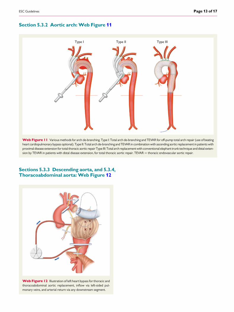
Type I Type II Type III
Web Figure 11 Various methods for arch de-branching. Type I: Total arch de-branching and TEVAR for off-pump total arch repair (use of beatingheart cardiopulmonary bypass optional). Type II: Total arch de-branching and TEVAR in combination with ascending aortic replacement in patients withproximal disease extension for total thoracic aortic repair Type III: Total arch replacement with conventional elephant trunk technique and distal exten-sion by TEVAR in patients with distal disease extension, for total thoracic aortic repair. TEVAR¼ thoracic endovascular aortic repair.
Web Figure 12 Illustration of left heart bypass for thoracic andthoracoabdominal aortic replacement, inflow via left-sided pul-monary veins, and arterial return via any downstream segment.
ESC Guidelines Page 13 of 17

Section 5.3.4 Thoracoabdominalaorta: Web Figure 13
Section 6.4.3 Natural history, morphologicalchanges, and complications: Web Figure 14
Web Figure 13 Illustration of left heart bypass for thoracic andthoracoabdominal aortic replacement showing selective visceralblood perfusion as well as selective bilateral cold saline perfusionof kidneys.
A B
Web Figure 14 Type-B IMH evolving with two localized, ulcer-like projections, 6 months after the acute onset (asterisks).
ESC GuidelinesPage 14 of 17

Section 7.2.4.2 Diagnostic imaging: Web Figure 15
A B
C
D E
WebFigure15 CTevaluationof aortic aneurysm. (A) Volume-rendered3Dreconstructionallowingqualitativeassessmentof thedimensionsofthe aneurysm and the relationship to side branches (e.g. renal or iliac arteries). It visualizes kinks and tortuosities and is useful for planning interven-tional procedures. (B) Modern 3D workstations with dedicated software for vascular analysis are recommended and allow the generation of a cen-treline along tortuous or kinked vessels. (C) Axial cross-section with several accepted methods of measuring the aneurysm diameter:(a) anteroposterior diameter, (b) transverse diameter, (c) maximum short-axis diameter (major axis), and (d) minimal short-axis diameter(minor axis). However, measurement of maximum aneurysm diameter should be performed perpendicular to the vessel centreline (D) ratherthan on axial cross-sections (particularly in tortuous aneurysms), to avoid over-estimation of maximum diameter, as shown in (C). In thisexample, maximum diameteron axial cross-section (c in C) is 64.2 mm, while the true maximum diameter is 60.5 mm (c in D). In partially thrombosedaneurysms, it is important to measure up to the outer contour of the aneurysm (C and D). (E) Straight multiplanar reformations are generated automat-ically upon centreline detection and can provide automatic diameter measurements at any site along the course of the vessel. 3D¼ three-dimensional;CT¼ computed tomography.
ESC Guidelines Page 15 of 17
Section 7.2.5.3 Follow-up of small abdominal aortic aneurysm: Web Table 2
Section 9.1.2 Diagnosis: Web Table 3
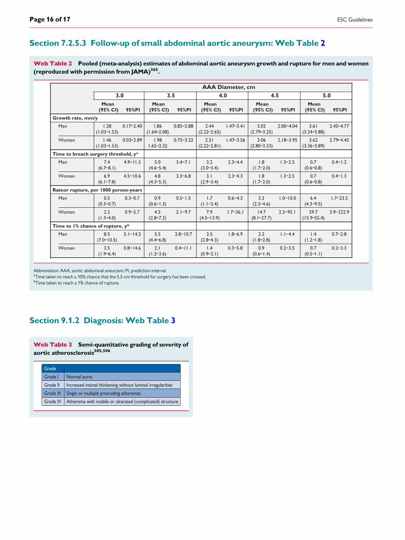
Web Table 2 Pooled (meta-analysis) estimates of abdominal aortic aneurysm growth and rupture for men and women(reproduced with permission from JAMA)365.
AAA Diameter, cm3.0 3.5 4.0 4.5 5.0
Mean(95% CI) 95%PI
Mean(95% CI) 95%PI
Mean(95% CI) 95%PI
Mean(95% CI) 95%PI
Mean(95% CI) 95%PI
Growth rate, mm/y
Men 1.28(1.03−1.53)
0.17−2.40 1.86(1.64−2.08)
0.85−2.88 2.44(2.22−2.65)
1.47−3.41 3.02(2.79−3.25)
2.00−4.04 3.61(3.34−3.88)
2.45−4.77
Women 1.46(1.03−1.53)
0.03−2.89 1.981.65−2.32
0.75−3.22 2.51(2.22−2.81)
1.47−3.56 3.06(2.80−3.33)
2.18−3.95 3.62(3.36−3.89)
2.79−4.45
Time to breach surgery threshold, ya
Men 7.4(6.7−8.1)
4.9−11.3 5.0(4.6−5.4)
3.4−7.1 3.2(3.0−3.4)
2.3−4.4 1.8(1.7−2.0)
1.3−2.5 0.7(0.6−0.8)
0.4−1.2
Women 6.9(6.1−7.8)
4.5−10.6 4.8(4.3−5.3)
3.3−6.8 3.1(2.9−3.4)
2.3−4.3 1.8(1.7−2.0)
1.3−2.5 0.7(0.6−0.8)
0.4−1.3
Rateor rupture, per 1000 person-years
Men 0.5(0.3−0.7)
0.3−0.7 0.9(0.6−1.3)
0.5−1.5 1.7(1.1−2.4)
0.6−4.3 3.2(2.2−4.6)
1.0−10.0 6.4(4.3−9.5)
1.7−23.5
Women 2.2(1.3−4.0)
0.9−5.7 4.5(2.8−7.2)
2.1−9.7 7.9(4.5−13.9)
1.7−36.1 14.7(8.1−27.7)
2.2−95.1 29.7(15.9−55.4)
3.9−222.9
Time to 1% chance of rupture, yb
Men 8.5(7.0−10.5)
5.1−14.2 5.5(4.4−6.8)
2.8−10.7 3.5(2.8−4.3)
1.8−6.9 2.2(1.8−2.8)
1.1−4.4 1.4(1.2−1.8)
0.7−2.8
Women 3.5(1.9−6.4)
0.8−14.6 2.1(1.2−3.6)
0.4−11.1 1.4(0.9−2.1)
0.3−5.8 0.9(0.6−1.4)
0.2−3.5 0.7(0.5−1.1)
0.2−3.3
Abbreviation: AAA, aortic abdominal aneurysm; Pl, prediction interval.aTime taken to reach a 10% chance that the 5.5-cm threshold for surgery has been crossed.bTime taken to reach a 1% chance of rupture.
Web Table 3 Semi-quantitative grading of severity ofaortic atherosclerosis505,506
Grade
Grade I Normal aorta
Grade II Increased intimal thickening without luminal irregularities
Grade III Single or multiple protruding atheromas
Grade IV Atheroma with mobile or ulcerated (complicated) structure
ESC GuidelinesPage 16 of 17

Section 10.2 Treatment: Web Table 4
Web Table 4 Inflammatory diseases associated with aortitis
Disease Diagnostic criteria
Giant cell arteritis540
• Age at onset >50 years• Recent-onset localized headache• Temporal artery tenderness or pulse attenuation• Elevated erythrocyte sedimentation rate >50 mm/h• Artery biopsy showing necrotizing vasculitis
Three or more criteria are present (sensitivity >90%;
Takayasu arteritis525
• Age at onset <40 years• Intermittent claudication• Diminished brachial artery pulse• Subclavian artery or carotid bruit• Systolic blood pressure variation of >10 mmHg between arms• Aortographic evidence of aorta or aortic branch stenosis
Three or more criteria are present (sensitivity 90.5%;
Behçet disease526
• Oral ulceration• Recurrent genital ulceration• Uveitis or retinal vasculitis• Skin lesions, erythema nodosum, pseudofolliculitis or pathergy
Oral ulceration plus two of the other three criteria
Ankylosing spondylitis527
• Onset of pain at age <40 years• Back pain for >3 months• Morning stiffness• Subtle symptom onset• Improvement with exercise
Four of the diagnostic criteria are present
BP ¼ blood pressure.
ESC Guidelines Page 17 of 17