2015 ESC/ERS Guidelines for the diagnosis and...
73
2015 ESC/ERS Guidelines for the diagnosis and treatment of pulmonary hypertension Nazzareno Galiè 1 (ESC Chairperson), Marc Humbert 2 (ERS Chairperson), Jean-Luc Vachiery 3 , Simon Gibbs 1 , Irene Lang 1 , Adam Torbicki 1 , Gérald Simonneau 2 , Andrew Peacock 2 , Anton Vonk Noordegraaf 2 , Maurice Beghetti 4 , Ardeschir Ghofrani 2 , Miguel Angel Gomez Sanchez 1 , Georg Hansmann 4 , Walter Klepetko 3 , Patrizio Lancellotti 1 , Marco Matucci 5 , Theresa McDonagh 1 , Luc A. Pierard 1 , Pedro T. Trindade 1 , Maurizio Zompatori 6 and Marius Hoeper 2 Affiliations: 1 Representing the European Society of Cardiology. 2 Representing the European Respiratory Society. 3 Representing the International Society for Heart and Lung Transplantation. 4 Representing the Association for European Paediatric and Congenital Cardiology. 5 Representing the European League Against Rheumatism. 6 Representing the European Society of Radiology. A full list of collaborators and document reviewers can be found in the Appendix. The Joint Task Force for the Diagnosis and Treatment of Pulmonary Hypertension of the European Society of Cardiology (ESC) and the European Respiratory Society (ERS) Endorsed by: Association for European Paediatric and Congenital Cardiology (AEPC), International Society for Heart and Lung Transplantation (ISHLT) Correspondence: Nazzareno Galiè, Dept of Experimental, Diagnostic and Specialty Medicine–DIMES, University of Bologna, Via Massarenti 9, 40138 Bologna, Italy. E-mail: [email protected] Marc Humbert, Service de Pneumologie, Hôpital Bicêtre, Université Paris-Sud, Assistance Publique Hôpitaux de Paris, 78 rue du Général Leclerc, 94270 Le Kremlin-Bicêtre, France. E-mail: [email protected] @ERSpublications 2015 ESC/ERS pulmonary hypertension guidelines incorporate changes and adaptations focusing on clinical management http://ow.ly/RiDLb Editorial Comments in Eur Respir J 2015; 46: 879–882 [DOI: 10.1183/13993003.01177-2015]. This article has been revised according to the erratum published in the December 2015 issue of the European Respiratory Journal. The content of these European Society of Cardiology (ESC) and European Respiratory Society (ERS) Guidelines has been published for personal and educational use only. No commercial use is authorized. No part of the ESC/ERS Guidelines may be translated or reproduced in any form without written permission from the ESC and/or ERS. Permission can be obtained upon submission of a written request to Oxford University Press, the publisher of the European Heart Journal or from the European Respiratory Society, the publisher of European Respiratory Journal and the party authorized to handle such permissions on behalf of the ESC and ERS. This article is being published concurrently in the European Heart Journal (10.1093/eurheartj/ehv317) and the European Respiratory Journal (10.1183/13993003.01032-2015). The articles are identical except for minor stylistic and spelling differences in keeping with each journal’s style. Either citation can be used when citing this article. Conflict of interest: Disclosures can be found alongside the online version of this article at erj.ersjournals.com Published on behalf of the European Society of Cardiology and European Respiratory Society. All rights reserved. © 2015 European Society of Cardiology & European Respiratory Society. Eur Respir J 2015; 46: 903–975 | DOI: 10.1183/13993003.01032-2015 903 ESC/ERS GUIDELINES PULMONARY HYPERTENSION
Transcript of 2015 ESC/ERS Guidelines for the diagnosis and...

2015 ESC/ERS Guidelines for thediagnosis and treatment of pulmonaryhypertension
Nazzareno Galiè1 (ESC Chairperson), Marc Humbert2 (ERS Chairperson),Jean-Luc Vachiery3, Simon Gibbs1, Irene Lang1, Adam Torbicki1,Gérald Simonneau2, Andrew Peacock2, Anton Vonk Noordegraaf2,Maurice Beghetti4, Ardeschir Ghofrani2, Miguel Angel Gomez Sanchez1,Georg Hansmann4, Walter Klepetko3, Patrizio Lancellotti1, Marco Matucci5,Theresa McDonagh1, Luc A. Pierard1, Pedro T. Trindade1, Maurizio Zompatori6
and Marius Hoeper2
Affiliations: 1Representing the European Society of Cardiology. 2Representing the European RespiratorySociety. 3Representing the International Society for Heart and Lung Transplantation. 4Representing theAssociation for European Paediatric and Congenital Cardiology. 5Representing the European League AgainstRheumatism. 6Representing the European Society of Radiology. A full list of collaborators and documentreviewers can be found in the Appendix.
The Joint Task Force for the Diagnosis and Treatment of PulmonaryHypertension of the European Society of Cardiology (ESC) and theEuropean Respiratory Society (ERS)
Endorsed by: Association for European Paediatric and Congenital Cardiology(AEPC), International Society for Heart and Lung Transplantation (ISHLT)
Correspondence:Nazzareno Galiè, Dept of Experimental, Diagnostic and SpecialtyMedicine–DIMES, University of Bologna, ViaMassarenti 9, 40138 Bologna, Italy. E-mail: [email protected] Humbert, Service de Pneumologie, Hôpital Bicêtre, Université Paris-Sud, Assistance PubliqueHôpitaux deParis, 78 rue du Général Leclerc, 94270 LeKremlin-Bicêtre, France. E-mail:[email protected]
@ERSpublications2015 ESC/ERS pulmonary hypertension guidelines incorporate changes and adaptationsfocusing on clinical management http://ow.ly/RiDLb
Editorial Comments in Eur Respir J 2015; 46: 879–882 [DOI: 10.1183/13993003.01177-2015].
This article has been revised according to the erratum published in the December 2015 issue of the EuropeanRespiratory Journal.
The content of these European Society of Cardiology (ESC) and European Respiratory Society (ERS) Guidelines has beenpublished for personal and educational use only. No commercial use is authorized. No part of the ESC/ERS Guidelinesmay be translated or reproduced in any form without written permission from the ESC and/or ERS. Permission can beobtained upon submission of a written request to Oxford University Press, the publisher of the European Heart Journalor from the European Respiratory Society, the publisher of European Respiratory Journal and the party authorized tohandle such permissions on behalf of the ESC and ERS.
This article is being published concurrently in the European Heart Journal (10.1093/eurheartj/ehv317) and the EuropeanRespiratory Journal (10.1183/13993003.01032-2015). The articles are identical except for minor stylistic and spellingdifferences in keeping with each journal’s style. Either citation can be used when citing this article.
Conflict of interest: Disclosures can be found alongside the online version of this article at erj.ersjournals.com
Published on behalf of the European Society of Cardiology and European Respiratory Society. All rights reserved. © 2015European Society of Cardiology & European Respiratory Society.
Eur Respir J 2015; 46: 903–975 | DOI: 10.1183/13993003.01032-2015 903
ESC/ERS GUIDELINESPULMONARY HYPERTENSION

Table of Contents
Abbreviation and acronyms1. Preamble ...................................................................................................................... 9052. Introduction .................................................................................................................. 9073. Definitions and classifications .................................................................................... 907
3.1 Definitions ............................................................................................................ 9073.2 Classifications...................................................................................................... 908
4. Epidemiology and genetics of pulmonary hypertension .......................................... 9094.1 Epidemiology and risk factors............................................................................ 9094.2 Genetics................................................................................................................ 911
5. Pulmonary hypertension diagnosis ............................................................................ 9115.1 Diagnosis.............................................................................................................. 911
5.1.1 Clinical presentation .............................................................................. 9135.1.2 Electrocardiogram .................................................................................. 9135.1.3 Chest radiograph .................................................................................... 9135.1.4 Pulmonary function tests and arterial blood gases ............................ 9135.1.5 Echocardiography .................................................................................... 9145.1.6 Ventilation/perfusion lung scan.............................................................. 9155.1.7 High-resolution computed tomography, contrast enhanced
computed tomography, and pulmonary angiography .......................... 9165.1.8 Cardiac magnetic resonance imaging .................................................. 9165.1.9 Blood tests and immunology.................................................................. 9175.1.10 Abdominal ultrasound scan.................................................................. 9175.1.11 Right heart catheterization and vasoreactivity .................................... 9175.1.12 Genetic testing ...................................................................................... 918
5.2 Diagnostic algorithm .......................................................................................... 9196. Pulmonary arterial hypertension (group 1) .............................................................. 920
6.1 Clinical characteristics........................................................................................ 9206.2 Evaluation of severity .......................................................................................... 920
6.2.1 Clinical parameters, imaging and haemodynamics ............................ 9206.2.2 Exercise capacity .................................................................................... 9226.2.3 Biochemical markers.............................................................................. 9226.2.4 Comprehensive prognostic evaluation and risk
assessment .............................................................................................. 9226.2.5 Definition of patient status...................................................................... 9236.2.6 Treatment goals and follow-up strategy................................................ 924
6.3 Therapy ................................................................................................................ 9246.3.1 General measures .................................................................................. 9256.3.1.1 Physical activity and supervised rehabilitation .................................. 9256.3.1.2 Pregnancy, birth control, and post-menopausal
hormonal therapy ................................................................................ 9266.3.1.3 Elective surgery .................................................................................... 9266.3.1.4 Infection prevention.............................................................................. 9266.3.1.5 Psychosocial support .......................................................................... 9266.3.1.6 Adherence to treatments .................................................................... 9276.3.1.7 Travel .................................................................................................... 9276.3.1.8 Genetic counselling .............................................................................. 9276.3.2 Supportive therapy .................................................................................. 9276.3.2.1 Oral anticoagulants .............................................................................. 9286.3.2.2 Diuretics ................................................................................................ 9286.3.2.3 Oxygen .................................................................................................. 9286.3.2.4 Digoxin and other cardiovascular drugs ............................................ 9286.3.2.5 Anaemia and iron status ...................................................................... 9286.3.3 Specific drug therapy .............................................................................. 9286.3.3.1 Calcium channel blockers .................................................................. 9286.3.3.2 Endothelin receptor antagonists ........................................................ 9296.3.3.3 Phosphodiesterase type 5 inhibitors and guanylate cyclase
stimulators ............................................................................................ 930
6.3.3.4 Prostacyclin analogues and prostacyclin receptor agonists ............ 9316.3.3.5 Experimental compounds and strategies .......................................... 9326.3.4 Combination therapy .............................................................................. 9336.3.5 Drug interactions .................................................................................... 9366.3.6 Balloon atrial septostomy ...................................................................... 9366.3.7 Advanced right ventricular failure.......................................................... 9366.3.7.1 Intensive care unit management ........................................................ 9366.3.7.2 Right ventricle assistance.................................................................... 9376.3.8 Transplantation ........................................................................................ 9376.3.9 Treatment algorithm .............................................................................. 9386.3.10 Diagnosis and treatment of pulmonary arterial
hypertension complications.................................................................. 9396.3.10.1 Arrhythmias ........................................................................................ 9396.3.10.2 Haemoptysis........................................................................................ 9406.3.10.3 Mechanical complications.................................................................. 9406.3.11 End of life care and ethical issues ...................................................... 940
7. Specific pulmonary (arterial) hypertension subsets ................................................ 9407.1 Paediatric pulmonary arterial hypertension .................................................... 940
7.1.1 Diagnosis.................................................................................................. 9417.1.2 Therapy .................................................................................................... 941
7.2 Pulmonary arterial hypertension associated with adultcongenital heart disease ...................................................................................... 9427.2.1 Diagnosis.................................................................................................. 9427.2.2 Therapy .................................................................................................... 942
7.3 Pulmonary arterial hypertension associated withconnective tissue disease...................................................................................... 9447.3.1 Diagnosis.................................................................................................. 9457.3.2 Therapy .................................................................................................... 945
7.4 Pulmonary arterial hypertension associated with portal hypertension ............ 9457.4.1 Diagnosis.................................................................................................. 9457.4.2 Therapy .................................................................................................... 946
7.5 Pulmonary arterial hypertension associated with humanimmunodeficiency virus infection ........................................................................ 9477.5.1 Diagnosis.................................................................................................. 9477.5.2 Therapy .................................................................................................... 948
7.6 Pulmonary veno-occlusive disease and pulmonarycapillary haemangiomatosis ................................................................................ 9487.6.1 Diagnosis.................................................................................................. 9497.6.2 Therapy .................................................................................................... 949
8. Pulmonary hypertension due to left heart disease (group 2) .................................... 9508.1 Diagnosis.............................................................................................................. 9518.2 Therapy ................................................................................................................ 952
9. Pulmonary hypertension due to lung diseases and/or hypoxia (group 3) ................ 9529.1 Diagnosis.............................................................................................................. 9539.2 Therapy ................................................................................................................ 954
10. Chronic thromboembolic pulmonary hypertension (group 4) ................................ 95410.1 Diagnosis............................................................................................................ 95410.2 Therapy .............................................................................................................. 95610.2.1 Surgical .......................................................................................................... 95610.2.2 Medical .......................................................................................................... 95610.2.3 Interventional ................................................................................................ 957
11. Pulmonary hypertension with unclear and/ormultifactorial mechanisms (group 5) ...................................................................... 958
12. Definition of a pulmonary hypertension referral centre ........................................ 95813. To do and not to do messages from the guidelines .............................................. 96014. Appendix .................................................................................................................... 96015. Web addenda .............................................................................................................. 96216. References.................................................................................................................. 962
Disclaimer: The ESC/ERS Guidelines represent the views of the ESC and ERS and were produced after carefulconsideration of the scientific and medical knowledge and the evidence available at the time of their publication. TheESC and ERS are not responsible in the event of any contradiction, discrepancy and/or ambiguity between the ESC/ERSGuidelines and any other official recommendations or guidelines issued by the relevant public health authorities, inparticular in relation to good use of healthcare or therapeutic strategies. Health professionals are encouraged to take theESC/ERS Guidelines fully into account when exercising their clinical judgment, as well as in the determination and theimplementation of preventive, diagnostic or therapeutic medical strategies; however, the ESC/ERS Guidelines do notoverride, in any way whatsoever, the individual responsibility of health professionals to make appropriate and accuratedecisions in consideration of each patient’s health condition and in consultation with that patient and, whereappropriate and/or necessary, the patient’s caregiver. Nor do the ESC/ERS Guidelines exempt health professionals fromtaking into full and careful consideration the relevant official updated recommendations or guidelines issued by thecompetent public health authorities, in order to manage each patient’s case in light of the scientifically accepted datapursuant to their respective ethical and professional obligations. It is also the health professional’s responsibility toverify the applicable rules and regulations relating to drugs and medical devices at the time of prescription.
904 DOI: 10.1183/13993003.01032-2015
PULMONARY HYPERTENSION | N. GALIÈ ET AL.
Abbreviations and acronyms
ALAT alanine aminotransferaseASAT aspartate aminotransferaseAPAH associated pulmonary arterial hypertensionBAS balloon atrial septostomyBMPR2 bone morphogenetic protein receptor 2BNP brain natriuretic peptideBPA balloon pulmonary angioplastyBREATHE Bosentan Randomised trial of Endothelin
Antagonist THErapyCAV1 caveolin-1CCB calcium channel blockercGMP cyclic guanosine monophosphateCHD congenital heart diseaseCI cardiac indexCMR cardiac magnetic resonanceCO cardiac outputCOPD chronic obstructive pulmonary diseaseCpc-PH combined post-capillary and pre-capillary pulmonary
hypertensionCPET cardiopulmonary exercise testingCPFE combined pulmonary fibrosis and emphysemaCT computed tomographyCTD connective tissue diseaseCTPA computed tomography pulmonary angiogramCTEPH chronic thromboembolic pulmonary
hypertensionDLCO diffusing capacity of the lung for carbon
monoxideDPAH drug-induced pulmonary arterial hypertensionDPG diastolic pressure gradient (diastolic PAP − mean PAWP)EACVI European association of cardiovascular imagingECG electrocardiogramECMO extracorporeal membrane oxygenationEIF2AK4 eukaryotic translation initiation factor 2 alpha kinase 4EMA European Medicines AgencyERA endothelin receptor antagonistFC functional classFDA US Food and Drug AdministrationHAART highly active antiretroviral therapyHIV human immunodeficiency virusHF-pEF heart failure with preserved left ventricular
ejection fractionHPAH heritable pulmonary arterial hypertensionHRCT high-resolution computed tomographyICU intensive care unitINR international normalized ratioIPAH idiopathic pulmonary arterial hypertensionIpc-PH isolated post-capillary pulmonary hypertensionIPF idiopathic pulmonary fibrosis
i.v. intravenousIVC inferior vena cavaLA left atrium/atrialLHD left heart diseaseLV left ventricle/ventricularMR magnetic resonanceNYHA New York Heart AssociationNO nitric oxideNT-proBNP N-terminal pro-brain natriuretic peptidePA pulmonary arteryPaCO2 arterial carbon dioxide pressurePaO2 arterial oxygen pressurePAH pulmonary arterial hypertensionPAP pulmonary arterial pressurePAPm mean pulmonary arterial pressurePAPs systolic pulmonary arterial pressurePAWP pulmonary artery wedge pressurePASP pulmonary artery systolic pressurePCH pulmonary capillary haemangiomatosisPDE-5i phosphodiesterase type 5 inhibitorPE pulmonary embolismPEA pulmonary endarterectomyPFTs pulmonary function testsPH pulmonary hypertensionPoPH porto-pulmonary hypertensionPPHN persistent pulmonary hypertension of the
newbornPVOD pulmonary veno-occlusive diseasePVR pulmonary vascular resistanceRA right atriumRAP right atrial pressureRCT randomized controlled trialRHC right heart catheterizationRV right ventricle/ventricular6MWD/6MWT 6-minute walking distance/6-minute walking testSCD sickle cell diseasesGC soluble guanylate cyclaseSSc systemic sclerosisSvO2 mixed venous oxygen saturationSVR systemic vascular resistanceTAPSE tricuspid annular plane systolic excursiont.i.d. three times a dayTGF-β transforming growth factor βTPG transpulmonary pressure gradient (mean PAP − mean
PAWP)TRV tricuspid regurgitant velocityVE/VCO2 minute ventilation – carbon dioxide production
relationshipV/Q ventilation/perfusionWHO-FC World Health Organization functional classWU Wood units
1. PreambleGuidelines summarize and evaluate all available evidence on a particular issue at the time of the writingprocess, with the aim of assisting health professionals in selecting the best management strategies for anindividual patient with a given condition, taking into account the impact on outcome, as well as the risk–benefit ratio of particular diagnostic or therapeutic means. Guidelines and recommendations should helphealth professionals to make decisions in their daily practice. However, the final decisions concerning anindividual patient must be made by the responsible health professional(s) in consultation with the patientand caregiver as appropriate.
A great number of Guidelines have been issued in recent years by the European Society of Cardiology(ESC) and by the European Respiratory Society (ERS), as well as by other societies and organisations.Because of the impact on clinical practice, quality criteria for the development of guidelines have beenestablished in order to make all decisions transparent to the user. The recommendations for formulatingand issuing ESC Guidelines can be found on the ESC website (http://www.escardio.org/Guidelines-&-Education/Clinical-Practice-Guidelines/Guidelines-development/Writing-ESC-Guidelines).ESC Guidelines represent the official position of the ESC on a given topic and are regularly updated.
Members of this Task Force were selected by the ESC and ERS to represent professionals involved with themedical care of patients with this pathology. Selected experts in the field undertook a comprehensive
DOI: 10.1183/13993003.01032-2015 905
PULMONARY HYPERTENSION | N. GALIÈ ET AL.

review of the published evidence for management (including diagnosis, treatment, prevention andrehabilitation) of a given condition according to ESC Committee for Practice Guidelines (CPG) policy andapproved by the ERS. A critical evaluation of diagnostic and therapeutic procedures was performed,including assessment of the risk–benefit ratio. Estimates of expected health outcomes for largerpopulations were included, where data exist. The level of evidence and the strength of the recommendationof particular management options were weighed and graded according to predefined scales, as outlined inTables 1 and 2.
The experts of the writing and reviewing panels provided declaration of interest forms for all relationshipsthat might be perceived as real or potential sources of conflicts of interest. These forms were compiled intoone file and can be found on the ESC website (http://www.escardio.org/guidelines). Any changes indeclarations of interest that arise during the writing period must be notified to the ESC and ERS andupdated. The Task Force received its entire financial support from the ESC and ERS without anyinvolvement from the healthcare industry.
The ESC CPG supervises and coordinates the preparation of new Guidelines produced by task forces, expertgroups or consensus panels. The Committee is also responsible for the endorsement process of theseGuidelines. The ESC Guidelines undergo extensive review by the CPG and external experts, and in this caseby ERS-appointed experts. After appropriate revisions the Guidelines are approved by all the experts involvedin the Task Force. The finalized document is approved by the CPG and by ERS for publication in theEuropean Heart Journal and in the European Respiratory Journal. The Guidelines were developed after carefulconsideration of the scientific and medical knowledge and the evidence available at the time of their dating.
The task of developing ESC/ERS Guidelines covers not only integration of the most recent research, butalso the creation of educational tools and implementation programmes for the recommendations. Toimplement the guidelines, condensed pocket guideline versions, summary slides, booklets with essentialmessages, summary cards for non-specialists and an electronic version for digital applications(smartphones, etc.) are produced. These versions are abridged and thus, if needed, one should always referto the full text version, which is freely available on the ESC website. The National Societies of the ESC areencouraged to endorse, translate and implement all ESC Guidelines. Implementation programmes areneeded because it has been shown that the outcome of disease may be favourably influenced by thethorough application of clinical recommendations.
Surveys and registries are needed to verify that real-life daily practice is in keeping with what isrecommended in the guidelines, thus completing the loop between clinical research, writing of guidelines,disseminating them and implementing them into clinical practice.
TABLE 1 Classes of recommendations
Classes of
recommendations
Definition Suggested wording to use
Class I Evidence and/or general agreement that a
given treatment or procedure is beneficial,
useful, effective.
Conflicting evidence and/or a divergence of
opinion about the usefulness/efficacy of the
given treatment or procedure.
Weight of evidence/opinion is in favour of usefulness/efficacy.
Usefulness/efficacy is less well established by evidence/opinion.
Evidence or general agreement that the
given treatment or procedure is not
useful/effective, and in some cases may be
harmful.
Is recommended/is indicated
Should be considered
May be considered
Is not recommended
Class II
Class IIa
Class IIb
Class III
906 DOI: 10.1183/13993003.01032-2015
PULMONARY HYPERTENSION | N. GALIÈ ET AL.

Health professionals are encouraged to take the ESC/ERS Guidelines fully into account when exercisingtheir clinical judgment, as well as in the determination and the implementation of preventive, diagnosticor therapeutic medical strategies. However, the ESC/ERS Guidelines do not override in any waywhatsoever the individual responsibility of health professionals to make appropriate and accurate decisionsin consideration of each patient’s health condition and in consultation with that patient and the patient’scaregiver where appropriate and/or necessary. It is also the health professional’s responsibility to verify therules and regulations applicable to drugs and devices at the time of prescription.
2. IntroductionPulmonary hypertension (PH) is a pathophysiological disorder that may involve multiple clinicalconditions and can complicate the majority of cardiovascular and respiratory diseases. The composition ofthe guidelines task force reflects the multidisciplinary nature of PH, including members of differentmedical societies, associations and working groups. The current document follows the two previous ESCand ERS Guidelines, published in 2004 and 2009, focusing on clinical management of PH. A systematicliterature review was performed from MEDLINE® to identify new studies published since 2009 concerningthe topic of PH. Task force members selected studies based on relevance and appropriateness. The mainchanges and adaptations as compared with the 2009 ESC and ERS PH guidelines are as follows:
• The table of contents structure has been simplified, with three initial general chapters includingclassifications, basic aspects and differential diagnosis, two chapters for pulmonary arterial hypertension(PAH) and one chapter each for PH due to left heart disease (LHD), lung disease and/or hypoxia, chronicthromboembolic pulmonary hypertension (CTEPH) and unclear and/or multifactorial mechanisms.
• New wordings and parameters for the haemodynamic definition of post-capillary PH subgroups havebeen adopted. Pulmonary vascular resistance (PVR) has been included in the haemodynamic definitionof PAH.
• An updated common clinical classification for adult and paediatric patients is reported.• New advances in pathology, pathobiology, genetics, epidemiology and risk factors are reported.• An updated diagnostic algorithm has been provided in an independent chapter and novel screening
strategies are proposed in the web addenda.• The importance of expert referral centres in the management of PH patients has been highlighted in
both the diagnostic and treatment algorithms.• New developments on PAH severity evaluation and on treatments and treatment goals are reported,
including combination therapy and two new recently approved drugs. The treatment algorithm has beenupdated accordingly.
• The chapters on PH due to LHD and lung diseases have been updated. The term ‘out of proportion PH’has been abandoned in both conditions.
• New diagnostic and treatment algorithms are reported in the CTEPH chapter, including general criteriafor operability and balloon pulmonary angioplasty (BPA) and a newly approved drug.
• A short chapter on PH due to unclear and/or multifactorial mechanisms has been added.
3. Definitions and classifications3.1 DefinitionsPH is defined as an increase in mean pulmonary arterial pressure (PAPm) ⩾25 mmHg at rest asassessed by right heart catheterization (RHC) [1]. Available data have shown that the normal PAPm at restis 14 ± 3 mmHg with an upper limit of normal of approximately 20 mmHg [1, 2]. The clinical significanceof a PAPm between 21 and 24 mmHg is unclear. Patients presenting with a pulmonary artery pressure(PAP) in this range should be carefully followed when they are at risk for developing PAH [e.g. patientswith connective tissue disease (CTD) or family members of patients with heritable PAH (HPAH)] [1].
TABLE 2 Level of evidence
Level of
evidence A
Level of
evidence B
Level of
evidence C
Data derived from multiple randomized clinical trials
or meta-analyses.
Data derived from a single randomized clinical trial or
large non-randomised studies.
Consensus of opinion of the experts and/or small
studies, retrospective studies, registries.
DOI: 10.1183/13993003.01032-2015 907
PULMONARY HYPERTENSION | N. GALIÈ ET AL.
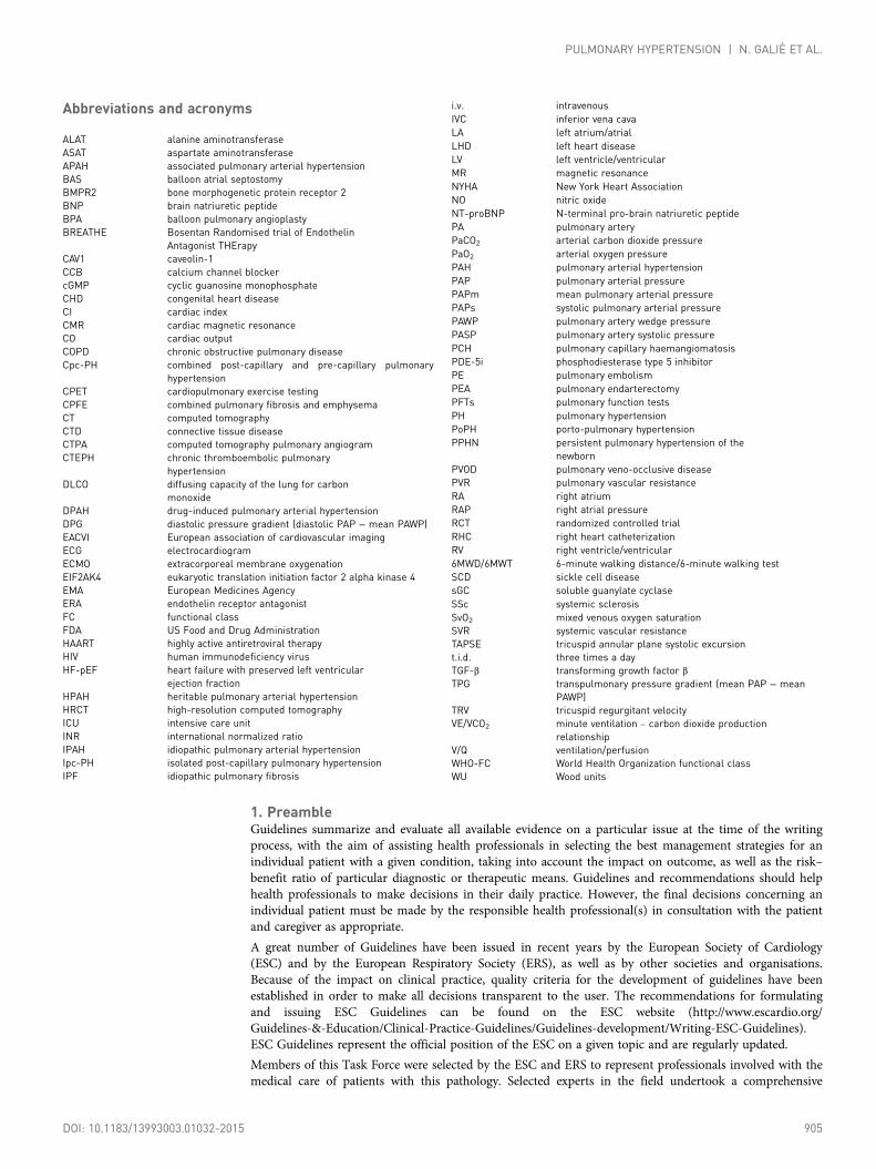
Due to the lack of reliable data that define which levels of exercise-induced changes in PAPm or PVRhave prognostic implications, a disease entity ‘PH on exercise’ cannot be defined and should not be used[1]. A recent retrospective study has proposed a definition of PH on exercise with the combination ofPAPm and total PVR data, but no outcome prospective validation has been provided [3].
The term PAH describes a group of PH patients characterized haemodynamically by the presence ofpre-capillary PH, defined by a pulmonary artery wedge pressure (PAWP) ⩽15 mmHg and a PVR>3 Wood units (WU) in the absence of other causes of pre-capillary PH such as PH due to lung diseases,CTEPH or other rare diseases [1].
According to various combinations of PAP, PAWP, cardiac output (CO), diastolic pressure gradient(DPG) and PVR, assessed in stable clinical conditions, different haemodynamic definitions of PH areshown in Table 3 together with their corresponding clinical classification (Table 4) [1, 4]. The reasons forthe updated definitions of post-capillary PH are reported in the specific section (8.0).
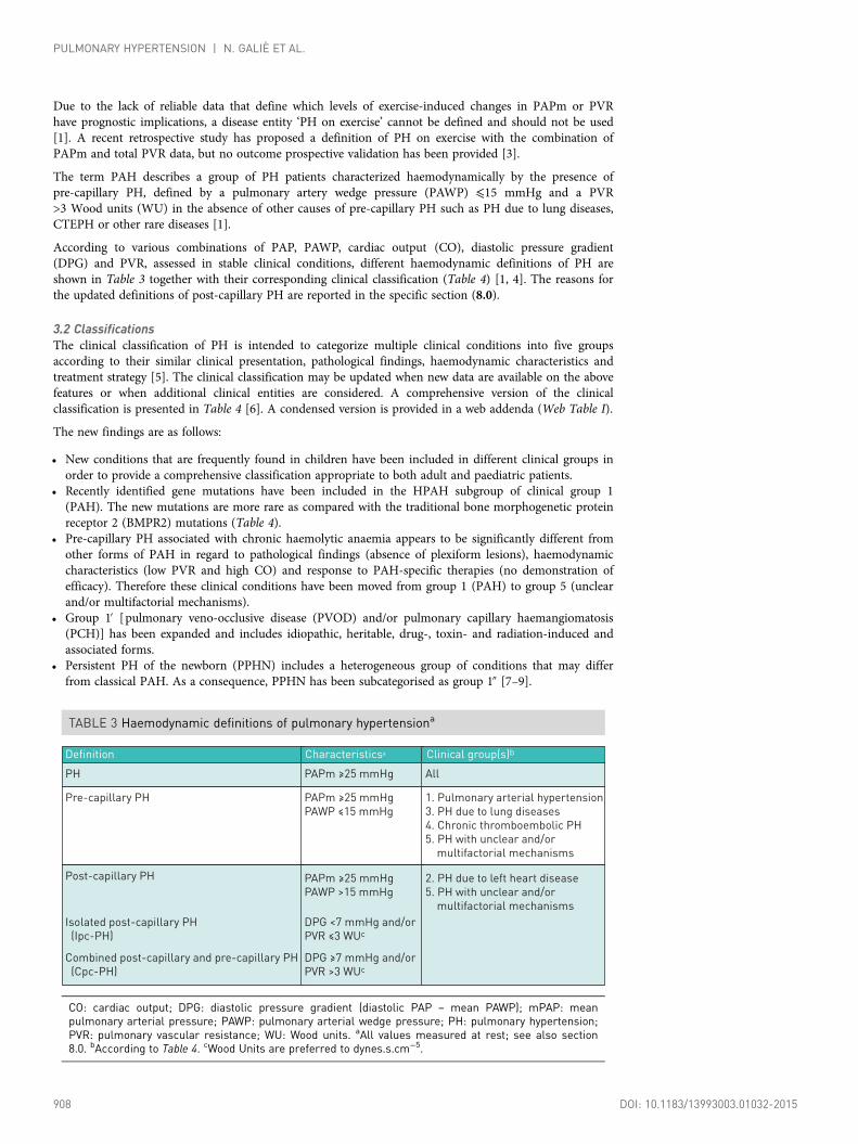
3.2 ClassificationsThe clinical classification of PH is intended to categorize multiple clinical conditions into five groupsaccording to their similar clinical presentation, pathological findings, haemodynamic characteristics andtreatment strategy [5]. The clinical classification may be updated when new data are available on the abovefeatures or when additional clinical entities are considered. A comprehensive version of the clinicalclassification is presented in Table 4 [6]. A condensed version is provided in a web addenda (Web Table I).
The new findings are as follows:
• New conditions that are frequently found in children have been included in different clinical groups inorder to provide a comprehensive classification appropriate to both adult and paediatric patients.
• Recently identified gene mutations have been included in the HPAH subgroup of clinical group 1(PAH). The new mutations are more rare as compared with the traditional bone morphogenetic proteinreceptor 2 (BMPR2) mutations (Table 4).
• Pre-capillary PH associated with chronic haemolytic anaemia appears to be significantly different fromother forms of PAH in regard to pathological findings (absence of plexiform lesions), haemodynamiccharacteristics (low PVR and high CO) and response to PAH-specific therapies (no demonstration ofefficacy). Therefore these clinical conditions have been moved from group 1 (PAH) to group 5 (unclearand/or multifactorial mechanisms).
• Group 1′ [pulmonary veno-occlusive disease (PVOD) and/or pulmonary capillary haemangiomatosis(PCH)] has been expanded and includes idiopathic, heritable, drug-, toxin- and radiation-induced andassociated forms.
• Persistent PH of the newborn (PPHN) includes a heterogeneous group of conditions that may differfrom classical PAH. As a consequence, PPHN has been subcategorised as group 1″ [7–9].
TABLE 3 Haemodynamic definitions of pulmonary hypertensiona
Definition
Pre-capillary PH
Post-capillary PH
Isolated post-capillary PH
(Ipc-PH)
Combined post-capillary and pre-capillary PH
(Cpc-PH)
Characteristicsa
PAPm ≥25 mmHg
PAWP ≤15 mmHg
PH PAPm ≥25 mmHg
PAPm ≥25 mmHg
PAWP >15 mmHg
DPG <7 mmHg and/or
PVR ≤3 WUc
DPG ≥7 mmHg and/or
PVR >3 WUc
Clinical group(s)b
2. PH due to left heart disease
5. PH with unclear and/or
multifactorial mechanisms
1. Pulmonary arterial hypertension
3. PH due to lung diseases
4. Chronic thromboembolic PH
5. PH with unclear and/or
multifactorial mechanisms
All
CO: cardiac output; DPG: diastolic pressure gradient (diastolic PAP – mean PAWP); mPAP: meanpulmonary arterial pressure; PAWP: pulmonary arterial wedge pressure; PH: pulmonary hypertension;PVR: pulmonary vascular resistance; WU: Wood units. aAll values measured at rest; see also section8.0. bAccording to Table 4. cWood Units are preferred to dynes.s.cm−5.
908 DOI: 10.1183/13993003.01032-2015
PULMONARY HYPERTENSION | N. GALIÈ ET AL.

• Paediatric heart diseases such as congenital or acquired left heart inflow or outflow tract obstruction andcongenital cardiomyopathies have been included in group 2 (PH due to LHD).
• No changes are proposed for group 3 (PH due to lung diseases and/or hypoxia).• Group 4 has been renamed as ‘CTEPH and other pulmonary artery (PA) obstructions’, which includes
CTEPH, pulmonary angiosarcoma, other intravascular tumours, arteritis, congenital pulmonary arteriesstenoses and parasites (Table 4).
• Segmental PH is observed in discrete lung areas perfused by aorto-pulmonary collaterals in congenitalheart diseases such as pulmonary or tricuspid atresia. This very unusual haemodynamic condition hasbeen included in group 5 (unclear and/or multifactorial mechanisms).
• Some pathological and pathophysiological information on the clinical groups are reported in the webaddenda.
Important pathophysiological and clinical definitions are reported in Table 5. A clinical classification ofPAH associated with congenital heart disease (CHD) is reported in Table 6.
An anatomical–pathophysiological classification of congenital systemic-to-pulmonary shunts associatedwith PAH is presented in Web Table II. A list of developmental lung diseases associated with PH ispresented in Web Table III.
4. Epidemiology and genetics of pulmonary hypertension4.1 Epidemiology and risk factorsReporting in the literature of PH incidence data at the global level is poor. In the UK, a prevalence of 97cases per million with a female:male ratio of 1.8 has been reported. The age-standardized death rate in theUSA ranges between 4.5 and 12.3 per 100,000 population. Comparative epidemiological data on theprevalence of the different groups of PH are not widely available, but it is clear that LHD (group 2) isbelieved to be the most common cause of PH, although severe PH is relatively uncommon in this setting.Although patients belonging to groups 2 and 3 represent an important part of the clinical practice, there isdisproportionately little information about the demographics and clinical course of this segment of the PHpopulation, suggesting that registry database methodology may be useful for these groups. Globally,schistosomiasis-associated PAH and high altitude–related PH represent an important burden to mankind.
• Group 1 (PAH): Several registries have described the epidemiology of PAH [10–12]. The lowest estimateof the prevalence of PAH and idiopathic PAH (IPAH) are 15 cases and 5.9 cases per million adultpopulation, respectively. The lowest estimate of PAH incidence is 2.4 cases per million adult populationper year. In Europe, PAH prevalence and incidence are in the range of 15–60 subjects per millionpopulation and 5–10 cases per million per year, respectively [11]. In registries, around half of PAHpatients have idiopathic, heritable or drug-induced PAH. In the subgroup of associated PAH conditions(APAH), the leading cause is CTD, mainly systemic sclerosis (SSc) [10].
PAH may occur in different settings depending on associated clinical conditions [13]. IPAH correspondsto sporadic disease, without any familial history of PAH or known triggering factor. While the mean age ofpatients with IPAH in the first US National Institutes of Health registry created in 1981 was 36 years, PAHis now more frequently diagnosed in elderly patients, resulting in a mean age at diagnosis between 50 and65 years in current registries. Furthermore, the female predominance is quite variable among registries andmay not be present in elderly patients, and survival appears to have improved over time.
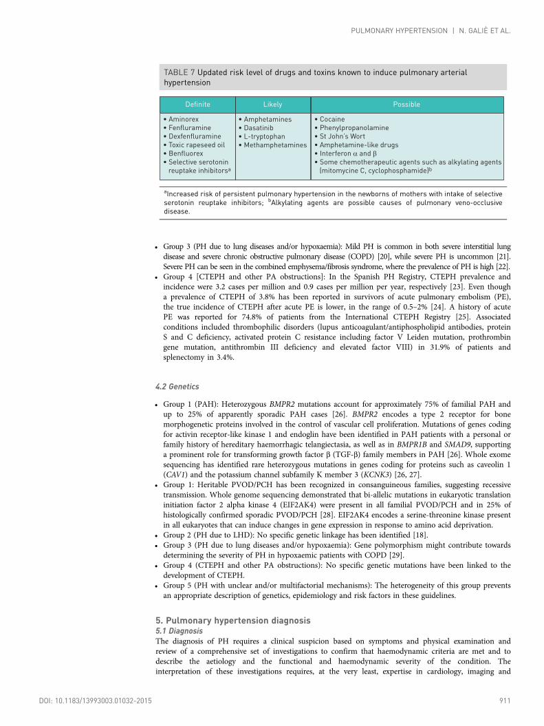
A number of risk factors for the development of PAH has been identified and are defined as any factoror condition that is suspected to play a predisposing or facilitating role in disease development. Risk factorswere classified as definite, likely or possible, based on the strength of their association with PH and theirprobable causal role [13]. A definite association is acknowledged in the case of either an epidemic, such asoccurred with appetite suppressants, or if large, multicentre epidemiological studies demonstrate anassociation between the clinical condition or drug and PAH. A likely association is acknowledged if asingle-centre case–control study or multiple case series demonstrate an association or if clinical andhaemodynamic recovery occurs after stopping exposure, such as occurred in dasatinib-induced PAH. Apossible association can be suspected, for example, for drugs with similar mechanisms of action as those inthe definite or likely category but which have not yet been studied, such as drugs used to treat attentiondeficit disorder. Definite clinical associations are listed among APAH in Table 4 and the risk level ofdifferent drugs and toxins are listed in Table 7 [6, 14–16].
• Group 2 (PH due to LHD): The prevalence of PH in patients with chronic heart failure increases withthe progression of functional class (FC) impairment. Up to 60% of patients with severe left ventricular(LV) systolic dysfunction and up to 70% of patients with heart failure with preserved ejection fractionmay present with PH. In left-sided valvular diseases, the prevalence of PH increases with the severity ofthe defect and of the symptoms. PH can be found in virtually all patients with severe symptomaticmitral valve disease and in up to 65% of those with symptomatic aortic stenosis [17–19].
DOI: 10.1183/13993003.01032-2015 909
PULMONARY HYPERTENSION | N. GALIÈ ET AL.
TABLE 5 Important pathophysiological and clinicaldefinitions
1. Pulmonary hypertension (PH) is a haemodynamic and
pathophysiological condition defined as an increase in mean
pulmonary arterial pressure ≥25 mmHg at rest as assessed by
right heart catheterization (Table 3). PH can be found in multiple
clinical conditions (Table 4).
3. There is no sufficient data to support the definition of ‘PH on
exercise’.
2. Pulmonary arterial hypertension (PAH, group 1) is a clinical
condition characterized by the presence of pre-capillary PH (Table
3) and pulmonary vascular resistance >3 Wood units, in the
absence of other causes of pre-capillary PH such as PH due to
lung diseases, chronic thromboembolic PH, or other rare diseases
(Table 4). PAH includes different forms that share a similar clinical
picture and virtually identical pathological changes of the lung
microcirculation (Table 4).
TABLE 4 Comprehensive clinical classification of pulmonaryhypertension (updated from Simonneau et al. [5])
1. Pulmonary arterial hypertension
1.1 Idiopathic
1.2 Heritable
1.2.1 BMPR2 mutation
1.2.2 Other mutations
1.3 Drugs and toxins induced
1.4 Associated with:
1.4.1 Connective tissue disease
1.4.2 Human immunodeficiency virus (HIV) infection
1.4.3 Portal hypertension
1.4.4 Congenital heart disease (Table 6)
1.4.5 Schistosomiasis
1’. Pulmonary veno-occlusive disease and/or pulmonary capillary
haemangiomatosis
1’.1 Idiopathic
1’.2 Heritable
1’.2.1 EIF2AK4 mutation
1’.2.2 Other mutations
1’.3 Drugs, toxins and radiation induced
1’.4 Associated with:
1’.4.1 Connective tissue disease
1’.4.2 HIV infection
2.1 Left ventricular systolic dysfunction
2.2 Left ventricular diastolic dysfunction
2.3 Valvular disease
2.4 Congenital/acquired left heart inflow/outflow tract obstruction
and congenital cardiomyopathies
2.5 Congenital/acquired pulmonary veins stenosis
1”. Persistent pulmonary hypertension of the newborn
2. Pulmonary hypertension due to left heart disease
3.1 Chronic obstructive pulmonary disease
3.2 Interstitial lung disease
3.3 Other pulmonary diseases with mixed restrictive and
obstructive pattern
3.4 Sleep-disordered breathing
3.5 Alveolar hypoventilation disorders
3.6 Chronic exposure to high altitude
3.7 Developmental lung diseases (Web Table III)
4.1 Chronic thromboembolic pulmonary hypertension
4.2 Other pulmonary artery obstructions
4.2.1 Angiosarcoma
4.2.2 Other intravascular tumors
4.2.3 Arteritis
4.2.4 Congenital pulmonary arteries stenoses
4.2.5 Parasites (hydatidosis)
5.1 Haematological disorders: chronic haemolytic anaemia,
myeloproliferative disorders, splenectomy
5.2 Systemic disorders: sarcoidosis, pulmonary histiocytosis,
lymphangioleiomyomatosis, neurofibromatosis
5.3 Metabolic disorders: glycogen storage disease, Gaucher disease,
thyroid disorders
5.4 Others: pulmonary tumoral thrombotic microangiopathy,
fibrosing mediastinitis, chronic renal failure (with/without
dialysis), segmental pulmonary hypertension
3. Pulmonary hypertension due to lung diseases and/or hypoxia
4. Chronic thromboembolic pulmonary hypertension and other
pulmonary artery obstructions
5. Pulmonary hypertension with unclear and/or multifactorial
mechanisms
BMPR2: bone morphogenetic protein receptor, type 2;EIF2AK4: eukaryotic translation initiation factor 2 alpha kinase4; HIV: human immunodeficiency virus.
TABLE 6 Clinical classification of pulmonary arterialhypertension associated with congenital heart disease(updated from Simonneau et al. [5])
1. Eisenmenger’s syndrome
Includes all large intra- and extra-cardiac defects which begin as
systemic-to-pulmonary shunts and progress with time to severe
elevation of PVR and to reversal (pulmonary-to-systemic) or
bidirectional shunting; cyanosis, secondary erythrocytosis, and
multiple organ involvement are usually present.
2. PAH associated with prevalent systemic-to-pulmonary shunts
• Correctablea
• Non-correctable
Includes moderate to large defects; PVR is mildly to moderately
increased, systemic-to-pulmonary shunting is still prevalent,
whereas cyanosis at rest is not a feature.
3. PAH with small/coincidental defectsb
Marked elevation in PVR in the presence of small cardiac defects
(usually ventricular septal defects <1 cm and atrial septal defects
<2 cm of effective diameter assessed by echo), which themselves do
not account for the development of elevated PVR; the clinical picture
is very similar to idiopathic PAH. Closing the defects is
contra-indicated.
4. PAH after defect correction
Congenital heart disease is repaired, but PAH either persists
immediately after correction or recurs/develops months or years
after correction in the absence of significant postoperative
haemodynamic lesions.
PAH: pulmonary arterial hypertension; PVR: pulmonary vascularresistance. aWith surgery or intravascular percutaneous procedure.bThe size applies to adult patients. However, also in adults thesimple diameter may be not sufficient for defining thehaemodynamic relevance of the defect and also the pressuregradient, the shunt size and direction, and the pulmonary tosystemic flows ratio should be considered (Web Table II).
910 DOI: 10.1183/13993003.01032-2015
PULMONARY HYPERTENSION | N. GALIÈ ET AL.
• Group 3 (PH due to lung diseases and/or hypoxaemia): Mild PH is common in both severe interstitial lungdisease and severe chronic obstructive pulmonary disease (COPD) [20], while severe PH is uncommon [21].Severe PH can be seen in the combined emphysema/fibrosis syndrome, where the prevalence of PH is high [22].
• Group 4 [CTEPH and other PA obstructions]: In the Spanish PH Registry, CTEPH prevalence andincidence were 3.2 cases per million and 0.9 cases per million per year, respectively [23]. Even thougha prevalence of CTEPH of 3.8% has been reported in survivors of acute pulmonary embolism (PE),the true incidence of CTEPH after acute PE is lower, in the range of 0.5–2% [24]. A history of acutePE was reported for 74.8% of patients from the International CTEPH Registry [25]. Associatedconditions included thrombophilic disorders (lupus anticoagulant/antiphospholipid antibodies, proteinS and C deficiency, activated protein C resistance including factor V Leiden mutation, prothrombingene mutation, antithrombin III deficiency and elevated factor VIII) in 31.9% of patients andsplenectomy in 3.4%.
4.2 Genetics
• Group 1 (PAH): Heterozygous BMPR2 mutations account for approximately 75% of familial PAH andup to 25% of apparently sporadic PAH cases [26]. BMPR2 encodes a type 2 receptor for bonemorphogenetic proteins involved in the control of vascular cell proliferation. Mutations of genes codingfor activin receptor-like kinase 1 and endoglin have been identified in PAH patients with a personal orfamily history of hereditary haemorrhagic telangiectasia, as well as in BMPR1B and SMAD9, supportinga prominent role for transforming growth factor β (TGF-β) family members in PAH [26]. Whole exomesequencing has identified rare heterozygous mutations in genes coding for proteins such as caveolin 1(CAV1) and the potassium channel subfamily K member 3 (KCNK3) [26, 27].
• Group 1: Heritable PVOD/PCH has been recognized in consanguineous families, suggesting recessivetransmission. Whole genome sequencing demonstrated that bi-allelic mutations in eukaryotic translationinitiation factor 2 alpha kinase 4 (EIF2AK4) were present in all familial PVOD/PCH and in 25% ofhistologically confirmed sporadic PVOD/PCH [28]. EIF2AK4 encodes a serine-threonine kinase presentin all eukaryotes that can induce changes in gene expression in response to amino acid deprivation.
• Group 2 (PH due to LHD): No specific genetic linkage has been identified [18].• Group 3 (PH due to lung diseases and/or hypoxaemia): Gene polymorphism might contribute towards
determining the severity of PH in hypoxaemic patients with COPD [29].• Group 4 (CTEPH and other PA obstructions): No specific genetic mutations have been linked to the
development of CTEPH.• Group 5 (PH with unclear and/or multifactorial mechanisms): The heterogeneity of this group prevents
an appropriate description of genetics, epidemiology and risk factors in these guidelines.
5. Pulmonary hypertension diagnosis5.1 DiagnosisThe diagnosis of PH requires a clinical suspicion based on symptoms and physical examination andreview of a comprehensive set of investigations to confirm that haemodynamic criteria are met and todescribe the aetiology and the functional and haemodynamic severity of the condition. Theinterpretation of these investigations requires, at the very least, expertise in cardiology, imaging and
TABLE 7 Updated risk level of drugs and toxins known to induce pulmonary arterialhypertension
Definite Likely Possible
• Aminorex
• Fenfluramine
• Dexfenfluramine
• Toxic rapeseed oil
• Benfluorex
• Selective serotonin
reuptake inhibitorsa
• Amphetamines
• Dasatinib
• L-tryptophan
• Methamphetamines
• Cocaine
• Phenylpropanolamine
• St John’s Wort
• Amphetamine-like drugs
• Interferon α and β• Some chemotherapeutic agents such as alkylating agents
(mitomycine C, cyclophosphamide)b
aIncreased risk of persistent pulmonary hypertension in the newborns of mothers with intake of selectiveserotonin reuptake inhibitors; bAlkylating agents are possible causes of pulmonary veno-occlusivedisease.
DOI: 10.1183/13993003.01032-2015 911
PULMONARY HYPERTENSION | N. GALIÈ ET AL.

respiratory medicine and may best be discussed at a multidisciplinary team meeting. This is particularlyimportant for identifying patients who may have more than one cause of PH. The main cause of PHshould be identified according to the clinical classification in Table 4. An algorithm for reaching adiagnosis is shown in Figure 1.
Symptoms, signs, history suggestive of PH
Echocardiographic probability of PH (Table 8)
CTD
Drugs - toxin
HIV
CHD
p y
Schistosomiasis
High or intermediate
Yes
NoYes
No
No
Yes
Yes
Low
Consider left heart disease and lung diseases
by symptoms, signs, risk factors, ECG,
PFT+DLCO, chest radiograph and HRCT,
arterial blood gases (Table 9)
Consider other causes and/or follow-up (Table 9)
Diagnosis of left heart diseases
or lung diseases confirmed?
Signs of severe PH/RV
dysfunction
Refer to PH expert centreV/Q scana
Mismatched perfusion defects?
No signs of severe
PH/RV dysfunction
Treat underlying disease
Refer to PH
expert centre
RHC (Table 10)
mPAP ≥25 mmHg, PAWP
≤15mmHg, PVR >3 Wood units
Group 5
Portopulmonary
Heritable
PVOD/PCH
Idiopathic
PVOD/PCH
Idiopathic
PAH
Heritable
PAH
Consider other
causes
CTEPH possible:
CT pulmonary angiography,
RHC +/- pulmonary angiography
PAH likely
Specific diagnostic tests
FIGURE 1 Diagnostic algorithm. CHD: congenital heart diseases; CT: computed tomography; CTD: connective tissue disease; CTEPH: chronicthromboembolic pulmonary hypertension; DLCO: carbon monoxide diffusing capacity; ECG: electrocardiogram; HIV: Human immunodeficiencyvirus; HRCT: high-resolution CT; mPAP: mean pulmonary arterial pressure; PA: pulmonary angiography; PAH: pulmonary arterial hypertension;PAWP: pulmonary artery wedge pressure; PFT: pulmonary function tests; PH: pulmonary hypertension; PVOD/PCH: pulmonary veno-occlusivedisease or pulmonary capillary hemangiomathosis; PVR: pulmonary vascular resistance; RHC: right heart catheterisation; RV: right ventricular;V/Q: ventilation/perfusion. aCT pulmonary angiography alone may miss diagnosis of chronic thromboembolic pulmonary hypertension.
912 DOI: 10.1183/13993003.01032-2015
PULMONARY HYPERTENSION | N. GALIÈ ET AL.

5.1.1 Clinical presentationThe symptoms of PH are non-specific and mainly related to progressive right ventricular (RV)dysfunction. Initial symptoms are typically induced by exertion. They include shortness of breath, fatigue,weakness, angina and syncope. Less commonly patients may also describe dry cough and exercise-inducednausea and vomiting. Symptoms at rest occur only in advanced cases. Abdominal distension and ankleoedema will develop with progressing RV failure. The presentation of PH may be modified by diseasesthat cause or are associated with PH as well as other concurrent diseases.
In some patients the clinical presentation may be related to mechanical complications of PH and theabnormal distribution of blood flow in the pulmonary vascular bed. These include haemoptysis related torupture of hypertrophied bronchial arteries, as well as symptoms attributable to pulmonary arterialdilatation such as hoarseness caused by compression of the left recurrent laryngeal nerve, wheeze causedby large airway compression and angina due to myocardial ischaemia caused by compression of the leftmain coronary artery. Significant dilation of the PA may result in its rupture or dissection, leading to signsand symptoms of cardiac tamponade.
The physical signs of PH include left parasternal lift, an accentuated pulmonary component of the secondheart sound, an RV third heart sound, a pansystolic murmur of tricuspid regurgitation and a diastolic murmurof pulmonary regurgitation. Elevated jugular venous pressure, hepatomegaly, ascites, peripheral oedema andcool extremities characterize patients with advanced disease. Wheeze and crackles are usually absent.
Clinical examination may suggest an underlying cause of PH. Telangiectasia, digital ulceration andsclerodactyly are seen in scleroderma, inspiratory crackles may point towards interstitial lung disease andspider naevi, testicular atrophy, and palmar erythema suggest liver disease. When digital clubbing isencountered, PVOD, cyanotic CHD, interstitial lung disease or liver disease should be considered.
5.1.2 ElectrocardiogramAn electrocardiogram (ECG) may provide supportive evidence of PH, but a normal ECG does not excludethe diagnosis. An abnormal ECG is more likely in severe rather than mild PH. ECG abnormalities mayinclude P pulmonale, right axis deviation, RV hypertrophy, RV strain, right bundle branch block, and QTcprolongation. While RV hypertrophy has insufficient sensitivity (55%) and specificity (70%) to be ascreening tool, RV strain is more sensitive [30]. Prolongation of the QRS complex and QTc suggest severedisease [31, 32]. The ECG differential diagnosis includes anterolateral myocardial ischaemia. In contrast toPH, ECG changes in ischaemia more commonly affect the lateral and inferior leads, and when present in theanterior chest leads are usually accompanied by a Q wave in V1 to V3, and rarely cause right axis deviation.
Supraventricular arrhythmias may occur in advanced disease, in particular atrial flutter, but also atrialfibrillation, with a cumulative incidence in 25% of patients after 5 years [33]. Atrial arrhythmias compromiseCO and almost invariably lead to further clinical deterioration. Ventricular arrhythmias are rare.
5.1.3 Chest radiographIn 90% of patients with IPAH the chest radiograph is abnormal at the time of diagnosis [34]. Findings inpatients with PAH include central pulmonary arterial dilatation, which contrasts with ‘pruning’ (loss) of theperipheral blood vessels. Right atrium (RA) and RV enlargement may be seen in more advanced cases. A chestradiograph may assist in differential diagnosis of PH by showing signs suggesting lung disease (group 3, Table 4)or pulmonary venous congestion due to LHD (group 2, Table 4). Chest radiography may help in distinguishingbetween arterial and venous PH by respectively demonstrating increased and decreased artery:vein ratios [35].
Overall, the degree of PH in any given patient does not correlate with the extent of radiographicabnormalities. As for ECG, a normal chest radiograph does not exclude PH.
5.1.4 Pulmonary function tests and arterial blood gasesPulmonary function tests and arterial blood gases identify the contribution of underlying airway orparenchymal lung disease. Patients with PAH have usually mild to moderate reduction of lung volumesrelated to disease severity [36, 37]. Although diffusion capacity can be normal in PAH, most patients havedecreased lung diffusion capacity for carbon monoxide (DLCO). An abnormal low DLCO, defined as<45% of predicted, is associated with a poor outcome [36, 37]. The differential diagnosis of a low DLCOin PAH includes PVOD, PAH associated with scleroderma and parenchymal lung disease. Althoughairflow obstruction is unusual, peripheral airway obstruction can be detected. Due to alveolarhyperventilation at rest, arterial oxygen pressure (PaO2) remains normal or is only slightly lower thannormal and arterial carbon dioxide pressure (PaCO2) is decreased [38].
COPD as a cause of hypoxic PH is diagnosed on the evidence of irreversible airflow obstruction together withincreased residual volumes and reduced DLCO [39]. Arterial blood gases of COPD patients show a decreased
DOI: 10.1183/13993003.01032-2015 913
PULMONARY HYPERTENSION | N. GALIÈ ET AL.

PaO2 with normal or increased PaCO2 [40]. A decrease in lung volume combined with decreased diffusioncapacity for carbon monoxide may indicate interstitial lung disease [39]. The severity of emphysema and ofinterstitial lung disease can be diagnosed using high-resolution computed tomography (CT). Combinedemphysema and pulmonary fibrosis may pseudonormalize spirometry, although the DLCO is almost alwaysreduced, emphasizing the need to interpret pulmonary function alongside lung imaging.
The prevalence of nocturnal hypoxaemia and central sleep apnoeas are high in PAH (70–80%) [41, 42].Overnight oximetry or polysomnography should be performed where obstructive sleep apnoea syndromeor hypoventilation are considered.
5.1.5 EchocardiographyTransthoracic echocardiography is used to image the effects of PH on the heart and estimate PAP fromcontinuous wave Doppler measurements. Echocardiography should always be performed when PH issuspected and may be used to infer a diagnosis of PH in patients in whom multiple differentechocardiographic measurements are consistent with this diagnosis. When treatment of PH itself is beingconsidered, echocardiography alone is not sufficient to support a treatment decision and cardiaccatheterization is required. Detailed guidelines describing the echocardiographic assessment of the right heartcan be found in documents created and/or endorsed by the European Association of Cardiovascular Imaging(EACVI), a registered branch of the ESC, and the reader is referred to these for further instruction [43, 44].
The estimation of systolic PAP is based on the peak tricuspid regurgitation velocity (TRV) taking intoaccount right atrial pressure (RAP) as described by the simplified Bernoulli equation. RAP can be estimatedby echocardiography based on the diameter and respiratory variation in diameter of the inferior vena cava(IVC): an IVC diameter <2.1 cm that collapses >50% with a sniff suggests a normal RA pressure of 3mmHg (range 0–5 mmHg), whereas an IVC diameter >2.1 cm that collapses <50% with a sniff or <20% onquiet inspiration suggests a high RA pressure of 15 mmHg (range 10–20 mmHg). In scenarios in which theIVC diameter and collapse do not fit this paradigm, an intermediate value of 8 mmHg (range 5–10 mmHg)may be used. The EACVI recommends such an approach rather than using a fixed value of 5 or 10 mmHgfor PA systolic pressure (PASP) estimations. However, given the inaccuracies of RAP estimation and theamplification of measurement errors by using derived variables, we recommend using the continuous waveDoppler measurement of peak TRV (and not the estimated PASP) as the main variable for assigning theechocardiographic probability of PH.
When peak TRV is technically difficult to measure (trivial or mild tricuspid regurgitation) somelaboratories use contrast echocardiography [e.g. agitated saline administered by intravenous (i.v.)injection], which may improve the Doppler signal, allowing measurement of peak TRV velocity.Unfortunately, despite the strong correlation of TRV with a tricuspid regurgitation pressure gradient,Doppler-derived pressure estimation may be inaccurate in the individual patient. In patients with severetricuspid regurgitation, TRV may be significantly underestimated and cannot be used to exclude PH.Overestimation may also occur [44]. PH cannot be reliably defined by a cut-off value of TRV.Consequently, estimation of PAP based solely on Doppler transthoracic echocardiography measurements isnot suitable for screening for mild, asymptomatic PH. Other echocardiographic variables that might raiseor reinforce suspicion of PH independent of TRV should always be sought.
Conclusions derived from an echocardiographic examination should aim to assign a level of probability ofPH. This ESC Guideline suggests grading the probability of PH based on TRV at rest and on the presenceof additional pre-specified echocardiographic variables suggestive of PH (Table 8A). The probability of PHmay then be judged as high, intermediate or low. When interpreted in a clinical context, theechocardiographic result is required to decide the need for cardiac catheterization in individual patients. Inorder to facilitate and standardize assignment to the level of probability of PH, several additionalechocardiographic signs are proposed in addition to criteria based on TRV (Table 8B). These signs provideassessment of the RV size and pressure overload, the pattern of blood flow velocity out of the RV, thediameter of the PA and an estimate of RAP [43–45]. Their measurement has been defined inrecommendations endorsed by the EACVI [43, 44].
The recommended plan for further patient investigation based on echocardiographic probability of PH isshown in Table 9 for symptomatic patients. In the Web addendum, a similar table (Web Table IX) forscreening for asymptomatic patients with risk factors for PAH or with incidental findings suggesting thepossibility of PH on ECG or lung imaging is provided.
Echocardiography can be helpful in detecting the cause of suspected or confirmed PH. Two-dimensional,Doppler and contrast examinations can be used to identify CHD. High pulmonary blood flow found onpulsed wave Doppler in the absence of a detectable shunt or significant dilatation of proximal PA despiteonly moderate PH may warrant transoesophageal examination with contrast or cardiac magnetic
914 DOI: 10.1183/13993003.01032-2015
PULMONARY HYPERTENSION | N. GALIÈ ET AL.
resonance (CMR) imaging to exclude sinus venosus atrial septal defect and/or anomalous pulmonaryvenous return. In cases of suspicion of LV diastolic dysfunction, Doppler echocardiographic signs shouldbe assessed even if their reliability is considered low. RHC should be considered when the diagnosisremains uncertain after non-invasive investigations (see section 8.1). The practical clinical value of exerciseDoppler echocardiography in the identification of cases with PH limited to exercise is uncertain because ofthe lack of validated criteria and prospective confirmatory data.
5.1.6 Ventilation/perfusion lung scanA ventilation/perfusion (V/Q) lung scan should be performed in patients with PH to look for CTEPH.The V/Q scan has been the screening method of choice for CTEPH because of its higher sensitivity
TABLE 8A Echocardiographic probability of pulmonaryhypertension in symptomatic patients with a suspicion ofpulmonary hypertension
Peak tricuspid
regurgitation
velocity (m/s)
Presence of other
echo ‘PH signs’aEchocardiographic
probability of pulmonary
hypertension
≤2.8 or not
measurableNo Low
≤2.8 or not
measurableYes
2.9–3.4 No
Intermediate
2.9–3.4 Yes
>3.4 Not requiredHigh
PH: pulmonary hypertension. aSee Table 8B.
TABLE 8B Echocardiographic signs suggesting pulmonaryhypertension used to assess the probability of pulmonaryhypertension in addition to tricuspid regurgitation velocitymeasurement in Table 8A
A: The ventriclesa B: Pulmonary arterya C: Inferior vena cava and
right atriuma
Right ventricle/left
ventricle basal
diameter ratio
>1.0
Right ventricular
outflow Doppler
acceleration time
<105 msec and/or
midsystolic notching
Inferior cava diameter
>21 mm with decreased
inspiratory collapse
(<50% with a sniff or
<20% with quiet
inspiration)
PA diameter >25 mm.
Flattening of the
interventricular
septum (left
ventricular
eccentricity index
>1.1 in systole
and/or diastole)
Early diastolic
pulmonary
regurgitation velocity
>2.2 m/sec
Right atrial area
(end-systole) >18 cm2
PA: pulmonary artery. aEchocardiographic signs from at least twodifferent categories (A/B/C) from the list should be present to alterthe level of echocardiographic probability of pulmonaryhypertension.
TABLE 9 Diagnostic management suggested according to echocardiographic probability of pulmonary hypertension inpatients with symptoms compatible with pulmonary hypertension, with or without risk factors for pulmonary arterialhypertension or chronic thromboembolic pulmonary hypertension
Echocardiographic
probability of PH
Without risk factors or associated
condition for PAH or CTEPHdClassa
Alternative diagnosis should be
considered
Alternative diagnosis, echo
follow-up, should be considered
Further investigation of PH may be
considerede
Further investigation of PH
(including RHCe) is recommended
Echo follow-up should be
considered
Further assessment of PH
including RHC should be
considerede
Further investigation of PHe
including RHC is recommended
IIa
IIa
IIb
I
Levelb
C
C
C
Classa
IIa
IIa
I
Levelb
C
B
C
Refc
[45, 46]
With risk factors or associated
conditions for PAH or CTEPHc
Low
Intermediate
High
CTEPH: chronic thromboembolic pulmonary hypertension; Echo: echocardiographic; PAH: pulmonary arterial hypertension; PH: pulmonaryhypertension; RHC: right heart catheterization. aClass of recommendation. bLevel of evidence. cReference(s) supporting recommendations.dThese recommendations do not apply to patients with diffuse parenchymal lung disease or left heart disease. eDepending on the presenceof risk factors for PH group 2, 3 or 5. Further investigation strategy may differ depending on whether risk factors/associated conditionssuggest higher probability of PAH or CTEPH – see diagnostic algorithm.
DOI: 10.1183/13993003.01032-2015 915
PULMONARY HYPERTENSION | N. GALIÈ ET AL.

compared with CT pulmonary angiogram (CTPA), especially in inexperienced centres [47]. A normal- orlow-probability V/Q scan effectively excludes CTEPH with a sensitivity of 90–100% and a specificity of94–100%; however, many V/Q scans are not diagnostic. While in PAH the V/Q lung scan may be normal,it may also show small peripheral unmatched and non-segmental defects in perfusion. A caveat is thatunmatched perfusion defects may also be seen in other pulmonary vascular disease such as PVOD. Whilea V/Q scan is still recommended as the screening test of choice, ventilation scans are often replaced witheither a recent chest radiograph or a recent high-resolution CT of the lungs, but such practices are notreally evidence-based. Also, CT is preferred in many centres since it is more readily available. A fewstudies suggest that single photon emission CT, also a nuclear medicine technique, could be superior to V/Q planar scan and CTPA, but these results need more extensive evaluation [48]. More recently, newertechniques such as three-dimensional magnetic resonance (MR) perfusion mapping, have beendemonstrated to be as sensitive as traditional perfusion scintigraphy in screening for CTEPH; MR can alsobe used as a radiation-free modality to assess both ventilation and perfusion in CTEPH [49].
5.1.7 High-resolution computed tomography, contrast-enhanced computed tomography, andpulmonary angiographyCT imaging is a widely available tool that can provide important information on vascular, cardiac,parenchymal and mediastinal abnormalities. It may suggest the diagnosis of PH (PA or RV enlargement),identify a cause of PH such as CTEPH or lung disease, provide clues as to the form of PAH(e.g. oesophageal dilation in SSc or congenital cardiac defects such as anomalous pulmonary venousdrainage) and also provide prognostic information [50].
CT may raise a suspicion of PH in symptomatic patients or those examined for unrelated indicationsby showing an increased PA diameter (⩾29 mm) and pulmonary:ascending aorta diameter ratio (⩾1.0). Asegmental artery:bronchus ratio >1 : 1 in three or four lobes has been reported to have high specificity forPH [51, 52].
High-resolution CT provides detailed views of the lung parenchyma and facilitates the diagnosis ofinterstitial lung disease and emphysema. High-resolution CT may also be very helpful where there is aclinical suspicion of PVOD. Characteristic changes of interstitial oedema with diffuse central ground-glassopacification and thickening of interlobular septa support the diagnosis of PVOD; additional findings mayinclude lymphadenopathy, pleural shadows and effusions [53]. Pulmonary capillary haemangiomatosis issuggested by diffuse bilateral thickening of the interlobular septa and the presence of small, centrilobular,poorly circumscribed nodular opacities. However, ground-glass abnormalities are also present in PAH,occurring in more than one-third of patients [50].
Contrast CT angiography of the PA is helpful in determining whether there is evidence of surgicallyaccessible CTEPH. It can delineate the typical angiographic findings in CTEPH, such as completeobstruction, bands and webs and intimal irregularities, as accurately and reliably as digital subtractionangiography [54, 55]. With this technique, collaterals from bronchial arteries can be identified.
Traditional pulmonary angiography is required in most patients for the workup of CTEPH to identifythose who may benefit from pulmonary endarterectomy (PEA) or BPA [56, 57]. Angiography can beperformed safely by experienced staff in patients with severe PH using modern contrast media andselective injections. Angiography may also be useful in the evaluation of possible vasculitis or pulmonaryarteriovenous malformations, but CT angiography has similar or even higher accuracy for both diagnoses,and is less invasive [58, 59].
5.1.8 Cardiac magnetic resonance imagingCMR imaging is accurate and reproducible in the assessment of RV size, morphology and function andallows non-invasive assessment of blood flow, including stroke volume, CO, pulmonary arterialdistensibility and RV mass.
In patients with suspected PH, the presence of late gadolinium enhancement, reduced pulmonary arterialdistensibility and retrograde flow have high predictive value for the identification of PH; however, nosingle CMR measurement can exclude PH [60–62]. In patients with PH, CMR may also be useful in casesof suspected CHD if echocardiography is not conclusive.
Contrast-enhanced and unenhanced MR angiography have a potential in the study of the pulmonaryvasculature in patients with suspected CTEPH, particularly in clinical scenarios such as suspected chronicembolism in pregnant women, young patients or when iodine-based contrast media injection iscontraindicated [63].
CMR provides useful prognostic information in patients with PAH both at baseline and at follow-up [64–66].
916 DOI: 10.1183/13993003.01032-2015
PULMONARY HYPERTENSION | N. GALIÈ ET AL.

5.1.9 Blood tests and immunologyBlood tests are not useful in diagnosing PH, but are required to identify the aetiology of some forms ofPH as well as end organ damage. Routine biochemistry, haematology and thyroid function tests arerequired in all patients, as well as a number of other specific blood tests. Liver function tests may beabnormal because of high hepatic venous pressure, liver disease and/or endothelin receptor antagonist(ERA) therapy. Hepatitis serology should be performed if clinical abnormalities are noted. Thyroid diseaseis common in PAH and may develop during the course of the disease. This should always be consideredin cases of abrupt deterioration.
Serological testing is required to detect underlying CTD, hepatitis and human immunodeficiency virus(HIV). Up to 40% of patients with IPAH have elevated antinuclear antibodies usually in a low titre (1:80).It is important to look for evidence of SSc since this disease has a relatively high prevalence of PAH.Limited scleroderma typically has antinuclear antibodies, including anti-centromere, dsDNA, anti-Ro,U3-RNP, B23, Th/To and U1-RNP. Diffuse scleroderma is typically associated with a positive U3-RNP.Patients with systemic lupus erythematosus may have anticardiolipin antibodies.
Patients with CTEPH should undergo thrombophilia screening, including antiphospholipid antibodies,anticardiolipin antibodies and lupus anticoagulant. HIV testing is required in PAH. N-terminal pro-brainnatriuretic peptide (NT-proBNP) may be elevated in patients with PH and is an independent riskpredictor in these patients.
5.1.10 Abdominal ultrasound scanSimilar to blood tests, abdominal ultrasound may be useful for identification of some of the clinical entitiesassociated with PAH. Abdominal ultrasound may confirm but not formally exclude portal hypertension.The use of contrast agents and the addition of a colour Doppler examination may improve the accuracy ofthe diagnosis [67]. Portal hypertension can be reliably confirmed or excluded by measurement of thegradient between free and occluded (wedge) hepatic vein pressure at the time of RHC [68].
5.1.11 Right heart catheterization and vasoreactivityRHC is required to confirm the diagnosis of PAH and CTEPH, to assess the severity of haemodynamicimpairment and to undertake vasoreactivity testing of the pulmonary circulation in selected patients(Table 10). When performed at expert centres, these procedures have low morbidity (1.1%) and mortality(0.055%) rates [69]. The threshold to perform left heart catheterization in addition to RHC should be lowin patients with clinical risk factors for coronary artery disease or heart failure with preserved ejectionfraction, as well as in patients with echocardiographic signs of systolic and/or diastolic LV dysfunction.Specific recommendations for catheterization of patients with LHD or lung disease in addition to Table 10are described in Tables 31 and 33, respectively. Measurement of LV end-diastolic pressure is alsoimportant to avoid misclassification of patients with an elevated PAWP when this is unexpected and maybe inaccurate [absence of risk factors for heart failure with preserved ejection fraction, normal left atrial(LA) size and absence of echocardiographic markers of elevated LV filling pressures].
The interpretation of invasive haemodynamics should be made in the context of the clinical picture andimaging, in particular echocardiography. Cardiac catheterization should be performed after the completionof other investigations so that it can answer specific questions that may arise from these investigations andavoid an unnecessary procedure where an alternative diagnosis is revealed.
RHC is a technically demanding procedure that requires meticulous attention to detail to obtain clinicallyuseful information. To obtain high-quality results and to be of low risk to patients, the procedure shouldbe limited to expert centres. Particular attention should be paid to the following issues:
• The external pressure transducer should be zeroed at the mid-thoracic line in a supine patient, halfwaybetween the anterior sternum and the bed surface [70]. This represents the level of the LA.
• Pressure measurements should be made in the PA, PA wedge position, RV and RA. Where a ballooncatheter is used, it should be inflated in the RA, from where the catheter should be advanced until itreaches the PAWP position. Repeated deflations and inflations of the balloon in the end pulmonaryarteries should be avoided because this has been associated with rupture of the pulmonary arteries. ThePAWP is a surrogate of LA pressure and should be recorded as the mean of three measurements. Bloodsampling should also be considered with the balloon inflated in the wedge position to confirm that atrue PAWP measurement has been taken, as this should have the same saturation as systemic blood. Allpressure measurements should be determined at the end of normal expiration (breath holding is notrequired). Alternatively, assuming that negative inspiratory and positive expiratory intrathoracicpressures cancel each other out, averaging pulmonary vascular pressures over several respiratory cycles isalso acceptable, except in dynamic hyperinflation states [70]. Ideally, high-fidelity tracings that can be
DOI: 10.1183/13993003.01032-2015 917
PULMONARY HYPERTENSION | N. GALIÈ ET AL.

printed on paper should be used rather than small moving traces on a cardiac monitor. Non-invasiveblood pressure should be recorded at the time of the procedure if left heart catheterization is notundertaken.
• Blood samples for oximetry should be taken from the high superior vena cava, IVC and PA at aminimum. Systemic arterial blood oxygen (O2) saturation should also be determined. A stepwiseassessment of O2 saturation should be performed in every patient with a pulmonary arterial O2
saturation >75% and whenever a left-to-right shunt is suspected.• CO should be measured using thermodilution or the direct Fick method. Thermodilution measured in
triplicate is the preferred method because it can provide reliable measurements even in patients with lowCO and/or severe tricuspid regurgitation [71]. In patients with intracardiac shunts, thermodilution maybe inaccurate because of early recirculation of the injectate. The direct Fick method requires directmeasurement of O2 uptake, a technique that is not widely available. The indirect Fick method, whichuses estimated values of O2 uptake, is acceptable but lacks reliability.
• Pulmonary vasoreactivity testing for identification of patients suitable for high-dose calcium channelblocker (CCB) treatment is recommended only for patients with IPAH, HPAH or drug-induced PAH. Itshould be performed at the time of RHC. In all other forms of PAH and PH the results can bemisleading and responders are rare. Inhaled nitric oxide (NO) at 10–20 parts per million (ppm) is thestandard of care for vasoreactivity testing, but i.v. epoprostenol, i.v. adenosine or inhaled iloprost can beused as alternatives (Web Table IV). A positive acute response is defined as a reduction of the meanPAP ⩾10 mmHg to reach an absolute value of mean PAP ⩽40 mmHg with an increased or unchangedCO. Only about 10% of patients with IPAH will meet these criteria. The use of CCBs, O2,phosphodiesterase type 5 inhibitors or other vasodilators for acute vasoreactivity testing is discouraged.
• Interpretation of the PAWP at a single point in time needs to be performed in a clinical context. Inmany patients with LHD, PAWP may be reduced to <15 mmHg with diuretics [72–74]. For this reason,the effect of an acute volume challenge on left heart filling pressures has been considered [75]. Limiteddata suggest that a fluid bolus of 500 ml appears to be safe and may discriminate patients withPAH from those with LV diastolic dysfunction [76, 77]. Further evaluation of administering a fluidchallenge is required before this can be considered for routine clinical practice. Similarly, exercisehaemodynamics to identify patients with LV diastolic dysfunction is likely to be useful [2, 78, 79],but lacks standardisation and requires further evaluation [17]. Furthermore, PAWP may underestimateLV end-diastolic pressure [80].
• Derived variables calculated from the RHC measurements should include transpulmonary pressuregradient (TPG) and PVR. A PVR >3 WU is required for the diagnosis of PAH [1]. PVR is commonlyused but has the disadvantage of being a composite variable that is highly sensitive to changes in bothflow and filling pressure and may not reflect changes in the pulmonary circulation at rest [81, 82]. TheDPG between the mean PAWP and diastolic PAP is less affected by flow and filling pressures [81] butmay not be of prognostic value [83]. DPG may have a role in patients suspected of having PH related toLHD, as discussed in section 8 [4].
• Coronary angiography may be required in the presence of angina, risk factors for coronary arterydisease and listing for PEA or lung transplantation. It may identify left main stem coronary arterycompression by an enlarged PA as well as coronary artery disease.
Recommendations for right and left heart catheterization and vasoreactivity testing are summarised in theTables 10 and 11.
5.1.12 Genetic testingThe availability of molecular genetic diagnosis has opened up a new field for patient care, includinggenetic counselling for PAH (developed in section 6.3.1.8) [26]. Genetic testing and counselling followsstrict local regulations that set the conditions for prescribing and conducting reviews of the geneticcharacteristics of a patient. The ethical principles are to inform patients properly to avoid harm, to allowpatients to preserve their autonomy (disclosure about the process, risks and benefits of the genetic testwithout external pressures) and to allow equal access to genetic counselling and testing. Patients withsporadic or familial PAH or PVOD/PCH should be advised about the availability of genetic testing andcounselling because of the strong possibility that they carry a disease-causing mutation. Trainedprofessionals should offer counselling and testing to the patient. Genetic counselling and BMPR2 mutationscreening (point mutations and large rearrangements) should be offered by referral centres to patientswith IPAH considered to be sporadic or induced by anorexigens and to patients with a family history ofPAH. When no BMPR2 mutations are identified in familial PAH patients or in IPAH patients <40 yearsold, or when PAH occurs in patients with a personal or familial history of hereditary haemorrhagictelangiectasia, screening of the ACVRL1 and ENG genes may be performed. If no mutations in theBMPR2, ACVRL1 and ENG genes are identified, screening of rare mutations may be considered (KCNK3,CAV1, etc.).
918 DOI: 10.1183/13993003.01032-2015
PULMONARY HYPERTENSION | N. GALIÈ ET AL.

Patients with sporadic or familial PVOD/PCH should be tested for EIF2AK4 mutations [28]. The presenceof a bi-allelic EIF2AK4 mutation is sufficient to confirm a diagnosis of PVOD/PCH without performing ahazardous lung biopsy for histological confirmation.
5.2 Diagnostic algorithmThe diagnostic algorithm is shown in Figure 1: the diagnostic process starts after the suspicion of PH andechocardiography compatible with PH (according to the different levels of PH probability reported inTables 8 and 9) and continues with the identification of the more common clinical groups of PH [group 2(LHD) and group 3 (lung diseases)], then distinguishes group 4 (CTEPH) and finally makes the diagnosisand recognizes the different types in group 1 (PAH) and the rarer conditions in group 5.
PAH should be considered in the differential diagnosis of exertional dyspnoea, syncope, angina and/orprogressive limitation of exercise capacity, particularly in patients without apparent risk factors, symptomsor signs of common cardiovascular and respiratory disorders. Special awareness should be directed towards
TABLE 10 Recommendations for right heartcatheterization in pulmonary hypertension
Recommendations
RHC is recommended to confirm the
diagnosis of pulmonary arterial
hypertension (group 1) and to support
treatment decisions
In patients with PH, it is recommended to
perform RHC in expert centres (see
section 12) as it is technically
demanding and may be associated with
serious complications
RHC should be considered in pulmonary
arterial hypertension (group 1) to assess
the treatment effect of drugs (Table 16)
RHC is recommended in patients with
congenital cardiac shunts to support
decisions on correction (Table 24)
RHC is recommended in patients with PH
due to left heart disease (group 2) or lung
disease (group 3) if organ transplantation
is considered
When measurement of PAWP is
unreliable, left heart catheterization
should be considered to measure LVEDP
RHC may be considered in patients with
suspected PH and left heart disease or
lung disease to assist in the differential
diagnosis and support treatment
decisions
RHC is indicated in patients with CTEPH
(group 4) to confirm the diagnosis and
support treatment decisions
Classa
I
I
IIa
I
I
IIa
IIb
I
Levelb
C
B
C
C
C
C
C
C
Ref.c
[69]
CTEPH: chronic thromboembolic pulmonary hypertension;LVEDP: left ventricular end-diastolic pressure; PAWP: pulmonaryartery wedge pressure; PH: pulmonary hypertension; RHC: rightheart catheterization. aClass of recommendation. bLevel ofevidence. cReference(s) supporting recommendations.
TABLE 11 Recommendations for vasoreactivity testing
Recommendations
Vasoreactivity testing is indicated only in
expert centres
Vasoreactivity testing is recommended in
patients with IPAH, HPAH and PAH
associated with drugs use to detect
patients who can be treated with high
doses of a CCB
Adenosine should be considered for
performing vasoreactivity testing as an
alternative
The use of oral or intravenous CCBs in
acute vasoreactvity testing is not
recommended
Vasoreactivity testing to detect patients
who can be safely treated with high doses
of a CCB is not recommended in patients
with PAH other than IPAH, HPAH and PAH
associated with drugs use and is not
recommended in PH groups 2, 3, 4 and 5
Inhaled iloprost may be considered for
performing vasoreactivity testing as an
alternative
Classa
I
I
IIa
III
IIb
Levelb
C
C
C
C
III C
Nitric oxide is recommended forperforming
vasoreactivity testingI C
Intravenous epoprostenol is recommended
for performing vasoreactivity testing as
an alternativeI C
A positive response to vasoreactivity
testing is defined as a reduction of mean
PAP ≥10 mmHg to reach an absolute
value of mean PAP ≤40 mmHg with an
increased or unchanged cardiac output
I C
C
Ref.c
[69]
[84, 85]
[85, 86]
[85, 86]
[85, 86]
[87, 88]
[89, 90]
CCB: calcium channel blocker; HPAH: heritable pulmonary arterialhypertension; IPAH: idiopathic pulmonary arterial hypertension;PAP: pulmonary arterial pressure; PAH: pulmonary arterialhypertension. aClass of recommendation. bLevel of evidence.cReference(s) supporting recommendations.
DOI: 10.1183/13993003.01032-2015 919
PULMONARY HYPERTENSION | N. GALIÈ ET AL.

patients with associated conditions and/or risk factors for the development of PAH, such as family history,CTD, CHD, HIV infection, portal hypertension or a history of drug or toxin intake known to induce PAH(Table 7). In everyday clinical practice such awareness may be low. More often PH is found unexpectedlyon transthoracic echocardiography requested for another indication.
If transthoracic echocardiography is compatible with a high or intermediate probability of PH (Table 9), aclinical history, symptoms, signs, ECG, chest radiograph, pulmonary function tests (PFTs, includingDLCO, arterial blood gases analysis and nocturnal oximetry, if required) and high-resolution CT of thechest are requested to identify the presence of group 2 (LHD) or group 3 (lung diseases) PH. In case of anechocardiographic low probability of PH (Table 9), no additional investigations are required and othercauses for the symptoms should be considered together with follow-up. If the diagnosis of left heart orlung diseases is confirmed, the appropriate treatment for these conditions should be considered. In thepresence of severe PH and/or RV dysfunction, the patient should be referred to a PH expert centre whereadditional causes of PH can be explored. If the diagnosis of left heart or lung diseases is not confirmed, aV/Q lung scan should be performed for the differential diagnosis between CTEPH and PAH. Concurrentlythe patient should be referred to a PH expert centre.
If the V/Q scan shows multiple segmental perfusion defects, a diagnosis of group 4 (CTEPH) PH shouldbe suspected [91]. The final diagnosis of CTEPH (and the assessment of suitability for PEA) will requireCT pulmonary angiography, RHC and selective pulmonary angiography. The CT scan may also showsigns suggestive of group 1′ (PVOD). If a V/Q scan is normal or shows only subsegmental ‘patchy’perfusion defects, a diagnosis of group 1 (PAH) or the rarer conditions of group 5 should be considered.In Table 9, further management according to the probability of PH is given, including indications forRHC. Additional specific diagnostic tests, including haematology, biochemistry, immunology, serology,ultrasonography and genetics, will allow the final diagnosis to be refined.
Open or thoracoscopic lung biopsy entails a substantial risk of morbidity and mortality [92]. Because ofthe low likelihood of altering the diagnosis and treatment, biopsy is not recommended in PAH patients.
The recommendations for a diagnostic strategy are reported in the Table 12.
The pulmonary arterial hypertension screening programme is reported in the Web Addenda.
6. Pulmonary arterial hypertension (group 1)6.1 Clinical characteristicsThe clinical characteristics of PAH are not specific and can be derived from the general description reportedin section 5.1.1. More detailed descriptions of the individual PAH subsets are reported in the section 7.
6.2 Evaluation of severity6.2.1 Clinical parameters, imaging and haemodynamicsClinical assessment remains a key part of the evaluation of patients with PH, as it provides valuable informationfor determining disease severity, improvement, deterioration or stability. Elementary parts of history takingbetween follow-up visits include changes in exercise capacity, episodes of chest pain, arrhythmia, haemoptysis orsyncope and changes in medications, as well as adherence to the prescribed drugs. Physical examinationprovides information on the presence or absence of peripheral or central cyanosis, enlarged jugular veins,oedema, ascites and pleural effusions and on heart rate, rhythm and blood pressure.
The World Health Organization functional class (WHO-FC) (Web Table V), despite its interobservervariability [95], remains one of the most powerful predictors of survival, not only at diagnosis, but alsoduring follow-up [96–98]. A worsening FC is one of the most alarming indicators of disease progression,which should trigger further diagnostic studies to identify the causes of clinical deterioration [97, 99].
As RV function is a key determinant of exercise capacity and outcome in patients with PH, echocardiographyremains an important follow-up tool. In contrast to common belief, the estimated systolic PAP (PAPs) at rest isusually not prognostic and not relevant for therapeutic decision making [96, 97, 100]. An increase in PAPsdoes not necessarily reflect disease progression and a decrease in PAPs does not necessarily signalimprovement. A comprehensive echocardiographic assessment includes a description of chamber sizes,particularly of the RA and RV area, the magnitude of tricuspid regurgitation, the LV eccentricity index and RVcontractility, which can be determined by several variables, including RV longitudinal systolic strain/strain rateand RV fractional area change, Tei index and tricuspid annular plane systolic excursion (TAPSE) [101–108].
Three-dimensional echocardiography may achieve a better estimation than standard two-dimensionalassessment, but underestimations of volumes and ejection fractions have been reported [109].
Speckle tracking improves the quantification of RV function [110]. Given the complex geometry of the RV,none of these variables alone is sufficient to describe RV function, and the overall impression of an
920 DOI: 10.1183/13993003.01032-2015
PULMONARY HYPERTENSION | N. GALIÈ ET AL.

experienced physician is often more important than single variables. Echocardiography during exerciseprovides additional information on RV function. Of note, a marked increase (>30 mmHg) of PAPs duringexercise reflects better RV function and is associated with a better long-term outcome than a modest or noincrease [111]. This so-called contractile reserve has recently been shown to be an independent prognosticmarker in patients with severe PH [111].
CMR imaging is more accurate for the assessment of RV morphology and function than echocardiographyand also allows measurement of stroke volume and CO. A number of CMR prognostic markers have beenidentified, including increased RV volume, reduced LV volume, reduced RV ejection fraction and reducedstroke volume. There is some evidence that follow-up CMR studies may have utility in the long-termmanagement of PAH by identifying RV failure prior to the development of clinical features [64, 66, 112, 113].
Haemodynamics assessed by RHC provide important prognostic information, both at the time ofdiagnosis and during follow-up. RA pressure, cardiac index (CI) and mixed venous oxygen saturation(SvO2) are the most robust indicators of RV function and prognosis, whereas PAPm provides littleprognostic information (except for CCB responders) [96, 97, 99, 100, 114]. Non-invasive assessment ofCO by rebreathing techniques [71] or bioreactance [115] has not yet been sufficiently validated to allowroutine clinical use and therapeutic decision making.
There are still uncertainties around the optimal timing of follow-up RHC. Strategies vary between centres,from regular invasive haemodynamic assessments to a predominantly non-invasive follow-up strategy.
TABLE 12 Recommendations for diagnostic strategy
Recommendations
Echocardiography is recommended as a
first-line non-invasive diagnostic
investigation in case of suspicion of PH
Ventilation/perfusion or perfusion lung
scan is recommended in patients with
unexplained PH to exclude CTEPH
Classa
I
I
Levelb
C
Contrast CT angiography of the PA is
recommended in the workup of patients
with CTEPHI C
C
Ref.c
[47]
Routine biochemistry. haematology,
immunology, HIV testing and thyroid
function tests are recommended in all
patients with PAH to identify the specific
associated condition
I C
Lung function test with DLCO is
recommended in the initial evaluation of
patients with PHI C
[93]
Abdominal ultrasound is recommended
for the screening of portal hypertensionI C [67]
High-resolution CT should be considered
in all patients with PHIIa C [94]
Open or thoracoscopic lung biopsy is not
recommended in patients with PAHIII C
[36]
Pulmonary angiography should be
considered in the workup of patients with
CTEPHIIa C
CT: computed tomography; CTEPH: chronic thromboembolicpulmonary hypertension; DLCO: diffusing capacity of the lung forcarbon monoxide; PAH: pulmonary arterial hypertension; PH:pulmonary hypertension. aClass of recommendation. bLevel ofevidence. cReference(s) supporting recommendations.
DOI: 10.1183/13993003.01032-2015 921
PULMONARY HYPERTENSION | N. GALIÈ ET AL.

There is no evidence that an approach involving regular RHC is associated with better outcomes than apredominantly non-invasive follow-up strategy. However, there is consensus among experts that RHCshould be performed whenever therapeutic decisions can be expected from the results, which may includechanges in medications and/or decisions regarding listing for transplantation.
6.2.2 Exercise capacityThe 6-minute walking test (6MWT), a submaximal exercise test, remains the most widely used exercise test inPH centres. The test is easy to perform, inexpensive and familiar to patients and centres. As with all PHassessments, 6MWT results must always be interpreted in the clinical context. The 6-minute walking distance(6MWD) is influenced by several factors, including sex, age, height, weight, co-morbidities, need for O2,learning curve and motivation. Nevertheless, test results are usually given in absolute numbers rather thanpercent predicted. Absolute values, but not changes in 6MWD, provide prognostic information, but there is nosingle threshold that is applicable for all patients (see below) [96, 99, 116–118]. It is recommended to use theBorg score at the end of the 6MWT to determine the level of effort. In addition, some studies suggest thatadding peripheral O2 measurements and heart rate response may improve the prognostic relevance, but thesefindings await independent confirmation [119, 120].
Cardiopulmonary exercise testing (CPET) is usually performed as a maximal exercise test and providesimportant information on exercise capacity as well as on gas exchange, ventilator efficacy and cardiacfunction during exercise. Most PH centres use an incremental ramp protocol, although the test has not yetbeen standardized for this patient population. Patients with PAH show a typical pattern with a lowend-tidal partial pressure of carbon dioxide (pCO2), high ventilator equivalents for carbon dioxide (VE/VCO2), low oxygen pulse (VO2/HR) and low peak oxygen uptake (peak VO2) [121]. Several variablesdetermined by CPET provide prognostic information, although peak VO2 is most widely used fortherapeutic decision making [106, 122–125]. The diagnostic and prognostic information provided byCPET add to that provided by the 6MWT [122].
6.2.3 Biochemical markersThere is still no specific marker for PAH or pulmonary vascular remodelling, although a wide variety ofbiomarkers have been explored in the field. These can be grouped into markers of vascular dysfunction[asymmetric dimethylarginine (ADMA), endothelin-1, angiopoeitins, von Willebrand factor] [126–131],markers of inflammation (C-reactive protein, interleukin 6, chemokines) [132–135], markers of myocardialstress (atrial natriuretic peptide, brain natriuretic peptide (BNP)/NT-proBNP, troponins) [97, 118,136–139], markers of low CO and/or tissue hypoxia [pCO2, uric acid, growth differentiation factor 15(GDF15), osteopontin] [38, 140–142] and markers of secondary organ damage (creatinine, bilirubin)[97, 137]. This list is constantly growing, but so far BNP and NT-proBNP remain the only biomarkers thatare widely used in the routine practice of PH centres as well as in clinical trials. BNP/NT-proBNP levelscorrelate with myocardial dysfunction and provide prognostic information at the time of diagnosis andduring follow-up assessments [143]. They are not specific for PH, but can be elevated in almost any heartdisease. BNP/NT-proBNP levels tend to have a high variability and should be interpreted in the clinicalcontext. There are no clear advantages of using BNP versus NT-proBNP. BNP appears to have a slightlytighter correlation with pulmonary haemodynamics and is less affected by kidney function, whereasNT-proBNP seems to be a stronger predictor of prognosis [137].
6.2.4 Comprehensive prognostic evaluation and risk assessmentRegular assessment of patients with PAH in expert PH centres is strongly recommended. A comprehensiveassessment is required since there is no single variable that provides sufficient diagnostic and prognosticinformation. The most important questions to be addressed at each visit are (i) is there any evidence ofclinical deterioration since the last assessment?; (ii) if so, is clinical deterioration caused by progression ofPH or by a concomitant illness?; (iii) is RV function stable and sufficient?; and (iv) is the current statuscompatible with a good long-term prognosis, i.e. does the patient meet the low-risk criteria (see below)?
In order to answer these questions, a multidimensional approach is needed. Table 13 lists the variables thatare most frequently used in PH centres. Not all of them need to be assessed at each visit. However, thebasic programme should include determination of the FC and at least one measurement of exercisecapacity, e.g. 6MWD or CPET. It is also recommended to obtain some information on RV function, eitherby measuring BNP/NT-proBNP or by performing echocardiography. Most of the proposed variables andcut-off values are based on expert opinion. They may provide prognostic information and may be used toguide therapeutic decisions, but application to individual patients must be done carefully. The indicatedmortality rates are crude estimates and the depicted variables have been studied mostly in patients withIPAH. Not all variables may be in the same risk group, and it is the comprehensive assessment ofindividual patients that should guide treatment decisions. The individual risk is further modified by other
922 DOI: 10.1183/13993003.01032-2015
PULMONARY HYPERTENSION | N. GALIÈ ET AL.

factors, such as the rate of disease progression and the presence or absence of signs of right heart failure,or syncope, and also by co-morbidities, age, sex, background therapy, and PAH subtype, among others.
Finally, the assessment of PAH patients should provide information on co-morbidities and diseasecomplications. ECGs should be obtained on a regular basis to detect clinically relevant arrhythmias, whichoccur frequently in this patient population [33]. Patients with PAH occasionally present with progressivehypoxaemia and may be candidates for long-term O2 therapy. In addition, a low PaCO2 is associated withreduced pulmonary blood flow and has prognostic implications [38]. Thus arterial or capillary blood gasesprovide important information and should be part of the regular clinical assessment, at least in cases of clinicaldeterioration. Alternatively the peripheral O2 saturation may be used, but it is less reliable and does not provideinformation on PaCO2. The recommended basic laboratory workup (in addition to BNP/NT-proBNP) includesblood counts and international normalized ratio (INR) (in patients receiving vitamin K antagonists), as well asserum sodium, potassium, creatinine, uric acid, aspartate aminotransferase (ASAT), alanine aminotransferase(ALAT) (in patients receiving ERAs) and bilirubin. In addition, troponin, uric acid, iron status and thyroidfunction should be checked at least once a year or whenever the patient presents with clinical worsening.Tables 14 and 15 provide detailed recommendations on the follow-up assessment of patients with PAH.
6.2.5 Definition of patient statusBased on the comprehensive assessment described in the last section, the patient can be classified as lowrisk, intermediate risk or high risk for clinical worsening or death (Table 13). Of course, there are severalother factors that have an impact on disease manifestation and prognosis that cannot be affected by PAHtherapy, including age, sex, underlying disease and co-morbidities. Although reliable individual predictionsare always difficult, patients categorized as low risk have an estimated 1-year mortality <5%. Basically thesepatients present with non-progressive disease in WHO-FC I or II with a 6MWD >440 m and no signs ofclinically relevant RV dysfunction. The estimated 1-year mortality in the intermediate-risk group is 5–10%.These patients typically present in WHO-FC III, with moderately impaired exercise capacity and signs of
TABLE 13 Risk assessment in pulmonary arterial hypertension
Determinants of prognosisa
(estimated 1-year mortality) Low risk <5%
Absent
No
No
I, II
Absent
Slow
Occasional syncopeb
III
165–440 m>440 m
Peak VO2 >15ml/min/kg
(>65% pred.)
VE/VCO2 slope <36
Peak VO2 11–15 ml/min/kg
(35–65% pred.)
VE/VCO2 slope 36–44.9
BNP 50–300 ng/l
NT-proBNP 300–1400 ng/l
RA area 18–26 cm2
No or minimal,
pericardial effusion
RAP 8–14 mmHg
CI 2.0–2.4 l/min/m2
SvO2 60–65%
BNP <50 ng/I
NT-proBNP <300 ng/l
RA area <18 cm2
No pericardial effusion
RAP <8 mmHg
CI ≥2.5 I/min/m2
SvO2 >65%
Intermediate risk 5–10%
Present
Rapid
Repeated syncopec
IV
<165 m
Peak VO2 <11 ml/min/kg
(<35% pred.)
VE/VCO2 slope ≥45
BNP >300 ng/l
NT-proBNP >1400 ng/l
RA area >26 cm2
pericardial effusion
RAP >14 mmHg
CI <2.0 l/min/m2
SvO2 <60%
High risk >10%
Clinical signs of right heart failure
Progression of symptoms
Syncope
WHO functional class
6MWD
Cardiopulmonary exercise testing
NT-proBNP plasma levels
Imaging (echocardiography, CMR imaging)
Haemodynamics
6MWD: 6-minute walking distance; BNP: brain natriuretic peptide; CI: cardiac index; CMR: cardiac magnetic resonance; NT-proBNP:N-terminal pro-brain natriuretic peptide; pred.: predicted; RA: right atrium; RAP: right atrial pressure; SvO2: mixed venous oxygensaturation; VE/VCO2: ventilatory equivalents for carbon dioxide; VO2: oxygen consumption; WHO: World Health Organization. aMost of theproposed variables and cut-off values are based on expert opinion. They may provide prognostic information and may be used to guidetherapeutic decisions, but application to individual patients must be done carefully. One must also note that most of these variableshave been validated mostly for IPAH and the cut-off levels used above may not necessarily apply to other forms of PAH. Furthermore,the use of approved therapies and their influence on the variables should be considered in the evaluation of the risk. bOccasionalsyncope during brisk or heavy exercise, or occasional orthostatic syncope in an otherwise stable patient. cRepeated episodes of syncope,even with little or regular physical activity.
DOI: 10.1183/13993003.01032-2015 923
PULMONARY HYPERTENSION | N. GALIÈ ET AL.

RV dysfunction, but not with RV failure. Patients in the high-risk group have an estimated 1-yearmortality >10%. These patients present in WHO-FC III or IV with progressive disease and signs of severeRV dysfunction, or with RV failure and secondary organ dysfunction.
The variables shown in Table 13 may not behave consistently, i.e. they may fall into different riskcategories. Again, it is the overall assessment that should drive therapeutic decisions.
6.2.6 Treatment goals and follow-up strategyThe overall treatment goal in patients with PAH is achieving a low-risk status (Table 13), which is usuallyassociated with good exercise capacity, good quality of life, good RV function and a low mortality risk.Specifically, this means bringing and/or keeping the patient in WHO-FC II whenever possible. In mostpatients, this will be accompanied by a near-normal or normal 6MWD. Several treatment goals for the6MWD have been proposed including >380 m, >440 m and >500 m [96, 99, 116–118, 144]. All of thesenumbers are based on survival analyses from selected cohorts or on expert opinion. The present guidelinesadopt a threshold of >440 m as suggested during the 5th World Symposium on Pulmonary Hypertension[145], because this number was derived from the largest cohort investigated so far [99]. Nevertheless,individual factors must be considered and lower values may be acceptable in elderly patients or patientswith co-morbidities, whereas even values >440 m may not be sufficient in younger, otherwise healthypatients. Especially in those patients, CPET should be regularly used, as it provides more objectiveinformation on exercise capacity and RV performance.
It should be noted that these treatment goals are not always realistic and may not be achievable in patientswith advanced disease, patients with severe co-morbidities or very old patients.
6.3 TherapyThe therapy for PAH patients has evolved progressively in the past decade, increasing in complexity andin evidence for efficacy [146–148]. The treatment process of PAH patients cannot be considered as a mere
TABLE 14 Suggested assessment and timing for the follow-up of patients with pulmonaryarterial hypertension
At baseline
+Medical assessment and
determination of functional class
+
+
+
ECG
6MWT/Borg dyspnoea score
CPET
+
+
Echo
Basic labb
+
+
Extended labc
Blood gas analysisd
+
In case of
clinical
worsening
+
+
+
+e
+
+
+
+
+e
Every 6–12
monthsa
+
+
+
+
+
+
+
+
+f
3–6 months
after changes
in therapya
+
+
+
+
+
+
+e
Every 3–6
monthsa
+
+
+
+
Right heart catheterization
ALAT: alanine aminotransferase; ASAT: aspartate aminotransferase; BGA: blood gas analysis; BNP:brain natriuretic peptide; CPET: cardiopulmonary exercise testing; Echo: echocardiography; ECG:electrocardiogram; ERAs: endothelin receptor antagonists; FC: functional class; INR: internationalnormalized ratio; lab: laboratory assessment; NT-proBNP: N-terminal pro-brain natriuretic peptide;RHC: right heart catheterization; TSH: thyroid stimulating hormone; 6MWT: 6-minute walking test.aIntervals to be adjusted according to patient needs. bBasic lab includes blood count, INR (in patientsreceiving vitamin K antagonists), serum creatinine, sodium, potassium, ASAT/ALAT (in patients receivingERAs), bilirubin and BNP/NT-proBNP. cExtended lab includes TSH, troponin, uric acid, iron status (iron,ferritin, soluble transferrin receptor) and other variables according to individual patient needs. dFromarterial or arterialized capillary blood; may be replaced by peripheral oxygen saturation in stablepatients or if BGA is not available. eShould be considered. fSome centres perform RHCs at regularintervals during follow-up.
924 DOI: 10.1183/13993003.01032-2015
PULMONARY HYPERTENSION | N. GALIÈ ET AL.

prescription of drugs, but is characterised by a complex strategy that includes the initial evaluation ofseverity and the subsequent response to treatment.
The current treatment strategy for PAH patients can be divided into three main steps [149]:
1. The initial approach includes general measures (physical activity and supervised rehabilitation,pregnancy, birth control and post-menopausal hormonal therapy, elective surgery, infection prevention,psychosocial support, adherence to treatments, genetic counselling and travel), supportive therapy (oralanticoagulants, diuretics, O2, digoxin), referral to expert centres and acute vasoreactivity testing for theindication of chronic CCB therapy.
2. The second step includes initial therapy with high-dose CCB in vasoreactive patients or drugs approved forPAH in non-vasoreactive patients according to the prognostic risk (Table 13) of the patient and the gradeof recommendation and level of evidence for each individual compound or combination of compounds.
3. The third part is related to the response to the initial treatment strategy; in the case of an inadequateresponse, the role of combinations of approved drugs and lung transplantation are proposed.
6.3.1 General measuresPatients with PAH require sensible advice about general activities of daily living and need to adapt to theuncertainty associated with a serious chronic life-threatening disease. The diagnosis usually confers adegree of social isolation [150]. Encouraging patients and their family members to join patient supportgroups can have positive effects on coping, confidence and outlook. The recommendations for generalmeasures are reported in the Table 16.
6.3.1.1 Physical activity and supervised rehabilitationThe 2009 PH guidelines suggested that PAH patients should be encouraged to be active within symptomlimits [151]. It was recommended that patients should avoid excessive physical activity that leads todistressing symptoms, but when physically deconditioned, patients may undertake supervised exerciserehabilitation. This was based on a randomized controlled trial (RCT) that demonstrated an improvement
TABLE 15 Recommendations for evaluation of the severityof pulmonary arterial hypertension and clinical response totherapy
Recommendations
It is recommended to evaluate the
severity of PAH patients with a panel of
data derived from clinical assessment,
exercise tests, biochemical markers and
echocardiographic and haemodynamic
evaluations (Tables 13 and 14)
It is recommended to perform regular
follow-up assessments every 3–6 months
in stable patients (Table 14)
Classa
I
I
Levelb
C
C
Ref.c
[96, 97,
99]
Achievement/maintenance of a low-risk
profile (Table 13) is recommended as an
adequate treatment response for
patients with PAH
[98]
Achievement/maintenance of an
intermediate-risk profile (Table 13)
should be considered an inadequate
treatment response for most patients
with PAH
IIa C [96–99]
I C [96–99]
PAH: pulmonary arterial hypertension. aClass of recommendation.bLevel of evidence. cReference(s) supporting recommendations.
TABLE 16 Recommendations for general measures
Recommendations
It is recommended that PAH patients
avoid pregnancy
Immunization of PAH patients against
influenza and pneumococcal infection is
recommended
Classa
I
I
Levelb
C
C
Ref.c
[160,
161]
Psychosocial support is recommended in
PAH patients
Supervised exercise training should be
considered in physically deconditioned
PAH patients under medical therapyIIa B
[153-
157]
In elective surgery, epidural rather than
general anaesthesia should be preferred
whenever possibleIIa C
In-flight O2 administration should
be considered for patients in WHO-FC III
and IV and those with arterial blood O2
pressure consistently <8 kPa (60 mmHg)
IIa C
Excessive physical activity that leads to
distressing symptoms is not
recommended in PAH patientsIII C
I C [168]
O2: oxygen; PAH: pulmonary arterial hypertension; WHO-FC: WorldHealth Organization functional class. aClass of recommendation.bLevel of evidence. cReference(s) supporting recommendations.
DOI: 10.1183/13993003.01032-2015 925
PULMONARY HYPERTENSION | N. GALIÈ ET AL.

in exercise and functional capacity and in quality of life in patients with PH who took part in a trainingprogramme as compared with an untrained control group [152]. Since then, additional uncontrolledexperiences have supported these data utilising different models of exercise training [153–157]. Twoadditional RCTs have been published reporting that trained PAH patients reached higher levels of physicalactivity, had decreased fatigue severity and showed improved 6MWD, cardiorespiratory function andpatient-reported quality of life as compared with untrained controls [158, 159]. The sample sizes of allthese studies are quite small (ranging from 19 to 183 patients) and all or the initial training was highlysupervised and in some instances conducted in an inpatient setting.
This recommendation is limited by gaps in the knowledge about the optimal method of exerciserehabilitation and the intensity and duration of the training. In addition, the characteristics of thesupervision and the mechanisms for the improvement of symptoms, exercise and functional capacity areunclear, as are the possible effects on prognosis. Exercise training programmes should be implemented bycentres experienced in both PAH patient care and rehabilitation of compromised patients. In addition,patients should be treated with the best standard of pharmacological treatment and in stable clinicalcondition before embarking on a supervised rehabilitation programme.
6.3.1.2 Pregnancy, birth control and post-menopausal hormonal therapyPregnancy remains associated with a substantial mortality rate in PAH. However, a recent report indicatesthat the outcome of pregnancies in PAH has improved, at least when PAH is well controlled, andparticularly in long-term responders to CCBs [160]. During a 3-year period, the 13 participating centresreported 26 pregnancies. Three women (12%) died and one (4%) developed right heart failure requiringurgent heart–lung transplantation. There were eight abortions; two spontaneous and six induced. Sixteenpregnancies (62%) were successful, i.e. the women delivered healthy babies without complications. A studyfrom the USA from five centres between 1999 and 2009 managed 18 pregnancies with three deaths (17%)[161]. These data must be confirmed by larger series before the general recommendation to avoid pregnancyin all patients with PAH is reconsidered. There is less consensus relating to the most appropriate methods ofbirth control. Barrier contraceptive methods are safe for the patient, but with an unpredictable effect.Progesterone-only preparations such as medroxyprogesterone acetate and etonogestrel are effectiveapproaches to contraception and avoid the potential issues of oestrogens such as those associated with theold-generation mini-pill [162]. It should be remembered that the ERA bosentan may reduce the efficacy oforal contraceptive agents. The levonorgestrel-releasing intrauterine coil is also effective but may rarely lead toa vasovagal reaction when inserted, which may be poorly tolerated in severe PAH [162]. A combination oftwo methods may also be utilised. The patient who becomes pregnant should be informed of the high risk ofpregnancy and termination of the pregnancy should be discussed. Those patients who choose to continuepregnancy should be treated with disease-targeted therapies, planned elective delivery and effective closecollaboration between obstetricians and the PAH team [163, 164].
It is unclear whether the use of hormonal therapy in post-menopausal women with PAH is advisable. Itmay be considered in cases of intolerable menopausal symptoms in conjunction with oral anticoagulation.
6.3.1.3 Elective surgeryElective surgery is expected to have an increased risk in patients with PAH. It is unclear as to which formof anaesthesia is preferable, but epidural is probably better tolerated than general anaesthesia [165–167].Patients usually maintained on oral therapy may require temporary conversion to i.v. or nebulizedtreatment until they are able to both swallow and absorb drugs taken orally.
6.3.1.4 Infection preventionPatients with PAH are susceptible to developing pneumonia, which is the cause of death in 7% of cases[34]. While there are no controlled trials, it is recommended to vaccinate against influenza andpneumococcal pneumonia.
6.3.1.5 Psychosocial supportPH is a disease with a significant impact on the psychological, social (including financial), emotional andspiritual functioning of patients and their families [168]. Teams managing these patients should have the skillsand expertise to assess and manage issues in all of these domains, with close links to colleagues in relevantdisciplines for those with severe problems, e.g. psychiatry, clinical psychology, welfare and social work. Patientsupport groups may also play an important role and patients should be advised to join such groups.
PH is a disease that may be severely life-limiting. In addition to psychological and social support there shouldbe proactive advanced care planning with referral to specialist palliative care services when appropriate.
926 DOI: 10.1183/13993003.01032-2015
PULMONARY HYPERTENSION | N. GALIÈ ET AL.
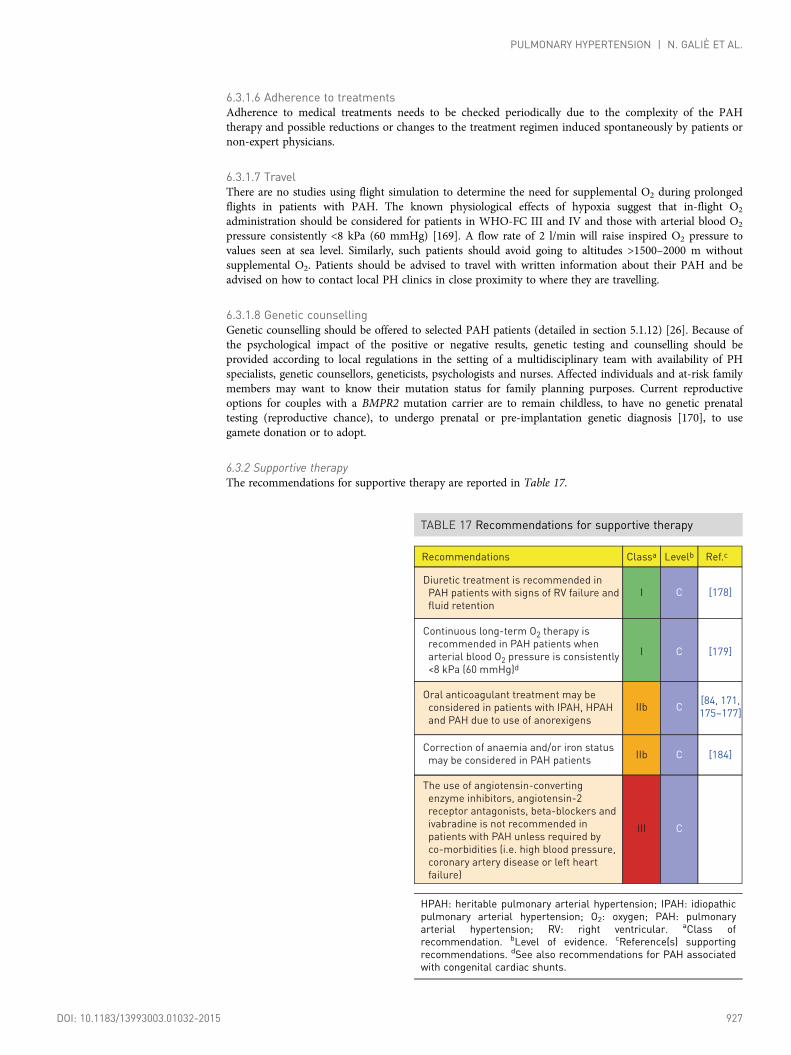
6.3.1.6 Adherence to treatmentsAdherence to medical treatments needs to be checked periodically due to the complexity of the PAHtherapy and possible reductions or changes to the treatment regimen induced spontaneously by patients ornon-expert physicians.
6.3.1.7 TravelThere are no studies using flight simulation to determine the need for supplemental O2 during prolongedflights in patients with PAH. The known physiological effects of hypoxia suggest that in-flight O2
administration should be considered for patients in WHO-FC III and IV and those with arterial blood O2
pressure consistently <8 kPa (60 mmHg) [169]. A flow rate of 2 l/min will raise inspired O2 pressure tovalues seen at sea level. Similarly, such patients should avoid going to altitudes >1500–2000 m withoutsupplemental O2. Patients should be advised to travel with written information about their PAH and beadvised on how to contact local PH clinics in close proximity to where they are travelling.
6.3.1.8 Genetic counsellingGenetic counselling should be offered to selected PAH patients (detailed in section 5.1.12) [26]. Because ofthe psychological impact of the positive or negative results, genetic testing and counselling should beprovided according to local regulations in the setting of a multidisciplinary team with availability of PHspecialists, genetic counsellors, geneticists, psychologists and nurses. Affected individuals and at-risk familymembers may want to know their mutation status for family planning purposes. Current reproductiveoptions for couples with a BMPR2 mutation carrier are to remain childless, to have no genetic prenataltesting (reproductive chance), to undergo prenatal or pre-implantation genetic diagnosis [170], to usegamete donation or to adopt.
6.3.2 Supportive therapyThe recommendations for supportive therapy are reported in Table 17.
TABLE 17 Recommendations for supportive therapy
Recommendations
Diuretic treatment is recommended in
PAH patients with signs of RV failure and
fluid retention
Continuous long-term O2 therapy is
recommended in PAH patients when
arterial blood O2 pressure is consistently
<8 kPa (60 mmHg)d
Classa
I
I
Levelb
C
C
Ref.c
[178]
[179]
Oral anticoagulant treatment may be
considered in patients with IPAH, HPAH
and PAH due to use of anorexigens
Correction of anaemia and/or iron status
may be considered in PAH patients
IIb
IIb
C
C
[84, 171,
175–177]
The use of angiotensin-converting
enzyme inhibitors, angiotensin-2
receptor antagonists, beta-blockers and
ivabradine is not recommended in
patients with PAH unless required by
co-morbidities (i.e. high blood pressure,
coronary artery disease or left heart
failure)
III C
[184]
HPAH: heritable pulmonary arterial hypertension; IPAH: idiopathicpulmonary arterial hypertension; O2: oxygen; PAH: pulmonaryarterial hypertension; RV: right ventricular. aClass ofrecommendation. bLevel of evidence. cReference(s) supportingrecommendations. dSee also recommendations for PAH associatedwith congenital cardiac shunts.
DOI: 10.1183/13993003.01032-2015 927
PULMONARY HYPERTENSION | N. GALIÈ ET AL.

6.3.2.1 Oral anticoagulantsThere is a high prevalence of vascular thrombotic lesions at post-mortem examination in patients withIPAH [171]. Abnormalities in coagulation and fibrinolytic pathways have also been reported [172–174].This, together with the non-specific increased risk factors for venous thromboembolism, including heartfailure and immobility, represents the rationale for oral anticoagulation in PAH. Evidence in favour of oralanticoagulation is confined to patients with IPAH, HPAH and PAH due to anorexigens and is generallyretrospective and based on single-centre experience [84, 171]. Registry and RCT data appear to beheterogeneous and inconclusive [175–177]. The potential benefits of oral anticoagulation in APAH is evenless clear. Generally patients with PAH receiving therapy with long-term i.v. prostaglandins areanticoagulated in the absence of contraindications due in part to the additional risk of catheter-associatedthrombosis. The role of the new oral anticoagulants in PAH is unknown. Additional information onAPAH is provided in the individual chapters.
6.3.2.2 DiureticsDecompensated right heart failure leads to fluid retention, raised central venous pressure, hepatic congestion,ascites and peripheral oedema. Although there are no RCTs on the use of diuretics in PAH, clinicalexperience shows clear symptomatic benefit in fluid overloaded patients treated with this therapy. The choiceand dose of diuretic therapy may be left to the PAH physician [178]. The addition of aldosterone antagonistsshould also be considered together with systematic assessments of electrolyte plasma levels. It is importantwith diuretic use to monitor renal function and blood biochemistry in patients to avoid hypokalaemia andthe effects of decreased intravascular volume leading to pre-renal failure.
6.3.2.3 OxygenAlthough O2 administration has been demonstrated to reduce the PVR in patients with PAH, there are norandomised data to suggest that long-term O2 therapy is beneficial. Most patients with PAH, except thosewith CHD and pulmonary-to-systemic shunts, have minor degrees of arterial hypoxaemia at rest unlessthey have a patent foramen ovale. There are data showing that nocturnal O2 therapy does not modify thenatural history of advanced Eisenmenger syndrome [179]. Guidance may be based on evidence in patientswith COPD; when arterial blood O2 pressure is consistently <8 kPa (60 mmHg; alternatively, <91% ofarterial O2 saturation) patients are advised to take O2 to achieve an arterial blood O2 pressure >8 kPa[169]. Ambulatory O2 may be considered when there is evidence of symptomatic benefit and correctabledesaturation on exercise.
6.3.2.4 Digoxin and other cardiovascular drugsDigoxin has been shown to improve CO acutely in IPAH, although its efficacy is unknown whenadministered chronically [180]. It may be given to slow ventricular rate in patients with PAH who developatrial tachyarrhythmias.
No convincing data are available on the usefulness and safety of angiotensin-converting enzyme inhibitors,angiotensin II receptor antagonists, beta-blockers or ivabradine in patients with PAH.
6.3.2.5 Anaemia and iron statusIron deficiency is common in patients with PAH and has been reported in 43% of patients with IPAH,46% of patients with SSc-PAH and 56% of patients with Eisenmenger syndrome [181–183]. In all of theseentities, preliminary data indicate that iron deficiency may be associated with reduced exercise capacity,and perhaps also with a higher mortality, independent of the presence or severity of anaemia [181, 182,184, 185]. Based on these data, regular monitoring of the iron status should be considered in patients withPAH and detection of an iron deficiency should trigger a search for potential reasons. Iron substitutionshould be considered in patients with iron deficiency. Some studies suggest that oral iron absorption isimpaired in patients with PAH, so i.v. iron administration may be preferable [181, 184, 186]. However,controlled trials are lacking.
6.3.3 Specific drug therapy6.3.3.1 Calcium channel blockersIt has been increasingly recognised that only a small number of patients with IPAH who demonstrate afavourable response to acute vasodilator testing (Table 11) at the time of RHC do well with CCBs [84, 85].The CCBs that have been predominantly used in reported studies are nifedipine, diltiazem andamlodipine, with particular emphasis on nifedipine and diltiazem [84, 85]. The choice of CCB is based onthe patient’s heart rate at baseline, with a relative bradycardia favouring nifedipine and amlodipine and arelative tachycardia favouring diltiazem. The daily doses of these drugs that have shown efficacy in IPAHare relatively high: 120–240 mg for nifedipine, 240–720 mg for diltiazem and up to 20 mg for amlodipine.
928 DOI: 10.1183/13993003.01032-2015
PULMONARY HYPERTENSION | N. GALIÈ ET AL.

It is advisable to start with an initial lower dose, e.g. 30 mg of slow release nifedipine twice a day or 60 mgof diltiazem three times a day (t.i.d.) or 2.5 mg of amlodipine once a day, and increase cautiously andprogressively to the maximum tolerated dose. Limiting factors for dose increase are usually systemichypotension and lower limb peripheral oedema. Patients with IPAH who meet the criteria for a positivevasodilator response and are treated with CCBs should be followed closely for reasons of both safety andefficacy, with a complete reassessment after 3–4 months of therapy including RHC.
If the patient does not show an adequate response, defined as being in WHO-FC I or II and with amarked haemodynamic improvement (near normalization), additional PAH therapy should be instituted.In some cases the combination of CCB with the approved PAH drugs is required because of furtherclinical deterioration in case of CCB withdrawal attempts. Patients who have not undergone avasoreactivity study or those with a negative study should not be started on CCBs because of potentialsevere side effects (e.g. hypotension, syncope and RV failure) [187].
Vasodilator responsiveness does not appear to predict a favourable long-term response to CCB therapy inpatients with PAH in the setting of CTD, HIV, porto-pulmonary hypertension (PoPH) and PVOD [188, 189].
The recommendations for CCB therapy are reported in Table 18. For specific approved doses of the drugs,please refer to the updated official prescription information.
6.3.3.2 Endothelin receptor antagonistsActivation of the endothelin system has been demonstrated in both plasma and lung tissue of PAH patients[190]. Although it is unclear if the increases in endothelin-1 plasma levels are a cause or a consequence ofPH [191], these data support a prominent role for the endothelin system in the pathogenesis of PAH [192].Endothelin-1 exerts vasoconstrictor and mitogenic effects by binding to two distinct receptor isoforms in the
TABLE 18 Recommendations for calcium channel blockertherapy in patients who respond to the acute vasoreactivity test
Recommendations
High doses of CCBs are recommended in
patients with IPAH, HPAH and DPAH who
are responders to acute vasoreactivity
testing
Close follow-up with complete
reassessment after 3–4 months of
therapy (including RHC) is
recommended in patients with IPAH,
HPAH and DPAH treated by high doses
of CCBs
Classa
I
I
Levelb
C
C
Ref.c
[84, 85]
Continuation of high doses of CCBs is
recommended in patients with IPAH,
HPAH and DPAH in WHO-FC I or II with
marked haemodynamic improvement
(near normalization)
I C [84, 85]
High doses of CCBs are not indicated in
patients without a vasoreactivity study or
non-responders unless standard doses
are prescribed for other indications (e.g.
Raynaud’s phenomenon)
III C
[84, 85]
Initiation of specific PAH therapy is
recommended in patients in WHO-FC III
or IV or those without marked
haemodynamic improvement (near
normalization) after high doses of CCBs
I C [84, 85]
CCB: calcium channel blocker; DPAH: drug-induced PAH; HPAH:heritable PAH; IPAH: idiopathic PAH; PAH: pulmonary arterialhypertension; RHC: right heart catheterization; RV: right ventricular;WHO-FC: World Health Organization functional class.
DOI: 10.1183/13993003.01032-2015 929
PULMONARY HYPERTENSION | N. GALIÈ ET AL.

pulmonary vascular smooth muscle cells, endothelin receptors type A and B. The characteristics of RCTswith PAH drugs interfering with the endothelin pathway are reported in Web Table VIA.
AmbrisentanAmbrisentan is an ERA that preferentially binds with endothelin receptor type A. Ambrisentan has beenevaluated in a pilot study [193] and in two large RCTs that have demonstrated efficacy on symptoms,exercise capacity, haemodynamics and time to clinical worsening of patients with IPAH and PAHassociated with CTD and HIV infection [194]. The incidence of abnormal liver function tests ranges from0.8 to 3%. Monthly liver function assessment is not mandated in the USA [195]. An increased incidenceof peripheral oedema has been reported with ambrisentan use.
BosentanBosentan is an oral active dual endothelin receptor type A and B antagonist and the first molecule of itsclass to be synthesized. Bosentan has been evaluated in PAH (idiopathic, associated with CTD andEisenmenger syndrome) in six RCTs (Study-351, BREATHE-1, BREATHE-2, BREATHE-5, EARLY andCOMPASS 2), which showed improvement in exercise capacity, FC, haemodynamics, echocardiographicand Doppler variables and time to clinical worsening [196–200]. Increases in hepatic aminotransferasesoccurred in approximately 10% of the patients and were found to be dose dependent and reversible afterdose reduction or discontinuation. For these reasons, liver function testing should be performed monthlyin patients receiving bosentan.
MacitentanThe dual ERA macitentan has been evaluated in an event-driven RCT [201]: 742 PAH patients weretreated with 3 mg or 10 mg macitentan as compared with placebo for an average of 100 weeks. Theprimary endpoint was the time from the initiation of treatment to the first occurrence of a compositeendpoint of death, atrial septostomy, lung transplantation, initiation of treatment with i.v. or subcutaneousprostanoids or worsening of PAH. Macitentan significantly reduced this composite endpoint of morbidityand mortality among patients with PAH and also increased exercise capacity. Benefits were shown both forpatients who had not received treatment previously and for those receiving additional therapy for PAH.While no liver toxicity was shown, reduction in blood haemoglobin ⩽8 g/dl was observed in 4.3% ofpatients receiving 10 mg of macitentan.
6.3.3.3 Phosphodiesterase type 5 inhibitors and guanylate cyclase stimulatorsInhibition of the cyclic guanosine monophosphate (cGMP) degrading enzyme phosphodiesterase type 5results in vasodilation through the NO/cGMP pathway at sites expressing this enzyme. Since thepulmonary vasculature contains substantial amounts of phosphodiesterase type 5, the potential clinicalbenefit of phosphodiesterase type 5 inhibitors (PDE-5is) has been investigated in PAH. In addition,PDE-5is exert antiproliferative effects [202, 203]. All three PDE-5is approved for the treatment of erectiledysfunction—sildenafil, tadalafil and vardenafil—cause significant pulmonary vasodilation, with maximumeffects observed after 60, 75–90 and 40–45 minutes, respectively [204]. The characteristics of RCTs withPAH drugs interfering with the NO pathway [soluble guanylate cyclase (sGC) stimulators, PDE-5is] arereported in Web Table VIB.
SildenafilSildenafil is an orally active, potent and selective inhibitor of phosphodiesterase type 5. Four RCTs in PAHpatients treated with sildenafil have confirmed favourable results on exercise capacity, symptoms and/orhaemodynamics [205–208]. An RCT addressing the effects of adding sildenafil to epoprostenol showedimprovements after 12 weeks in 6MWD and time to clinical worsening. Of note, seven deaths occurred inthis trial, all in the placebo group [209]. The approved dose of sildenafil is 20 mg t.i.d. Most side effects ofsildenafil are mild to moderate and mainly related to vasodilation (headache, flushing, epistaxis). Based onpharmacokinetic data, an i.v. formulation of sildenafil [210] has been proposed as a bridge for PAHpatients on long-term oral treatment who are temporarily unable to ingest tablets.
TadalafilTadalafil is a once-daily dispensed selective PDE-5i. An RCT in 406 PAH patients (53% on backgroundbosentan therapy) treated with tadalafil 2.5, 10, 20 or 40 mg once daily has shown favourable results onexercise capacity, symptoms, haemodynamics and time to clinical worsening at the highest dose [211]. Theside-effect profile was similar to that of sildenafil.
930 DOI: 10.1183/13993003.01032-2015
PULMONARY HYPERTENSION | N. GALIÈ ET AL.

VardenafilVardenafil is a twice-daily dispensed PDE-5i. An RCT in 66 treatment-naive PAH patients treated withvardenafil 5 mg twice daily has shown favourable results on exercise capacity, haemodynamics and time toclinical worsening [212]. The side-effect profile was similar to that of sildenafil.
RiociguatWhile PDE-5is such as sildenafil, tadalafil and vardenafil enhance the NO–cGMP pathway, slowing cGMPdegradation, sGC stimulators enhance cGMP production [213]. Moreover, pre-clinical studies with sGCstimulators have antiproliferative and antiremodelling properties in various animal models.
An RCT [214] in 443 PAH patients (44% and 6% on background therapy with ERAs or prostanoids,respectively) treated with riociguat up to 2.5 mg t.i.d. has shown favourable results on exercise capacity,haemodynamics, WHO-FC and time to clinical worsening. The increase in exercise capacity was alsodemonstrated in patients on background therapy. The most common serious adverse event in the placebogroup and in the 2.5-mg group was syncope (4% and 1%, respectively). The combination of riociguat andPDE-5i is contraindicated due to hypotension and other relevant side effects detected in the open-labelphase of an RCT study [215].
6.3.3.4 Prostacyclin analogues and prostacyclin receptor agonistsProstacyclin is produced predominantly by endothelial cells and induces potent vasodilation of all vascularbeds. This compound is the most potent endogenous inhibitor of platelet aggregation and also appears tohave both cytoprotective and antiproliferative activities [216]. Dysregulation of the prostacyclin metabolicpathways has been shown in patients with PAH as assessed by a reduction of prostacyclin synthaseexpression in the pulmonary arteries and of prostacyclin urinary metabolites [217]. The clinical use ofprostacyclin in patients with PAH has been extended by the synthesis of stable analogues that possessdifferent pharmacokinetic properties but share qualitatively similar pharmacodynamic effects.
The characteristics of RCTs with PAH drugs interfering with the prostacyclin pathway (prostanoids andprostacyclin IP receptor agonists) are reported in Web Table VIC.
BeraprostBeraprost is the first chemically stable and orally active prostacyclin analogue. An RCT [218] in Europeand a second in the USA [219] have shown an improvement in exercise capacity that persists up to 3–6months. There were no haemodynamic improvements or long-term outcome benefits. The most frequentadverse events were headache, flushing, jaw pain and diarrhoea.
EpoprostenolEpoprostenol (synthetic prostacyclin) has a short half-life (3–5 minutes) and is stable at room temperaturefor only 8 hours; it requires cooling and continuous administration by means of an infusion pump and apermanent tunnelled catheter. The efficacy of continuous i.v. administration of epoprostenol has beentested in three unblinded RCTs in patients with WHO-FC III and IV IPAH [220, 221] and in those withPAH associated with the scleroderma spectrum of diseases [222]. Epoprostenol improves symptoms,exercise capacity and haemodynamics in both clinical conditions and is the only treatment shown toreduce mortality in IPAH in a single RCT study [221]. The meta-analysis for total mortality of the threeepoprostenol RCTs [220–222] has shown a risk reduction for mortality of about 70%. Long-termpersistence of efficacy has also been shown [96, 107] in IPAH as well as in other APAH conditions[223–225] and in non-operable CTEPH [226].
Treatment with epoprostenol is initiated at a dose of 2–4 ng/kg/min, with doses increasing at a rate limitedby side effects (flushing, headache, diarrhoea, leg pain). The optimal dose varies between individualpatients, ranging in the majority between 20 and 40 ng/kg/min [96, 107].
Serious adverse events related to the delivery system include pump malfunction, local site infection,catheter obstruction and sepsis. Guidelines for the prevention of central venous catheter bloodstreaminfections have been proposed [227]. Abrupt interruption of the epoprostenol infusion should be avoided,because in some patients this may lead to a PH rebound with symptomatic deterioration and even death.A thermostable formulation of epoprostenol is also available and does not usually require cooling packs tomaintain stability beyond 8–12 hours [228].
IloprostIloprost is a chemically stable prostacyclin analogue available for i.v., oral or aerosol administration.Inhaled iloprost has been evaluated in one RCT in which daily repetitive iloprost inhalations (six to ninetimes, 2.5–5 µg/inhalation, median 30 µg daily) were compared with placebo inhalation in patients with
DOI: 10.1183/13993003.01032-2015 931
PULMONARY HYPERTENSION | N. GALIÈ ET AL.

PAH and CTEPH [229]. The study showed an increase in exercise capacity and improvement insymptoms, PVR and clinical events in enrolled patients. A second RCT involving 60 patients alreadytreated with bosentan showed an increase in exercise capacity (P < 0.051) in the subjects randomized tothe addition of inhaled iloprost compared with placebo [230]. Another similar study was terminatedprematurely for futility [231]. Overall, inhaled iloprost was well tolerated, with flushing and jaw pain beingthe most frequent side effects. Continuous i.v. administration of iloprost appeared to be as effective asepoprostenol in a small series of patients with PAH and CTEPH [232]. The effects of oral iloprost havenot been assessed in PAH.
TreprostinilTreprostinil is a tricyclic benzidine analogue of epoprostenol, with sufficient chemical stability to beadministered at ambient temperature. These characteristics allow administration of the compound by i.v.and subcutaneous routes. The subcutaneous administration of treprostinil can be accomplished by amicro-infusion pump and a small subcutaneous catheter. The effects of treprostinil in PAH were assessedin an RCT and showed improvements in exercise capacity, haemodynamics and symptoms [233]. Thegreatest exercise improvement was observed in patients who were more compromised at baseline and insubjects who could tolerate the upper quartile dose (>13.8 ng/kg/min). Infusion site pain was the mostcommon adverse effect of treprostinil, leading to discontinuation of the treatment in 8% of cases on activedrug and limiting dose increases in an additional proportion of patients [233]. Treatment withsubcutaneous treprostinil is initiated at a dose of 1–2 ng/kg/min, with doses increasing at a rate limited byside effects (local site pain, flushing, headache). The optimal dose varies between individual patients,ranging in the majority between 20 and 80 ng/kg/min.
An RCT was performed with i.v. treprostinil in PAH patients, but the enrolment of this trial was closedbecause of safety considerations after 45 (36%) of the planned 126 patients had been randomized [234].The data generated from 31 (25%) survivors after the randomized phase (23 active and 8 placebo) are notconsidered reliable. The dose of i.v. treprostinil is two to three times higher than the dose of i.v.epoprostenol [235, 236].
An RCT with inhaled treprostinil in PAH patients on background therapy with either bosentan or sildenafilshowed improvements in the 6MWD by 20 m at peak dose and 12 m at trough dose, NT-proBNP andquality of life measures [237].
Oral treprostinil has been evaluated in two RCTs in PAH patients on background therapy with bosentanand/or sildenafil and in both trials the primary endpoint 6MWD did not reach statistical significance [238,239]. An additional RCT in treatment-naive PAH patients showed improvement in the 6MWD by 26 m atpeak dose and 17 m at trough dose [240].
SelexipagSelexipag is an orally available, selective prostacyclin IP receptor agonist. Although selexipag and itsmetabolite have modes of action similar to that of endogenous prostacyclin (IP receptor agonism), they arechemically distinct from prostacyclin with a different pharmacology. In a pilot RCT in PAH patients(receiving stable ERA and/or PDE-5i therapy), selexipag reduced PVR after 17 weeks [241]. Anevent-driven phase 3 RCT that enrolled 1156 patients [248] has shown that selexipag alone or on top ofmono- or double therapy with ERAs and/or PDE-5i was able to reduce by 40% (hazard ratio 0.60, P <0.0001) a composite morbidity and mortality endpoint (including death from all causes, hospitalization forworsening of PAH, worsening of PAH resulting in the need for lung transplantation or atrial septostomy,initiation of parenteral prostanoids or chronic O2 for worsening of PAH and disease progression).
Recommendations for efficacy of specific drug monotherapies are reported in the Table 19.
6.3.3.5 Experimental compounds and strategiesDespite progress in the treatment of PAH, the functional limitation and survival of these patients remainunsatisfactory. There are three well-known pathways that contribute to the pathogenesis of PAH: theendothelin, NO and prostacyclin pathways. Treatments targeting these pathways are well established inclinical practice, such as ERAs, PDE-5is and prostanoids. Additional therapeutic strategies targeted atdiverse pathobiological changes are being explored in order to further improve symptoms and prognosis.Three pathways have been explored with unsatisfactory results using the following compounds: inhaledvasoactive intestinal peptide, tyrosine kinase inhibitors (platelet-derived growth factor inhibitors) andserotonin antagonists. The following additional compounds are in an earlier stage of development: rhokinase inhibitors, vascular endothelial growth factor receptor inhibitors, angiopoietin-1 inhibitors andelastase inhibitors. Gene therapy strategies have been tested in animal models. Stem cell therapy hasproven to be effective in the monocrotaline rat model and is currently being tested in a proof-of-concept
932 DOI: 10.1183/13993003.01032-2015
PULMONARY HYPERTENSION | N. GALIÈ ET AL.
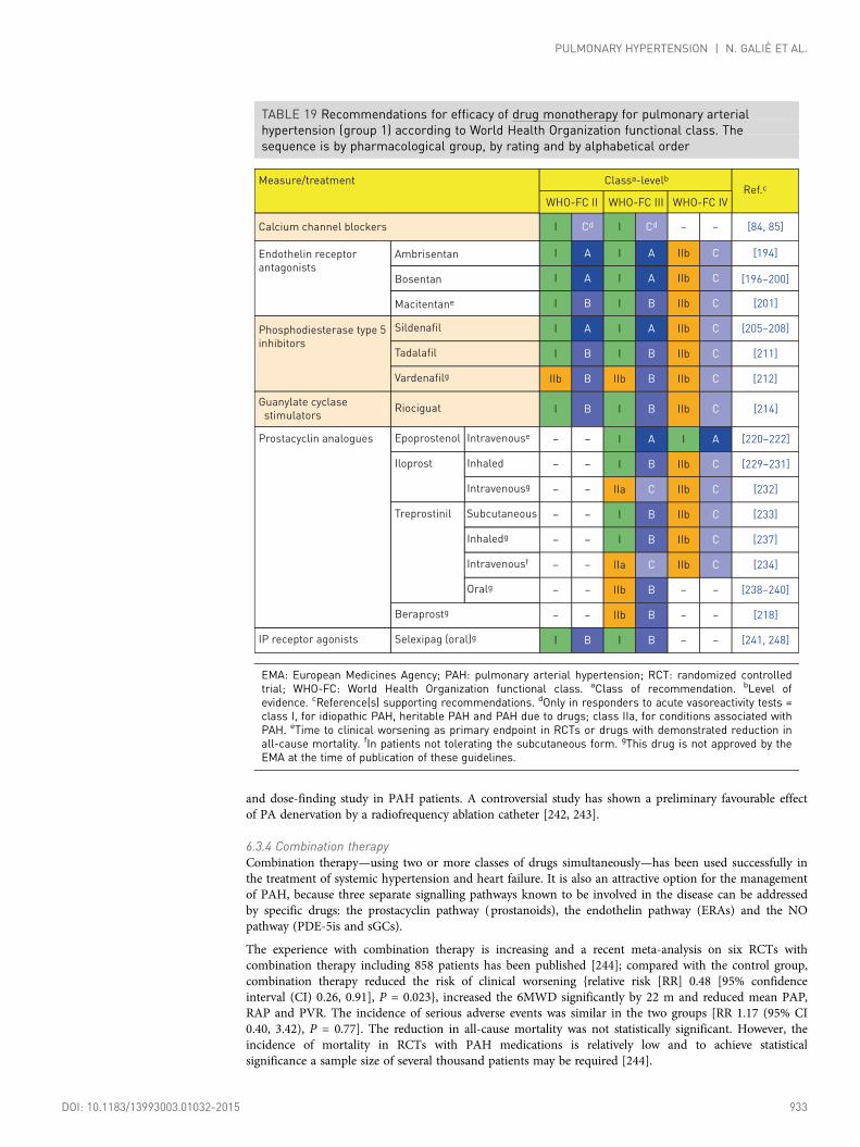
and dose-finding study in PAH patients. A controversial study has shown a preliminary favourable effectof PA denervation by a radiofrequency ablation catheter [242, 243].
6.3.4 Combination therapyCombination therapy—using two or more classes of drugs simultaneously—has been used successfully inthe treatment of systemic hypertension and heart failure. It is also an attractive option for the managementof PAH, because three separate signalling pathways known to be involved in the disease can be addressedby specific drugs: the prostacyclin pathway (prostanoids), the endothelin pathway (ERAs) and the NOpathway (PDE-5is and sGCs).
The experience with combination therapy is increasing and a recent meta-analysis on six RCTs withcombination therapy including 858 patients has been published [244]; compared with the control group,combination therapy reduced the risk of clinical worsening {relative risk [RR] 0.48 [95% confidenceinterval (CI) 0.26, 0.91], P = 0.023}, increased the 6MWD significantly by 22 m and reduced mean PAP,RAP and PVR. The incidence of serious adverse events was similar in the two groups [RR 1.17 (95% CI0.40, 3.42), P = 0.77]. The reduction in all-cause mortality was not statistically significant. However, theincidence of mortality in RCTs with PAH medications is relatively low and to achieve statisticalsignificance a sample size of several thousand patients may be required [244].
TABLE 19 Recommendations for efficacy of drug monotherapy for pulmonary arterialhypertension (group 1) according to World Health Organization functional class. Thesequence is by pharmacological group, by rating and by alphabetical order
Measure/treatment
Calcium channel blockers
Phosphodiesterase type 5
inhibitors
Sildenafil
Tadalafil
Vardenafilg
Endothelin receptor
antagonists
Ambrisentan
Bosentan
Macitentane
Classa-levelb
WHO-FC II WHO-FC III WHO-FC IV
I Cd I Cd
I A I A IIb C
– –
Ref.c
[84, 85]
[194]
I A I A IIb C [196–200]
I
I
I
IIb
B I B IIb C [201]
A I A IIb C [205–208]
B I B IIb C [211]
Riociguat
Epoprostenol Intravenouse
Intravenousg
Iloprost
Treprostinil
Beraprostg
Selexipag (oral)gIP receptor agonists
Inhaled
Subcutaneous
Intravenousf
Oralg
Inhaledg
Guanylate cyclase
stimulators
Prostacyclin analogues
I B
– –
– –
– –
– –
– –
– –
– –
– –
I B
I A
IIb C [214]
I A [220–222]
I B IIb C [229–231]
I B IIb C [233]
I B IIb C [237]
IIa C IIb C [232]
IIb B – – [238–240]
IIb B – – [218]
I B I B – – [241, 248]
IIa C IIb C [234]
B IIb B IIb C [212]
EMA: European Medicines Agency; PAH: pulmonary arterial hypertension; RCT: randomized controlledtrial; WHO-FC: World Health Organization functional class. aClass of recommendation. bLevel ofevidence. cReference(s) supporting recommendations. dOnly in responders to acute vasoreactivity tests =class I, for idiopathic PAH, heritable PAH and PAH due to drugs; class IIa, for conditions associated withPAH. eTime to clinical worsening as primary endpoint in RCTs or drugs with demonstrated reduction inall-cause mortality. fIn patients not tolerating the subcutaneous form. gThis drug is not approved by theEMA at the time of publication of these guidelines.
DOI: 10.1183/13993003.01032-2015 933
PULMONARY HYPERTENSION | N. GALIÈ ET AL.

Combination therapy may be applied sequentially or initially (upfront).
Sequential combination therapy is the most widely utilised strategy both in RCTs and in clinical practice:from monotherapy there is an addition of a second and then a third drug in cases of inadequate clinicalresults or in cases of deterioration. A structured prospective programme to evaluate the adequacy of clinicalresults is the so-called goal-oriented therapy, a treatment strategy that uses known prognostic indicators astreatment targets. The therapy is considered adequate only if the targets are met. The key difference betweengoal-oriented therapy and non-structured approaches is that patients who are stabilised, or even those whoimprove slightly, can still receive additional therapy if treatment goals are not met. The goal-orientedtreatment strategy utilises different targets, including WHO-FC I or II, and the near-normalization of restingCI and/or of NT-proBNP plasma levels. A recent study has confirmed a better prognosis in patientsachieving these goals as compared with patients who did not [97].
Recommendations and evidence on the use of specific drugs for initial combination therapy and forsequential combination therapy for PAH according to WHO-FC are provided in Table 20 and Table 21,respectively.
The rationale for initial or upfront combination therapy is based on the known mortality of PAH, which isreminiscent of many malignancies, and the fact that malignancies and critical medical illnesses (heart failure,malignant hypertension) are not treated with a stepwise approach to therapy, but rather with pre-emptivecombination therapy. The experience in RCTs with initial combination therapy started with the smallBREATHE-2 (Web Table VID) study, which failed to demonstrate any significant difference between patientstreated initially with the combination epoprostenol and bosentan as compared with epoprostenol alone[198]. In a more recent experience, 23 treatment-naive PAH patients were treated with the initialcombination of epoprostenol and bosentan and compared with a matched historical control group treatedwith epoprostenol [245]. The study showed a statistically significant greater decrease in PVR in the initialcombination therapy group, but this haemodynamic benefit did not translate into a statistically significant
TABLE 20 Recommendations for efficacy of initial drugcombination therapy for pulmonary arterial hypertension(group 1) according to World Health Organization functionalclass. Sequence is by rating
Measure/treatment
Ambrisentan +
tadalafild
Other ERA + PDE-5i
Bosentan +
sildenafil +
i.v. epoprostenol
Other ERA or
PDE-5i +
s.c. treprostinil
Bosentan + i.v. epoprostenol
Other ERA or
PDE-5i + other
i.v. prostacyclin
analogues
Classa-Levelb
WHO-FC II WHO-FC III WHO-FC IV
I B I B IIb C
Ref.c
[247]
IIa C IIa C IIb C –
– – IIa C IIa C [246]
– – IIa C IIa C [198, 245]
IIb C IIb C –
IIb C IIb C –
ERA: endothelin receptor antagonist; i.v.: intravenous; PDE-5i:phosphodiesterase type 5 inhibitor; RCT: randomized controlledtrial; s.c.: subcutaneous; WHO-FC: World Health Organizationfunctional class. aClass of recommendation. bLevel of evidence.cReference(s) supporting recommendations. dTime to clinicalfailure as primary endpoint in RCTs or drugs with demonstratedreduction in all-cause mortality (prospectively defined).
934 DOI: 10.1183/13993003.01032-2015
PULMONARY HYPERTENSION | N. GALIÈ ET AL.

difference in survival or in transplant-free survival. A pilot study on an initial triple combination in 19WHO-FC class III and IV patients has provided preliminary evidence of the long-term benefits of upfronttriple combination therapy in patients with severe PAH [246]. A recent multicentre, multinational, blinded,placebo-controlled trial (Web Table VID) compared first-line monotherapy with tadalafil or monotherapywith ambrisentan with upfront combination therapy with tadalafil and ambrisentan in de novo WHO-FC IIand III PAH patients [247]. The primary endpoint was a composite of clinical failure events (including
TABLE 21 Recommendations for efficacy of sequential drugcombination therapy for pulmonary arterial hypertension(group 1) according to World Health Organization functionalclass. Sequence is by rating and by alphabetical order
Measure/treatment
Macitentan added to
sildenafild
Classa-Levelb
WHO-FC II WHO-FC III WHO-FC IV
I B I B IIa C
Ref.c
[201]
Riociguat added
to bosentanI B I B IIa C [214]
Iloprost inhaled
added to bosentanIIb B IIb B IIb C [230, 231]
Ambrisentan added
to sildenafilIIb C IIb C IIb C [249]
Bosentan added to
sildenafilIIb C IIb C IIb C [251, 252]
Other double
combinationsIIb C IIb C IIb C –
Riociguat added to
sildenafil or other
PDE-5i
III B III B III B [215]
Selexipage added to
ERA and/or PDE-5idI B I B IIa C [241, 248]
Treprostinil inhaled
added to sildenafil
or bosentan
IIa B IIa B IIa C [237]
Tadalafil added
to bosentanIIa C IIa C IIa C [211]
Bosentan added to
epoprostenol– – IIb C IIb C [250]
Sildenafil added
to bosentanIIb C IIb C IIb C [252]
Other triple
combinationsIIb C IIb C IIb C –
Sildenafil added
to epoprostenol– – I B IIa B [209]
EMA: European Medicines Agency; ERA: endothelin receptorantagonist; PAH: pulmonary arterial hypertension; PDE-5i:phosphodiesterase type 5 inhibitor; RCT: randomized controlledtrial; WHO-FC: World Health Organization functional class. aClass ofrecommendation. bLevel of evidence. cReference(s) supportingrecommendations. dTime to clinical worsening as primary endpointin RCTs or drugs with demonstrated reduction in all-cause mortality(prospectively defined). eThis drug was not approved by the EMA atthe time of publication of these guidelines.
DOI: 10.1183/13993003.01032-2015 935
PULMONARY HYPERTENSION | N. GALIÈ ET AL.

death, hospitalization, PAH progression and unsatisfactory clinical status). The study was positive, with a50% reduction in events in the combination group. In addition, improvements were observed in exercisecapacity, rate of satisfactory clinical response and NT-proBNP plasma levels [247].
6.3.5 Drug interactionsSignificant drug interactions involving the disease-targeted therapies for PAH are shown in Web Table VII.This table highlights known important interactions but does not include theoretical untested interactions,which may still be clinically important. In addition, updated official prescribing information for eachcompound should be read.
Bosentan is an inducer of cytochrome P450 isoenzymes CYP3A4 and CYP2C9. Plasma concentrations ofdrugs metabolised by these isoenzymes will be reduced when co-administered with bosentan. Bosentan is alsometabolised by these enzymes so that their inhibition may increase the plasma concentration of bosentan. Inaddition to interactions shown in Web Table VII, a combination of a potent CYP3A4 inhibitor (ketoconazole,ritonavir) and/or a CYP2C9 inhibitor (e.g. amiodarone, fluconazole) with bosentan may cause a significantincrease in plasma bosentan levels and thus is contraindicated. Interactions may theoretically occur withitraconazole, tacrolimus, sirolimus, carbamazepine, phenytoin, phenobarbitone, dapsone and St John’s wort.
Sildenafil is metabolised by cytochrome P450 isoenzymes CYP3A4 (major route) and CYP2C9 (minorroute). There is an increase in sildenafil bioavailability and reduced clearance with CYP3A4 substrates andinhibitors and CYP3A4 substrates plus beta-adrenoceptor blockers. CYP3A4 inducers such ascarbamazepine, phenytoin, phenobarbital, rifampicin and St John’s wort may significantly lower sildenafillevels. Sildenafil levels are modestly increased by fresh grapefruit juice, a weak inhibitor of CYP3A4.
Finally, care is needed when PAH-targeted medications are co-administered with antihypertensive drugssuch as beta-adrenoceptor blockers, angiotensin-converting enzyme inhibitors, etc., to avoid excessivesystemic hypotension.
6.3.6 Balloon atrial septostomyThe creation of an inter-atrial right-to-left shunt can decompress the right heart chambers and increase LVpreload and CO [253, 254]. In addition, this improves systemic O2 transport despite arterial O2 desaturation[253] and decreases sympathetic hyperactivity. The recommended technique is graded balloon dilation atrialseptostomy, which produces equivalent improvements in haemodynamics and symptoms but reduced riskcompared with the original blade technique. Other techniques are considered experimental [255].
A careful pre-procedure risk assessment ensures reduced mortality. Balloon atrial septostomy (BAS)should be avoided in end-stage patients presenting with a baseline mean RAP >20 mmHg and O2
saturation at rest <85% on room air. Patients should be on optimal medical therapy, which may includepreconditioning with i.v. inotropic drugs, prior to considering BAS. Published reports suggest a benefit inpatients who are in WHO-FC IV with right heart failure refractory to medical therapy or with severesyncopal symptoms [253, 254]. It may also be considered in patients awaiting lung transplantation withunsatisfactory clinical response on maximal medical therapy or when medical therapy is not available.Studies show improvements in CI and decreases in RAP with improvement in 6MWD [253, 254].The impact of BAS on long-term survival has not been established in RCTs [253, 254]. BAS should beregarded as a palliative or bridging procedure to be performed only in centres with experience inthe method [256]. Since BAS is performed very rarely, it has not been included in the treatmentalgorithm (Figure 2).
6.3.7 Advanced right ventricular failure6.3.7.1 Intensive care unit managementPatients with PH may require intensive care unit (ICU) treatment for a co-morbid condition (includingmajor surgery), right heart failure or both. In a series from France, the mortality among PAH patientsadmitted to the ICU was 41% [257], underscoring the poor prognosis. Hence PAH patients requiring ICUtreatment should be managed at specialized centres whenever possible. Basic monitoring includes vitalsigns (heart rate, blood pressure, body temperature and O2 saturation), urine production, central venouspressure, central venous O2 saturation and blood lactate levels. The combination of a low central venousO2 saturation (<60%) with rising lactate levels and low or absent urine production signals imminent rightheart failure. In certain situations, placement of a right heart catheter might be required to allowcomprehensive haemodynamic monitoring. The basic principles of ICU management of patients with PHand RV failure include the treatment of triggering factors (such as anaemia, arrhythmias, infections orother co-morbidities), optimization of fluid balance (usually with i.v. diuretics), reduction of RV afterload(usually with parenteral prostacyclin analogues, but sometimes also with other PAH drugs), improvementof CO with inotropes (with dobutamine being the preferred inotrope to treat RV failure) and maintenance
936 DOI: 10.1183/13993003.01032-2015
PULMONARY HYPERTENSION | N. GALIÈ ET AL.

of systemic blood pressure with vasopressors, if necessary [258–260]. Intubation should be avoided inpatients with RV failure, as it frequently results in haemodynamic collapse.
6.3.7.2 Right ventricle assistanceThe use of veno-arterial extracorporeal membrane oxygenation (ECMO) should be considered for selectedpatients with PH and RV failure. A veno-venous approach may improve oxygenation but does not unloadthe RV, which makes it unsuitable for this patient population. There are two basic concepts for the use ofECMO in these patients: bridge to recovery and bridge to transplantation. Few reports have beenpublished on the bridge to recovery concept [261], which is only reasonable in patients for whom a cleartherapeutic concept with a realistic chance of recovery exists. There are, however, several reports on thesuccessful use of ECMO as a bridge to transplantation, especially when used in awake patients [261–263].An alternative approach involves connecting a pumpless device to the pulmonary circulation [264, 265].All of these procedures are available only in highly specialized centres.
6.3.8 TransplantationThe advent of disease-targeted therapy for severe PAH has reduced and delayed patient referral for lungtransplant programmes [256]. The long-term outcomes of medically treated patients remain uncertain and
Treatment naïve patientPAH confirmed by
expert centre
General measures
(table 16)
Supportive therapy
(table 17)
Acute vasoreactivity test
(IPAH/HPAH/DPAH only)CCB therapy
(table 18)
Vasoreactive
Non-vasoreactive
Low or intermediate risk
(WHO FC II–III)aHigh risk
(WHO FC IV)a
Initial combination
including i.v. PCAc
(table 20)
Initial oral
combinationb
(table 20)
Initial
monotherapyb
(table 19)
Patient already
on treatment
Inadequate clinical response
(table 15)
Double or triple sequential combination
(table 21)
Consider listing for lung transplantationd
(table 22)
Inadequate clinical response
(table 15)
Consider referral for lung
transplantation
FIGURE 2 Evidence based treatment algorithm for pulmonary arterial hypertension patients (for group 1patients only; see description in the text). CCB: calcium channel blockers; DPAH: drug-induced PAH; HPAH:heritable PAH; IPAH: idiopathic PAH; i.v.: intravenous; PAH: pulmonary arterial hypertension; PCA:prostacyclin analogues; WHO-FC: World Health Organization functional class. aSome WHO-FC III patients maybe considered high risk (see Table 13). bInitial combination with ambrisentan plus tadalafil has proven to besuperior to initial monotherapy with ambrisentan or tadalafil in delaying clinical failure. cIntravenousepoprostenol should be prioritised as it has reduced the 3 months rate for mortality in high risk PAH patientsalso as monotherapy. dConsider also balloon atrial septostomy.
DOI: 10.1183/13993003.01032-2015 937
PULMONARY HYPERTENSION | N. GALIÈ ET AL.
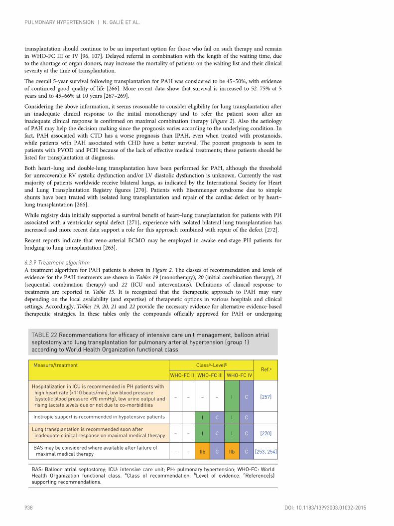
transplantation should continue to be an important option for those who fail on such therapy and remainin WHO-FC III or IV [96, 107]. Delayed referral in combination with the length of the waiting time, dueto the shortage of organ donors, may increase the mortality of patients on the waiting list and their clinicalseverity at the time of transplantation.
The overall 5-year survival following transplantation for PAH was considered to be 45–50%, with evidenceof continued good quality of life [266]. More recent data show that survival is increased to 52–75% at 5years and to 45–66% at 10 years [267–269].
Considering the above information, it seems reasonable to consider eligibility for lung transplantation afteran inadequate clinical response to the initial monotherapy and to refer the patient soon after aninadequate clinical response is confirmed on maximal combination therapy (Figure 2). Also the aetiologyof PAH may help the decision making since the prognosis varies according to the underlying condition. Infact, PAH associated with CTD has a worse prognosis than IPAH, even when treated with prostanoids,while patients with PAH associated with CHD have a better survival. The poorest prognosis is seen inpatients with PVOD and PCH because of the lack of effective medical treatments; these patients should belisted for transplantation at diagnosis.
Both heart–lung and double-lung transplantation have been performed for PAH, although the thresholdfor unrecoverable RV systolic dysfunction and/or LV diastolic dysfunction is unknown. Currently the vastmajority of patients worldwide receive bilateral lungs, as indicated by the International Society for Heartand Lung Transplantation Registry figures [270]. Patients with Eisenmenger syndrome due to simpleshunts have been treated with isolated lung transplantation and repair of the cardiac defect or by heart–lung transplantation [266].
While registry data initially supported a survival benefit of heart–lung transplantation for patients with PHassociated with a ventricular septal defect [271], experience with isolated bilateral lung transplantation hasincreased and more recent data support a role for this approach combined with repair of the defect [272].
Recent reports indicate that veno-arterial ECMO may be employed in awake end-stage PH patients forbridging to lung transplantation [263].
6.3.9 Treatment algorithmA treatment algorithm for PAH patients is shown in Figure 2. The classes of recommendation and levels ofevidence for the PAH treatments are shown in Tables 19 (monotherapy), 20 (initial combination therapy), 21(sequential combination therapy) and 22 (ICU and interventions). Definitions of clinical response totreatments are reported in Table 15. It is recognized that the therapeutic approach to PAH may varydepending on the local availability (and expertise) of therapeutic options in various hospitals and clinicalsettings. Accordingly, Tables 19, 20, 21 and 22 provide the necessary evidence for alternative evidence-basedtherapeutic strategies. In these tables only the compounds officially approved for PAH or undergoing
TABLE 22 Recommendations for efficacy of intensive care unit management, balloon atrialseptostomy and lung transplantation for pulmonary arterial hypertension (group 1)according to World Health Organization functional class
Measure/treatment
Hospitalization in ICU is recommended in PH patients with
high heart rate (>110 beats/min), low blood pressure
(systolic blood pressure <90 mmHg), low urine output and
rising lactate levels due or not due to co-morbidities
Classa-Levelb
WHO-FC II WHO-FC III WHO-FC IV
– – – – I C
Ref.c
[257]
Lung transplantation is recommended soon after
inadequate clinical response on maximal medical therapy – – I C I C [270]
Inotropic support is recommended in hypotensive patients I C I C
BAS may be considered where available after failure of
maximal medical therapy IIb C IIb C– – [253, 254]
BAS: Balloon atrial septostomy; ICU: intensive care unit; PH: pulmonary hypertension; WHO-FC: WorldHealth Organization functional class. aClass of recommendation. bLevel of evidence. cReference(s)supporting recommendations.
938 DOI: 10.1183/13993003.01032-2015
PULMONARY HYPERTENSION | N. GALIÈ ET AL.
regulatory approval in at least one country are included. A four-level hierarchy for endpoints in RCTs hasbeen proposed by experts according to the level of evidence regarding efficacy [273, 274]. According to thishierarchy, drugs or combinations of drugs with time to clinical failure or to clinical worsening as the primaryendpoint in RCTs or drugs with a demonstrated reduction in all-cause mortality (prospectively defined) havebeen highlighted with a footnote in Tables 19, 20 and 21. The PAH treatment algorithm does not apply topatients in other clinical groups, and in particular it does not apply to patients with PH associated with group2 (LHD) or group 3 (lung diseases). In addition, the different treatments have been evaluated by RCTsmainly in IPAH, HPAH, PAH due to drugs and in PAH associated with CTD or with CHD (surgicallycorrected or not).
Treatment algorithm description (Figure 2)
• After confirmation of the diagnosis of the treatment-naive PAH patient in an expert centre, thesuggested initial approach is the adoption of general measures and the initiation of supportive therapy ifneeded (Tables 16 and 17).
• Acute vasoreactivity testing should be performed only in patients with IPAH, HPAH and PAHassociated with drugs or toxins use. Vasoreactive patients should be treated with high doses(progressively titrated) of CCB; adequate response should be confirmed after 3–4 months of treatment(Table 18). Patient responders without an adequate clinical response to CCB treatment should be treatedwith approved PAH medications according to the non-vasoreactive patient’s treatment strategy.
• Non-responders to acute vasoreactivity testing who are at low or intermediate risk (Table 13) can betreated with either initial monotherapy (Table 19) or initial oral combination therapy (Table 20).
• If initial monotherapy is chosen, since head-to-head comparisons among different compounds are notavailable, no evidence-based first-line monotherapy can be proposed. In this case the choice of the drugmay depend on a variety of factors, including the approval status, labelling, route of administration,side-effect profile, potential interaction with background therapies, patient preferences, co-morbidities,physician experience and cost.
• Since head-to-head comparison between initial combination therapy with ambrisentan plus tadalafil hasproven to be superior to initial monotherapy with ambrisentan or tadalafil in delaying clinical failure, ahigher grade of recommendation has been given to this initial combination (Table 20) [247].
• In non-vasoreactive and treatment-naive patients at high risk (Table 13), initial combination therapyincluding i.v. prostacyclin analogues should be considered (Table 20). I.V. epoprostenol should beprioritised since it has reduced the 3-month rate of mortality in high-risk PAH patients also asmonotherapy (Table 19) [149]. Alternative types of initial combination therapy may be considered(Table 20).
• In case of inadequate clinical response to initial combination therapy or initial monotherapy (Table 15),sequential double or triple combination therapy is recommended according to Table 21. Thecombination of riociguat and PDE-5i is contraindicated.
• In case of inadequate clinical response with sequential double combination therapy, triple combinationtherapy should be attempted (Tables 20 and 21).
• It seems reasonable to consider eligibility for lung transplantation after an inadequate clinical responseto the initial monotherapy or initial combination therapy and to refer the patient for lungtransplantation soon after the inadequate clinical response is confirmed on maximal combinationtherapy. BAS should be regarded as a palliative or bridging procedure in patients deteriorating despitemaximal medical therapy.
6.3.10 Diagnosis and treatment of pulmonary arterial hypertension complications6.3.10.1 ArrhythmiasArrhythmias are an increasing clinical problem in PAH patients. In particular, the presence of symptomaticatrial arrhythmia might portend a poor prognosis [275]. In contrast to patients with LHD, malignantventricular arrhythmias such as ventricular tachycardia, ventricular flutter and ventricular fibrillation arerare in PAH patients. In a series of 132 witnessed cardiac arrests in patients with PAH, ventricularfibrillation was observed in only 8% of the cases [276]. Another series of 231 patients with PAH or CTEPHfollowed over a 6-year period did not report any case of malignant ventricular arrhythmia [275]. In thatseries, supraventricular tachyarrhythmias occurred with an annual incidence of 2.8%. Atrial flutter andatrial fibrillation were equally common and both arrhythmias invariably led to clinical deterioration withsigns of right heart failure. Treatment of atrial flutter proved to be more successful than treatment of atrialfibrillation. Restoration of a stable sinus rhythm was associated with a favourable long-term survival, whilepersistent atrial fibrillation was associated with a 2-year mortality >80% [275]. Supraventricular arrhythmiasrepresent an indication for oral anticoagulation with vitamin K antagonists or new oral anticoagulants. Bothelectric cardioversion and radiofrequency ablation in refractory cases have proven to be effective [277].
DOI: 10.1183/13993003.01032-2015 939
PULMONARY HYPERTENSION | N. GALIÈ ET AL.

Although prospective and controlled data are lacking, these findings suggest that maintenance of a stablesinus rhythm after cardioversion should be considered an important treatment goal in patients with PAH.In order to achieve a stable sinus rhythm, prophylaxis with antiarrhythmic drugs without negativeinotropic effects, such as oral amiodarone (see interactions in Web Table VII), should also be considered,even if specific data regarding efficacy are lacking.
6.3.10.2 HaemoptysisHaemoptysis is a recognised complication of PH, represents a risk factor for mortality and appears to bemore frequent in specific types, such as HPAH, PAH associated with CHD and CTEPH. Haemoptysisseverity ranges from mild to very severe leading to sudden death. Reported prevalence is variable, from 1%to 6% [278]. Bronchial artery embolization is suggested as an acute emergency procedure in the case ofsevere haemoptysis or as elective intervention in cases of frequent mild or moderate episodes. Haemoptysismay represent a contraindication to anticoagulant therapy.
6.3.10.3 Mechanical complicationsMechanical complications in PAH patients are usually related to progressive dilatation of the PA andinclude PA aneurysms, rupture and dissection and compression of different intrathoracic structures such asthe left main coronary artery, pulmonary veins, main bronchi and the recurrent laryngeal nerves [279–281].Symptoms and signs are not specific and are variable according to the different complications, includingchest pain (angina-like or not), dyspnoea, regional lung oedema and sudden death. Contrast-enhanced andhigh-resolution CT scan represent the best imaging option to detect mechanical complications.
Treatment options are not well established in cases of PA aneurysms, pseudo-aneurysms and dissection[279, 280].
The indications and results of a traditional surgical approach in the presence of PH are unknown and thepredictable risks may be very high. The percutaneous approach (stenting) faces relevant technical issuesmainly due to early branching of the main and lobar PA. A possible approach is double-lung or heart–lung transplantation, but this solution can be applied only in chronic stabilised cases, and clear indicationsare lacking. In patients with left main coronary artery compression, a percutaneous stenting procedureshould be performed [282].
6.3.11 End of life care and ethical issuesThe clinical course of PH is one of progressive deterioration interspersed with episodes of acutedecompensation. It is difficult to predict when patients will die since death may come either suddenly orslowly because of progressive heart failure. It has been shown that physicians caring for patients tend to beoveroptimistic in their prognostication and frequently misunderstand their patients’ wishes.
Open and sensitive communication with patients allows advanced planning and discussion of their fears,concerns and wishes, which is essential to good care. Opportunities to discuss prognosis should be createdat the time of initial diagnosis. Recognition that cardiopulmonary resuscitation in severe PH has a pooroutcome may enable a do-not-resuscitate order. This may increase the chance of patients being in theirpreferred place of care at the end of life.
Patients approaching the end of life require frequent assessment of their full needs by a multidisciplinaryteam. Attention should be given to controlling the distressing symptoms and prescribing appropriate drugswhile withdrawing unnecessary medication. Well-informed psychological, social and spiritual support isalso vital. Specialist palliative care should be consulted about patients whose needs are beyond theexpertise of the PH team.
PH is a disease that may be severely life-limiting. In addition to psychological and social support, there shouldbe proactive advanced care planning with referral to specialist palliative care services when appropriate.
7. Specific pulmonary (arterial) hypertension subsets7.1 Paediatric pulmonary arterial hypertensionPH can present at any age from the neonatal period to adulthood. Paediatric PH has some unique featuresthat are not found in adult PH, including prenatal aetiological factors, and postnatal parenchymal andvascular abnormalities in lung development [283]. Before the epoprostenol era, the prognosis had beenworse in children, with a median survival estimated at 10 months, compared to 2.8 years in adults;however, with new targeted therapies the outcome has improved significantly [9, 284].
Recent data from registries have shed light on the prevalence and incidence of paediatric PH. In theNetherlands, the incidence and prevalence of IPAH is 0.7 and 4.4 per million children, respectively [285].Similar numbers have been observed in the UK, with annual cases of IPAH of 0.48 per million and a
940 DOI: 10.1183/13993003.01032-2015
PULMONARY HYPERTENSION | N. GALIÈ ET AL.

prevalence of 2.1 per million [286]. National and large-scale registries either including children [284] orexclusively dedicated to paediatrics [287, 288] have described the different aetiologies of PH, with IPAH,HPAH and CHD-PAH as the most common. However, PH associated with respiratory disease is alsonoted to be important and may be underreported [287].
Although the 2009 Dana Point classification included most of the paediatric causes, it was felt thatpaediatric aetiologies should be better described. The 2011 Panama classification was recently proposed,with 10 different subgroups of paediatric PH [289]. The 2013 Nice classification includes new groups andaetiologies specific to children [9], such as congenital and acquired left heart inflow/outflow tractobstruction and segmental PH; it has been further adapted for the current ESC/ERS Guidelines (Table 4and Web Table I) with provision of clinical (Table 6) and anatomical–pathophysiological classification ofcongenital systemic-to-pulmonary shunts associated with PAH (Web Table II) and of developmental lungdisease (Web Table III).
PPHN remains in the PAH group but has been moved to a subgroup, as it is considered to be a specificentity with a more transient course (Table 4 and Web Table IV) in most cases.
7.1.1 DiagnosisDyspnoea, fatigue and failure to thrive are common symptoms; syncope is more common in children, butovert RV failure is a late event and the child may die of sudden death before the occurrence of RV failure[284, 287]. A specific diagnostic workup has been recently recommended, and even if some causes arerare, they should be excluded before a definite diagnosis of IPAH [284]. A thorough family and personalhistory, including pregnancy, delivery and postnatal details, is essential [290]. Diagnosis must beconfirmed by heart catheterization, and vasoreactivity testing should be performed. Recent reports suggestthat cardiac catheterization may be a greater risk in children compared with adults, with younger age (<1year) and worse FC (WHO-FC IV) as risk factors. Cardiac catheterization performed in expert centres isrecommended [290]. The general scheme of the diagnostic algorithm for adult patients with PH (Figure 1)may also be adopted in children, with some adaptation related to the different epidemiology [9].
As in adults, clinical evidence of RV failure, progression of symptoms, WHO-FC III/IV and elevated BNPlevels are recognized as being associated with higher risk of death. In children, failure to thrive, haemodynamicparameters such as the PAPm:systemic artery pressure ratio, RAP >10 mmHg and PVR index >20 WU/m2
have also been associated with a higher risk of death, while the 6MWD was not a prognostic parameter.
7.1.2 TherapyThere is a lack of randomized trials in paediatrics, making it difficult to deliver strong guidelines [291,292]. A specific treatment algorithm, similar to that used in adults (Figure 2), has been recommended.Determinants of risk and risk stratification have also been suggested [9]. CCBs are used in responders, butclose follow-up is mandatory, as some patients may fail long-term therapy.
Indications for epoprostenol are similar to those for adults. The optimal dose varies between patients andthus individual adaptation is required [293, 294]. Use of i.v. iloprost and trepostinil, as well assubcutaneous trepostinil [295], has been reported. Oral beraprost is used in some countries, but lack ofproof of efficacy is a problem. Inhaled iloprost is difficult to deliver, but some reports have shown efficacy,mostly in combination with other therapies [296].
Bosentan pharmacokinetics have been assessed in two studies [297, 298]. Several uncontrolled studies haveshown positive results similar to adults, with survival rates around 80–90% at 1 year [298]. A paediatricformulation is available in Europe [299]. Data with ambrisentan are scarce and a study is ongoing.
Sildenafil has shown efficacy [300] and has been approved in Europe for children 1–17 years of age.Increased mortality using high doses has raised concerns; therefore high doses should not be used inchildren (high individual doses of sildenafil on a three daily dosing not recommended: >10 mg/dose with abodyweight of 8–20 kg, >20 mg/dose in children with a bodyweight >20 kg or >1 mg/kg/dose in infantsand small children) [301].
Data on tadalafil also suggest efficacy [302]; a trial is currently under way to define the specific dosage forchildren.
An increasing number of paediatric patients are on combination therapies even though evidence is stilllimited [303].
RV decompression strategies include atrioseptostomy [304], ductal stenting in cases of patent ductusarteriosus [305] and a surgical Potts shunt [306]. Transcatheter Potts shunt creation has also beenproposed [307]. Lung transplantation remains an important option for paediatric PH patients.
DOI: 10.1183/13993003.01032-2015 941
PULMONARY HYPERTENSION | N. GALIÈ ET AL.

Specific goals for treatment should be applied for children. Some have been extrapolated from defined riskfactors in children but still require validation in large cohorts [9]. Recently FC, TAPSE and NT-proBNPhave been recognized as treatment goals [308].
The recommendations for paediatric PH are reported in the Table 23.
7.2. Pulmonary arterial hypertension associated with adult congenital heart diseasePAH associated with adult CHD is included in group 1 of the PH clinical classification (Table 4) andrepresents a very heterogeneous patient population. A specific clinical classification (Table 6) and ananatomical–pathophysiological classification (Web Table II) are provided to better characterize eachindividual patient with PAH associated with adult CHD [13, 309]. Some malformations, such as patentductus arteriosus, sinus venosus atrial septal defect or partial anomalous pulmonary venous return, areoften concealed and patients are misclassified as suffering from IPAH. Hence these congenital anomaliesshould be specifically sought.
Epidemiological data remain scarce, as no studies have been designed to assess the prevalence of PAH inadult CHD, although the figure of 5–10% was reported in a European survey [310]. Persistent exposure ofthe pulmonary vasculature to increased blood flow due to systemic-to-pulmonary shunts, as well asincreased pressure, may result in a typical pulmonary obstructive arteriopathy (identical to other PAHforms) leading to an increase in PVR. If PVR approaches or exceeds systemic vascular resistance (SVR),the shunt is reversed (Eisenmenger syndrome) [311].
7.2.1. DiagnosisAs indicated in Table 6, the clinical presentation of adult patients with PAH may differ. Eisenmengersyndrome is a multisystem disorder and the most severe form of PAH in adult CHD. The signs andsymptoms of Eisenmenger syndrome result from PH, low arterial O2 saturation and haematologicalchanges, including secondary erythrocytosis, thrombocytopenia and at times leucopenia. They includedyspnoea, fatigue and syncope. In patients with PAH associated with adult CHD without reversal of theshunt, the degree of cyanosis and erythrocytosis may be mild to moderate. Eisenmenger syndrome patientsalso present with haemoptysis, cerebrovascular accidents, brain abscesses, coagulation abnormalities andsudden death. Subjects with Eisenmenger syndrome have reduced life expectancy, although many surviveinto their third or fourth decade, with some even surviving up to their seventh decade [312]. In patientslisted for lung or heart–lung transplantation when no medical treatment was available, Eisenmengersyndrome had better survival compared with IPAH, with a 3-year survival rate of 77% compared with 35%for untreated IPAH [313]. In a recent study on the different clinical groups of PAH patients associatedwith CHD (Table 6), the worst survival was observed in patients with PAH after defect repair or withsmall/coincidental defects as compared with patients with Eisenmenger syndrome or those with prevalentsystemic-to-pulmonary shunts [314]. Improved survival may result from preservation of RV function, asthe RV does not undergo remodelling at birth and remains hypertrophied [315]. The RV is also relievedby the right-to-left shunt, sustaining CO at the expense of hypoxaemia and cyanosis.
Of all patients with CHD, those with Eisenmenger syndrome are the most severely compromised in termsof exercise intolerance [314, 316].
Patients with CHD (in particular those without shunts) may also develop PH due to LHD (group 2,Table 4) or concomitant lung diseases (group 3, Table 4). In these cases, a comprehensive diagnosticworkup, as reported in the section 7.1.1, is recommended.
7.2.2. TherapyOperability may be considered in patients with prevalent systemic-to-pulmonary shunting (Table 6). Thecriteria for shunt closure based on baseline PVR have been proposed (Table 24) based on availableliterature data [317–319]. Additional criteria include the type of the defect, age, the PVR:SVR ratio and theQp:Qs ratio [320]. No prospective data are available on the usefulness of vasoreactivity testing, closure testor lung biopsy for operability assessment [320]. Surgical or percutaneous intervention is contraindicated inpatients with Eisenmenger syndrome and likely is useless in patients with small/coincidental defects.
The medical treatment strategy for patients with PAH associated with CHD, and in particular for subjectswith Eisenmenger syndrome, is mainly based on the clinical experience of experts rather than beingformally evidence-based [311]. A specific treatment algorithm has been proposed [309].
PAH associated with CHD patients should be managed in specialised centres. Patient education, behaviouralmodifications and awareness of potential medical risk factors are important aspects of management.
942 DOI: 10.1183/13993003.01032-2015
PULMONARY HYPERTENSION | N. GALIÈ ET AL.

Patients with PAH associated with CHD may present with clinical deterioration in different circumstances,such as during non-cardiac surgery requiring general anaesthesia, dehydration, lung infections and at highaltitudes. It is recommended to avoid strenuous exercise, but mild activities seem to be beneficial.Pregnancy is associated with very high risk to both the mother and foetus and should be discouraged.Hence effective contraception is mandatory. Dual contraception is indicated in patients taking ERAs inlight of the interaction with progesterone-based compounds.
Long-term home O2 therapy may improve symptoms but has not been shown to modify survival, at leastwhen given only at night [179]. The use of supplemental O2 therapy is recommended in cases in which itproduces a consistent increase in arterial O2 saturation and reduces symptoms.
The use of oral anticoagulant treatment in Eisenmenger syndrome is controversial: a high incidence of PAthrombosis and stroke is reported, but there is also an increased risk of haemorrhage and haemoptysis[321]. No data exist on this issue and thus definite recommendations cannot be made. Oral anticoagulanttreatment may be considered in patients with PA thrombosis, signs of heart failure and absent or onlymild haemoptysis [321].
Secondary erythrocytosis is beneficial for adequate O2 transport and delivery and routine phlebotomyshould be avoided. If symptoms of hyperviscosity are present, phlebotomy with isovolumic replacementshould be performed, usually when the haematocrit is >65%. Iron deficiency should be corrected. No cleardata support the use of CCBs in patients with Eisenmenger syndrome and the empirical use of CCBs isdangerous and should be avoided.
One RCT is available with specific drug therapy in Eisenmenger syndrome patients: bosentan has been shownto improve 6MWT and decrease PVR after 16 weeks of treatment in WHO-FC III patients. Although abeneficial effect of bosentan has been shown on exercise capacity and quality of life in this group of patients, aneffect on mortality remains uncertain [200]. Long-term follow-up (40 weeks) showed sustained improvement[322]. Bosentan is currently approved in Europe for WHO-FC III Eisenmenger syndrome patients.
Experiences with other ERAs [323] and the PDE-5is sildenafil [314] and tadalafil [324] show favourablefunctional and haemodynamic results in patients with PAH associated with CHD and Eisenmenger syndrome.
The use of i.v. epoprostenol has been reported in Eisenmenger syndrome patients, with favourable effectson haemodynamics and exercise capacity, although central lines expose the patients to the risk ofparadoxical embolism and sepsis [223]. No data are available on the use of other prostanoids.
There are a few published data on combination therapy, but the rationale is the same as in IPAH [207, 314].The use of PAH therapy to achieve the operability criteria in PAH with systemic-to-pulmonary cardiovascularshunts (Table 24), allowing defect correction (‘treat-to-close’ concept), is not supported by available data.
Heart–lung or lung transplantation with heart surgery is an option in special cases not responsive tomedical treatment, but is limited by organ availability. Short- and long-term survival rates following heart–
TABLE 23 Recommendations for paediatric pulmonaryhypertension
Recommendations
A PH diagnostic algorithm workup is
recommended for diagnosis and
definition of the specific aetiology
group in paediatric PH patientsd
Classa Levelb
I C
Ref.c
[9, 290]
Combination therapy should be
considered in paediatric PH patientsIIa C
[295, 298,
302]
A PAH-specific therapeutic algorithm is
recommended in paediatric PH
patientsdI C [9]
Specific paediatric determinants of risk
should be consideredd IIa C [9, 308]
PAH: pulmonary arterial hypertension; PH: pulmonaryhypertension. aClass of recommendation. bLevel of evidence.cReference(s) supporting recommendations. dSee Ivy D et al. J AmColl Cardiol 2013; 62(25): D117–D126.
TABLE 24 Recommendations for correction of congenitalheart disease with prevalent systemic-to-pulmonary shunts
Recommendations
PVRi
(WU·m2)
PVR
(WU)Correctabled
<4
Classa Levelb
<2.3 Yes CIIa
Ref.c
[317]
>8 >4.6 No CIIa [317]
4–8 2.3–4.6
Individual
patient
evaluation in
tertiary
centres
CIIa [317]
PVR: pulmonary vascular resistance; PVRi: pulmonary vascularresistance index; WU: Wood units. aClass of recommendation.bLevel of evidence. cReference(s) supporting recommendations.dWith surgery or intravascular percutaneous procedure.
DOI: 10.1183/13993003.01032-2015 943
PULMONARY HYPERTENSION | N. GALIÈ ET AL.

lung transplantation are similar to other forms of PAH. The prolonged estimated survival of patients withEisenmenger syndrome makes the decision as to if and when patients should be listed difficult [309].
Recommendations for PAH associated with CHD are reported in the Table 25.
7.3 Pulmonary arterial hypertension associated with connective tissue diseasePAH is a well-known complication of CTDs such as SSc, systemic lupus erythematosus, mixed CTD and, to alesser extent, rheumatoid arthritis, dermatomyositis and Sjögren’s syndrome [325–329]. PAH associated withCTD is the second most prevalent type of PAH after IPAH in Western countries [10]. SSc, particularly in itslimited variant, represents the main CTD associated with PAH in Europe and the USA (systemic lupuserythematosus is more common in Asia) [325, 329]. The prevalence of haemodynamically proven pre-capillaryPH in large cohorts of SSc patients ranges from 5 to 12% [46, 325, 330, 331]. In these patients, PH may occurin association with interstitial lung disease or as a result of an isolated pulmonary vascular disease, which mayaffect pre-capillary arterioles (PAH) and post-capillary venules (PVOD) [326, 332]. In addition, group 2pulmonary venous hypertension from LHD may be present [76, 326, 333]. It is thus imperative to determinewhich mechanism is operative since this dictates treatment in the context of a multifaceted disease.
TABLE 25 Recommendations for pulmonary arterialhypertension associated with congenital heart disease
Recommendations
Bosentan is recommended in
WHO-FC III patients with Eisenmenger
syndrome
Classa Levelb
BI
Ref.c
[200, 322]
In the absence of significant
haemoptysis, oral anticoagulant
treatment may be considered in
patients with PA thrombosis or signs
of heart failure
CIIb
If symptoms of hyperviscosity are
present, phlebotomy with isovolumic
replacement should be considered,
usually when the haematocrit is >65%
CIIa
Other ERAs, PDE-5is and prostanoids
should be considered in patients with
Eisenmenger syndromeCIIa
[223, 314,
323, 324]
The use of supplemental O2 therapy
should be considered in cases in which
it produces a consistent increase in
arterial O2 saturation and reduces
symptoms
CIIa [179]
The use of supplemental iron treatment
may be considered in patients with low
ferritin plasma levelsCIIb [184]
The use of CCBs is not recommended
in patients with Eisenmenger
syndromeCIII [189]
[183]
Combination drug therapy may be
considered in patients with
Eisenmenger syndromeCIIb [207, 314]
CCBs: calcium channel blockers; ERAs: endothelin receptorantagonists; O2: oxygen; PA: pulmonary artery; PDE-5is:phosphodiesterase type 5 inhibitors; WHO-FC: World HealthOrganization functional class. aClass of recommendation. bLevel ofevidence. cReference(s) supporting recommendations.
944 DOI: 10.1183/13993003.01032-2015
PULMONARY HYPERTENSION | N. GALIÈ ET AL.

7.3.1. DiagnosisCompared with IPAH, patients with CTD and PAH are mainly women (female:male ratio 4:1), are older(mean age at diagnosis >60 years), may present concomitant disorders (interstitial lung disease, LHD) andhave shorter survival times [326, 330, 334–336]. The unadjusted risk of death for SSc-PAH compared withIPAH is 2.9 and the predictors of outcome are broadly similar as for IPAH [336]. Symptoms and clinicalpresentation are very similar to IPAH and occasional patients thought to have IPAH can be identified ashaving an associated CTD by immunological screening tests. High-resolution tomography of the chest ishelpful for evaluating the presence of associated interstitial lung disease and/or PVOD [326, 332, 337]. Anisolated reduction of DLCO is a frequent abnormality in SSc associated with PAH [327, 338].
Resting echocardiography is recommended as a screening test in asymptomatic SSc patients, followed byannual screening with echocardiography, DLCO and biomarkers [325]. A two-step composite score hasbeen proposed in the DETECT study to select patients who should have RHC [327]. Specificrecommendations for screening/early detection are provided in Web Table IX. The cost-effectiveness ofthese strategies has not been clarified as compared with symptom-based detection. Echocardiography isrecommended in the presence of symptoms in other CTDs. As in other forms of PAH, RHC isrecommended in all cases of suspected PAH associated with CTD to confirm the diagnosis, determineseverity and rule out left-sided heart disease.
7.3.2 TherapyTreatment of patients with CTD-associated PAH is more complex than that of IPAH. Immunosuppressivetherapy combining glucocorticosteroids and cyclophosphamide may result in clinical improvement inpatients with PAH associated with systemic lupus erythematosus or mixed CTD [339].
Long-term favourable response to CCB treatment is reported in <1% of cases [189]. In SSc, the long-term risk:benefit ratio of oral anticoagulation is less favourable than in IPAH because of an increased risk of bleeding [175].
Treatment of patients with CTD and PAH should follow the same treatment algorithm as in IPAH(Figure 2). This recommendation is based on the fact that patients with CTD have been included in mostof the major RCTs for regulatory approval of PAH therapy, including those with combination therapy.
Subgroup analysis of patients with SSc enrolled in the RCTs performed with bosentan, macitentan,sildenafil, riociguat and subcutaneous treprostinil have shown favourable effects. In some of these trials themagnitude of the response in the PAH subgroup associated with CTD was lower than in IPAH [340]. Thechoice of PAH therapy in the context of SSc and its generalized microangiopathy must take into accountother vascular damage such as past/current digital ulcers.
Continuous i.v. epoprostenol therapy was shown to improve exercise capacity, symptoms andhaemodynamics in a 3-month RCT in SSc-PAH [222]. However, retrospective analysis shows a bettereffect of i.v. epoprostenol on survival in IPAH as compared with SSc-PAH. This may be due, at least inpart, to co-morbidities such as cardiopulmonary involvement.
SSc should not be considered as an a priori contraindication for lung transplantation [341]. Amultidisciplinary approach optimizing SSc patient management before, during and after surgery isrecommended [341]. Indications and contraindications for transplantation have to be adapted to thespecificities of SSc with a special focus on digestive (gastro-oesophageal reflux disease and intestinaldisease), cardiac, renal and cutaneous involvement.
The recommendations for PAH associated with CTD are reported in the Table 26.
7.4 Pulmonary arterial hypertension associated with portal hypertensionPAH associated with portal hypertension is commonly referred to as PoPH. This entity should not beconfused with hepatopulmonary syndrome, which is characterized by abnormal pulmonary vasodilationand hypoxaemia [342]. However, overlaps between both conditions may occur [343]. As implied by theterm, PoPH is associated with the presence of portal hypertension, not necessarily with the presence ofliver disease. However, as cirrhotic liver disease is by far the most common cause of portal hypertension,PoPH is most frequently encountered in patients with cirrhosis. Approximately 1–5% of patients withportal hypertension develop PAH [344] and the risk seems to be largely independent from the aetiology ofliver disease and from the impairment of hepatic function [345]. Although some genetic risk factors havebeen described [346], the pathogenetic link between portal and PH remains unclear.
7.4.1 DiagnosisIn general, the signs and symptoms of PoPH are similar or identical to most other forms of PAH, withprogressive dyspnoea on exertion being the chief complaint. The clinical picture may be obscured by the
DOI: 10.1183/13993003.01032-2015 945
PULMONARY HYPERTENSION | N. GALIÈ ET AL.
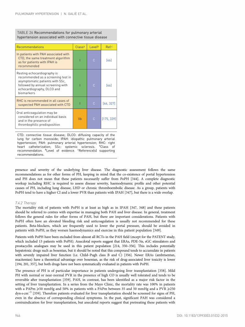
presence and severity of the underlying liver disease. The diagnostic assessment follows the samerecommendations as for other forms of PH, keeping in mind that the co-existence of portal hypertensionand PH does not mean that these patients necessarily suffer from PoPH [344]. A complete diagnosticworkup including RHC is required to assess disease severity, haemodynamic profile and other potentialcauses of PH, including lung disease, LHD or chronic thromboembolic disease. As a group, patients withPoPH tend to have a higher CI and a lower PVR than patients with IPAH [347], but there is a wide overlap.
7.4.2 TherapyThe mortality risk of patients with PoPH is at least as high as in IPAH [347, 348] and these patientsshould be referred to centres with expertise in managing both PAH and liver disease. In general, treatmentfollows the general rules for other forms of PAH, but there are important considerations. Patients withPoPH often have an elevated bleeding risk and anticoagulation is usually not recommended for thesepatients. Beta-blockers, which are frequently used to lower the portal pressure, should be avoided inpatients with PoPH, as they worsen haemodynamics and exercise in this patient population [349].
Patients with PoPH have been excluded from almost all RCTs in the PAH field (except for the PATENT study,which included 13 patients with PoPH). Anecdotal reports suggest that ERAs, PDE-5is, sGC stimulators andprostacyclin analogues may be used in this patient population [214, 350–356]. This includes potentiallyhepatotoxic drugs such as bosentan, but it should be noted that this compound tends to accumulate in patientswith severely impaired liver function (i.e. Child–Pugh class B and C) [356]. Newer ERAs (ambrisentan,macitentan) have a theoretical advantage over bosentan, as the risk of drug-associated liver toxicity is lower[194, 201, 357], but both drugs have not been systematically evaluated in patients with PoPH.
The presence of PH is of particular importance in patients undergoing liver transplantation [358]. MildPH with normal or near-normal PVR in the presence of high CO is usually well tolerated and tends to bereversible after transplantation [359]. PAH, in contrast, has been identified as a major risk factor in thesetting of liver transplantation. In a series from the Mayo Clinic, the mortality rate was 100% in patientswith a PAPm ⩾50 mmHg and 50% in patients with a PAPm between 35 and 50 mmHg and a PVR ⩾250dyn·s·cm−5 [359]. Therefore patients evaluated for liver transplantation should be screened for signs of PH,even in the absence of corresponding clinical symptoms. In the past, significant PAH was considered acontraindication for liver transplantation, but anecdotal reports suggest that pretreating these patients with
TABLE 26 Recommendations for pulmonary arterialhypertension associated with connective tissue disease
Recommendations
In patients with PAH associated with
CTD, the same treatment algorithm
as for patients with IPAH is
recommended
Classa Levelb
CI
Ref.c
[46]
RHC is recommended in all cases of
suspected PAH associated with CTD CI [46, 327]
Resting echocardiography is
recommended as a screening test in
asymptomatic patients with SSc,
followed by annual screening with
echocardiography, DLCO and
biomarkers
CI [46]
Oral anticoagulation may be
considered on an individual basis
and in the presence of
thrombophilic predisposition
CIIb [175, 339]
CTD: connective tissue disease; DLCO: diffusing capacity of thelung for carbon monoxide; IPAH: idiopathic pulmonary arterialhypertension; PAH: pulmonary arterial hypertension; RHC: rightheart catheterization; SSc: systemic sclerosis. aClass ofrecommendation. bLevel of evidence. cReference(s) supportingrecommendations.
946 DOI: 10.1183/13993003.01032-2015
PULMONARY HYPERTENSION | N. GALIÈ ET AL.

PAH drugs might improve the outcome after liver transplantation [360–363]. There is not yet sufficientevidence to allow general recommendations and the transplant decision should be made bymultidisciplinary teams in expert centres. A few specialized centres offer combined liver–lung or liver–heart–lung transplantation for carefully selected patients [364].
The recommendations for PAH associated with portal hypertension are reported in the Table 27.
7.5 Pulmonary arterial hypertension associated with human immunodeficiency virus infectionThe use of highly active antiretroviral therapy (HAART) and aggressive management of opportunisticinfections has contributed to increased life expectancy in HIV-infected patients [366, 367]. Consequently,the spectrum of complications has shifted towards other long-term conditions, including PAH. It is likelythat modern HIV management with HAART has also resulted in improved survival and decreasedincidence of HIV-associated PAH [368]. Taken together, these effects on survival and incidence haveresulted in a stable PAH prevalence in HIV-infected patients over recent decades. A population studyindicated that the minimal prevalence of HIV-related PAH was 0.46%, very similar to the prevalencebefore the HAART era [369]. The pathogenesis of HIV-related PAH remains unclear. The absence of viralparticles in the complex plexiform lesions found from these patients suggests that an indirect action ofviral infection on inflammation and growth factors may act as a trigger in a predisposed patient.
7.5.1 DiagnosisHIV-associated PAH shares a similar clinical presentation with IPAH. At the time of diagnosis, a majorityof patients are in an advanced New York Heart Association (NYHA)-FC III or IV. Patients may present
TABLE 27 Recommendations for pulmonary arterialhypertension associated with portal hypertension
Recommendations
Echocardiographic assessment for
signs of PH is recommended in
symptomatic patients with liver
disease or portal hypertension and
in all candidates for liver
transplantation
Classa Levelb
BI
Ref.c
[344]
It is recommended that the
treatment algorithm for patients
with other forms of PAH should be
applied to patients with PAH
associated with portal
hypertension, taking into account
the severity of liver disease
CI[214,
350–356]
Liver transplantation may be
considered in selected patients
responding well to PAH therapyCIIb [361–363]
It is recommended that patients
affected by PAH associated with
portal hypertension should be
referred to centres with expertise in
managing both conditions
CI [344]
Anticoagulation is not recommended
in patients with PH associated with
portal hypertensionCIII [365]
Liver transplantation is
contraindicated in patients with
severe and uncontrolled PAH
CIII [361–363]
PAH: pulmonary arterial hypertension; PH: pulmonary hypertension.aClass of recommendation. bLevel of evidence. cReference(s)supporting recommendations.
DOI: 10.1183/13993003.01032-2015 947
PULMONARY HYPERTENSION | N. GALIÈ ET AL.
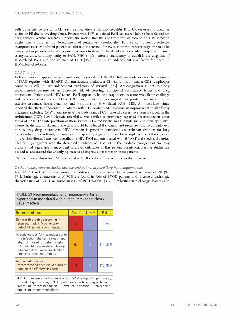
with other risk factors for PAH, such as liver disease (chronic hepatitis B or C), exposure to drugs ortoxins or PE due to i.v. drug abuse. Patients with HIV-associated PAH are more likely to be male and i.v.drug abusers. Animal research supports the notion that the additive effect of cocaine on HIV infectionmight play a role in the development of pulmonary arteriopathy. Because of its low prevalence,asymptomatic HIV-infected patients should not be screened for PAH. However, echocardiography must beperformed in patients with unexplained dyspnoea to detect HIV-related cardiovascular complications suchas myocarditis, cardiomyopathy or PAH. RHC confirmation is mandatory to establish the diagnosis ofHIV-related PAH and the absence of LHD [369]. PAH is an independent risk factor for death inHIV-infected patients.
7.5.2 TherapyIn the absence of specific recommendations, treatment of HIV-PAH follows guidelines for the treatmentof IPAH together with HAART. On multivariate analysis, a CI >2.8 l/min/m2 and a CD4 lymphocytecount >200 cells/ml are independent predictors of survival [225]. Anticoagulation is not routinelyrecommended because of an increased risk of bleeding, anticipated compliance issues and druginteractions. Patients with HIV-related PAH appear to be non-responders to acute vasodilator challengeand thus should not receive CCBs [189]. Uncontrolled studies suggest that prostacyclins may improveexercise tolerance, haemodynamics and symptoms in HIV-related PAH [218]. An open-label studyreported the effects of bosentan in patients with HIV-related PAH, showing an improvement in all efficacymeasures, including 6MWT and invasive haemodynamics [370]. Sporadic cases have been included in theambrisentan RCTs [194]. Hepatic tolerability was similar to previously reported observations in otherforms of PAH. The interpretation of these studies is limited by the small sample size and their open-labelnature. In the case of sildenafil, the dose should be reduced if ritonavir and saquinavir are co-administereddue to drug–drug interactions. HIV infection is generally considered an exclusion criterion for lungtransplantation, even though in some centres specific programmes have been implemented. Of note, casesof reversible disease have been described in HIV-PAH patients treated with HAART and specific therapies.This finding, together with the decreased incidence of HIV-PH in the modern management era, mayindicate that aggressive management improves outcomes in this patient population. Further studies areneeded to understand the underlying reasons of improved outcomes in these patients.
The recommendations for PAH associated with HIV infection are reported in the Table 28.
7.6 Pulmonary veno-occlusive disease and pulmonary capillary haemangiomatosisBoth PVOD and PCH are uncommon conditions but are increasingly recognized as causes of PH [92,371]. Pathologic characteristics of PCH are found in 73% of PVOD patients and, inversely, pathologiccharacteristics of PVOD are found in 80% of PCH patients [372]. Similarities in pathologic features and
TABLE 28 Recommendations for pulmonary arterialhypertension associated with human immunodeficiencyvirus infection
Recommendations
Echocardiographic screening in
asymptomatic HIV patients to
detect PH is not recommended
Classa Levelb
CIII
Ref.c
[369]
Anticoagulation is not
recommended because of a lack of
data on the efficacy:risk ratioCIII [175, 367]
In patients with PAH associated with
HIV infection, the same treatment
algorithm used for patients with
PAH should be considered, taking
into consideration co-morbidities
and drug–drug interactions
CIIa [194, 367]
HIV: human immunodeficiency virus; IPAH: idiopathic pulmonaryarterial hypertension; PAH: pulmonary arterial hypertension.aClass of recommendation. bLevel of evidence. cReference(s)supporting recommendations.
948 DOI: 10.1183/13993003.01032-2015
PULMONARY HYPERTENSION | N. GALIÈ ET AL.

clinical characteristics and the risk of drug-induced pulmonary oedema with PAH therapy [371, 373]suggest that these two conditions overlap, and it has been proposed that PCH could be a secondaryangioproliferative process caused by post-capillary obstruction of PVOD rather than a distinct disease [6,372]. Thus PVOD and PCH have been classified together in a specific subgroup of the clinicalclassification next to PAH (Table 4, group 1′) because of their pathological, genetic and clinical similaritiesand differences with PAH [6]. The true incidence of PVOD/PCH remains unknown because many casesare still misclassified as PAH [374]. The proportion of idiopathic cases of PAH that in reality fulfil thecriteria for PVOD/PCH is likely to be around 10% (lowest estimates of PVOD/PCH incidence andprevalence are <1 case/million) [92, 374]. In contrast to IPAH, there is a male predominance in PVODand the prognosis appears to be worse [371]. Familial occurrence of PVOD has been reported, but BMPR2mutations have rarely been found in patients with this disease [28, 371, 375]. While PAH that is due toBMPR2 mutations segregates as an autosomal dominant trait with incomplete penetrance, familial cases ofPVOD/PCH typically occur in the young siblings of one generation with unaffected parents, indicatingthat the disease segregates as a recessive trait [28]. In these families, PVOD/PCH is caused by bi-allelicmutations in EIF2AK4 [28]. Like PAH, PVOD/PCH may complicate the course of associated conditions(SSc, HIV infection, etc.) and exposure to drugs or toxins (cyclophosphamide, mitomycin, etc.).
7.6.1 DiagnosisThe diagnosis of PVOD/PCH can be established with a high probability by the combination of clinicalsuspicion, physical examination, bronchoscopy and radiological findings [371]. This non-invasiveapproach may avoid the need to perform a hazardous lung biopsy (the gold standard to confirm ahistological diagnosis of PVOD/PCH), which is no longer recommended in most cases [371]. In heritablecases, identification of a bi-allelic EIF2AK4 mutation is sufficient to confirm a diagnosis of PVOD/PCHwithout histological confirmation [28, 376]. Most patients complain of dyspnoea on exertion and fatigue, aclinical presentation that is indistinguishable from PAH [371, 374]. Physical examination may revealdigital clubbing and bi-basal crackles on lung auscultation, these being unusual in PAH [371]. Case seriesindicate that patients with PVOD/PCH are more severely hypoxaemic and have much lower DLCO levelsthan in other forms of PAH [92, 371]. This can be explained by the presence of chronic interstitialpulmonary oedema and pulmonary capillary proliferation typical of PVOD/PCH.
Chest radiography may reveal Kerley B lines, mediastinal lymph node enlargement and peripheralinterstitial infiltrate, in addition to other signs of PH [92, 371]. High-resolution CT of the chest is theinvestigation of choice. Typical findings suggestive of PVOD/PCH are the presence of subpleural thickenedseptal lines, centrilobular ground-glass opacities and mediastinal lymphadenopathy [92, 371]. Theassociation of these three findings was found to be 100% specific for PVOD/PCH in cases of PAH, with66% sensitivity. In addition, their presence appears to closely correlate with the risk of pulmonary oedemawith PAH drugs. V/Q lung scanning is not useful in discriminating PVOD/PCH from IPAH [377].
Because PVOD/PCH may be associated with occult alveolar haemorrhage, bronchoscopy withbronchoalveolar lavage may be a useful tool in the diagnostic strategy. In a retrospective study, the resultsof bronchoalveolar lavage performed in 19 patients with PAH or PVOD/PCH were analysed. Comparedwith IPAH, the eight cases of PVOD presented with a significantly elevated cell count, a higher percentageof hemosiderin-laden macrophages and a markedly elevated Golde score [378].
The haemodynamic presentation of PVOD is similar to IPAH [92, 371]. Importantly, PAWP is almostinvariably normal, because the pathological changes occur in small venulae and capillaries and do notaffect the larger pulmonary veins [92, 371]. Vasoreactivity testing may be complicated by acute pulmonaryoedema [92, 371].
7.6.2 TherapyThere is no established medical therapy for PVOD/PCH [92]. Most importantly, vasodilators and, inparticular, i.v. epoprostenol must be used with great caution because of the high risk of severedrug-induced pulmonary oedema [92, 373]. However, there are reports of sustained clinical improvementin individual patients treated with these medications [379]. High-dose diuretics, O2 therapy and slowincreases in epoprostenol doses are recommended [379]. Therefore therapy for PVOD/PCH should beundertaken only at centres with extensive experience in the management of PH and patients should befully informed about the risks. Investigational use of angiogenesis inhibitors such as interferon alfa-2a hasbeen considered in these patients but is not currently recommended [379]. The only curative therapy forPVOD/PCH is lung transplantation, and there are no reports of recurrence of disease followingtransplantation. Eligible patients with PVOD/PCH should be referred to a transplant centre for evaluationas soon as the diagnosis is established [379].
The recommendations for PVOD/PCH are reported in the Table 29.
DOI: 10.1183/13993003.01032-2015 949
PULMONARY HYPERTENSION | N. GALIÈ ET AL.

8. Pulmonary hypertension due to left heart disease (group 2)PH is a common complication of LHDs (PH-LHD), frequently occurring as a ‘symptom’ of the underlyingcondition [4, 380]. and often related to disease severity. PH-LHD can complicate any left heart disorder,such as valvular heart diseases and congenital defects. However, it has been most often studied in patientswith heart failure and with preserved or reduced ejection fraction [4, 380]. When present, PH-LHD resultsin more severe symptoms and worse exercise tolerance and exerts a negative impact on outcome [4, 380,381]. Compared with PAH, patients with PH-LHD [especially in the case of heart failure with preservedLV ejection fraction (HF-pEF)] are often older, female, with a higher prevalence of cardiovascularco-morbidities [382] and most, if not all, of the features of metabolic syndrome [383].
The true prevalence of PH-LHD in heart failure remains unknown, mostly because the definition of PH inepidemiological studies has been based on echocardiography, with a variety of cut-off values [4, 384].Invasive haemodynamics were reported in a limited number of single-centre reports [4]. In a retrospectiveanalysis performed in a large PH centre, LHD was identified as the cause of PH in 36% of all patientsreferred for evaluation, of which 55% had ‘passive’ PH, defined as a TPG <12 mmHg [151, 385].
PH develops in LHD in response to a passive backward transmission of filling pressures, mainly driven byLV diastolic function, enhanced by exercise-induced mitral regurgitation and a loss of LA compliance [4].In some patients, these purely mechanical components of venous congestion may trigger a superimposedcomponent, combining pulmonary vasoconstriction, decreased NO availability, increased endothelinexpression, desensitization to natriuretic peptide–induced vasodilation and vascular remodelling [4, 380,386]. This results in a further increase in PAPm in excess of the elevation of PAWP that may lead topulmonary vascular disease, increased RV afterload and RV failure [4].
The definitions of PH and the difference between pre- and post-capillary PH are based on pressurecut-off. This explains why TPG, i.e. the difference between PAPm and PAWP, has been used to distinguish‘passive’ PH (TPG <12 mmHg) from ‘reactive’ PH (TPG ⩾12 mmHg) [151]. However, this definition andthe associated terminology have been unsatisfactory, to such an extent that ‘out-of-proportion’ PH-LHDhas often been used to characterize a subset of patients with significant changes in pulmonary circulation[4, 151]. Ideally the latter should be defined by a haemodynamic variable that would (i) be a marker ofdisease, (ii) be least dependent on changes in PAWP and stroke volume and (iii) take into account the
TABLE 29 Recommendations for pulmonary veno-occlusivedisease and pulmonary capillary haemangiomatosis
Recommendations
A combination of clinical findings,
physical examination,
bronchoscopy and radiological
findings is recommended to
diagnose PVOD/PCH
Classa Levelb
CI
Ref.c
[92, 371,
378]
Referral of eligible patients with
PVOD/PCH to a transplant centre
for evaluation is indicated as soon
as the diagnosis is established
CI [371]
Identification of a bi-allelic EIF2AK4
mutation is recommended to
confirm a diagnosis of heritable
PVOD/PCH without histological
confirmation
BI [28, 376]
Patients with PVOD/PCH should be
managed only in centres with
extensive experience in PH due to
the risk of lung oedema after the
initiation of PAH therapy
CIIa[371, 373,
379]
PAH: pulmonary arterial hypertension; PCH: pulmonary capillaryhaemangiomatosis; PH: pulmonary hypertension; PVOD:pulmonary veno-occlusive disease. aClass of recommendation.bLevel of evidence. cReference(s) supporting recommendations.
950 DOI: 10.1183/13993003.01032-2015
PULMONARY HYPERTENSION | N. GALIÈ ET AL.
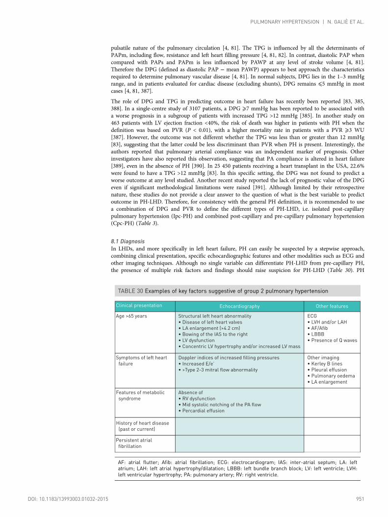
pulsatile nature of the pulmonary circulation [4, 81]. The TPG is influenced by all the determinants ofPAPm, including flow, resistance and left heart filling pressure [4, 81, 82]. In contrast, diastolic PAP whencompared with PAPs and PAPm is less influenced by PAWP at any level of stroke volume [4, 81].Therefore the DPG (defined as diastolic PAP − mean PAWP) appears to best approach the characteristicsrequired to determine pulmonary vascular disease [4, 81]. In normal subjects, DPG lies in the 1–3 mmHgrange, and in patients evaluated for cardiac disease (excluding shunts), DPG remains ⩽5 mmHg in mostcases [4, 81, 387].
The role of DPG and TPG in predicting outcome in heart failure has recently been reported [83, 385,388]. In a single-centre study of 3107 patients, a DPG ⩾7 mmHg has been reported to be associated witha worse prognosis in a subgroup of patients with increased TPG >12 mmHg [385]. In another study on463 patients with LV ejection fraction <40%, the risk of death was higher in patients with PH when thedefinition was based on PVR (P < 0.01), with a higher mortality rate in patients with a PVR ⩾3 WU[387]. However, the outcome was not different whether the TPG was less than or greater than 12 mmHg[83], suggesting that the latter could be less discriminant than PVR when PH is present. Interestingly, theauthors reported that pulmonary arterial compliance was an independent marker of prognosis. Otherinvestigators have also reported this observation, suggesting that PA compliance is altered in heart failure[389], even in the absence of PH [390]. In 25 450 patients receiving a heart transplant in the USA, 22.6%were found to have a TPG >12 mmHg [83]. In this specific setting, the DPG was not found to predict aworse outcome at any level studied. Another recent study reported the lack of prognostic value of the DPGeven if significant methodological limitations were raised [391]. Although limited by their retrospectivenature, these studies do not provide a clear answer to the question of what is the best variable to predictoutcome in PH-LHD. Therefore, for consistency with the general PH definition, it is recommended to usea combination of DPG and PVR to define the different types of PH-LHD, i.e. isolated post-capillarypulmonary hypertension (Ipc-PH) and combined post-capillary and pre-capillary pulmonary hypertension(Cpc-PH) (Table 3).
8.1 DiagnosisIn LHDs, and more specifically in left heart failure, PH can easily be suspected by a stepwise approach,combining clinical presentation, specific echocardiographic features and other modalities such as ECG andother imaging techniques. Although no single variable can differentiate PH-LHD from pre-capillary PH,the presence of multiple risk factors and findings should raise suspicion for PH-LHD (Table 30). PH
TABLE 30 Examples of key factors suggestive of group 2 pulmonary hypertension
Clinical presentation
Age >65 years
Echocardiography Other features
Structural left heart abnormality
• Disease of left heart valves
• LA enlargement (>4.2 cm)
• Bowing of the IAS to the right
• LV dysfunction
• Concentric LV hypertrophy and/or increased LV mass
ECG
• LVH and/or LAH
• AF/Afib
• LBBB
• Presence of Q waves
Symptoms of left heart
failure
Doppler indices of increased filling pressures
• Increased E/e’
• >Type 2-3 mitral flow abnormality
Other imaging
• Kerley B lines
• Pleural effusion
• Pulmonary oedema
• LA enlargement
Features of metabolic
syndrome
Absence of
• RV dysfunction
• Mid systolic notching of the PA flow
• Percardial effusion
History of heart disease
(past or current)
Persistent atrial
fibrillation
AF: atrial flutter; Afib: atrial fibrillation; ECG: electrocardiogram; IAS: inter-atrial septum; LA: leftatrium; LAH: left atrial hypertrophy/dilatation; LBBB: left bundle branch block; LV: left ventricle; LVH:left ventricular hypertrophy; PA: pulmonary artery; RV: right ventricle.
DOI: 10.1183/13993003.01032-2015 951
PULMONARY HYPERTENSION | N. GALIÈ ET AL.

should be suspected when patients present with otherwise unexplained symptoms, signs of right heartfailure and co-morbidities associated with PH, such as sleep apnoea syndrome, COPD, previous PE andrisk factors for PAH.
The respective role of fluid loading and exercise testing to uncover PH-LHD in patients with HF-pEF iscurrently not standardized and normal values are lacking to provide a clear recommendation to clinicians.In addition, it has been suggested that patients with a diagnosis of PAH may present an abnormal increasein PAWP in response to fluid loading [77].
The indications for RHC in PH-LHD, preferably electively in stable condition, are reported in Table 10.
8.2 TherapyThe primary goal of therapy in PH-LHD must be to improve global management of the underlyingcondition prior to considering specific measures to treat PH. This includes repair of valvular heart diseasewhen indicated and aggressive therapy for heart failure with reduced systolic function [4, 392]. Somepatients may also benefit from non-specific vasodilators such as nitrates and hydralazine, althoughevidence supporting this strategy is limited [4, 392]. In severe heart failure, optimizing volume status is ofcritical importance and may require invasive monitoring [4, 393]. In addition, the implantation of an LVassist device has been shown to lower pulmonary pressures through LV unloading without increasing therisk of post-implantation RV failure [4, 394, 395]. Risk factors for cardiovascular diseases and features ofmetabolic syndrome should be controlled [4, 392]. Concomitant disorders leading to PH should beidentified and treated, including COPD, sleep apnoea syndrome and PE. In contrast, there is no strongevidence-based recommendation for the treatment for HF-pEF [392].
The rationale to use PAH therapies in PH-LHD has been supported by acute or short-term studies usingprostanoids, ERAs and PDE-5is. Most of these studies consistently reported improvements inhaemodynamics, exercise capacity and symptoms [4, 396]. However, these studies carry significantmethodological limitations (small sample size, single centre, unclear or no randomization process) that donot provide enough evidence to support the use of these drugs in the clinical management of patients [4].In addition, there is no evidence suggesting that acute changes in pulmonary circulation may have a valueoutside of the setting of major cardiac surgery such as heart transplantation and/or LV assist deviceimplantation [4, 380].
In a recent multicentre, placebo-controlled trial [397], 201 patients with PH due to systolic heart failurewere randomized in four arms comparing three doses of riociguat (0.5, 1 and 2 mg t.i.d.) with placeboover 16 weeks. No effect on the primary endpoint (a change in PAPm after 16 weeks) was observed at anydose of riociguat compared with placebo [397].
Two multicentre clinical trials in PH-LHD are currently under way with sildenafil [SilHF (NCT01616381)]and macitentan [Melody-1 (NCT02070991)], the latter being the only study requiring RHC validation.
Therefore there is no new evidence supporting the use of PAH therapies in PH-LHD, due in part to theabsence of studies specifically stratifying patients for PH and/or targeting this specific condition. Suchtrials are necessary to address this area of unmet medical need and patients with Cpc-PH should beincluded in clinical trials. Recommendations for the management of PH-LHD are listed in Table 31.
9. Pulmonary hypertension due to lung diseases and/or hypoxia (group 3)The pathology, pathophysiology and epidemiology of these conditions have been discussed previously (seesection 4). The most common lung diseases associated with PH are COPD, interstitial lung disease andcombined pulmonary fibrosis and emphysema (CPFE). Rare conditions such as Langerhans cellgranulomatosis or sarcoidosis are reported in Web Table VIII. In any lung disease, the development of PHis accompanied by a deterioration of exercise capacity, worsening of hypoxaemia and shorter survival[398–400]. The severity of PH is usually poorly associated with the severity of the underlying lung disease[401, 402]. The most common indicators for the presence of PH in these patients are a disproportionallylow DLCO and a low pCO2 [401, 402].
The haemodynamic classification of PH associated with lung disease is shown in Table 32 [20]. In case ofsevere PH, other potential causes such as LHD or CTEPH should be excluded. In some patients with lungdisease and PH, especially in patients with mild lung disease but severe PH, it may be difficult todetermine whether PH is due to lung disease or whether the patient suffers from two diseases, i.e. PAHand chronic lung disease. Those patients should be referred to a PH centre where there is also expertise inlung diseases.
952 DOI: 10.1183/13993003.01032-2015
PULMONARY HYPERTENSION | N. GALIÈ ET AL.

9.1 DiagnosisClinical symptoms and physical signs of PH may be difficult to identify in patients with respiratory disorders.In addition, in patients with lung disease, peripheral oedema may not necessarily indicate RV failure, as it mayalso result from the effects of hypoxaemia and hypercapnia on the renin-angiotensin-aldosterone system.Furthermore, concomitant LHD, which is frequently found in patients with chronic respiratory diseases, mayalso contribute to PH. As a general rule, patients presenting with symptoms that are more severe than expectedbased on their PFT results should be further evaluated, in particular by echocardiography, to search forconcomitant LHD or PH.
Echocardiography remains the most widely used non-invasive diagnostic tool for the assessment of PH.Indications for echocardiography in patients with lung diseases include the clinical suspicion of significantPH or the evaluation of concomitant LHD. It should be noted, however, that the accuracy ofechocardiography in patients with advanced respiratory diseases is low [403–405]. Patients with clinical orechocardiographic signs of severe PH and/or severe RV dysfunction should be referred to a PH centre.
A definite diagnosis of PH relies on measurements obtained during RHC. Potential indications for RHCin advanced lung disease are (i) proper diagnosis or exclusion of PH in candidates for surgical treatments(transplantation, lung volume reduction), (ii) suspected PAH or CTEPH, (iii) episodes of RV failureand (iv) inconclusive echocardiographic findings in cases with a high level of suspicion and potentialtherapeutic implications.
TABLE 31 Management of pulmonary hypertension in leftheart disease
Recommendations
Optimization of the treatment of the
underlying condition is recommended
before considering assessment of PH-LHD
(i.e. treating structural heart disease)
Classa Levelb
BI
Ref.c
[396]
It is recommended to perform invasive
assessment of PH in patients on optimized
volume statusCI
The importance and role of vasoreactivity
testing is not established in PH-LHD,
except in patients who are candidates for
heart transplantation and/or LV assist
device implantation
CIII
It is recommended to identify other causes
of PH (i.e. COPD, sleep apnoea syndrome,
PE, CTEPH) and to treat them when
appropriate before considering assessment
of PH-LHD
CI [344]
The use of PAH-approved therapies is not
recommended in PH-LHD CIII [396]
[396]
Patients with PH-LHD and a severe
pre-capillary component as indicated by a
high DPG and/or high PVR should be
referred to an expert PH centre for a
complete diagnostic workup and an
individual treatment decision
CIIa
COPD: chronic obstructive pulmonary disease; CTEPH: chronicthromboembolic pulmonary hypertension; DPG: diastolic pressuregradient; LHD: left heart disease; LV: left ventricular; PE:pulmonary embolism; PH: pulmonary hypertension; PVR:pulmonary vascular resistance. aClass of recommendation. bLevelof evidence. cReference(s) supporting recommendations.
TABLE 32 Haemodynamic classification of pulmonaryhypertension due to lung disease [9]
Haemodynamics
(right heart catheterization)
Terminology
COPD/IPF/CPFE without PH PAPm <25 mmHg
COPD/IPF/CPFE with severe PH PAPm >35 mmHg, or
PAPm ≥25 mmHg in the presence
of a low cardiac output
(CI <2.5 L/min/m2, not explained
by other causes)
COPD/IPF/CPFE with PH PAPm ≥25 mmHg
CI: cardiac index; COPD: chronic obstructive pulmonary disease;CPFE: combined pulmonary fibrosis and emphysema; IPF:idiopathic pulmonary fibrosis; PAP: pulmonary artery pressure;PAPm: mean pulmonary arterial pressure; PH: pulmonaryhypertension.
DOI: 10.1183/13993003.01032-2015 953
PULMONARY HYPERTENSION | N. GALIÈ ET AL.

9.2 TherapyCurrently there is no specific therapy for PH associated with lung diseases. Long-term O2 administrationhas been shown to partially reduce the progression of PH in COPD. Nevertheless, PAP rarely returns tonormal values and the structural abnormalities of pulmonary vessels remain unaltered [169]. In interstitiallung diseases, the role of long-term O2 therapy on PH progression is less clear.
Treatment with conventional vasodilators such as CCBs is not recommended because they may impair gasexchange due to the inhibition of hypoxic pulmonary vasoconstriction [406–408] and because of their lackof efficacy after long-term use [409, 410].
Published experience with targeted PAH drug therapy is scarce, and so far there is no evidence from RCTssuggesting that PAH drugs result in improved symptoms or outcomes in patients with lung disease [411–416].
In summary, patients with lung disease and PH who are hypoxaemic should receive long-term O2 therapy,adapting the general recommendations for COPD. Treatment of the underlying lung disease should beoptimized. The use of drugs approved for PAH is not recommended for patients with PH due to lungdisease. Patients with suspected PAH in addition to their lung diseases (characterized by mild lungparenchymal abnormalities, symptoms insufficiently explained by lung mechanical disturbances and ahaemodynamic ‘PAH phenotype’, i.e. severe PH with high PVR and low CO) may be treated according tothe recommendations for PAH, keeping in mind the potential implications of the co-existing lung diseaseon symptoms and response to therapy.
The recommendations for PH due to lung diseases are reported in the Table 33.
10. Chronic thromboembolic pulmonary hypertension (group 4)CTEPH is a disease of obstructive PA remodelling as a consequence of major vessel thromboembolism.CTEPH has been reported with a cumulative incidence of 0.1–9.1% within the first 2 years after asymptomatic PE event [417]. The large margin of error is probably due to referral bias, a paucity of earlysymptoms and difficulty in differentiating acute PE from symptoms of pre-existing CTEPH [418].Although the exact prevalence and annual incidence of CTEPH are unknown, some data suggest that thiscondition may occur in approximately 5 individuals per million population per year [419].
In the differential diagnosis of CTEPH the following conditions have to be considered and treatedaccording to current best knowledge. These conditions include PA sarcoma, tumour cell embolism,parasites (hydatid cyst), foreign body embolism and congenital or acquired PA stenosis (Table 4).
10.1 DiagnosisRoutine screening for CTEPH after PE is not supported by current evidence; a significant number ofCTEPH cases develop in the absence of previous acute PE.
The median age of patients at diagnosis is 63 years and both genders are equally affected [25]; paediatriccases are rare [287, 420]. Clinical symptoms and signs are non-specific or absent in early CTEPH, withsigns of right heart failure only becoming evident in advanced disease. Thus early diagnosis remains achallenge in CTEPH, with a median time of 14 months between symptom onset and diagnosis in expertcentres [421]. When present, the clinical symptoms of CTEPH may resemble those of acute PE or IPAH;in the latter context, oedema and haemoptysis occur more often in CTEPH, while syncope is morecommon in IPAH [422].
The diagnosis of CTEPH is based on findings obtained after at least 3 months of effective anticoagulationin order to discriminate this condition from ‘subacute’ PE. These findings are mean PAP ⩾25 mmHg withPAWP ⩽15 mmHg, mismatched perfusion defects on lung scan and specific diagnostic signs for CTEPHseen by multidetector CT angiography, MR imaging or conventional pulmonary cineangiography, such asring-like stenoses, webs/slits and chronic total occlusions (pouch lesions or tapered lesions).
Some patients, particularly those with complete unilateral obstruction, may present with normalpulmonary haemodynamics at rest despite symptomatic disease. These patients are also considered to haveCTEPH and are managed accordingly. A suitable terminology to describe this condition of chronicthromboembolic pulmonary vascular disease is still lacking.
An algorithm for CTEPH diagnosis is shown in Figure 3. While CT pulmonary angiography is theinvestigation of choice for the diagnosis of acute PE, planar V/Q lung scan remains the main first-lineimaging modality for CTEPH, as it carries a 96–97% sensitivity and a 90–95% specificity for the diagnosis[47]. In contrast, in IPAH and PVOD, perfusion scans typically show non-segmental defects or are normal[377]. More recent work suggests that both V/Q scanning and modern CT pulmonary angiography maybe accurate methods for the detection of CTEPH, with excellent diagnostic efficacy in expert hands (100%sensitivity, 93.7% specificity and 96.5% accuracy for V/Q and 96.1%, 95.2% and 95.6%, respectively, forCT pulmonary angiography) [93, 423, 424].
954 DOI: 10.1183/13993003.01032-2015
PULMONARY HYPERTENSION | N. GALIÈ ET AL.

Multidetector CT pulmonary angiography has become an established imaging modality for confirmingCTEPH [93], however, this investigation alone cannot exclude the disease [47]. CT pulmonary angiographymay help to identify complications of the disease such as PA dilatation resulting in left main coronaryartery compression and hypertrophied bronchial arterial collaterals, which may lead to haemoptysis.
A high-resolution CT scan of the chest delivers images of the lung parenchyma and identifies emphysema,bronchial disease or interstitial lung disease, as well as infarcts, vascular and pericardial malformations andthoracic wall deformities. Perfusion inequalities manifest as a mosaic parenchymal pattern, with dark areascorresponding to relatively decreased perfusion. Although a mosaic pattern is frequent in CTEPH, it can also beobserved in up to 12% of patients with PAH [425]. MR imaging of the pulmonary vasculature is still consideredinferior to CT [426], but this modality, as well as cone beam CT [427], angioscopy [428], intravascular ultrasoundor optical coherence tomography, may be complimentary and used according to local experience and practice.
RHC is an essential diagnostic tool. Preoperative and immediate postoperative PVR is a long-term predictorof prognosis [429]. The final step in the diagnostic pathway is selective pulmonary angiography in theanterior–posterior and lateral projections illustrating ring-like stenosis, webs (‘slits’), pouches, wallirregularities, complete vascular obstructions as well as bronchial collaterals, and supports the technicalassessment of operability.
TABLE 33 Recommendations for pulmonary hypertensiondue to lung diseases
Recommendations
Echocardiography is recommended
for the non-invasive diagnostic
assessment of suspected PH in
patients with lung disease
Classa Levelb
CI
Ref.c
[403, 405]
The optimal treatment of the
underlying lung disease, including
long-term O2 therapy in patients
with chronic hypoxaemia, is
recommended in patients with PH
due to lung diseases
CI [169]
RHC is not recommended for
suspected PH in patients with lung
disease, unless therapeutic
consequences are to be expected
(e.g. lung transplantation,
alternative diagnoses such as PAH
or CTEPH, potential enrolment in a
clinical trial)
CIII [169]
Referral to an expert centre is
recommendedd in patients with
echocardiographic signs of severe
PH and/or severe right ventricular
dysfunction
CI
Referral to PH expert center should
be considered for patients with signs
of severe PH/severe RV failure for
individual-based treatment
CIIa
The use of drugs approved for PAH is
not recommended in patients with
PH due to lung diseasesCIII [411–416]
CTEPH: chronic thromboembolic pulmonary hypertension; PAH:pulmonary arterial hypertension; PH: pulmonary hypertension;RHC: right heart catheterization. aClass of recommendation. bLevelof evidence. cReference(s) supporting recommendations. dThisrecommendation does not apply to patients with end-stage lungdisease who are not considered candidates for lung transplantation.
DOI: 10.1183/13993003.01032-2015 955
PULMONARY HYPERTENSION | N. GALIÈ ET AL.

10.2 Therapy10.2.1 SurgicalPEA is the treatment of choice for CTEPH (Figure 4). In Europe, in-hospital mortality is currently as lowas 4.7% [430], and even lower in high-volume single centres [431]. The majority of patients experiencesubstantial relief from symptoms and near normalization of haemodynamics [430–432]. In contrast tosurgical embolectomy for acute PE, treatment of CTEPH necessitates a true bilateral endarterectomythrough the medial layer of the pulmonary arteries, which is performed under deep hypothermia andcirculatory arrest [431], without the need for cerebral perfusion [433].
Operability of patients with CTEPH is determined by multiple factors that cannot easily be standardized;these are related to the suitability of the patient, the expertise of the surgical team and available resources.General criteria include preoperative WHO-FC II–IV and surgical accessibility of thrombi in the main,lobar or segmental pulmonary arteries. Advanced age per se is not a contraindication for surgery. There isno PVR threshold or measure of RV dysfunction that can be considered to preclude PEA.
Postoperative ECMO is recommended as a standard of care in PEA centres for severe cases [434–436].Early postoperative reperfusion oedema may require veno-arterial ECMO, and severe persistent PH maybe bridged to emergency lung transplantation with veno-venous ECMO.
Patients who do not undergo PEA or suffer from persistent or recurrent PH after PEA (post-PEA PH)face a poor prognosis.
10.2.2 MedicalOptimal medical treatment for CTEPH consists of anticoagulants and diuretics, and O2 in cases of heartfailure or hypoxaemia. Lifelong anticoagulation is recommended, even after PEA, though no data exist onthe efficacy and safety of new oral anticoagulants. Although there is no consensus, routine cava filterplacement is not justified by the available evidence. Pulmonary microvascular disease in CTEPH hasprovided the rationale for off-label use of drugs approved for PAH [25]. Some non-randomized studies haveprovided evidence for improvement in exercise capacity and haemodynamics [437–439]. Medical treatmentof CTEPH with targeted therapy may be justified in technically non-operable patients or in the presence ofan unacceptable surgical risk:benefit ratio (Figure 2). Patients with persistent or recurrent PH after PEA mayalso be candidates for targeted medical therapy. The use of targeted therapy in operable patients with severehaemodynamic compromise as a bridge to PEA has not yet been supported by scientific evidence.
Symptoms, signs, history suggestive of CTEPH
Echocardiographic probability of PH
(Table 8)
High or intermediate probability of PH
V/Q scana
Mismatched perfusion defects?
Yes No
CTEPH ruled outCTEPH possible
Refer to PH/CTEPH
expert centre
CT pulmonary angiography
Right heart catheterisation
+/- pulmonary angiography
Work-up for PH/PAH
(figure 1)
FIGURE 3 Diagnostic algorithm for chronic thromboembolic pulmonary hypertension. CT: computedthomography; CTEPH: chronic thromboembolic pulmonary hypertension; PAH: pulmonary arterial hypertension;PH: pulmonary hypertension; V/Q: ventilation/perfusion. aCT pulmonary angiography alone may miss diagnosisof chronic thromboembolic pulmonary hypertension.
956 DOI: 10.1183/13993003.01032-2015
PULMONARY HYPERTENSION | N. GALIÈ ET AL.

The dual endothelin antagonist bosentan was evaluated in 157 patients with inoperable CTEPH orpersistent/recurrent PH after PEA over 16 weeks; the primary combined endpoint of a decrease in PVRand an increase in the 6MWD was not met [440]. However, an oral sGC stimulator, riociguat, wasadministered to 261 of 446 screened patients with non-operable CTEPH or persistent/recurrent PH afterPEA for 16 weeks and led to a mean increase of 39 m in the 6MWD (P < 0.001, primary endpoint) and toa least squares mean difference of 246 dyn·cm·s−5 in PVR (P < 0.001, secondary endpoint); the time toclinical worsening remained unchanged [441].
Preoperative medical treatment is uncertain because the magnitude of effects was small in one RCT [442].One retrospective study indicated no difference in outcome, but there was a delay in surgery for patientstreated medically [442]. Prospective RCTs are needed in patients with potential treatment benefit; forexample, patients with a high PVR and technically challenging anatomy.
After PEA, patients should be followed in CTEPH centres, with at least one haemodynamic assessment tobe considered at 6–12 months after the intervention.
10.2.3 InterventionalIn 2001 Feinstein et al. [443]. published a series of 18 patients with non-operable CTEPH who had beensubjected to balloon dilatation of the pulmonary arteries. Despite a significant decrease in PAPm, 11patients developed reperfusion pulmonary oedema and 3 required mechanical ventilation. Recently,Japanese investigators have refined BPA by using smaller balloons, by cautiously limiting the number ofballoon inflations per session to one or two pulmonary vascular segments and by the use of intravascularimaging [444–446]. An average number of 4.8 sessions is needed per patient to improve parameters of RVfunction [57]. A careful approach with targeting only one lobe during each session and very cautiousballoon sizing have reduced the incidence of reperfusion pulmonary oedema to 2% in individual centres[447]. While BPA is still not extensively used, [448]. it is rapidly gaining attention worldwide. BPA shouldonly be performed in experienced and high-volume CTEPH centres.
The recommendations for CTEPH are summarised in Table 34.
Diagnosis confirmed by
CTEPH expert centre
Operability assessment by a
multidisciplinary CTEPH team
Technically non-operableTechnically operable
Acceptable
risk/benefit ratio
Non-acceptable
risk/benefit ratioa
Persistent
symptomatic PH
Persistent severe
symptomatic PH
Consider BPA in
expert centreb
Targeted medical
therapy
Consider lung
transplantation
Pulmonary
endarterectomy
Lifelong anticoagulation
FIGURE 4 Treatment algorithm for chronic thromboembolic pulmonary hypertension. BPA: balloon pulmonaryangioplasty; CTEPH: chronic thromboembolic pulmonary hypertension; PH: pulmonary hypertension. aTechnicallyoperable patients with non-acceptable risk/benefit ratio can be considered also for BPA. bIn some centersmedical therapy and BPA are initiated concurrently.
DOI: 10.1183/13993003.01032-2015 957
PULMONARY HYPERTENSION | N. GALIÈ ET AL.

An algorithm for the treatment of CTEPH is provided in Figure 4.
11. Pulmonary hypertension with unclear and/or multifactorial mechanisms (group 5)PH with unclear and/or multifactorial mechanisms (group 5, Table 4) includes several disorders with multiplepatho-aetiologies. A common feature of these diseases is that the mechanisms of PH are poorly understoodand may include pulmonary vasoconstriction, proliferative vasculopathy, extrinsic compression, intrinsicocclusion, high-output cardiac failure, vascular obliteration and left heart failure as causes (Web Table VIII).
These patients need careful diagnosis. Treatment is tailored for that diagnosis; treatment of PH issecondary. The axiom should be ‘Treat the lung not the pressure’. There are no RCTs regarding the use ofPAH-approved drugs in the treatment of group 5 disorders [450]. Of particular importance is that some ofthe diseases described in Web Table VII may have a venous component (PVOD) that could be madeworse by the use of pulmonary arterial vasodilators.
12. Definition of a pulmonary hypertension referral centrePAH is a rare disease. Since, in general, medical centres with a high volume of patients tend to obtain thebest outcomes, the establishment of expert referral centres is clinically and economically highly desirableand supported by patient organisations. The purpose of a referral centre is to receive new referrals andundertake assessment and investigation of all causes of PH, routinely manage appropriate patients withPAH- and CTEPH-specific drug therapies, work closely with other healthcare providers to obtain the bestoutcomes for patients and undertake audits, research and education.
Referral centres should have sufficient patients on chronic therapy as well as new referrals to warrant thisstatus. The ideal number of patients seen by an adult centre each year is recommended to be no fewerthan 200, of which at least half have a final diagnosis of PAH. In countries with a population >10 million,adult centres should ideally expand to accommodate >300 patients annually. It is recommended that areferral centre, as a minimum, should follow at least 50 patients with PAH or CTEPH and receive at leasttwo new referrals per month with documented PAH or CTEPH. Paediatric centres are recommended tosee 30–50 patients per year. These numbers can be adapted according to specific country characteristics(population distribution, geographical constraints, etc.).
12.1 Facilities and skills required for a referral centre
1. Referral centres are recommended to provide care by an interprofessional team that should, as aminimum, comprise [451–456]:
(a) two consultant physicians (normally from either or both cardiology and respiratory medicine)experienced in and with a special interest in PH with dedicated PH clinical sessions for outpatients,inpatients and a multidisciplinary team meeting
(b) clinical nurse specialist(c) radiologist with expertise in pulmonary hypertension imaging(d) cardiologist or PH physician with expertise in echocardiography(e) cardiologist or PH physician with expertise in RHC and vasoreactivity testing(f ) access to psychological and social work support(g) appropriate on-call cover and expertise
2. For referral centres, access to the following facilities is recommended:
(a) a ward where staff has special expertise in PH(b) an intensive therapy unit with relevant expertise(c) a specialist outpatient service(d) emergency care(e) diagnostic investigations including echocardiography, CT scanning, nuclear scanning, MR imaging,
ultrasound, exercise testing, lung function testing and a cardiac catheterization laboratory(f ) access to the full range of specific PAH and CTEPH drug therapy available in their country
3. Referral centres are recommended to have established networks (e.g. referral criteria, patient pathway andclinical management protocols) with other services that may not necessarily be on the same site [452]:(a) genetics(b) CTD(c) family planning(d) PEA(e) lung transplantation(f ) adult CHD
958 DOI: 10.1183/13993003.01032-2015
PULMONARY HYPERTENSION | N. GALIÈ ET AL.

4. Referral centres should consider undertaking a programme of clinical audit of adherence to guidelinesand clinical outcomes that includes survival analysis. Audits should also carry out comparisons withinthe same country where there is more than one referral centre.
5. Referral centres should consider participating in collaborative clinical research in PAH and CTEPH thatincludes phase II and phase III clinical trials.
6. Referral centres should consider raising awareness about referral criteria and provide regular educationabout all aspects of PH to appropriate healthcare professionals. In particular, education should beaimed at junior doctors in training as well as senior colleagues.
7. Referral centres should consider participating in the development and running of a network of PHcentres within their own country where there is more than one referral centre.
8. Referral centres should consider having a link to their national and/or European PH patients’ associations.The recommendations for pulmonary hypertension referral centres are reported in the Table 35.
TABLE 34 Recommendations for chronic thromboembolicpulmonary hypertension
Recommendations
In PE survivors with exercise dyspnoea,
CTEPH should be considered
Classa Levelb
CIIa
Ref.c
[449]
It is recommended that in all patients
with CTEPH the assessment of
operability and decisions regarding
other treatment strategies should be
made by a multidisciplinary team of
experts
CI [91]
Riociguat is recommended in
symptomatic patients who have been
classified as having
persistent/recurrent CTEPH after
surgical treatment or inoperable
CTEPH by a CTEPH team including at
least one experienced PEA surgeon
BI [441]
Interventional BPA may be considered
in patients who are technically
non-operable or carry an unfavourable
risk:benefit ratio for PEA
CIIb
[57,
444–446,
448]
Life-long anticoagulation is
recommended in all patients with
CTEPHCI [91]
Surgical PEA in deep hypothermia
circulatory arrest is recommended for
patients with CTEPHCI [91]
Off-label use of drugs approved for PAH
may be considered in symptomatic
patients who have been classified as
having inoperable CTEPH by a CTEPH
team including at least one
experienced PEA surgeon
BIIb [437–440]
Screening for CTEPH in asymptomatic
survivors of PE is currently not
recommendedCIII [417]
BPA: balloon pulmonary angioplasty; CTEPH: chronic thromboembolicpulmonary hypertension; PAH: pulmonary arterial hypertension; PE:pulmonary embolism; PEA: pulmonary endarterectomy. aClass ofrecommendation. bLevel of evidence. cReference(s) supportingrecommendations.
TABLE 35 Recommendations for pulmonary hypertensionreferral centres
Recommendations
It is recommended for referral centres to provide
care by a multiprofessional team (cardiology and
respiratory medicine physicians, clinical nurse
specialist, radiologists, psychological and social
work support, appropriate on-call expertise)
Classa Levelb
CI
It should be considered that a referral centre follow
at least 50 patients with PAH or CTEPH and should
receive at least two new referrals per month with
documented PAH or CTEPH
CIIa
Referral centres should participate in collaborative
clinical research in PAH, including phase II and
phase III clinical trialsCIIa
It is recommended for referral centres to have
direct links and quick referral patterns to other
services (such as CTD, family planning, PEA, lung
transplantation, adult congenital heart disease)
CI
It should be considered that a referral centre
perform at least 20 vasoreactivity tests in IPAH,
HPAH or DPAH patients per yearCIIa
CTD: connective tissue disease; CTEPH: chronic thromboembolicpulmonary hypertension; DPAH: drug-induced pulmonary arterialhypertension; HPAH: heritable pulmonary arterial hypertension;IPAH: idiopathic pulmonary arterial hypertension; PAH: pulmonaryarterial hypertension; PEA: pulmonary endarterectomy. aClass ofrecommendation. bLevel of evidence. cReference(s) supportingrecommendations.
DOI: 10.1183/13993003.01032-2015 959
PULMONARY HYPERTENSION | N. GALIÈ ET AL.

14. AppendixTask Force Members: Nazzareno Galiè (ESC Chairperson) (Italy), Marc Humbert (ERS Chairperson)(France), Jean-Luc Vachiery (Belgium), Simon Gibbs (UK), Irene Lang (Austria), Adam Torbicki (Poland),Gérald Simonneau (France), Andrew Peacock (UK), Anton Vonk Noordegraaf (The Netherlands), Maurice
13. To do and not to do messages from the guidelines
Pulmonary hypertension diagnosis
Pulmonary arterial hypertension severity
Pulmonary arterial hypertension general measures
Pulmonary arterial hypertension therapy
Recommendations for left heart disease and lung diseases
Recommendations for chronic thromboembolic pulmonary hypertension
Right heart catheterization is recommended to confirm the
diagnosis of pulmonary arterial hypertension (PAH -
Group 1) and to support treatment decisionsCI
Initial approved drugs monotherapy is recommended in
treatment naïve, low or intermediate risk patients with
pulmonary arterial hypertension (Table 19)AI
Sequential drugs combination therapy is recommended in
patients with inadequate treatment response to initial
monotherapy or to initial double combination therapy
(Table 21)
BI
Surgical pulmonary endarterectomy in deep hypothermia
circulatory arrest is recommended for patients with
CTEPH and it is recommended that the assessment of
operability and decisions regarding other treatment
strategies (drugs therapy or balloon pulmonary
angioplasty) be made by a multidisciplinary team of
experts
CI
The use of PAH approved therapies is not recommended in
patients with pulmonary hypertension due to left heart
disease or lung diseasesCIII
Vasoreactivity testing is recommended in patients with
IPAH, HPAH and PAH induced by drugs use to detect
patients who can be treated with high doses of a calcium
channel blocker
CI
It is recommended to evaluate the severity of PAH patients
with a panel of data derived from clinical assessment,
exercise tests, biochemical markers, and
echocardiographic and haemodynamic evaluation (Tables
13 and 14) and to perform regular follow-up assessments
every 3-6 months in stable patients (Table 14)
CI
It is recommended for referral centres to provide care by a
multi-professional team (cardiology and respiratory
medicine physicians, clinical nurse specialist,
radiologists, psychological and social work support,
appropriate on-call expertise)
CI
Initial approved oral drugs combination therapy is
recommended in treatment naïve, low or intermediate
risk patients with pulmonary arterial hypertension (Table
20)
BI
It is recommended to avoid pregnancy in patients with PAH CI
960 DOI: 10.1183/13993003.01032-2015
PULMONARY HYPERTENSION | N. GALIÈ ET AL.

Beghetti (Switzerland), Ardeschir Ghofrani (Germany), Miguel Angel Gomez Sanchez (Spain), GeorgHansmann (Germany), Walter Klepetko (Austria), Patrizio Lancellotti (Belgium), Marco Matucci (Italy),Theresa McDonagh (UK), Luc A. Pierard (Belgium), Pedro T. Trindade (Switzerland), Maurizio Zompatori(Italy) and Marius Hoeper (Germany). Document reviewers: Victor Aboyans (CPG Review Coordinator)(France), Antonio Vaz Carneiro (CPG Review Coordinator) (Portugal), Stephan Achenbach (Germany),Stefan Agewall (Norway), Yannick Allanore (France), Riccardo Asteggiano (Italy), Luigi Paolo Badano (Italy),Joan Albert Barberà (Spain), Hélène Bouvaist (France), Héctor Bueno (Spain), Robert A. Byrne (Germany),Scipione Carerj (Italy), Graςa Castro (Portugal), Çetin Erol (Turkey), Volkmar Falk (Germany), ChristianFunck-Brentano (France), Matthias Gorenflo (Germany), John Granton (Canada), Bernard Iung (France),David G. Kiely (UK), Paulus Kirchhof (Germany/UK), Barbro Kjellstrom (Sweden), Ulf Landmesser(Switzerland), John Lekakis (Greece), Christos Lionis (Greece), Gregory Y.H. Lip (UK), Stylianos E. Orfanos(Greece), Myung H. Park (USA), Massimo F. Piepoli (Italy), Piotr Ponikowski (Poland), Marie-Pierre Revel(France), David Rigau (ERS methodologist) (Switzerland), Stephan Rosenkranz (Germany), Heinz Völler(Germany), and Jose Luis Zamorano (Spain).
ESC Associations: Acute Cardiovascular Care Association (ACCA), European Association forCardiovascular Prevention and Rehabilitation (EACPR), European Association of Cardiovascular Imaging(EACVI), European Association of Percutaneous Cardiovascular Interventions (EAPCI), European HeartRhythm Association (EHRA), Heart Failure Association (HFA).
ESC Councils: Council for Cardiology Practice (CCP), Council on Cardiovascular Nursing and AlliedProfessions (CCNAP), Council on Cardiovascular Primary Care (CCPC).
ESC Working Groups: Cardiovascular Pharmacotherapy, Cardiovascular Surgery, Grown-up CongenitalHeart Disease, Pulmonary Circulation and Right Ventricular Function, Valvular Heart Disease.
ESC Committee for Practice Guidelines (CPG): Jose Luis Zamorano (Chairperson) (Spain), VictorAboyans (France), Stephan Achenbach (Germany), Stefan Agewall (Norway), Lina Badimon (Spain),Gonzalo Barón-Esquivias (Spain), Helmut Baumgartner (Germany), Jeroen J. Bax (The Netherlands),Héctor Bueno (Spain), Scipione Carerj (Italy), Veronica Dean (France), Çetin Erol (Turkey), DonnaFitzsimons (UK), Oliver Gaemperli (Switzerland), Paulus Kirchhof (Germany/UK), Philippe Kolh(Belgium), Patrizio Lancellotti (Belgium), Gregory Y.H. Lip (UK), Petros Nihoyannopoulos (UK),Massimo F. Piepoli (Italy), Piotr Ponikowski (Poland), Marco Roffi (Switzerland), Adam Torbicki(Poland), Antonio Vaz Carneiro (Portugal), Stephan Windecker (Switzerland).
ESC National Cardiac Societies actively involved in the review process of the 2015 ESC/ERS Guidelinesfor the diagnosis and treatment of pulmonary hypertension:
Albania: Albanian Society of Cardiology, Sokol Myftiu; Austria: Austrian Society of Cardiology, DianaBonderman; Azerbaijan: Azerbaijan Society of Cardiology, Ibrahimov Firdovsi; Belarus: BelorussianScientific Society of Cardiologists, Irina Lazareva; Belgium: Belgian Society of Cardiology, Michel DePauw; Bosnia & Herzegovina: Association of Cardiologists of Bosnia & Herzegovina, Šekib Sokolović;Bulgaria: Bulgarian Society of Cardiology, Vasil Velchev; Croatia: Croatian Cardiac Society, Maja Čikeš;Cyprus: Cyprus Society of Cardiology, Josef Antoniou Moutiris; Czech Republic: Czech Society ofCardiology, Pavel Jansa; Denmark: Danish Society of Cardiology, Jens Erik Nielsen-Kudsk; Estonia:Estonian Society of Cardiology, Ly Anton; Finland: Finnish Cardiac Society, Pertti Jääskeläinen; France:French Society of Cardiology, Fabrice Bauer; Georgia: Georgian Society of Cardiology, ArchilChukhrukidze; Germany: German Cardiac Society, Christian Opitz; Greece: Hellenic CardiologicalSociety, George Giannakoulas; Hungary: Hungarian Society of Cardiology, Kristóf Karlócai; Iceland:Icelandic Society of Cardiology, Hjörtur Oddsson; Ireland: Irish Heart Foundation, Sean Gaine; Israel:Israel Heart Society, Doron Menachemi; Italy: Italian Federation of Cardiology, Michele Emdin;Kyrgyzstan: Kyrgyz Society of Cardiology, Talant Sooronbaev; Latvia: Latvian Society of Cardiology,Ainars Rudzītis; Lithuania: Lithuanian Society of Cardiology, Lina Gumbiene; Luxembourg: LuxembourgSociety of Cardiology, Frederic Lebrun; Malta: Maltese Cardiac Society, Josef Micallef; Moldavia:Moldavian Society of Cardiology, Victor Botnaru; Morocco: Moroccan Society of Cardiology, LatifaOukerraj; Norway: Norwegian Society of Cardiology, Arne K. Andreassen; Poland: Polish Cardiac Society,Marcin Kurzyna; Portugal: Portuguese Society of Cardiology, Maria João Ribeiro Leite Baptista; Romania:Romanian Society of Cardiology, Ioan Mircea Coman; Russia: Russian Society of Cardiology, OlgaMoiseeva; Serbia: Cardiology Society of Serbia, Branislav S. Stefanović; Slovakia: Slovak Society ofCardiology, Iveta Šimková; Sweden: Swedish Society of Cardiology, Gerhard Wikström; Switzerland: SwissSociety of Cardiology, Markus Schwerzmann; The Former Yugoslav Republic of Macedonia: MacedonianFYR Society of Cardiology, Elizabeta Srbinovska-Kostovska; The Netherlands: Netherlands Society ofCardiology, Arie P. J. van Dijk; Tunisia: Tunisian Society of Cardiology and Cardio-Vascular Surgery,Abdallah Mahdhaoui; Turkey: Turkish Society of Cardiology, Cihangir Kaymaz; UK: BritishCardiovascular Society, Gerry Coghlan; Ukraine: Ukrainian Association of Cardiology, Yuriy Sirenko.
DOI: 10.1183/13993003.01032-2015 961
PULMONARY HYPERTENSION | N. GALIÈ ET AL.
15. Web addendaThis article has supplementary materials available from erj.ersjournals.com
16. References1 Hoeper MM, Bogaard HJ, Condliffe R, et al. Definitions and diagnosis of pulmonary hypertension. J Am Coll
Cardiol 2013; 62(Suppl):D42–D50.2 Kovacs G, Berghold A, Scheidl S, et al. Pulmonary arterial pressure during rest and exercise in healthy subjects A
systematic review. Eur Respir J 2009; 34: 888–894.3 Herve P, Lau E, Sitbon O, et al. Criteria for diagnosis of exercise pulmonary hypertension. Eur Respir J 2015; 46:
728–737.4 Vachiery JL, Adir Y, Barbera JA, et al. Pulmonary hypertension due to left heart disease. J Am Coll Cardiol 2013;
62: D100–D108.5 Simonneau G, Galiè N, Rubin LJ, et al. Clinical classification of pulmonary hypertension. J Am Coll Cardiol
2004;43(Suppl 1):S5–S12.6 Simonneau G, Gatzoulis MA, Adatia I, et al. Updated clinical classification of pulmonary hypertension. J Am
Coll Cardiol 2013; 62: D34–D41.7 Dhillon R. The management of neonatal pulmonary hypertension. Arch Dis Child Fetal Neonatal Ed 2012; 97:
F223–F228.8 Porta NF, Steinhorn RH. Pulmonary vasodilator therapy in the NICU: inhaled nitric oxide, sildenafil, and other
pulmonary vasodilating agents. Clin Perinatol 2012; 39: 149–164.9 Ivy DD, Abman SH, Barst RJ, et al. Pediatric pulmonary hypertension. J Am Coll Cardiol 2013; 62: D117–D126.10 Humbert M, Sitbon O, Chaouat A, et al. Pulmonary arterial hypertension in France: results from a national
registry. Am J Respir Crit Care Med 2006; 173: 1023–1030.11 Peacock AJ, Murphy NF, McMurray JJV, et al. An epidemiological study of pulmonary arterial hypertension. Eur
Respir J 2007; 30: 104–109.12 McGoon MD, Benza RL, Escribano-Subias P, et al. Pulmonary arterial hypertension: epidemiology and registries.
J Am Coll Cardiol 2013;62(Suppl):D51–D59.13 Simonneau G, Robbins I, Beghetti M, et al. Updated clinical classification of pulmonary hypertension. J Am Coll
Cardiol 2009;54(Suppl):S43–S54.14 Montani D, Bergot E, Günther S, et al. Pulmonary arterial hypertension in patients treated by dasatinib.
Circulation 2012; 125: 2128–2137.15 Savale L, Chaumais MC, Cottin V, et al. Pulmonary hypertension associated with benfluorex exposure. Eur
Respir J 2012; 40: 1164–1172.16 Savale L, Sattler C, Gunther S, et al. Pulmonary arterial hypertension in patients treated with interferon. Eur
Respir J 2014; 44: 1627–1634.17 Badesch BD, Champion HC, Gomez-Sanchez MA, et al. Diagnosis and assessment of pulmonary arterial
hypertension. J Am Coll Cardiol 2009;54(Suppl):S55–S56.18 Oudiz RJ. Pulmonary hypertension associated with left-sided heart disease. Clin Chest Med 2007; 28: 233–241.19 Vahanian A, Alfieri O, Andreotti F, et al. Guidelines on the management of valvular heart disease (version
2012). Eur Heart J 2012; 33: 2451–2496.20 Seeger W, Adir Y, Barberà JA, et al. Pulmonary hypertension in chronic lung diseases. J Am Coll Cardiol 2013;62
(Suppl):D109–D116.21 Hurdman J, Condliffe R, Elliot CA, et al. Pulmonary hypertension in COPD: results from the ASPIRE registry.
Eur Respir J 2013; 41: 1292–1301.22 Cottin V, Nunes H, Brillet PY, et al. Combined pulmonary fibrosis and emphysema: a distinct underrecognised
entity. Eur Respir J 2005; 26: 586–593.23 Escribano-Subias P, Blanco I, Lopez-Meseguer M, et al. Survival in pulmonary hypertension in Spain: insights
from the Spanish registry. Eur Respir J 2012; 40: 596–603.24 Pengo V, Lensing AW, Prins MH, et al. Incidence of chronic thromboembolic pulmonary hypertension after
pulmonary embolism. N Engl J Med 2004; 350: 2257–2264.25 Pepke-Zaba J, Delcroix M, Lang I, et al. Chronic thromboembolic pulmonary hypertension (CTEPH): results from
an international prospective registry. Circulation 2011; 124: 1973–1981.26 Soubrier F, Chung WK, Machado R, et al. Genetics and genomics of pulmonary arterial hypertension. J Am Coll
Cardiol 2013;62(Suppl):D13–D21.27 Ma L, Roman-Campos D, Austin ED, et al. A novel channelopathy in pulmonary arterial hypertension. N Engl J
Med 2013; 369: 351–361.28 Eyries M, Montani D, Girerd B, et al. EIF2AK4 mutations cause pulmonary veno-occlusive disease, a recessive
form of pulmonary hypertension. Nat Genet 2014; 46: 65–69.29 Eddahibi S, Chaouat A, Morrell N, et al. Polymorphism of the serotonin transporter gene and pulmonary
hypertension in chronic obstructive pulmonary disease. Circulation 2003; 108: 1839–1844.30 Bonderman D, Wexberg P, Martischnig AM, et al. A noninvasive algorithm to exclude pre-capillary pulmonary
hypertension. Eur Respir J 2011; 37: 1096–1103.31 Rich JD, Thenappan T, Freed B, et al. QTc prolongation is associated with impaired right ventricular function
and predicts mortality in pulmonary hypertension. Int J Cardiol 2013; 167: 669–676.32 Sun PY, Jiang X, Gomberg-Maitland M, et al. Prolonged QRS duration: a new predictor of adverse outcome in
idiopathic pulmonary arterial hypertension. Chest 2012; 141: 374–380.33 Olsson KM, Nickel NP, Tongers J, et al. Atrial flutter and fibrillation in patients with pulmonary hypertension.
Int J Cardiol 2013; 167: 2300–2305.34 Rich S, Dantzker DR, Ayres SM, et al. Primary pulmonary hypertension: a national prospective study. Ann Intern
Med 1987; 107: 216–223.35 Milne EN. Forgotten gold in diagnosing pulmonary hypertension: the plain chest radiograph. Radiographics
2012; 32: 1085–1087.
962 DOI: 10.1183/13993003.01032-2015
PULMONARY HYPERTENSION | N. GALIÈ ET AL.

36 Trip P, Nossent EJ, de Man FS, et al. Severely reduced diffusion capacity in idiopathic pulmonary arterialhypertension: patient characteristics and treatment responses. Eur Respir J 2013; 42: 1575–1585.
37 Sun XG, Hansen JE, Oudiz RJ, et al. Pulmonary function in primary pulmonary hypertension. J Am Coll Cardiol2003; 41: 1028–1035.
38 Hoeper MM, Pletz MW, Golpon H, et al. Prognostic value of blood gas analyses in patients with idiopathicpulmonary arterial hypertension. Eur Respir J 2007; 29: 944–950.
39 Pellegrino R, Viegi G, Brusasco V, et al. Interpretative strategies for lung function tests. Eur Respir J 2005; 26:948–968.
40 Holverda S, Bogaard HJ, Groepenhoff H, et al. Cardiopulmonary exercise test characteristics in patients withchronic obstructive pulmonary disease and associated pulmonary hypertension. Respiration 2008; 76: 160–167.
41 Jilwan FN, Escourrou P, Garcia G, et al. High occurrence of hypoxemic sleep respiratory disorders in precapillarypulmonary hypertension and mechanisms. Chest 2013; 143: 47–55.
42 Rafanan AL, Golish JA, Dinner DS, et al. Nocturnal hypoxemia is common in primary pulmonary hypertension.Chest 2001; 120: 894–899.
43 Rudski LG, Lai WW, Afilalo J, et al. Guidelines for the echocardiographic assessment of the right heart in adults:a report from the American Society of Echocardiography endorsed by the European Association ofEchocardiography, a registered branch of the European Society of Cardiology, and the Canadian Society ofEchocardiography. J Am Soc Echocardiogr 2010; 23: 685–713.
44 Lang RM, Badano LP, Mor-Avi V, et al. Recommendations for cardiac chamber quantification byechocardiography in adults: an update from the American Society of Echocardiography and the EuropeanAssociation of Cardiovascular Imaging. Eur Heart J Cardiovasc Imaging 2015; 16: 233–271.
45 Foale R, Nihoyannopoulos P, McKenna W, et al. Echocardiographic measurement of the normal adult rightventricle. Br Heart J 1986; 56: 33–44.
46 Hachulla E, Gressin V, Guillevin L, et al. Early detection of pulmonary arterial hypertension in systemic sclerosis:a French nationwide prospective multicenter study. Arthritis Rheum 2005; 52: 3792–3800.
47 Tunariu N, Gibbs SJR, Win Z, et al. Ventilation-perfusion scintigraphy Is more sensitive than multidetectorCTPA in detecting chronic thromboembolic pulmonary disease as a treatable cause of pulmonary hypertension. JNucl Med 2007; 48: 680–684.
48 Meng JJ, Zhang LJ, Wang Q, et al. [A comparison of ventilation/perfusion single photon emission CT and CTpulmonary angiography for diagnosis of pulmonary embolism]. Zhonghua Jie He He Hu Xi Za Zhi 2013; 36:177–181.
49 Rajaram S, Swift AJ, Telfer A, et al. 3D contrast-enhanced lung perfusion MRI is an effective screeningtool for chronic thromboembolic pulmonary hypertension: results from the ASPIRE Registry. Thorax 2013; 68:677–678.
50 Rajaram S, Swift AJ, Condliffe R, et al. CT features of pulmonary arterial hypertension and its major subtypes: asystematic CT evaluation of 292 patients from the ASPIRE Registry. Thorax 2015; 70: 382–387.
51 Shen Y, Wan C, Tian P, et al. CT-base pulmonary artery measurement in the detection of pulmonaryhypertension: a meta-analysis and systematic review. Medicine (Baltimore) 2014; 93: e256.
52 Tan RT, Kuzo R, Goodman LR, et al. Utility of CT scan evaluation for predicting pulmonary hypertension inpatients with parenchymal lung disease. Chest 1998; 113: 1250–1256.
53 Resten A, Maitre S, Humbert M, et al. Pulmonary hypertension: CT of the chest in pulmonary venoocclusivedisease. Am J Roentgenol 2004; 183: 65–70.
54 Dartevelle P, Fadel E, Mussot S, et al. Chronic thromboembolic pulmonary hypertension. Eur Respir J 2004; 23:637–648.
55 Reichelt A, Hoeper MM, Galanski M, et al. Chronic thromboembolic pulmonary hypertension: evaluation with64-detector row CT versus digital substraction angiography. Eur J Radiol 2008; 71: 49–54.
56 Fedullo PF, Auger WR, Kerr KM, et al. Chronic thromboembolic pulmonary hypertension. N Engl J Med 2001;345: 1465–1472.
57 Fukui S, Ogo T, Morita Y, et al. Right ventricular reverse remodelling after balloon pulmonary angioplasty. EurRespir J 2014; 43: 1394–1402.
58 Castaner E, Alguersuari A, Andreu M, et al. Imaging findings in pulmonary vasculitis. Semin Ultrasound CT MR2012; 33: 567–579.
59 Nawaz A, Litt HI, Stavropoulos SW, et al. Digital subtraction pulmonary arteriography versus multidetector CTin the detection of pulmonary arteriovenous malformations. J Vasc Interv Radiol 2008; 19: 1582–1588.
60 Peacock AJ, Vonk Noordegraaf A. Cardiac magnetic resonance imaging in pulmonary arterial hypertension. EurRespir Rev 2013; 22: 526–534.
61 Swift AJ, Rajaram S, Condliffe R, et al. Diagnostic accuracy of cardiovascular magnetic resonance imaging ofright ventricular morphology and function in the assessment of suspected pulmonary hypertension results fromthe ASPIRE registry. J Cardiovasc Magn Reson 2012; 14: 40–50.
62 Swift AJ, Rajaram S, Hurdman J, et al. Noninvasive estimation of PA pressure, flow, and resistance with CMRimaging: derivation and prospective validation study from the ASPIRE registry. JACC Cardiovasc Imaging 2013;6: 1036–1047.
63 Ley S, Kauczor HU, Heussel CP, et al. Value of contrast-enhanced MR angiography and helical CT angiographyin chronic thromboembolic pulmonary hypertension. Eur Radiol 2003; 13: 2365–2371.
64 van Wolferen SA, Marcus JT, Boonstra A, et al. Prognostic value of right ventricular mass, volume, and functionin idiopathic pulmonary arterial hypertension. Eur Heart J 2007; 28: 1250–1257.
65 Peacock AJ, Crawley S, McLure L, et al. Changes in right ventricular function measured by cardiac magneticresonance imaging in patients receiving pulmonary arterial hypertension–targeted therapy: the EURO-MR Study.Circ Cardiovasc Imaging 2014; 7: 107–114.
66 van de Veerdonk MC, Kind T, Marcus JT, et al. Progressive right ventricular dysfunction in patients withpulmonary arterial hypertension responding to therapy. J Am Coll Cardiol 2011; 58: 2511–2519.
67 Albrecht T, Blomley MJ, Cosgrove DO, et al. Non-invasive diagnosis of hepatic cirrhosis by transit-time analysisof an ultrasound contrast agent. Lancet 1999; 353: 1579–1583.
68 Naeije R. Hepatopulmonary syndrome and portopulmonary hypertension. Swiss Med Wkly 2003; 133: 163–169.
DOI: 10.1183/13993003.01032-2015 963
PULMONARY HYPERTENSION | N. GALIÈ ET AL.

69 Hoeper MM, Lee SH, Voswinckel R, et al. Complications of right heart catheterization procedures in patientswith pulmonary hypertension in experienced centers. J Am Coll Cardiol 2006; 48: 2546–2552.
70 Kovacs G, Avian A, Pienn M, et al. Reading pulmonary vascular pressure tracings. How to handle the problemsof zero leveling and respiratory swings. Am J Respir Crit Care Med 2014; 190: 252–257.
71 Hoeper MM, Maier R, Tongers J, et al. Determination of cardiac output by the Fick method, thermodilution,and acetylene rebreathing in pulmonary hypertension. Am J Respir Crit Care Med 1999; 160: 535–541.
72 Frost AE, Farber HW, Barst RJ, et al. Demographics and outcomes of patients diagnosed with pulmonaryhypertension with pulmonary capillary wedge pressures 16 to 18 mm Hg: insights from the REVEAL Registry.Chest 2013; 143: 185–195.
73 Abraham WT, Adamson PB, Bourge RC, et al. Wireless pulmonary artery haemodynamic monitoring in chronicheart failure: a randomised controlled trial. Lancet 2011; 377: 658–666.
74 Prasad A, Hastings JL, Shibata S, et al. Characterization of static and dynamic left ventricular diastolic function inpatients with heart failure with a preserved ejection fraction. Circ Heart Fail 2010; 3: 617–626.
75 Fujimoto N, Borlaug BA, Lewis GD, et al. Hemodynamic responses to rapid saline loading: the impact of age, sex, andheart failure. Circulation 2013; 127: 55–62.
76 Fox BD, Shimony A, Langleben D, et al. High prevalence of occult left heart disease in scleroderma-pulmonaryhypertension. Eur Respir J 2013; 42: 1083–1091.
77 Robbins IM, Hemnes AR, Pugh ME, et al. High prevalence of occult pulmonary venous hypertension revealed byfluid challenge in pulmonary hypertension. Circ Heart Fail 2014; 7: 116–122.
78 Borlaug BA, Nishimura RA, Sorajja P, et al. Exercise hemodynamics enhance diagnosis of early heart failure withpreserved ejection fraction. Circ Heart Fail 2010; 3: 588–595.
79 Hager WD, Collins I, Tate JP, et al. Exercise during cardiac catheterization distinguishes between pulmonary andleft ventricular causes of dyspnea in systemic sclerosis patients. Clin Respir J 2013; 7: 227–236.
80 Halpern SD, Taichman DB. Misclassification of pulmonary hypertension due to reliance on pulmonary capillarywedge pressure rather than left ventricular end-diastolic pressure. Chest 2009; 136: 37–43.
81 Naeije R, Vachiery JL, Yerly P, et al. The transpulmonary pressure gradient for the diagnosis of pulmonaryvascular disease. Eur Respir J 2013; 41: 217–223.
82 Provencher S, Herve P, Sitbon O, et al. Changes in exercise haemodynamics during treatment in pulmonaryarterial hypertension. Eur Respir J 2008; 32: 393–398.
83 Tedford RJ, Beaty CA, Mathai SC, et al. Prognostic value of the pre-transplant diastolic pulmonary arterypressure-to-pulmonary capillary wedge pressure gradient in cardiac transplant recipients with pulmonaryhypertension. J Heart Lung Transplant 2014; 33: 289–297.
84 Rich S, Kaufmann E, Levy PS. The effect of high doses of calcium-channel blockers on survival in primarypulmonary hypertension [see comments]. N Engl J Med 1992; 327: 76–81.
85 Sitbon O, Humbert M, Jaïs X, et al. Long-term response to calcium channel blockers in idiopathic pulmonaryarterial hypertension. Circulation 2005; 111: 3105–3111.
86 Barst R, McGoon M, Torbicki A, et al. Diagnosis and differential assessment of pulmonary arterial hypertension.J Am Coll Cardiol 2004;43(Suppl 1):S40–S47.
87 Morgan JM, McCormack DG, Griffiths MJ, et al. Adenosine as a vasodilator in primary pulmonary hypertension[see comments]. Circulation 1991; 84: 1145–1149.
88 Nootens M, Schrader B, Kaufmann E, et al. Comparative acute effects of adenosine and prostacyclin in primarypulmonary hypertension. Chest 1995; 107: 54–57.
89 Hoeper MM, Olschewski H, Ghofrani HA, et al. A comparison of the acute hemodynamic effects of inhalednitric oxide and aerosolized iloprost in primary pulmonary hypertension. J Am Coll Cardiol 2000; 35: 176–182.
90 Opitz CF, Wensel R, Bettmann M, et al. Assessment of the vasodilator response in primary pulmonary hypertension.Comparing prostacyclin and iloprost administered by either infusion or inhalation. Eur Heart J 2003; 24: 356–365.
91 Kim NH, Delcroix M, Jenkins DP, et al. Chronic thromboembolic pulmonary hypertension. J Am Coll Cardiol2013; 62: D92–D99.
92 Montani D, Price LC, Dorfmuller P, et al. Pulmonary veno-occlusive disease. Eur Respir J 2009; 33: 189–200.93 He J, Fang W, Lv B, et al. Diagnosis of chronic thromboembolic pulmonary hypertension: comparison of
ventilation/perfusion scanning and multidetector computed tomography pulmonary angiography withpulmonary angiography. Nucl Med Commun 2012; 33: 459–463.
94 Cottin V, Le Pavec J, Prevot G, et al. Pulmonary hypertension in patients with combined pulmonary fibrosis andemphysema syndrome. Eur Respir J 2010; 35: 105–111.
95 Taichman DB, McGoon MD, Harhay MO, et al. Wide variation in clinicians’ assessment of New York HeartAssociation/World Health Organization functional class in patients with pulmonary arterial hypertension. MayoClin Proc 2009; 84: 586–592.
96 Sitbon O, Humbert M, Nunes H, et al. Long-term intravenous epoprostenol infusion in primary pulmonaryhypertension: prognostic factors and survival. J Am Coll Cardiol 2002; 40: 780–788.
97 Nickel N, Golpon H, Greer M, et al. The prognostic impact of follow-up assessments in patients with idiopathicpulmonary arterial hypertension. Eur Respir J 2012; 39: 589–596.
98 Barst RJ, Chung L, Zamanian RT, et al. Functional class improvement and 3-year survival outcomes in patientswith pulmonary arterial hypertension in the REVEAL Registry. Chest 2013; 144: 160–168.
99 Benza RL, Miller DP, Gomberg-Maitland M, et al. Predicting survival in pulmonary arterial hypertension:insights from the Registry to Evaluate Early and Long-Term Pulmonary Arterial Hypertension DiseaseManagement (REVEAL). Circulation 2010; 122: 164–172.
100 McLaughlin VV, Sitbon O, Badesch DB, et al. Survival with first-line bosentan in patients with primarypulmonary hypertension. Eur Respir J 2005; 25: 244–249.
101 Sachdev A, Villarraga HR, Frantz RP, et al. Right ventricular strain for prediction of survival in patients withpulmonary arterial hypertension. Chest 2011; 139: 1299–1309.
102 Raymond RJ, Hinderliter AL, Willis PW, et al. Echocardiographic predictors of adverse outcomes in primarypulmonary hypertension. J Am Coll Cardiol 2002; 39: 1214–1219.
103 Bustamante-Labarta M, Perrone S, De La Fuente RL, et al. Right atrial size and tricuspid regurgitation severity predictmortality or transplantation in primary pulmonary hypertension. J Am Soc Echocardiogr 2002; 15: 1160–1164.
964 DOI: 10.1183/13993003.01032-2015
PULMONARY HYPERTENSION | N. GALIÈ ET AL.

104 Forfia PR, Fisher MR, Mathai SC, et al. Tricuspid annular displacement predicts survival in pulmonaryhypertension. Am J Respir Crit Care Med 2006; 174: 1034–1041.
105 Thenappan T, Shah SJ, Rich S, et al. Survival in pulmonary arterial hypertension: a reappraisal of the NIH riskstratification equation. Eur Respir J 2010; 35: 1079–1087.
106 Wensel R, Opitz CF, Anker SD, et al. Assessment of survival in patients with primary pulmonary hypertension:importance of cardiopulmonary exercise testing. Circulation 2002; 106: 319–324.
107 McLaughlin VV, Shillington A, Rich S. Survival in primary pulmonary hypertension: the impact of epoprostenoltherapy. Circulation 2002; 106: 1477–1482.
108 Fine NM, Chen L, Bastiansen PM, et al. Outcome prediction by quantitative right ventricular functionassessment in 575 subjects evaluated for pulmonary hypertension. Circ Cardiovasc Imaging 2013; 6: 711–721.
109 Shimada YJ, Shiota M, Siegel RJ, et al. Accuracy of right ventricular volumes and function determined bythree-dimensional echocardiography in comparison with magnetic resonance imaging: a meta-analysis study.J Am Soc Echocardiogr 2010; 23: 943–953.
110 Smith BC, Dobson G, Dawson D, et al. Three-dimensional speckle tracking of the right ventricle: toward optimalquantification of right ventricular dysfunction in pulmonary hypertension. J Am Coll Cardiol 2014; 64: 41–51.
111 Grunig E, Tiede H, Enyimayew EO, et al. Assessment and prognostic relevance of right ventricular contractilereserve in patients with severe pulmonary hypertension. Circulation 2013; 128: 2005–2015.
112 Swift AJ, Rajaram S, Marshall H, et al. Black blood MRI has diagnostic and prognostic value in the assessment ofpatients with pulmonary hypertension. Eur Radiol 2012; 22: 695–702.
113 Swift AJ, Rajaram S, Campbell MJ, et al. Prognostic value of cardiovascular magnetic resonance imagingmeasurements corrected for age and sex in idiopathic pulmonary arterial hypertension. Circ Cardiovasc Imaging2014; 7: 100–106.
114 Sitbon O, McLaughlin VV, Badesch DB, et al. Survival in patients with class III idiopathic pulmonary arterialhypertension treated with first line oral bosentan compared with an historical cohort of patients started onintravenous epoprostenol. Thorax 2005; 60: 1025–1030.
115 Rich JD, Archer SL, Rich S. Noninvasive cardiac output measurements in patients with pulmonary hypertension.Eur Respir J 2013; 42: 125–133.
116 Savarese G, Paolillo S, Costanzo P, et al. Do changes of 6-minute walk distance predict clinical events in patients withpulmonary arterial hypertension?: a meta-analysis of 22 randomized trials. J Am Coll Cardiol 2012; 60: 1192–1201.
117 Gabler NB, French B, Strom BL, et al. Validation of 6-minute walk distance as a surrogate end point inpulmonary arterial hypertension trials. Circulation 2012; 126: 349–356.
118 Fritz JS, Blair C, Oudiz RJ, et al. Baseline and follow-up 6-min walk distance and brain natriuretic peptidepredict 2-year mortality in pulmonary arterial hypertension. Chest 2013; 143: 315–323.
119 Paciocco G, Martinez F, Bossone E, et al. Oxygen desaturation on the six-minute walk test and mortality inuntreated primary pulmonary hypertension. Eur Respir J 2001; 17: 647–652.
120 Provencher S, Chemla D, Herve P, et al. Heart rate responses during the 6-minute walk test in pulmonaryarterial hypertension. Eur Respir J 2006; 27: 114–120.
121 Sun XG, Hansen JE, Oudiz RJ, et al. Exercise pathophysiology in patients with primary pulmonary hypertension.Circulation 2001; 104: 429–435.
122 Wensel R, Francis DP, Meyer FJ, et al. Incremental prognostic value of cardiopulmonary exercise testing andresting haemodynamics in pulmonary arterial hypertension. Int J Cardiol 2013; 167: 1193–1198.
123 Blumberg FC, Arzt M, Lange T, et al. Impact of right ventricular reserve on exercise capacity and survival inpatients with pulmonary hypertension. Eur J Heart Fail 2013; 15: 771–775.
124 Diller GP, Dimopoulos K, Okonko D, et al. Exercise intolerance in adult congenital heart disease: comparativeseverity, correlates, and prognostic implication. Circulation 2005; 112: 828–835.
125 Arena R, Lavie CJ, Milani RV, et al. Cardiopulmonary exercise testing in patients with pulmonary arterialhypertension: an evidence-based review. J Heart Lung Transplant 2010; 29: 159–173.
126 Pullamsetti S, Kiss L, Ghofrani HA, et al. Increased levels and reduced catabolism of asymmetric and symmetricdimethylarginines in pulmonary hypertension. FASEB J 2005; 19: 1175–1177.
127 Kielstein JT, Bode-Boger SM, Hesse G, et al. Asymmetrical dimethylarginine in idiopathic pulmonary arterialhypertension. Arterioscler Thromb Vasc Biol 2005; 25: 1414–1418.
128 Kielstein JT, Impraim B, Simmel S, et al. Cardiovascular effects of systemic nitric oxide synthase inhibition withasymmetrical dimethylarginine in humans. Circulation 2004; 109: 172–177.
129 Kawut SM, Horn EM, Berekashvili KK, et al. von Willebrand factor independently predicts long-term survival inpatients with pulmonary arterial hypertension. Chest 2005; 128: 2355–2362.
130 Kumpers P, Nickel N, Lukasz A, et al. Circulating angiopoietins in idiopathic pulmonary arterial hypertension.Eur Heart J 2010; 31: 2291–2300.
131 Rubens C, Ewert R, Halank M, et al. Big endothelin-1 and endothelin-1 plasma levels are correlated with the severity ofprimary pulmonary hypertension. Chest 2001; 120: 1562–1569.
132 Quarck R, Nawrot T, Meyns B, et al. C-reactive protein: a new predictor of adverse outcome in pulmonaryarterial hypertension. J Am Coll Cardiol 2009; 53: 1211–1218.
133 Balabanian K, Foussat A, Dorfmuller P, et al. CX(3)C chemokine fractalkine in pulmonary arterial hypertension.Am J Respir Crit Care Med 2002; 165: 1419–1425.
134 Dorfmuller P, Zarka V, Durand-Gasselin I, et al. Chemokine RANTES in severe pulmonary arterial hypertension.Am J Respir Crit Care Med 2002; 165: 534–539.
135 Humbert M, Monti G, Brenot F, et al. Increased interleukin-1 and interleukin-6 serum concentrations in severeprimary pulmonary hypertension. Am J Respir Crit Care Med 1995; 151: 1628–1631.
136 Nagaya N, Nishikimi T, Uematsu M, et al. Plasma brain natriuretic peptide as a prognostic indicator in patientswith primary pulmonary hypertension. Circulation 2000; 102: 865–870.
137 Leuchte HH, El Nounou M, Tuerpe JC, et al. N-terminal pro-brain natriuretic peptide and renal insufficiency aspredictors of mortality in pulmonary hypertension. Chest 2007; 131: 402–409.
138 Fijalkowska A, Kurzyna M, Torbicki A, et al. Serum N-terminal brain natriuretic peptide as a prognosticparameter in patients with pulmonary hypertension. Chest 2006; 129: 1313–1321.
139 Torbicki A, Kurzyna M, Kuca P, et al. Detectable serum cardiac troponin T as a marker of poor prognosisamong patients with chronic precapillary pulmonary hypertension. Circulation 2003; 108: 844–848.
DOI: 10.1183/13993003.01032-2015 965
PULMONARY HYPERTENSION | N. GALIÈ ET AL.

140 Nickel N, Kempf T, Tapken H, et al. Growth differentiation factor-15 in idiopathic pulmonary arterialhypertension. Am J Respir Crit Care Med 2008; 178: 534–541.
141 Nagaya N, Uematsu M, Satoh T, et al. Serum uric acid levels correlate with the severity and the mortality ofprimary pulmonary hypertension. Am J Respir Crit Care Med 1999; 160: 487–492.
142 Lorenzen JM, Nickel N, Kramer R, et al. Osteopontin in patients with idiopathic pulmonary hypertension. Chest2011; 139: 1010–1017.
143 Warwick G, Thomas PS, Yates DH. Biomarkers in pulmonary hypertension. Eur Respir J 2008; 32: 503–512.144 Hoeper MM, Markevych I, Spiekerkoetter E, et al. Goal-oriented treatment and combination therapy for
pulmonary arterial hypertension. Eur Respir J 2005; 26: 858–863.145 McLaughlin VV, Gaine SP, Howard LS, et al. Treatment goals of pulmonary hypertension. J Am Coll Cardiol
2013;62(Suppl):D73–D81.146 Galiè N, Manes A, Negro L, et al. A meta-analysis of randomized controlled trials in pulmonary arterial
hypertension. Eur Heart J 2009; 30: 394–403.147 Bai Y, Sun L, Hu S, et al. Combination therapy in pulmonary arterial hypertension: a meta-analysis. Cardiology
2011; 120: 157–165.148 Galiè N, Simonneau G. The Fifth World Symposium on Pulmonary Arterial Hypertension. J Am Coll Cardiol
2013;62(Suppl):D1–D3.149 Galiè N, Corris P, Frost A, et al. Updated treatment algorithm of pulmonary hypertension. J Am Coll Cardiol
2013;62(Suppl):D60–D72.150 Löwe B, Gräfe K, Ufer C, et al. Anxiety and depression in patients with pulmonary hypertension. Psychosom
Med 2004; 66: 831–836.151 Galiè N, Hoeper M, Humbert M, et al. Guidelines on diagnosis and treatment of pulmonary hypertension: the
Task Force on Diagnosis and Treatment of Pulmonary Hypertension of the European Society of Cardiology andof the European Respiratory Society. Eur Heart J 2009; 30: 2493–2537.
152 Mereles D, Ehlken N, Kreuscher S, et al. Exercise and respiratory training improve exercise capacity and qualityof life in patients with severe chronic pulmonary hypertension. Circulation 2006; 114: 1482–1489.
153 de Man FS, Handoko ML, Groepenhoff H, et al. Effects of exercise training in patients with idiopathicpulmonary arterial hypertension. Eur Respir J 2009; 34: 669–675.
154 Grunig E, Ehlken N, Ghofrani A, et al. Effect of exercise and respiratory training on clinical progression andsurvival in patients with severe chronic pulmonary hypertension. Respiration 2011; 81: 394–401.
155 Grunig E, Maier F, Ehlken N, et al. Exercise training in pulmonary arterial hypertension associated withconnective tissue diseases. Arthritis Res Ther 2012; 14: R148.
156 Grunig E, Lichtblau M, Ehlken N, et al. Safety and efficacy of exercise training in various forms of pulmonaryhypertension. Eur Respir J 2012; 40: 84–92.
157 Becker-Grunig T, Klose H, Ehlken N, et al. Efficacy of exercise training in pulmonary arterial hypertensionassociated with congenital heart disease. Int J Cardiol 2013; 168: 375–381.
158 Weinstein AA, Chin LMK, Keyser RE, et al. Effect of aerobic exercise training on fatigue and physical activity inpatients with pulmonary arterial hypertension. Respir Med 2013; 107: 778–784.
159 Chan L, Chin LM, Kennedy M, et al. Benefits of intensive treadmill exercise training on cardiorespiratoryfunction and quality of life in patients with pulmonary hypertension. Chest 2013; 143: 333–343.
160 Jaïs X, Olsson KM, Barbera JA, et al. Pregnancy outcomes in pulmonary arterial hypertension in the modernmanagement era. Eur Respir J 2012; 40: 881–885.
161 Duarte AG, Thomas S, Safdar Z, et al. Management of pulmonary arterial hypertension during pregnancy: aretrospective, multicenter experience. Chest 2013; 143: 1330–1336.
162 Thorne S, Nelson-Piercy C, MacGregor AJ, et al. Pregnancy and contraception in heart disease and pulmonaryarterial hypertension. J Fam Plann Reprod Health Care 2006; 32: 75–81.
163 Bendayan D, Hod M, Oron G, et al. Pregnancy outcome in patients with pulmonary arterial hypertensionreceiving prostacyclin therapy. Obstet Gynecol 2005; 106: 1206–1210.
164 Bonnin M, Mercier FJ, Sitbon O, et al. Severe pulmonary hypertension during pregnancy: mode of delivery andanesthetic management of 15 consecutive cases. Anesthesiology 2005; 102: 1133–1137.
165 Meyer S, McLaughlin VV, Seyfarth HJ, et al. Outcomes of noncardiac, nonobstetric surgery in patients withPAH: an international prospective survey. Eur Respir J 2013; 41: 1302–1307.
166 Olofsson C, Bremme K, Forssell G, et al. Cesarean section under epidural ropivacaine 0.75% in a parturient withsevere pulmonary hypertension. Acta Anaesthesiol Scand 2001; 45: 258–260.
167 Raines DE, Liberthson RR, Murray JR. Anesthetic management and outcome following noncardiac surgery innonparturients with Eisenmenger’s physiology. J Clin Anesth 1996; 8: 341–347.
168 Guillevin L, Armstrong I, Aldrighetti R, et al. Understanding the impact of pulmonary arterial hypertension onpatients’ and carers’ lives. Eur Respir Rev 2013; 22: 535–542.
169 Weitzenblum E, Sautegeau A, Ehrhart M, et al. Long-term oxygen therapy can reverse the progression ofpulmonary hypertension in patients with chronic obstructive pulmonary disease. Am Rev Respir Dis 1985; 131:493–498.
170 Frydman N, Steffann J, Girerd B, et al. Pre-implantation genetic diagnosis in pulmonary arterial hypertensiondue to BMPR2 mutation. Eur Respir J 2012; 39: 1534–1535.
171 Fuster V, Steele PM, Edwards WD, et al. Primary pulmonary hypertension: natural history and the importanceof thrombosis. Circulation 1984; 70: 580–587.
172 Herve P, Humbert M, Sitbon O, et al. Pathobiology of pulmonary hypertension: the role of platelets andthrombosis. Clin Chest Med 2001; 22: 451–458.
173 Hoeper MM, Sosada M, Fabel H. Plasma coagulation profiles in patients with severe primary pulmonaryhypertension. Eur Respir J 1998; 12: 1446–1449.
174 Huber K, Beckmann R, Frank H, et al. Fibrinogen, t-PA, and PAI-1 plasma levels in patients with pulmonaryhypertension. Am J Respir Crit Care Med 1994; 150: 929–933.
175 Olsson KM, Delcroix M, Ghofrani HA, et al. Anticoagulation and survival in pulmonary arterial hypertension:results from the Comparative, Prospective Registry of Newly Initiated Therapies for Pulmonary Hypertension(COMPERA). Circulation 2014; 129: 57–65.
966 DOI: 10.1183/13993003.01032-2015
PULMONARY HYPERTENSION | N. GALIÈ ET AL.

176 Galiè N, Delcroix M, Ghofrani A, et al. Anticoagulant therapy does not influence long-term outcomes in patientswith pulmonary arterial hypertension (PAH): insights from the randomised controlled SERAPHIN trial ofmacitentan. Eur Heart J 2014; 35: 10.
177 Preston RJ, Roberts KE, Miller DP, et al. Effect of warfarin treatment on survival of patients with pulmonaryarterial hypertension (PAH) in the Registry to Evaluate Early and Long-Term PAH Disease Management(REVEAL). Am J Respir Crit Care Med 2014; 189: A2464.
178 Cohn JN. Optimal diuretic therapy for heart failure. Am J Med 2001; 111: 577.179 Sandoval J, Aguirre JS, Pulido T, et al. Nocturnal oxygen therapy in patients with the Eisenmenger syndrome.
Am J Respir Crit Care Med 2001; 164: 1682–1687.180 Rich S, Seidlitz M, Dodin E, et al. The short-term effects of digoxin in patients with right ventricular dysfunction
from pulmonary hypertension. Chest 1998; 114: 787–792.181 Ruiter G, Lankhorst S, Boonstra A, et al. Iron deficiency is common in idiopathic pulmonary arterial
hypertension. Eur Respir J 2011; 37: 1386–1391.182 Ruiter G, Lanser IJ, de Man FS, et al. Iron deficiency in systemic sclerosis patients with and without pulmonary
hypertension. Rheumatology (Oxford) 2014; 53: 285–292.183 Broberg CS, Bax BE, Okonko DO, et al. Blood viscosity and its relationship to iron deficiency, symptoms, and
exercise capacity in adults with cyanotic congenital heart disease. J Am Coll Cardiol 2006; 48: 356–365.184 Rhodes CJ, Howard LS, Busbridge M, et al. Iron deficiency and raised hepcidin in idiopathic pulmonary arterial
hypertension clinical prevalence, outcomes, and mechanistic insights. J Am Coll Cardiol 2011; 58: 300–309.185 Van De Bruaene A, Delcroix M, Pasquet A, et al. Iron deficiency is associated with adverse outcome in
Eisenmenger patients. Eur Heart J 2011; 32: 2790–2799.186 Viethen T, Gerhardt F, Dumitrescu D, et al. Ferric carboxymaltose improves exercise capacity and quality of life in
patients with pulmonary arterial hypertension and iron deficiency: a pilot study. Int J Cardiol 2014; 175: 233–239.187 Galié N, Ussia G, Passarelli P, et al. Role of pharmacologic tests in the treatment of primary pulmonary
hypertension. Am J Cardiol 1995; 75: 55A–62A.188 Mukerjee D, St George D, Coleiro B, et al. Prevalence and outcome in systemic sclerosis associated pulmonary
arterial hypertension: application of a registry approach. Ann Rheum Dis 2003; 62: 1088–1093.189 Montani D, Savale L, Natali D, et al. Long-term response to calcium-channel blockers in non-idiopathic pulmonary
arterial hypertension. Eur Heart J 2010; 31: 1898–1907.190 Giaid A, Yanagisawa M, Langleben D, et al. Expression of endothelin-1 in the lungs of patients with pulmonary
hypertension. N Engl J Med 1993; 328: 1732–1739.191 Stewart DJ, Levy RD, Cernacek P, et al. Increased plasma endothelin-1 in pulmonary hypertension: marker or
mediator of disease? Ann Intern Med 1991; 114: 464–469.192 Galié N, Manes A, Branzi A. The endothelin system in pulmonary arterial hypertension. Cardiovasc Res 2004; 61:
227–237.193 Galié N, Badesch BD, Oudiz R, et al. Ambrisentan therapy for pulmonary arterial hypertension. J Am Coll
Cardiol 2005; 46: 529–535.194 Galié N, Olschewski H, Oudiz RJ, et al. Ambrisentan for the treatment of pulmonary arterial hypertension.
Results of the ambrisentan in pulmonary arterial hypertension, randomized, double-blind, placebo-controlled,multicenter, efficacy (ARIES) study 1 and 2. Circulation 2008; 117: 3010–3019.
195 McGoon M, Frost A, Oudiz R, et al. Ambrisentan therapy in patients with pulmonary arterial hypertension whodiscontinued bosentan or sitaxsentan due to liver function test abnormalities. Chest 2009; 135: 122–129.
196 Channick RN, Simonneau G, Sitbon O, et al. Effects of the dual endothelin-receptor antagonist bosentan inpatients with pulmonary hypertension: a randomised placebo-controlled study. Lancet 2001; 358: 1119–1123.
197 Rubin LJ, Badesch DB, Barst RJ, et al. Bosentan therapy for pulmonary arterial hypertension. N Engl J Med 2002;346: 896–903.
198 Humbert M, Barst RJ, Robbins IM, et al. Combination of bosentan with epoprostenol in pulmonary arterialhypertension: BREATHE-2. Eur Respir J 2004; 24: 353–359.
199 Galiè N, Rubin LJ, Hoeper M, et al. Treatment of patients with mildly symptomatic pulmonary arterialhypertension with bosentan (EARLY study): a double-blind, randomised controlled trial. Lancet 2008; 371:2093–2100.
200 Galiè N, Beghetti M, Gatzoulis MA, et al. Bosentan therapy in patients with Eisenmenger syndrome: amulticenter, double-blind, randomized, placebo-controlled study. Circulation 2006; 114: 48–54.
201 Pulido T, Adzerikho I, Channick RN, et al. Macitentan and morbidity and mortality in pulmonary arterialhypertension. N Engl J Med 2013; 369: 809–818.
202 Wharton J, Strange JW, Moller GMO, et al. Antiproliferative effects of phosphodiesterase type 5 inhibition inhuman pulmonary artery cells. Am J Respir Crit Care Med 2005; 172: 105–113.
203 Tantini B, Manes A, Fiumana E, et al. Antiproliferative effect of sildenafil on human pulmonary artery smoothmuscle cells. Basic Res Cardiol 2005; 100: 131–138.
204 Ghofrani HA, Voswinckel R, Reichenberger F, et al. Differences in hemodynamic and oxygenation responses tothree different phosphodiesterase-5 inhibitors in patients with pulmonary arterial hypertension: a randomizedprospective study. J Am Coll Cardiol 2004; 44: 1488–1496.
205 Galiè N, Ghofrani HA, Torbicki A, et al. the Sildenafil Use in Pulmonary Arterial Hypertension (SUPER) StudyGroup. Sildenafil citrate therapy for pulmonary arterial hypertension. N Engl J Med 2005; 353: 2148–2157.
206 Sastry BKS, Narasimhan C, Reddy NK, et al. Clinical efficacy of sildenafil in primary pulmonary hypertension: arandomized, placebo-controlled, double-blind, crossover study. J Am Coll Cardiol 2004; 43: 1149–1153.
207 Iversen K, Jensen AS, Jensen TV, et al. Combination therapy with bosentan and sildenafil in Eisenmengersyndrome: a randomized, placebo-controlled, double-blinded trial. Eur Heart J 2010; 31: 1124–1131.
208 Singh T, Rohit M, Grover A, et al. A randomized, placebo-controlled, double-blind, crossover study to evaluate theefficacy of oral sildenafil therapy in severe pulmonary artery hypertension. Am Heart J 2006; 151: 851.e1–851.e5.
209 Simonneau G, Rubin L, Galiè N, et al. Addition of sildenafil to long-term intravenous epoprostenol therapy inpatients with pulmonary arterial hypertension. Ann Intern Med 2008; 149: 521–530.
210 Vachiery JL, Huez S, Gillies H, et al. Safety, tolerability and pharmacokinetics of an intravenous bolus ofsildenafil in patients with pulmonary arterial hypertension. Br J Clin Pharmacol 2011; 71: 289–292.
DOI: 10.1183/13993003.01032-2015 967
PULMONARY HYPERTENSION | N. GALIÈ ET AL.

211 Galiè N, Brundage BH, Ghofrani HA, et al. Tadalafil therapy for pulmonary arterial hypertension. Circulation2009; 119: 2894–2903.
212 Jing ZC, Yu ZX, Shen JY, et al. Vardenafil in pulmonary arterial hypertension: a randomized, double-blind,placebo-controlled study. Am J Respir Crit Care Med 2011; 183: 1723–1729.
213 Giaid A, Saleh D. Reduced expression of endothelial nitric oxide synthase in the lungs of patients withpulmonary hypertension. N Engl J Med 1995; 333: 214–221.
214 Ghofrani HA, Galiè N, Grimminger F, et al. Riociguat for the treatment of pulmonary arterial hypertension. NEngl J Med 2013; 369: 330–340.
215 Galiè N, Muller K, Scalise AV, et al. PATENT PLUS: a blinded, randomised and extension study of riociguat plussildenafil in PAH. Eur Respir J 2015; 45: 1314–1322.
216 Jones DA, Benjamin CW, Linseman DA. Activation of thromboxane and prostacyclin receptors elicits opposingeffects on vascular smooth muscle cell growth and mitogen-activated protein kinase signaling cascades. MolPharmacol 1995; 48: 890–896.
217 Galiè N, Manes A, Branzi A. Prostanoids for pulmonary arterial hypertension. Am J Respir Med 2003; 2: 123–137.218 Galiè N, Humbert M, Vachiery JL, et al. Effects of beraprost sodium, an oral prostacyclin analogue, in patients
with pulmonary arterial hypertension: a randomised, double-blind placebo-controlled trial. J Am Coll Cardiol2002; 39: 1496–1502.
219 Barst RJ, McGoon M, McLaughlin VV, et al. Beraprost therapy for pulmonary arterial hypertension. J Am CollCardiol 2003; 41: 2119–2125.
220 Rubin LJ, Mendoza J, Hood M, et al. Treatment of primary pulmonary hypertension with continuousintravenous prostacyclin (epoprostenol). Results of a randomized trial. Ann Intern Med 1990; 112: 485–491.
221 Barst RJ, Rubin LJ, Long WA, et al. A comparison of continuous intravenous epoprostenol (prostacyclin) withconventional therapy for primary pulmonary hypertension [see comments]. N Engl J Med 1996; 334: 296–302.
222 Badesch DB, Tapson VF, McGoon MD, et al. Continuous intravenous epoprostenol for pulmonary hypertensiondue to the scleroderma spectrum of disease. A randomized, controlled trial [see comments]. Ann Intern Med2000; 132: 425–434.
223 Rosenzweig EB, Kerstein D, Barst RJ. Long-term prostacyclin for pulmonary hypertension with associatedcongenital heart defects. Circulation 1999; 99: 1858–1865.
224 Krowka MJ, Frantz RP, McGoon MD, et al. Improvement in pulmonary hemodynamics during intravenousepoprostenol (prostacyclin): a study of 15 patients with moderate to severe portopulmonary hypertension.Hepatology 1999; 30: 641–648.
225 Nunes H, Humbert M, Sitbon O, et al. Prognostic factors for survival in human immunodeficiencyvirus-associated pulmonary arterial hypertension. Am J Respir Crit Care Med 2003; 167: 1433–1439.
226 Cabrol S, Souza R, Jaïs X, et al. Intravenous epoprostenol in inoperable chronic thromboembolic pulmonaryhypertension. J Heart Lung Transplant 2007; 26: 357–362.
227 Doran AK, Ivy DD, Barst RJ, et al. Guidelines for the prevention of central venous catheter-related blood streaminfections with prostanoid therapy for pulmonary arterial hypertension. Int J Clin Pract Suppl 2008; 160: 5–9.
228 Sitbon O, Delcroix M, Bergot E, et al. EPITOME-2: an open-label study assessing the transition to a newformulation of intravenous epoprostenol in patients with pulmonary arterial hypertension. Am Heart J 2014; 167:210–217.
229 Olschewski H, Simonneau G, Galiè N, et al. Inhaled iloprost in severe pulmonary hypertension. N Engl J Med 2002;347: 322–329.
230 McLaughlin VV, Oudiz RJ, Frost A, et al. Randomized study of adding inhaled iloprost to existing bosentan inpulmonary arterial hypertension. Am J Respir Crit Care Med 2006; 174: 1257–1263.
231 Hoeper M, Leuchte H, Halank M, et al. Combining inhaled iloprost with bosentan in patients with idiopathicpulmonary arterial hypertension. Eur Respir J 2006; 4: 691–694.
232 Higenbottam T, Butt AY, McMahon A, et al. Long-term intravenous prostaglandin (epoprostenol or iloprost) fortreatment of severe pulmonary hypertension. Heart 1998; 80: 151–155.
233 Simonneau G, Barst RJ, Galiè N, et al. Continuous subcutaneous infusion of treprostinil, a prostacyclin analogue,in patients with pulmonary arterial hypertension. A double-blind, randomized, placebo-controlled trial. Am JRespir Crit Care Med 2002; 165: 800–804.
234 Hiremath J, Thanikachalam S, Parikh K, et al. Exercise improvement and plasma biomarker changes withintravenous treprostinil therapy for pulmonary arterial hypertension: a placebo-controlled trial. J Heart LungTransplant 2010; 29: 137–149.
235 Tapson VF, Gomberg-Maitland M, McLaughlin VV, et al. Safety and efficacy of IV treprostinil for pulmonaryarterial hypertension: a prospective, multicenter, open-label, 12-week trial. Chest 2006; 129: 683–688.
236 Sitbon O, Manes A, Jaïs X, et al. Rapid switch from intravenous epoprostenol to intravenous treprostinil inpatients with pulmonary arterial hypertension. J Cardiovasc Pharmacol 2007; 49: 1–5.
237 McLaughlin V, Rubin L, Benza RL, et al. Addition of inhaled treprostinil to oral therapy for pulmonary arterialhypertension: a randomized controlled clinical trial. J Am Coll Cardiol 2010; 55: 1915–1922.
238 Tapson VF, Torres F, Kermeen F, et al. Oral treprostinil for the treatment of pulmonary arterial hypertension inpatients on background endothelin receptor antagonist and/or phosphodiesterase type 5 inhibitor therapy (theFREEDOM-C study): a randomized controlled trial. Chest 2012; 142: 1383–1390.
239 Tapson VF, Jing ZC, Xu KF, et al. Oral treprostinil for the treatment of pulmonary arterial hypertension inpatients on background endothelin receptor antagonist and/or phosphodiesterase type 5 inhibitor therapy (theFREEDOM-C2 study): a randomized controlled trial. Chest 2013; 144: 952–958.
240 Jing ZC, Parikh K, Pulido T, et al. Efficacy and safety of oral treprostinil monotherapy for the treatment ofpulmonary arterial hypertension: a randomized, controlled trial. Circulation 2013; 127: 624–633.
241 Simonneau G, Torbicki A, Hoeper MM, et al. Selexipag, an oral, selective prostacyclin receptor agonist for thetreatment of pulmonary arterial hypertension. Eur Respir J 2012; 40: 874–880.
242 Chen SL, Zhang FF, Xu J, et al. Pulmonary artery denervation to treat pulmonary arterial hypertension: thesingle-center, prospective, first-in-man PADN-1 study (first-in-man pulmonary artery denervation for treatment ofpulmonary artery hypertension). J Am Coll Cardiol 2013; 62: 1092–1100.
968 DOI: 10.1183/13993003.01032-2015
PULMONARY HYPERTENSION | N. GALIÈ ET AL.

243 Galiè N, Manes A. New treatment strategies for pulmonary arterial hypertension: hopes or hypes? J Am CollCardiol 2013; 62: 1101–1102.
244 Galiè N, Palazzini M, Manes A. Pulmonary arterial hypertension: from the kingdom of the near-dead to multipleclinical trial meta-analyses. Eur Heart J 2010; 31: 2080–2086.
245 Kemp K, Savale L, O’Callaghan DS, et al. Usefulness of first-line combination therapy with epoprostenoland bosentan in pulmonary arterial hypertension: an observational study. J Heart Lung Transplant 2012; 31:150–158.
246 Sitbon O, Jaïs X, Savale L, et al. Upfront triple combination therapy in pulmonary arterial hypertension: a pilotstudy. Eur Respir J 2014; 43: 1691–1697.
247 Galiè N, Barbera JA, Frost A, et al. Initial Use of Ambrisentan plus Tadalafil in Pulmonary Arterial Hypertension.New Engl J Med 2015; 379: 834–844.
248 McLaughlin VV, Channick R, Chin KM, et al. Effect of selexipag on morbidity/mortality in pulmonary arterialhypertension: results of the GRIPHON study. J Am Coll Cardiol 2015;65(Suppl A):A380.
249 Badesch BD, Feldman J, Keogh A, et al. ARIES-3: ambrisentan therapy in a diverse population of patients withpulmonary hypertension. Cardiovasc Ther 2012; 30: 93–99.
250 Provencher S, Sitbon O, Humbert M, et al. Long-term outcome with first-line bosentan therapy in idiopathicpulmonary arterial hypertension. Eur Heart J 2006; 27: 589–595.
251 McLaughlin V, Channick RN, Ghofrani HA, et al. Bosentan added to sildenafil therapy in patients withpulmonary arterial hypertension. Eur Respir J 2015; 46: 405–413.
252 Dardi F, Manes A, Palazzini M, et al. Combining bosentan and sildenafil in pulmonary arterial hypertensionpatients failing monotherapy: real-world insights. Eur Respir J 2015; 46: 414–421.
253 Sandoval J, Gaspar J, Pulido T, et al. Graded balloon dilation atrial septostomy in severe primary pulmonaryhypertension. A therapeutic alternative for patients nonresponsive to vasodilator treatment. J Am Coll Cardiol1998; 32: 297–304.
254 Kurzyna M, Dabrowski M, Bielecki D, et al. Atrial septostomy in treatment of end-stage right heart failure inPatients with pulmonary hypertension. Chest 2007; 131: 977–983.
255 Althoff TF, Knebel F, Panda A, et al. Long-term follow-up of a fenestrated Amplatzer atrial septal occluder inpulmonary arterial hypertension. Chest 2008; 133: 283–285.
256 Keogh A, Benza RL, Corris P, et al. Interventional and surgical modalities of treatment in pulmonary arterialhypertension. J Am Coll Cardiol 2009;54(Suppl):S67–S77.
257 Sztrymf B, Souza R, Bertoletti I, et al. Prognostic factors of acute heart failure in patients with pulmonary arterialhypertension. Eur Respir J 2010; 35: 1286–1293.
258 Zamanian RT, Haddad F, Doyle RL, et al. Management strategies for patients with pulmonary hypertension inthe intensive care unit. Crit Care Med 2007; 35: 2037–2050.
259 Price LC, Wort SJ, Finney SJ, et al. Pulmonary vascular and right ventricular dysfunction in adult critical care:current and emerging options for management: a systematic literature review. Crit Care 2010; 14: R169.
260 Hoeper MM, Granton J. Intensive care unit management of patients with severe pulmonary hypertension andright heart failure. Am J Respir Crit Care Med 2011; 184: 1114–1124.
261 Rosenzweig EB, Brodie D, Abrams DC, et al. Extracorporeal membrane oxygenation as a novel bridging strategyfor acute right heart failure in group 1 pulmonary arterial hypertension. ASAIO J 2014; 60: 129–133.
262 Olsson KM, Simon A, Strueber M, et al. Extracorporeal membrane oxygenation in nonintubated patients asbridge to lung transplantation. Am J Transplant 2010; 10: 2173–2178.
263 Fuehner T, Kuehn C, Hadem J, et al. Extracorporeal membrane oxygenation in awake patients as bridge to lungtransplantation. Am J Respir Crit Care Med 2012; 185: 763–768.
264 de Perrot M, Granton JT, McRae K, et al. Impact of extracorporeal life support on outcome in patients withidiopathic pulmonary arterial hypertension awaiting lung transplantation. J Heart Lung Transplant 2011; 30:997–1002.
265 Strueber M, Hoeper MM, Fischer S, et al. Bridge to thoracic organ transplantation in patients with pulmonaryarterial hypertension using a pumpless lung assist device. Am J Transplant 2009; 9: 853–857.
266 Trulock EP, Edwards LB, Taylor DO, et al. Registry of the International Society for Heart and LungTransplantation: twenty-third official adult lung and heart lung transplantation report—2006. J Heart LungTransplant 2006; 25: 880–892.
267 Toyoda Y, Thacker J, Santos R, et al. Long-term outcome of lung and heart-lung transplantation for idiopathicpulmonary arterial hypertension. Ann Thorac Surg 2008; 86: 1116–1122.
268 Fadel E, Mercier O, Mussot S, et al. Long-term outcome of double-lung and heart−lung transplantation forpulmonary hypertension: a comparative retrospective study of 219 patients. Eur J Cardiothorac Surg 2010; 38:277–284.
269 de Perrot M, Granton JT, McRae K, et al. Outcome of patients with pulmonary arterial hypertension referred forlung transplantation: a 14-year single-center experience. J Thorac Cardiovasc Surg 2012; 143: 910–918.
270 Christie JD, Edwards LB, Kucheryavaya AY, et al. The Registry of the International Society for Heart and LungTransplantation: 29th adult lung and heart-lung transplant report—2012. J Heart Lung Transplant 2012;31:1073–1086.
271 Waddell TK, Bennett L, Kennedy R, et al. Heart-lung or lung transplantation for Eisenmenger syndrome. J HeartLung Transplant 2002; 21: 731–737.
272 Choong CK, Sweet SC, Guthrie TJ, et al. Repair of congenital heart lesions combined with lung transplantationfor the treatment of severe pulmonary hypertension: a 13-year experience. J Thorac Cardiovasc Surg 2005; 129:661–669.
273 Fleming TR, Powers JH. Biomarkers and surrogate endpoints in clinical trials. Statistic Med 2012; 31: 2973–2984.274 Fleming TR. Surrogate endpoints and FDA accelerated approval process. Health Affairs 2005; 24: 67–78.275 Tongers J, Schwerdtfeger B, Klein G, et al. Incidence and clinical relevance of supraventricular tachyarrhythmias
in pulmonary hypertension. Am Heart J 2007; 153: 127–132.276 Hoeper MM, Galiè N, Murali S, et al. Outcome after cardiopulmonary resuscitation in patients with pulmonary
arterial hypertension. Am J Respir Crit Care Med 2002; 165: 341–344.
DOI: 10.1183/13993003.01032-2015 969
PULMONARY HYPERTENSION | N. GALIÈ ET AL.

277 Showkathali R, Tayebjee MH, Grapsa J, et al. Right atrial flutter isthmus ablation is feasible and results in acuteclinical improvement in patients with persistent atrial flutter and severe pulmonary arterial hypertension. Int JCardiol 2011; 149: 279–280.
278 Zylkowska J, Kurzyna M, Pietura R, et al. Recurrent hemoptysis: an emerging life-threatening complication inidiopathic pulmonary arterial hypertension. Chest 2011; 139: 690–693.
279 Zylkowska J, Kurzyna M, Florczyk M, et al. Pulmonary artery dilatation correlates with the risk of unexpecteddeath in chronic arterial or thromboembolic pulmonary hypertension. Chest 2012; 142: 1406–1416.
280 Russo V, Zompatori M, Galiè N. Extensive right pulmonary artery dissection in a young patient with chronicpulmonary hypertension. Heart 2012; 98: 265–266.
281 Demerouti EA, Manginas AN, Athanassopoulos GD, et al. Complications leading to sudden cardiac death inpulmonary arterial hypertension. Respiratory Care 2013; 58: 1246–1254.
282 Lee MS, Oyama J, Bhatia R, et al. Left main coronary artery compression from pulmonary artery enlargementdue to pulmonary hypertension: a contemporary review and argument for percutaneous revascularization.Catheter Cardiovasc Interv 2010; 76: 543–550.
283 Barst RJ, Ertel SI, Beghetti M, et al. Pulmonary arterial hypertension: a comparison between children and adults.Eur Respir J 2011; 37: 665–677.
284 Barst RJ, McGoon MD, Elliott CG, et al. Survival in childhood pulmonary arterial hypertension. Circulation2012; 125: 113–122.
285 van Loon RL, Roofthooft MTR, Hillege HL, et al. Pediatric pulmonary hypertension in the Netherlands:epidemiology and characterization during the period 1991 to 2005. Circulation 2011; 124: 1755–1764.
286 Moledina S, Hislop AA, Foster H, et al. Childhood idiopathic pulmonary arterial hypertension: a national cohortstudy. Heart 2010; 96: 1401–1406.
287 Berger RM, Beghetti M, Humpl T, et al. Clinical features of paediatric pulmonary hypertension: a registry study.Lancet 2012; 379: 537–546.
288 Hansmann G, Hoeper MM. Registries for paediatric pulmonary hypertension. Eur Respir J 2013; 42: 580–583.289 Cerro MJ, Abman S, Diaz G, et al. A consensus approach to the classification of pediatric pulmonary hypertensive
vascular disease: report from the PVRI Pediatric Taskforce, Panama 2011. Pulm Circ 2011; 1: 286–298.290 Beghetti M, Berger RM, Schulze-Neick I, et al. Diagnostic evaluation of paediatric pulmonary hypertension in
current clinical practice. Eur Respir J 2013; 42: 689–700.291 Schulze-Neick I, Beghetti M. Issues related to the management and therapy of paediatric pulmonary
hypertension. Eur Respir Rev 2010; 19: 331–339.292 Adatia I, Haworth SG, Wegner M, et al. Clinical trials in neonates and children: report of the Pulmonary
Hypertension Academic Research Consortium Pediatric Advisory Committee. Pulm Circ 2013; 3: 252–266.293 Lammers AE, Hislop AA, Flynn Y, et al. Epoprostenol treatment in children with severe pulmonary
hypertension. Heart 2007; 93: 739–743.294 Barst RJ, Maislin G, Fishman AP. Vasodilator therapy for primary pulmonary hypertension in children.
Circulation 1999; 99: 1197–1208.295 Levy M, Celermajer DS, Bourges-Petit E, et al. Add-on therapy with subcutaneous treprostinil for refractory
pediatric pulmonary hypertension. J Pediatr 2011; 158: 584–588.296 Ivy DD, Doran AK, Smith KJ, et al. Short- and long-term effects of inhaled iloprost therapy in children with
pulmonary arterial hypertension. J Am Coll Cardiol 2008; 51: 161–169.297 Barst R, Ivy D, Widlitz AC, et al. Pharmacokinetics, safety, and efficacy of bosentan in pediatric patients with
pulmonary arterial hypertension. Clin Pharmacol Ther 2003; 73: 372–382.298 Rosenzweig EB, Ivy DD, Widlitz A, et al. Effects of long-term bosentan in children with pulmonary arterial
hypertension. J Am Coll Cardiol 2005; 46: 697–704.299 Beghetti M, Haworth SG, Bonnet D, et al. Pharmacokinetic and clinical profile of a novel formulation of
bosentan in children with pulmonary arterial hypertension: the FUTURE-1 study. Br J Clin Pharmacol 2009; 68:948–955.
300 Barst RJ, Ivy DD, Gaitan G, et al. A randomized, double-blind, placebo-controlled, dose-ranging study of oralsildenafil citrate in treatment-naive children with pulmonary arterial hypertension. Circulation 2012; 125:324–334.
301 Barst RJ, Beghetti M, Pulido T, et al. STARTS-2: long-term survival with oral sildenafil monotherapy intreatment-naive pediatric pulmonary arterial hypertension. Circulation 2014; 129: 1914–1923.
302 Takatsuki S, Calderbank M, Ivy DD. Initial experience with tadalafil in pediatric pulmonary arterialhypertension. Pediatr Cardiol 2012; 33: 683–688.
303 Douwes JM, Roofthooft MT, Van Loon RL, et al. Sildenafil add-on therapy in paediatric pulmonary arterialhypertension, experiences of a national referral centre. Heart 2014; 100: 224–230.
304 Micheletti A, Hislop AA, Lammers A, et al. Role of atrial septostomy in the treatment of children withpulmonary arterial hypertension. Heart 2006; 92: 969–972.
305 Boudjemline Y, Patel M, Malekzadeh-Milani S, et al. Patent ductus arteriosus stenting (transcatheter Potts shunt) forpalliation of suprasystemic pulmonary arterial hypertension: a case series. Circ Cardiovasc Interv 2013; 6: e18–e20.
306 Baruteau AE, Belli E, Boudjemline Y, et al. Palliative Potts shunt for the treatment of children withdrug-refractory pulmonary arterial hypertension: updated data from the first 24 patients. Eur J Cardiothorac Surg2015; 47: e105–e110.
307 Esch JJ, Shah PB, Cockrill BA, et al. Transcatheter Potts shunt creation in patients with severe pulmonary arterialhypertension: initial clinical experience. J Heart Lung Transplant 2013; 32: 381–387.
308 Ploegstra MJ, Douwes JM, Roofthooft MT, et al. Identification of treatment goals in paediatric pulmonary arterialhypertension. Eur Respir J 2014; 44: 1616–1626.
309 Galiè N, Manes A, Palazzini M, et al. Management of pulmonary arterial hypertension associated with congenitalsystemic-to-pulmonary shunts and Eisenmenger’s syndrome. Drugs 2008; 68: 1049–1066.
310 Engelfriet PM, Duffels MGJ, Moller T, et al. Pulmonary arterial hypertension in adults born with a heart septaldefect: the Euro Heart Survey on adult congenital heart disease. Heart 2007; 93: 682–687.
311 Beghetti M, Galiè N. Eisenmenger syndrome: a clinical perspective in a new therapeutic era of pulmonary arterialhypertension. J Am Coll Cardiol 2009; 53: 733–740.
970 DOI: 10.1183/13993003.01032-2015
PULMONARY HYPERTENSION | N. GALIÈ ET AL.

312 Daliento L, Somerville J, Presbitero P, et al. Eisenmenger syndrome. Factors relating to deterioration and death.Eur Heart J 1998; 19: 1845–1855.
313 Hopkins WE, Ochoa LL, Richardson GW, et al. Comparison of the hemodynamics and survival of adults with severeprimary pulmonary hypertension or Eisenmenger syndrome. J Heart Lung Transplant 1996; 15: 100–105.
314 Manes A, Palazzini M, Leci E, et al. Current era survival of patients with pulmonary arterial hypertensionassociated with congenital heart disease: a comparison between clinical subgroups. Eur Heart J 2014; 35: 716–724.
315 Hopkins WE. The remarkable right ventricle of patients with Eisenmenger syndrome. Coron Artery Dis 2005; 16:19–25.
316 Diller GP, Gatzoulis MA. Pulmonary vascular disease in adults with congenital heart disease. Circulation 2007;115: 1039–1050.
317 Moller JH, Patton C, Varco RL, et al. Late results (30 to 35 years) after operative closure of isolated ventricularseptal defect from 1954 to 1960. Am J Cardiol 1991; 68: 1491–1497.
318 van Albada ME, Berger RM. Pulmonary arterial hypertension in congenital cardiac disease—the need forrefinement of the Evian-Venice classification. Cardiol Young 2008; 18: 10–17.
319 Schulze-Neick I, Beghetti M. Classifying pulmonary hypertension in the setting of the congenitally malformedheart—cleaning up a dog’s dinner. Cardiol Young 2008; 18: 22–25.
320 Lopes AA, Leary PW. Measurement, interpretation and use of hemodynamic parameters. Cardiol Young 2009;19(Suppl 1):8–12.
321 Broberg CS, Ujita M, Prasad S, et al. Pulmonary arterial thrombosis in Eisenmenger syndrome is associated withbiventricular dysfunction and decreased pulmonary flow velocity. J Am Coll Cardiol 2007; 50: 634–642.
322 Gatzoulis MA, Beghetti M, Galiè N, et al. Longer-term bosentan therapy improves functional capacity inEisenmenger syndrome: results of the BREATHE-5 open-label extension study. Int J Cardiol 2007; 127: 27–32.
323 Zuckerman WA, Leaderer D, Rowan CA, et al. Ambrisentan for pulmonary arterial hypertension due tocongenital heart disease. J Am Coll Cardiol 2011; 107: 1381–1385.
324 Mukhopadhyay S, Sharma M, Ramakrishnan S, et al. Phosphodiesterase-5 inhibitor in Eisenmenger syndrome: apreliminary observational study. Circulation 2006; 114: 1807–1810.
325 Khanna D, Gladue H, Channick R, et al. Recommendations for screening and detection of connective tissuedisease-associated pulmonary arterial hypertension. Arthritis Rheum 2013; 65: 3194–3201.
326 Gashouta MA, Humbert M, Hassoun PM. Update in systemic sclerosis-associated pulmonary arterialhypertension. Presse Med 2014; 43: e293–e304.
327 Coghlan JG, Denton CP, Gruenig E, et al. Evidence-based detection of pulmonary arterial hypertension in systemicsclerosis: the DETECT study. Ann Rheum Dis 2014; 73: 1340–1349.
328 Humbert M, Yaici A, de Groote P, et al. Screening for pulmonary arterial hypertension in patients with systemicsclerosis: clinical characteristics at diagnosis and long-term survival. Arthritis Rheum 2011; 63: 3522–3530.
329 Hao YJ, Jiang X, Zhou W, et al. Connective tissue disease-associated pulmonary arterial hypertension in Chinesepatients. Eur Respir J 2014; 44: 963–972.
330 Hachulla E, Carpentier P, Gressin V, et al, ItinerAIR-Sclerodermie Study Investigators. Risk factors for death andthe 3-year survival of patients with systemic sclerosis: the French ItinerAIR-Sclerodermie study. Rheumatology2009; 48: 304–308.
331 Avouac J, Airo P, Meune C, et al. Prevalence of pulmonary hypertension in systemic sclerosis in EuropeanCaucasians and metaanalysis of 5 studies. J Rheumatol 2010; 37: 2290–2298.
332 Gunther S, Jaïs X, Maitre S, et al. Computed tomography findings of pulmonary venoocclusive disease inscleroderma patients presenting with precapillary pulmonary hypertension. Arthritis Rheum 2012; 64: 2995–3005.
333 Coghlan G. Does left heart disease cause most systemic sclerosis associated pulmonary hypertension? Eur Respir J2013; 42: 888–890.
334 Humbert M, Sitbon O, Yaïci A, et al. Survival in incident and prevalent cohorts of patients with pulmonaryarterial hypertension. Eur Respir J 2010; 36: 549–555.
335 Fisher MR, Mathai SC, Champion HC, et al. Clinical differences between idiopathic and scleroderma-relatedpulmonary hypertension. Arthritis Rheum 2006; 54: 3043–3050.
336 Launay D, Sitbon O, Hachulla E, et al. Survival in systemic sclerosis-associated pulmonary arterial hypertensionin the modern management era. Ann Rheum Dis 2013; 72: 1940–1946.
337 O’Callaghan DS, Dorfmuller P, Jaïs X, et al. Pulmonary veno-occlusive disease: the bete noire of pulmonaryhypertension in connective tissue diseases? Presse Med 2011; 40: e65–e78.
338 Demling RH, Smith M, Gunther R, et al. Pulmonary injury and prostaglandin production during endotoxemia inconscious sheep. Am J Physiol 1981; 240: H348–H353.
339 Jaïs X, Launay D, Yaici A, et al. Immunosuppressive therapy in lupus- and mixed connective tissuedisease-associated pulmonary arterial hypertension: a retrospective analysis of twenty-three cases. ArthritisRheum 2008; 58: 521–531.
340 Avouac J, Wipff J, Kahan A, et al. Effects of oral treatments on exercise capacity in systemic sclerosis relatedpulmonary arterial hypertension: a meta-analysis of randomised controlled trials. Ann Rheum Dis 2008; 67:808–814.
341 Launay D, Savale L, Berezne A, et al. Lung and heart-lung transplantation for systemic sclerosis patients. Amonocentric experience of 13 patients, review of the literature and position paper of a multidisciplinary workinggroup. Presse Med 2014; 43: e345–e363.
342 Hoeper MM, Krowka MJ, Strassburg CP. Portopulmonary hypertension and hepatopulmonary syndrome. Lancet2004; 363: 1461–1468.
343 Zopey R, Susanto I, Barjaktarevic I, et al. Transition from hepatopulmonary syndrome to portopulmonaryhypertension: a case series of 3 patients. Case Rep Pulmonol 2013; 2013: 561870.
344 Krowka MJ, Swanson KL, Frantz RP, et al. Portopulmonary hypertension: results from a 10-year screeningalgorithm. Hepatology 2006; 44: 1502–1510.
345 Kawut SM, Krowka MJ, Trotter JF, et al. Clinical risk factors for portopulmonary hypertension. Hepatology 2008;48: 196–203.
346 Roberts KE, Fallon MB, Krowka MJ, et al. Genetic risk factors for portopulmonary hypertension in patients withadvanced liver disease. Am J Respir Crit Care Med 2009; 179: 835–842.
DOI: 10.1183/13993003.01032-2015 971
PULMONARY HYPERTENSION | N. GALIÈ ET AL.

347 Krowka MJ, Miller DP, Barst RJ, et al. Portopulmonary hypertension: a report from the US-based REVEALRegistry. Chest 2012; 141: 906–915.
348 Le Pavec J, Souza R, Herve P, et al. Portopulmonary hypertension: survival and prognostic factors. Am J Respir CritCare Med 2008; 178: 637–643.
349 Provencher S, Herve P, Jaïs X, et al. Deleterious effects of beta-blockers on exercise capacity and hemodynamicsin patients with portopulmonary hypertension. Gastroenterology 2006; 130: 120–126.
350 Hoeper MM, Seyfarth HJ, Hoeffken G, et al. Experience with inhaled iloprost and bosentan in portopulmonaryhypertension. Eur Respir J 2007; 30: 1096–1102.
351 Halank M, Knudsen L, Seyfarth HJ, et al. Ambrisentan improves exercise capacity and symptoms in patientswith portopulmonary hypertension. Z Gastroenterol 2011; 49: 1258–1262.
352 Reichenberger F, Voswinckel R, Steveling E, et al. Sildenafil treatment for portopulmonary hypertension. EurRespir J 2006; 28: 563–567.
353 Kuo PC, Johnson LB, Plotkin JS, et al. Continuous intravenous infusion of epoprostenol for the treatment ofportopulmonary hypertension. Transplantation 1997; 63: 604–606.
354 Swanson KL, Wiesner RH, Nyberg SL, et al. Survival in portopulmonary hypertension: Mayo Clinic experiencecategorized by treatment subgroups. Am J Transplant 2008; 8: 2445–2453.
355 Sakai T, Planinsic RM, Mathier MA, et al. Initial experience using continuous intravenous treprostinil to managepulmonary arterial hypertension in patients with end-stage liver disease. Transpl Int 2009; 22: 554–561.
356 Savale L, Magnier R, Le Pavec J, et al. Efficacy, safety and pharmacokinetics of bosentan in portopulmonaryhypertension. Eur Respir J 2013; 41: 96–103.
357 Humbert M, Segal ES, Kiely DG, et al. Results of European post-marketing surveillance of bosentan inpulmonary hypertension. Eur Respir J 2007; 30: 338–344.
358 Kia L, Shah SJ, Wang E, et al. Role of pretransplant echocardiographic evaluation in predicting outcomesfollowing liver transplantation. Am J Transplant 2013; 13: 2395–2401.
359 Krowka MJ, Plevak DJ, Findlay JY, et al. Pulmonary hemodynamics and perioperative cardiopulmonary-relatedmortality in patients with portopulmonary hypertension undergoing liver transplantation. Liver Transplant 2000;6: 443–450.
360 Ashfaq M, Chinnakotla S, Rogers L, et al. The impact of treatment of portopulmonary hypertension on survivalfollowing liver transplantation. Am J Transplant 2007; 7: 1258–1264.
361 Raevens S, De Pauw M, Reyntjens K, et al. Oral vasodilator therapy in patients with moderate to severeportopulmonary hypertension as a bridge to liver transplantation. Eur J Gastroenterol Hepatol 2013; 25: 495–502.
362 Plotkin JS, Kuo PC, Rubin LJ, et al. Successful use of chronic epoprostenol as a bridge to liver transplantation insevere portopulmonary hypertension. Transplantation 1998; 65: 457–459.
363 Austin MJ, McDougall NI, Wendon JA, et al. Safety and efficacy of combined use of sildenafil, bosentan, andiloprost before and after liver transplantation in severe portopulmonary hypertension. Liver Transplant 2008; 14:287–291.
364 Grannas G, Neipp M, Hoeper MM, et al. Indications for and outcomes after combined lung and livertransplantation: a single-center experience on 13 consecutive cases. Transplantation 2008; 85: 524–531.
365 Robalino BD, Moodie DS. Association between primary pulmonary hypertension and portal hypertension: analysis ofits pathophysiology and clinical, laboratory and hemodynamic manifestations. J Am Coll Cardiol 1991; 17: 492–498.
366 Barbaro G, Lucchini A, Pellicelli AM, et al. Highly active antiretroviral therapy compared with HAART andbosentan in combination in patients with HIV-associated pulmonary hypertension. Heart 2006; 92: 1164–1166.
367 Degano B. HIV-associated pulmonary arterial hypertension: survival and prognostic factors in the moderntherapeutic era. AIDS 2010; 24: 67–75.
368 Opravil M, Pechère M, Speich R, et al. HIV-associated primary pulmonary hypertension. A case control study.Swiss HIV Cohort Study. Am J Respir Crit Care Med 1997; 155: 990–995.
369 Sitbon O, Lascoux-Combe C, Delfraissy JF, et al. Prevalence of HIV-related pulmonary arterial hypertension inthe current antiretroviral therapy era. Am J Respir Crit Care Med 2008; 177: 108–113.
370 Sitbon O, Gressin V, Speich R, et al. Bosentan for the treatment of human immunodeficiency virus-associatedpulmonary arterial hypertension. Am J Respir Crit Care Med 2004; 170: 1212–1217.
371 Montani D, Achouh L, Dorfmuller P, et al. Pulmonary veno-occlusive disease: clinical, functional, radiologic, andhemodynamic characteristics and outcome of 24 cases confirmed by histology. Medicine (Baltimore) 2008; 87:220–233.
372 Lantuejoul S, Sheppard MN, Corrin B, et al. Pulmonary veno-occlusive disease and pulmonary capillaryhemangiomatosis: a clinicopathologic study of 35 cases. Am J Surg Pathol 2006; 30: 850–857.
373 Humbert M, Maître S, Capron F, et al. Pulmonary edema complicating continuous intravenous prostacyclin inpulmonary capillary hemangiomatosis. Am J Respir Crit Care Med 1998; 157: 1681–1685.
374 Mandel J, Mark EJ, Hales CA. Pulmonary veno-occlusive disease. Am J Respir Crit Care Med 2000; 162:1964–1973.
375 Langleben D, Heneghan JM, Batten AP, et al. Familial pulmonary capillary hemangiomatosis resulting inprimary pulmonary hypertension. Ann Intern Med 1988; 109: 106–109 [published erratum appears in AnnIntern Med 1988;109:439].
376 Best DH, Sumner KL, Austin ED, et al. EIF2AK4 mutations in pulmonary capillary hemangiomatosis. Chest2014; 145: 231–236.
377 Seferian A, Helal B, Jaïs X, et al. Ventilation/perfusion lung scan in pulmonary veno-occlusive disease. Eur RespirJ 2012; 40: 75–83.
378 Rabiller A, Jaïs X, Hamid A, et al. Occult alveolar haemorrhage in pulmonary veno-occlusive disease. Eur RespirJ 2006; 27: 108–113.
379 Montani D, Jaïs X, Price LC, et al. Cautious epoprostenol therapy is a safe bridge to lung transplantation inpulmonary veno-occlusive disease. Eur Respir J 2009; 34: 1348–1356.
380 Fang JC, DeMarco T, Givertz MM, et al. World Health Organization Pulmonary Hypertension Group 2:pulmonary hypertension due to left heart disease in the adult—a summary statement from the PulmonaryHypertension Council of the International Society for Heart and Lung Transplantation. J Heart Lung Transplant2012; 31: 913–933.
972 DOI: 10.1183/13993003.01032-2015
PULMONARY HYPERTENSION | N. GALIÈ ET AL.

381 Ghio S, Gavazzi A, Campana C, et al. Independent and additive prognostic value of right ventricular systolicfunction and pulmonary artery pressure in patients with chronic heart failure. J Am Coll Cardiol 2001; 37: 183–188.
382 Thenappan T, Shah SJ, Gomberg-Maitland M, et al. Clinical characteristics of pulmonary hypertension in patients withheart failure and preserved ejection fraction. Circ Heart Fail 2011; 4: 257–265.
383 Robbins IM, Newman JH, Johnson RF, et al. Association of the metabolic syndrome with pulmonary venoushypertension. Chest 2009; 136: 31–36.
384 Bursi F, McNallan SM, Redfield MM, et al. Pulmonary pressures and death in heart failure: a community study. JAm Coll Cardiol 2012; 59: 222–231.
385 Gerges C, Gerges M, Lang MB, et al. Diastolic pulmonary vascular pressure gradient: a predictor of prognosis in“out-of-proportion” pulmonary hypertension. Chest 2013; 143: 758–766.
386 Moraes DL, Colucci WS, Givertz MM. Secondary pulmonary hypertension in chronic heart failure: the role ofthe endothelium in pathophysiology and management. Circulation 2000; 102: 1718–1723.
387 Rapp AH, Lange RA, Cigarroa JE, et al. Relation of pulmonary arterial diastolic and mean pulmonary arterial wedgepressures in patients with and without pulmonary hypertension. Am J Cardiol 2001; 88: 823–824.
388 Miller WL, Grill DE, Borlaug BA. Clinical features, hemodynamics, and outcomes of pulmonary hypertensiondue to chronic heart failure with reduced ejection fraction: pulmonary hypertension and heart failure. JACCHeart Fail 2013; 1: 290–299.
389 Tampakakis E, Leary PJ, Selby VN, et al. The diastolic pulmonary gradient does not predict survival in patientswith pulmonary hypertension due to left heart disease. JACC Heart Fail 2015; 3: 9–16.
390 Pellegrini P, Rossi A, Pasotti M, et al. Prognostic relevance of pulmonary arterial compliance in patients withchronic heart failure. Chest 2014; 145: 1064–1070.
391 Chatterjee NA, Lewis GD. Characterization of pulmonary hypertension in heart failure using the diastolicpressure gradient: limitations of a solitary measurement. JACC Heart Fail 2015; 3: 17–21.
392 McMurray JJ, Adamopoulos S, Anker SD, et al. ESC Guidelines for the diagnosis and treatment of acute andchronic heart failure 2012: the Task Force for the Diagnosis and Treatment of Acute and Chronic Heart Failure2012 of the European Society of Cardiology. Developed in collaboration with the Heart Failure Association (HFA)of the ESC. Eur Heart J 2012; 33: 1787–1847.
393 Khush KK, Tasissa G, Butler J, et al. Effect of pulmonary hypertension on clinical outcomes in advanced heartfailure: analysis of the Evaluation Study of Congestive Heart Failure and Pulmonary Artery CatheterizationEffectiveness (ESCAPE) database. Am Heart J 2009; 157: 1026–1034.
394 Patel ND, Weiss ES, Schaffer J, et al. Right heart dysfunction after left ventricular assist device implantation: acomparison of the pulsatile HeartMate I and axial-flow HeartMate II devices. Ann Thorac Surg 2008; 86: 832–840.
395 Torre-Amione G, Southard RE, Loebe MM, et al. Reversal of secondary pulmonary hypertension by axial andpulsatile mechanical circulatory support. J Heart Lung Transplant 2010; 29: 195–200.
396 Barnett CF, DeMarco T. Pulmonary hypertension associated with left-sided heart disease. Heart Fail Clin 2012; 8:447–459.
397 Bonderman D, Ghio S, Felix SB, et al. Riociguat for patients with pulmonary hypertension caused by systolic leftventricular dysfunction: a phase IIb double-blind, randomized, placebo-controlled, dose-ranging hemodynamicstudy. Circulation 2013; 128: 502–511.
398 Oswald-Mammosser M, Weitzenblum E, Quoix E, et al. Prognostic factors in COPD patients receiving long-termoxygen therapy. Importance of pulmonary artery pressure. Chest 1995; 107: 1193–1198.
399 Kessler R, Faller M, Weitzenblum E, et al. “Natural history” of pulmonary hypertension in a series of 131patients with chronic obstructive lung disease. Am J Respir Crit Care Med 2001; 164: 219–224.
400 Lettieri CJ, Nathan SD, Barnett SD, et al. Prevalence and outcomes of pulmonary arterial hypertension inadvanced idiopathic pulmonary fibrosis. Chest 2006; 129: 746–752.
401 Thabut G, Dauriat G, Stern JB, et al. Pulmonary hemodynamics in advanced COPD candidates for lung volumereduction surgery or lung transplantation. Chest 2005; 127: 1531–1536.
402 Chaouat A, Bugnet AS, Kadaoui N, et al. Severe pulmonary hypertension and chronic obstructive pulmonary disease.Am J Respir Crit Care Med 2005; 172: 189–194.
403 Arcasoy SM, Christie JD, Ferrari VA, et al. Echocardiographic assessment of pulmonary hypertension in patientswith advance lung disease. Am J Respir Crit Care Med 2003; 167: 735–740.
404 Fisher MR, Forfia PR, Chamera E, et al. Accuracy of Doppler echocardiography in the hemodynamic assessmentof pulmonary hypertension. Am J Respir Crit Care Med 2009; 179: 615–621.
405 Nathan SD, Shlobin OA, Barnett SD, et al. Right ventricular systolic pressure by echocardiography as a predictorof pulmonary hypertension in idiopathic pulmonary fibrosis. Respir Med 2008; 102: 1305–1310.
406 Agusti AG, Barbera JA, Roca J, et al. Hypoxic pulmonary vasoconstriction and gas exchange during exercise inchronic obstructive pulmonary disease. Chest 1990; 97: 268–275.
407 Barbera JA, Roger N, Roca J, et al. Worsening of pulmonary gas exchange with nitric oxide inhalation in chronicobstructive pulmonary disease. Lancet 1996; 347: 436–440.
408 Simonneau G, Escourrou P, Duroux P, et al. Inhibition of hypoxic pulmonary vasoconstriction by nifedipine. NEngl J Med 1981; 304: 1582–1585.
409 Morrell NW, Higham MA, Phillips PG, et al. Pilot study of losartan for pulmonary hypertension in chronicobstructive pulmonary disease. Respir Res 2005; 6: 88–95.
410 Saadjian A, Philip-Joet F, Paganelli F, et al. Long-term effects of cicletanine on secondary pulmonaryhypertension. J Cardiovasc Pharmacol 1998; 31: 364–371.
411 Stolz D, Rasch H, Linka A, et al. A randomised, controlled trial of bosentan in severe COPD. Eur Respir J 2008;32: 619–628.
412 Blanco I, Gimeno E, Munoz PA, et al. Hemodynamic and gas exchange effects of sildenafil in patients with chronicobstructive pulmonary disease and pulmonary hypertension. Am J Respir Crit Care Med 2010; 181: 270–278.
413 Zisman DA, Schwarz M, Anstrom KJ, et al. A controlled trial of sildenafil in advanced idiopathic pulmonaryfibrosis. N Engl J Med 2010; 363: 620–628.
414 Blanco I, Santos S, Gea J, et al. Sildenafil to improve respiratory rehabilitation outcomes in COPD: a controlledtrial. Eur Respir J 2013; 42: 982–992.
DOI: 10.1183/13993003.01032-2015 973
PULMONARY HYPERTENSION | N. GALIÈ ET AL.

415 Lederer DJ, Bartels MN, Schluger NW, et al. Sildenafil for chronic obstructive pulmonary disease: a randomizedcrossover trial. COPD 2012; 9: 268–275.
416 Goudie AR, Lipworth BJ, Hopkinson PJ, et al. Tadalafil in patients with chronic obstructive pulmonary disease: arandomised, double-blind, parallel-group, placebo-controlled trial. Lancet Respir Med 2014; 2: 293–300.
417 Lang IM, Pesavento R, Bonderman D, et al. Risk factors and basic mechanisms of chronic thromboembolicpulmonary hypertension: a current understanding. Eur Respir J 2013; 41: 462–468.
418 Guerin L, Couturaud F, Parent F, et al. Prevalence of chronic thromboembolic pulmonary hypertension after acutepulmonary embolism. Prevalence of CTEPH after pulmonary embolism. Thromb Haemost 2014; 112: 598–605.
419 Pepke-Zaba J, Jansa P, Kim NH, et al. Chronic thromboembolic pulmonary hypertension: role of medicaltherapy. Eur Respir J 2013; 41: 985–990.
420 Madani MM, Wittine LM, Auger WR, et al. Chronic thromboembolic pulmonary hypertension in pediatricpatients. J Thorac Cardiovasc Surg 2011; 141: 624–630.
421 Pepke-Zaba J, Hoeper MM, Humbert M. Chronic thromboembolic pulmonary hypertension: advances frombench to patient management. Eur Respir J 2013; 41: 8–9.
422 Lang IM, Simonneau G, Pepke-Zaba JW, et al. Factors associated with diagnosis and operability of chronicthromboembolic pulmonary hypertension. A case-control study. Thromb Haemost 2013; 110: 83–91.
423 Lang IM, Plank C, Sadushi-Kolici R, et al. Imaging in pulmonary hypertension. JACC Cardiovasc Imaging 2010;3: 1287–1295.
424 Hoeper MM, Mayer E, Simonneau G, et al. Chronic thromboembolic pulmonary hypertension. Circulation 2006;113: 2011–2020.
425 Sherrick AD, Swensen SJ, Hartman TE. Mosaic pattern of lung attenuation on CT scans: frequency amongpatients with pulmonary artery hypertension of different causes. Am J Roentgenol 1997; 169: 79–82.
426 Ley S, Ley-Zaporozhan J, Pitton MB, et al. Diagnostic performance of state-of-the-art imaging techniques formorphological assessment of vascular abnormalities in patients with chronic thromboembolic pulmonaryhypertension (CTEPH). Eur Radiol 2012; 22: 607–616.
427 Sugiyama M, Fukuda T, Sanda Y, et al. Organized thrombus in pulmonary arteries in patients with chronicthromboembolic pulmonary hypertension; imaging with cone beam computed tomography. Jpn J Radiol 2014;32: 375–382.
428 Shure D, Gregoratos G, Moser KM. Fiberoptic angioscopy: role in the diagnosis of chronic pulmonary arterialobstruction. Ann Intern Med 1985; 103: 844–850.
429 Skoro-Sajer N, Marta G, Gerges C, et al. Surgical specimens, haemodynamics and long-term outcomes afterpulmonary endarterectomy. Thorax 2014; 69: 116–122.
430 Mayer E, Jenkins D, Lindner J, et al. Surgical management and outcome of patients with chronicthromboembolic pulmonary hypertension: results from an international prospective registry. J Thorac CardiovascSurg 2011; 141: 702–710.
431 Madani MM, Auger WR, Pretorius V, et al. Pulmonary endarterectomy: recent changes in a single institution’sexperience of more than 2,700 patients. Ann Thorac Surg 2012; 94: 97–103.
432 Jenkins D, Mayer E, Screaton N, et al. State-of-the-art chronic thromboembolic pulmonary hypertensiondiagnosis and management. Eur Respir Rev 2012; 21: 32–39.
433 Vuylsteke A, Sharples L, Charman G, et al. Circulatory arrest versus cerebral perfusion during pulmonaryendarterectomy surgery (PEACOG): a randomised controlled trial. Lancet 2011; 378: 1379–1387.
434 Thistlethwaite PA, Madani MM, Kemp AD, et al. Venovenous extracorporeal life support after pulmonaryendarterectomy: indications, techniques, and outcomes. Ann Thorac Surg 2006; 82: 2139–2145.
435 Berman M, Tsui S, Vuylsteke A, et al. Successful extracorporeal membrane oxygenation support after pulmonarythromboendarterectomy. Ann Thorac Surg 2008; 86: 1261–1267.
436 Mydin M, Berman M, Klein A, et al. Extracorporeal membrane oxygenation as a bridge to pulmonaryendarterectomy. Ann Thorac Surg 2011; 92: e101–e103.
437 Hughes RJ, Jaïs X, Bonderman D, et al. The efficacy of bosentan in inoperable chronic thromboembolicpulmonary hypertension: a 1-year follow-up study. Eur Respir J 2006; 28: 138–143.
438 Skoro-Sajer N, Bonderman D, Wiesbauer F, et al. Treprostinil for severe inoperable chronic thromboembolicpulmonary hypertension. Thromb Haemost 2007; 5: 483–489.
439 Reichenberger F, Voswinckel R, Enke B, et al. Long-term treatment with sildenafil in chronic thromboembolicpulmonary hypertension. Eur Respir J 2007; 30: 922–927.
440 Jaïs X, D’Armini AM, Jansa P, et al. Bosentan for treatment of inoperable chronic thromboembolic pulmonaryhypertension: BENEFiT (Bosentan Effects in iNopErable Forms of chronIc Thromboembolic pulmonaryhypertension), a randomized, placebo-controlled trial. J Am Coll Cardiol 2008; 52: 2127–2134.
441 Ghofrani HA, D’Armini AM, Grimminger F, et al. Riociguat for the treatment of chronic thromboembolicpulmonary hypertension. N Engl J Med 2013; 369: 319–329.
442 Reesink HJ, Surie S, Kloek JJ, et al. Bosentan as a bridge to pulmonary endarterectomy for chronicthromboembolic pulmonary hypertension. J Thorac Cardiovasc Surg 2010; 139: 85–91.
443 Feinstein JA, Goldhaber SZ, Lock JE, et al. Balloon pulmonary angioplasty for treatment of chronicthromboembolic pulmonary hypertension. Circulation 2001; 103: 10–13.
444 Kataoka M, Inami T, Hayashida K, et al. Percutaneous transluminal pulmonary angioplasty for the treatment ofchronic thromboembolic pulmonary hypertension. Circ Cardiovasc Interv 2012; 5: 756–762.
445 Mizoguchi H, Ogawa A, Munemasa M, et al. Refined balloon pulmonary angioplasty for inoperable patients withchronic thromboembolic pulmonary hypertension. Circ Cardiovasc Interv 2012; 5: 748–755.
446 Sugimura K, Fukumoto Y, Satoh K, et al. Percutaneous transluminal pulmonary angioplasty markedly improvespulmonary hemodynamics and long-term prognosis in patients with chronic thromboembolic pulmonaryhypertension. Circ J 2012; 76: 485–488.
447 Inami T, Kataoka M, Shimura N, et al. Pulmonary edema predictive scoring index (PEPSI), a new index topredict risk of reperfusion pulmonary edema and improvement of hemodynamics in percutaneous transluminalpulmonary angioplasty. JACC Cardiovasc Interv 2013; 6: 725–736.
448 Andreassen AK, Ragnarsson A, Gude E, et al. Balloon pulmonary angioplasty in patients with inoperable chronicthromboembolic pulmonary hypertension. Heart 2013; 99: 1415–1420.
974 DOI: 10.1183/13993003.01032-2015
PULMONARY HYPERTENSION | N. GALIÈ ET AL.

449 Rubin LJ, Hoeper MM, Klepetko W, et al. Current and future management of chronic thromboembolicpulmonary hypertension: from diagnosis to treatment responses. Proc Am Thorac Soc 2006; 3: 601–607.
450 Peacock A. Pulmonary hypertension due to chronic hypoxia. BMJ 1990; 300: 763.451 Recommendations on the management of pulmonary hypertension in clinical practice. Heart 2001; 86(Suppl 1):
I1–I13.452 Consensus statement on the management of pulmonary hypertension in clinical practice in the UK and Ireland.
Thorax 2008; 63(Suppl 2):ii1–ii41.453 Barbera JA, Escribano P, Morales P, et al. [Standards of care in pulmonary hypertension. Consensus statement of
the Spanish Society of Pulmonology and Thoracic Surgery (SEPAR) and the Spanish Society of Cardiology(SEC)]. Rev Esp Cardiol 2008; 61: 170–184.
454 Armstrong I, Rochnia N, Harries C, et al. The trajectory to diagnosis with pulmonary arterial hypertension: aqualitative study. BMJ Open 2012; 2: e000806.
455 Ghofrani HA, Distler O, Gerhardt F, et al. Treatment of pulmonary arterial hypertension (PAH): updatedRecommendations of the Cologne Consensus Conference 2011. Int J Cardiol 2011;154(Suppl 1):S20–S33.
456 Vachiery JL, Gaine S. Challenges in the diagnosis and treatment of pulmonary arterial hypertension. Eur RespirRev 2012; 21: 313–320.
DOI: 10.1183/13993003.01032-2015 975
PULMONARY HYPERTENSION | N. GALIÈ ET AL.