12 Wartime Penetrating Injuries - Thieme Medical Publishers · The current treatment of penetrating...
17
Historical Background The current treatment of penetrating brain injury in mili- tary conflict has evolved from the principles established at the end of World War I (WWI) by Dr. Harvey Cushing. 1 Since that time, the strategy of radical debridement uti- lized in World Wars I and II, 2 the Korean War, 4 the Vietnam War, and the Iran–Iraq War 5 has been followed by an approach of conservative debridement during the Israeli- Lebanon conflict of the 1980s. 6 During Operation Iraqi Freedom (OIF), a method of early radical decompression through the use of hemicraniectomy with conservative debridement and duraplasty has been applied to blast- induced penetrating brain injuries. Although a formal analysis of all casualties is not complete, the immediate impression is that early decompression results in increased survivability and neurological improvement. 7 Ultimately, long-term follow-up will be necessary to determine if early decompression actually improves functional outcome (see Fig. 12.1). The multitude of head injuries associated with trench warfare in WWI challenged early neurosurgeons unlike any prior civil-military conflict. 8 The field of neurosurgery was in its infancy and was unprepared for the complexi- ties of these injuries. Cushing’s observations and reports were instrumental during this time in establishing guide- lines for treatments. He noted that decreased infection rates limited the major cause of mortality at the time. 1 However, due to the lack of axial imaging and delays in the evacuation process, few operations were actually performed for immediate “life-saving” interventions. Despite these obstacles, Dr. Cushing developed a process of radical debridement of the scalp and skull and irrigation of the track with a catheter, attempting to remove all for- eign bodies. This was then followed by a watertight scalp closure without drains. The application of these tech- niques in a well-equipped center, usually remote from the front, was preferable in his mind to the “frontline” sur- gery that risked overwhelming infectious morbidity. His classification of penetrating injuries provided the founda- tion for the concept of limiting secondary injury and pro- moting eventual reconstruction (Table 12.1). These concepts evolved with improved training and technology during WWII. In a summary of procedures from WWII, Dr. Donald Matson clearly outlined the pur- pose of far-forward neurosurgery. 9 The tenets of those lessons still hold true in today’s interventions and are summarized as follows: (1) the immediate saving of life (hematoma evacuation, brain stem decompression), (2) the prevention of infection, (3) the preservation of the nervous function, and (4) the restoration of anatomic structure. 9 He also attributed the success of medical care in WWII to forward neurosurgical care with specialized equipment, rapid evacuation of casualties to these hospi- tals permitting early surgery, availability of blood in large amounts in the forward area, and the universal application of antibiotics. The application of these lessons in the cur- rent conflict will be the focus of this chapter (Table 12.2). Over the past 5.5 years, our experience has included the treatment of nearly 200 severe, penetrating brain injuries. This population includes a total of 38 patients with severe, traumatic vasospasm, 40 patients with Wartime Penetrating Injuries Rocco A. Armonda, Randy S. Bell, Samuel Critides, and Alexander H. Vo 12 238 Strategy. Aggressive debridement Conservative debridement Aggressive decompression, conservative debridement, watertight closure WWI WWII Korean Vietnam Iran/Iraq Israel/Lebanon Operation Iraqi Freedom Fig. 12.1 Evolution of neurosurgical approach to wartime penetrating brain injury. The views presented are the professional opinions of the authors and do not represent the views of the Department of Defense, Department of the Army, or Department of the Navy.
Transcript of 12 Wartime Penetrating Injuries - Thieme Medical Publishers · The current treatment of penetrating...

Historical Background
The current treatment of penetrating brain injury in mili-tary conflict has evolved from the principles established atthe end of World War I (WWI) by Dr. Harvey Cushing.1
Since that time, the strategy of radical debridement uti-lized in World Wars I and II,2 the Korean War,4 the VietnamWar, and the Iran–Iraq War5 has been followed by anapproach of conservative debridement during the Israeli-Lebanon conflict of the 1980s.6 During Operation IraqiFreedom (OIF), a method of early radical decompressionthrough the use of hemicraniectomy with conservativedebridement and duraplasty has been applied to blast-induced penetrating brain injuries. Although a formalanalysis of all casualties is not complete, the immediateimpression is that early decompression results in increasedsurvivability and neurological improvement.7 Ultimately,long-term follow-up will be necessary to determine if earlydecompression actually improves functional outcome(see Fig. 12.1).
The multitude of head injuries associated with trenchwarfare in WWI challenged early neurosurgeons unlikeany prior civil-military conflict.8 The field of neurosurgerywas in its infancy and was unprepared for the complexi-ties of these injuries. Cushing’s observations and reportswere instrumental during this time in establishing guide-lines for treatments. He noted that decreased infectionrates limited the major cause of mortality at the time.1
However, due to the lack of axial imaging and delays inthe evacuation process, few operations were actuallyperformed for immediate “life-saving” interventions.
Despite these obstacles, Dr. Cushing developed a processof radical debridement of the scalp and skull and irrigationof the track with a catheter, attempting to remove all for-eign bodies. This was then followed by a watertight scalpclosure without drains. The application of these tech-niques in a well-equipped center, usually remote from thefront, was preferable in his mind to the “frontline” sur-gery that risked overwhelming infectious morbidity. Hisclassification of penetrating injuries provided the founda-tion for the concept of limiting secondary injury and pro-moting eventual reconstruction (Table 12.1).
These concepts evolved with improved training andtechnology during WWII. In a summary of proceduresfrom WWII, Dr. Donald Matson clearly outlined the pur-pose of far-forward neurosurgery.9 The tenets of thoselessons still hold true in today’s interventions and aresummarized as follows: (1) the immediate saving of life(hematoma evacuation, brain stem decompression), (2)the prevention of infection, (3) the preservation of thenervous function, and (4) the restoration of anatomicstructure.9 He also attributed the success of medical carein WWII to forward neurosurgical care with specializedequipment, rapid evacuation of casualties to these hospi-tals permitting early surgery, availability of blood in largeamounts in the forward area, and the universal applicationof antibiotics. The application of these lessons in the cur-rent conflict will be the focus of this chapter (Table 12.2).
Over the past 5.5 years, our experience has includedthe treatment of nearly 200 severe, penetrating braininjuries. This population includes a total of 38 patientswith severe, traumatic vasospasm, 40 patients with
Wartime Penetrating InjuriesRocco A. Armonda, Randy S. Bell, Samuel Critides, and Alexander H. Vo12
238
Strategy.
Aggressive debridement
Conservative debridement
Aggressive decompression, conservative debridement,
watertight closure
WWI WWII Korean Vietnam Iran/Iraq Israel/Lebanon Operation Iraqi Freedom
Fig. 12.1 Evolution of neurosurgical approach to wartime penetrating brain injury.
The views presented are the professional opinions of the authors and do not represent the views of the Department of Defense, Department of the Army, or Department of the Navy.

traumatic aneurysms, and well over 100 patients whohave received decompressive hemicraniectomy. The addi-tion of routine cerebral angiography and transcranialDoppler ultrasonography (US) has augmented patientcare. A specific review of our population has revealed that30% of patients presenting with an initial Glasgow ComaScale (GCS) of 3 to 5 have good functional outcomes; 60%of patients with GCS �5 have good functional outcomes.
Missiles and Mechanisms of WartimePenetrating Injuries
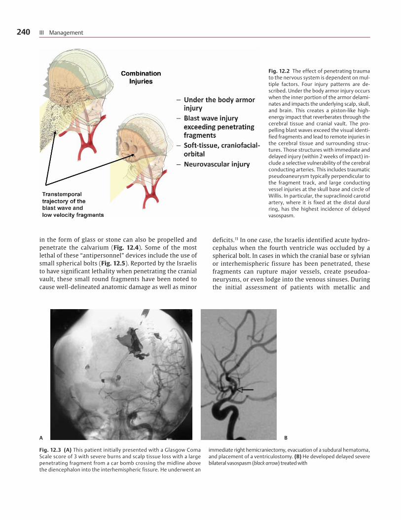
The effect of penetrating trauma to the nervous system isdependent on multiple factors (Fig. 12.2). As seen in re-cent conflicts, the incidence of survivable missile injuries(i.e., AK-47 round) to the brain remains low. Recent en-gagements have identified the use of even higher velocityrounds with longer metal jackets and higher muzzle ve-locities (i.e., AK-74), which are used as a sniper’s weaponof choice. The majority of these wounds is still fatal. This isrelated to the high likelihood of perforation, global cranialvault disruption, and high cavitation pressures. However,the majority of injuries during Operation Iraqi Freedomhave been from roadside “IEDs” or improvised explosive
devices. These include vehicle-borne delivery systemscommonly referred to as either “car bombs” or “suicidebombers.” These munitions are variable in their designand delivery of injury. The injuries are dependent on theexplosive that is used, the distance from the explosion, theshape of the projectile, and lastly the viscoelastic proper-ties of the impacted tissue. Such projectiles are propelledby enormous blast-overpressure forces, which may ac-count for the injury force beyond the flying projectiles orthe terminal impact. Syndromes of central nervous system(CNS) dysfunction associated with blast injuries have beenidentified and classified since WWII.10 During the explo-sion of such devices, flying projectiles include the materi-als used to make the bomb (primary projectile) andadditional materials (i.e., nails and other metallic objects,rocks, glass, body parts) packed around the device by theenemy (secondary projectiles). These fragments, althoughtraveling with lower terminal velocity compared with thesniper’s round, inflict significant destruction due to theirabnormal size, shape, and porosity.
Unlike the previously discussed metal fragments, non-metallic fragments may lead to delayed abscess formationand secondary sepsis. In the case of a vehicle-borne IED(VBIED), the metal from the auto can act as a secondaryprojectile (Fig. 12.3). Debris from surrounding buildings
12 Wartime Penetrating Injuries 239
Table 12.1 Cushing’s Classification of Penetrating Brain Injury (1918)1
Grade Description No. of WWI Cases % Mortality
I Scalp lacerations with intact skull 22 4.5
II Wounds with skull fractures/intact dura/ � depression 54 9.2
III Wounds with depressed skull fracture/dural laceration 18 11.8
IV Wounds (guttering type) with in-driven fragments, usually protruding brain 25 24
V Penetrating wound, lodged projectile, brain usually protruding 41 36.6
VI Wounds penetrating ventricles with either (a) bone fragments or (b) projectiles a)14 a)42.8b)16 b)100
VII Wounds involving orbitonasal or auropetrosal region with extruding brain 15 73.3
VIII Perforating wounds, cerebral injury severe 5 80
IX Craniocerebral injury with massive skull fracture 10 50
Table 12.2 Matson’s Tenets
Matson’s Tenets9 Current Application
I. Save life Application of ATLS/ACLS/far forward homeostasis and hemicraniectomy
II. Prevent infection Watertight dural closure
III. Preserve nervous system function Prevention of secondary neurologic injury through advanced neurocritical and neurointerventional care (i.e., meningitis, seizures, stroke)
IV. Restore anatomic function Restore anatomic protection and contour (i.e., cranioplasty)
Abbreviations: ACLS, advanced cardiac life support; ATLS, advanced trauma life support.
in the form of glass or stone can also be propelled andpenetrate the calvarium (Fig. 12.4). Some of the mostlethal of these “antipersonnel” devices include the use ofsmall spherical bolts (Fig. 12.5). Reported by the Israelisto have significant lethality when penetrating the cranialvault, these small round fragments have been noted tocause well-delineated anatomic damage as well as minor
deficits.11 In one case, the Israelis identified acute hydro-cephalus when the fourth ventricle was occluded by aspherical bolt. In cases in which the cranial base or sylvianor interhemispheric fissure has been penetrated, thesefragments can rupture major vessels, create pseudoa-neurysms, or even lodge into the venous sinuses. Duringthe initial assessment of patients with metallic and
240 III Management
Fig. 12.2 The effect of penetrating traumato the nervous system is dependent on mul-tiple factors. Four injury patterns are de-scribed. Under the body armor injury occurswhen the inner portion of the armor delami-nates and impacts the underlying scalp, skull,and brain. This creates a piston-like high-energy impact that reverberates through thecerebral tissue and cranial vault. The pro-pelling blast waves exceed the visual identi-fied fragments and lead to remote injuries inthe cerebral tissue and surrounding struc-tures. Those structures with immediate anddelayed injury (within 2 weeks of impact) in-clude a selective vulnerability of the cerebralconducting arteries. This includes traumaticpseudoaneurysm typically perpendicular tothe fragment track, and large conductingvessel injuries at the skull base and circle ofWillis. In particular, the supraclinoid carotidartery, where it is fixed at the distal duralring, has the highest incidence of delayedvasospasm.
A B
Fig. 12.3 (A) This patient initially presented with a Glasgow ComaScale score of 3 with severe burns and scalp tissue loss with a largepenetrating fragment from a car bomb crossing the midline abovethe diencephalon into the interhemispheric fissure. He underwent an
immediate right hemicraniectomy, evacuation of a subdural hematoma,and placement of a ventriculostomy. (B) He developed delayed severebilateral vasospasm (black arrow) treated with

nonmetallic foreign body penetration, the question ofremoval must be considered and may be influenced bymultiple variables. Ventricular or paraventricular locationof such metallic or nonmetallic porous material has beenassociated with delayed infections and late neurologicaldeterioration.12 Overall, if there is evidence of fragmentmovement, contact with the cerebrospinal fluid (CSF)within either a cisternal or ventricular location, or loca-tion adjacent to a vascular structure, it may be advisableto remove the foreign body (Table 12.3). The exceptionmay be interhemispheric bone fragments without vesselabnormality. Regardless of approach, the fragmentsshould be followed radiographically to assess for any evi-dence of delayed movement or abscess formation. Thisconservative approach is acceptable because reoperationto remove fragments has not been shown to reduce theseizure rate or the incidence of late infections but hasincreased the neurological morbidity. 6,13
Management of Wartime PenetratingInjuries
Initial Resuscitation
The application of Matson’s tenets begins at the point ofinjury. Combat medical personnel are faced with multiplechallenges, not least of which is resuscitating the patientwhile under enemy fire. Unlike the civilian environment,the care of the military casualty is often hindered by theongoing threat to the unit. Medical teams are specificallytargeted by the enemy to discourage, demoralize, and
12 Wartime Penetrating Injuries 241
Fig. 12.3 (Continued) (C) microballoon angioplasty and nicardipine(black arrow). (D) He was taken back to the operating room for removal ofthe large metal fragment measuring �4 cm. He underwent a cranioplasty
with tissue expanders previously placed, yet required a latissum dorsiflap due to tissue breakdown. At 36 months postinjury, he is ambulatingindependently, effectively communicating, and feeding himself.
C D
Fig. 12.4 Debris surrounding the explosion is propelled as secondary frag-ments. In a vehicle or building this occurs in the form of twisted metal,glass, or roadside stones and can penetrate the calvarium via the orbit andmidface. In a frontal direction significant anatomic disruption results to theanterior skull base, orbit, midface, airway, and bilateral frontal lobes as wellas the anterior cerebral artery complex in the interhemispheric fissure. Thesoft tissue, supporting bony framework, and anatomic continuity are lostfrom the skull base to the orbit and infratemporal fossa.

242 III Management
A
C
B
D
Fig. 12.5 (A) This soldier had an initial GlasgowComa Scale score of 7 with a transorbital sphericalbolt penetration deposited into the pineal region(black arrow, B). (C) He received a ventriculostomythen delayed left hemicraniectomy and (D) subse-quent cerebral angiogram demonstrating an ante-rior communicating artery pseudoaneurysm. Thepatient reruptured this pseudoaneurysm followingrapid enlargement and expired.

deter the unit’s combat effectiveness. Therefore, a conceptof removing the casualty from the “kill-zone” is essentialprior to focused resuscitation. In a direct firefight, themedic’s first priority may be to return fire in an attemptto suppress the enemy before evacuating the casualty.Because most of the injuries during OIF have occurredfrom unmanned roadside bombs (i.e., IEDs), the medicalplan is typically adjusted. Unlike civilian trauma and pre-vious military conflicts, immediate evacuation from the“kill box” is of the utmost importance. After mobilizationto a safer area, initial resuscitation and medical evacua-tion to the next level of care are conducted.
Early airway and hemorrhage control combined withrapid evacuation is the first stage in the resuscitation of acasualty with severe neurotrauma. Direct transport toneurosurgeons located in the combat support hospital(CSH) has allowed immediate intervention, leading to im-proved survivability. The exact magnitude of increasedsurvival is difficult to evaluate because, with such rapidevacuations, a higher proportion of expectant wounds areseen by the neurosurgeon than in prior conflicts.
Far Forward Neuroimaging and Neurosurgery
The challenges of complex, severe military penetratingbrain injury (PBI) are addressed by the coordinated effortsof physicians, nurses, and technicians at the CSH. In theUnited States military medical model, the CSH is the firstlocation where both neurosurgery and computerizedtomography (CT) scanning are available. After the initialairway, breathing, and circulation have been managed, ahemodynamically stable patient must undergo appropri-ate imaging. At this stage, it is imperative that no unneces-sary delay prevents appropriate cranial decompression fora life-threatening lesion. Occasionally, life-threateningextracranial bleeding must first be treated. Multiple op-tions exist with the most practical and efficient includingsimultaneous cranial/corporeal intervention or delayedimaging after hemodynamic stability has been achieved.Delayed neuroimaging is used when faced with a closedinjury, a neurologically stable patient, or patients under-going prolonged extracranial procedures without thebenefit of an immediate postoperative examination.
The approach to the severely brain injured patient hasevolved throughout the current conflict. Because of thelong transport flights that must occur, the practice haschanged to include wide decompressive hemicraniectomywith subsequent duraplasty and watertight closure asearly as possible. The thought is that the decompressionmay mitigate or reduce incidence of secondary neurologi-cal deficits that occur from malignant intracranial hyper-tension. Nevertheless, as in civilian neurotrauma, mostcranial interventions will include early postoperativeimaging and intracranial pressure (ICP) monitoring whereappropriate.
Medical Evacuation
The medical evacuation of the severely injured soldier ormarine to the United States currently involves a stop inGermany and includes over 7,200 miles of travel. The med-ical hazards of this trip must be taken into considerationand include the effects of delayed cerebral edema, hydro-cephalus, or hemorrhage, which may occur during transfersor flight. To address these issues, critical care air transportteams have been instrumental in the strategic evacuationsof patients from Baghdad to Germany and beyond. Manage-ment of elevated ICP, hypoxia, and hypotension is their pri-mary focus; each team consists of a physician, nurse, andrespiratory technician and is rarely supplemented with aneurosurgeon or neurologist. Out of over 21,000 casualties,over 500 intubated neurotrauma patients have been trans-ported in this fashion. Additional operational challengesinclude enemy activity, weather, and airframe function.
Description of Injuries
Patterns of penetrating trauma in both civilian and mili-tary have been classically described based on the fragmentpath. The key element is the unseen force propelling thefragment. Rarely is this force completely characterized in abomb blast. Typically, all that is seen are the fragments,spall, or retained overlying clothing driven into the cranialvault. A complete physical examination allows the abilityto identify points of foreign body entry or exit. The mostcommonly missed region of fragment entry includes theretroauricular and suboccipital regions. Fragment entriesfrom these sites are particularly hazardous, with the in-creased risk of vascular, cranial nerve, or brain stem injury.
Perforating
These injuries typically carry the worst prognosis, especiallywhen associated with high-velocity injuries or when theinjuries cross the midline or are transhemispheric. In a large
12 Wartime Penetrating Injuries 243
Table 12.3 Criteria for Removal of Intracranial Fragment
• Movement of fragment
• Abscess formation
• Vessel compression or contact
• Porous material in contact with cerebrospinal fluid (i.e., rock, wood)
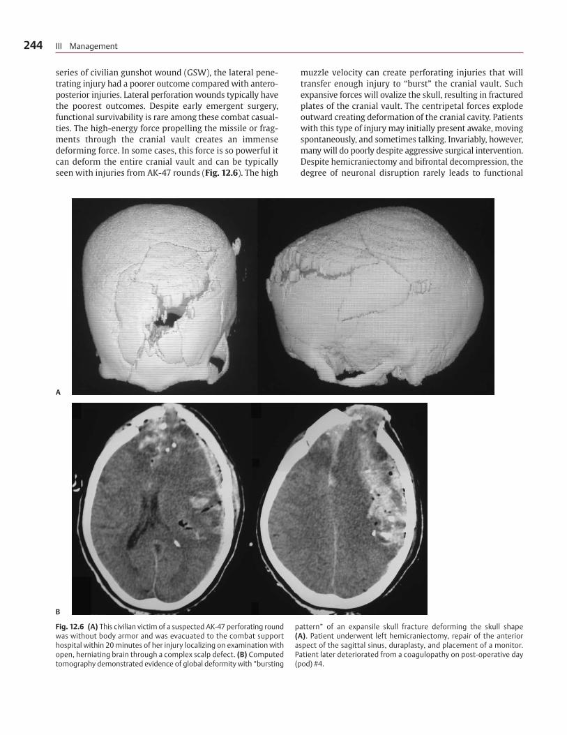
series of civilian gunshot wound (GSW), the lateral pene-trating injury had a poorer outcome compared with antero-posterior injuries. Lateral perforation wounds typically havethe poorest outcomes. Despite early emergent surgery,functional survivability is rare among these combat casual-ties. The high-energy force propelling the missile or frag-ments through the cranial vault creates an immensedeforming force. In some cases, this force is so powerful itcan deform the entire cranial vault and can be typicallyseen with injuries from AK-47 rounds (Fig. 12.6). The high
muzzle velocity can create perforating injuries that willtransfer enough injury to “burst” the cranial vault. Suchexpansive forces will ovalize the skull, resulting in fracturedplates of the cranial vault. The centripetal forces explodeoutward creating deformation of the cranial cavity. Patientswith this type of injury may initially present awake, movingspontaneously, and sometimes talking. Invariably, however,many will do poorly despite aggressive surgical intervention.Despite hemicraniectomy and bifrontal decompression, thedegree of neuronal disruption rarely leads to functional
244 III Management
A
B
Fig. 12.6 (A) This civilian victim of a suspected AK-47 perforating roundwas without body armor and was evacuated to the combat supporthospital within 20 minutes of her injury localizing on examination withopen, herniating brain through a complex scalp defect. (B) Computedtomography demonstrated evidence of global deformity with “bursting
pattern” of an expansile skull fracture deforming the skull shape (A). Patient underwent left hemicraniectomy, repair of the anterioraspect of the sagittal sinus, duraplasty, and placement of a monitor.Patient later deteriorated from a coagulopathy on post-operative day(pod) #4.
survival. In some cases, rapid decompression may lead to anassociated hypotension especially in hypovolemic patientswhose blood pressure will typically drop during decom-pression. Communicating with your anesthesiologist willallow appropriate anticipation of this response.
Penetrating
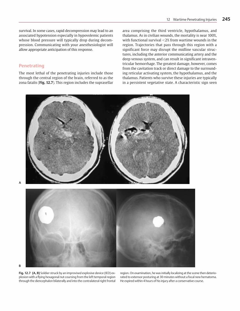
The most lethal of the penetrating injuries include thosethrough the central region of the brain, referred to as thezona fatalis (Fig. 12.7). This region includes the suprasellar
area comprising the third ventricle, hypothalamus, andthalamus. As in civilian wounds, the mortality is near 100%,with functional survival �2% from wartime wounds in theregion. Trajectories that pass through this region with asignificant force may disrupt the midline vascular struc-tures, including the anterior communicating artery and thedeep venous system, and can result in significant intraven-tricular hemorrhage. The greatest damage, however, comesfrom the cavitation track or direct damage to the surround-ing reticular activating system, the hypothalamus, and thethalamus. Patients who survive these injuries are typicallyin a persistent vegetative state. A characteristic sign seen
12 Wartime Penetrating Injuries 245
A
B
Fig. 12.7 (A, B) Soldier struck by an improvised explosive device (IED) ex-plosion with a flying hexagonal nut coursing from the left temporal regionthrough the diencephalon bilaterally and into the contralateral right frontal
region. On examination, he was initially localizing at the scene then deterio-rated to extensor posturing at 30 minutes without a focal new hematoma.He expired within 4 hours of his injury after a conservative course.

246 III Management
Table 12.4 Complications of Wartime Penetrating Brain Injury
Time Type of Complications Treatment
0–24 h ICP increased Hemicraniectomy
Hematoma Evacuation/coagulation correction
Ischemia Decompression/ID occlusion
Anatomic defect Anatomic closure
Hypoxia Airway/pulmonary correction
Hypotension Overt or occult EBL
PRBC/FFP/PLTS vs. whole blood vs. hypotonic saline
24–48 h ICP increased Hemicraniectomy
Hematoma Evacuation/coagulation correction
Hydrocephalus Ventriculostomy
Edema Decompression
Seizure Antiepileptics/cEEG monitoring
72 h–1st wk Edema Medical/surgical decompression
ICH (Contusion) Correct coagulopathy
Hydrocephalus Ventriculostomy
CSF leak Repair/CSF diversion
Ischemia Medical/endovascular Tx
Pseudoaneurysm Surgical/endovascular Tx
Seizures Antiepileptics/cEEG monitoring
2–3rd wk Infections R/O abcess, CSF infection
Vasospasm TCDs, PbO2, cEEG, CBF monitoring with combined HHH versus angioplasty
Pseudoaneurysm Endovascular versus microsurgery
Seizures Antiepileptics
Delayed hydrocephalus VP shunt (low-pressure consider use of programmable valve)
1–6 mo Infection R/O abscess, meningitis
Low-pressure hydrocephalus VP shunt (programmable valve)
Syndrome of trephine Reconstructive cranioplasty
Seizures Antiepileptics
Cranioplasty complications
Temporalis atrophy Resuspension/implant/fat graft
Infection Prosthesis removal
Hydrocephalus VP shunt
Epidural/subgaleal Drainage
Hygroma/hematoma ICH Evacuation
Scalp necrosis Free-flap
Abbreviations: CBF, cerebral blod flow; cEEG, continuous electroencephalogram; CSF, cerebrospinal fluid; EBL, estimated blood loss; FFP, freshfrozen plasma; ICP, intracranial pressure; ICH, intracranial hematoma; HHH, hypervolemic, hypertensive, hyperdynamic; PLTS, platelets; PRBC, packed red blood cells; R/O, rule out; TCD, transcranial doppler; Tx, treatment; VP, ventriculoperitoneal.

in civilian injuries, and occasionally in lower-caliberwartime injuries, is the “tram-track” sign. This representsthe cavitation tract and is associated with significant energytransfer, significant edema, and poor outcome. This out-come is commonly repeated in the transhemispheric,transventricular wound. Multiple hemisphere injury andcrossing the midline at the level of the corpus callosum orbelow portends a high mortality and poor functional out-come.14 As stated earlier, these missile tracts have beenassociated with pseudoaneurysms. Typically occurringperpendicular to the long axis of the tract, they are associ-ated with ischemia or delayed rupture if not appropriatelytreated. The M1 segment is particularly vulnerable in someof these paths. Deeply embedded metals are not classicallyretrieved unless they are in the ventricular system, inmotion, compressing a large vascular structure, creatinghydrocephalus, or associated with a delayed abscess. Again,the early use of radical hemicraniectomy with duraplastyhas allowed a higher survival and earlier, improved func-tional outcome in this population than previously predicted.In many of these cases, a majority may have presented ini-tially talking before massive edema, shift, and cerebraldysfunction occurred. The patients typically have a poorerexamination after the first 48 to 72 hours, usually due todelayed cerebral edema or hydrocephalus. In selected cases,delayed blast-induced vasospasm may occur, although thisis typically seen after the first week.
Injury Patterns and Management
Complex cranial–facial injuries are typical in the OIF con-flict. Comparable to WWI trench-warfare, the head andneck is a region of selective vulnerability. Injuries thatbridge the craniocervical junction, associated orbitofacialinjuries, and injuries to the neck have been particularlychallenging to treat. Cranial-basal injuries have a ten-dency to have a higher association with neurovascularinjuries with a profound risk for delayed stroke anddeath. Additionally, this region is also associated with ahigh rate of CSF leaks, fistulae, and infections. The disrup-tion of the cranial base with communication with theorbit, pharynx, and infratemporal fossa may be associatedwith cranial nerve injuries, blindness, and globe disrup-tion. Avoiding associated complications begins with ahigh index of suspicion followed by an aggressive role forneuroangiography, meningitis monitoring, and cranialnerve evaluation (Table 12.4).
Orbitofacial Injuries
Orbitofacial injuries in this conflict are highly associatedwith neurovascular injuries, CSF leakage, and death15
(Fig. 12.5). Biomechanical studies of penetrating traumato the maxilla and mandible have demonstrated signifi-cant force transmission to the brain. In particular, the pres-sure waves in the brain were greatest when Chinese M193or M56 military bullets were used in animal models com-pared with 1.03 grain spheres at 1,400 m/s or at 800 m/s.16
Transorbital intracranial entry risks injury to the internalcarotid, cavernous sinus, anterior communicating arterycomplex, optic nerve, and cranial nerves II to VI (Fig. 12.8).This is most common when the medial aspect of the orbitis penetrated. Diffuse intracranial air associated with atransorbital injury strongly increases irreversible brainstem injury, transorbital cerebral herniation, and increasesthe risk of death. Disruption of the orbital roof can createa communication with the intracranial cavity, leading toassociated CSF leaks, encephalocele, intracranial abscesses,or delayed orbital reconstruction difficulties. In extremeblast cases, the maxillary sinus, orbit, and anterior cranialvault will all communicate through a traumatic disrup-tion, exposing the brain to the sinus mucosa. In such cases,it is usually necessary to re-create surgically the cranialbase, orbit, and maxillary sinus to protect the brain andobtain a cosmetically acceptable result. The use of tita-nium mesh fixation for the anterior skull base floor intheater has allowed subsequent surgeons to then use thatfoundation to keep the cerebral-orbital spaces separate.This closure is reinforced with pericranium (when avail-able), fascia lata, temporalis fascia, fat, and occasionallysplit-thickness skull bone graft.
Surgical Considerations
The overall management goals include acute decompres-sion and hemorrhage control. This is typically accom-plished with a bifrontal craniotomy or craniectomy. Incases with disruption of the anterior cranial floor andfrontal sinus with obvious risk for CSF leakage, a sinusexenteration, skull base reconstruction with watertightdural closure is usually performed. In restricted situationssuch as those akin to combat conditions involving masscasualties, lack of imaging, and lack of ophthalmologysupport, a limited procedure may be initially performed.This includes epidural hematoma evacuation followed bytransfer to another neurosurgeon within 24 hours for amore definitive anterior skull base reconstruction. Thiswas the case during the attack on the United Nationsheadquarters in Baghdad where over 150 casualtiesarrived at the combat support hospital and 30 underwentopen surgeries. Half of these required cranial or cervicalsurgery to remove glass embedded within the cranialvault, face, orbit, or neck. The possible array of penetratingfragments includes glass, rocks, metal, and occasionallythe fragments of the suicide bomber. Plain films and the
12 Wartime Penetrating Injuries 247

physical examination are particularly helpful in under-standing the global distribution of the fragments, the pathof injury, and the best surgical approach. Unlike metal, anattempt is made early to remove glass, depressed bone overair sinus, clothing, body armor, and rocks from the cranialvault. However, deeply embedded fragments are not pur-sued unless there is documented delayed movement orvascular compromise. This is in keeping with avoidance ofsecondary injury through missile tract exploration.
Transtemporal Injuries
Those injuries that penetrate the frontotemporal region ofthe cranial cavity may include underlying injury to thefrontotemporal lobes, internal carotid and middle cere-bral arteries, and lateral ventricles with intraventricular
hemorrhage (Fig. 12.9). Additionally, those associatedwith a significant force to the skull base may destroy thepetrous bone; petrous carotid artery; facial, auditory, andtrigeminal nerves as well as the lateral orbit and opticnerve. This can lead to CSF leaks, pseudoaneurysms,blindness, loss of usable hearing, and facial paralysis.
Surgical Considerations
Injuries in the region of the lateral skull base should in-clude proximal exposure and control of the cervical carotidartery and its branches. In cases of intractable epistaxis,endovascular methods are preferred to obtain proximalcontrol but may not be possible in an austere environ-ment. Reconstruction of the petrous carotid artery is par-ticularly challenging in a combat environment. Multiple
248 III Management
Fig. 12.8 This patient had an initial Glasgow Coma Scale score of 3 witha penetrating right suboccipital fragment passing transtentorially intothe occipital lobe on the left, then into the right occipital parietal junc-tion. The patient underwent a suboccipital craniectomy, right hemi-craniectomy, transverse sinus ligation preserving the vein of Labbé, andplacement of a ventriculostomy. He demonstrated delayed recurrent
severe bilateral internal carotid artery, middle cerebral artery vasospasmrequiring microballoon angioplasty and nicardipine. His examinationimproved with following commands, speaking spontaneously, and mov-ing all four extremities. He demonstrated delayed hydrocephalus requiringa ventriculoperitoneal shunt.

12 Wartime Penetrating Injuries 249
A
B C
Fig. 12.9 (A) This soldier presented with a Glasgow Coma Scale scoreof 3 with a lateral temporal penetrating fragment coursing throughthe sylvian fissure, central diencephalon bilaterally, and third ventriclewith significant SAH, IVH, and temporal lobe hematoma. He underwenta left hemicraniectomy, clipping of a transected left middle cerebral
artery (B; white arrow, C; black arrow), evacuation of the temporal lobehematoma, and placement of a ventriculostomy (B, C). The postoper-ative course was complicated by posterior circulation delayed severevasospasm requiring microballoon angioplasty and intraarterialnicardipine 1.
(Continued on page 250)
constraints include the lack of intraoperative angiography,the absence of a high-definition operating microscope andmicroinstruments, the reduced availability of grafts dueto extremity injuries, and most critically, the presence ofa swollen, edematous, and hemorrhagic brain. In somecases, it may be more reasonable to perform a proximaland distal supraclinoid internal carotid artery (ICA) liga-tion to prevent a thromboembolic middle cerebral artery(MCA) stroke.
Laterally displaced entrance wounds may create asignificant amount of soft tissue loss. This will challengeboth the initial closure as well as the delayed reconstruction.
In an attempt to preserve the known vascular pedicles, itmay be preferable to base a curvilinear incision behindthe ear to the anterior forehead (Fig. 12.10). The superiortemporal artery will play an important role in scalp viabilitywith a large hemicraniectomy flap.
Preservation of the vein of Labbé and viable MCAbranches is an important part of the surgical decompres-sion. Adequate bone removal insures that venous com-promise from swelling at the bone edge will not occur.Following dural opening, a careful examination of thesylvian and cortical MCA branches should take placebecause the branches of the middle cerebral artery most

250 III Management
Fig. 12.10 (A) Alternative hemicraniectomy incision with midline curvilin-ear incision with vertical bisecting incision to the root of the zygoma. Origi-nally described by Dr. Ludwig Kempe at the Walter Reed Army MedicalCenter during procedures for hemispherectomy and reintroduced in thecurrent conflict by Major Jon Martin, MD, while serving in Balad, Iraq, in
spring 2007 [Kempe, L. Operative Neurosurgery Vol 1. New York: SpringerVerlag; 1968; 180–189]. Brain is covered by synthetic dura after placementof an ipsilateral intracranial pressure monitor, tunneled at the midline, alarge 7fr subgaleal drain is then placed before closing the scalp (B, C).
A
B
C
Fig. 12.9 (Continued) (D, E). Delayed cranioplasty was performed at6 months and required ventriculoperitoneal shunting for delayedhydrocephalus. The patient’s best examination at 8 months remains
minimally reactive localizing, but the patient is noncommunicativewith a right hemiplegia.
D E

commonly injured include those in the distal corticalsurface or lateral fissure. Typically, pseudoaneurysms areperpendicular to the fragment path in the zone of cavita-tion adjacent to the track and, if encountered, should beexcluded from the normal circulation. Suspicion for trau-matic aneurysms should arise when a sylvian fissurehematoma, focal parenchymal blood (i.e., gyrus rectushematoma), or a hematoma remote from the fragmentare present.
Delayed complications from injuries in this regioninclude CSF leaks, pseudoaneurysm rupture, thromboem-bolic strokes, and flap necrosis associated with devascu-larization (Table 12.4). Commonly, CSF leaks include adisruption of the petrous skull base with underlying low-pressure hydrocephalus. In the presence of a hemi-craniectomy, a distended flap may lead to CSF egressthrough the disrupted petrous ridge, subgaleal space, andpossibly through the wound. In an effort to decrease thisoccurrence, we routinely place ventriculostomies to de-compress the hemicraniectomy flap and afford anotherpathway for CSF egress while the disrupted skull base issealing. Except in extreme cases with multiple ventricu-lostomies, we have avoided the routine use of early lum-bar drainage due to concerns with cranial spinal pressuredissociation, meningitis, and lumbar overdrainage.
Pseudoaneurysm management has challenged cur-rent practice patterns during the current conflict. Morepseudoaneurysms have been detected and treated in thefirst 2 years of this war than in the entire 10 years of theIran–Iraq conflict. 3,17 Unfortunately, early in the conflict,delayed rupture resulted in death, coma, progressiveparalysis, and near-fatal cardiac arrest from epistaxis inpatients demonstrating early recovery from their initialneurological injury. This observation has prompted aconcerted effort toward early detection and treatment.An aggressive screening process composed of early CT
and cerebral angiography performed upon arrival to astateside hospital by an experienced neurointervention-alist is now our standard of care. CT angiography (CTA)alone has been inadequate secondary to technical limita-tions stemming from metal artifacts, poor timing of thecontrast bolus with venous contamination, and contrastdiverted from stenosed conductance vessels. The criteriafor a screening angiogram are outlined in Table 12.5. Ifthe pseudoaneurysm is associated with a well-definedneck and is endovascularly accessible, the preference atour institution is early exclusion with either coils orstent-assisted coiling. In cases with distal pericallosalor MCA aneurysms, early microsurgery is the preferredtreatment. The recurrence rate for endovascularlytreated aneurysms approaches 30% in the senior author’sseries and requires close follow-up (Fig. 12.11). Repeatangiography at 3 months followed by either repeatendovascular treatment or open microsurgery has beenthe current strategy.
Suboccipital or Occipital Injuries
These injuries can be some of the most lethal due to theextent of injury to the brain stem, venous sinuses, andmultiple intracranial compartments. Low-velocity frag-ments or high-velocity missiles that pass from the poste-rior fossa into the supratentorial compartment may createa path of injury that includes as many as three cerebralcompartments (i.e., the ipsilateral cerebellum, occipital-temporal lobe, and contralateral parietal-occipital lobes)(Fig. 12.8). Additional injuries to the cervical spinal cord,vertebrobasilar circulation, and cranial nerves are possible.In one specific case in our series, an extracranial, suboccip-ital fragment resulted in a proximal posterior inferiorcerebellar artery (PICA) traumatic aneurysm that subse-quently ruptured.
12 Wartime Penetrating Injuries 251
Table 12.5 Evolution of Criteria for Intracranial Angiography following Penetrating Injury
Iran–Iraq War* Operation Iraqi Freedom†
Penetrating injury through pterion, orbit, posterior fossa Previous criteria plus:
Penetrating fragment with intracranial hematoma Known cerebral artery sacrifice and/or pseudoaneurysm at the time of initial exploration
Known cerebral artery sacrifice and/or pseudoaneurysm at the time of initial exploration
Blast-induced penetrating injury with GCS �8
TCD evidence of posttraumatic vasospasm
Spontaneous decrease in PBrO2
Source: Data from *Aarabi B. Traumatic aneurysms of brain due to high velocity missile head wounds. Neurosurgery 1988;22(6 Pt 1):1056–1063and †Armonda RA, Bell RS, Vo AH, et al. Wartime traumatic cerebral vasospasm. Recent reviews of combat casualties. Neurosurgery 2006 Dec:59: 1215–1225. Discussion 1225.
Abbreviations: GCS, Glasgow Coma Scale, PBrO2, partial pressure brain tissue oxygen; TCD, transcranial doppler.

252 III Management
B C
D E
A
Surgical Considerations
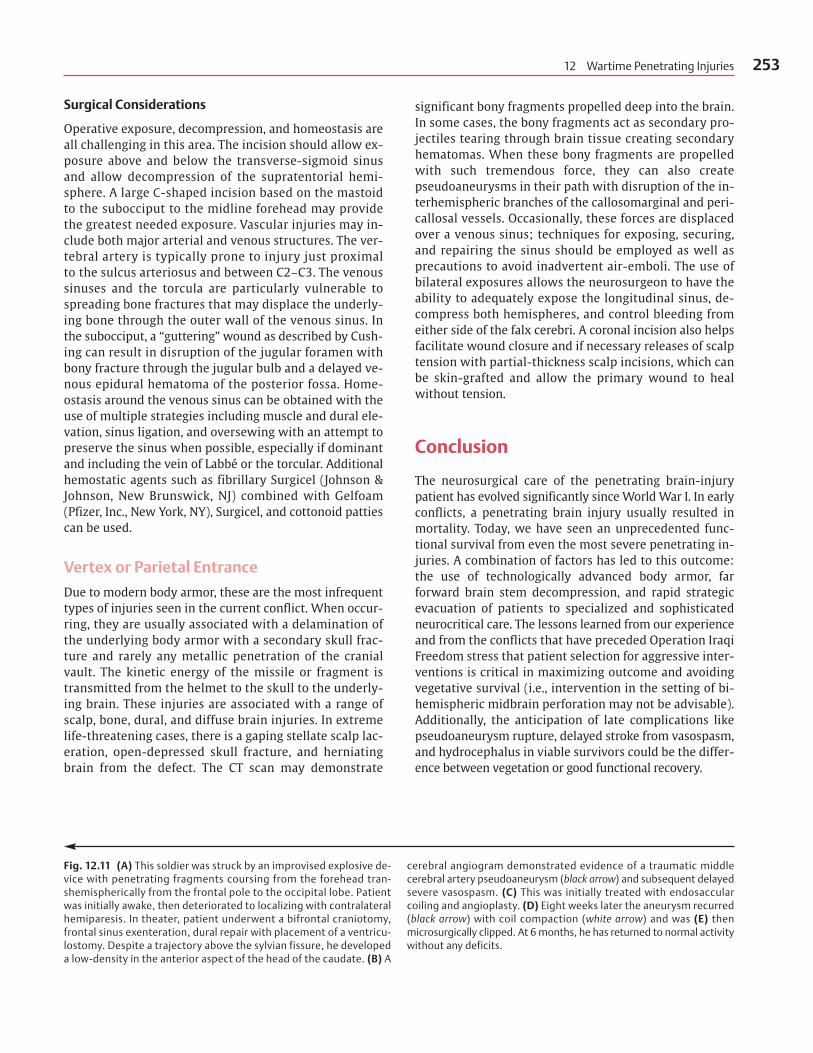
Operative exposure, decompression, and homeostasis areall challenging in this area. The incision should allow ex-posure above and below the transverse-sigmoid sinusand allow decompression of the supratentorial hemi-sphere. A large C-shaped incision based on the mastoidto the subocciput to the midline forehead may providethe greatest needed exposure. Vascular injuries may in-clude both major arterial and venous structures. The ver-tebral artery is typically prone to injury just proximalto the sulcus arteriosus and between C2–C3. The venoussinuses and the torcula are particularly vulnerable tospreading bone fractures that may displace the underly-ing bone through the outer wall of the venous sinus. Inthe subocciput, a “guttering” wound as described by Cush-ing can result in disruption of the jugular foramen withbony fracture through the jugular bulb and a delayed ve-nous epidural hematoma of the posterior fossa. Home-ostasis around the venous sinus can be obtained with theuse of multiple strategies including muscle and dural ele-vation, sinus ligation, and oversewing with an attempt topreserve the sinus when possible, especially if dominantand including the vein of Labbé or the torcular. Additionalhemostatic agents such as fibrillary Surgicel (Johnson &Johnson, New Brunswick, NJ) combined with Gelfoam(Pfizer, Inc., New York, NY), Surgicel, and cottonoid pattiescan be used.
Vertex or Parietal Entrance
Due to modern body armor, these are the most infrequenttypes of injuries seen in the current conflict. When occur-ring, they are usually associated with a delamination ofthe underlying body armor with a secondary skull frac-ture and rarely any metallic penetration of the cranialvault. The kinetic energy of the missile or fragment istransmitted from the helmet to the skull to the underly-ing brain. These injuries are associated with a range ofscalp, bone, dural, and diffuse brain injuries. In extremelife-threatening cases, there is a gaping stellate scalp lac-eration, open-depressed skull fracture, and herniatingbrain from the defect. The CT scan may demonstrate
significant bony fragments propelled deep into the brain.In some cases, the bony fragments act as secondary pro-jectiles tearing through brain tissue creating secondaryhematomas. When these bony fragments are propelledwith such tremendous force, they can also createpseudoaneurysms in their path with disruption of the in-terhemispheric branches of the callosomarginal and peri-callosal vessels. Occasionally, these forces are displacedover a venous sinus; techniques for exposing, securing,and repairing the sinus should be employed as well asprecautions to avoid inadvertent air-emboli. The use ofbilateral exposures allows the neurosurgeon to have theability to adequately expose the longitudinal sinus, de-compress both hemispheres, and control bleeding fromeither side of the falx cerebri. A coronal incision also helpsfacilitate wound closure and if necessary releases of scalptension with partial-thickness scalp incisions, which canbe skin-grafted and allow the primary wound to healwithout tension.
Conclusion
The neurosurgical care of the penetrating brain-injurypatient has evolved significantly since World War I. In earlyconflicts, a penetrating brain injury usually resulted inmortality. Today, we have seen an unprecedented func-tional survival from even the most severe penetrating in-juries. A combination of factors has led to this outcome:the use of technologically advanced body armor, farforward brain stem decompression, and rapid strategicevacuation of patients to specialized and sophisticatedneurocritical care. The lessons learned from our experienceand from the conflicts that have preceded Operation IraqiFreedom stress that patient selection for aggressive inter-ventions is critical in maximizing outcome and avoidingvegetative survival (i.e., intervention in the setting of bi-hemispheric midbrain perforation may not be advisable).Additionally, the anticipation of late complications likepseudoaneurysm rupture, delayed stroke from vasospasm,and hydrocephalus in viable survivors could be the differ-ence between vegetation or good functional recovery.
12 Wartime Penetrating Injuries 253
Fig. 12.11 (A) This soldier was struck by an improvised explosive de-vice with penetrating fragments coursing from the forehead tran-shemispherically from the frontal pole to the occipital lobe. Patientwas initially awake, then deteriorated to localizing with contralateralhemiparesis. In theater, patient underwent a bifrontal craniotomy,frontal sinus exenteration, dural repair with placement of a ventricu-lostomy. Despite a trajectory above the sylvian fissure, he developeda low-density in the anterior aspect of the head of the caudate. (B) A
cerebral angiogram demonstrated evidence of a traumatic middlecerebral artery pseudoaneurysm (black arrow) and subsequent delayedsevere vasospasm. (C) This was initially treated with endosaccularcoiling and angioplasty. (D) Eight weeks later the aneurysm recurred(black arrow) with coil compaction (white arrow) and was (E) then microsurgically clipped. At 6 months, he has returned to normal activitywithout any deficits.

References1. Cushing H. A series of wounds involving the brain and its enveloping
structures. Br J Surg 1918;5:558–684
2. Eden K. Mobile neurosurgery in warfare, experiences in the Eighth
Army’s Campaign in Cyrenaica, Tripolitania, and Tunisia. Lancet
1943;2:689–692
3. Aarabi B. Traumatic aneurysms of brain due to high velocity missile
head wounds. Neurosurgery 1988;22(6 Pt 1):1056–1063
4. Meirowsky AM. Penetrating wounds of the brain. In: JB Costes, ed.
Neurological Surgery of Trauma. Washington, DC: Office of the
Surgeon General, Department of the Army; 1965:103–136
5. Carey ME, Young H, Mathis JL, Forsythe J. A bacteriologic study of cran-
iocerebral missile wounds from Vietnam. J Neurosurg 1971;34:145–154
6. Brandvold B, Levy L, Feinsod M, George E. Penetrating craniocerebral
injuries in the Israeli involvement in the Lebanese conflict,
1982–1985: an analysis of a less aggressive approach. J Neurosurg
1990;72:15–21
7. Bell R, Vo A, Porter C, et al. Wartime neurovascular injuries: review of
the effectiveness of early, aggressive, endovascular management in the
setting of blast-related cerebral vasospasm. Neurosurgery 2006;59(2):
455–456
8. Simpson DA, David D. The genesis of craniomaxillofacial surgery. ANZ
J Surg 2004;74:71–77
9. Matson D. The management of acute craniocerebral injuries due to mis-
siles. In: Spurling G, Woodhall B, eds. Surgery in WWII: Neurosurgery
Vol 1. Washington, DC: Office of the Surgeon General, Department of the
Army; 1958
10. Cramer F. Blast concussion and cerebral injuries due to explosion
waves. In: Spurling G, Woodhall B, eds. Surgery in WWII: Neuro-
surgery Vol 1. Washington, DC: Office of the Surgeon General, Depart-
ment of the Army; 1958
11. Roth J, Mayo A, Elran H, Razon N, Kluger Y. Brain injuries caused by
spherical bolts. J Neurosurg 2005;102:864–869
12. Rosenwasser RH, Andrews W, Jimenez F. Penetrating craniocerebral
trauma. Surg Clin North Am 1991;71:305–316
13. Amirjamshidi A, Abbassioun K, Rahmat H. Minimal debridement or
simple wound closure as the only surgical treatment in war victims
with low-velocity penetrating head injuries: indications and manage-
ment protocol based upon more than 8 years follow-up of 99 cases from
Iran-Iraq conflict. Surg Neurol 2003;60(2):105–110, discussion 10–1
14. Aldrich EF, Eisenberg HM, Saydjari C, et al. Predictors of mortality in se-
verely head-injured patients with civilian gunshot wounds: a report from
the NIH Traumatic Coma Data Bank. Surg Neurol 1992;38(6):418–423
15. Dillon JD Jr, Meirowsky A. Facio-orbito-cranial missile wounds. Surg
Neurol 1975;4:515–518
16. Tan Y, Zhou S, Jiang H. Biomechanical changes in the head associated
with penetrating injuries of the maxilla and mandible: an experimen-
tal investigation. J Oral Maxillofac Surg 2002;60(5):552–556
17. Amirjamshidi A, Rahmat H, Abbassioun K. Traumatic aneurysms and
arteriovenous fistulas of intracranial vessels associated with penetrating
head injuries occurring during war: principles and pitfalls in diagnosis
and management: a survey of 31 cases and review of the literature.
J Neurosurg 1996;84(5):769–780
254 III Management