Guidelines & Recommendations - Thieme
13
EFSUMB 2020 Proposal for a Contrast-Enhanced Ultrasound-Adapted Bosniak Cyst Categorization – Position Statement EFSUMB 2020 – Vorschlag für eine an den kontrastverstärkten Ultraschall adaptierte Bosniak-Klassifikation von Zysten – Eine Stellungnahme Authors Vito Cantisani 1 , Michele Bertolotto 2 , Dirk-André Clevert 3 , Jean-Michel Correas 4 , Francesco Maria Drudi 5 , Thomas Fischer 6 , Odd Helge Gilja 7 , Antonio Granata 8 , Ole Graumann 9 , Christopher J. Harvey 10 , Andre Ignee 11 , Christian Jenssen 12 , Markus Herbert Lerchbaumer 13 , Matthew Ragel 14 , Adrian Saftoiu 15 , Andreas L. Serra 16 , Konrad Friedrich Stock 17 , Jolanta Webb 18 , Paul S. Sidhu 19 Affiliations 1 Department of Radiology, “Sapienza” University of Rome, Rome, Italy 2 Department of Radiology, University of Trieste, Ospedale di Cattinara, Trieste, IT 3 Department of Clinical Radiology, University of Munich- Großhadern Campus, Munich, Germany 4 Service de Radiologie adultes, Hôpital Necker, Université Paris Descartes, Paris, France 5 Department of Radiology, University La Sapienza, Italy 6 Department of Radiology, University Berlin, Charité, Berlin, Germany 7 Haukeland University Hospital, National Centre for Ultrasound in Gastroenterology, Bergen, Norway 8 Nephrology and Dialysis Unit, Emergency Hospital “Cannizzaro”, Catania – Italy 9 Research and Innovation Unit of Radiology, University of Southern Denmark, Odense C, Denmark 10 Department of Imaging, Imperial College NHS Healthcare Trust, London, United Kingdom of Great Britain and Northern Ireland 11 Innere Medizin 2, Caritas-Krankenhaus, Bad Mergentheim, Germany 12 Klinik für Innere Medizin, Krankenhaus Märkisch Oderland Strausberg/Wriezen, Germany 13 Department of Radiology, Charité Centrum 6 – Diagnostische und interventionelle Radiologie und Nuklearmedizin, Berlin, Germany 14 Radiology Department, Aintree University Hospitals NHS Foundation Trust, Liverpool, United Kingdom of Great Britain and Northern Ireland 15 Research Center in Gastroenterology and Hepatology, University of Medicine and Pharmacy Craiova, Romania 16 Department of Internal Medicine and Nephrology, Klinik Hirslanden, Zürich, Switzerland 17 Abteilung für Nephrologie, Klinikum rechts der Isar der TU München, München, Germany 18 Radiology Department, Aintree University Hospitals NHS Foundation Trust, Liverpool, United Kingdom of Great Britain and Northern Ireland 19 Department of Radiology, King’s College Hospital London, United Kingdom of Great Britain and Northern Ireland Key words urinary tract, renal cyst, Bosniak classification, CEUS, cystic renal lesions received 24.04.2020 accepted 20.10.2020 published online 11.12.2020 Bibliography Ultraschall in Med 2021; 42: 154–166 DOI 10.1055/a-1300-1727 ISSN 0172-4614 © 2020. Thieme. All rights reserved. Georg Thieme Verlag KG, Rüdigerstraße 14, 70469 Stuttgart, Germany Correspondence Dr. Vito Cantisani Department of Radiology, “Sapienza” University of Rome, Viale Regina Elena, 324, 00161 Rome, Italy Tel.: +39/34 71 74 39 47 Fax: +39/0 64 45 56 02 [email protected] ABSTRACT The well-established Bosniak renal cyst classification is based on contrast-enhanced computed tomography determining the malignant potential of cystic renal lesions. Ultrasound has not been incorporated into this pathway. However, the development of ultrasound contrast agents coupled with the superior resolution of ultrasound makes it possible to redefine the imaging of cystic renal lesions. In this position statement, an EFSUMB Expert Task Force reviews, analyzes, and describes the accumulated knowledge and limitations and presents the Guidelines & Recommendations 154 Cantisani V et al. EFSUMB 2020 Proposal… Ultraschall in Med 2021; 42: 154–166 | © 2020. Thieme. All rights reserved. This document was downloaded for personal use only. Unauthorized distribution is strictly prohibited. Published online: 2020-12-11
Transcript of Guidelines & Recommendations - Thieme

EFSUMB 2020 Proposal for a Contrast-Enhanced Ultrasound-AdaptedBosniak Cyst Categorization – Position Statement
EFSUMB 2020 – Vorschlag für eine an den kontrastverstärktenUltraschall adaptierte Bosniak-Klassifikation von Zysten –Eine Stellungnahme
Authors
Vito Cantisani1, Michele Bertolotto2 , Dirk-André Clevert3, Jean-Michel Correas4, Francesco Maria Drudi5,
Thomas Fischer6, Odd Helge Gilja7, Antonio Granata8, Ole Graumann9 , Christopher J. Harvey10, Andre Ignee11,
Christian Jenssen12, Markus Herbert Lerchbaumer13, Matthew Ragel14, Adrian Saftoiu15 , Andreas L. Serra16,
Konrad Friedrich Stock17, Jolanta Webb18, Paul S. Sidhu19
Affiliations
1 Department of Radiology, “Sapienza” University of Rome,
Rome, Italy
2 Department of Radiology, University of Trieste,
Ospedale di Cattinara, Trieste, IT
3 Department of Clinical Radiology, University of Munich-
Großhadern Campus, Munich, Germany
4 Service de Radiologie adultes, Hôpital Necker, Université
Paris Descartes, Paris, France
5 Department of Radiology, University La Sapienza, Italy
6 Department of Radiology, University Berlin, Charité, Berlin,
Germany
7 Haukeland University Hospital, National Centre
for Ultrasound in Gastroenterology, Bergen, Norway
8 Nephrology and Dialysis Unit, Emergency Hospital
“Cannizzaro”, Catania – Italy
9 Research and Innovation Unit of Radiology, University
of Southern Denmark, Odense C, Denmark
10 Department of Imaging, Imperial College NHS Healthcare
Trust, London, United Kingdom of Great Britain
and Northern Ireland
11 Innere Medizin 2, Caritas-Krankenhaus, Bad Mergentheim,
Germany
12 Klinik für Innere Medizin, Krankenhaus Märkisch Oderland
Strausberg/Wriezen, Germany
13 Department of Radiology, Charité Centrum 6 –
Diagnostische und interventionelle Radiologie
und Nuklearmedizin, Berlin, Germany
14 Radiology Department, Aintree University Hospitals NHS
Foundation Trust, Liverpool, United Kingdom of Great
Britain and Northern Ireland
15 Research Center in Gastroenterology and Hepatology,
University of Medicine and Pharmacy Craiova, Romania
16 Department of Internal Medicine and Nephrology, Klinik
Hirslanden, Zürich, Switzerland
17 Abteilung für Nephrologie, Klinikum rechts der Isar
der TU München, München, Germany
18 Radiology Department, Aintree University Hospitals NHS
Foundation Trust, Liverpool, United Kingdom of Great
Britain and Northern Ireland
19 Department of Radiology, King’s College Hospital London,
United Kingdom of Great Britain and Northern Ireland
Key words
urinary tract, renal cyst, Bosniak classification, CEUS, cystic
renal lesions
received 24.04.2020
accepted 20.10.2020
published online 11.12.2020
Bibliography
Ultraschall in Med 2021; 42: 154–166
DOI 10.1055/a-1300-1727
ISSN 0172-4614
© 2020. Thieme. All rights reserved.
Georg Thieme Verlag KG, Rüdigerstraße 14,
70469 Stuttgart, Germany
Correspondence
Dr. Vito Cantisani
Department of Radiology, “Sapienza” University of Rome,
Viale Regina Elena, 324, 00161 Rome, Italy
Tel.: +39/34 71 74 39 47
Fax: +39/0 64 45 56 02
ABSTRACT
The well-established Bosniak renal cyst classification is based
on contrast-enhanced computed tomography determining
the malignant potential of cystic renal lesions. Ultrasound
has not been incorporated into this pathway. However, the
development of ultrasound contrast agents coupled with the
superior resolution of ultrasound makes it possible to redefine
the imaging of cystic renal lesions. In this position statement,
an EFSUMB Expert Task Force reviews, analyzes, and describes
the accumulated knowledge and limitations and presents the
Guidelines & Recommendations
154 Cantisani V et al. EFSUMB 2020 Proposal… Ultraschall in Med 2021; 42: 154–166 | © 2020. Thieme. All rights reserved.
Thi
s do
cum
ent w
as d
ownl
oade
d fo
r pe
rson
al u
se o
nly.
Una
utho
rized
dis
trib
utio
n is
str
ictly
pro
hibi
ted.
Published online: 2020-12-11

current position on the use of ultrasound contrast agents in
the evaluation of cystic renal lesions.
ZUSAMMENFASSUNG
Die gut etablierte Bosniak-Klassifikation von Nierenzysten ba-
siert auf der kontrastverstärkten Computertomografie, die
das maligne Potenzial zystischer Nierenläsionen bestimmt.
Die Sonografie wurde bei diesem Verfahren nicht berücksich-
tigt. Die Entwicklung von Ultraschallkontrastmitteln in Verbin-
dung mit der überlegenen Auflösung im Ultraschall ermö-
glicht es jedoch, die Bildgebung zystischer Nierenläsionen
neu zu definieren. In dieser Stellungnahme überprüft, analy-
siert und beschreibt eine Expertengruppe der EFSUMB das
gesammelte Wissen und die Grenzen und präsentiert die ak-
tuelle Position bezüglich des Einsatzes von Ultraschallkon-
trastmitteln bei der Bewertung zystischer Nierenläsionen.
Introduction
It remains a challenge for an imaging modality to accurately differ-entiate benign from malignant complex renal cysts. This isimportant as up to 6% of all asymptomatic renal lesions are cysticrenal cell carcinomas [1, 2]. Bosniak established a classification as atool for the characterization of cystic renal lesions detected by con-trast-enhanced computed tomography (CECT), and this has be-come the gold standard [3]. The detection of cystic renal lesionshas dramatically increased with the continued growth in cross-sec-tional imaging, and these lesions are often incidental findings inasymptomatic patients. B-mode ultrasound (US) can reliably cate-gorize cystic renal lesions as simple or complex cysts. The introduc-tion of ultrasound contrast agents (UCA) has significantly improvedthe capability of US to further evaluate any indeterminate lesion al-lowing definitive characterization [4–6]. Ultrasound practitionershave adapted this CT-based classification when evaluating complexcystic renal lesions using UCA with some success. In the recentEFSUMB Guidelines regarding non-contrast-enhanced ultrasound(CEUS) of the liver, it was recommended to use a UCA to character-ize any complex renal cysts and to apply a Bosniak categorization[4]. However, with the higher temporal and spatial resolution ofCEUS compared to CECT, there is a need to adapt the Bosniak clas-sification according to the CEUS findings [7, 8].
This position paper provides guidance for the use of CEUS forthe evaluation of renal cysts, following a literature review with anexpert opinion in keeping with the EFSUMB guideline policy [9].
Imaging of Cystic Renal Lesions: The Challenge
While simple renal cysts have a typical appearance on B-mode US,characterization of complex cystic lesions as benign or malignantmay be problematic. Malignancy may be missed within non-tumorechogenic content and, in turn, this echogenic material may simu-late the presence of malignancy. Conventional Doppler US tech-niques can be used to evaluate vascularity of septations and solidcomponents, but Doppler US often fails to detect slow flow in smallvessels [10, 11]. Renal cystic lesions that are not unequivocallycharacterized as benign with conventional US techniques requirefurther assessment with a contrast examination. Although CECT isused more often, there is evidence that contrast-enhanced mag-netic resonance imaging (CEMRI) and CEUS are at least as effectiveas CECT imaging for cystic lesion characterization. Benign lesionsdo not demonstrate internal enhancement, while the presence ofenhancing soft-tissue components is strongly predictive for malig-
nancy [10, 12, 13]. Contrast-enhanced ultrasound has the advan-tage of being able to monitor lesion vascularity in real time forseveral minutes at high frame rates [14, 15].
POSITION STATEMENT 1
Contrast-enhanced ultrasound can be used to characterize
cystic renal lesions.
The Bosniak Classification System
The classification of cystic renal lesions introduced by Bosniak forCECT in 1986 [3] and recently modified by Silvermann et al. in2019 [16] remains pertinent to the CECT diagnosis and manage-ment of complex cystic lesions [12, 17]. Cysts are classified basedon the presence of certain imaging features that determine thelikelihood of malignancy including hyperdensity, septations, calci-fications, wall thickening, and enhancement characteristics. A“Bosniak” score is assigned to reflect the interpretation, with anincreasing likelihood of malignancy [3, 18, 19]:▪ Category I–II: The cystic lesion is a simple or a minimally
complex cyst, regarded as “clearly benign” with no furtherevaluation required. The prevalence of malignancy in Bosniakcategories I and II is reported at 3.2 % (95% CI 0–6.8) and 6.0 %(95% CI 2.7–9.3), respectively [20]. In surgically treatedBosniak category II, a malignancy rate of around 9.0 % (5–14%)is reported [21].
▪ Category IIF: The cystic lesion is “presumably benign” withimaging surveillance advised [17]. The malignancy rate forBosniak category IIF is 6.7% (95% CI 5–8.4) [20] or 18% (12–26%) in surgical cohorts [21]. During imaging surveillance,re-classification to Bosniak category III/IV was necessary in12% (8–17%) with a malignancy rate as high as 85% (74–92%)in re-classified cystic lesions.
▪ Category III: The cystic lesion is “indeterminate” for malig-nancy. The malignancy rate in Bosniak category III is 55.1 %(95% CI 45.7–64.5) [20], and in a surgical cohort is 51% (42–61%), and in radiological cohorts 54% (45–63%) [21].
▪ Category: IV: The cystic lesion is likely malignant. The malig-nancy rate in Bosniak category IV is 91% (95% CI 87.7–94.2)[20], with no difference between surgical cohorts (malignancyin 86% of cases) and radiological cohorts (malignancy in 95%of cases) [21].
155Cantisani V et al. EFSUMB 2020 Proposal… Ultraschall in Med 2021; 42: 154–166 | © 2020. Thieme. All rights reserved.
Thi
s do
cum
ent w
as d
ownl
oade
d fo
r pe
rson
al u
se o
nly.
Una
utho
rized
dis
trib
utio
n is
str
ictly
pro
hibi
ted.

The Bosniak classification system is not intended to be used aloneto guide the management of patients with complex renal cysts.Management of complex renal cystic lesions is dependent on theindividual patient’s combination of imaging findings, clinical fac-tors, and available treatment options [16]. The treatment decisionlies with the urologist and/or the multidisciplinary renal cancerteams, based on current clinical practice and scientific evidenceprovided by urological guidelines [22]
POSITION STATEMENT 2
To guide the management of patients with renal cystic le-
sions, the Bosniak classification of imaging findings should al-
ways be used in conjunction with the assessment of clinical
data and individual treatment options.
How accurate is risk assessment of renalcystic lesions using the CT-based Bosniakclassification?
The crucial distinction is between Bosniak categories IIF and III, ascategory IIF may be followed-up but, in the case of category III,surgery is often indicated. The reported overall sensitivity of theCECT Bosniak cyst classification is 93% (95% CI 89–95) with a spe-cificity of 67% (95% CI 59–76) [20]. Magnetic resonance imagingis superior to CECT in identifying lesion septations and enhance-ment, resulting in a higher category, most often upgrading fromBosniak category II and IIF to IIF and III, respectively [23–25].There is reported discrepancy regarding the performance amongreporting radiologists in categorization. This discordance wasgreatest in the challenging categories II and III. The introductionof category IIF reduced this difficulty [24, 26]. A recent refine-ment of the CECT and CEMRI criteria may be helpful but requiresvalidation [16].
POSITION STATEMENT 3
Risk assessment of renal cystic lesions using the CT-based
Bosniak classification can be challenging and is subject to
interobserver variability.
How accurate is risk assessment of renalcystic lesions using the MR-based Bosniakclassification?
Both MR and CT imaging have similar results in the evaluation ofthe Bosniak categories [27, 28]. The pooled sensitivity and speci-ficity for MR imaging were 0.92 and 0.91, respectively, with anAUROC of 94.7 % [29]. Using enhancement subtraction imaging,the sensitivity was improved to 95 % [30], and combined mural
irregularity and intense mural enhancement is a strong predictorof malignancy [31]. Magnetic resonance imaging led to categorymigration with a change in the management of complex renalcysts in a significant proportion of cases; upgrades with MR ima-ging in 40% [32], 23% [28], or 10% [27]. An inherent artifact withMR imaging is the depiction of thicker septa than on CECT [17, 25,32, 33]. Diffusion-weighted MRI (DWI) can provide additional in-formation (on the presence of a tumor tissue component thatmay help differentiate certain cases of complex renal cysts fromcystic carcinomas) [34].
POSITION STATEMENT 4
CEMRI and CECT have similar accuracy in the evaluation of
renal cystic lesions using the Bosniak classification in the
majority of cases.
How good is the interobserver agreementbetween CT, CEUS, and MR imagingclassification of renal cystic lesions?
There was excellent interobserver agreement for Bosniak classifi-cation for both CECT (kappa score k = 0.87) and CEMRI (k = 0.93)between two readers [21]. Conversely, there was considerabledisagreement among three radiologists for CECT [21, 26]. Com-paring CT, MR, and CEUS imaging, there was agreement betweenCT and MR imaging in 78% (k = 0.91) of the cases and agreementbetween CT and CEUS imaging in 79% (k = 0.86) with discordanceonly in Bosniak classes II and IIF [25].
POSITION STATEMENT 5
Considerable interobserver variability in Bosniak classification
of renal cystic lesions with CECT exists which may have a
significant impact on clinical decision making.
Ultrasound Examination of Bosniak Cysts:Contrast Agents and Dose Administered
Different UCAs have been used in different studies, with no com-parative studies being published. The greatest experience is withthe sulfur hexafluoride–filled microbubble SonoVue (Bracco SpA,Italy), the agent used almost exclusively in Europe for CEUS of ab-dominal organs [4, 35]. The recommended dose for intravenoususe of SonoVue in renal cyst characterization ranges between 0.6to 2.4mL, but depends on the US system and patient habitus [36].If needed, a second dose of the UCA may be safely administeredto reexamine the kidney or for further examination of the contra-lateral kidney.
156 Cantisani V et al. EFSUMB 2020 Proposal… Ultraschall in Med 2021; 42: 154–166 | © 2020. Thieme. All rights reserved.
Guidelines & Recommendations
Thi
s do
cum
ent w
as d
ownl
oade
d fo
r pe
rson
al u
se o
nly.
Una
utho
rized
dis
trib
utio
n is
str
ictly
pro
hibi
ted.

POSITION STATEMENT 6
Different ultrasound contrast agents can be used to evaluate
renal cystic lesions with CEUS, with sulfur hexafluoride-filled
microbubbles being the agent with the greatest amount of
documentation regarding efficacy.
Ultrasound Examination of Bosniak Cysts:Contrast Agent Safety
Ultrasound contrast agents are administered safely in various ap-plications with minimal risk to patients [37–40]. The risk of ananaphylactoid reaction is low (1:7000 patients, 0.014 %) andsignificantly lower compared to iodinated CT contrast agents(35–95:100 000 patients, 0.035–0.095 %), and comparable tothe rate of severe anaphylactoid reactions associated with gadoli-nium-based contrast agents at 0.001–0.010%. Serious anaphylac-toid reactions to UCAs are observed in approximately 1:10 000 ex-posures. In most cases allergy-like events and hypotensionoccurred within a few minutes following the injection of the UCA[37, 41–43]. Due to the fact that UCAs are not excreted throughthe kidneys, there is no need for renal function blood tests prior toUCA injection [44, 45]. There is no evidence of any effect on renalfunction. Patients with renal insufficiency have no risk of contrast-related nephropathy. Most observed adverse events were mildand resolved spontaneously within a short time without sequelae.The safety profile in children reflects that in adults [46, 47].
POSITION STATEMENT 7
Microbubble contrast agents for ultrasound imaging are safe
and should be considered particularly in children and in
patients with renal insufficiency.
Ultrasound Examination of Bosniak Cysts:Equipment
Successful CEUS examinations require use of high performancecontrast-specific software, which enables separate processing ofnon-linear microbubble signals and linear signals emitted bynormal tissue. A low mechanical index (MI) should be used inorder to minimize non-linear soft tissue signals and to avoidunintentional microbubble destruction. Generally, a low MI exam-ination is typically considered < 0.3 not only to minimize micro-bubble disruption, but also to reduce tissue harmonics andartifacts. The optimum MI values vary with the different US man-ufacturers. Modern US machines can display a real-time dual-screen view, comprising a CEUS image alongside the B-mode USimage. Particularly, this is helpful for the CEUS investigation ofsmaller lesions. Importantly, the B-mode US image is formedusing low MI and is of inferior quality compared to the normallyused B-mode image. Additionally, some equipment provides the
possibility of a single screen presentation mode, displaying theCEUS image in an overlay mode together with the B-mode USimage.
POSITION STATEMENT 8
Use of ultrasound equipment with high-performance con-
trast-specific modes is essential for CEUS investigation of renal
cystic lesions.
Ultrasound Examination of Bosniak Cysts:Investigator Training
In order to ensure high quality of CEUS imaging, EFSUMB suggeststhat CEUS should be performed by practitioners with at least com-petence level 1 (preferably level 2 for the kidneys), as the diagnosticperformance of CEUS is operator-dependent and correlates withthe experience of the operator [48–50]. Additionally, physiciansshould ensure that their US machine is configured for adequateCEUS imaging and data post-processing. Familiarity with adminis-tration of the available UCA as well as knowledge of potential con-traindications and side effects is mandatory. The operator mustalso be aware of the local national medico-legal regulations.
POSITION STATEMENT 9
The diagnostic performance of CEUS depends on the compe-
tence and skill of the examiner.
Ultrasound Examination of Bosniak Cysts:Examination Techniques
The renal cystic lesion may have been found on a routine US ex-amination, or have been seen on CT without adequate character-ization, for instance during a CT examination for possible renalcolic. A curved array transducer, with a frequency between 1 and9MHz is deployed although linear transducers with a higher fre-quency can also be used in the detection of superficial renal cysts[51]. Following B-mode US to identify lesion location and colorDoppler US to assess vascularity, the best approach to performthe CEUS examination is determined. Prior to the administrationof the UCA, it is good practice to get oral or written informed con-sent for the use of intravenous contrast agents, according to localregulations. The examination should be performed with both thepatient and the examiner in a comfortable position, with a view ofthe lesion in a longitudinal plane to allow continuous observationduring respiration.
The UCA is administered by an assistant, and the examinationis recorded continuously for at least 60 seconds and still imagesthereafter [52]. The kidneys enhance rapidly and intensely afterUCA administration, with potential to assess both the macro-and the microvasculature, the former immediately after UCA arri-
157Cantisani V et al. EFSUMB 2020 Proposal… Ultraschall in Med 2021; 42: 154–166 | © 2020. Thieme. All rights reserved.
Thi
s do
cum
ent w
as d
ownl
oade
d fo
r pe
rson
al u
se o
nly.
Una
utho
rized
dis
trib
utio
n is
str
ictly
pro
hibi
ted.

val. The arterial pedicle and main arterial branches enhance first,followed rapidly by the segmental, interlobar, arcuate and inter-lobular arteries and then complete cortical enhancement. Medul-lary enhancement follows, with the outer medulla enhancing first,followed by gradual fill-in of the pyramids [53]. As UCAs are notexcreted by the kidneys, there is no UCA in the renal collectingsystem. With CEUS only two enhancement phases occur: a corti-cal phase, 15–30 s after UCA administration with cortical en-hancement seen, and a parenchymal phase, when both the cortexand medulla enhance at 25 s – 4mins after UCA administration.There is normally excellent depiction of renal perfusion through-out the kidney, superior to color Doppler US techniques. Contrastenhancement is reported to be less intense and fades earlier inpatients with chronic renal disease [54]. Any abnormal enhance-ment pattern, when compared with the marked enhancement ofthe cortex, should be observed for subsequent wash-out, thoughtto be an indicator of malignancy [4, 36]. It is important to recordthe examination as a dynamic cine clip, and to review the exami-nation carefully following completion of the examination [52].
POSITION STATEMENT 10
An appropriate examination technique is important to evalu-
ate complex renal cystic masses accurately both with conven-
tional US modes and CEUS.
POSITION STATEMENT 11
CEUS precisely depicts renal vascularization and its changes in
pathological conditions.
Bosniak Cyst Classification on MultiparametricImaging
The Bosniak categorization is a scale of increasing probability ofcancer based upon imaging features and works well for cystic renallesion evaluation in clinical practice [55]. The CECT-based Bosniakcyst classification system has been used to categorize cystic renallesions on CEMRI and CEUS, with comparable results [7, 8, 56–58],but both CEMRI and CEUS tend to upgrade complex renal cystic le-sions [59]. Imaging methods evaluate the various aspects of renalcystic lesions in different ways, and the single features are valuedwith different degrees of sensitivity and specificity. This must beconsidered when assigning the Bosniak category based on CECT,CEMRI, or multiparametric US. In particular, a CEUS examinationperforms better than CECT in the detection of lesion vascularity[59, 60], depicts more septa and is superior in depicting the degreeof both septal and wall thickening, septal enhancement and en-hancement of solid components within the lesion compared withCECT [7, 56, 61, 62]. CEUS is extremely sensitive in revealing eventhe tiny capillaries that feed hair-line thin septa with a superior tem-poral and spatial resolution compared to any other imaging modal-ity [63], with the potential to falsely upgrade lesions when applying
the original Bosniak criteria for categorization [8, 24, 56, 61–66].Contrast-enhanced ultrasound inherently demonstrates more com-plexity in cystic lesions and has the potential to improve lesion char-acterization and change therapeutic management effectively [5,14]. Cystic renal lesions initially categorized on CECT can be subjectto CEUS to improve diagnostic accuracy [67, 68]. Modified or newdiagnostic Bosniak categorizations for CEUS, to improve specificityand overall performance, have been advocated but these are incon-sistent [8, 14, 15, 63, 68]. Most of the investigators who use aCEUS-modified Bosniak category actually use the Bosniak scoringsystem but assign the Bosniak scores through imaging criteriaspecific for CEUS. Any modified categorization using CEUS shoulddefine these criteria unequivocally, rather than developing a sepa-rate classification.
POSITION STATEMENT 12
Caution should be used when applying the criteria developed
for CECT to CEUS as the criteria for Bosniak categorization vary
depending on which imaging technique is used.
POSITION STATEMENT 13
Medical reports must state which imaging technique was
used to classify a particular renal cystic lesion.
Bosniak Cyst Classification:Unique Features of CEUS
The key features to be considered are the presence of enhancingwall and septa, with or without irregularities, and intralesional en-hancing masses or nodules. Areas of calcification pose a difficultyfor imaging with US and sometimes with MRI and CT, interferingwith the assessment of enhancement [69]. Time intensity curveanalysis of an administered contrast agent has no establishedrole for the classification of renal cystic lesions [36, 61]. The char-acteristics of contrast enhancement on CEUS and CECT are differ-ent. The UCA agent is strictly intra-vascular, while agents used inCEMR and CECT have an equilibrium phase in which contrast leaksout of vessels. Therefore, the criteria used to score the lesions onCECT must be adapted to the CEUS technique.
These are the most relevant differences between CEUS andCECT:▪ Attenuation is a specific criterion for CECT scanning. The pres-
ence of echogenic content can act as a surrogate for high at-tenuation [15], although it is not equivalent, since hyperdensecysts can show anechoic content on B-mode US [12, 60].
▪ CEUS cannot differentiate between perceived and measurableenhancement, as enhancement is either present or not. Ofnote, perceived enhancement is no longer considered in thecurrent CECT/CEMRI categorization [16]. A surrogate could bethe identification of single microbubbles running within tiny
158 Cantisani V et al. EFSUMB 2020 Proposal… Ultraschall in Med 2021; 42: 154–166 | © 2020. Thieme. All rights reserved.
Guidelines & Recommendations
Thi
s do
cum
ent w
as d
ownl
oade
d fo
r pe
rson
al u
se o
nly.
Una
utho
rized
dis
trib
utio
n is
str
ictly
pro
hibi
ted.

vessels in the wall and septa, a phenomenon which is believedto be responsible for perceived enhancement [12].
▪ CEUS is superior to CECT in detecting enhancement. Septa canappear thicker, and subtle wall/septa irregularities are more evi-dent on CEUS.Moreover, thin septa with faint enhancement canappear thicker and with heavy enhancement if an excessive doseof UCA is injected (microbubble piling and blooming artifact).
▪ The presence of cyst wall calcification, with acoustic shadowing,may hamper the visualization of any deeper enhancing nodulesor septa, making lesion categorization ineffective [70].
▪ Large patient habitus or overlying bowel gas may also obscurevisualization with US.
▪ Nodules are only seen in Bosniak IV complex renal cysts and areeasily distinguished from localized wall or septal thickening ona CEUS examination.
Bosniak Cyst Classification:Scoring Criteria on Multiparametric US
The criteria for a US-based Bosniak category assessment havebeen reported with notable differences from the CT-based cate-
gories [6, 15, 51, 61, 70–72]. While characterization of simplecysts (category I) and of a subgroup of minimally complicated be-nign cysts (category II) is obtained on B-mode US, the majority ofcomplex renal cysts are effectively characterized on CEUS. Thecriteria described below represent a synthesis of those reportedin the different studies. ▶ Table 1 shows how Bosniak scoring isobtained using multiparametric US, following the recommenda-tions of the present paper.▪ Category I: Simple benign cysts. These cysts meet the sono-
graphic criteria for simple cysts anywhere in the body: thin(< 2mm) wall, sharp margins without irregularities and calcifi-cations; anechoic content; posterior acoustic enhancement[68]. These lesions are fully characterized as benign on B-modeUS; no UCA administration is needed (▶ Fig. 1).
▪ Category II: Minimally complex benign cysts. These cysts pres-ent with one of the following appearances: Cysts that meet thecriteria of simple cysts, but with a few [1–3] thin (< 2mm)septa without irregularities [68]. Calcification of the wall and/or septa may be present which do not hamper evaluation ofthe cystic content. These lesions are characterized as benignon B-mode US (▶ Fig. 2). No UCA administration is needed,but, if used, individual microbubbles are demonstrated within
▶ Table 1 Bosniak renal cyst classification on multiparametric US.
B-mode appearance CEUS appearance Bosniak score onmultiparametric US
Simple cysts with thin wall (< 2mm), sharpmargins without irregularities and calcifications;anechoic content; posterior acoustic enhancement
CEUS notnecessary
Thin wall without irregularities that show noenhancement on CEUS, or individual microbub-bles running within tiny vessels in the wall
I
Cysts that otherwise meet the criteria of simplecysts but are characterized by 1–3 thin septa(< 2mm) without irregularities. Calcifications ofthe wall and/or septa may be present which donot hamper evaluation of the cystic content
CEUS notnecessary
Thin wall and septa without irregularities show-ing no enhancement, or individual microbubblesrunning within tiny vessels in the wall and septa
II
Cysts with internal debris, echogenic content, ormixed appearance
CEUSnecessary
Thin wall and septa without irregularities show-ing no enhancement, or individual microbubblesrunning within tiny vessels in the wall and septa
II
Cysts with multiple septa, internal debris,echogenic content, or mixed appearance. Calci-fications of the wall and/or septa may be presentslightly hampering the evaluation of the cystwall, content, and septa
CEUSnecessary
Multiple septa, thin or minimally thickened(2–3mm). Smooth or minimally thickened wall
IIF
Totally intrarenal cysts otherwise meeting thecategory II criteria
CEUSnecessary
Thin septa without irregularities may be present,showing no enhancement, or individual micro-bubbles running within tiny vessels. Differentia-tion between non-enhancing and enhancingwall cannot be achieved
IIF
Cysts with multiple septa, internal debris,echogenic content, or mixed appearance
CEUSnecessary
Enhancing smooth thick (≥ 4mm) wall or septa,and/or enhancing irregular (> 3mm) walls and/or septa. No nodules are seen
III
Cysts with multiple septa, internal debris,echogenic content, or mixed appearance
CEUSnecessary
Enhancing smooth thick (≥ 4mm) wall or septa,and/or enhancing irregular (> 3mm) walls and/or septa. Enhancing soft-tissue protrusions,either nodules with obtuse margins (≥4mm) orwith acute margins of any size
IV
159Cantisani V et al. EFSUMB 2020 Proposal… Ultraschall in Med 2021; 42: 154–166 | © 2020. Thieme. All rights reserved.
Thi
s do
cum
ent w
as d
ownl
oade
d fo
r pe
rson
al u
se o
nly.
Una
utho
rized
dis
trib
utio
n is
str
ictly
pro
hibi
ted.

tiny vessels in the wall and septa [6, 7, 73]. Cysts with internaldebris, echogenic content, or a mixed appearance with thinwall that show no enhancement on CEUS (▶ Fig. 3), a limitednumber of thin septa [1–3] without irregularities, or fewmicrobubbles identified in the wall or septa [36].
▪ Category IIF – Presumably benign, imaging surveillance is ad-vised. Cysts with multiple thin septa, minimally thickened (2–3mm) smooth septa and cyst border (▶ Fig. 4). Internal debris,echogenic or mixed content, and calcification may be present[6, 58, 68, 73]. Cysts meeting the category II criteria withexisting calcification slightly hampering the evaluation of thecyst wall, content, and septa. Totally intrarenal cysts otherwisemeeting the category II criteria for which differentiationbetween non-enhancing and enhancing border cannot beachieved (▶ Fig. 4C) [15].
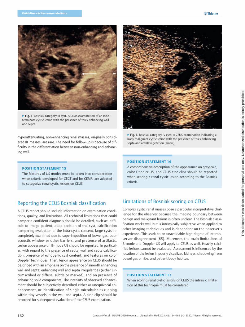
▪ Category III – Indeterminate lesions. Cystic lesions withenhancing smooth thick (≥ 4mm) wall or septa, and/or withenhancing irregular (> 3mm) walls and/or septa (▶ Fig. 5).No nodules are seen [6, 15, 36, 58, 68, 70, 73].
▪ Category IV – Likely malignant cystic tumors. Cystic lesionswith the characteristics of category III cysts, which also containenhancing soft-tissue protrusions (▶ Fig. 6), either noduleswith obtuse margins (≥ 4mm), or with acute margins of anysize [6, 15, 36, 63, 70, 73].
POSITION STATEMENT 14
The likelihood of malignancy of complex renal cystic lesions
can be assessed using CEUS-based criteria.
▶ Fig. 1 Bosniak category I cyst. A The cyst (arrow) shows anechoic content with posterior acoustic enhancement, a thin (< 2mm) border with sharpmargins, no irregularities, calcifications, or septa. Simple cysts are fully characterized as benign on B-mode US.B CEUS is not needed to confirm thefindings, but if an incidental cyst is encountered during an examination for a separate renal lesion, the findings on B-mode US is confirmed.
▶ Fig. 2 Bosniak category II cyst A Minimally complex benign cyst displaying 2 thin septa (arrows) without irregularities. Category II cyst not re-quiring further investigation on CEUS.B Cyst displaying thin wall calcifications (arrow). Category II cyst not requiring further investigation on CEUS.C CEUS is not needed to confirm the findings, but if an incidental septated cyst is encountered during an examination for a separate renal lesion, thevascularization of the septa (arrow) is clearly identified as thin enhancing linear areas within the cyst.
160 Cantisani V et al. EFSUMB 2020 Proposal… Ultraschall in Med 2021; 42: 154–166 | © 2020. Thieme. All rights reserved.
Guidelines & Recommendations
Thi
s do
cum
ent w
as d
ownl
oade
d fo
r pe
rson
al u
se o
nly.
Una
utho
rized
dis
trib
utio
n is
str
ictly
pro
hibi
ted.

Specific points regarding the CEUS Bosniakclassification
Cysts with thick or nodular calcifications, without hampering eval-uation of cyst content, are placed in category IIF; calcification isnot a sign of malignancy provided that there is no associated sus-picious lesion [18, 51, 73]. There is no definition for a thresholdfor labelling a septum as ‘thick’ [32]; septa ≤ 1mm are consideredthin, by the majority of investigators [6, 62, 70, 73]. Others sug-gest a 2mm threshold [8, 51, 72, 74]. A “hairline septum” is a
subjective assessment, dependent on the US equipment used,for which a precise thickness threshold cannot be assigned effec-tively. There is no threshold to differentiate between “few septa”and “multiple septa” with an arbitrary threshold at three septa.“Few” equals 1–3 and “many” is ≥ 4 septa [16, 32, 75]. Lesionsize is not a consideration for cyst categorization with conflictingresults for predicting malignancy [76–80]. On CECT, totally intra-renal non-enhancing high-attenuation cysts < 3 cm are assignedto category II, while cysts with the same characteristics > 3 cmare assigned to category IIF [12]. Large (≥ 3 cm) homogeneous,
▶ Fig. 3 Bosniak category IIF cyst A B-mode US shows a lesion with mixed appearance, with both a solid (arrow) and cystic component (arrowhead).B CEUS examination demonstrates no enhancement within the lesion, a thin wall without irregularities (arrow), and no septa.
▶ Fig. 4 Bosniak category IIF cyst. A CEUS examination of an intrarenal cyst with multiple, minimally thickened, enhancing septa (arrows). B CEUSexamination of an intrarenal cyst with a focal thickening (2mm) of the wall (arrow).
161Cantisani V et al. EFSUMB 2020 Proposal… Ultraschall in Med 2021; 42: 154–166 | © 2020. Thieme. All rights reserved.
Thi
s do
cum
ent w
as d
ownl
oade
d fo
r pe
rson
al u
se o
nly.
Una
utho
rized
dis
trib
utio
n is
str
ictly
pro
hibi
ted.
hyperattenuating, non-enhancing renal masses, originally consid-ered IIF masses, are rare. The need for follow-up is because of dif-ficulty in the differentiation between non-enhancing and enhanc-ing wall.
POSITION STATEMENT 15
The features of US modes must be taken into consideration
when criteria developed for CECT and for CEMRI are adapted
to categorize renal cystic lesions on CEUS.
Reporting the CEUS Bosniak classification
A CEUS report should include information on examination condi-tions, quality, and limitations. All technical limitations that couldhamper a confident diagnosis should be detailed, such as: diffi-cult-to-image patient, deep position of the cyst, calcificationhampering evaluation of the intra-cystic content, large cysts in-completely examined due to superimposition of bowel gas, pooracoustic window or other barriers, and presence of artifacts.Lesion appearance on B-mode US should be reported, in particul-ar, with regard to the presence of septa, wall and septa calcifica-tion, presence of echogenic cyst content, and features on colorDoppler techniques. Then, lesion appearance on CEUS should bedescribed with an emphasis on the presence of smooth enhancingwall and septa, enhancing wall and septa irregularities (either cir-cumscribed or diffuse, subtle or marked), and on presence ofenhancing solid components. The intensity of observed enhance-ment should be subjectively described either as unequivocal en-hancement, or identification of single microbubbles runningwithin tiny vessels in the wall and septa. A cine clip should berecorded for subsequent evaluation of the CEUS examination.
POSITION STATEMENT 16
A comprehensive description of the appearance on grayscale,
color Doppler US, and CEUS cine clips should be reported
when scoring a renal cystic lesion according to the Bosniak
criteria.
Limitations of Bosniak scoring on CEUS
Complex cystic renal masses pose a particular interpretative chal-lenge for the observer because the imaging boundary betweenbenign and malignant lesions is often unclear. The Bosniak classi-fication works well but is intrinsically subjective when applied toother imaging techniques and is dependent on the observer’sexperience. This leads to an unavoidable high degree of interob-server disagreement [65]. Moreover, the main limitations ofB-mode and Doppler US will apply to CEUS as well. Heavily calci-fied lesions cannot be evaluated. Assessment is influenced by thelocation of the lesion in poorly visualized kidneys, shadowing frombowel gas or ribs, and patient body habitus.
POSITION STATEMENT 17
When scoring renal cystic lesions on CEUS the intrinsic limita-
tion of this technique must be considered.
▶ Fig. 5 Bosniak category III cyst. A CEUS examination of an inde-terminate cystic lesion with the presence of thick enhancing walland septa.
▶ Fig. 6 Bosniak category IV cyst. A CEUS examination indicating alikely malignant cystic lesion with the presence of thick enhancingsepta and a wall vegetation (arrow).
162 Cantisani V et al. EFSUMB 2020 Proposal… Ultraschall in Med 2021; 42: 154–166 | © 2020. Thieme. All rights reserved.
Guidelines & Recommendations
Thi
s do
cum
ent w
as d
ownl
oade
d fo
r pe
rson
al u
se o
nly.
Una
utho
rized
dis
trib
utio
n is
str
ictly
pro
hibi
ted.

Controversies with CEUS of Bosniak Categories
Does the CEUS-based classification upgradeor downgrade the risk of malignancy of renalcystic lesions?
The presence of enhancement, indicating neovascularization, isthe most important factor in determining the need for surgery incystic renal lesions [12, 13]. Improved CT resolution when com-pared to the original CECT categories has resulted in fewer inde-terminate cyst findings and increased specificity [81]. With CEUS,31 % of renal cysts were attributed a higher Bosniak categorycompared to CECT [7, 8, 13, 56, 65]. The increased contrast en-hancement and better temporal and spatial resolution of CEUS(and MR imaging) demonstrate previously undetected features.Minimal septa enhancement is not indicative of malignancy, andan increased sensitivity of CEUS demonstrating enhancing no-dules not seen with CECT has been noted [56, 82]. A similar higherBosniak category with MR imaging has been seen [10, 28, 32] butan apparent wall thickening artifact is an issue [66]. Both upgrad-ing and downgrading of Bosniak categories with MRI and CEUScompared to CT imaging is apparent in > 20% of cases [25], withCEUS demonstrating lower specificity but improved sensitivityand accuracy compared to MR imaging [57].
POSITION STATEMENT 18
The imaging criteria used to assign the Bosniak categories are
developed for CECT and must be adapted to be successfully
applied for scoring on CEMRI and CEUS, as changes in cate-
gory will occur.
Should CEUS be considered an equivalent,complimentary, or alternative technique to contrast-enhanced CT for renal cystic lesions?
Although CECT is the reference standard for Bosniak categories ofrenal cystic malignancy risk, CECT is inherently inaccurate, with areported sensitivity of 89.6 % and specificity of 65.1 % in distin-guishing between benign and malignant renal cysts [20]. Thecomparability of CEUS with the reference standard of CECT hasbeen addressed with excellent agreement [7, 56, 83] with a singlestudy indicating that experience in CEUS interpretation is crucial[65]. There was a potential for CEUS to overestimate the Bosniakcategory, with the ‘real-time’ examination able to demonstrateminor enhancement (a marker of malignant potential). The cur-rent view suggests that this is an advantage, rather than a draw-back. This requires, however, a fundamental change in imagingassessment of renal cysts, centered on CEUS demonstration of le-sion vascularity [5]. When CEUS is inconclusive due to poor visua-lization (i. e., due to patient habitus or poor acoustic window),CECT usually permits better characterization and furthermoreallows staging of a malignant renal lesion. There is better demon-stration of calcification on CECT which could affect the Bosniak
category on US [18, 84]. Using CEUS only in cases of contraindica-tions or non-acceptance of CECT is not justified based on the cur-rent knowledge of the potential of this technique and would bedetrimental to acquiring further cumulative experience in CEUS.
Characteristics of non-progressive BosniakCategory IIF cysts
The initial cyst size, change in lesion size (increase or decrease),and growth rate [growth rate = (follow-up size minus initial size)/years between measurements] were not found to correlate withprogression. A multilobulated border of the lesion was not foundto correlate with progression and no lesions with calcification pro-gressed. Growth rates in cystic lesions are often a consequence offluid accumulation (downgrading to a Bosniak category II). Pro-gression to malignancy is based on the appearance of enhancingsolid portions, an increase in number, thickness or irregularity ofenhancing septa, and on an increase in thickness of the enhancingwall [85]. There is no difference in progression to malignancy onfollow-up CECT imaging compared with MR imaging [16, 86–88].When there is indeterminate enhancement on CECT, CEMR orCEUS imaging can be the next imaging stage [59, 89, 90].
POSITION STATEMENT 19
Follow-up of cystic renal lesions can be carried out effectively
with CECT, CEMR, or CEUS imaging. The current evidence
shows similar performance for the three techniques.
When should CEUS be supplemented by CTor MR imaging for follow-up?
A CEUS examination is suited for the follow-up of nonsurgical le-sions to detect any morphologic changes such as thickening ofsepta, appearance of a solid nodule, or contrast-enhanced altera-tions indicative of progression of the disease. A CEUS examinationhas at least the same diagnostic accuracy as CECT for renal cystcategorization but image acquisition is influenced by the locationof the lesion in poorly visualized kidneys, shadowing from bowelgas or ribs, patient’s constitution, and wall calcification [29, 57,91]. Smaller lesions localized within the renal parenchyma maybe difficult to characterize with CEUS, as these lesions often disap-pear (‘masked’) during a CEUS examination due to the prominentvascularity of the renal cortex, with the possibility of a lower doseof UCA being helpful. With these issues, further CTor MR imagingis necessary [25].
POSITION STATEMENT 20
CEUS-based Bosniak categorization must be supplemented by
CT or MR imaging when there is inadequate visualization of
the cystic renal lesion.
163Cantisani V et al. EFSUMB 2020 Proposal… Ultraschall in Med 2021; 42: 154–166 | © 2020. Thieme. All rights reserved.
Thi
s do
cum
ent w
as d
ownl
oade
d fo
r pe
rson
al u
se o
nly.
Una
utho
rized
dis
trib
utio
n is
str
ictly
pro
hibi
ted.

Conclusion
The Bosniak categorization was originally formulated for CECT,and then applied to MR imaging and CEUS without adapting thecriteria developed primarily for CT. With differences in imagingspecifics, an inaccurate assignment of the Bosniak category andultimately inappropriate treatment could result from the CEUSand MR imaging interpretation. The criteria used to assign theBosniak category scores on CEUS have been reviewed, redefined,and standardized, taking into account the unique characteristicsof the CEUS examination. The aim is to allow for clearly definedcriteria allowing for a better assessment of the performance ofCEUS in the categorization of complex renal cysts. The CEUS cate-gorization is not intended to replace, but rather to complementthe current Bosniak CT categorization, thereby improving itsaccuracy in the assessment of malignancy in each category. TheBosniak categorization system is used worldwide and provides acommon language but is intrinsically subjective, a shortcomingwhich will likely be reduced when using the redefined scoringcriteria which include a CEUS examination.
Conflict of interest
Vito Cantisani: lecturer fees from Bracco, Samsung, Toshiba.Paul Sidhu: lecture fees from Bracco.Adrian Saftoiu: lecture fees from Bracco.Dirk-André Clevert: speaker bureau Bracco, Siemens, Esaote, Samsung,Philips.
Acknowledgements
We would like to thank Dr D. Fresilli for his help during all the phasesrelated to the submission.
References
[1] Parienty RA, Pradel J, Parienty I. Cystic renal cancers: CT characteristics.Radiology 1985; 157: 741–744
[2] Warren KS, McFarlane J. The Bosniak classification of renal cystic masses.BJU International 2005; 95: 939–942
[3] Bosniak MA. The current radiological approach to renal cysts. Radiology1986; 158: 1–10
[4] Sidhu PS, Cantisani V, Dietrich CF et al. The EFSUMB Guidelines and Re-commendations for the Clinical Practice of Contrast-Enhanced Ultrasound(CEUS) in Non-Hepatic Applications: Update 2017 (Short Version).Ultraschall in Med 2018; 39: 154–180
[5] Barr RG, Peterson C, Hindi A. Evaluation of indeterminate renal massesswith contras-enhanced US: a diagnostic performance study. Radiology2014; 271: 133–142
[6] Rubenthaler J, Bogner F, Reiser M et al. Contrast-Enhanced Ultrasound(CEUS) of the Kidneys by Using the Bosniak Classification. Ultraschall inMed 2017; 37: 234–251
[7] Ascenti G, Mazziotti S, Zimbaro G et al. Complex Cystic Renal Masses:Characterization with Contrast-enhanced US. Radiology 2007; 243: 158–165
[8] Quaia E, Bertolotto M, Cioffi V et al. Comparison of Contrast-EnhancedSonography with Unenhanced Sonography and Contrast-Enhanced CT inthe Diagnosis of Malignancy in Complex Cystic Renal Masses. Am JRoentgenol 2008; 191: 1239–1249
[9] Jenssen C, Gilja OH, Serra AL et al. European Federation of Societies forUltrasound in Medicine and Biology (EFSUMB) Policy Document Devel-opment Strategy: Clinical Practice Guidelines, Position Statements andTechnological Reviews. Ultrasound Int Open 2019; 05: E2–E10
[10] Hindman NM. Imaging of Cystic Renal Masses. Urologic Clinics of NorthAmerica 2018; 45: 331–349
[11] Kazmierski B, Deurdulian C, Tchelepi H et al. Applications of contrast-enhanced ultrasound in the kidney. Abdominal Radiology 2017; 43:880–898
[12] Israel GM, Bosniak MA. An update of the Bosniak renal cyst classificationsystem. Urology 2005; 66: 484–488
[13] Robbin ML, Lockhart ME, Barr RG. Renal imaging with ultrasound con-trast: current status. Radiologic Clinics of North America 2003; 41: 963–978
[14] Barr RG. Is there a need to modify the Bosnaik renal mass classificationwith the addition of contrast-enhanced sonography? J Ultrasound Med2017; 36: 865–868
[15] Chang EH, Chong WK, Kasoji SK et al. Diagnostic accuracy of contrast-enhanced ultrasound for characterization of kidney lesions in patientswith and without chronic kidney disease. BMC Nephrol 2017; 18: 266
[16] Silverman SG, Pedrosa I, Ellis JH et al. Bosniak Classification of CysticRenal Masses, Version 2019: An Update Proposal and Needs Assess-ment. Radiology 2019; 292: 475–488
[17] Bosniak MA. The Bosniak Renal Cyst Classification: 25 Years Later.Radiology 2012; 262: 781–785
[18] Israel GM, Bosniak MA. Calcification in Cystic Renal Masses: Is It Importantin Diagnosis? Radiology 2003; 226: 47
[19] Israel GM, Bosniak MA. How I Do It: Evaluating Renal Masses. Radiology2005; 236: 441–450
[20] Sevcenco S, Spick C, Helbich TH et al. Malignancy rates and diagnosticperformance of the Bosniak classification for the diagnosis of cystic renallesions in computed tomography – a systematic review and meta-analysis. Eur Radiol 2017; 27: 2239–2247
[21] Schoots IG, Zaccai K, Hunink MG et al. Bosniak Classification for ComplexRenal Cysts Reevaluated: A Systematic Review. Journal of Urology 2017;198: 12–21
[22] Ljungberg B, Albiges L, Abu-Ghanem Y et al. European Association ofUrology Guidelines on Renal Cell Carcinoma: The 2019 Update. EuropeanUrology 2019; 75: 799–810
[23] Silverman SG, Israel GM, Herts BR et al. Management of the IncidentalRenal Mass. Radiology 2008; 249: 16–31
[24] Graumann O, Osther SS, Karstoft J et al. Bosniak classification system:inter-observer and intra-observer agreement among experienceduroradiologists. Acta Radiologica 2015; 56: 374–383
[25] Graumann O, Osther SS, Karstoft J et al. Bosniak classification system: aprospective comparison of CT, contrast-enhanced US, and MR for cate-gorizing complex renal cystic masses. Acta Radiologica 2015; 57: 1409–1417
[26] Siegel CL, McFarland EG, Brink JA et al. CTof cystic renal masses: analysisof diagnostic performance and interobserver variation. Am J Roentgenol1997; 169: 813–818
[27] Israel GM, Hindman N, Bosniak MA. Evaluation of Cystic Renal Masses:Comparison of CT and MR Imaging by Using the Bosniak ClassificationSystem. Radiology 2004; 231: 365–371
[28] Zhong J, Cao F, Guan X et al. Renal cyst masses (Bosniak category II–III)may be over evaluated by the Bosniak criteria based on MR findings.Medicine (Baltimore) 2017; 96: e9361
[29] Zhou L, Tang L, Yang T et al. Comparison of contrast-enhanced ultra-sound with MRI in the diagnosis of complex cystic renal masses: a meta-analysis. Acta Radiologica 2018; 59: 1254–1263
164 Cantisani V et al. EFSUMB 2020 Proposal… Ultraschall in Med 2021; 42: 154–166 | © 2020. Thieme. All rights reserved.
Guidelines & Recommendations
Thi
s do
cum
ent w
as d
ownl
oade
d fo
r pe
rson
al u
se o
nly.
Una
utho
rized
dis
trib
utio
n is
str
ictly
pro
hibi
ted.

[30] Hecht EM, Israel GM, Krinsky GA et al. Renal Masses: Quantitative Anal-ysis of Enhancement with Signal Intensity Measurements versus Quali-tative Analysis of Enhancement with Image Subtraction for DiagnosingMalignancy at MR Imaging. Radiology 2004; 232: 373–378
[31] Balci NC, Semelka RC, Patt RH et al. Complex renal cysts: findings on MRimaging. Am J Roentgenol 1999; 172: 1495–1500
[32] Ferreira AM, Reis RB, Kajiwara PP et al. MRI evaluation of complex renalcysts using the Bosniak classification: a comparison to CT. AbdominalRadiology 2016; 41: 2011–2019
[33] Weibl P, Klatte T, Kollarik B et al. Interpersonal variability and presentdiagnostic dilemmas in Bosniak classification system. ScandinavianJournal of Urology and Nephrology 2011; 45: 239–244
[34] Inci E, Hocaoglu E, Aydin S et al. Diffusion-weighted magnetic resonanceimaging in evaluation of primary solid and cystic renal masses using theBosniak classification. European Journal of Radiology 2012; 81: 815–820
[35] Dietrich CF, Nolsøe CP, Barr RG et al. Guidelines and Good Clinical Prac-tice Recommendations for Contrast Enhanced Ultrasound (CEUS) in theLiver – Update 2020 – WFUMB in Cooperation with EFSUMB, AFSUMB,AIUM, and FLAUS. Ultraschall in Med 2020; 41: 562–585
[36] Harvey CJ, Alsafi A, Kuzmich S et al. Role of US contrast agents in theassessment of indeterminate solid and cystic lesions in native andtransplant kidneys. Radiographics 2015; 35: 1419–1430
[37] Piscaglia F, Bolondi L. The safety of SonoVue in abdominal applications:retrospective analysis of 23188 investigations. Ultrasound in Med Biol2006; 32: 1369–1375
[38] Main ML. Ultrasound contrast agent safety: from anecdote to evidence.JACC Cardiovasc Imaging 2009; 2: 1057–1059
[39] Main ML, Goldman JH, Grayburn PA. Thinking Outside the box: TheUltrasound Contrast Controversy. J Am Coll Cardiol 2007; 50: 2434–2437
[40] Tang C, Fang K, Guo Y et al. Safety of Sulfur Hexafluoride Microbubblesin Sonography of Abdominal and Superficial Organs: RetrospectiveAnalysis of 30222 Cases. J Ultrasound Med 2017; 36: 531–538
[41] Kitzman DW, Goldman ME, Gilliam LD et al. Efficacy and safety of thenovel ultrasound contrast agent perflutren (Definity) in patients withsuboptimal baseline left ventricular echocardiographic images. Am JCardiol 2000; 86: 669–674
[42] Wilson SR, Burns PN. Microbubble-enhanced US in body imaging: Whatrole? Radiology 2010; 257: 24–39
[43] ter Haar G. Ultrasonic contrast agents: safety considerations reviewed.Eur J Radiol 2002; 41: 217–221
[44] Girometti R, Stocca E, Granata A et al. Impact of contrast-enhancedultrasound in patients with renal function impairment. World J Radiol2017; 28: 10–16
[45] Putz FJ, Erlmeier A, Wiesinger I et al. Contrast-enhanced ultrasound(CEUS) in renal imaging at an interdisciplinary ultrasound centre:Possibilities of dynamic microvascularisation and perfusion. ClinHemorheol Microcircl 2017; 66: 293–302. doi:10.3233/CH-179103
[46] Yusuf GT, Sellars ME, Deganello A et al. Retrospective Analysis of theSafety and Cost Implications of Pediatric Contrast-Enhanced Ultrasoundat a Single Center. Am J Roentgenol 2016; 208: 446–452
[47] Sidhu PS, Cantisani V, Deganello A et al. Role of contrast-enhancedultrasound (CEUS) in paediatric practice: An EFSUMB positionstatement. Ultraschall in Med 2017; 38: 33–43
[48] Education and Practical Standards Committee E. Minimal Training recom-mendations for the practice of medical ultrasound. Ultraschall in Med2006; 27: 79–95
[49] Education and Practical Standards Committee E. Minimum TrainingRequirements for the Practice of Medical Ultrasound in Europe.Appendix 14: (CEUS) Contrast Enhanced Ultrasound. Ultraschall in Med2010; 31: 426–427
[50] Jacobsen N, Nolsoe CP, Konge L et al. Contrast-Enhanced Ultrasound:Development of Syllabus for Core Theoretical and Practical Competen-cies. Ultrasound in Medicine & Biology 2020; 46: 2287–2292
[51] Tay SY, Tiu CM, Hu B et al. Characterization and management of variousrenal cystic lesions by sonographic features. Journal of the ChineseMedical Association 2018; 81: 1017–1026
[52] Deganello A, Sellars ME, Yusuf GT et al. How Much Should I RecordDuring a CEUS Examination? Practical Aspects of the Real-Time Featureof a Contrast Ultrasound Study. Ultraschall in Med 2018; 39: 484–486
[53] Correas JM, Claudon M, Tranquart F et al. The kidney: imaging withmicrobubble contrast agents. Ultrasound Q 2006; 22: 53–66
[54] Tsuruoka K, Yasuda T, Koitabashi K et al. Evaluation of Renal Microcircu-lation by Contrast-Enhanced Ultrasound With SonazoidTM as a ContrastAgent Comparison Between Normal Subjects and Patients With ChronicKidney Disease. Int Heart J 2010; 51: 176–182
[55] Narayanasamy S, Krishna S, Prasad Shanbhogue AK et al. Contemporaryupdate on imaging of cystic renal masses with histopathological corre-lation and emphasis on patient management. Clinical Radiology 2019;74: 83–94
[56] Park BK, Kim B, Kim SH et al. Assessment of cystic renal masses based onBosniak classification: comparison of CT and contrast-enhanced US. Eur JRadiol 2007; 61: 310–314
[57] Chen Y, Wu N, Xue T et al. Comparison of contrast-enhanced sonogra-phy with MRI in the diagnosis of complex cystic renal masses. Journal ofClinical Ultrasound 2015; 43: 203–209
[58] Edenberg J, Gloersen K, Osman HA et al. The role of contrast-enhancedultrasound in the classification of CT-indeterminate renal lesions. Scan-dinavian Journal of Urology 2016; 50: 445–451
[59] Bertolotto M, Cicero C, Perrone R et al. Renal masses with equivocal en-hamcement at CT: characterization with contrast-enhanced ultrasound.Am J Roentgenol 2015; 205: W557–W565
[60] Siddaiah M, Krishna S, McInnes MDF et al. Is Ultrasound Useful forFurther Evaluation of Homogeneously Hyperattenuating Renal LesionsDetected on CT? Am J Roentgenol 2017; 209: 604–610
[61] Gulati M, King KG, Gill IS et al. Contrast-enhanced ultrasound (CEUS) ofcystic and solid renal lesions: a review. Abdominal Imaging 2015; 40:1982–1996
[62] Defortescu G, Cornu JN, Bejar S et al. Diagnostic performance ofcontrast-enhanced ultrasonography and magnetic resonance imagingfor the assessment of complex renal cysts: A prospective study. Int J Urol2017; 24: 184–189
[63] Nicolau C, Bunesch L, Sebastia C. Renal complex cysts in adults: contrast-enhanced ultrasound. Abdominal Imaging 2011; 36: 742–752
[64] Clevert DA, Minaifar N, Weckbach S et al. Multislice computed tomog-raphy versus contrast-enhanced ultrasound in evaluation of complexcystic renal masses using the Bosniak classification system. Clin Hemor-heol Microcirc 2008; 39: 171–178
[65] Ragel M, Nedumaran A, Makowska-Webb J. Prospective comparison ofuse of contrast-enhanced ultrasound and contrast-enhanced computedtomography in the Bosniak classification of complex renal cysts.Ultrasound 2016; 24: 6–16
[66] Helenon O, Crosnier A, Verkarre V et al. Simple and complex renal cystsin adults: Classification system for renal cystic masses. Diagnostic andInterventional Imaging 2018; 99: 189–218
[67] Ignee A, Straub B, Brix D et al. The value of contrast enhanced ultrasound(CEUS) in the characterisation of patients with renal masses. ClinHemorheol Microcirc 2010; 46: 275–290
[68] Oon SF, Foley RW, Quinn D et al. Contrast-enhanced ultrasound of thekidney: a single-institution experience. Irish Journal of Medical Science(1971) 2018; 187: 795–802
[69] Israel GM, Bosniak MA. Pitfalls in Renal Mass Evaluation and How toAvoid Them. Radiographics 2008; 28: 1325–1338
165Cantisani V et al. EFSUMB 2020 Proposal… Ultraschall in Med 2021; 42: 154–166 | © 2020. Thieme. All rights reserved.
Thi
s do
cum
ent w
as d
ownl
oade
d fo
r pe
rson
al u
se o
nly.
Una
utho
rized
dis
trib
utio
n is
str
ictly
pro
hibi
ted.

[70] Nicolau C, Bunesch L, Pano B et al. Prospective evaluation of CT indeter-minate renal masses using US and contrast-enhanced ultrasound.Abdominal Imaging 2015; 40: 542–551
[71] Rubenthaler J, Mueller-Peltzer K, Negrúo de Figueiredo G et al. CEUSdiagnostic workup of cystic renal lesions. Der Radiologe 2018; 58: 545–552
[72] Xue LY, Lu Q, Huang BJ et al. Contrast-enhanced ultrasonography forevaluation of cystic renal mass: in comparison to contrast-enhanced CTand conventional ultrasound. Abdominal Imaging 2014; 39: 1274–1283
[73] Rubenthaler J, Negrao de Figueiredo G, Mueller-Peltzer K et al. Evaluationof renal lesions using contrast-enhanced ultrasound (CEUS); a 10-yearretrospective European single-centre analysis. European Radiology 2018;28: 4542–4549
[74] Xu Y, Zhang S, Wei X et al. Contrast enhanced ultrasonography predic-tion of cystic renal mass in comparison to histopathology; 2014; 58:429–438
[75] Destefani MH, Elias J Jr, Serra Negra Trazzi AM et al. Minimally ComplexRenal Cysts: Outcomes and Ultrasound Evaluation Compared withContrast-Enhanced Cross-Sectional Imaging Bosniak Classification.Ultrasound in Medicine and Biology 2017; 43: 2167–2173
[76] Han HH, Choi KH, Oh YT et al. Differential diagnosis of complex renalcysts based on lesion size along with the Bosniak renal cyst classification.Yonsei Med J 2012; 53: 729–733
[77] Goenka AH, Remer EM, Smith AD et al. Development of a ClinicalPrediction Model for Assessment of Malignancy Risk in Bosniak III RenalLesions. Urology 2013; 82: 630–635
[78] Hindman NM. Approach to Very Small (< 1.5 cm) Cystic Renal Lesions:Ignore, Observe, or Treat? Am J Roentgenol 2015; 204: 1182–1189
[79] Oh TH, Lee YH, Seo IY. Diagnostic Efficacy of Contrast-EnhancedUltrasound for Small Renal Masses. Korean J Urol 2014; 55: 587–592
[80] Lam CJ, Kapoor A. The true malignancy risk of Bosniak III cystic renallesions: Active surveillance or surgical resection? Can Urol Assoc J 2018;2018/02/23: E276–E280
[81] Lang EK, Macchia RJ, Gayle B et al. CT-guided biopsy of indeterminaterenal cystic masses (Bosniak 3 and 2F): accuracy and impact on clinicalmanagement. European Radiology 2002; 12: 2518–2524
[82] Sanz E, Hevia V, Gomez V et al. Renal Complex Cystic Masses: Usefulnessof Contrast-Enhanced Ultrasound (CEUS) in Their Assessment and ItsAgreement with Computed Tomography. Current Urology Reports2016; 17: 89
[83] Clevert DA, Bensler S, Stickel M et al. Contrast enhanced ultrasoundeases interpretation of an unclear renal tumor in addition to CT, MRI andhistological findings – A case report in a young patient. Clin HemorrMicrocircul 2007; 36: 313–318
[84] Herts BR, Silverman SG, Hindman NM et al. Management of the Inci-dental Renal Masses;on CT: A White Paper of the ACR; Incidental Find-ings Committee. Journal of the American College of Radiology 2018; 15:264–273
[85] Hwang JH, Lee CK, Yu HS et al. Clinical Outcomes of Bosniak Category IIFComplex Renal Cysts in Korean Patients. Korean J Urol 2012; 2012/06/19: 386–390
[86] El-Mokadem I, Budak M, Pillai S et al. Progression, interobserver agree-ment, and malignancy rate in complex renal cysts (> Bosniak category IIF).Urologic Oncology: Seminars and Original Investigations 2014; 32: 24
[87] O’Malley RL, Godoy G, Hecht EM et al. Bosniak Category IIF Designationand Surgery for Complex Renal Cysts. Journal of Urology 2009; 182:1091–1095
[88] Smith AD, Remer EM, Cox KL et al. Bosniak Category IIF and III CysticRenal Lesions: Outcomes and Associations. Radiology 2012; 262: 152–160
[89] Graumann O, Osther SS, Karstoft J et al. Evaluation of Bosniak category IIFcomplex renal cysts. Insights Imaging 2013; 4: 471–480
[90] Adey GS, Pedrosa I, Rofsky NM et al. Lower Limits of Detection UsingMagnetic Resonance Imaging for Solid Components in Cystic RenalNeoplasms. Urology 2008; 71: 47–51
[91] Lan D, Qu HC, Li N et al. The Value of Contrast-Enhanced Ultrasonogra-phy and Contrast-Enhanced CT in the Diagnosis of Malignant RenalCystic Lesions: A Meta-Analysis. PLoS ONE 2016; 11: e0155857
166 Cantisani V et al. EFSUMB 2020 Proposal… Ultraschall in Med 2021; 42: 154–166 | © 2020. Thieme. All rights reserved.
Guidelines & Recommendations
Thi
s do
cum
ent w
as d
ownl
oade
d fo
r pe
rson
al u
se o
nly.
Una
utho
rized
dis
trib
utio
n is
str
ictly
pro
hibi
ted.