1 Zofia Goc, 1 Monika Martiniaková, 2 Radoslav Omelka, 2 ... · Magdalena Semla,1 Zofia Goc,1...
31
Acrylamide a common food toxin related to physiological functions and health Magdalena Semla, 1 Zofia Goc, 1 Monika Martiniaková, 2 Radoslav Omelka, 2 Grzegorz Formicki 1 * 1 Pedagogical University of Cracow, Institute of Biology, Podbrzezie 3, 31-054 Kraków, Poland 2 Constantine the Philosopher University in Nitra, Faculty of Natural Sciences, Nábrežie mládeže 91, 949 74 Nitra, Slovak Republic *Corresponding author: Grzegorz Formicki, Institute of Biology, Pedagogical University of Cracow, Podbrzezie 3, 31-054 Kraków, Poland Short title: Physiological effects of acrylamide
Transcript of 1 Zofia Goc, 1 Monika Martiniaková, 2 Radoslav Omelka, 2 ... · Magdalena Semla,1 Zofia Goc,1...

Acrylamide a common food toxin related to physiological functions and health
Magdalena Semla,1 Zofia Goc,1 Monika Martiniaková,2 Radoslav Omelka,2 Grzegorz
Formicki1*
1 Pedagogical University of Cracow, Institute of Biology, Podbrzezie 3, 31-054 Kraków,
Poland
2 Constantine the Philosopher University in Nitra, Faculty of Natural Sciences, Nábrežie
mládeže 91, 949 74 Nitra, Slovak Republic
*Corresponding author: Grzegorz Formicki, Institute of Biology, Pedagogical University of
Cracow, Podbrzezie 3, 31-054 Kraków, Poland
Short title: Physiological effects of acrylamide
Zdenka.Stadnikova
Pre-press

Summary
Acrylamide (AA) is a highly reactive organic compound capable of polimeryzation to form
polyacrylamide, which is commonly used throughout a variety of industries. Given its toxic
effect on humans and animals, the last 20 years have seen an increased interest in research
devoted to the AA. One of the main sources of AA is food. AA appears in heated food
following the reaction between aminoacids and reduced sugars. Large concentrations of AA
can be found in popular staples such as coffee, bread or potato products. An average daily
consumption of AA is between 0.3-2.0 µg/kg b.w. Inhalation of acrylamide is related with
occupational exposure. AA delivered with food is metabolized in the liver by cytochrome
P450. AA biotransformation and elimination results in formation of toxic glycidamide (GA).
Both, AA and GA can be involved in the coupling reaction with the reduced glutathione
(GSH) forming glutathione conjugates which are excreted with urine. Biotransformation of
AA leads to the disturbance in the redox balance. Numerous research proved that AA and GA
have significant influence on physiological functions including signal propagation in
peripheral nerves, enzymatic and hormonal regulation, functions of muscles, reproduction etc.
In addition AA and GA show neurotoxic, genotoxic and cancerogenic properties. In 1994,
International Agency for Research on Cancer (IARC) classified acrylamide as a potentially
carcinogenic substance to human.
Key words: acrylamide, neurotoxicity, genotoxicity, reproductive toxicity, oxidative stress

Introduction
Food should deliver all the ingredients necessary for the organism to function
properly. Organic and inorganic compounds present in food are used by the organism as
energetic, regulatory and/or building substances. Unfortunately, food consumed by people is
often a source of harmful substances. Acrylamide (AA) is one of the most common toxins in
food. It occurs in food containing high concentrations of hydrocarbons subjected to high
temperature (Mottram et al., 2002). High concentration of acrylamide may be found in food
products such as potato chips, fried potatoes, cornflakes or bread. Thus acrylamide is present
in every day diet of most people. To make matters worse, some of the products containing
acrylamide are attractive to children and young people.
In 2001, the Scientific Committee on Toxicity, Ecotoxicity and the Environment
demonstrated its neurotoxicity, genotoxicity, carcinogenicity and reproductive toxicity
(Keramat et al., 2011; Carere, 2006). Toxic effects of acrylamide are mediated by the
formation of genotoxic metabolites, oxidative stress, affected propagation of neural signals,
ultrastructural and histological defects in central neural system (LoPachin, 2004; El-Sayyad et
al., 2011; Pingot et al., 2013). The International Agency for Research on Cancer (IARC,
1994) classified acrylamide as potentially carcinogenic substance to human.
Most of the mechanisms of AA toxicity are well recognized. Nonetheless some
aspects of acrylamide toxicity remain still unclear. First of all, the doses used in animal
experiments are much higher than mean acrylamide intake in food. Thus it is not known
whether acrylamide ingested with every day diet pose real risk to consumers’ health. On the
other hand, the known incidences of occupational exposure were probably related with the
intoxication with very high acrylamide doses (Pennisi et al., 2013). The studies conducted in
chemical plants revealed that personal breathing zone air samples contained up to 984 µg of
AA per m3 of air. Acrylamide and polyacrylamide production operators had hemoglobin AA
adducts in the concentration up to 1884 pmol/g, whereas hemoglobin AA adducts level in
administrative workers was 97.9 pmol/g (Moorman et al., 2012). It is still unclear whether
toxic effects of chronic exposure to acrylamide may accumulate in the organism in the long
term. Very little is known about the risk of fetal exposure to AA and their potential effects to
the prenatal and postnatal development. Thus the aim of this article is to bring together data
from different studies in order to analyze the risk of exposure to AA and related health risks.
Acrylamide in food
Acrylamide (CH2=CH–CO–NH2, according to IUPAC: 2-propenamid) is a highly
reactive, organic, white and crystal substance, with molecular weight of 71.08 g (Żyżelewicz
et al., 2010). AA is a polar substance which easily dissolves in water or other polar solvents
e.g. in methanol or ethanol (Jankowska et al., 2009). High reactivity of AA is connected with
the double bond and amide group. The compound may create hydrogen bonds and can react
both with amide and vinyl groups (Girma et al., 2005; Żyżelewicz et al., 2010). Acrylamide is
polymerized under the influence of temperature and UV radiation. These reactions result in
creation of new chemical compounds called polyacrylamides.
Recent years revealed a considerable increase in investigation of acrylamide as a
potentially dangerous substance to people. In early 2000s, Swedish researchers proved that
certain foods might contain large concentrations of acrylamide (Lofstedt, 2003). The research
by Tareke et al. (2002) indicated that food processing has influence on acrylamide formation.
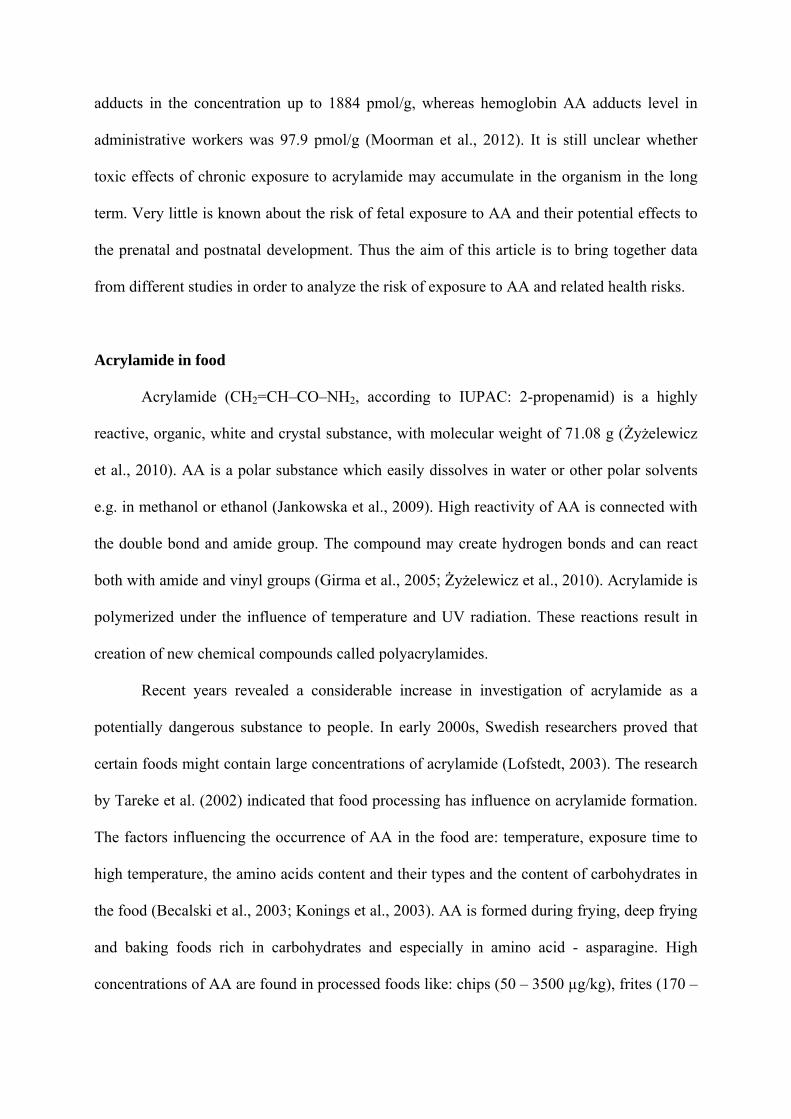
The factors influencing the occurrence of AA in the food are: temperature, exposure time to
high temperature, the amino acids content and their types and the content of carbohydrates in
the food (Becalski et al., 2003; Konings et al., 2003). AA is formed during frying, deep frying
and baking foods rich in carbohydrates and especially in amino acid - asparagine. High
concentrations of AA are found in processed foods like: chips (50 – 3500 µg/kg), frites (170 –

2287 µg/kg), coffee (170 – 350 µg/kg), bread (70 – 430 µg/kg) or corn flakes (30 – 1400
µg/kg) (Friedman, 2003). Acrylamide concentration in selected foodstuffs together with
methods of measurements of acrylamide concentration in food are presented in table 1.
The mechanism of AA formation in food has not been clearly described yet.
Numerous research has shown only hypothetical ways in which AA is being formed in
comestible products (Edegaard et al., 2008; Mestdagh et al., 2008). Most of the research point
to asparagine presence as a significant factor contributing to AA formation (Zhang et al.,
2009; Taeymans et al., 2004). The reaction between glucose (reducing sugar) and asparagine
gives a product responsible for the food’s flavor and color. This reaction is known as a
Millard’s reaction and it has a higher rate at the temperature exceeding 120°C (Friedman,
2003; Tareke et al., 2002). The content of AA increases considerably during frying, grilling
and roasting. Popular foodstuffs such as coffee, high-in-starch potato products and cereal
products contain large amounts of AA (Claus et al., 2008; Tajner-Czopek et al., 2012). People
are also exposed to harmful effects of AA by consuming natural unprocessed products rich in
asparagines, including asparagus, cocoa beans or cereals (Rachwał and Nebesny, 2012).
According to the European Food Safety Authority (EFSA) report, the level of AA in food
ranges from under 30 μg/kg to 4700 μg/kg, depending on the product (EFSA, 2009; Mojska
and Gielecińska, 2012). Research also shows that exposure to AA varies and depends mainly
on the population, age of consumers and their eating preferences. In European populations,
mean daily intake of acrylamide goes from 0.14 to 1.31 µg/kg body weight. Similar mean
intake (0.43-1.1 µg/kg body weight per day) was indicated in the United States (Dybing and
Sanner, 2003). The research conducted in Kraków, Poland by Jankowska et al. (2009)
indicated that AA was excessively consumed by children and teenagers. Among children and
adults, bread - a product eaten on a daily basis, is the main source of AA. Other Polish
research indicated that an average AA consumption in children aged 1-6 was about 0.47 μg/kg

b.w. per day and among children aged 7-18 – 0.34 μg/kg b.w. per day (Mojska and
Gielecińska, 2012). Maximum intake of acrylamide reaching 7.9 and 8.1 μg/kg b.w. per day
was estimated in 13 years old Norwegian boys and girls respectively (Dybing and Sanner,
2003). Food-related exposure of human populations to acrylamide together with methods used
for estimation of exposure levels are listed in table 2. Analysis of acrylamide studies is given
in Fig. 1.
Other kinds of exposure to acrylamide
Polyacrylamides are widely used in the industry for water treatment (as flocculator),
component of mortars, adhesives, dyes or in the textile and cosmetic industries. Furthermore,
polyacrylamide is used in laboratories, e.g. gel electrophoresis (Friedman, 2003). AA is used
for selective modification of protein’s sulfhydryl groups (SH) and the electrophoretic
separation of nucleic acids and proteins in the laboratories (Szczerbina, 2005).
Initially, polyacrylamide added to fertilizers or used as coagulant for water treatment was
claimed as the main source of AA (Szczerbina, 2005; Żyżelewicz et al., 2010). Cosmetic,
tobacco industry and plastics were indicated as the other sources of AA (Szczerbina, 2005).
Occupational exposure mostly affects chemical plant workers, laboratory workers,
construction industry workers, miners or workers of coal preparation plants (Pennisi et al.,
2013). The most publicized incidences of exposure include exposure to ground water
contaminated with acrylamide and N-methyloacrylamide in tunnel workers in Norway (Kjuus
et al., 2004; Goffeng et al., 2008) or exposures to aqueous solution of acrylamide in workers
of chemical manufactories in China (He et al., 1989). Typical symptoms of exposure are
manifested by paresthesia of the extremities, muscle weakness, ataxia, increased sweating.
Most of them result from peripheral neuropathy. In some cases impaired vision was diagnosed
(Pennisi et al., 2013). Interestingly, similar neurological effects were indicated in laboratory

studies in rats after 4 weeks of every third day exposures to doses of acrylamide as high as 40
mg/kg b.w. (Zhu et al., 2008).
High exposure to acrylamide occurs in tobacco smokers. Total amount of acrylamide
in the smoke from a single cigarette is around 1 µg or higher. Cigarettes and other tobacco
products like snuff, tobacco sticks or strips contain acrylamide in the range from below 100 to
367 ng/g (Moldoveanu and Gerardi, 2011). Thus tobacco related acrylamide intake depends
on the number of cigarettes smoked per day and/or the type of tobacco product consumed.
Measurements of the hemoglobin adducts with acrylamide suggest several times higher
exposure to AA in tobacco smokers than in non-smokers (Schettgen et al., 2004). Median
value of acrylamide intake in smoking pregnant women was estimated in the level of 91.1
µg/day (Brantsæter et al., 2008).
Absorption, metabolism and distribution of acrylamide
There are three ways by which AA is transmitted into the body: digestive system,
respiratory system (e.g. cigarette smoke) and skin absorption (e.g. cosmetics) (Carere, 2006;
Vesper et al., 2007). Irrespective of route, exposure to acrylamide rapidly occurs in blood
plasma with a peak concentration of 60-90 min in rats. Its epoxide form occurs later. In rats,
the peak concentration of GA is delayed by about 100 min in relation to AA plasma peak
(Barber et al., 2001). Both AA and glycidamide may create adducts with hemoglobin
following the reaction with sulfhydryl groups. The level of adducts is often used as indicator
of exposure to acrylamide as their formation is proportional to the acrylamide dose ingested,
inhaled or absorbed through the skin (Pingot et al., 2013, Tareke et al., 2008; Vikström et al.,
2012). Relatively large concentrations of AA and GA are distributed into muscle and neural
tissues (Barber et al., 2001). AA delivered by the oral route is metabolized in the liver. The
biotransformation takes place with the cytochrome P450. As a result of acrylamide

biotransformation, its epoxide form glycidamide (2,3-epoksypropan amide) is formed (Tareke
et al., 2008). During the second phase of biotransformation, AA and glycidamide (GA) are
coupled with reduced glutathione (GSH) by enzymes from the family of glutathione S-
transferase (GST) which leads to formation the glutathione conjugates (Friedman, 2003). The
final products of the glutathione conjugates reaction are the derivatives of N-acetylcysteine
excreted in urine (Pingot et al., 2013). As a result of reaction with GSH, AA and its
derivatives lose their toxic properties and may be more easily excreted from the organism.
Only about 50% of the AA daily dose is depurated from the organism, mainly in the urine
(EFSA, 2008). The half-life of AA in human organism is 2-7 hours which shows how slowly
this substance is being removed from the body (Sörgel et al., 2002).
Acrylamide and oxidative stress
Oxidative stress occurs when the rate of generation of free oxygen radicals (ROS) is
larger than the rate of their neutralization. An excess of free radicals may cause oxidation of
biological molecules namely lipid peroxidation, oxidation of enzymes and oxidation of DNA
bases. This leads to damage to cell organelles, impaired cell metabolism, DNA fragmentation
and cell death. Free radicals take part in pathogenesis of numerous diseases including
diabetes, neurodegeneration, diseases of cardiovascular system, neoplasm formation (Rahman
et al., 2012; Greń, 2013). Under normal physiological conditions, the occurrence and
metabolism of free radicals is controlled by antioxidative system which is composed of
enzymatic and nonenzymatic antioxidants. Major enzymatic antioxidants include superoxide
dismutase (SOD) catalyzing dismutation of superoxide anion to molecular oxygen and
hydrogen peroxide; catalase (CAT) catalyzing the decomposition of hydrogen peroxide;
glutathione peroxidase (GPx) catalyzing reduction of hydrogen peroxide accompanied by
oxidation of the reduced glutathione (GSH). Nonenzymatic antioxidants like reduced

glutathione, vitamins, thioredoxin, α-tocopherol etc., take part in neutralization of free
radicals by donation of electrons (Lobo et al., 2010).
There is evidence to suggest increased generation of free radicals and hydroperoxides
accompanied by lipid peroxidation in animals exposed to acrylamide (Prased and
Muralidhara, 2012). Increased activity of SOD in blood plasma, liver, testes, kidneys and
brain of acrylamide exposed rats hint at increased rate of formation of superoxide anion in the
whole organism (Yousef and El-Demerdash, 2006). Other studies indicated increased activity
of GPx accompanied by depletion of GSH which suggest adaptation of the antioxidative
system to increased H2O2 generation in different structures of neural system in rats (Zhu et al.,
2008). In general, depletion of GSH is a common phenomenon in animals treated with
acrylamide. The depletion of GSH results from higher rate of its consumption in reactions
with hydrogen peroxide (Zhu et al., 2008; Kopańska et al., 2015) and conjugation with
acrylamide and/or glycidamide in the phase II reactions catalyzed by glutathione s-transferase
(Paulsson et al., 2005). Glutathione is a major cell antioxidant whose shortage may be an
additional factor contributing to redox imbalance. Indeed, the data suggest that AA may
overwhelm the antioxidative system and cause symptoms of oxidative stress. For instance
Yousef and El-Demerdash (2006) indicated systemic increase of concentration of
thiobarbituric acid reactive substances in rats orally exposed to acrylamide. In similar fashion,
our team observed increased concentration of malondialdehyde in different brain areas of rats
intraperitoneally injected with AA solutions, all to suggest redox imbalance and increased
peroxidation of lipids (Kopańska et. al., 2015). Moreover, Zhu et al., (2008) found decreased
activity of SOD in neural system of rats after 10 weeks of exposure to acrylamide applied
every third day. This effect probably resulted from oxidation of SOD by excessively
generated superoxide ion.

All of these imply that acrylamide induces higher activity of antioxidative system, and that
high doses of acrylamide applied for longer time period induce symptoms of oxidative stress.
The data presented here were obtained in animal studies using the acrylamide doses in the
range 0.5 µg to 40 mg/kg body weight. First symptoms of affected redox balance were found
after 10 weeks of exposure to acrylamide doses of 25 µg/kg b.w. (Yousef and El-Demerdash,
2006). Such doses are only several times higher than the maximum acrylamide doses possibly
ingested with food by some human populations. Thus it is not clear whether people exposed
to acrylamide concentrations typically occurring in food will experience affected regulation of
redox reactions.
Genotoxicity and cytotoxicity of acrylamide
Oxidative imbalance induced by exposure to acrylamide may lead to cytotoxic and
genotoxic effects. Free radicals may cause damage to mitochondria and other cell organelles.
They induce apoptosis and cause oxidation of DNA bases, leading to fragmentation of the
double strand. All of these may cause cell death or neoplastic transformation (Valko et al.,
2004). The ROS related mechanism of cytotoxicity and/or mutations is attributed to all the
factors capable of inducing oxidative stress. On the other hand, acrylamide is known to
exhibit more specific effects on cells. AA was found to form 7-formamidoethyl adducts with
guanine. The formation of adducts with other acid bases is also probable, although they show
decreasing stability as guanine>adenine>uracil (Solomon et al., 1985; Shelkovsky et al.,
2002). The product of acrylamide biotransformation, glycidamide, shows higher affinity to
acid bases of nucleic acids than acrylamide. Nonetheless, both compounds were found to form
the strongest adducts with guanine at N-7 position (Atay et al., 2005). N7-dG-glycidamide is
the main DNA adduct. It has large pro-mutagenic properties because of formation of G-T
transversions during DNA replication (Besaratinia and Pfeifer, 2004). In embryonic

fibroblasts of transgenic Big Blue mice exposed in vitro to acrylamide dose of 320 μM, A-G
transitions and G-C tranversions were found (Besaratinia and Pfeifer, 2003). In human
lymphocytes, exposure to AA caused DNA strand to break, induced caspases-3 activity, and
apoptosis. Moreover, AA was found to disrupt DNA repair (Blasiak et al., 2004).
The studies over genotoxicity of chronic doses of acrylamide indicated significant increase of
glycidamide-DNA adducts in spermatocytes of mice exposed to doses of acrylamide as low as
0.01 µg per ml of drinking water given every day for 9-12 months. These animals also
showed increased number of incidences of double-strand breaks in DNA of the germ cells
(Nixon et al., 2012).
The genotoxic effects of acrylamide indicate that it may also play significant role in
neoplastic transformation. The carcinogenic potency of acrylamide was proven by Friedman’s
et al. (1995) studies. The study was conducted in males and females of Fischer 344 rats. AA
was administered in the drinking water, throughout the 106-week period, at the dose ranging
from 0.1 to 0.3 mg/kg b.w. per day. The results indicated a significant increase in the
frequency of thyroid follicular cell adenomas and adenocarcinomas in male rats from the
high-dose group. Moreover, that group witnessed incidence of mesothelioma of the tunica of
the testes. The females group saw a significant increase in the frequency of mammary gland
fibroadenoma and adenocarcinomas. This study also reported the occurrence of the thyroid
and mammary glands tumors after exposure to AA (Friedman et al., 1995). Another long term
studies were performed in females of swiss-ICR mice. The specimens were exposed to AA
doses going from 2.5 to 50.0 mg/kg b.w., administered orally, every second day. After 1-year
observation, the development of skin tumor in mice exposed to the highest AA doses was
observed (squamous cell papilloma and carcinoma). Moreover, incidences of lung cancer
were noted (Bull et al., 1984).

The pro-oncogenic activity of acrylamide in humans is not evident. Epidemiological studies
did not indicate any relation between exposure to acrylamide and cancer incidences in human
(Marsh et al., 1999). On the other hand, the doses of acrylamide inducing genotoxic effects in
animals well correspond with the AA doses ingested by high consumers of food containing
acrylamide. This is why European Union classification of Carcinogens placed acrylamide in
the second category, as carcinogen and mutagen (Szczerbina, 2005). Moreover, International
Agency for Research on Cancer classified AA as a potentially carcinogenic substance for
people (IARC, 1994).
Reproductive toxicity of acrylamide
As a low molecular weight compound easily dissolving in water, acrylamide passes
through the placenta in animals and human organism. It was also found in breast milk of
women. Thus it may have the influence on the normal prenatal and early postnatal
development of infants (Sӧrgel et al., 2002). Nonetheless, the data on the risk of the harmful
influence of acrylamide on the early development of human has not been assessed so far.
The food frequency questionnaire estimated medial acrylamide intake in pregnant women as
33.7 µg/day. The median excretion of acrylamide based on urine metabolites in this group of
women was 11.2 µg/24h (range: 3.3-75.6 µg/24h). Assuming that about 55% of acrylamide is
depurated in urine as mercapturic acid metabolites, this would correspond to a median
exposure of 20.3 µg/24h (Brantsæter et al., 2008). According to the above calculation, the
maximum exposure to acrylamide in pregnant women may be as high as 137.5 µg/24h. It was
estimated that about 50% of dietary acrylamide may be transferred through the placental
blood into the embryo (Sӧrgel et al., 2002). According to the questionnaire-based studies, the
main dietary sources of acrylamide to pregnant women were potato crisps, crisp bread,
biscuits, breakfast cereals and bakery products (Brantsæter et al., 2008).

The embryotoxic effects of acrylamide were studied in animal models. Exposure of pregnant
females of rodents to AA doses ≥5mg/kg b.w./day, administered orally, resulted in increased
post-implantation loss of embryos and decreased number of live pups. Exposure of pregnant
females to higher doses of AA (≤ 15 mg/kg b.w./day) resulted in reduced pup weight and
survival (NTP, 2011). Interesting studies over acrylamide influence on embryonic and early
postnatal development of rats were performed by El-Sayyad et al., (2011). In this study,
pregnant females were orally exposed to high acrylamide doses of 30 mg/kg b.w. from day 6
of gestation until parturition and throughout lactation. The young pups derived from
acrylamide exposed females had lower body size and weight and lower brain size in
comparison to control animals. They also suffered from muscular dystrophy and
ultrastructural changes in cerebral cortex.
The reproductive toxicity of acrylamide is also manifested by its influence on animal male
infertility. According to Scientific Committee on Food (SCF, 2002), the impaired fertility may
involve affected sperm count and sperm motility parameters. The increased number of
glycidamide-DNA adducts and fragmentation of DNA in germ cells of male mice exposed
chronically to low AA doses was proved by Nixon et al., 2012. This suggests increased risk of
DNA lesions in male reproductive material and their possible introduction into zygote.
Indeed, it was found that exposure of male rats to acrylamide doses of 19 mg/kg for eight
days and next mated to unexposed females led to reduced fertility rates and increased
frequency of resorption of embryos (Sakamoto and Hashimoto, 1986). Moreover, exposure of
rats to acrylamide (dose of 100 ppm) resulted in disrupted mating performance, ejaculatory
processes and subsequent transport of sperm (Zenick et al., 1986).
Neurotoxicity of acrylamide

The only toxic effects of acrylamide well documented in human were manifested by
peripheral neuropathy related to occupational exposure (Pennisi et al., 2013). Symptoms of
peripheral neuropathy were also described in animal studies. In monkeys, chronic oral
exposure to AA doses of 10 mg/kg b.w./day for up to 12 weeks was associated with clinical
signs of peripheral neuropathy like muscle weakness or ataxia of limbs (SCF, 2002). In rats,
neurotoxic AA effects were manifested by abnormal gait, shown as foot splay, ataxia and
weakness of the hindlimb skeletal muscle. Complete paralysis of hindlimbs occurred after 10
weeks of AA administration in a dose of 40 mg/kg b.w. every second day. The behavioral
effects were accompanied by serious alteration of electrophysiology of the sciatic nerve which
may suggest alteration of the myelin capsule and/or altered activity of axolemmal Na/K-
ATPase (Zhu et al., 2008).
In animal studies, toxic effects of acrylamide were also indicated in central nervous system.
Rat pups born by mothers exposed to acrylamide (30 mg/kg bw) and fed with milk from
lactating females exposed to acrylamide showed serious ultrastructural changes in cerebral
cortex. They were manifested by massive increases of pyknotic neuronal cells separated by
widened spaces, increased number of apoptotic cells, death of Purkinje cells and granular
neuronal cells (El-Sayyad et al, 2011). It is reasonable to suggest that ultrastructural changes
in brain may be followed by functional effects. Our team has indicated decreased activity of
acetylcholinesterase, an enzyme playing regulatory function in cholinergic transmission, in
cerebrum, cerebellum and medulla oblongata of mice exposed to acrylamide doses of 20 and
40 mg/kg for 24h, 48h and 8 days. This may hint at longer time of residence of acetylcholine
in cholinergic synapses and higher excitation of cholinergic nerves engaged in memory
formation, behavior, muscle controlling, controlling of autonomic functions etc., (Kopańska et
al., 2015).

There are probably several mechanisms of acrylamide neurotoxicity. It is generally accepted
that the most important one is related with conjugation of AA with cysteine residues of
presynaptic membrane proteins engaged in neurotransmitter release. Consequently, the flow
of nerve impulses may be inhibited, coupled with subsequent degeneration of neurons
(LoPachin and Barber, 2006; Pingot et al., 2013). Important role in neurotoxicity of
acrylamide is probably played by oxidative stress. Zhu et al. (2008) indicated that peripheral
neuropathy and altered electrophysiology of the sciatic nerve were accompanied by the
symptoms of redox imbalance. Similarly, in our studies, the affected activity of
acetylcholinesterase were accompanied by depletion of albumins and –SH group
concentrations and elevated content of malondialdehyde in brain of mice exposed to AA
which also suggest induction of redox imbalance (Kopańska et al., 2015).
The redox imbalance in brain of animals exposed to acrylamide is an important observation as
free radicals are known to contribute to neurodegeneration. The increased level of
malondialdehyde, the product of peroxidation of lipids, was found in erythrocytes, blood
serum and neurofibrillary tangles in brains of Alzheimer’s Disease patients (Matveychuk et
al., 2011). Assuming that neurodegeneration results from cumulative damage to neuronal cells
induced by free radicals (Praticò, 2005), it may be reasonably established that food
ingredients capable of inducing redox imbalance in brain may participate in etiology of
neurodegenerative diseases. The relation between the consumption of acrylamide-rich food
and risk of neurodegeneration has not been studied so far, although this seems to be an
interesting problem for toxicological and epidemiological studies.
CONCLUSION
Acrylamide belongs to the most common toxins in human diet. It shows relatively
high concentrations in asparagine rich foods processed at high temperature. Its mean

consumption depending on the population and age of consumers usually reaches
approximately 1 µg/kg body weight daily, although in high AA consumers its maximum
intake may be above 8 µg/kg body weight per day. This corresponds with the AA doses
inducing peroxidation of lipids and DNA lesions in long term animal studies. Genetic
disorders may affect male fertility, embryonic and fetal development and neoplastic
transformation. On the other hand, realistic data indicating the relation between consumption
of AA rich food and health risk for human are still missing. The only well documented health
disorders occurred as a result of occupational exposure and were manifested by peripheral
neuropathy symptoms.
Acknowledgements
This work was supported by the Slovak Research and Development Agency
under the contract no. SK-PL-2015-0032.
REFERENCES:
1. ATAY NZ, ÇALGAN D, ÖZAKAT E, VARNALI T: Acrylamide and glycidamide
adducts with guanine. J Mol Struc-THEOCHEM 728: 249-251, 2005.
2. BARBER DS, HUNT JR, EHRICH MF, LEHNING EJ, LOPACHIN RM:
Metabolism, toxicokinetics and hemoglobin adducts formation in rats following
subacute and subchronic acrylamide dosing. NeuroTox 22: 341-353, 2001.
3. BCS, BUREAU OF CHEMICAL SAFETY: Health Canada’s Revised Exposure
Assessment of Acrylamide in Food. Food Directorate, Health Products and Food
Branch, 1-19, 2012. www.hc-sc.gc.ca
4. BECALSKI A, LAU B, LEWIS D, SEAMAN S: Acrylamide in foods: occurrence,
sources, and modeling. J Agric Food Chem 51: 802–808, 2003.

5. BESARATINIA A, PFEIFER GP: Weak Yet Distinct Mutagenicity of Acrylamide in
Mammalian Cells. J Natl Cancer Inst 95: 12, 2003.
6. BESARATINIA A, PFEIFER GP: Genotoxicity of acrylamide ad glycidamide. J Natl
Cancer Inst 96: 1023-1029, 2004.
7. BFR, FEDERAL INSTITUTE FOR RISK ASSESSMENT: Assessment of acrylamide
intake from foods containing high acrylamide levels in Germany, 2003,
http//www.bfr.bund.de
8. BLASIAK J, GLOC E, WOZNIAK K, CZECHOWSKA A: Genotoxicity of
acrylamide in human lymphocytes. Chem Biol Interact 149: 137-149, 2004.
9. BRANTSÆTER AL, HEUGEN M, DE MUL A, BJELLAAS T, BECHER G, VAN
KLAVEREN J, ALEXANDER J, MELTZER HM: Exploration of different methods
to assess dietary acrylamide exposure in pregnant women participating in the
Norwegian Mother and Child Cohort Study (MoBa). Food Chem Toxicol 46: 2808-
2814, 2008.
10. BULL RJ, ROBINSON M, STOBER JA: Carcinogenic activity of acrylamide in the
skin and lung of Swiss-ICR mice. Cancer Lett 24: 209-212, 1984.
11. CARERE A: Genotoxicity and carcinogenicity of acrylamide: a critical review. Ann
Ist Super Sanita 42: 144-155, 2006.
12. CLAUS A, CARLE R, SCHIEBER A: Acrylamide in cereal products: a review. J
Cereal Sci 47: 118–133, 2008.
13. CLAEYS W, BAERT K, MESTDAGH F, VERCAMMEN J, DAENENS P, DE
MEULENAER B, MAGHUIN-ROGISTER G, HUYGHEBAERT A: Assessment of
the acrylamide intake of the Belgian population and the effect of mitigation strategies.
Food Addit Contam Part A Chem Anal Control Expo Risk Assess 27: 1199-2070,
2010.

14. DYBING E, SANNER T: Risk Assessment of Acrylamide in Foods. Toxicol Sci 75:
7–15, 2003.
15. EFSA, EUROPEAN FOOD SAFETY AUTHORITY: Results on the monitoring of
acrylamide levels in food. Scientific Report, 285: 1-26, 2009.
http://www.efsa.europa.eu/sites/default/files/scientific_output/files/main_documents/2
85r.pdf
16. EFSA, EUROPEAN FOOD SAFETY AUTHORITY: Acrylamide carcinogenicity –
new evidence in relation to dietary exposure. Scientific Colloquium, Summary Report
11: 1–27, 2008.
17. FRIEDMAN MA, DULAK LH, STEDHAM MA: A lifetime oncogenicity study in
rats with acrylamide. Fundam Appl Toxicol 27: 95-105, 1995.
18. FRIEDMAN M: Chemistry, Biochemistry, and Safety of Acrylamide. J Agric Food
Chem 51: 4504–4526, 2003.
19. GIRMA KB, LORENZ V, BLAUROCK S, EDELMANN F: Coordination chemistry
of acrylamide. Coord Chem Rev 249: 1283–1293, 2005.
20. GOFFENG LO, KJUUS H, HEIER MS, ALVESTRAND M, ULVESTAD B,
SKAUG V: Colour vision and light sensitivity in tunnel workers previously exposed
to acrylamide and N-methylolacrylamide containing grouting agents. NeuroTox 29:
31–39. 2008.
21. GREŃ A: Effects of vitamin E, C and D supplementation on inflammation and
oxidative stress in streptozotocin-induced diabetic mice. Int J Vitam Nutr Res 83: 168-
175, 2013.
22. EDEGAARD RV, GRANBY K, FRANDSEN H, THYGESEN J, SKIBSTED LH:
Acrylamide in bread: Effect of prooxidants and antioxidants. Eur Food Res Technol
227: 519-525, 2008.

23. EL-SAYYAD HI, EL-GAMMAL HL, HABAK LA, ABDEL-GALIL HM,
FERNANDO A, GAUR RL, OUHTIT A: Structural and ultrastructural evidence of
neurotoxic effects of fried potato chips on rat postnatal development. Nutrition 27:
1066-1075, 2011.
24. HE FS, ZHANG SL, Wang HL, Li G, Zhang ZM, Li FL, Dong XM, Hu FR:
Neurological and electroneuromyographic assessment of the adverse effects of
acrylamide on occupationally exposed workers. Scand J Work Environ Health 15:
125–129, 1989.
25. IARC, INTERNATIONAL AGENCY FOR RESEARCH ON CANCER: Some
industrial chemicals. Monographs on the Evaluation of Carcinogenic Risk for
Chemicals to Humans. Lyon, France. 60: 435, 1994.
26. JANKOWSKA J, HELBIN J, POTOCKI A: Akryloamid jako substancja obca w
żywności. Probl Hig Epidemiol 90: 171–174, 2009.
27. KERAMAT J, LEBAIL A, PROST C, JAFARI M: Acrylamide in baking products:
A review article. Food Bioprocess Tech 4: 530–543, 2011.
28. KJUUS H, GOFFENG LO, HEIER MS, SJÖHOLM H, OVREBØ S, SKAUG V,
PAULSSON B, TÖRNQVIST M, BRUDAL S: Effects on the peripheral nervous
system of tunnel workers exposed to acrylamide and N-methylolacrylamide. Scand J
Work Environ Health 30: 21–29, 2004.
29. KONINGS E, BAARS A, KLAVEREN J, SPANJER M, RENSEN P, HIEMSTRA M,
KOOIJ JA, PETERS PWJ: Acrylamide exposure from foods of the Dutch population
and an assessment of the consequent risks. Food Chem Toxicol 41: 1569–1579, 2003.
30. KOPAŃSKA M, LUKÁČ N, KAPUSTA E, FORMICKI G: Acrylamide Influence on
Activity of Acetylcholinesterase, Thiol Groups, and Malondialdehyde Content in the
Brain of Swiss Mice. J Biochem Molec Toxicol 29: 472-478, 2015.

31. LOBO V, PATIL A, PHATAK A, CHANDRA N: Free radicals, antioxidants and
functional foods: impact on human health. Pharmacogn Rev 4: 118-126, 2010.
32. LOFSTEDT RE: Science communication and the Swedish acrylamide “alarm. J
Health Commun 8: 407-432, 2003.
33. LOPACHIN RM: The changing view of acrylamide neurotoxicity. NeuroTox 25: 617-
630, 2004.
34. LOPACHIN RM, BARBER DS: Synaptic cysteine sulfhydryl groups as targets of
electrophilic neurotoxicants. Toxicol Sci 94: 240–255, 2006.
35. MARSH GM, LUCAS LJ, YOUK AO, SCHALL LC: Mortality patterns among
workers exposed to acrylamide: 1994 follow up. Occup Environ Med 56: 181–190,
1999.
36. MATVEYCHUK D, DURSUN SM, WOOD PL, BAKER GB: Reactive aldehydes
and neurodegenerative disorders. Bull Clin Psychopharmacol 21: 277–288, 2011.
37. MESTDAGH F, CASTELEIN P, PETEGHEM C, MEULENAER B: Importance of
Oil Degradation Components in the Formation of Acrylamide in Fried Foodstuffs. J
Agric Food Chem 56: 6141–6144, 2008.
38. MOJSKA H, GIELECIŃSKA I: Assessment of exposure of children and youth to
acrylamide in fast foods and snacks. Probl Hig Epidemiol 93: 613-617, 2012.
39. MOLDOVEANU SC, GERARDI AR: Acrylamide analysis in tobacco, alternative
tobacco products and cigarette smoke. J Chromatogr Sci 49: 234-242, 2011.
40. MOORMAN WJ, REUTMAN SS, SHAW PB, BLADE LM, MARLOW D, VESPER
H, CLARK JC, SCHRADER SM: Occupational exposure to acrylamide in closed
system production plants: air levels and biomonitoring. J Toxicol Environ Health, A
75: 100-111, 2012.

41. MOTTRAM DS, WEDZICHA BL, DODSON AT: Food chemistry: Acrylamide is
formed in the Maillard reaction. Nature 419: 448-449, 2002.
42. NIXON BJ, STANGER SJ, NIXON B, ROMAN SD: Chronic exposure to acrylamide
induces DNA damage in male germ cells of mice. Toxicol Sci 129: 135-145, 2012.
43. NTP, NATIONAL TOXICOLOGY PROGRAM: Toxicology and carcinogenesis
studies of acrylamide in F344/N rats and B6C3F1 mice. Technical Report, NIH 575: 1-
252, 2011.
44. PAULSSON B, RANNUG A, HENDERSON AP, GOLDING BT, TÖRNQVIST M,
WARHOLM M: In vitro studies of the influence of glutathione transferases and
epoxide hydrolase on the detoxification of acrylamide and glycidamide in blood. Mut
Res 580: 53-59, 2005.
45. PENNISI M, MALAGUARNERA G, PUGLISI V, VINCIGUERRA L, VACANTE
M, MALAGUARNERA M: Neurotoxicity of Acrylamide in Exposed Workers. Int J
Environ Res Public Health 10: 3843-3854, 2013.
46. PINGOT D, PYRZANOWSKI K, MICHAŁOWICZ J, BUKOWSKA B: Toksyczność
akrylamidu i jego metabolitu – glicydamidu. Med Pr 64: 259–270, 2013.
47. PRASED SN, MURALIDHARA: Evidence of acrylamide induced oxidative stress
and neurotoxicity in Drosophila melanogaster - Its amelioration with spice active
enrichment: Relevance to neuropathy. NeuroTox, 33: 1254-1264, 2012.
48. PRATICÒ D: Peripheral biomarkers of oxidative damage in Alzheimer’s disease: the
road ahead. Neurobiol Aging 26: 581-583, 2005.
49. RACHWAŁ D, NEBESNY E: Redukcja zawartości akrylamidu w produktach
spożywczych. Bromat Chem Toksykol 2: 219–227, 2012.
50. RAHMAN T, HOSEN I, ISLAM MMT, SHEKHAR HU: Oxidative stress and human
health. Adv Biosci Biotechnol 3: 997-1019, 2012.

51. SAKAMOTO J, HASHIMOTO K: Reproductive toxicity of acrylamide and related
compounds in mice--effects on fertility and sperm morphology. Arch Toxicol 59: 201–
205, 1986.
52. SCF, SCIENTIFIC COMMITTEE ON FOOD: Opinion of the Scientific Committee
on Food on new findings regarding the presence of acrylamide in food.
SCF/CS/CNTM/CONT/4 Final, Belgium, 3 July 2002.
http://ec.europa.eu/food/fs/sc/scf/out131_en.pdf
53. SCHETTGEN T, ROSSBACH B, KÜTTING B, LETZEL S, DREXLER H,
ANGERER J: Determination of haemoglobin adducts of acrylamide and glycidamide
in smoking and non-smoking persons of the general population. Int J Hyg Environ
Health 207: 531-539, 2004.
54. SHELKOVSKY V, STEPANIAN SG, GALETICH IK, KOSEVICH MV,
ADAMOWICZ L: Modelling of recognition sites of nucleic acid bases and amide side
chains of amino acids. Combination of experimental and theoretical approaches. Eur
Phys J D 20: 421-430, 2002.
55. SIROT V, HOMMET F, TARD A, LEBLANC JC: Dietary acrylamide exposure of
the French population: results of the second French Total Diet Study. Food Chem
Toxicol 50: 889-894, 2012.
56. SOLOMON JJ, FEDYK J, MUKAI F, SEGAL: Direct alkylation of 2’-
doxynucleosides and DNA following in vitro reaction with acrylamide. Cancer Res
45: 3465-3470, 1985.
57. SӦRGEL F, WEISSENBACHER R, KINZIG-SCHIPPERS M, HOFMANN A,
ILLAUER M, SKOTT A, LANDERSDORFER C: Acrylamide: increased
concentrations in homemade food and first evidence of its variable absorption from

food, variable metabolism and placental and breast milk transfer in humans.
Chemotherapy 48: 267–274, 2002.
58. SZCZERBINA T: Akrylamid – potencjalnie rakotwórcza substancja występująca w
żywności. Kosmos Probl Nauk Biol 54: 367–372, 2005.
59. TAEYMANS D, WOOD J, ASHBY P, BLANK I, STUDER A, STADLER RH,
GONDÉ P, VAN EIJCK P, LALLJIE S, LINGNERT H, LINDBLOM M,
MATISSEK R, MÜLLER D, TALLMADGE D, O'BRIEN J, THOMPSON S,
SILVANI D, WHITMORE T: A Review of acrylamide: an industry perspective on
research, analysis, formation, and control. Crit Rev Food Sci Nutr 44: 323–347, 2004.
60. TAJNER-CZOPEK A, RYTEL E, KITA A, PĘKSA A, MIEDZIANKA J: Wpływ
parametrów obróbki termicznej na zawartość akrylamidu w wybranych przetworach
ziemniaczanych. Bromat Chem Toksykol 3: 320–325, 2012.
61. TAREKE E, LYN-COOK B, RONINSON B, ALI S: Acrylamide: A dietary
carcinogen formed in vivo? J Agric Food Chem 56: 6020–6023, 2008.
62. TAREKE E, RYDBERG P, KARLSSON P, ERICSSON S, TORNQVIST M:
Analysis of Acrylamide, a Carcinogen Formed in Heated Foodstuffs. J Agric Food
Chem 50: 4998–5006, 2002.
63. VALKO M, IZAKOVIC M, MAZUR M, RHODES CJ, TELSER J: Role of oxygen
radicals in DNA damage and cancer incidence. Mol Cell Biochem 266: 37-56, 2004.
64. VESPER HW, BERNERT JT, OSPINA M, MEYERS T, INGHAM L, SMITH A,
MYERS GL: Assessment of the Relation between Biomarkers for Smoking and
Biomarkers for Acrylamide Exposure in Humans. Cancer Epidemiol Biomarkers Prev
16: 2471-2478, 2007.
65. VIKSTRÖM AC, WARHOLM M, PAULSSON B, AXMON A, WIRFÄLT E,
TÖRNQVIST M: Hemoglobin adducts as a measure of variations in exposure to

acrylamide in food and comparison to questionnaire data. Food Chem Toxicol 50:
2531–2539, 2012.
66. YOUSEF MI, EL-DEMERDASH: Acrylamide-induced oxidative stress and
biochemical perturbations in rats. Toxicology 219: 133-141, 2006.
67. ZAJĄC J, BOJAR I, HELBIN J, KOLARZYK E, POTOCKI A,STRZEMECKA J,
OWOC A: Dietary acrylamide exposure in chosen population of South Poland. Ann
Agric Environ Med 20: 351-355, 2013.
68. ZENICK H, HOPE E, SMITH MK: Reproductive toxicity associated with acrylamide
treatment in male and female rats. J Toxicol Environ Health 17: 457-472, 1986.
69. ZHANG Y, REN Y, ZHANG Y: New Research Developments on Acrylamide:
Analytical Chemistry, Formation Mechanism, and Mitigation Recipes. Chem Rev 109:
4375–4397, 2009.
70. ZHU Y, ZENG T, ZHU Y, YU S, WANG Q, ZHANG L, GUO X, XIE K: Effects of
acrylamide on the nervous tissue antioxidant system and sciatic nerve
electrophysiology in the rat. Neurochem Res 33: 2310-2317, 2008.
71. ŻYŻELEWICZ D, NEBESNY E, ORACZ J: Akrylamid – powstawanie, właściwości
fizykochemiczne i biologiczne. Bromat Chem Toksykol, 3: 415–427, 2010.

Fig. 1. Acrylamide studies according to PubMed (data from 1958 till 2016-08-23)

Tab. 1. Acrylamide content in analyzed products
LC‐MS: Liquid chromatography–mass spectrometry LC‐MS‐MS: Liquid chromatography tandem mass spectrometry GCQ‐MS/MS: Gas chromatography with tandem mass spectrometry LC‐MS/MS: Liquid chromatography with tandem mass spectrometry
Author Number of food samples
Analysis of acrylamide in food
Acrylamide content Products with the highest acrylamide content
Highest mean acrylamide content (µg/kg)
Claeys et al., 2010
1725 LC–MS
34-2814 (mean) Coffee substitute 2814±1045 Instant coffee 694±81 Potato crisps 525±477 Gingerbread 431±455
Sirot et al., 2012
192 LC–MS 2-954 µg/kg (range) Potato chips 954±240 French fries 724±358 Cocktail biscuits (salted)
697±430
Chocolate biscuits 139±100 Konings et al., 2003
341 LC–MS–MS <30-3100 µg/kg (range)
Potato crisps 1249±656
Cocktail snacks 1060±950
Gingerbread: 890±393
Chips (deep-fried) 351±297
Mojska and Gielecińska, 2012
111 GCQ–MS/MS LC–MS/MS
2-516 µg/kg (range)
Follow-on formula 73±78 Infant biscuits 219 ±139

Tab. 2. Exposure of human population to acrylamide
Authors Method used Population (n, total population)
Age years/months (n, group size)
Daily doses ingested Contribution of food in daily acrylamide exposure
Zając et al., 2013 Semi-quantitative food frequency questionnaire
Polish (n=1470) 6–12 years (n=300)
mean: 1.51 μg/kg b.w.
Baked goods: (42%) Crisps: (25%) Cookies: (14%)
13–19 years (n=296)
mean: 0.89 μg/kg b.w.
Baked goods: (46%) Cookies: (25%) French fries: (12%)
20–30 years (n=296)
mean: 0.61 μg/kg b.w.
Baked goods: (55%) Cookies: (15%) Coffee: (9%)
31–41 years (n=278)
mean: 0.56 μg/kg b.w.
Baked goods: (55%) Coffee: (16%) Cookies: (15%)
42–60 years (n=300)
mean: 0.67 μg/kg b.w. Baked goods: (38%) Crisps: (22%) Coffee: (16%)
Claeys et al., 2010
Probabilistic approach ‘Monte Carlo Risk Analysis Programme’
Belgium (n=662)
2.5-6.5 years (n=662)
mean: 0.72 μg/kg b.w. Biscuits: (26%) French fries: (25%) Bread & rolls:(20,2%)
BfR*, 2003 Questionnaire German (n=1085)
15–18 years (n=1085)
mean: 1.10 μg/kg b.w. Toast: (-%) Fried potatoes (-%)

BCS**, 2012 Probabilistic dietary exposure to acrylamide based on the AA measured in samples of each food along with individual consumption data
Canada (n=32 088) < 1 year (n=279)
mean: 0.211 μg/kg b.w.
-
1–3 years (n=2096)
mean: 0.609 μg/kg b.w.
French fries (30.0%) Snack chips (14.9%) Coffee (12.3%) 4–8 years
(n=3047) mean: 0.597 μg/kg b.w.
9–13 years (n=3883)
mean: 0.442 μg/kg b.w.
14 – 18 years (n=4423)
mean: 0.356 μg/kg b.w.
19 – 30 years (n=3713)
mean: 0.288 μg/kg b.w.
31 – 50 years (n=5125)
mean: 0.248 μg/kg b.w.
51 – 70 years (n=5533)
mean: 0.187 μg/kg b.w.
≥ 71 years (n=3989)
mean: 0.157 μg/kg b.w.

Sirot et al., 2012 Probabilistic approach on the basis of the weekly food consumption
French (n=336)
18–79 years (n=191)
mean: 0.43±0,33 μg/kg b.w.
French fries: (44,8%) Coffee: (29,5%) Biscuits: (9,4%)
3–17 years (n=145)
mean: 0.69±0,58 μg/kg b.w.
French fries: (60,8%) Biscuits: (18,8%) Cakes and other sweetened pastrie: (3.3%)
Konings et al., 2003
Probabilistic approach ‘Monte Carlo Risk Analysis Programme’
Dutch (n=6250)
1-6 years (n, not given)
mean: 1.04 μg/kg b.w.
Crisps: (40%) Dutch spiced cake: (20%) Chips and comparable products: (18%)
7-18 years (n, not given)
mean: 0.71 μg/kg b.w. Crisps: (46%) Dutch spiced cake: (23%) Chips and comparable products: (11%)
1-97 years (n=6250)
mean: 0.48 μg/kg bw Crisps: (31%) Chips and comparable products: (21%) Dutch spiced cake: (16%)

Dybing and Sanner, 2003
Food Frequency Questionnaire (FFQ)
Norway (n=2672) 16-79 years (n=2672)
mean: 0.46 - 0.49 μg/kg b.w.
Coffee: (28-28.6 %) Potato crisps: (17.6-17.4 %) Soft bread: (13.0-11.9 %)
Probabilistic approach based on UNGKOST 2000 data
Norway (n=6736) 9 years (n=2957)
mean: 0.32 – 0.36 μg/kg b.w.
Potato crisps,butter biscuits, sweet biscuits: (55–65% )
13 years (n=3779)
mean: 0.49 – 0.52 μg/kg b.w.
Mojska and Gielecińska, 2012
Probabilistic approach based on the theoretical number of food portions
Polish (n, not given) 6 month (n, not given)
mean: 17.46 µg/person/day
Jarred baby food: (56,7%) Follow-on formula: (43,3%)
7 month (n, not given)
mean: 20.87 µg/person/day
Jarred baby food: (52,7%) Follow-on formula: (27,2%)
8 month (n, not given)
mean: 21,65 µg/person/day
Jarred baby food: (50,8%) Follow-on formula: (26,2%)
9 month (n, not given)
mean: 29,06 µg/person/day
Jarred baby food: (37,9%) Follow-on formula: (21,7%)

* Federal Institute for Risk Assessment (BFR)
** Bureau of Chemical Safety, Food Directorate, Health Products and Food Branch (BCS)
10-12 months (n, not given)
mean: 38,05 µg/person/day
Jarred baby food: (55,2%) Infant cereals: (18,2%)
Brantsæter et al., 2008
Probabilistic data based on the food frequency questionnaire (FFQ)
Norwegian, pregnant women (n=19, age 23-44)
n=19 median: 33.7 µg/person
Crispbread (10-22%) Potato crisps (14-16%) Bread (8-11%) Biscuits (5-10%) Breakfast cereals (6-8%)
Probabilistic data based on the food diary (FD)
n=19 median: 28.5 µg/person
Probabilistic data based on the AA metabolite concentration in urine (non-smokers)
n=16 median: 20.3 µg/person
Probabilistic data based on the metabolite concentration in urine (smokers)
n=3 median: 91.1 µg/person