1 Visual Anatomy & Physiology First Edition Martini & Ober Chapter 13 Brain and Cranial Nerves...
44
1 Visual Anatomy & Physiology First Edition Martini & Ober Chapter 13 Brain and Cranial Nerves Lecture 20
-
Upload
silvester-fields -
Category
Documents
-
view
217 -
download
0
Transcript of 1 Visual Anatomy & Physiology First Edition Martini & Ober Chapter 13 Brain and Cranial Nerves...

1
Visual Anatomy & PhysiologyFirst Edition
Martini & Ober
Chapter 13
Brain and Cranial Nerves
Lecture 20

2
Overview of the Brain
Functions• regulates visceral activities• coordinates muscular movements• interprets sensations• determines perception• stores memory• carries out reasoning• makes decisions• determines personality
Major Parts• cerebrum (two hemispheres)• diencephalon
• thalamus• hypothalamus
• brain stem• midbrain (mesencephalon)• pons• medulla oblongata
• cerebellum
3
Protection of the Brain
• The brain is protected– Mechanically by
• The skull bones
• The meninges
• The cerebrospinal (CSF) fluid
– Biochemically by the blood-brain barrier• Capillaries interconnected by tight junctions
• Astrocytes/ependymal cells control permeability of general capillaries/choroid capillaries
• May be obstacle to delivery of drugs
• May become more permeable during stress

4
Meninges of the Brain
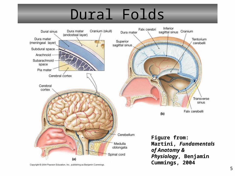
- dura mater – outer, tough (anchoring dural folds)
- arachnoid mater – web-like
- pia mater – inner, delicate
- Subdural space – like interstitial fluid
- Subarachnoid space – CSF
*Singular of meninges is meninx
Figure from: Hole’s Human A&P, 12th edition, 2010
5
Dural Folds
Figure from: Martini, Fundamentals of Anatomy & Physiology, Benjamin Cummings, 2004

6
Ventricles of the Brain
• interconnected cavities• within cerebral hemispheres and brain stem• continuous with central canal of spinal cord• filled with cerebrospinal fluid (CSF)
• lateral ventricles (1, 2)• third ventricle (3)• fourth ventricle (4)• cerebral aqueduct
Figure from: Hole’s Human A&P, 12th edition, 2010

7
Cerebrospinal Fluid• secreted by choroid plexus of ventricles (~500 ml/day)
• circulates in ventricles, central canal of spinal cord, and subarachnoid space
• completely surrounds brain and spinal cord
• clear liquid (more Na+ and Cl-, but less K+, Ca2+, glucose, and protein than plasma)
• nutritive and protective
• helps maintain stable ion concentrations in CNS
Figure from: Hole’s Human A&P, 12th edition, 2010

8
Structure of Cerebrum
• corpus callosum• connects hemispheres
• gyri• bumps or convolutions
• sulci• grooves
• longitudinal fissure• separates hemispheres
• transverse fissure• separates cerebrum from cerebellum
• lateral sulcus• separates the frontal from the temporal lobes
Figures from: Saladin, Anatomy & Physiology, McGraw Hill, 2007

10
Functions of Cerebrum• interpretation• initiating voluntary movements• storing memory• retrieving memory• reasoning• center for intelligence and personality
The cerebrum can be divided into several functional areas:
- Motor (frontal cortex) - Sensory (parietal, occipital, and temporal cortex)- Association (all lobes)
Points to keep in mind: - Each cerebral hemisphere receives information from, and sends information to, the opposite side of the body - Although symmetrical, the cerebral hemispheres are not entirely equal in function
11
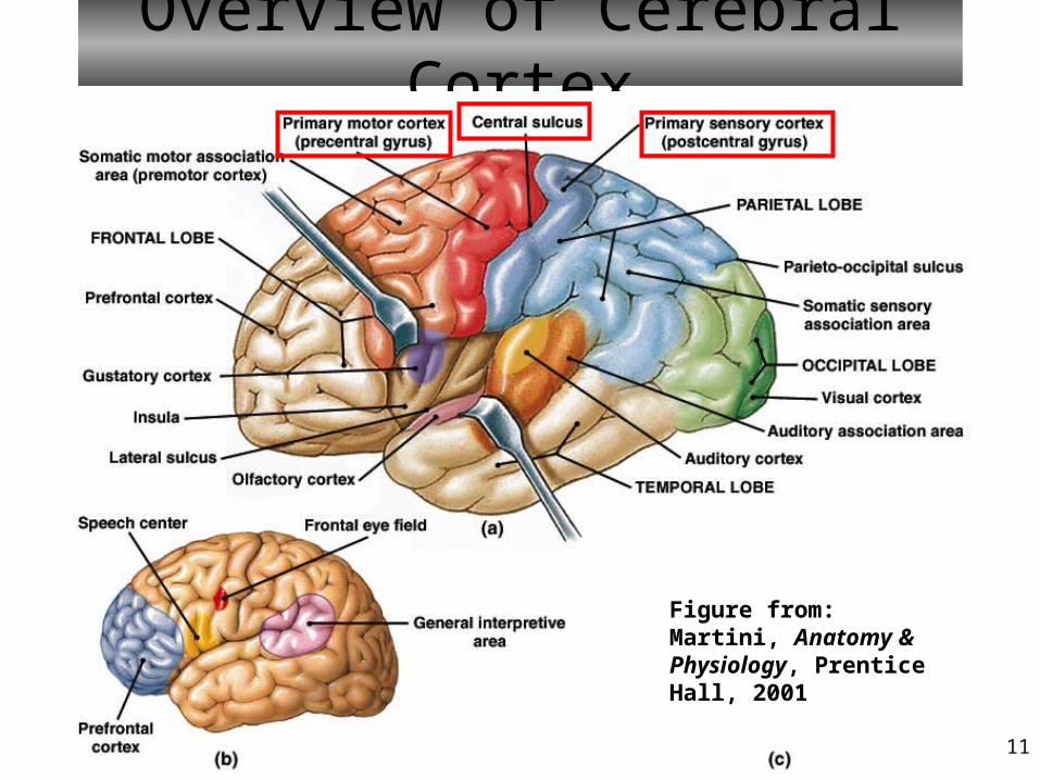
Overview of Cerebral Cortex
Figure from: Martini, Anatomy & Physiology, Prentice Hall, 2001
12
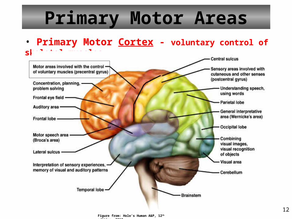
Primary Motor Areas• Primary Motor Cortex - voluntary control of skeletal muscles
Figure from: Hole’s Human A&P, 12th edition, 2010

13
Motor Areas of the CortexNotice the relative amount of cortical tissue devoted to each motor function. What would this be proportional to?
Figures from: Saladin, Anatomy & Physiology, McGraw Hill, 2007
The Motor “Homunculus”
?

14
Broca’s Area (Motor)• Broca’s Area
• in one (dominant, usually left) hemisphere• controls muscles needed for speech
Figure from: Hole’s Human A&P, 12th edition, 2010
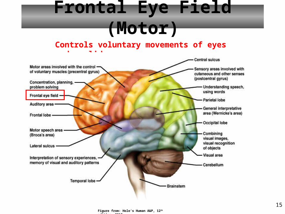
15
Frontal Eye Field (Motor)Controls voluntary movements of eyes and eyelids
Figure from: Hole’s Human A&P, 12th edition, 2010

16
Sensory Areas
Visual Area• occipital lobe• interprets vision
Auditory Area• temporal lobe• interprets hearing
Cutaneous Sensory Area• parietal lobe• interprets sensations on skin
Figure from: Hole’s Human A&P, 12th edition, 2010

17
Sensory Areas of the CortexNotice the relative amount of cortical tissue devoted to each sensory function.
The Somatosensory “Homunculus”
Figures from: Saladin, Anatomy & Physiology, McGraw Hill, 2007

18
Association Areas• regions of cortex that are not primary motor or primary sensory areas• widespread throughout the cerebral cortex• analyze and interpret sensory experiences; coordinate motor responses• memory, reasoning, verbalization, judgment, emotions
Figure from: Hole’s Human A&P, 12th edition, 2010
20
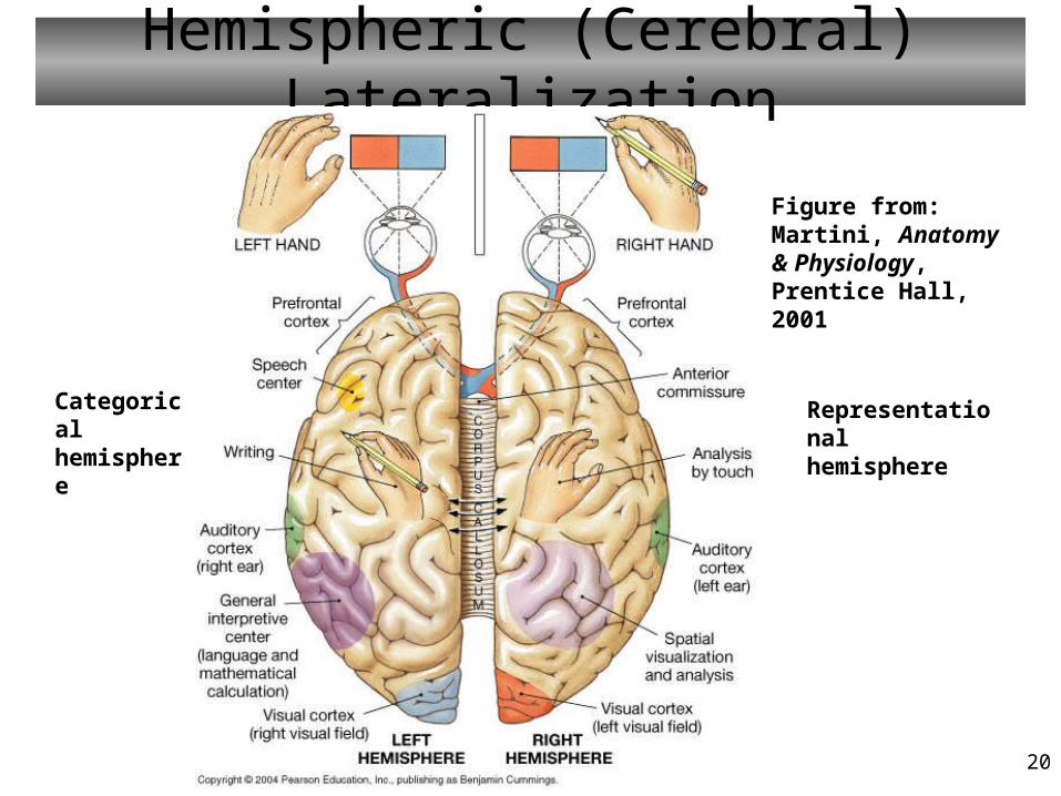
Hemispheric (Cerebral) Lateralization
Figure from: Martini, Anatomy & Physiology, Prentice Hall, 2001
Categorical hemisphere
Representational hemisphere

21
Basal Nuclei• nuclei are masses of gray matter in CNS
• deep within cerebral hemispheres
• caudate nucleus, putamen, globus pallidus (together called the corpus striatum)
• subconscious control certain muscular activities, e.g., learned movement patterns
Relay motor impulses originating in the cerebral cortex and substantia nigra of the midbrain.
Figure from: Hole’s Human A&P, 12th edition, 2010

22
Limbic SystemConsists of
• portions of frontal lobe• portions of temporal lobe• hypothalamus• thalamus• basal nuclei• other deep nuclei• associated with sense of smell (less significant)
The motivational system
Figure from: Saladin, Anatomy & Physiology, McGraw Hill, 2007
Functions• controls emotions• produces feelings• interprets sensory impulses• facilitates memory storage and retrieval (learning!)

23
Memory• A “Memory” is the persistence of knowledge that can be
accessed (we hope!) at a later time.• Memories are not stored in individual “memory cells” or
neurons; they are stored as pathways called engrams, or memory traces that use strengthened or altered synapses.
• Immediate memory lasts a few seconds, e.g., remembering the earliest part of a sentence to make sense of it.
• Short-term memory (STM) lasts a few seconds to a few hours– Working memory is a form of this (repeating a phone number over
to yourself just long enough to dial it – and then forget it!)– Limited to a few ‘bits’ of information (about 7-9). So, ‘chunk up’!
• Long-term memory (LTM) can last a lifetime– Can hold much more information that STM– Declarative (events and facts)– Procedural (motor skills)

24
Diencephalon• between cerebral hemispheres and brainstem• surrounds third ventricle
• thalamus• hypothalamus• epithalamus
• optic tracts• optic chiasm• infundibulum• posterior pituitary• mammillary bodies• pineal gland
(Tectum)
Figure from: Hole’s Human A&P, 12th edition, 2010

25
DiencephalonThalamus
• gateway for sensory impulses heading to cerebral cortex• receives all sensory impulses (except smell)• channels impulses to appropriate part of cerebral cortex and to the basal nuclei for interpretation
Hypothalamus• maintains homeostasis by regulating visceral activities
- Heart rate and blood pressure- Body temperature - Water and electrolyte balance- Hunger and body weight- Movement/secretions of glands and intestines- Stimulation of the pituitary (links nervous and endocrine)- Sleep and wakefulness

27
Brain Stem
Three Parts1. Midbrain2. Pons3. Medulla Oblongata
(Tectum)
Figure from: Hole’s Human A&P, 12th edition, 2010

28
Midbrain• between diencephalon and pons
• contains bundles of fibers that join lower parts of brainstem and spinal cord with higher part of brain
• cerebral aqueduct
• cerebral peduncles – bundles of nerve fibers
• contains red nucleus (rubro-) and substantia nigra
• corpora quadrigemina – centers for visual and auditory reflexes
(Tectum)
Figure from: Saladin, Anatomy & Physiology, McGraw Hill, 2007
Major connecting center between spinal cord and brain and parts of brainstem

29
Pons
• rounded bulge on underside of brainstem
• between medulla oblongata and midbrain
• helps regulate rate and depth of breathing
• relays nerve impulses to and from medulla oblongata and cerebellum
Ventral view Dorsal view
(Tectum)
Figure from: Hole’s Human A&P, 12th edition, 2010

30
Medulla Oblongata• enlarged continuation of spinal cord
• conducts ascending (olive) and descending (pyramids) impulses between brain and spinal cord
• contains cardiac, vasomotor, and respiratory control centers
• contains various nonvital reflex control centers (coughing, sneezing, vomiting)
Ventral view Dorsal view
(Tectum)
Figure from: Hole’s Human A&P, 12th edition, 2010

31
Reticular Formation• complex network of nerve fibers scattered throughout the brain stem
• extends into the diencephalon
• connects to centers of hypothalamus, basal nuclei, cerebellum, and cerebrum
• filters incoming sensory information; habituation
• modulates pain
• arouses cerebral cortex into state of wakefulness
Ascending portion is called the ‘reticular activating system’
(prefix = reticulo-)
Figure from: Hole’s Human A&P, 12th edition, 2010

32
Cerebellum• integrates sensory information concerning position of body parts
• coordinates skeletal muscle activity
• maintains posture
•May also be involved in several sensory, linguistic, emotional and non-motor functions
Figure from: Saladin, Anatomy & Physiology, McGraw Hill, 2007

33
Peripheral Nervous System
CNS PNS
You are here
Figure from: Hole’s Human A&P, 12th edition, 2010

34
Peripheral Nervous System
• Cranial nerves arising from the brain• Somatic fibers connecting to the skin and skeletal muscles• Autonomic fibers connecting to viscera
• Spinal nerves arising from the spinal cord• Somatic fibers connecting to the skin and skeletal muscles• Autonomic fibers connecting to viscera

35
Cranial Nerves
Paired. Numbered (roughly) in the order of their occurrence from anterior to posterior. Abbreviated using N or CN.
Figure from: Hole’s Human A&P, 12th edition, 2010

36
The Cranial Nerves
Numeral Name
Function Sensory, Motor, or Both (Mixed Nerve)
I OLFACTORY (OLD) OLFACTION/SMELL SENSORY (SOME)
II OPTIC (OPIE) VISION SENSORY (SAY)
III OCULOMOTOR (OCCASIONALLY) MOVE EYE MOTOR (MARRY)
IV TROCHLEAR (TRIES) MOVE EYE (superior oblique) MOTOR (MONEY)
V TRIGEMINAL (TRIGONOMETRY) MAJOR SENSORY NERVE FROM FACE
BOTH (BUT)
VI ABDUCENS (AND) MOVE EYE (lateral rectus) MOTOR (MY)
VII FACIAL (FEELS) MAJOR MOTOR NERVE OF FACE BOTH (BROTHER)
VIII VESTIBULOCOCHLEAR (VERY) HEARING AND EQUILIBRIUM SENSORY (SAYS)
IX GLOSSOPHARYNGEAL (GLOOMY) MOVE MUSCLES OF TONGUE AND PHARYNX
BOTH (BIG)
X VAGUS (VAGUE) INNERVATE VISCERAL SMOOTH MUSCLE; MUSCLES OF SPEECH
BOTH (BOOBS)
XI ACCESSORY (AND) MOVE NECK MUSCLES MOTOR (MATTER)
XII HYPOGLOSSAL (HYPOACTIVE) MOVE TONGUE MOTOR (MOST)
You should know this table
37
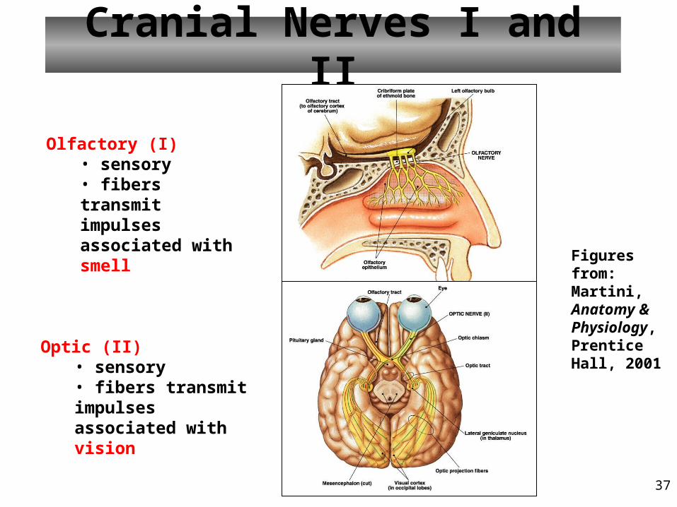
Cranial Nerves I and II
Olfactory (I)• sensory• fibers transmit impulses associated with smell
Optic (II)• sensory• fibers transmit impulses associated with vision
Figures from: Martini, Anatomy & Physiology, Prentice Hall, 2001

38
Cranial Nerves III, IV, and VI
Trochlear (IV)• primarily motor• motor impulses to the superior oblique (SO) muscles that move the eyes
Oculomotor (III)• primarily motor• motor impulses to muscles that
• raise eyelids• move the eyes• focus lens•adjust light entering eye
Figure from: Martini, Anatomy & Physiology, Prentice Hall, 2001
What’s a ganglion?
Abducens (VI)• primarily motor• motor impulses to the lateral rectus (LR) muscles that move the eyes

39
Cranial Nerve V
Trigeminal (V)• both sensory and motor• opthalmic division
• sensory from surface of eyes, tear glands, scalp, forehead, and upper eyelids
• maxillary division• sensory from upper teeth, upper gum, upper lip, palate, and skin of face
• mandibular division• sensory from scalp, skin of jaw, lower teeth, lower gum, and lower lip• motor to muscles of mastication and muscles in floor of mouth
Major sensory nerve of face
Figure from: Hole’s Human A&P, 12th edition, 2010

40
Cranial Nerve VII
Facial (VII)• both sensory and motor• sensory from taste receptors• motor to muscles of facial expression, tear glands, and salivary glands
Major MOTOR nerve of face
Figures from: Saladin, Anatomy & Physiology, McGraw Hill, 2007

41
Cranial Nerves VIII and IXVestibulocochlear (VIII)
• sensory• sensory from equilibrium receptors of ear• sensory from hearing receptors
Glossopharyngeal (IX)• both sensory and motor• sensory from pharynx, tonsils, tongue, and carotid arteries• motor to salivary glands and muscles of pharynx
Figures from: Martini, Anatomy & Physiology, Prentice Hall, 2001

42
Cranial Nerve X
Vagus (X)• both sensory and motor• somatic motor to muscles of speech and swallowing• autonomic motor (parasympathetic) to viscera of thorax and abdomen• sensory from pharynx, larynx, esophagus, and viscera of thorax and abdomen
Figure from: Saladin, Anatomy & Physiology, McGraw Hill, 2007

43
Cranial Nerves XI and XII
Accessory (XI)• primarily motor• motor to muscles of soft palate, pharynx, larynx, neck, and back
Hypoglossal (XII)• primarily motor• motor to muscles of the tongue
Figure from: Martini, Fundamentals of Anatomy & Physiology, Pearson Education, 2004

44
Review• The brain is protected by the
– Skull bones– Meninges– CSF– Blood-brain barrier
• The meninges of the brain and spinal cord consist of the– Dura mater– Arachnoid (membrane)– Pia mater

45
Review• Important motor areas of cerebral cortex
– Precentral gyrus (Primary motor area)– Broca’s area– Frontal eye field
• Important sensory areas of cerebral cortex– Postcentral gyrus (Primary cutaneous sensory)– Visual area (occipital lobe)– Auditory area (temporal lobe)

46
ReviewTable from: Hole’s Human A&P, 12th edition, 2010

47
The Cranial NervesNumeral
Name
Function Sensory, Motor, or Both (Mixed Nerve)
I OLFACTORY (OLD) OLFACTION/SMELL SENSORY (SOME)
II OPTIC (OPIE) VISION SENSORY (SAY)
III OCULOMOTOR (OCCASIONALLY) MOVE EYE MOTOR (MARRY)
IV TROCHLEAR (TRIES) MOVE EYE MOTOR (MONEY)
V TRIGEMINAL (TRIGONOMETRY) CHEWING, MASTICATION AND SENSORY FROM FACE
BOTH (BUT)
VI ABDUCENS (AND) MOVE EYE MOTOR (MY)
VII FACIAL (FEELS) FACIAL EXPRESSION BOTH (BROTHER)
VIII VESTIBULOCOCHLEAR (VERY) HEARING AND EQUILIBRIUM SENSORY (SAYS)
IX GLOSSOPHARYNGEAL (GLOOMY) MOVE MUSCLES OF TONGUE AND PHARYNX
BOTH (BIG)
X VAGUS (VAGUE) INNERVATE VISCERAL SMOOTH MUSCLE
BOTH (BOOBS)
XI ACCESSORY (AND) MOVE NECK MUSCLES MOTOR (MATTER)
XII HYPOGLOSSAL (HYPOACTIVE) MOVE TONGUE MOTOR (MOST)