1 Interpretation of Laboratory Tests: A Case-Oriented Review of Clinical Laboratory Diagnosis Roger...
77
1 Interpretation of Laboratory Tests: A Case-Oriented Review of Clinical Laboratory Diagnosis Roger L. Bertholf, Ph.D. Associate Professor of Pathology University of Florida Health Science Center/Jacksonville
-
Upload
ibrahim-sutcliffe -
Category
Documents
-
view
225 -
download
2
Transcript of 1 Interpretation of Laboratory Tests: A Case-Oriented Review of Clinical Laboratory Diagnosis Roger...
1
Interpretation of Laboratory Tests:A Case-Oriented Review of Clinical
Laboratory Diagnosis
Roger L. Bertholf, Ph.D.Associate Professor of Pathology
University of Florida Health Science Center/Jacksonville
2
Case 1: Oliguria and hematuria
R. Bertholf American Society of Clinical
Pathologists3
Case 1: Oliguria and hematuria
A 7-year-old boy was brought to the pediatrician because of vomiting and malaise. On physical examination he was slightly flushed, and had some noticeable swelling of his hands and feet. The patient was uncomfortable, and complained of pain “in his tummy”. He had a slight fever. Heart was normal and lungs were clear. His past medical history did not include any chronic diseases. The mother noted that he had a severe sore throat “about two weeks ago”, but that it had cleared up on its own. The child was not taking any medications. There were no masses in the abdomen, and lymphadenopathy was not present. The child had some difficulty producing a urine specimen, but finally was able to produce a small amount of urine, which was dipstick-positive for blood and protein.
R. Bertholf American Society of Clinical
Pathologists4
Questions. . .
• What is the differential diagnosis in this case?
• What laboratory tests might be helpful in establishing the diagnosis?
R. Bertholf American Society of Clinical
Pathologists5
What do the kidneys do?
• Regulate body fluid osmolality and volume
• Regulate electrolyte balance
• Regulate acid-base balance
• Excrete metabolic products and foreign substances
• Produce and excrete hormones

R. Bertholf American Society of Clinical
Pathologists6
The kidneys as regulatory organs
“The kidney presents in the highest degree the phenomenonof sensibility, the power of reacting to various stimuli in a
direction which is appropriate for the survival of the organism;a power of adaptation which almost gives one the idea that its
component parts must be endowed with intelligence.”
E. Starling (1909)
R. Bertholf American Society of Clinical
Pathologists7
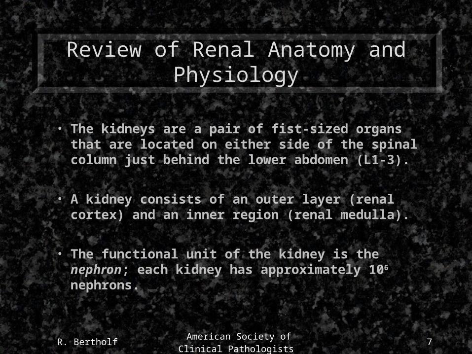
Review of Renal Anatomy and Physiology
• The kidneys are a pair of fist-sized organs that are located on either side of the spinal column just behind the lower abdomen (L1-3).
• A kidney consists of an outer layer (renal cortex) and an inner region (renal medulla).
• The functional unit of the kidney is the nephron; each kidney has approximately 106 nephrons.
R. Bertholf American Society of Clinical
Pathologists8
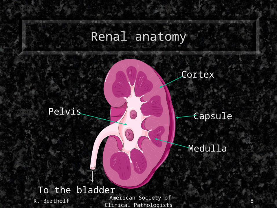
Renal anatomy
Cortex
Medulla
Pelvis
To the bladder
Capsule

R. Bertholf American Society of Clinical
Pathologists9
The Nephron
Renal artery
Glomerulus
Bowman’s capsule
Proximal tubule
Distal tubule
Collecting duct
Henle’s Loop
Afferent arteriole
R. Bertholf American Society of Clinical
Pathologists10
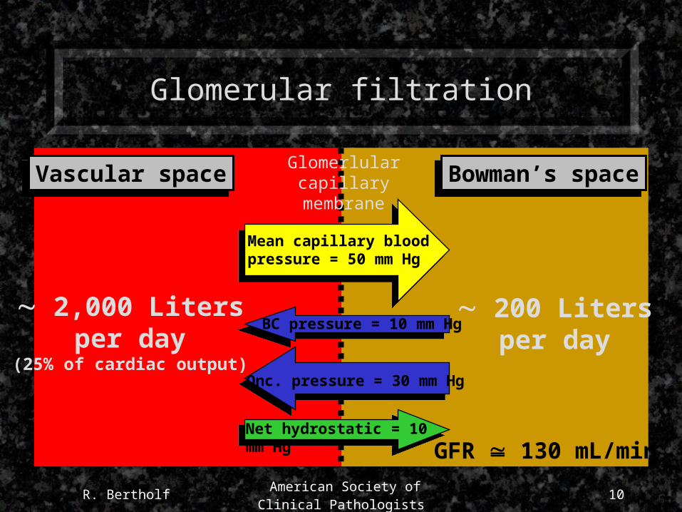
Glomerular filtration
Glomerlularcapillary
membraneVascular spaceVascular space Bowman’s spaceBowman’s space
Mean capillary bloodpressure = 50 mm Hg
BC pressure = 10 mm Hg
Onc. pressure = 30 mm Hg
Net hydrostatic = 10 mm Hg
2,000 Litersper day
(25% of cardiac output)
200 Litersper day
GFR 130 mL/min
R. Bertholf American Society of Clinical
Pathologists11
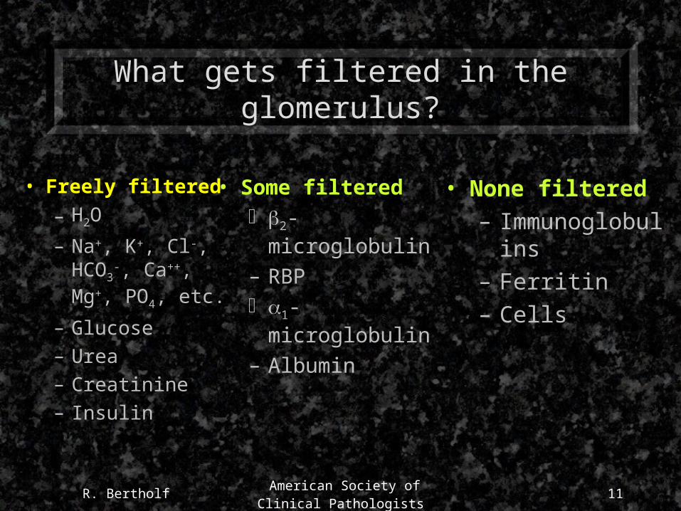
What gets filtered in the glomerulus?
• Freely filtered
– H2O
– Na+, K+, Cl-, HCO3
-, Ca++, Mg+, PO4, etc.
– Glucose
– Urea
– Creatinine
– Insulin
• Some filtered 2-microglobulin
– RBP 1-microglobulin
– Albumin
• None filtered
– Immunoglobulins
– Ferritin
– Cells

R. Bertholf American Society of Clinical
Pathologists12
Then what happens?
• If 200 liters of filtrate enter the nephrons each day, but only 1-2 liters of urine result, then obviously most of the filtrate (99+ %) is reabsorbed.
• Reabsorption can be active or passive, and occurs in virtually all segments of the nephron.

R. Bertholf American Society of Clinical
Pathologists13
Reabsorption from glomerular filtrate
% ReabsorbedWater 99.2
Sodium 99.6Potassium 92.9Chloride 99.5
Bicarbonate 99.9Glucose 100Albumin 95-99
Urea 50-60Creatinine 0 (or negative)
R. Bertholf American Society of Clinical
Pathologists14
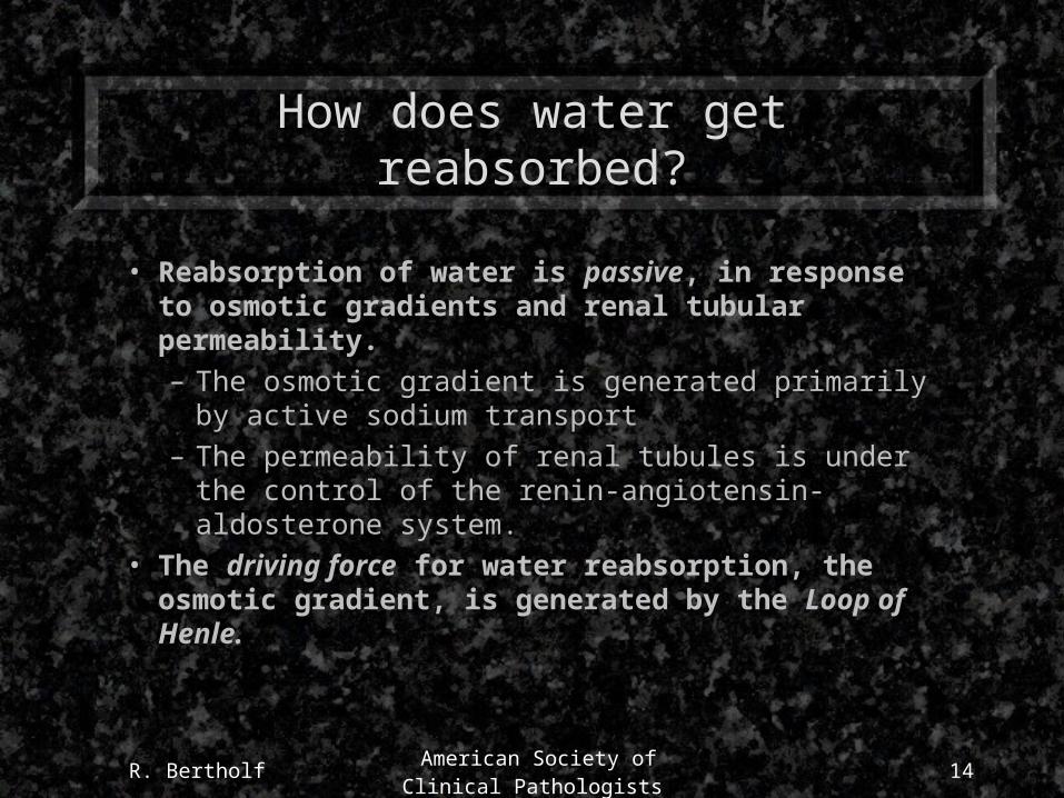
How does water get reabsorbed?
• Reabsorption of water is passive, in response to osmotic gradients and renal tubular permeability.– The osmotic gradient is generated primarily by active
sodium transport– The permeability of renal tubules is under the control
of the renin-angiotensin-aldosterone system.• The driving force for water reabsorption, the osmotic
gradient, is generated by the Loop of Henle.
R. Bertholf American Society of Clinical
Pathologists15
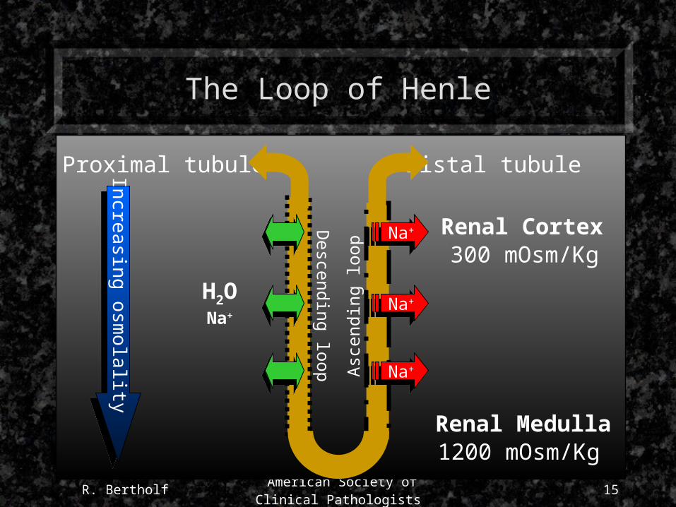
The Loop of Henle
Proximal tubule Distal tubule
Descending loop A
scen
ding
loop
Increasing osmolality
Renal Cortex
Renal Medulla
Na+Na+
Na+Na+
Na+Na+
H2ONa+
1200 mOsm/Kg
300 mOsm/Kg
R. Bertholf American Society of Clinical
Pathologists16
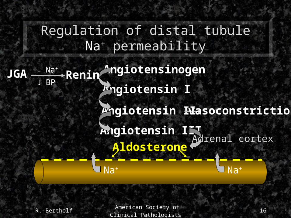
Regulation of distal tubule Na+ permeability
JGA Renin Na+
BP
Angiotensinogen
Angiotensin I
Angiotensin II
Angiotensin III
vasoconstriction
AldosteroneAdrenal cortex
Na+ Na+

R. Bertholf American Society of Clinical
Pathologists17
Regulation of H2O reabsorption
Pituitary
ADH (vasopressin)
Plasmahyperosmolality
H2OH2O
Renal Medulla (osmolality 1200 mOsm/Kg)

R. Bertholf American Society of Clinical
Pathologists18
Summary of renal physiology
Filtration - Reabsorption + Secretion = Elimination
GFR (Filtered but not reabsorbed or secreted)
TRPF (Filtered and secreted)
R. Bertholf American Society of Clinical
Pathologists19
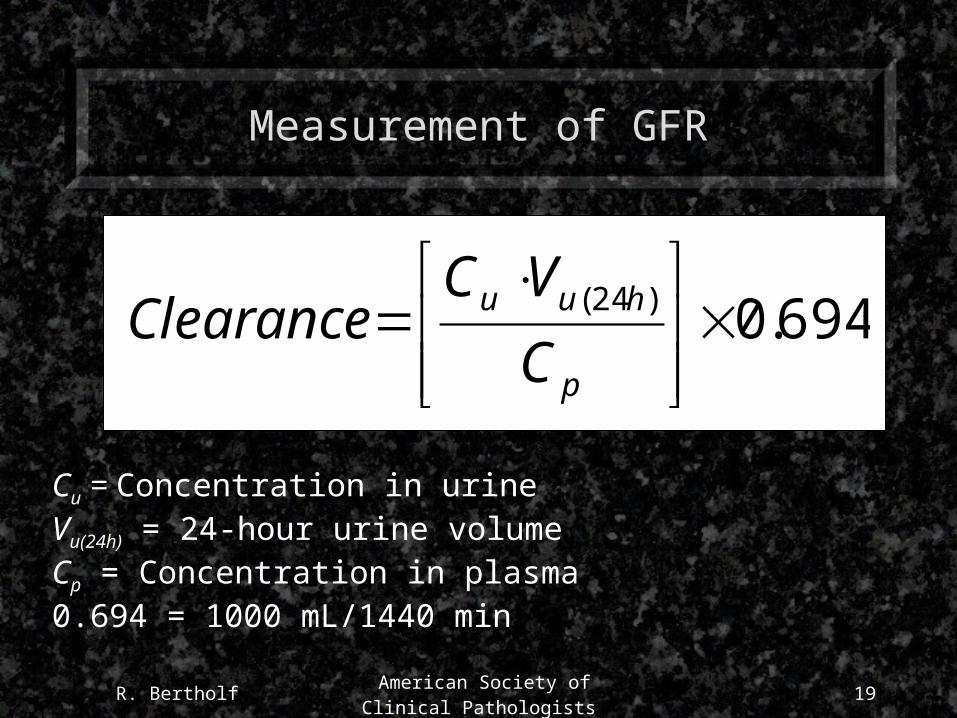
Measurement of GFR
694.0)24(
p
huu
C
VCClearance
Cu = Concentration in urineVu(24h) = 24-hour urine volumeCp = Concentration in plasma0.694 = 1000 mL/1440 min
R. Bertholf American Society of Clinical
Pathologists20
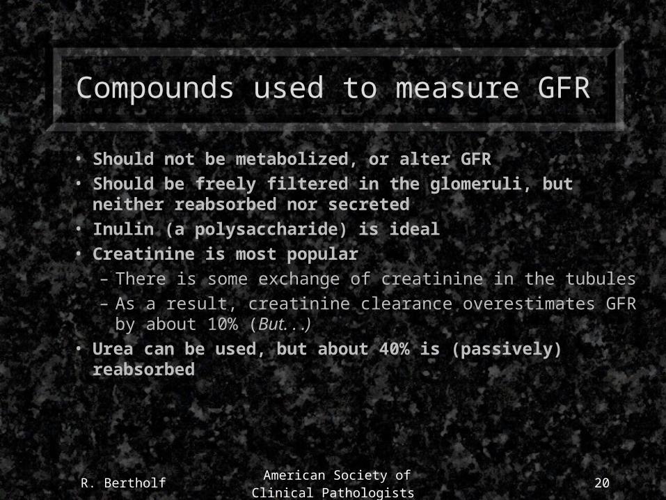
Compounds used to measure GFR
• Should not be metabolized, or alter GFR• Should be freely filtered in the glomeruli, but neither
reabsorbed nor secreted• Inulin (a polysaccharide) is ideal• Creatinine is most popular
– There is some exchange of creatinine in the tubules– As a result, creatinine clearance overestimates GFR by about
10% (But. . .)• Urea can be used, but about 40% is (passively) reabsorbed
R. Bertholf American Society of Clinical
Pathologists21
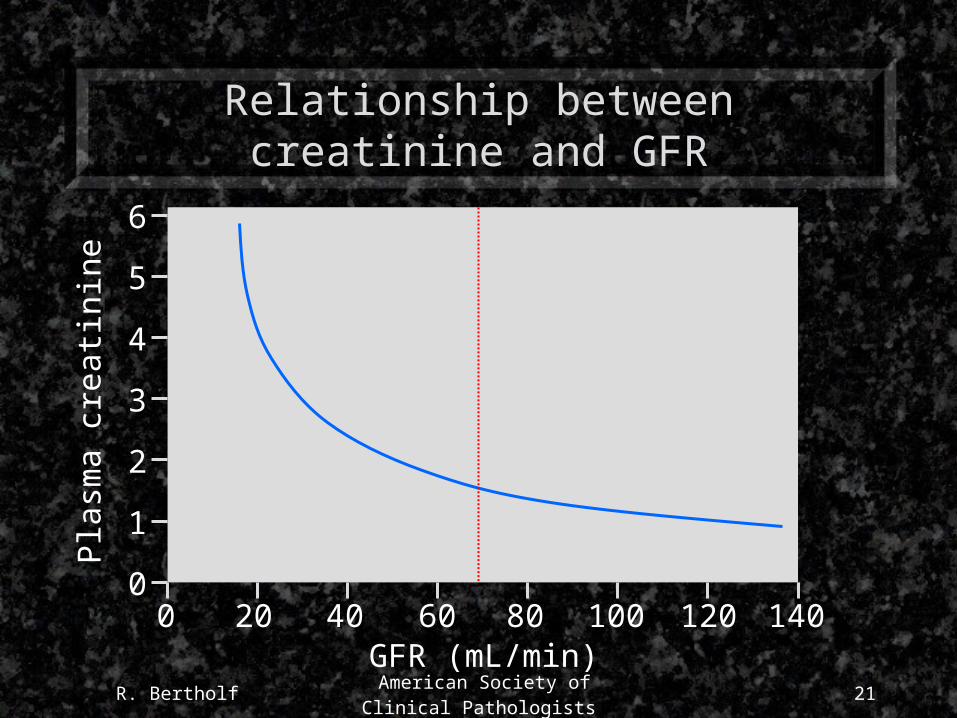
Relationship between creatinine and GFRP
lasm
a cr
eati
nine
GFR (mL/min)
1
2
3
4
5
6
00 20 40 60 80 100 120 140

R. Bertholf American Society of Clinical
Pathologists22
Measurement of TRPF
• Para-aminohippurate (PAH) is freely filtered in the glomeruli and actively secreted in the tubules.
• PAH clearance gives an estimate of the total amount of plasma from which a constituent can be removed.

R. Bertholf American Society of Clinical
Pathologists23
Creatinine
O
N
CH3HN
NH
HO
O CH3
NH
NH2
- H2ON
Creatine Creatinine
1-2% of creatine is hydrolyzed to creatinine each day
R. Bertholf American Society of Clinical
Pathologists24
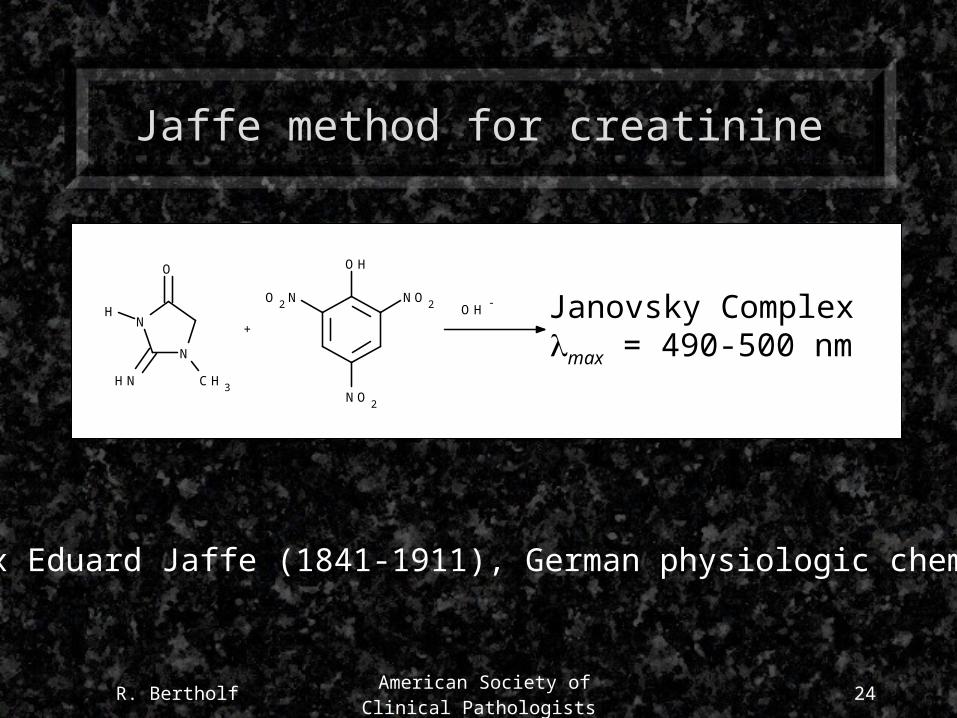
Jaffe method for creatinine
O
N
CH3HN
NH
O2N NO2
NO2
OH
OH -
+Janovsky Complexmax = 490-500 nm
Max Eduard Jaffe (1841-1911), German physiologic chemist
R. Bertholf American Society of Clinical
Pathologists25
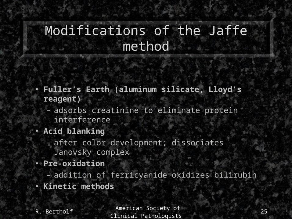
Modifications of the Jaffe method
• Fuller’s Earth (aluminum silicate, Lloyd’s reagent)
– adsorbs creatinine to eliminate protein interference
• Acid blanking
– after color development; dissociates Janovsky complex
• Pre-oxidation
– addition of ferricyanide oxidizes bilirubin
• Kinetic methods
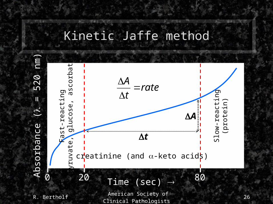
R. Bertholf American Society of Clinical
Pathologists26
Kinetic Jaffe methodA
bsor
banc
e (
= 5
20 n
m)
Time (sec) 0 8020
Fast
-rea
ctin
g(p
yruv
ate,
glu
cose
, asc
orba
te)
Slow
-rea
ctin
g(p
rote
in)
t
A
ratet
A
creatinine (and -keto acids)

R. Bertholf American Society of Clinical
Pathologists27
Enzymatic creatinine methods
• Creatininase
– creatininecreatineCKADPPKLD
• Creatinase
– creatininecreatinesarcosinesarcosine oxidaseperoxideperoxidase reaction
• Creatinine deaminase (iminohydrolase)
– most common
R. Bertholf American Society of Clinical
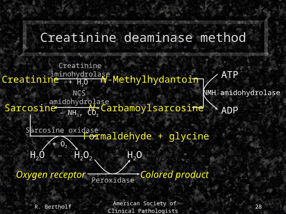
Pathologists28
Creatinine deaminase method
Creatinine
Creatinineiminohydrolase
+ H2O N-Methylhydantoin ATP
ADP
NMH amidohydrolase
N-Carbamoylsarcosine
H2O
PeroxidaseOxygen receptor Colored product
Sarcosine
NCSamidohydrolase
- NH3, CO2
+ O2
Sarcosine oxidase
H2O H2O2
Formaldehyde + glycine
R. Bertholf American Society of Clinical
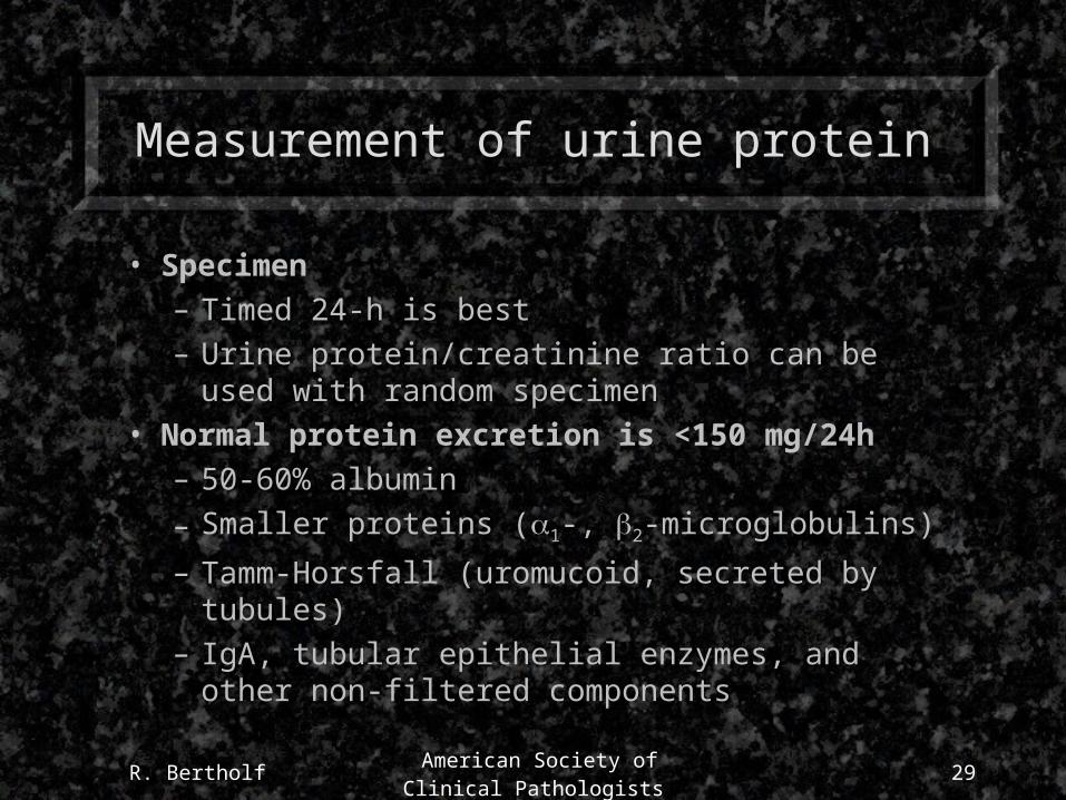
Pathologists29
Measurement of urine protein
• Specimen– Timed 24-h is best– Urine protein/creatinine ratio can be used with
random specimen• Normal protein excretion is <150 mg/24h
– 50-60% albumin
– Smaller proteins (1-, 2-microglobulins)
– Tamm-Horsfall (uromucoid, secreted by tubules)– IgA, tubular epithelial enzymes, and other non-
filtered components

R. Bertholf American Society of Clinical
Pathologists30
Dipstick method for urine protein
• Method is based on protein association with pH indicator
• Test pad contains dye tetrabromphenol blue at pH=3
• If protein binds to the pH indicator, H+ is displaced and the color changes from yellow to green (or blue)
• Most sensitive to albumin (poor method for detecting tubular proteinuria)
R. Bertholf American Society of Clinical
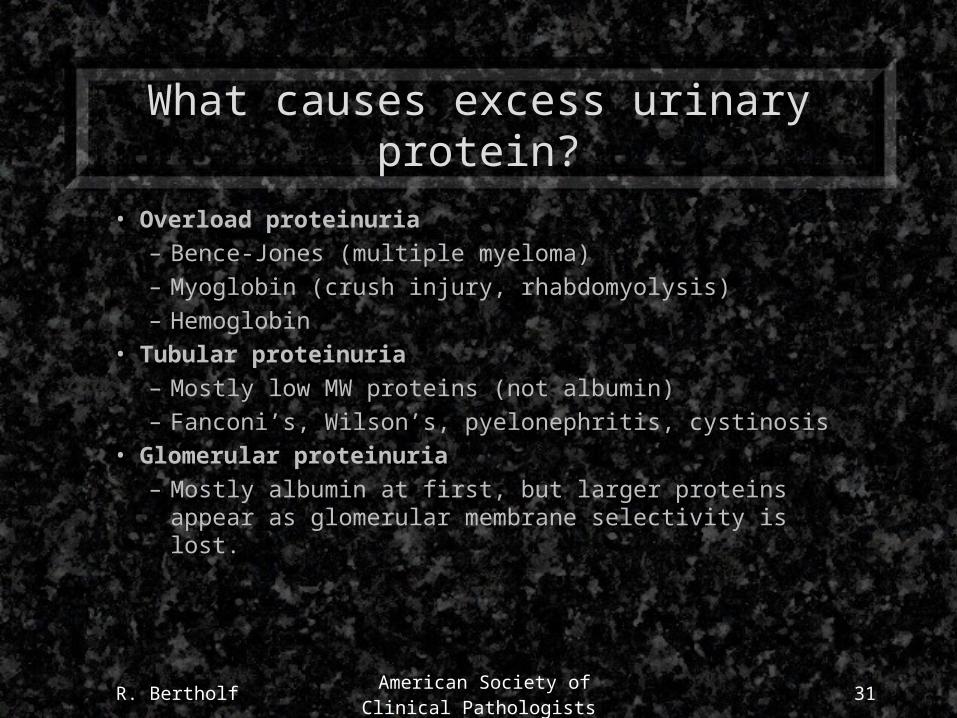
Pathologists31
What causes excess urinary protein?
• Overload proteinuria– Bence-Jones (multiple myeloma)– Myoglobin (crush injury, rhabdomyolysis)– Hemoglobin
• Tubular proteinuria– Mostly low MW proteins (not albumin)– Fanconi’s, Wilson’s, pyelonephritis, cystinosis
• Glomerular proteinuria– Mostly albumin at first, but larger proteins appear as
glomerular membrane selectivity is lost.
R. Bertholf American Society of Clinical
Pathologists32
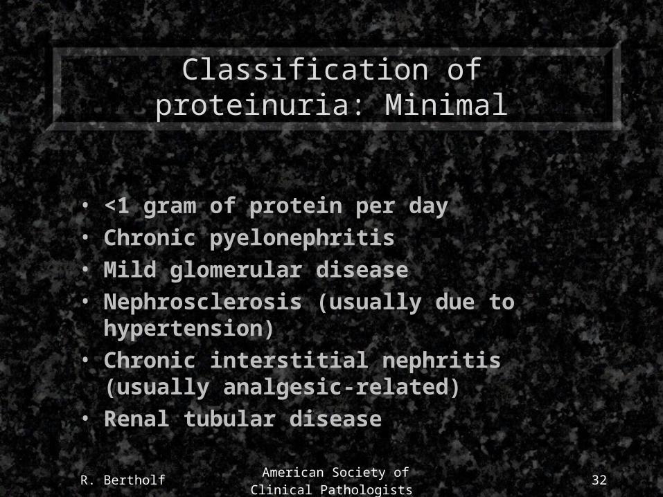
Classification of proteinuria: Minimal
• <1 gram of protein per day
• Chronic pyelonephritis
• Mild glomerular disease
• Nephrosclerosis (usually due to hypertension)
• Chronic interstitial nephritis (usually analgesic-related)
• Renal tubular disease
R. Bertholf American Society of Clinical
Pathologists33
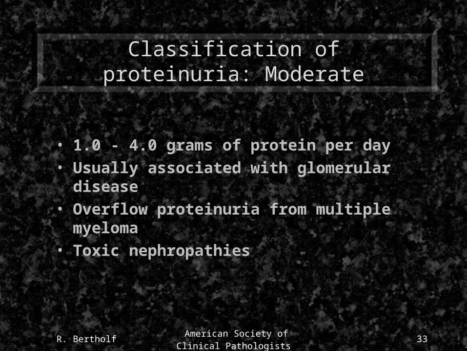
Classification of proteinuria: Moderate
• 1.0 - 4.0 grams of protein per day
• Usually associated with glomerular disease
• Overflow proteinuria from multiple myeloma
• Toxic nephropathies
R. Bertholf American Society of Clinical
Pathologists34
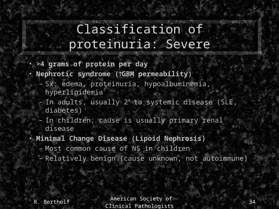
Classification of proteinuria: Severe
• >4 grams of protein per day
• Nephrotic syndrome (GBM permeability)
– Sx: edema, proteinuria, hypoalbuminemia, hyperlipidemia
– In adults, usually 2 to systemic disease (SLE, diabetes)
– In children, cause is usually primary renal disease
• Minimal Change Disease (Lipoid Nephrosis)
– Most common cause of NS in children
– Relatively benign (cause unknown, not autoimmune)
R. Bertholf American Society of Clinical
Pathologists35
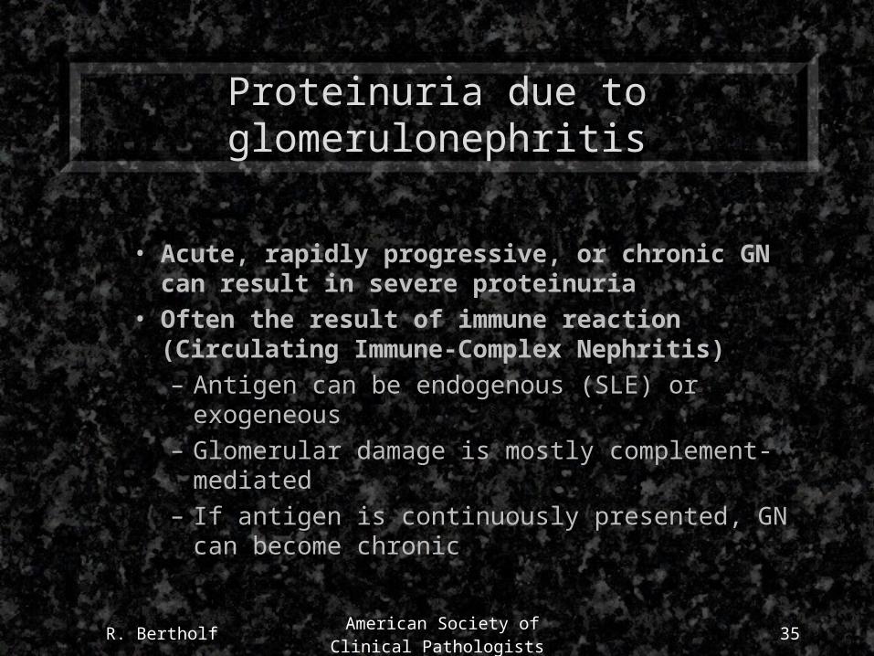
Proteinuria due to glomerulonephritis
• Acute, rapidly progressive, or chronic GN can result in severe proteinuria
• Often the result of immune reaction (Circulating Immune-Complex Nephritis)
– Antigen can be endogenous (SLE) or exogeneous
– Glomerular damage is mostly complement-mediated
– If antigen is continuously presented, GN can become chronic

R. Bertholf American Society of Clinical
Pathologists36
How do red blood cells get in urine?
• Hematuria can result from bleeding anywhere in the kidneys or urinary tract
– Disease, trauma, toxicity
• Hemoglobinuria can result from intravascular hemolysis
– Disease, trauma, toxicity
R. Bertholf American Society of Clinical
Pathologists37
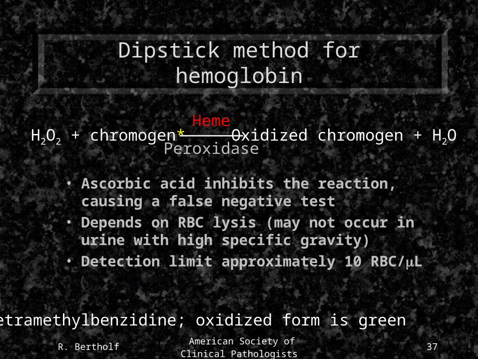
Dipstick method for hemoglobin
• Ascorbic acid inhibits the reaction, causing a false negative test
• Depends on RBC lysis (may not occur in urine with high specific gravity)
• Detection limit approximately 10 RBC/L
H2O2 + chromogen* Oxidized chromogen + H2OHeme
Peroxidase
*tetramethylbenzidine; oxidized form is green
R. Bertholf American Society of Clinical
Pathologists38
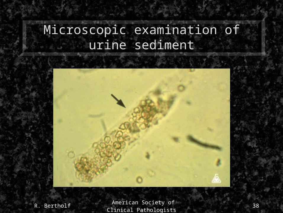
Microscopic examination of urine sediment
R. Bertholf American Society of Clinical
Pathologists39
Significance of RBC casts in urine
• Indicative of blood crossing the GBM
• Casts form in the distal tubules
• Stasis produces brown, granular casts
• RBC casts almost always reflect glomerular disease
R. Bertholf American Society of Clinical
Pathologists40
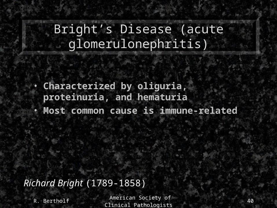
Bright’s Disease (acute glomerulonephritis)
• Characterized by oliguria, proteinuria, and hematuria
• Most common cause is immune-related
Richard Bright (1789-1858)
R. Bertholf American Society of Clinical
Pathologists41
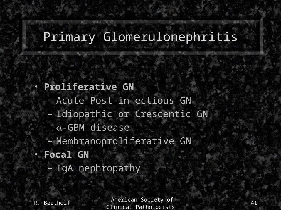
Primary Glomerulonephritis
• Proliferative GN
– Acute Post-infectious GN
– Idiopathic or Crescentic GN -GBM disease
– Membranoproliferative GN
• Focal GN
– IgA nephropathy
R. Bertholf American Society of Clinical
Pathologists42
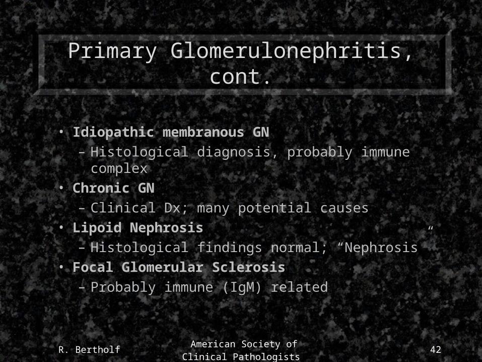
Primary Glomerulonephritis, cont.
• Idiopathic membranous GN
– Histological diagnosis, probably immune complex
• Chronic GN
– Clinical Dx; many potential causes
• Lipoid Nephrosis
– Histological findings normal; “Nephrosis”
• Focal Glomerular Sclerosis
– Probably immune (IgM) related
R. Bertholf American Society of Clinical
Pathologists43
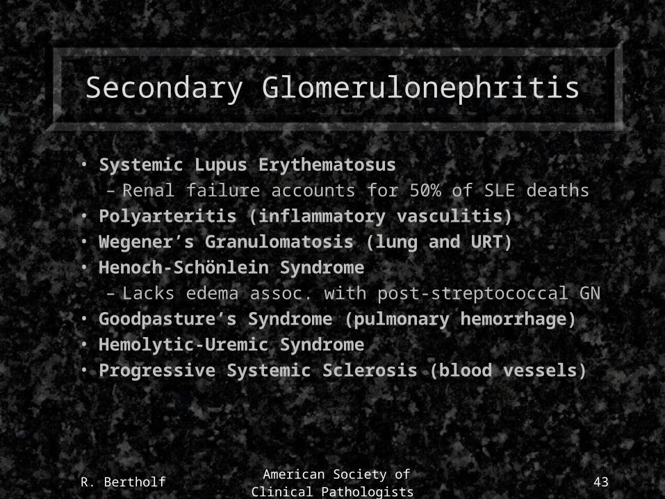
Secondary Glomerulonephritis
• Systemic Lupus Erythematosus– Renal failure accounts for 50% of SLE deaths
• Polyarteritis (inflammatory vasculitis)• Wegener’s Granulomatosis (lung and URT)• Henoch-Schönlein Syndrome
– Lacks edema assoc. with post-streptococcal GN• Goodpasture’s Syndrome (pulmonary hemorrhage)• Hemolytic-Uremic Syndrome• Progressive Systemic Sclerosis (blood vessels)
44
Case 3: Chest Pain

R. Bertholf American Society of Clinical
Pathologists45
Case 3: Chest Pain
A 63 year old male was brought to the emergency departmentafter complaining of severe chest pain that had lasted for twohours. He had been mowing his lawn when the pain developed,and he became concerned when the pain did not subside afterhe stopped the activity. He had no previous history of heartdisease. On presentation he was moderately overweight, dia-phoretic, and in obvious discomfort. He described his chestpain as “beginning in the center of my chest, then my arms, neck, and jaw began to ache too.”
Diagnostic procedures were performed.

R. Bertholf American Society of Clinical
Pathologists46
Questions
• What is the most important consideration in the triage of this patient?
• What tests should be ordered?
R. Bertholf American Society of Clinical
Pathologists47
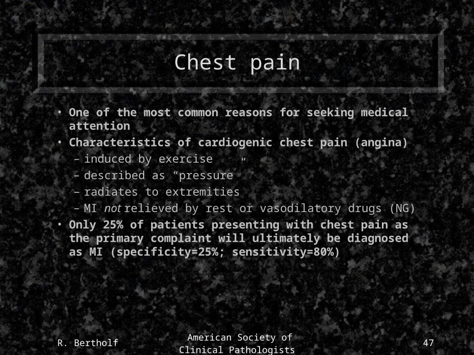
Chest pain
• One of the most common reasons for seeking medical attention
• Characteristics of cardiogenic chest pain (angina)– induced by exercise– described as “pressure”– radiates to extremities– MI not relieved by rest or vasodilatory drugs (NG)
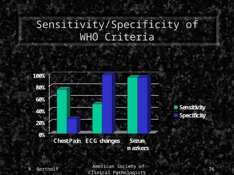
• Only 25% of patients presenting with chest pain as the primary complaint will ultimately be diagnosed as MI (specificity=25%; sensitivity=80%)
R. Bertholf American Society of Clinical
Pathologists48
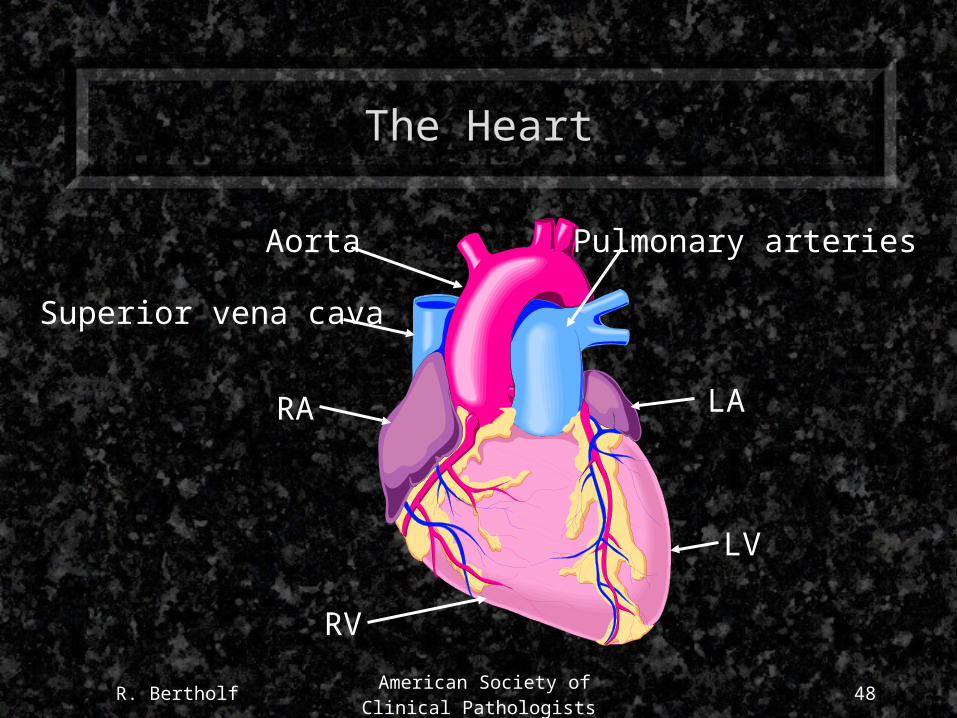
The Heart
Aorta
Superior vena cava
RA LA
RV
LV
Pulmonary arteries
R. Bertholf American Society of Clinical
Pathologists49
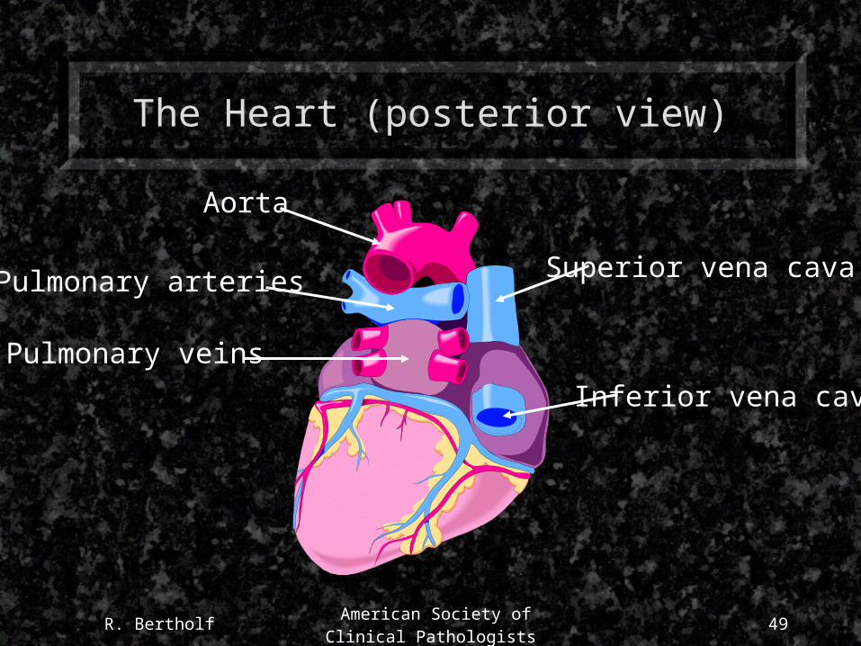
The Heart (posterior view)
Aorta
Superior vena cava
Inferior vena cava
Pulmonary veins
Pulmonary arteries
R. Bertholf American Society of Clinical
Pathologists50
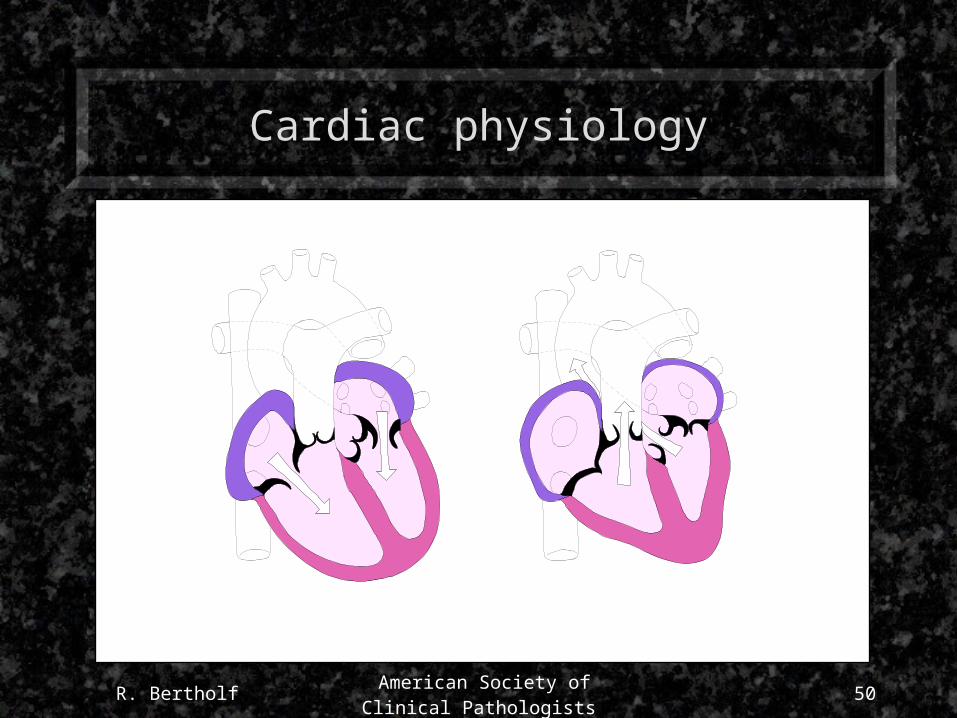
Cardiac physiology
R. Bertholf American Society of Clinical
Pathologists51
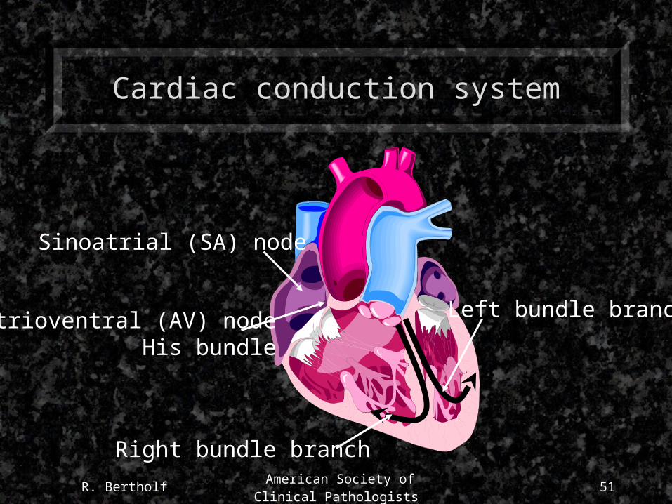
Cardiac conduction system
Sinoatrial (SA) node
Atrioventral (AV) nodeHis bundle
Right bundle branch
Left bundle branch
R. Bertholf American Society of Clinical
Pathologists52
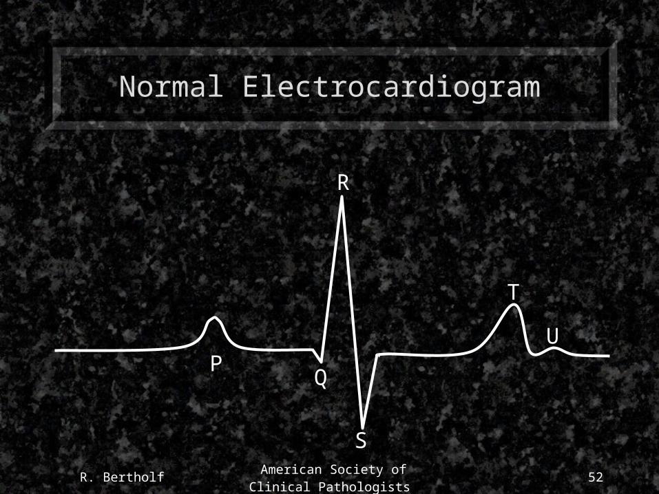
Normal Electrocardiogram
PQ
R
S
T
U

R. Bertholf American Society of Clinical
Pathologists53
Myocardial infarction
Right coronary artery
Left coronary artery
Anterior left ventricle
R. Bertholf American Society of Clinical
Pathologists54
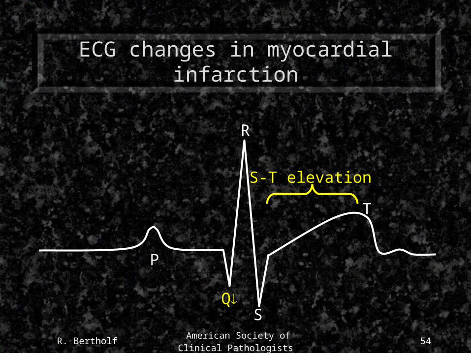
ECG changes in myocardial infarction
S
P
R
T
Q
S-T elevation

R. Bertholf American Society of Clinical
Pathologists55
Diagnostic value of ECG
• ECG changes depend on the location and severity of myocardial necrosis
• Virtually 100% of patients with characteristic Q-wave and S-T segment changes are diagnosed with myocardial infarction (100% specificity)
• However, as many as 50% of myocardial infarctions do not produce characteristic ECG changes (sensitivity 50%)
• ECG may be insensitive for detecting prognostically significant ischemia
R. Bertholf American Society of Clinical
Pathologists56
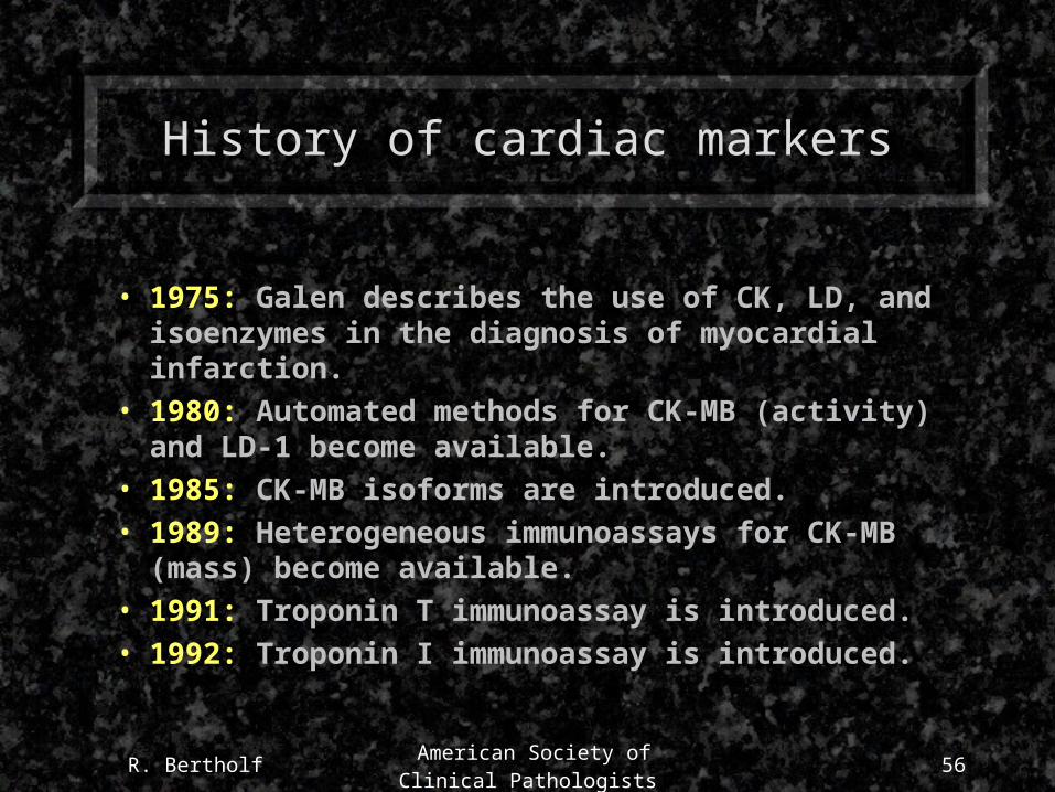
History of cardiac markers
• 1975: Galen describes the use of CK, LD, and isoenzymes in the diagnosis of myocardial infarction.
• 1980: Automated methods for CK-MB (activity) and LD-1 become available.
• 1985: CK-MB isoforms are introduced.
• 1989: Heterogeneous immunoassays for CK-MB (mass) become available.
• 1991: Troponin T immunoassay is introduced.
• 1992: Troponin I immunoassay is introduced.

R. Bertholf American Society of Clinical
Pathologists57
Enzyme markers
• Aspartate transaminase (AST; SGOT)
• 2-Hydroxybutyrate dehydrogenase
• Lactate dehydrogenase
– Five isoenzymes, composed of combinations of H (heart) and M (muscle) subunits
• Creatine kinase
– Three isoenzymes, composed of combinations of M (muscle) and B (brain) subunits
R. Bertholf American Society of Clinical
Pathologists58
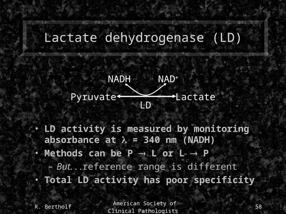
Lactate dehydrogenase (LD)
Pyruvate
• LD activity is measured by monitoring absorbance at = 340 nm (NADH)
• Methods can be P L or L P
– But. . .reference range is different
• Total LD activity has poor specificity
LactateLD
NAD+NADH
R. Bertholf American Society of Clinical
Pathologists59
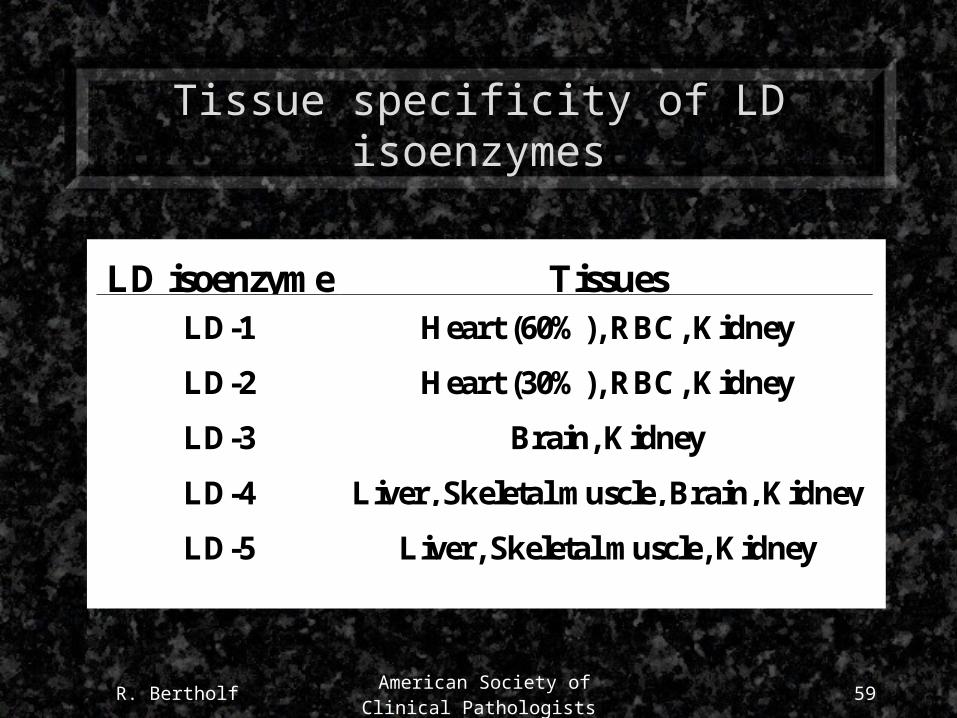
Tissue specificity of LD isoenzymes
LD isoenzyme TissuesLD-1 Heart (60%), RBC, Kidney
LD-2 Heart (30%), RBC, Kidney
LD-3 Brain, Kidney
LD-4 Liver, Skeletal muscle, Brain, Kidney
LD-5 Liver, Skeletal muscle, Kidney
R. Bertholf American Society of Clinical
Pathologists60
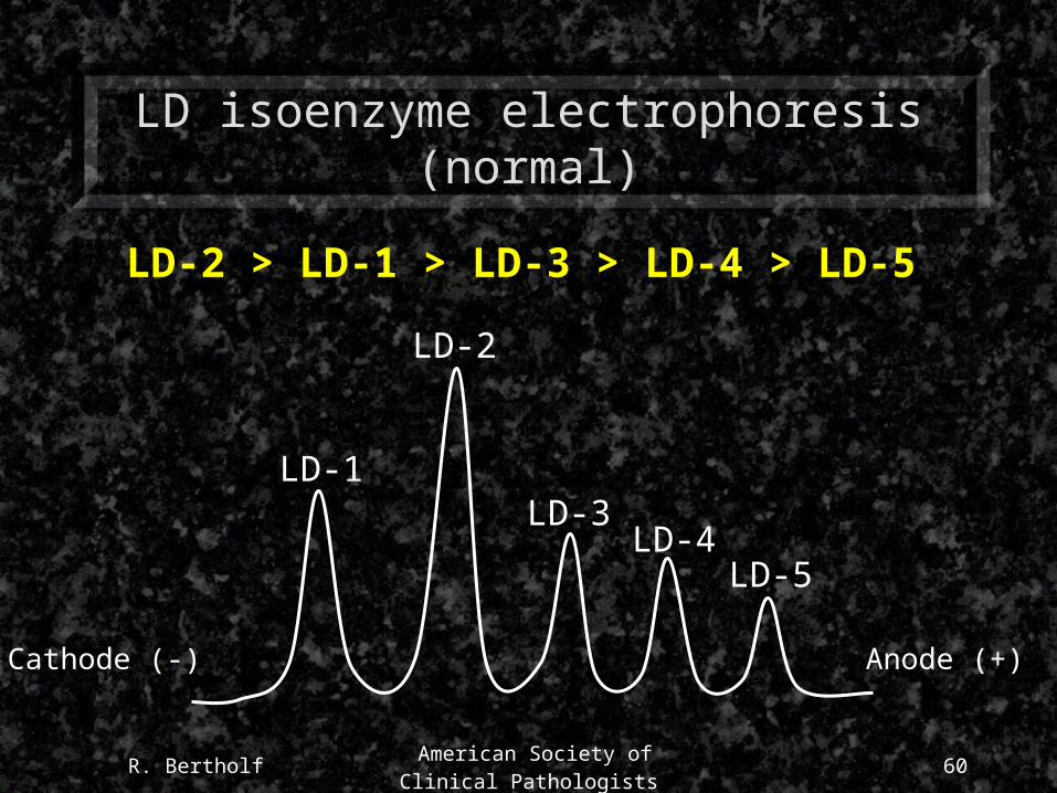
LD isoenzyme electrophoresis (normal)
LD-1
LD-2
LD-3LD-4
LD-5
LD-2 > LD-1 > LD-3 > LD-4 > LD-5
Cathode (-) Anode (+)

R. Bertholf American Society of Clinical
Pathologists61
LD isoenzyme electrophoresis (abnormal)
LD-1
LD-2
LD-3LD-4
LD-5
LD-1 > LD-2
Cathode (-) Anode (+)
R. Bertholf American Society of Clinical
Pathologists62
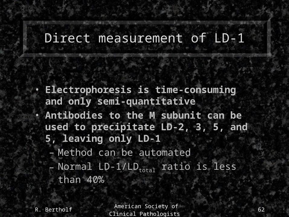
Direct measurement of LD-1
• Electrophoresis is time-consuming and only semi-quantitative
• Antibodies to the M subunit can be used to precipitate LD-2, 3, 5, and 5, leaving only LD-1
– Method can be automated
– Normal LD-1/LDtotal ratio is less than 40%

R. Bertholf American Society of Clinical
Pathologists63
Sensitivity and specificity of LD-1
• Sensitivity and specificity of the LD 1:2 “flip”, or LD-1 > 40% of total, are 90+% within 24 hours of MI, but. . .
– May be normal for 12 or more hours after symptoms appear (peak in 72-144 hours)
– May not detect minor infarctions
• Elevations persist for up to 10 days
• Even slight hemolysis can cause non-diagnostic elevations in LD-1
R. Bertholf American Society of Clinical
Pathologists64
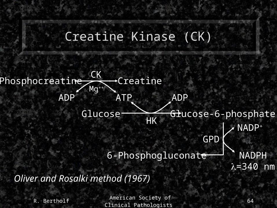
Creatine Kinase (CK)
Phosphocreatine
ADP
HKGlucose Glucose-6-phosphate
NADPH=340 nm
NADP+
GPD
6-Phosphogluconate
Oliver and Rosalki method (1967)
CreatineCK
ADP ATPMg++
R. Bertholf American Society of Clinical
Pathologists65
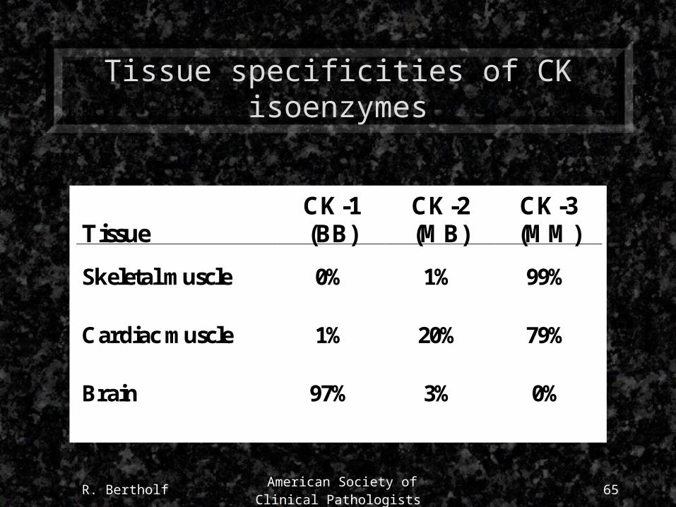
Tissue specificities of CK isoenzymes
TissueCK-1(BB)
CK-2(MB)
CK-3(MM)
Skeletal muscle 0% 1% 99%
Cardiac muscle 1% 20% 79%
Brain 97% 3% 0%
R. Bertholf American Society of Clinical
Pathologists66
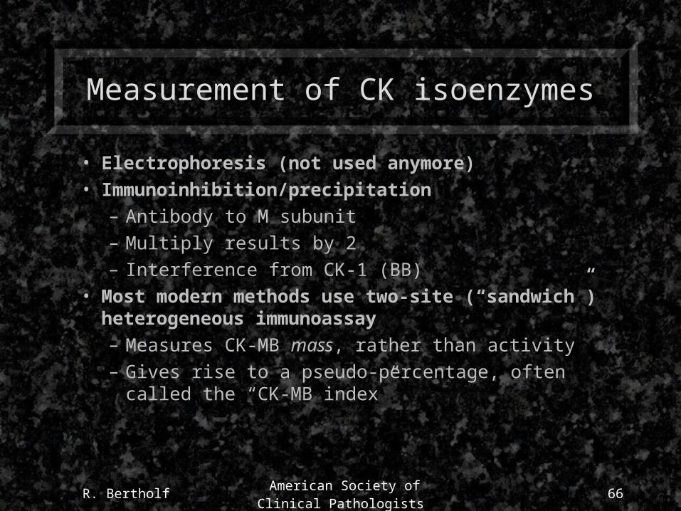
Measurement of CK isoenzymes
• Electrophoresis (not used anymore)• Immunoinhibition/precipitation
– Antibody to M subunit– Multiply results by 2– Interference from CK-1 (BB)
• Most modern methods use two-site (“sandwich”) heterogeneous immunoassay– Measures CK-MB mass, rather than activity– Gives rise to a pseudo-percentage, often called the “CK-MB
index”
R. Bertholf American Society of Clinical
Pathologists67
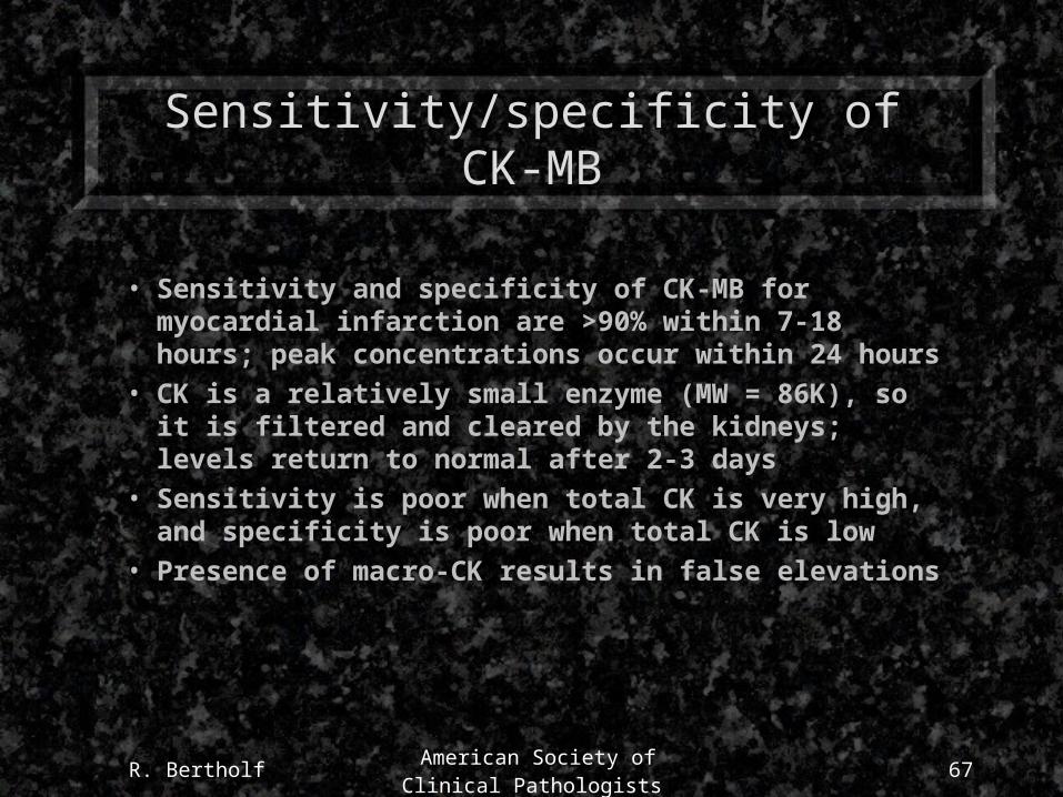
Sensitivity/specificity of CK-MB
• Sensitivity and specificity of CK-MB for myocardial infarction are >90% within 7-18 hours; peak concentrations occur within 24 hours
• CK is a relatively small enzyme (MW = 86K), so it is filtered and cleared by the kidneys; levels return to normal after 2-3 days
• Sensitivity is poor when total CK is very high, and specificity is poor when total CK is low
• Presence of macro-CK results in false elevations
R. Bertholf American Society of Clinical
Pathologists68
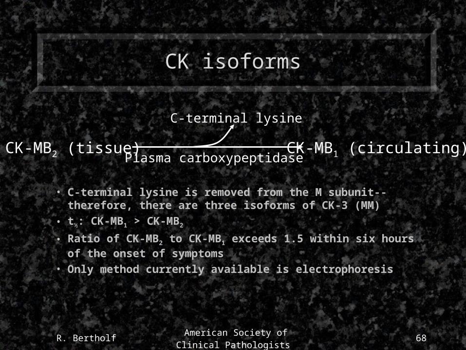
CK isoforms
• C-terminal lysine is removed from the M subunit--therefore, there are three isoforms of CK-3 (MM)
• t½: CK-MB1 > CK-MB2
• Ratio of CK-MB2 to CK-MB1 exceeds 1.5 within six hours of the onset of symptoms
• Only method currently available is electrophoresis
CK-MB2 (tissue) CK-MB1 (circulating)
C-terminal lysine
Plasma carboxypeptidase
R. Bertholf American Society of Clinical
Pathologists69
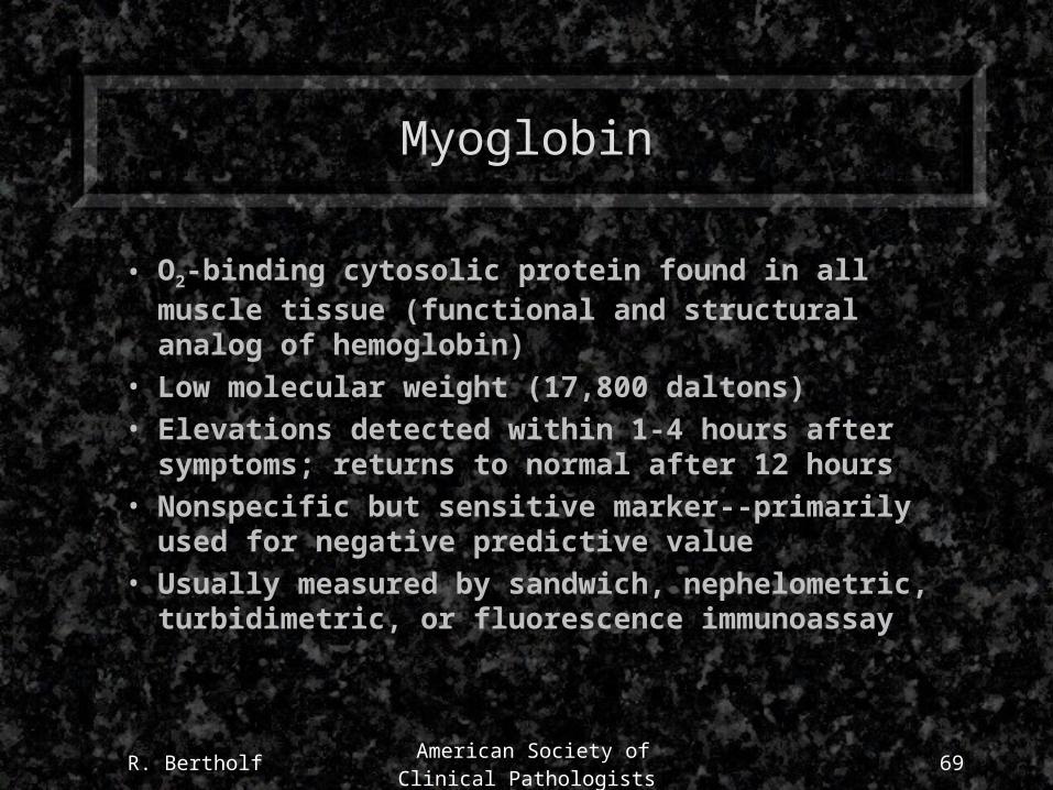
Myoglobin
• O2-binding cytosolic protein found in all muscle tissue (functional and structural analog of hemoglobin)
• Low molecular weight (17,800 daltons)• Elevations detected within 1-4 hours after symptoms;
returns to normal after 12 hours• Nonspecific but sensitive marker--primarily used for
negative predictive value• Usually measured by sandwich, nephelometric,
turbidimetric, or fluorescence immunoassay
R. Bertholf American Society of Clinical
Pathologists70
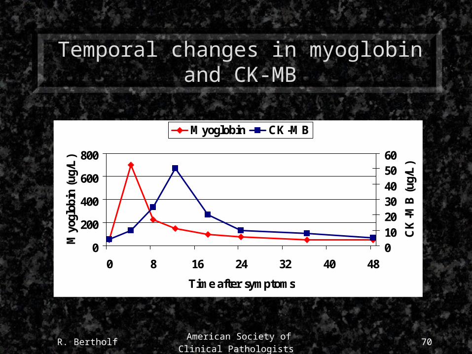
Temporal changes in myoglobin and CK-MB
0
200
400
600
800
0 8 16 24 32 40 48
Time after symptoms
Myo
glob
in (
ug/L
)
0102030405060
CK
-MB
(ug
/L)
Myoglobin CK-MB
R. Bertholf American Society of Clinical
Pathologists71
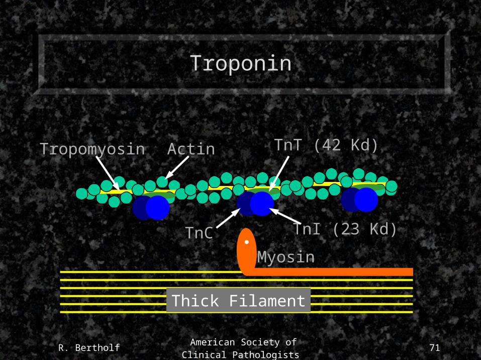
Troponin
Thick Filament
Myosin
Tropomyosin Actin
TnC
TnT (42 Kd)
TnI (23 Kd)
R. Bertholf American Society of Clinical
Pathologists72
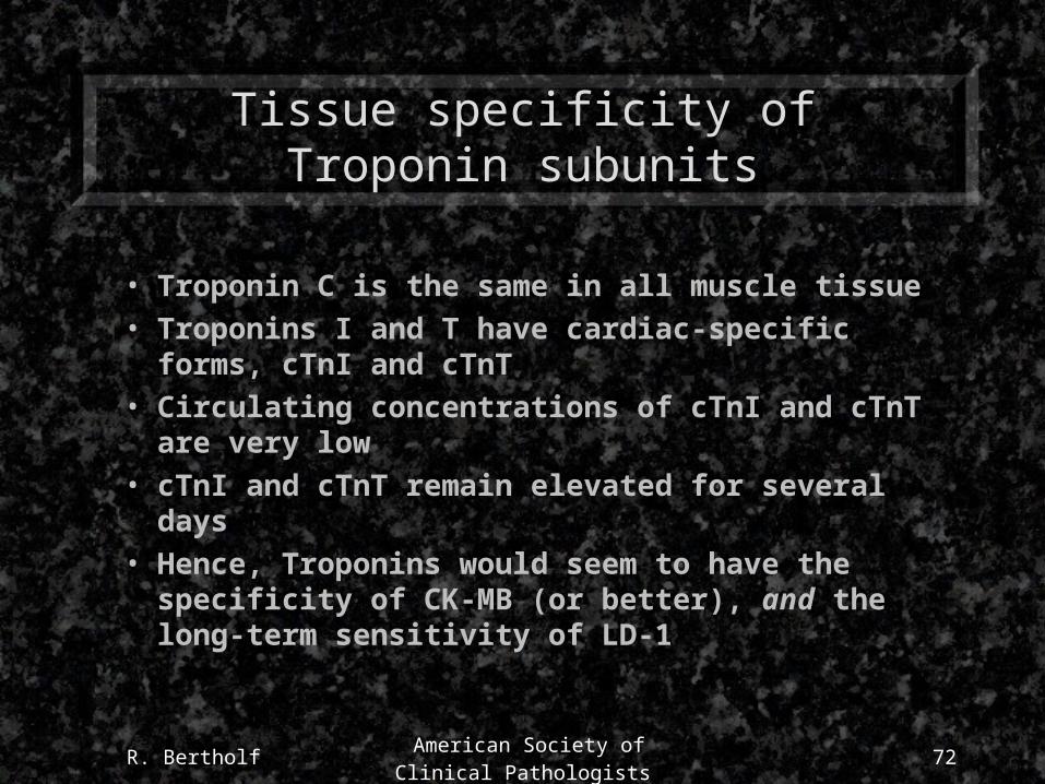
Tissue specificity of Troponin subunits
• Troponin C is the same in all muscle tissue
• Troponins I and T have cardiac-specific forms, cTnI and cTnT
• Circulating concentrations of cTnI and cTnT are very low
• cTnI and cTnT remain elevated for several days
• Hence, Troponins would seem to have the specificity of CK-MB (or better), and the long-term sensitivity of LD-1
R. Bertholf American Society of Clinical
Pathologists73
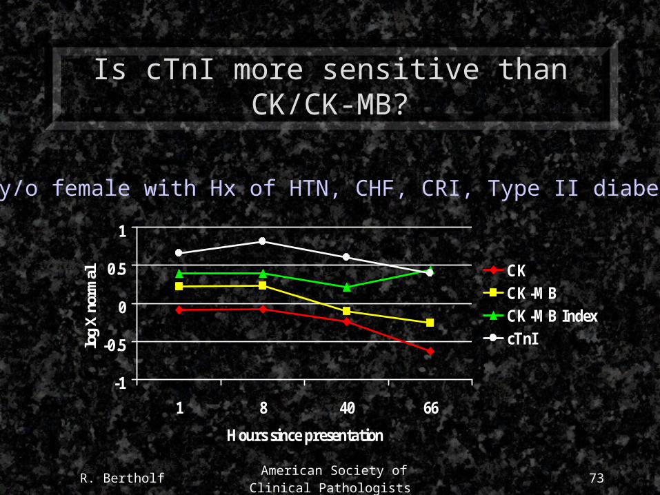
Is cTnI more sensitive than CK/CK-MB?
-1
-0.5
0
0.5
1
1 8 40 66
Hours since presentation
log
X n
orm
al CKCK-MBCK-MB IndexcTnI
79 y/o female with Hx of HTN, CHF, CRI, Type II diabetes
R. Bertholf American Society of Clinical
Pathologists74
Measurement of cTnI and cTnT
• All methods are immunochemical (ELISA, MEIA, CIA, ECIA)
• Roche Diagnostics (formerly BMC) is the sole manufacturer of cTnT assays
– First generation assay may have had some cross-reactivity with skeletal muscle TnT
– Second generation assay is cTnT-specific
– Also available in qualitative POC method
• Many diagnostics companies have cTnI methods
R. Bertholf American Society of Clinical
Pathologists75
W.H.O. has a Myocardial Infarction?
• A clinical history of ischemic-type chest discomfort
• Changes on serially obtained ECG tracings
• A rise and fall in serum cardiac markers
A patient presenting with any two of the following:
Source JACC 28;1996:1328-428
R. Bertholf American Society of Clinical
Pathologists76
Sensitivity/Specificity of WHO Criteria
0%
20%
40%
60%
80%
100%
Chest Pain ECG changes Serummarkers
SensitivitySpecificity
R. Bertholf American Society of Clinical
Pathologists77
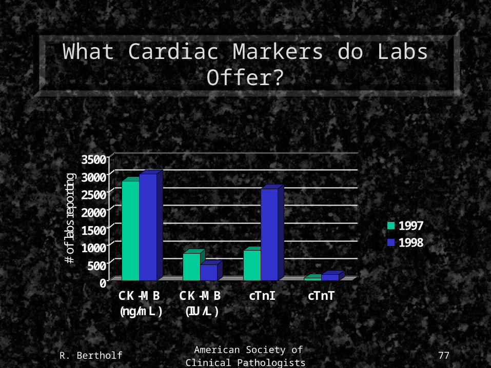
What Cardiac Markers do Labs Offer?
0
500
1000
1500
2000
2500
3000
3500
# of
labs
rep
ortin
g
CK-MB(ng/mL)
CK-MB(IU/L)
cTnI cTnT
19971998