1 CHAPTER 14 The Brain and Cranial Nerves. 2 INTRODUCTION Brain = center for registering sensations,...
21
1 CHAPTER 14 The Brain and Cranial Nerves
-
Upload
raymond-small -
Category
Documents
-
view
217 -
download
0
Transcript of 1 CHAPTER 14 The Brain and Cranial Nerves. 2 INTRODUCTION Brain = center for registering sensations,...

1
CHAPTER 14
The Brain and Cranial Nerves

2
INTRODUCTION• Brain = center for registering sensations, correlating them
with one another and with stored information, making decisions, and taking action
• Center for intellect, emotions, behavior, and memory
• Directs our behavior towards others
• In this chapter we will consider the principal parts of the brain, how the brain is protected and nourished, and how it is related to the spinal cord and to the 12 pairs of cranial nerves.

3
ORGANIZATION/PROTECTION/VASCULARIZATION
• Four major parts to brain– brain stem– cerebellum– diencephalon– cerebrum
• Protection– cranium (skull)– cranial meninges
• continuous w/ spinal meninges• dura mater has 2 layers
– fused xcpt where they separate to form dural venous sinuses
– no epidural space– 3 extensions separate parts of brain

4
ORGANIZATION/PROTECTION/VASCULARIZATION
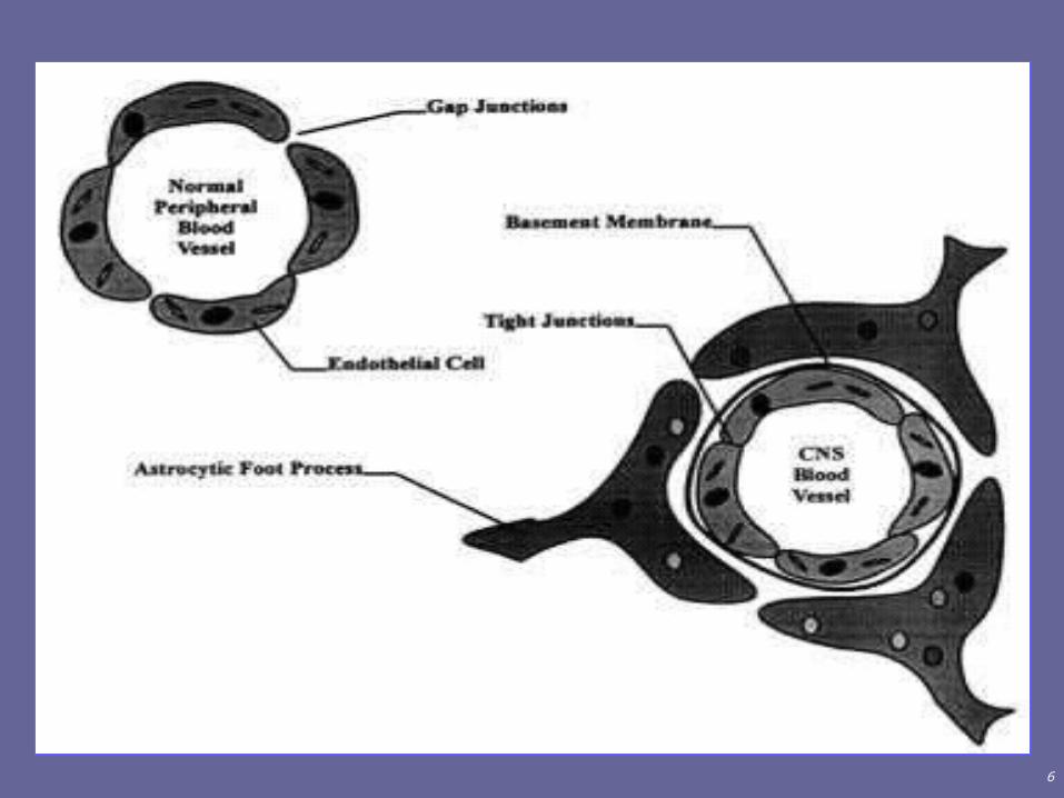
• Blood Flow & Blood-Brain Barrier– to brain via internal carotid & vertebral arteries– from brain via internal jugular vein– absolute requirement for O2 & glucose to make ATP
• no glucose storage requires constant supply• lack of O2 for 4 min causes permanent injury
– blood-brain barrier (BBB)• prevents passage of large molecules, harmful
substances & pathogens• tight junctions seal together capillary cells & basement
membrane surrounds• astrocyte processes cover capillaries & regulate
permeability of tight junctions

5
…VASCULARIZATION
– BBB• crossing BBB
– lipid-solubles cross easily» Oxygen, CO2, caffeine, alcohol, anesthetics
– proteins/antibiotics do not cross
– glucose is transported by facilitated or active transport
– breaching BBB presents hurdle for introducing drugs into brain for treatment of pathologies
6
7
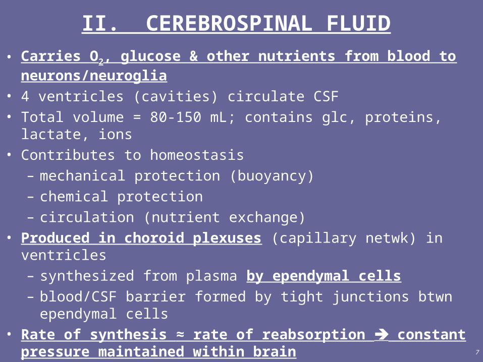
II. CEREBROSPINAL FLUID
• Carries O2, glucose & other nutrients from blood to neurons/neuroglia
• 4 ventricles (cavities) circulate CSF• Total volume = 80-150 mL; contains glc, proteins, lactate, ions• Contributes to homeostasis
– mechanical protection (buoyancy)– chemical protection– circulation (nutrient exchange)
• Produced in choroid plexuses (capillary netwk) in ventricles– synthesized from plasma by ependymal cells– blood/CSF barrier formed by tight junctions btwn ependymal
cells• Rate of synthesis ≈ rate of reabsorption constant
pressure maintained within brain

8
III. THE BRAIN STEM
• Medulla (oblongata)– inferior part of brain stem, continuous w/ spinal cord
– white matter (pyramids) contains all sensory/motor tracts btwn spinal cord & other parts of brain
– decussation of pyramids: point where 90% of axons from L/R sides crossover why L side of brain controls R side of body, & vice versa
– nuclei w/in gray matter control vital body functions• heart, breathing rates (cardiovasc & medull rhyth ctrs)• reflexes for vomiting, coughing, sneezing• sensations for touch, proprioception• associated w/ 5 pr cranial nerves
9
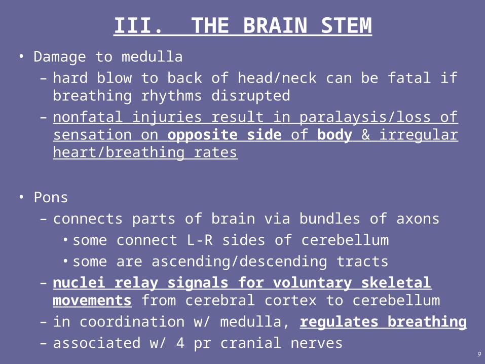
III. THE BRAIN STEM• Damage to medulla
– hard blow to back of head/neck can be fatal if breathing rhythms disrupted
– nonfatal injuries result in paralaysis/loss of sensation on opposite side of body & irregular heart/breathing rates
• Pons – connects parts of brain via bundles of axons
• some connect L-R sides of cerebellum• some are ascending/descending tracts
– nuclei relay signals for voluntary skeletal movements from cerebral cortex to cerebellum
– in coordination w/ medulla, regulates breathing– associated w/ 4 pr cranial nerves

10
III. THE BRAIN STEM
• Midbrain– cerebral peduncles (ascending/descending tracts)
• motor tracts carry impulses from cerebrum to spinal cord, pons, medulla
• sensory tracts from medulla to thalamus– contains reflex centers for visual activities, head
movements– controls subconscious muscle activity
• some neurons (substantia nigra) release dopamine• loss/damage of these neurons results in loss of tone
Parkinson’s disease– associated w/ 2 pr cranial nerves

11
III. THE BRAIN STEM
• Reticular Formation
– netlike arrangement of white/gray matter
– reticular activating system• alerts cerebral cortex to sensory signals to awaken
from sleep• maintains consciousness & helps keep you awake
with stimuli from ears, eyes, skin and muscles
– motor function: maintains muscle tone

12
IV. CEREBELLUM
• Evaluates & coordinates voluntary movement
• Maintains posture & balance
• FB mechanism allows for correction of error in movements makes smooth/controlled movements
• Ataxia = loss of muscle coordination due to trauma/disease– unable to coordinate movement w/ sense of body
position– altered speech patterns– XS alcohol can cause ataxia also (slurred
speech/decreased breathing rate)
13
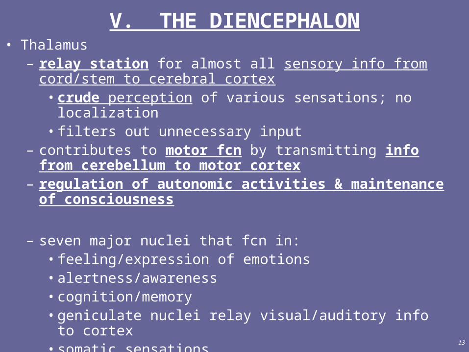
V. THE DIENCEPHALON• Thalamus
– relay station for almost all sensory info from cord/stem to cerebral cortex
• crude perception of various sensations; no localization• filters out unnecessary input
– contributes to motor fcn by transmitting info from cerebellum to motor cortex
– regulation of autonomic activities & maintenance of consciousness
– seven major nuclei that fcn in:• feeling/expression of emotions• alertness/awareness• cognition/memory• geniculate nuclei relay visual/auditory info to cortex• somatic sensations
14
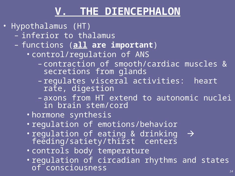
V. THE DIENCEPHALON• Hypothalamus (HT)
– inferior to thalamus– functions (all are important)
• control/regulation of ANS– contraction of smooth/cardiac muscles & secretions
from glands– regulates visceral activities: heart rate, digestion– axons from HT extend to autonomic nuclei in brain
stem/cord• hormone synthesis• regulation of emotions/behavior• regulation of eating & drinking feeding/satiety/thirst
centers• controls body temperature• regulation of circadian rhythms and states of
consciousness
15
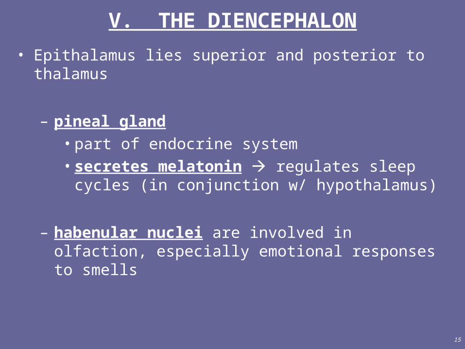
V. THE DIENCEPHALON
• Epithalamus lies superior and posterior to thalamus
– pineal gland • part of endocrine system• secretes melatonin regulates sleep cycles (in
conjunction w/ hypothalamus)
– habenular nuclei are involved in olfaction, especially emotional responses to smells
16
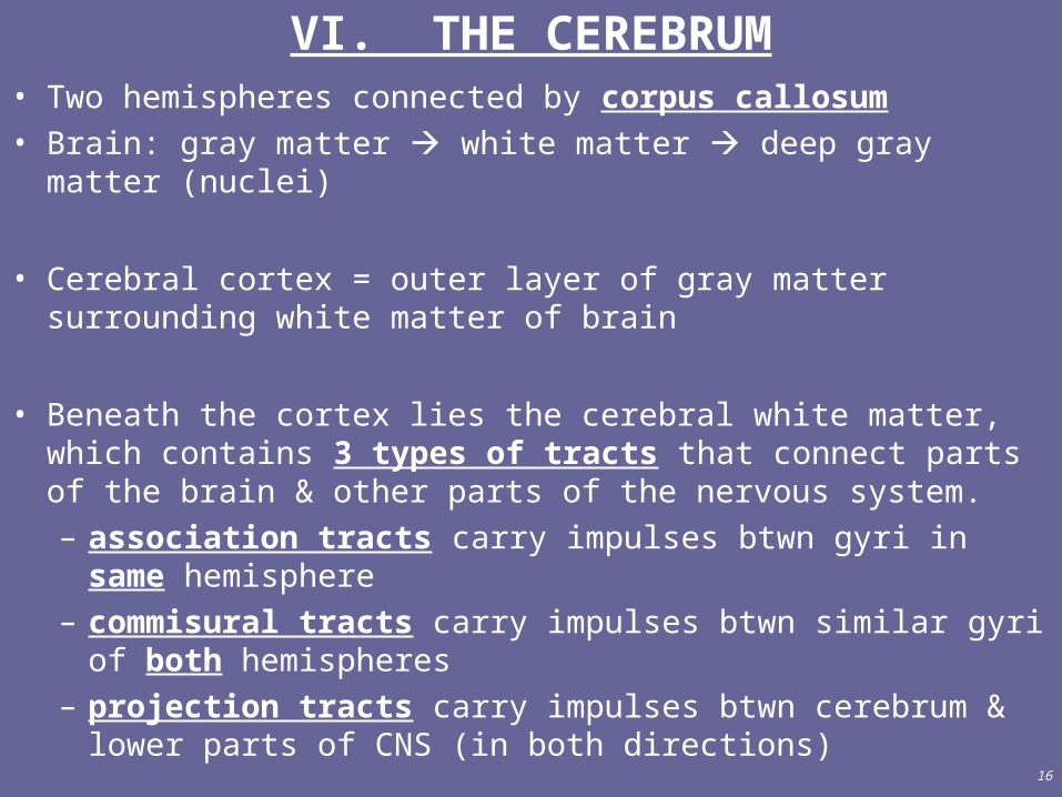
VI. THE CEREBRUM• Two hemispheres connected by corpus callosum• Brain: gray matter white matter deep gray matter (nuclei)
• Cerebral cortex = outer layer of gray matter surrounding white matter of brain
• Beneath the cortex lies the cerebral white matter, which contains 3 types of tracts that connect parts of the brain & other parts of the nervous system.– association tracts carry impulses btwn gyri in same
hemisphere– commisural tracts carry impulses btwn similar gyri of both
hemispheres– projection tracts carry impulses btwn cerebrum & lower
parts of CNS (in both directions)
17
VI. THE CEREBRUM• Basal ganglia (nuclei)
– deep gray matter of cerebrum– receive input from cortex– output to motor parts of cortex via thalamus
– functions:• regulates initiation/termination of body movements• regulates muscle tone, subconscious contraction
of skeletal muscles• initiation/termination of some cognitive processes
– damage results in tremors, muscle rigidity & involuntary movements (which are, you guessed it, hallmarks of…. Parkinson’s disease)
18
VI. THE CEREBRUM• Brain Injuries
– result from head trauma & subsequent ischemia (↓blood flow) or oxygen deprivation (hypoxia)
– concussion = abrupt (temporary) loss of consciousness associated w/ vision & equilibrium problems
• caused by sudden blow to head• signs: vomiting, lack of concentration, confusion
– contusion = bruising of brain due to trauma, includes leakage of blood from small vessels
• usually associated w/ concussion• immediate loss of consciousness & reflexes,
fluctuations in breathing, ↓ blood pressure– laceration = tear of brain tissue
• rupture of large vessels causes cerebral hematoma, edema & increased pressure in brain

19
VII. FUNCTIONAL ORGANIZATION OF CEREBRAL CORTEX
• Occipital lobe contains primary visual areas (input/perception)
• Temporal lobes contain primary auditory areas– sound input & perception– faster than visual pathway because fewer synapses
• Parietal lobes contain primary somatosensory areas– receives/processes nerve impulses for touch, pressure, heat,
cold, pain, proprioception– allows localization of sensations
• Frontal lobes contain regions for voluntary motor activity and speaking ability

20
VII. FUNCTIONAL ORGANIZATION OF CEREBRAL CORTEX
• Frontal lobes– voluntary motor activity
• primary motor cortex controls voluntary contractions of specific muscles/groups of muscles
• more cortical area is devoted to skilled, fine movements
– speaking ability• Broca’s area controls muscles used for speech
(articulation) • ***Wernicke’s area responsible for forming speech
patterns sends info to Broca for execution– receives visual & auditory input cannot read
without “hearing” words as you read them****

21
VII. FUNCTIONAL ORGANIZATION OF CEREBRAL CORTEX
• Association areas consist of sensory & motor areas connected by association tracts– prefrontal association
• personality traits• planning ability (for voluntary activity)• weighs consequences of future actions
– somatosensory-visual-auditory association areas• integrate somatic, visual & auditory info for
comprehension• functions in distinguishing language from noise
– premotor area• stores info about programmed skeletal movements• communicates w/ primary motor area to generate
complex, sequential movements