1 Blood (rev 11/11) The circulatory system consists of the heart, blood vessels and the blood...
55
1 Blood (rev 11/11) • The circulatory system consists of the heart, blood vessels and the blood itself. • The circulatory system is essential to supply all cells with what they need and removing substances they no longer need. BIO 102-Blood- Cardiovascular
-
Upload
derick-bates -
Category
Documents
-
view
216 -
download
0
Transcript of 1 Blood (rev 11/11) The circulatory system consists of the heart, blood vessels and the blood...

1
Blood (rev 11/11)
• The circulatory system consists of the heart, blood vessels and the blood itself.
• The circulatory system is essential to supply all cells with what they need and removing substances they no longer need.
BIO 102-Blood-Cardiovascular

2
The Functions of Blood
Blood is actually a liquid body tissue and is classified as a connective tissue
Blood carries out three essential tasks:
• Transportation: oxygen, nutrients, waste, hormones
• Regulation: body temperature, volume of water in the body, pH of body fluids
• Defense: contains specialized defense cells to protect against illness and excessive bleeding through clotting mechanisms
These are necessary for homeostasis.
BIO 102-Blood-Cardiovascular
3
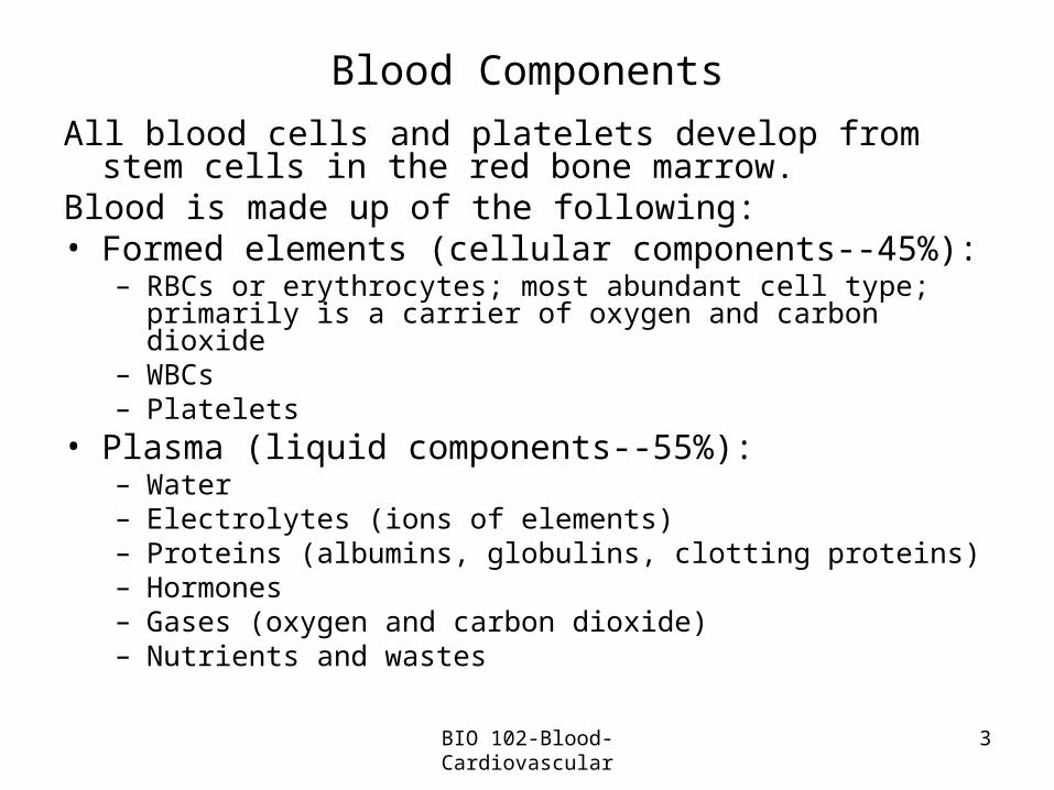
Blood Components
All blood cells and platelets develop from stem cells in the red bone marrow.
Blood is made up of the following: • Formed elements (cellular components--45%):
– RBCs or erythrocytes; most abundant cell type; primarily is a carrier of oxygen and carbon dioxide
– WBCs– Platelets
• Plasma (liquid components--55%):– Water– Electrolytes (ions of elements)– Proteins (albumins, globulins, clotting proteins)– Hormones– Gases (oxygen and carbon dioxide)– Nutrients and wastes
BIO 102-Blood-Cardiovascular
4
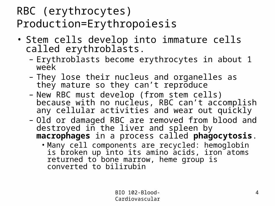
RBC (erythrocytes) Production=Erythropoiesis
• Stem cells develop into immature cells called erythroblasts.– Erythroblasts become erythrocytes in about 1 week– They lose their nucleus and organelles as they
mature so they can’t reproduce– New RBC must develop (from stem cells) because
with no nucleus, RBC can’t accomplish any cellular activities and wear out quickly
– Old or damaged RBC are removed from blood and destroyed in the liver and spleen by macrophages in a process called phagocytosis.
• Many cell components are recycled: hemoglobin is broken up into its amino acids, iron atoms returned to bone marrow, heme group is converted to bilirubin
BIO 102-Blood-Cardiovascular
5
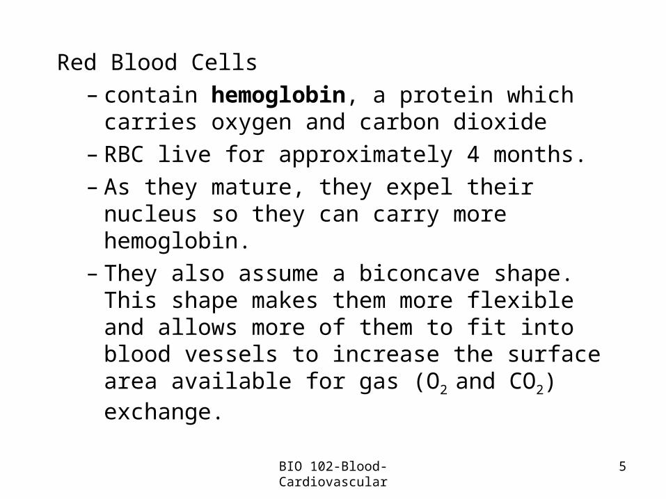
Red Blood Cells– contain hemoglobin, a protein which
carries oxygen and carbon dioxide– RBC live for approximately 4 months. – As they mature, they expel their nucleus so
they can carry more hemoglobin.– They also assume a biconcave shape.
This shape makes them more flexible and allows more of them to fit into blood vessels to increase the surface area available for gas (O2 and CO2) exchange.
BIO 102-Blood-Cardiovascular

6
Hemoglobin Molecule• Hemoglobin is an oxygen binding protein
which consists of 4 polypeptide chains coiled around a “heme group”
• The “heme group” has an iron atom in its center. This combines easily with oxygen at the lungs AND lets go of the oxygen when reaching body tissues.
BIO 102-Blood-Cardiovascular
7
Hematocrit• is a measure of the oxygen carrying capacity
of blood • is obtained by spinning down blood and
measuring the amount of formed elements • RBCs make up nearly 99% of formed
elements• Normal hematocrit
– men: 43-49% women: 37-43%
BIO 102-Blood-Cardiovascular
8
• Regulation of RBC production is a negative feedback control loop which maintains homeostasis.
• Cells in the kidneys check the availability of oxygen. If levels are low, these cells are signaled to secrete the hormone erythropoietin. This is carried to red bone marrow where more RBC are produced.
• When the oxygen levels are appropriate, the kidney cells stop production of erythropoietin.
BIO 102-Blood-Cardiovascular

9
White Blood Cells-WBC or leukocytes
Functions: protection from infection, regulation of the inflammatory reaction
• Types:– Granular:
• neutrophils, eosinophils, basophils• Mature in the red bone marrow• Granules are actually vesicles filled with proteins
and enzymes– Agranular:
• lymphocytes, monocytes• monocytes mature in red bond marrow;
lymphocytes mature in the thymus gland
BIO 102-Blood-Cardiovascular
10
Circulating levels of WBC rise whenever the body is threatened by viruses, bacteria, or other health challenges
• Each type of WBC can produce chemicals which travel, via the blood, to the bone marrow where they stimulate the production of more WBC
Most WBC remain in the blood vessels, but some circulate in the intercellular fluid and the lymphatic system.
BIO 102-Blood-Cardiovascular

11
Types of WBC • Neutrophils-most common WBC- approximately
60% of WBC are neutrophils
• see in acute infections; are the first WBC to combat infection
• main function is phagocytosis (bacteria and fungi)
– Eosinophils- approximately 2-4% of WBCs
• see in parasitic infections (such as worms) and in allergic reactions (they release chemicals that diminish the severity of these reactions)
BIO 102-Blood-Cardiovascular
12
– Basophils-are rare, 0.5% of WBCs• initiate the inflammatory response—
granules in the cell cytoplasm contain histamine which starts the inflammatory response
– Lymphocytes-second most common WBC, about 30%; found in tonsils, blood, spleen, lymph nodes, thymus
• manufactures antibodies and eliminates anything foreign to the body
• play crucial role in immune response– Monocytes-about 5%
• active in phagocytosis• elevated in chronic infections
BIO 102-Blood-Cardiovascular
13
Platelets• are small cell fragments (not complete cells) which
play an essential role in the process of blood clotting• platelet production is regulated by hormone
thrombopoietin• platelets are stable as they circulate, but when they
encounter a “rough surface” they form a temporary plug and initiate the clotting mechanism
• the body also requires vitamin K for normal blood clotting
BIO 102-Blood-Cardiovascular
14
• Clotting process or hemostasis (stopping blood)
– damage to a blood vessel triggers a vasospasm or constriction of the damaged blood vessel
– platelets in the area swell, become sticky, adhere to the damaged area and produce a plug which will become the clot
– platelets also release chemicals to help in clot formation
• prothrombin activator converts prothrombin (a plasma protein) into thrombin
• thrombin converts the fibrinogen molecules, to fibrin which traps blood cells, forms a clot and seals the hole
BIO 102-Blood-Cardiovascular

15
• Blood Typing– Each of us has one of 4 types of blood--A, B, AB, O--
along with some specific glycoproteins or antigens• Our cells have surface proteins that the immune
system can recognize as “self” or “non-self”. The immune system will recognize foreign cells as non-self .
• An antigen is a non-self cell protein that causes the immune system to defend itself.
• The immune system builds antibodies-an opposing protein which can kill the non-self cells.
– and causes them to stick together so it can be destroyed. So, the transfused blood clumps or clots within our blood vessels.
– This can be fatal
BIO 102-Blood-Cardiovascular
16
• Another antigen found in blood is the Rh antigen--if you have it, your blood is classified as Rh positive. If you do not have this, your blood is classified as Rh negative.
Blood Typing Tests– Based on the interaction between antigens
and antibodies– performed with anti-sera which contain
high concentrations of anti-A and anti-B antibodies
– blood samples are mixed with each anti-sera
BIO 102-Blood-Cardiovascular

17
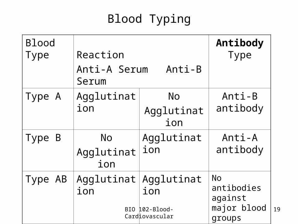
– if agglutination or clumping (similar to clotting) occurs with
anti-A sera, you have type A blood
anti-B sera, you have type B blood– if clumping occurs with both anti-A and
anti-B, you have type AB blood– if no clumping occurs with either anti-A and
anti-B sera, you have type O blood• so, the antibodies you have in your body are
the opposite of your blood type
BIO 102-Blood-Cardiovascular

BIO 102-Blood-Cardiovascular 18
Blood Types Determine Blood Compatibility
Figure 7.12
19
Blood Typing
Blood Type
Reaction
Anti-A Serum Anti-B Serum
Antibody Type
Type A Agglutination No
Agglutination
Anti-B antibody
Type B No
Agglutination
Agglutination Anti-A antibody
Type AB Agglutination Agglutination No antibodies against major blood groups
Type O No
Agglutination
No
Agglutination
Anti-A & Anti-B antibodies
BIO 102-Blood-Cardiovascular

20
Blood Disorders
• Carbon monoxide poisoning: competes with oxygen
Anemia: reduction in oxygen-carrying capacityTypes of Anemia• Iron deficiency anemia occurs when there is insufficient
iron ingested so fewer hemoglobin molecules are available.
• Aplastic anemia where the bone marrow doesn’t produce enough stem cells
• Hemorrhagic anemia is caused by extreme blood loss
BIO 102-Blood-Cardiovascular
21
• Pernicious Anemia where the body is unable to absorb vitamin B12 from the digestive tract. The body uses B12 to produce normal RBC.
• Sickle cell Anemia is an inherited disorder in which the RBC become sickle or crescent shaped when the oxygen concentration of the blood is low. This shape doesn’t travel easily through blood vessels because the cells clump, get stuck in the vessels and cause a great deal of pain. – Sickle shaped cells can’t carry a normal
amount of oxygen.
BIO 102-Blood-Cardiovascular
22
• Leukemia is a form of cancer where you see an uncontrolled production of abnormal or immature WBC in the bone marrow. This crowds out the production of normal WBC, RBC, and platelets.
• Multiple myeloma: a form of cancer where abnormal plasma cells in the bone marrow increase production. These cells are important for the manufacture of antibodies.
• Mononucleosis is a contagious infection of lymphocytes caused by the Epstein-Barr virus.
• Septicemia can also be called “blood poisoning”. It occurs when organisms invade the blood, overpower our body’s defenses and multiply rapidly in the blood.
• Thrombocytopenia is a reduction in the number of platelets.
BIO 102-Blood-Cardiovascular
23
Polycythemia is a term used to describe an abnormally high RBC count– this increases the thickness of blood and
slows down the flow of blood.
Hemophilia is an inherited condition caused by a deficiency of one or more clotting factors (known as clotting factor VIII)– When a blood vessel is damaged, blood either clots
very slowly or not at all
BIO 102-Blood-Cardiovascular
24
The Cardiovascular System
Blood Vessels: Arteries, VeinsArterial systemStructure: • Endothelium: thin inner layer of squamous epithelial cells• Middle: thick layer of smooth muscle woven with elastic
connective tissue• Outer layer: tough supportive layer of connective tissue,
primarily collagen– anchors vessels to surrounding tissues and helps protect them
from injury• Aneurysm: ballooning of the arterial wall
– Endothelium of blood vessel becomes damaged and blood seeps through and accumulates between the middle and outer layers of the blood vessel
BIO 102-Blood-Cardiovascular
25
The Cardiovascular System
• Functions:– Arteries: carry blood away from heart– Need thicker muscular wall due to pressure of blood
being pumped by aorta – Because blood pressure is less by the time blood has
reached the arterioles, they do not have the outermost layer of connective tissue and the muscular layer is thinner.
• precapillary sphincter, a band of smooth muscle, is located where the arteriole meets the capillary and controls the blood flow to each capillary
• Capillaries: thin walled blood vessels; branching design allows exchange of gases, nutrients, waste, and defensive cells between vessel and tissue
BIO 102-Blood-Cardiovascular
26
The Cardiovascular System
– vasoconstriction– vasodilation
BIO 102-Blood-Cardiovascular
27
Venous system• Functions: carry blood to the heart • Structure: veins: three layers, thin-walled
– like the walls of arteries, the walls of veins consist of 3 layers of tissue.
• Outer 2 layers are much thinner than those of arteries
• veins have larger diameters (lumen) than arteries – the pressure in veins is much lower than that in
arteries which is why their walls are not as strong as arteries
• Blood pressure lower in veins than in capillaries– veins can act as a blood volume reservoir
BIO 102-Blood-Cardiovascular
28
– the larger diameter of veins allows them to stretch to accommodate large volumes of blood at low pressures
– because veins can stretch, it is more difficult for them to return blood to the heart against the force of gravity
– people who spend a lot of time on their feet may get varicose veins because of this
• Factors which help veins to return blood to heart
• Contraction of skeletal muscles-skeletal muscle pump
• as we move and muscles contract and relax, they press against veins and help push blood to the heart
BIO 102-Blood-Cardiovascular

29
– One-way valves—blood can only flow in one direction
• Open passively to allow blood to move toward the heart and cloose whenever blood begins to flow backward
– the work of the skeletal muscles helps the valves pump blood. This is called a skeletal muscle pump
BIO 102-Blood-Cardiovascular
30
Pressure changes associated with breathing– movements associated with breathing also help
pump blood. This is called a respiratory pump and helps to push blood from the abdomen to the chest and to the heart.
• when we breathe, there are pressure changes in the thoracic and abdominal cavities
• during inhalation, abdominal pressure increases and squeezes abdominal veins
• simultaneously, pressure within the thoracic cavity decreases which dilates the thoracic veins and thus propels the blood.
BIO 102-Blood-Cardiovascular
31
Lymphatic System
Function: maintains proper volume of blood and interstitial fluid; also functions in immune system– Picks up objects in interstitial fluid that are too large to
diffuse into capillaries• Lipid droplets absorbed during digestion• Invading organisms
– Transports these to larger lymphatic vessels which return the fluid to veins near the heart
• Structure:– Lymphatic vessels– Lymph
BIO 102-Blood-Cardiovascular

32
The Heart
Structure: composed of cardiac muscle enclosed by pericardium, a fibrous sac– Pericardium protects the heart, anchors it to
surrounding structures, prevents it from overfilling with blood
– Pericardial cavity separates it from heart muscle itself and contains a tiny amount of fluid to allow heart and pericardium to glide smoothly every time the heart contracts
– Heart beat rate determined by the SA Node
BIO 102-Blood-Cardiovascular
33
The Heart• Layers: Epicardium: thin, outermost layer
made up of epithelial and connective tissue– Myocardium: thick layer primarily of cardiac
muscle – Endocardium: innermost layer of endothelial
tissue resting on a layer of connective tissue; is continuous with the endothelium that lines blood vessels
BIO 102-Blood-Cardiovascular
34
The Heart• Pericarditis:
– A layer of the heart wall becomes inflamed• Endocarditis: inflammation of the endocardium• 4 Chambers: two atrias, two ventricles
– Atrias are on the top– Ventricles are the 2, more muscular chambers
on the bottom• Septum, a muscular partition, separates the right
and left sides of the heart
BIO 102-Blood-Cardiovascular
35
The Heart• Valves: prevent blood from flowing backward
– Two atrioventricular valves: tricuspid (right) and bicuspid (mitral--left)• Chordae tendinae: strands of connective
tissue which connect to muscular extensions of the ventricle wall, called papillary tendons
• These prevent the valves from being pushed backward
– Two semilunar valves: pulmonary and aortic• Have 3 flaps
BIO 102-Blood-Cardiovascular
36
Flow of blood through the heart:
• Deoxygenated blood through the vena cava to the right atrium
• Deoxygenated blood through the right atrioventricular valve to the right ventricle
• Deoxygenated blood through the pulmonary semilunar valve to the pulmonary trunk and the lungs
• Oxygenated blood through the pulmonary veins to the left atrium
• Oxygenated blood through the left atrioventricular valve to the left ventricle
BIO 102-Blood-Cardiovascular
37
• Oxygenated blood through the aortic semilunar valve to the aorta
Blood flow through the tissues:• Oxygenated blood through branching
arteries and arterioles to the tissues• Oxygenated blood through the arterioles to
capillaries• Deoxygenated blood from capillaries into
venules and veins• Ultimately to the vena cava and into the
right atrium
BIO 102-Blood-Cardiovascular

38
• Coronary arteries supply the heart muscle with blood (myocardium is too thick to be able to be supplied with oxygen and nutrients by diffusion from the blood passing through it)
• Coronary arteries branch from the aorta as it leaves the heart and encircle the heart’s surface
• Cardiac veins bring the blood back to the right atrium
BIO 102-Blood-Cardiovascular
39
Cardiac cycle is a measure of the blood pumped with each beat multiplied by the number of heart beats per minute
• Steps summarized:1. Heart relaxes and all four chambers fill; blood is
sucked in as the heart muscle expands
2. Atrial contraction: more blood into the already filled ventricles
3. Ventricular contraction: blood is ejected into the aorta and pulmonary trunk
• Systole refers to the contraction pressure• Relaxation of the entire heart = diastole
BIO 102-Blood-Cardiovascular

40
Heart Sounds and Heart Valves
• Lub-dub – Lub signals the closure of the 2 AV valves– Dub signals the aortic and pulmonary
semilunar valves closing
• Heart murmurs are created by obstructions the blood encounters as it flows through the heart
BIO 102-Blood-Cardiovascular
41
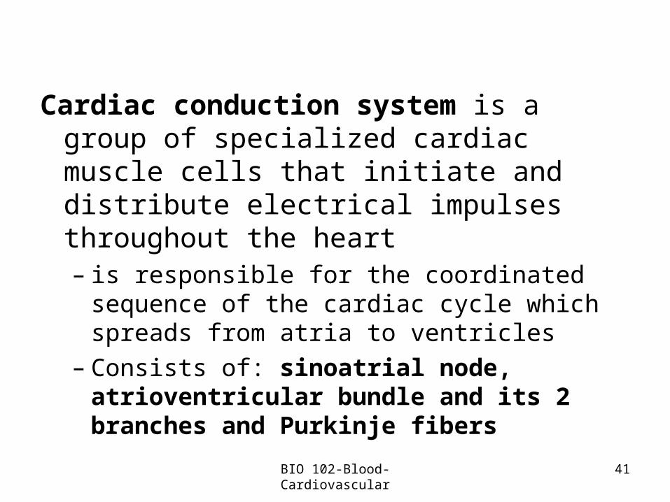
Cardiac conduction system is a group of specialized cardiac muscle cells that initiate and distribute electrical impulses throughout the heart– is responsible for the coordinated sequence of
the cardiac cycle which spreads from atria to ventricles
– Consists of: sinoatrial node, atrioventricular bundle and its 2 branches and Purkinje fibers
BIO 102-Blood-Cardiovascular
42
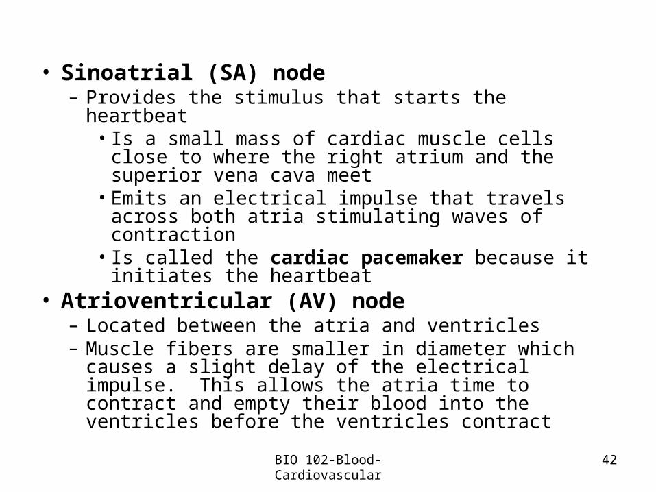
• Sinoatrial (SA) node– Provides the stimulus that starts the heartbeat
• Is a small mass of cardiac muscle cells close to where the right atrium and the superior vena cava meet
• Emits an electrical impulse that travels across both atria stimulating waves of contraction
• Is called the cardiac pacemaker because it initiates the heartbeat
• Atrioventricular (AV) node– Located between the atria and ventricles– Muscle fibers are smaller in diameter which causes a
slight delay of the electrical impulse. This allows the atria time to contract and empty their blood into the ventricles before the ventricles contract
BIO 102-Blood-Cardiovascular
43
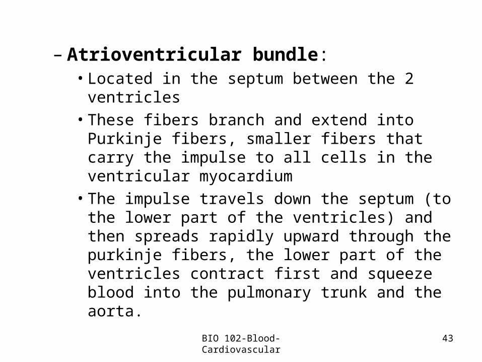
– Atrioventricular bundle:• Located in the septum between the 2 ventricles • These fibers branch and extend into Purkinje
fibers, smaller fibers that carry the impulse to all cells in the ventricular myocardium
• The impulse travels down the septum (to the lower part of the ventricles) and then spreads rapidly upward through the purkinje fibers, the lower part of the ventricles contract first and squeeze blood into the pulmonary trunk and the aorta.
BIO 102-Blood-Cardiovascular

44
Cardiac Conduction System Coordinates Contraction
• SA node: cardiac pacemaker
• AV node: relay impulse
• AV bundle and Purkinje fibers: carry impulse to ventricles
Figure 8.14BIO 102-Blood-Cardiovascular
45
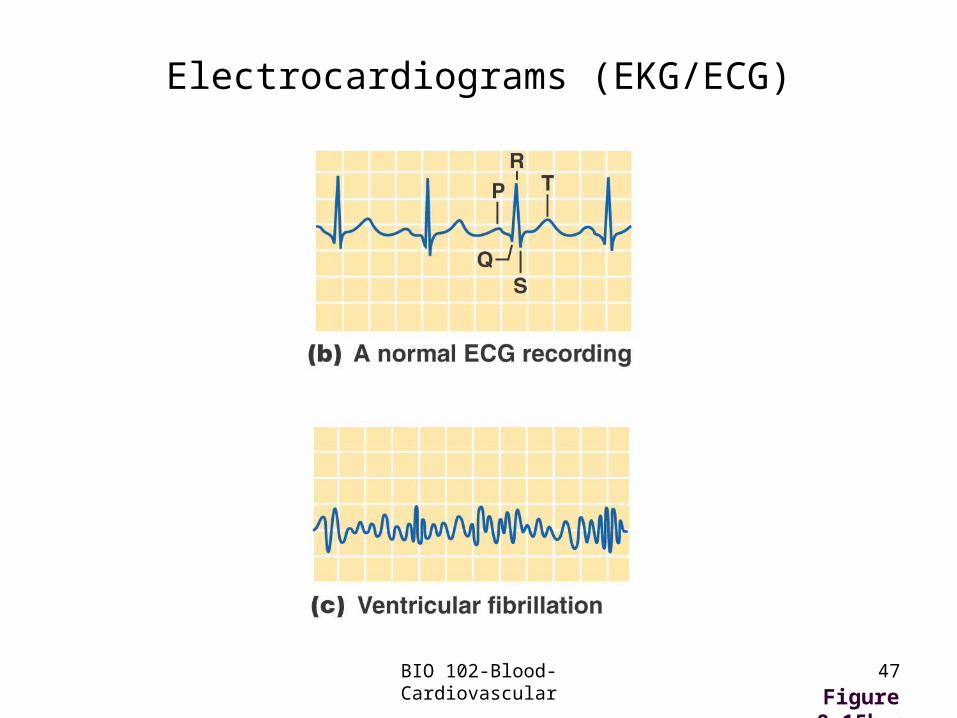
Electrocardiograms (EKG/ECG)
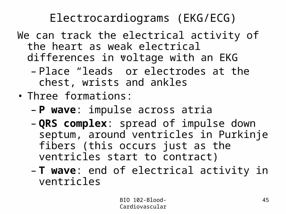
We can track the electrical activity of the heart as weak electrical differences in voltage with an EKG– Place “leads” or electrodes at the chest, wrists
and ankles• Three formations:
– P wave: impulse across atria– QRS complex: spread of impulse down
septum, around ventricles in Purkinje fibers (this occurs just as the ventricles start to contract)
– T wave: end of electrical activity in ventricles
BIO 102-Blood-Cardiovascular
46
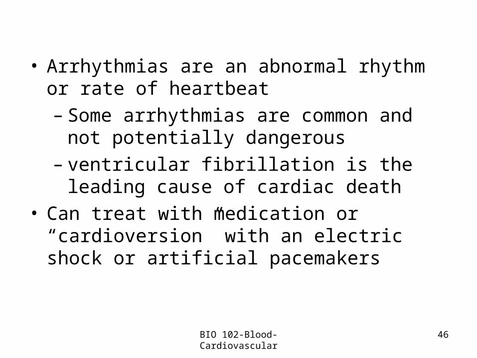
• Arrhythmias are an abnormal rhythm or rate of heartbeat– Some arrhythmias are common and not
potentially dangerous– ventricular fibrillation is the leading cause of
cardiac death• Can treat with medication or “cardioversion” with
an electric shock or artificial pacemakers
BIO 102-Blood-Cardiovascular
47
Electrocardiograms (EKG/ECG)
Figure 8.15b,cBIO 102-Blood-Cardiovascular

48
Blood Pressure
• Force that blood exerts on the wall of a blood vessel as a result of the pumping action of the heart
• Definitions: “normal”:– Systolic pressure: highest pressure, pressure reached
during ventricular contraction to eject blood from theheart
– Diastolic pressure: lowest pressure, pressure when the ventricles relax
• Arteries store energy generated during systole and during diastole they use that stored energy to supply blood to the tissues
• Measurement: sphygmomanometer
BIO 102-Blood-Cardiovascular
49
• Hypertension: high blood pressure:– Definition– The silent killer– Risk factors
• Hypotension: blood pressure too low so blood can’t be pushed throughout the body and back to the heart; generally thought of as reducing blood flow to the brain– Clinical signs: dizziness, fainting– Causes: orthostatic, severe burns, blood loss
BIO 102-Blood-Cardiovascular
50
Regulation of the Cardiovascular System: Baroreceptors
Baroreceptors: pressure receptors in aorta and carotid arteries which help maintain arterial blood pressure
• Steps in mechanism:– Blood pressure rises, arterial vessels stretched– Signals sent to cardiovascular center in the brain – Heart signaled to lower heart rate and force of
contraction– Cardiac output (amount of blood and rate that the
heart pumps into the aorta) lowered– Arterioles vasodilate (increasing arteriole diameter)
and thus increasing blood flow to tissues– Combined effect lowers blood pressure
The opposite happens when blood pressure is too low
BIO 102-Blood-Cardiovascular
51
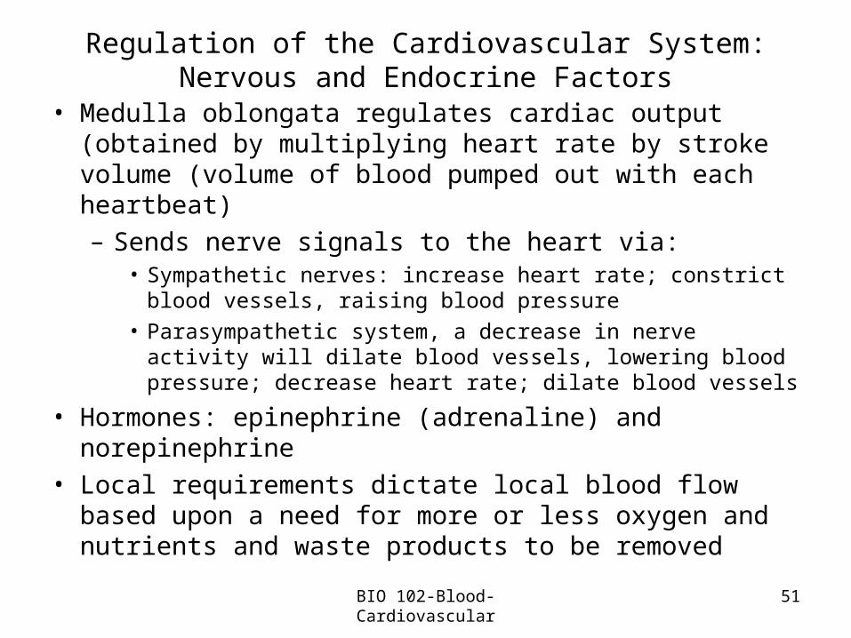
Regulation of the Cardiovascular System: Nervous and Endocrine Factors
• Medulla oblongata regulates cardiac output (obtained by multiplying heart rate by stroke volume (volume of blood pumped out with each heartbeat)– Sends nerve signals to the heart via:
• Sympathetic nerves: increase heart rate; constrict blood vessels, raising blood pressure
• Parasympathetic system, a decrease in nerve activity will dilate blood vessels, lowering blood pressure; decrease heart rate; dilate blood vessels
• Hormones: epinephrine (adrenaline) and norepinephrine• Local requirements dictate local blood flow based upon a
need for more or less oxygen and nutrients and waste products to be removed
BIO 102-Blood-Cardiovascular
52
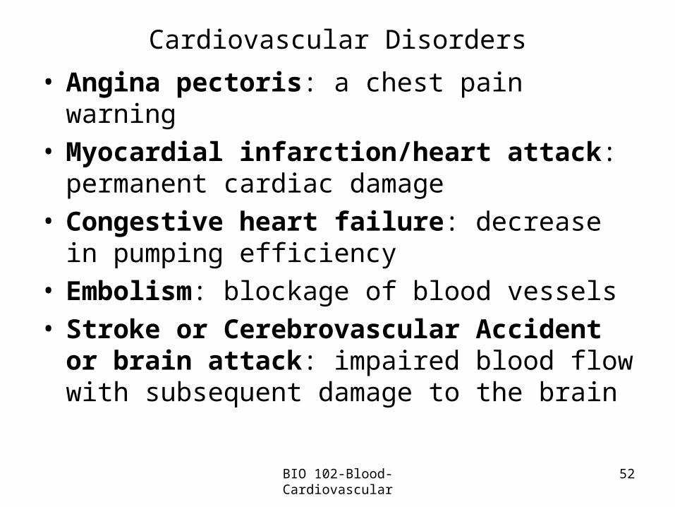
Cardiovascular Disorders
• Angina pectoris: a chest pain warning• Myocardial infarction/heart attack: permanent
cardiac damage• Congestive heart failure: decrease in pumping
efficiency• Embolism: blockage of blood vessels• Stroke or Cerebrovascular Accident or brain
attack: impaired blood flow with subsequent damage to the brain
BIO 102-Blood-Cardiovascular
• Pericarditis:– Inflammation of the pericardium (sac which
surrounds the heart)• Endocarditis: inflammation of the endocardium
BIO 102-Blood-Cardiovascular 53

54
Reducing the Risk of Cardiovascular Disease
• Smoking: don’t • Blood lipids: monitor cholesterol levels• Exercise: regular and moderate• Blood pressure: treat hypertension• Weight: being overweight increases risk of heart
attack and stroke• Control of diabetes mellitus: early diagnosis and
treatment delays onset of related problems• Stress: avoid chronic stress
BIO 102-Blood-Cardiovascular
55
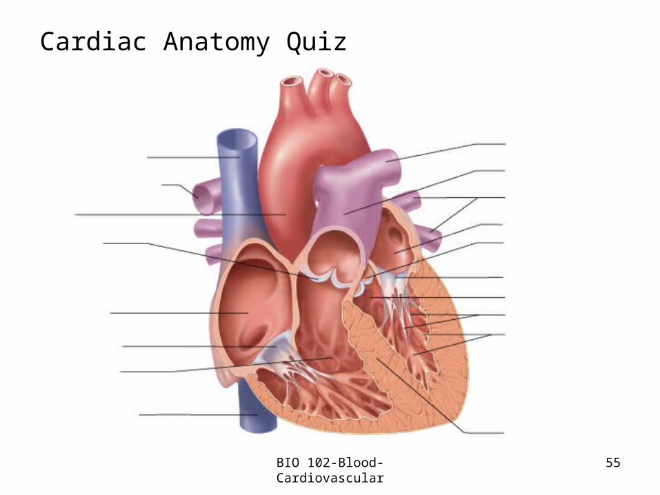
Cardiac Anatomy Quiz
BIO 102-Blood-Cardiovascular