0DWHULDO (6, IRU&KHP&RPP 7KLV - rsc.org · S7 3.3. Synthesis of peptide amides Peptide amides 3, 7,...
39
S1 Total chemical synthesis of the site-selective azide-labeled [I66A]HIV-1 protease Yun-Kun Qi, ‡ab Hao-Nan Chang, ‡b Kai-Mai Pan b and Ji-Shen Zheng *ab a.High Magnetic Field Laboratory, Chinese Academy of Sciences Hefei 230031, China. b. Department of Chemistry, Tsinghua University, Beijing 100084, China ‡ These authors contributed equally to this work. E-mail: [email protected]. Table of Contents 1. Reagents and materials ............................................................................................S3 2. HPLC, mass spectrometry (MS) ..............................................................................S3 3. Fmoc-based solid-phase peptide synthesis (Fmoc-SPPS) .......................................S3 3.1. General procedures for Fmoc-SPPS .................................................................................S3 3.2. Synthesis of peptide hydrazides .......................................................................................S3 3.3. Synthesis of peptide amides .............................................................................................S7 4. The attempt to synthesize azide-labled HIV-1 protease ..........................................S9 5. The attempt to synthesize alkyne-labled HIV-1 protease ......................................S10 5.1. The study of the side reaction during free-radical-based desulfurization ......................S10 5.2. The attempts to prevent the side reaction during free radical desulfurization ................S20 6. The model of [I66A]HIV-1 protease precursor with solubilizing tag ...................S23 7. Synthesis of compound 14 .....................................................................................S23 8. General procedure for the native chemical ligation of peptide hydrazids .............S24 8.1. Ligation of peptide hydrazide 1 with peptide hydrazide 2 .............................................S24 8.2. The failed ligation of peptide hydrazide 4 with peptide amide 3 ...................................S25 8.3. Ligation of peptide hydrazide 1 with peptide hydrazide 6 .............................................S26 8.4. Ligation of peptide hydrazide 8 with peptide amide 7 ...................................................S27 8.5. Ligation of peptide hydrazide 11 with peptide hydrazide 12 .........................................S28 Electronic Supplementary Material (ESI) for ChemComm. This journal is © The Royal Society of Chemistry 2015
Transcript of 0DWHULDO (6, IRU&KHP&RPP 7KLV - rsc.org · S7 3.3. Synthesis of peptide amides Peptide amides 3, 7,...
S1
Total chemical synthesis of the site-selective azide-labeled [I66A]HIV-1 protease
Yun-Kun Qi,‡ab Hao-Nan Chang,‡b Kai-Mai Panb and Ji-Shen Zheng*ab
a.High Magnetic Field Laboratory, Chinese Academy of Sciences Hefei 230031, China.
b. Department of Chemistry, Tsinghua University, Beijing 100084, China
‡ These authors contributed equally to this work.
E-mail: [email protected].
Table of Contents
1. Reagents and materials ............................................................................................S3
2. HPLC, mass spectrometry (MS) ..............................................................................S3
3. Fmoc-based solid-phase peptide synthesis (Fmoc-SPPS) .......................................S3
3.1. General procedures for Fmoc-SPPS .................................................................................S3
3.2. Synthesis of peptide hydrazides .......................................................................................S3
3.3. Synthesis of peptide amides .............................................................................................S7
4. The attempt to synthesize azide-labled HIV-1 protease ..........................................S9
5. The attempt to synthesize alkyne-labled HIV-1 protease ......................................S10
5.1. The study of the side reaction during free-radical-based desulfurization ......................S10
5.2. The attempts to prevent the side reaction during free radical desulfurization................S20
6. The model of [I66A]HIV-1 protease precursor with solubilizing tag ...................S23
7. Synthesis of compound 14 .....................................................................................S23
8. General procedure for the native chemical ligation of peptide hydrazids .............S24
8.1. Ligation of peptide hydrazide 1 with peptide hydrazide 2 .............................................S24
8.2. The failed ligation of peptide hydrazide 4 with peptide amide 3 ...................................S25
8.3. Ligation of peptide hydrazide 1 with peptide hydrazide 6 .............................................S26
8.4. Ligation of peptide hydrazide 8 with peptide amide 7 ...................................................S27
8.5. Ligation of peptide hydrazide 11 with peptide hydrazide 12 .........................................S28
Electronic Supplementary Material (ESI) for ChemComm.This journal is © The Royal Society of Chemistry 2015
S2
8.6. Ligation of peptide hydrazide 16 with peptide amide 13 ...............................................S29
9. Substitution reaction between peptide 15 and compound 14.................................S32
10. Protein folding .....................................................................................................S33
11. Substrate hydrolysis by synthetic azide-labeled [I66A]HIV-1 protease..............S34
12. Substrate hydrolysis by the commercial HIV-1 protease ....................................S35
13. Characterization of peptides 4, 15, 16, 17, 18 .....................................................S36
14. ESI-MS spectra of peptides 19, 20 and 21...........................................................S39
15. References............................................................................................................S39
S3
1. Reagents and materials
Rink amide AM resin and 2-Chlorotrityl resin were purchased from Hecheng Technology
(Tianjing, China). Fmoc-amino acids were purchased from GL Biochem (Shanghai, China), C S Bio
or Bo Mai Jie Technology (Beijing, China). Other reagents and materials were purchased as
previously described.[1]
2. HPLC and mass spectrometry (MS)
Reversed phase HPLC was all performed on Shimadzu Prominence HPLC. For peptide analysis,
Vydac C18 (4.6 × 150 mm) and C8 (4.6 × 150 mm) columns were used at a flow rate of 1.0 mL/min.
For peptide purification, Vydac C18 (10 × 250 mm), C8 (10 × 250 mm) and C4 (10 × 250 mm)
columns were used at a flow rate of 3-4 mL/min. The UV absorption at 214 nm and 254 nm were
monitored for the injections.
ESI-MS spectra were recorded on an Agilent 1200 Series HPLC system with LC-MS (Agilent
6340 ion trap as the mass spectrometer) or an Esquire~LC iron trap mass spectrometer. MALDI-
TOF/MS was performed on an Applied Biosystems 4800PLUS MALDI-TOF/TOF mass
spectrometer in the Center of Biomedical Analysis, Tsinghua University.
3. Fmoc-based solid-phase peptide synthesis (Fmoc-SPPS)
3.1. General procedures for Fmoc-SPPS
Peptides were synthesized manually by Fmoc-SPPS.[1] The Rink amide AM resin was swelled
in DCM/DMF (1:1). After 30 min, the Fmoc group of the resin was removed by treatment with 20%
piperidine in DMF (twice: 5 min, 10 min). Then the coupling was carried out using a solution of the
Fmoc-amino acid (5 equiv.), O-(6-Chlorobenzotriazol-1-yl)-N,N,N’,N’-tetramethyluronium
hexafluorophosphate (HCTU) (4.5 equiv.) and DIPEA (10 equiv.) in DMF (the Fmoc-amino acid
was activated for about 15 s before addition to the resin). 1.0-1.3 h reaction at 30 °C is enough for
most amino acids. Double coupling could be carried out when needed (for high sterically hindered
amino acids and the amino acid right after proline). After the coupling reaction, the resin was
thoroughly washed with DMF and DCM. The Fmoc group was removed by 20% piperidine in DMF
S4
(twice: 5 min and then 10 min). Then the resin was thoroughly washed with DMF and DCM. Then
the next coupling reaction could be conducted. After all the coupling reactions completed, the N-
terminal Fmoc group was removed by treatment with 20% piperidine in DMF (twice: 5 min, 10 min).
After washing the resin thoroughly with DMF and DCM, the cleavage and deprotection steps was
carried out by cocktail K (TFA/phenol/water/thioanisole/EDT, 82.5:5:5:5:2.5) or B
(TFA/phenol/water/TIPS, 88:5:5:2) for 2.5-3.0 h. Then the TFA solutions were concentrated by pure
argon. The crude peptide was then precipitated with cold Et2O and then centrifuged (three times).
Finally, the crude peptide was dissolved in H2O/CH3CN and purified by semi-preparative RP-HPLC.
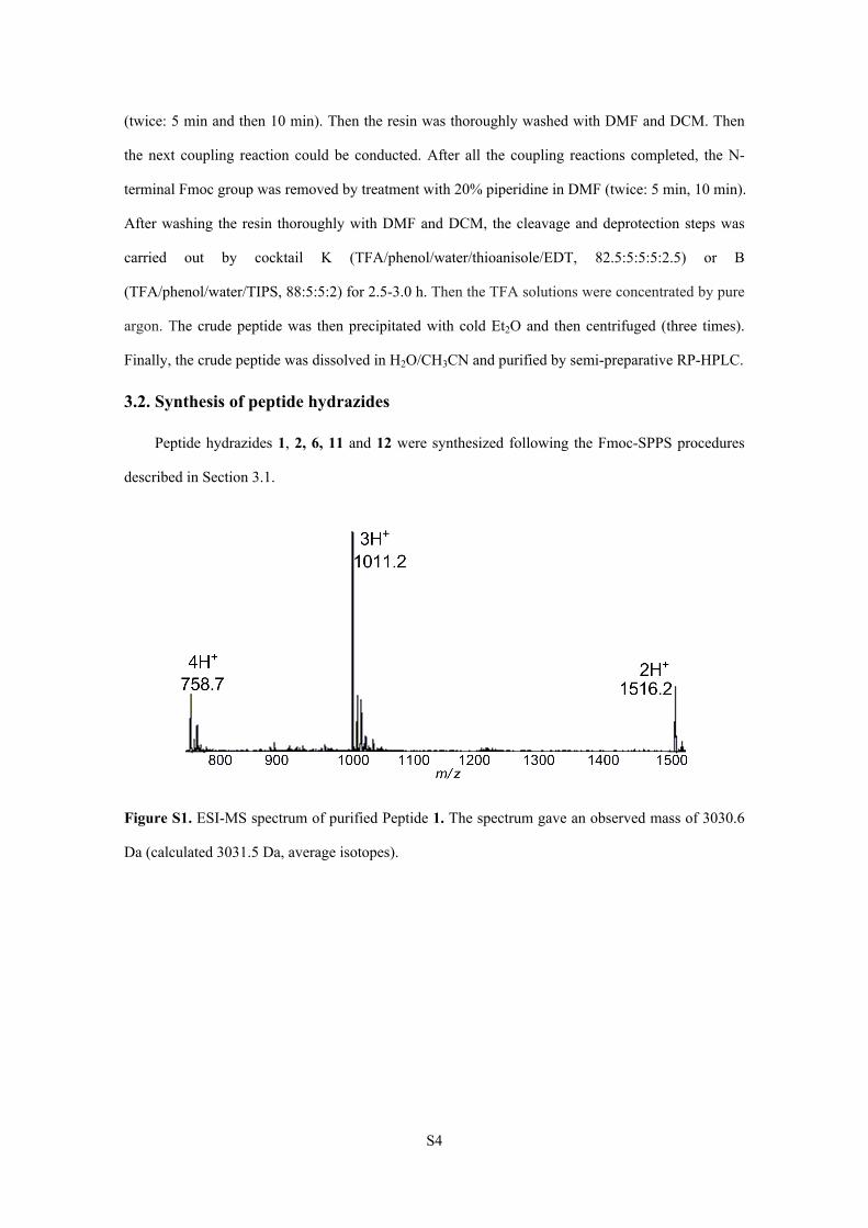
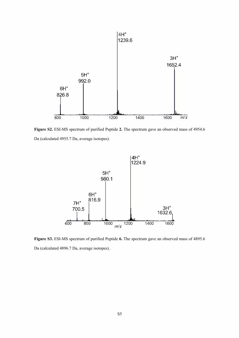
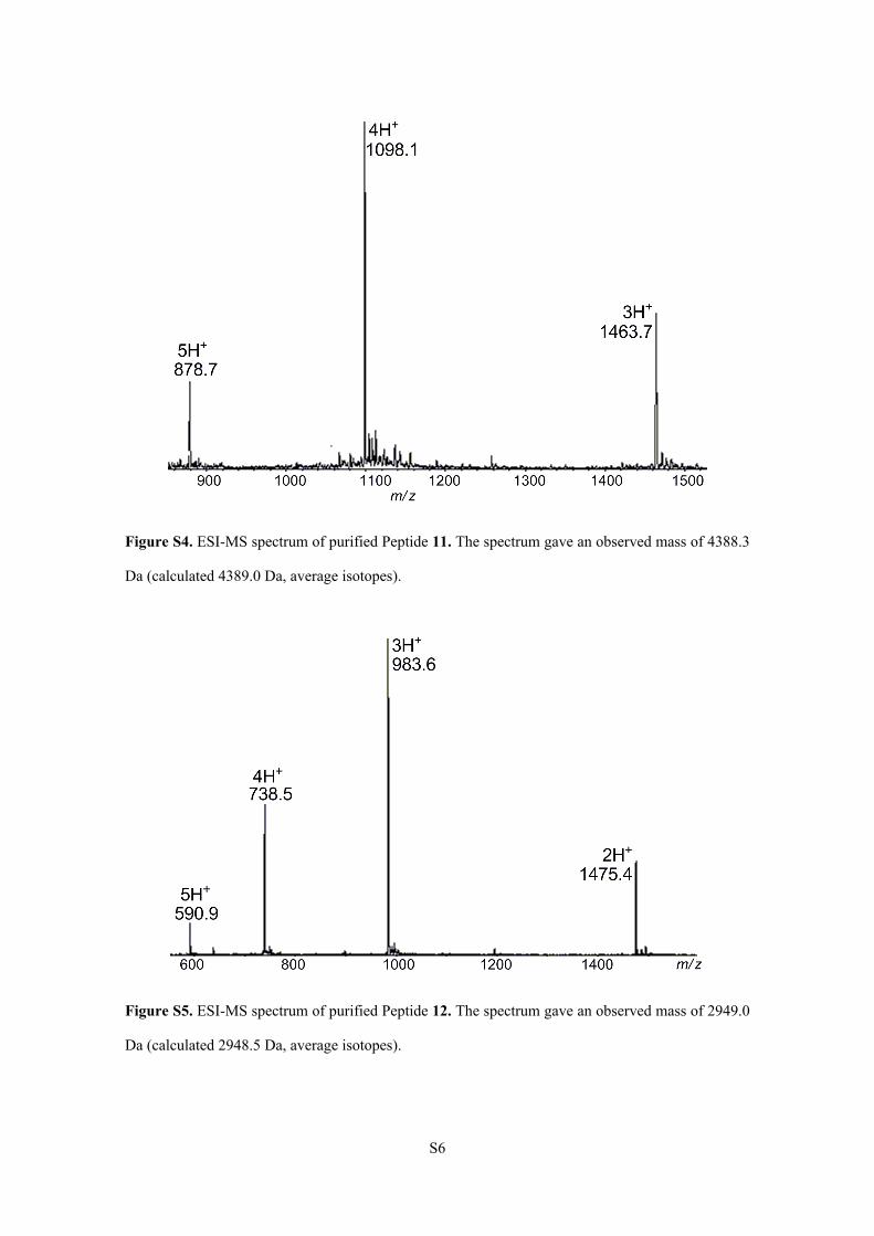
3.2. Synthesis of peptide hydrazides
Peptide hydrazides 1, 2, 6, 11 and 12 were synthesized following the Fmoc-SPPS procedures
described in Section 3.1.
Figure S1. ESI-MS spectrum of purified Peptide 1. The spectrum gave an observed mass of 3030.6
Da (calculated 3031.5 Da, average isotopes).
S5
Figure S2. ESI-MS spectrum of purified Peptide 2. The spectrum gave an observed mass of 4954.6
Da (calculated 4955.7 Da, average isotopes).
Figure S3. ESI-MS spectrum of purified Peptide 6. The spectrum gave an observed mass of 4895.6
Da (calculated 4896.7 Da, average isotopes).
S6
Figure S4. ESI-MS spectrum of purified Peptide 11. The spectrum gave an observed mass of 4388.3
Da (calculated 4389.0 Da, average isotopes).
Figure S5. ESI-MS spectrum of purified Peptide 12. The spectrum gave an observed mass of 2949.0
Da (calculated 2948.5 Da, average isotopes).
S7
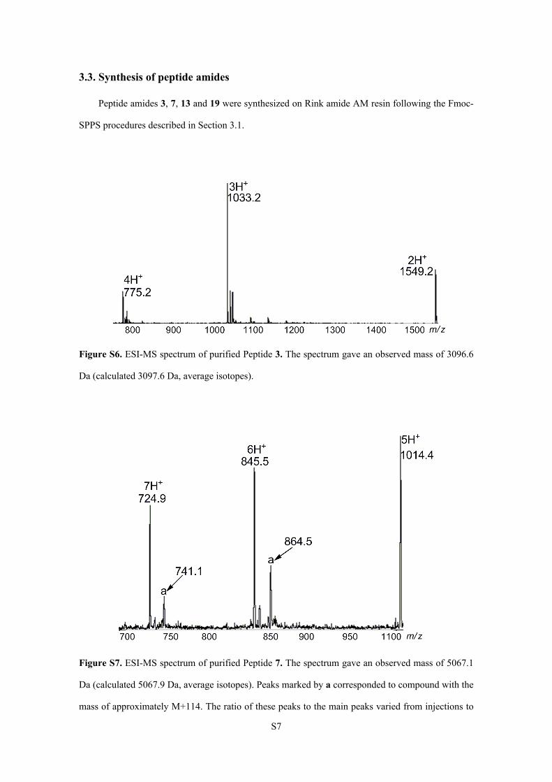
3.3. Synthesis of peptide amides
Peptide amides 3, 7, 13 and 19 were synthesized on Rink amide AM resin following the Fmoc-
SPPS procedures described in Section 3.1.
Figure S6. ESI-MS spectrum of purified Peptide 3. The spectrum gave an observed mass of 3096.6
Da (calculated 3097.6 Da, average isotopes).
Figure S7. ESI-MS spectrum of purified Peptide 7. The spectrum gave an observed mass of 5067.1
Da (calculated 5067.9 Da, average isotopes). Peaks marked by a corresponded to compound with the
mass of approximately M+114. The ratio of these peaks to the main peaks varied from injections to
S8
injections. These peaks were thought to be associated with the C-terminus Arg6 tag because these
peaks were observed only in ESI-MS spectra of peptides containing the Arg6 tag.2 Because these
peaks may disappear after the detachment of the Arg6 tag from the peptide, our synthesis process
was not hampered by this problem.
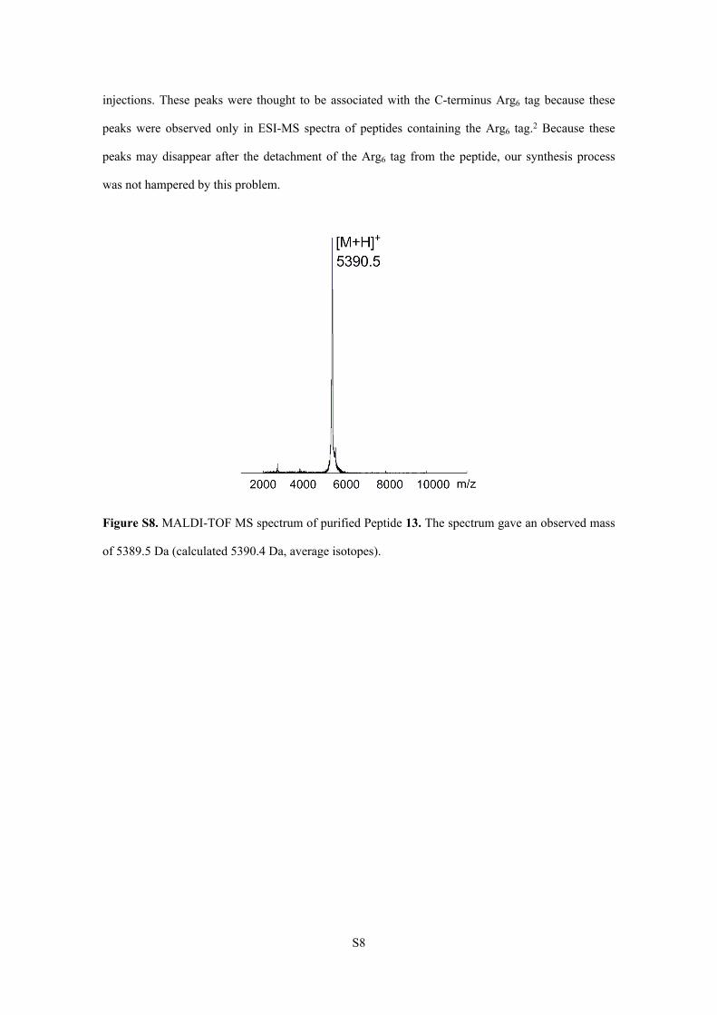
Figure S8. MALDI-TOF MS spectrum of purified Peptide 13. The spectrum gave an observed mass
of 5389.5 Da (calculated 5390.4 Da, average isotopes).
S9
4. The attempts to synthesize azide-labled HIV-1 protease
Analytic RP-HPLC chromatogram ( = 214 nm) of purified 5 (Figure 1E in main text). HPLC
conditions: a linear gradient of 20-60% acetonitrile (with 0.08-0.1% TFA) in water (with 0.1% TFA)
over 30 min (5% for 2 min, then 20%-60% for 30 min) on a Vydac C8 (4.6 × 150 mm) column.
Although the free-radical-based desulfurization reaction is often used in protein chemical
synthesis, the understanding of the compatibility of azide groups with free-radical-based
desulfurization reaction is limited. To test the compatibility of azide groups with free-radical-based
desulfurization reaction, we treated PR[Pro1−Lys(N3)41−Lys70]-NHNH2 4 with aqueous TCEP (500
mM) containing 6 M Gn·HCl and 0.2 M Na2HPO4 (pH = 6.6), tBuSH and 2,2’-azobis[2-(2-
imidazolin-2-yl)propane]dihydrochloride (VA-044).1 After stirred at room temperature for 2 min,
PR[Pro1−Lys(N3)41−Lys70]-NHNH2 4 was completely converted to PR[Pro1−Lys41−Lys70]-NHNH2 5.
ESI-MS analysis of 5 showed an exact 26 Da decrease, which could be attributed to the reduction of
azide by high concentration of TCEP. Through the initial attempts, we found out that although azide
group could survive the TCEP during NCL because of the low TCEP concentration and a quick
operation, it can not bear such a high concentration of TCEP during free-radical-based
desulfurization reaction.
S10
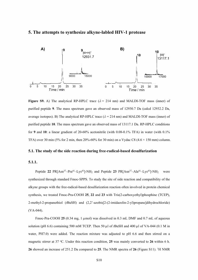
5. The attempts to synthesize alkyne-labled HIV-1 protease
Figure S9. A) The analytical RP-HPLC trace (= 214 nm) and MALDI-TOF mass (inner) of
purified peptide 9. The mass spectrum gave an observed mass of 12930.7 Da (calcd 12932.2 Da,
average isotopes). B) The analytical RP-HPLC trace ( = 214 nm) and MALDI-TOF mass (inner) of
purified peptide 10. The mass spectrum gave an observed mass of 13117.1 Da. RP-HPLC conditions
for 9 and 10: a linear gradient of 20-60% acetonitrile (with 0.08-0.1% TFA) in water (with 0.1%
TFA) over 30 min (5% for 2 min, then 20%-60% for 30 min) on a Vydac C8 (4.6 × 150 mm) column.
5.1. The study of the side reaction during free-radical-based desulfurization
5.1.1.
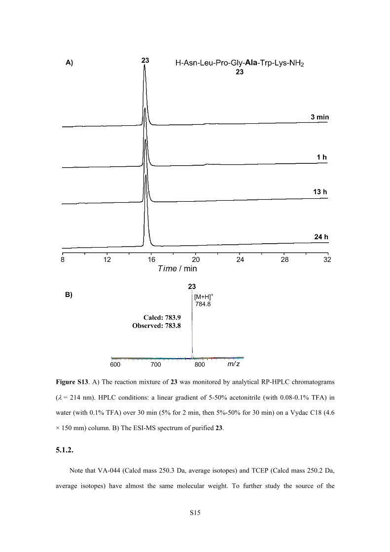
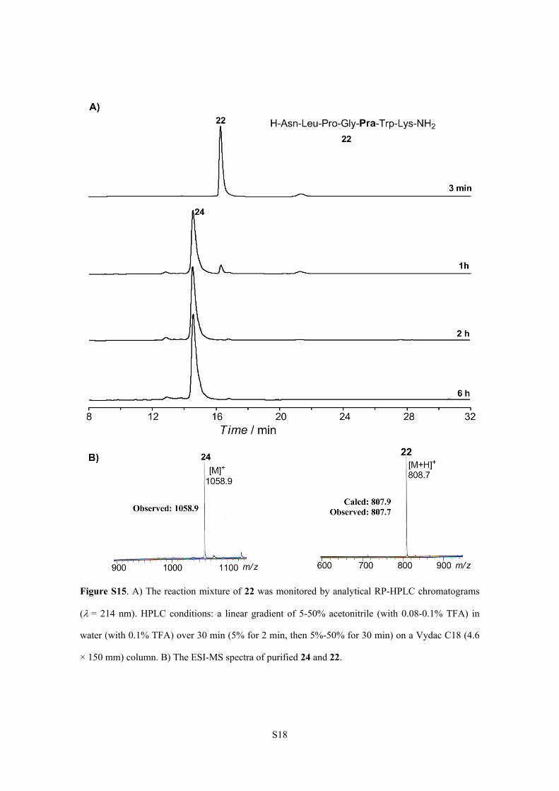
Peptide 22 PR[Asn37–Pra41–Lys43]-NH2 and Peptide 23 PR[Asn37–Ala41–Lys43]-NH2 were
synthesized through standard Fmoc-SPPS. To study the site of side reaction and compatibility of the
alkyne groups with the free-radical-based desulfurization reaction often involved in protein chemical
synthesis, we treated Fmoc-Pra-COOH 25, 22 and 23 with Tris(2-carboxyethyl)phosphine (TCEP),
2-methyl-2-propanethiol (tBuSH) and (2,2’-azobis[2-(2-imidazolin-2-yl)propane]dihydrochloride)
(VA-044).
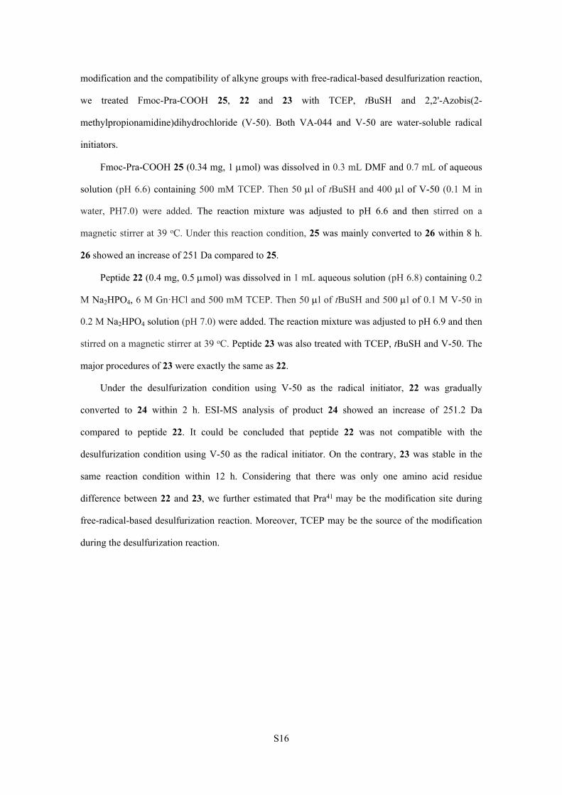
Fmoc-Pra-COOH 25 (0.34 mg, 1 mol) was dissolved in 0.3 mL DMF and 0.7 mL of aqueous
solution (pH 6.6) containing 500 mM TCEP. Then 50 l of tBuSH and 400 l of VA-044 (0.1 M in
water, PH7.0) were added. The reaction mixture was adjusted to pH 6.6 and then stirred on a
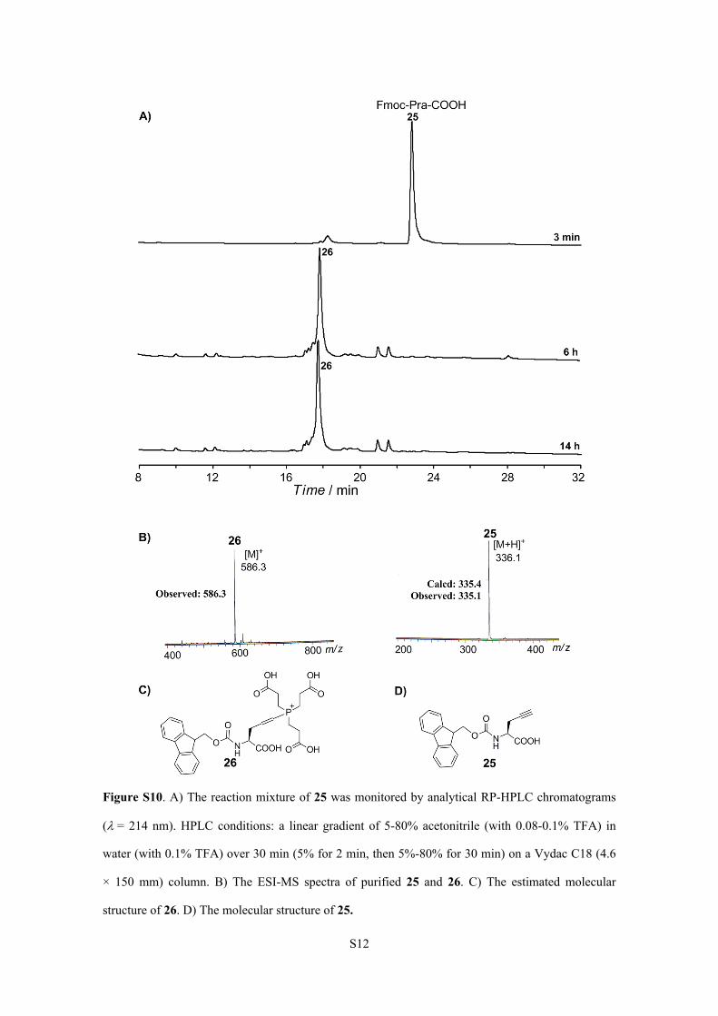
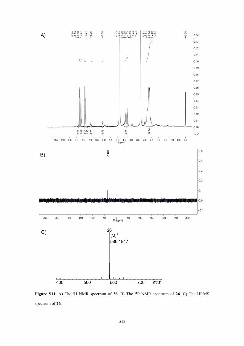
magnetic stirrer at 37 oC. Under this reaction condition, 25 was mainly converted to 26 within 6 h.
26 showed an increase of 251.2 Da compared to 25. The NMR spectra of 26 (Figure S11). 1H NMR
S11
(600 MHz, [D4]CH3OH): δ (ppm) = 7.80 (d, J = 6 Hz, 2H), 7.67 (d, J = 6 Hz, 2H), 7.40-7.31 (m, 4H),
6.95 (m, 1H), 6.08 (m, 1H), 4.43-4.23 (m, 4H), 2.81-2.53 (m, 14H). 31P NMR (243 MHz,
[D6]DMSO): δ (ppm) = + 35.80. HRMS (Positive ESI) Calcd. For C29H33NO10P+: 586.1842, Found:
586.1847. It was speculated that TCEP (Calcd mass 250.2 Da, average isotopes) was added to the
alkyne group of 26 under the free-radical-based desulfurization condition.
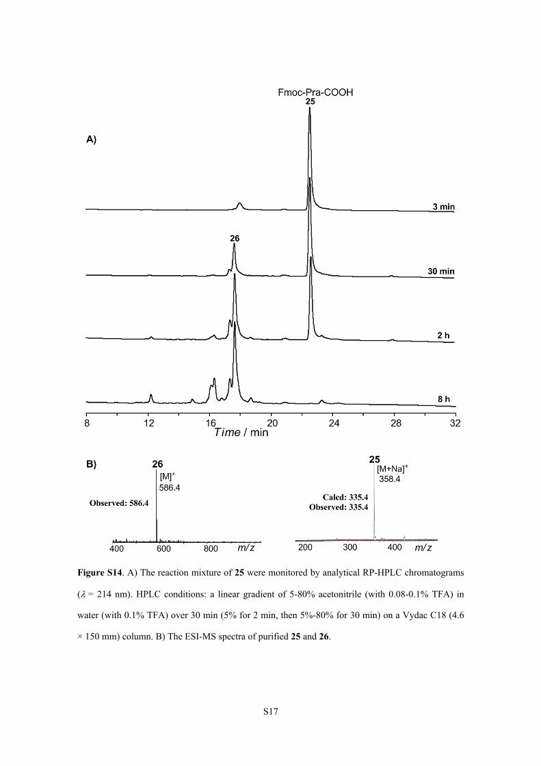
Peptide 22 (0.4 mg, 0.5 mol) was dissolved in 1 mL aqueous solution (pH 6.8) containing 0.2
M Na2HPO4, 6 M Gn·HCl and 500 mM TCEP. Then 50 l of tBuSH and 500 l of 0.1 M VA-044 in
0.2 M Na2HPO4 solution (pH 7.0) were added. The reaction mixture was adjusted to pH 6.9 and then
stirred on a magnetic stirrer at 37 oC. Peptide 23 was also treated with TCEP, tBuSH and VA-044.
The major procedures of 23 were exactly the same as 22.
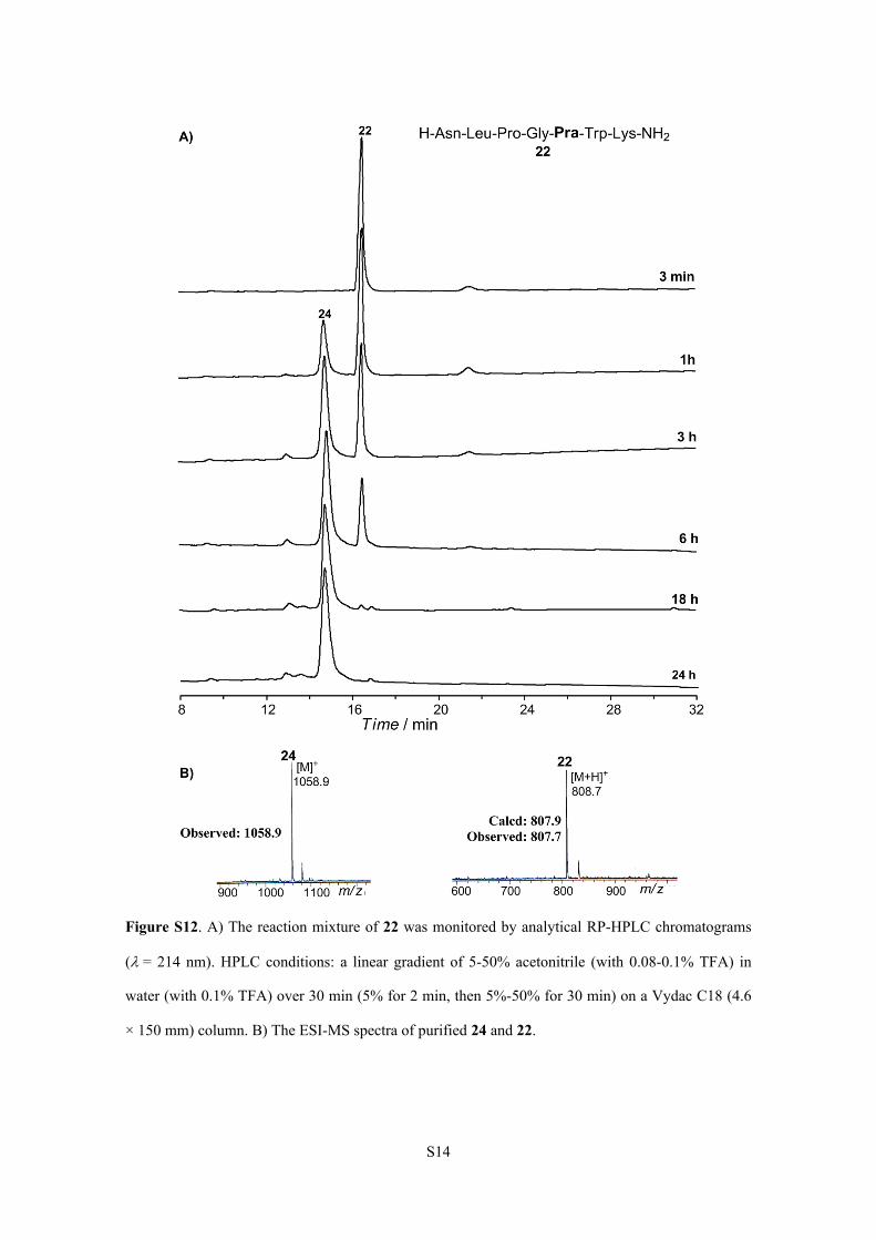
Under the commonly used free-radical-based desulfurization condition, 22 was gradually
converted to 24 within 18 h. ESI-MS analysis of product 24 showed an increase of 251.2 Da
compared to peptide 22. It could be concluded that peptide 22 was not compatible with the
commonly used free-radical-based desulfurization condition. On the contrary, 23 was stable in the
same reaction condition within 24 h. Considering that there was only one amino acid residue
difference between 22 and 23, we concluded that Pra41 may be the modification site during free-
radical-based desulfurization reaction.
S12
Figure S10. A) The reaction mixture of 25 was monitored by analytical RP-HPLC chromatograms
(= 214 nm). HPLC conditions: a linear gradient of 5-80% acetonitrile (with 0.08-0.1% TFA) in
water (with 0.1% TFA) over 30 min (5% for 2 min, then 5%-80% for 30 min) on a Vydac C18 (4.6
× 150 mm) column. B) The ESI-MS spectra of purified 25 and 26. C) The estimated molecular
structure of 26. D) The molecular structure of 25.
S13
Figure S11. A) The 1H NMR spectrum of 26. B) The 31P NMR spectrum of 26. C) The HRMS
spectrum of 26.
S14
Figure S12. A) The reaction mixture of 22 was monitored by analytical RP-HPLC chromatograms
(= 214 nm). HPLC conditions: a linear gradient of 5-50% acetonitrile (with 0.08-0.1% TFA) in
water (with 0.1% TFA) over 30 min (5% for 2 min, then 5%-50% for 30 min) on a Vydac C18 (4.6
× 150 mm) column. B) The ESI-MS spectra of purified 24 and 22.
S15
Figure S13. A) The reaction mixture of 23 was monitored by analytical RP-HPLC chromatograms
(= 214 nm). HPLC conditions: a linear gradient of 5-50% acetonitrile (with 0.08-0.1% TFA) in
water (with 0.1% TFA) over 30 min (5% for 2 min, then 5%-50% for 30 min) on a Vydac C18 (4.6
× 150 mm) column. B) The ESI-MS spectrum of purified 23.
5.1.2.
Note that VA-044 (Calcd mass 250.3 Da, average isotopes) and TCEP (Calcd mass 250.2 Da,
average isotopes) have almost the same molecular weight. To further study the source of the
S16
modification and the compatibility of alkyne groups with free-radical-based desulfurization reaction,
we treated Fmoc-Pra-COOH 25, 22 and 23 with TCEP, tBuSH and 2,2'-Azobis(2-
methylpropionamidine)dihydrochloride (V-50). Both VA-044 and V-50 are water-soluble radical
initiators.
Fmoc-Pra-COOH 25 (0.34 mg, 1 mol) was dissolved in 0.3 mL DMF and 0.7 mL of aqueous
solution (pH 6.6) containing 500 mM TCEP. Then 50 l of tBuSH and 400 l of V-50 (0.1 M in
water, PH7.0) were added. The reaction mixture was adjusted to pH 6.6 and then stirred on a
magnetic stirrer at 39 oC. Under this reaction condition, 25 was mainly converted to 26 within 8 h.
26 showed an increase of 251 Da compared to 25.
Peptide 22 (0.4 mg, 0.5 mol) was dissolved in 1 mL aqueous solution (pH 6.8) containing 0.2
M Na2HPO4, 6 M Gn·HCl and 500 mM TCEP. Then 50 l of tBuSH and 500 l of 0.1 M V-50 in
0.2 M Na2HPO4 solution (pH 7.0) were added. The reaction mixture was adjusted to pH 6.9 and then
stirred on a magnetic stirrer at 39 oC. Peptide 23 was also treated with TCEP, tBuSH and V-50. The
major procedures of 23 were exactly the same as 22.
Under the desulfurization condition using V-50 as the radical initiator, 22 was gradually
converted to 24 within 2 h. ESI-MS analysis of product 24 showed an increase of 251.2 Da
compared to peptide 22. It could be concluded that peptide 22 was not compatible with the
desulfurization condition using V-50 as the radical initiator. On the contrary, 23 was stable in the
same reaction condition within 12 h. Considering that there was only one amino acid residue
difference between 22 and 23, we further estimated that Pra41 may be the modification site during
free-radical-based desulfurization reaction. Moreover, TCEP may be the source of the modification
during the desulfurization reaction.
S17
Figure S14. A) The reaction mixture of 25 were monitored by analytical RP-HPLC chromatograms
(= 214 nm). HPLC conditions: a linear gradient of 5-80% acetonitrile (with 0.08-0.1% TFA) in
water (with 0.1% TFA) over 30 min (5% for 2 min, then 5%-80% for 30 min) on a Vydac C18 (4.6
× 150 mm) column. B) The ESI-MS spectra of purified 25 and 26.
S18
Figure S15. A) The reaction mixture of 22 was monitored by analytical RP-HPLC chromatograms
(= 214 nm). HPLC conditions: a linear gradient of 5-50% acetonitrile (with 0.08-0.1% TFA) in
water (with 0.1% TFA) over 30 min (5% for 2 min, then 5%-50% for 30 min) on a Vydac C18 (4.6
× 150 mm) column. B) The ESI-MS spectra of purified 24 and 22.
S19
Figure S16. A) The reaction mixture of 23 was monitored by analytical RP-HPLC chromatograms
(= 214 nm). HPLC conditions: a linear gradient of 5-50% acetonitrile (with 0.08-0.1% TFA) in
water (with 0.1% TFA) over 30 min (5% for 2 min, then 5%-50% for 30 min) on a Vydac C18 (4.6
× 150 mm) column. B) The ESI-MS spectrum of purified 23.
S20
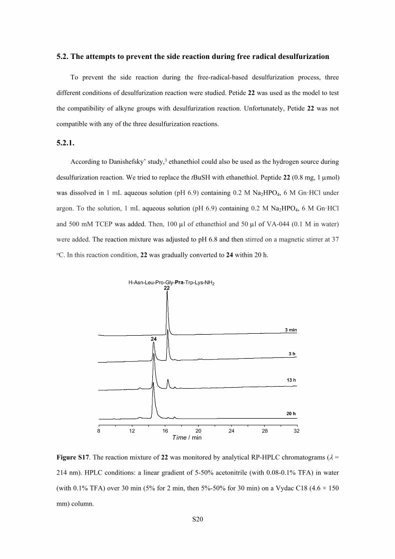
5.2. The attempts to prevent the side reaction during free radical desulfurization
To prevent the side reaction during the free-radical-based desulfurization process, three
different conditions of desulfurization reaction were studied. Petide 22 was used as the model to test
the compatibility of alkyne groups with desulfurization reaction. Unfortunately, Petide 22 was not
compatible with any of the three desulfurization reactions.
5.2.1.
According to Danishefsky’ study,3 ethanethiol could also be used as the hydrogen source during
desulfurization reaction. We tried to replace the tBuSH with ethanethiol. Peptide 22 (0.8 mg, 1 mol)
was dissolved in 1 mL aqueous solution (pH 6.9) containing 0.2 M Na2HPO4, 6 M Gn·HCl under
argon. To the solution, 1 mL aqueous solution (pH 6.9) containing 0.2 M Na2HPO4, 6 M Gn·HCl
and 500 mM TCEP was added. Then, 100 l of ethanethiol and 50 l of VA-044 (0.1 M in water)
were added. The reaction mixture was adjusted to pH 6.8 and then stirred on a magnetic stirrer at 37
oC. In this reaction condition, 22 was gradually converted to 24 within 20 h.
Figure S17. The reaction mixture of 22 was monitored by analytical RP-HPLC chromatograms (=
214 nm). HPLC conditions: a linear gradient of 5-50% acetonitrile (with 0.08-0.1% TFA) in water
(with 0.1% TFA) over 30 min (5% for 2 min, then 5%-50% for 30 min) on a Vydac C18 (4.6 × 150
mm) column.
S21
5.2.2.
We tried to reduce the concentration of VA-044 during desulfurization reaction. Peptide 22 (0.4
mg, 0.5 mol) was dissolved in 1 mL aqueous solution (pH 6.8) containing 0.2 M Na2HPO4, 6 M
Gn·HCl and 500 mM TCEP. Then 50 l of tBuSH and 500 l of 0.02 M VA-044 in 0.2 M Na2HPO4
solution (pH 7.0) were added. The reaction mixture was adjusted to pH 6.9 and then stirred on a
magnetic stirrer at 37 oC. In this reaction condition, 22 was gradually converted to 24 within 15 h.
Figure S18. The reaction mixture of 22 was monitored by analytical RP-HPLC chromatograms (=
214 nm). HPLC conditions: a linear gradient of 5-50% acetonitrile (with 0.08-0.1% TFA) in water
(with 0.1% TFA) over 30 min (5% for 2 min, then 5%-50% for 30 min) on a Vydac C18 (4.6 × 150
mm) column.
5.2.3.
We tried to change the pH value of desulfurization reaction. Peptide 22 (0.4 mg, 0.5 mol) was
dissolved in 1 mL aqueous solution (pH 4.0) containing 0.2 M Na2HPO4, 6 M Gn·HCl and 500 mM
TCEP. Then 50 l of tBuSH and 500 l of 0.1 M VA-044 in 0.2 M Na2HPO4 solution (pH 7.0) were
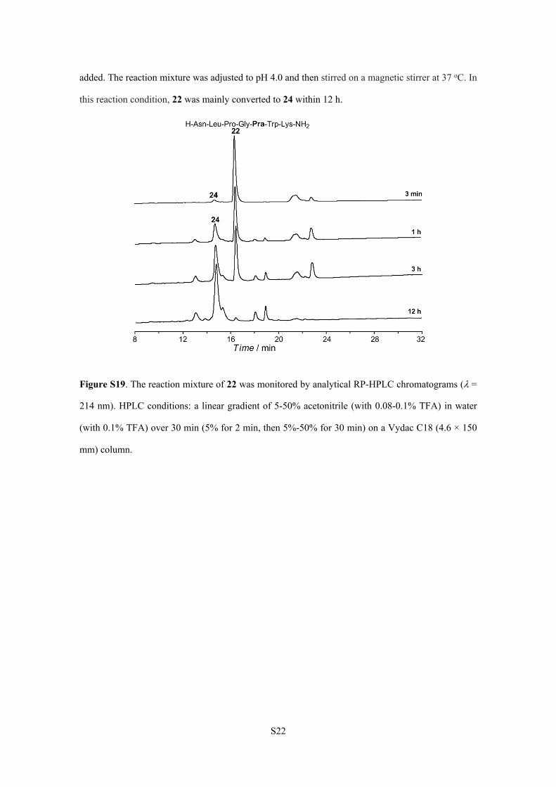
S22
added. The reaction mixture was adjusted to pH 4.0 and then stirred on a magnetic stirrer at 37 oC. In
this reaction condition, 22 was mainly converted to 24 within 12 h.
Figure S19. The reaction mixture of 22 was monitored by analytical RP-HPLC chromatograms (=
214 nm). HPLC conditions: a linear gradient of 5-50% acetonitrile (with 0.08-0.1% TFA) in water
(with 0.1% TFA) over 30 min (5% for 2 min, then 5%-50% for 30 min) on a Vydac C18 (4.6 × 150
mm) column.
S23
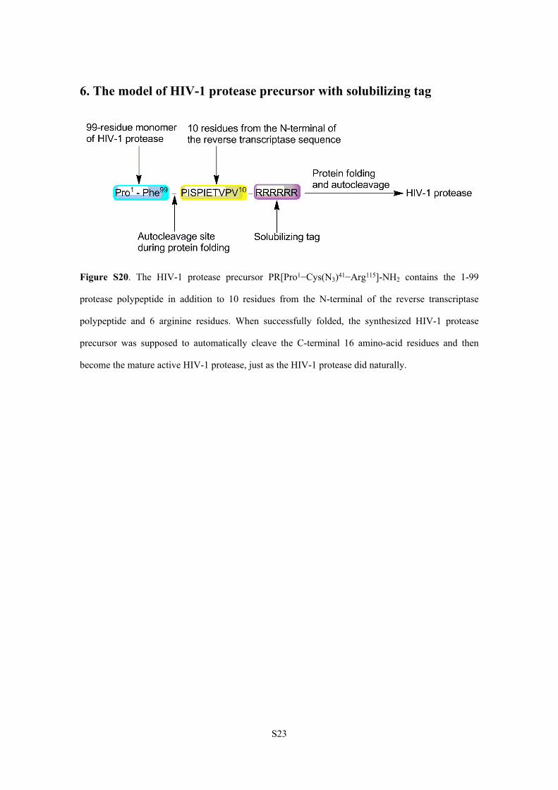
6. The model of HIV-1 protease precursor with solubilizing tag
Figure S20. The HIV-1 protease precursor PR[Pro1−Cys(N3)41−Arg115]-NH2 contains the 1-99
protease polypeptide in addition to 10 residues from the N-terminal of the reverse transcriptase
polypeptide and 6 arginine residues. When successfully folded, the synthesized HIV-1 protease
precursor was supposed to automatically cleave the C-terminal 16 amino-acid residues and then
become the mature active HIV-1 protease, just as the HIV-1 protease did naturally.
S24
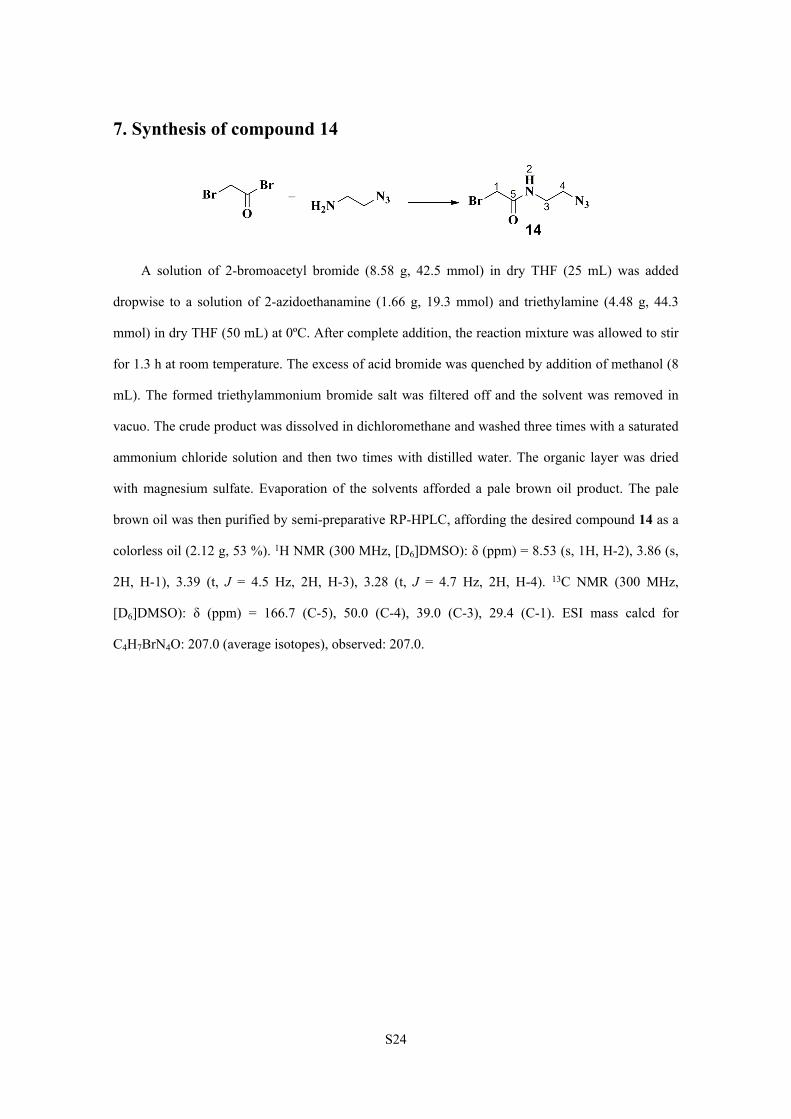
7. Synthesis of compound 14
A solution of 2-bromoacetyl bromide (8.58 g, 42.5 mmol) in dry THF (25 mL) was added
dropwise to a solution of 2-azidoethanamine (1.66 g, 19.3 mmol) and triethylamine (4.48 g, 44.3
mmol) in dry THF (50 mL) at 0ºC. After complete addition, the reaction mixture was allowed to stir
for 1.3 h at room temperature. The excess of acid bromide was quenched by addition of methanol (8
mL). The formed triethylammonium bromide salt was filtered off and the solvent was removed in
vacuo. The crude product was dissolved in dichloromethane and washed three times with a saturated
ammonium chloride solution and then two times with distilled water. The organic layer was dried
with magnesium sulfate. Evaporation of the solvents afforded a pale brown oil product. The pale
brown oil was then purified by semi-preparative RP-HPLC, affording the desired compound 14 as a
colorless oil (2.12 g, 53 %). 1H NMR (300 MHz, [D6]DMSO): δ (ppm) = 8.53 (s, 1H, H-2), 3.86 (s,
2H, H-1), 3.39 (t, J = 4.5 Hz, 2H, H-3), 3.28 (t, J = 4.7 Hz, 2H, H-4). 13C NMR (300 MHz,
[D6]DMSO): δ (ppm) = 166.7 (C-5), 50.0 (C-4), 39.0 (C-3), 29.4 (C-1). ESI mass calcd for
C4H7BrN4O: 207.0 (average isotopes), observed: 207.0.
S25
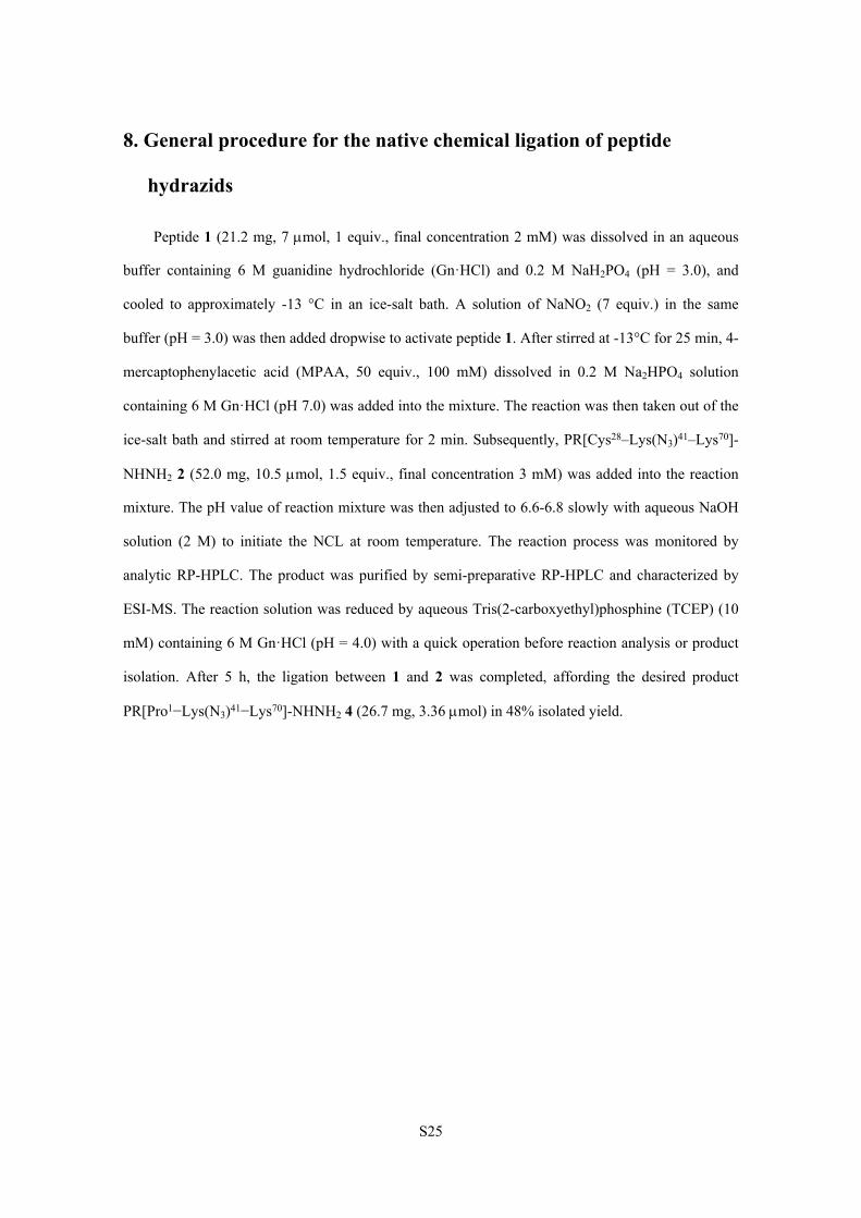
8. General procedure for the native chemical ligation of peptide
hydrazids
Peptide 1 (21.2 mg, 7 mol, 1 equiv., final concentration 2 mM) was dissolved in an aqueous
buffer containing 6 M guanidine hydrochloride (Gn·HCl) and 0.2 M NaH2PO4 (pH = 3.0), and
cooled to approximately -13 °C in an ice-salt bath. A solution of NaNO2 (7 equiv.) in the same
buffer (pH = 3.0) was then added dropwise to activate peptide 1. After stirred at -13°C for 25 min, 4-
mercaptophenylacetic acid (MPAA, 50 equiv., 100 mM) dissolved in 0.2 M Na2HPO4 solution
containing 6 M Gn·HCl (pH 7.0) was added into the mixture. The reaction was then taken out of the
ice-salt bath and stirred at room temperature for 2 min. Subsequently, PR[Cys28–Lys(N3)41–Lys70]-
NHNH2 2 (52.0 mg, 10.5 mol, 1.5 equiv., final concentration 3 mM) was added into the reaction
mixture. The pH value of reaction mixture was then adjusted to 6.6-6.8 slowly with aqueous NaOH
solution (2 M) to initiate the NCL at room temperature. The reaction process was monitored by
analytic RP-HPLC. The product was purified by semi-preparative RP-HPLC and characterized by
ESI-MS. The reaction solution was reduced by aqueous Tris(2-carboxyethyl)phosphine (TCEP) (10
mM) containing 6 M Gn·HCl (pH = 4.0) with a quick operation before reaction analysis or product
isolation. After 5 h, the ligation between 1 and 2 was completed, affording the desired product
PR[Pro1−Lys(N3)41−Lys70]-NHNH2 4 (26.7 mg, 3.36 mol) in 48% isolated yield.
S26
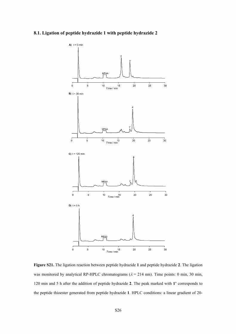
8.1. Ligation of peptide hydrazide 1 with peptide hydrazide 2
Figure S21. The ligation reaction between peptide hydrazide 1 and peptide hydrazide 2. The ligation
was monitored by analytical RP-HPLC chromatograms (= 214 nm). Time points: 0 min, 30 min,
120 min and 5 h after the addition of peptide hydrazide 2. The peak marked with 1’ corresponds to
the peptide thioester generated from peptide hydrazide 1. HPLC conditions: a linear gradient of 20-
S27
60% acetonitrile (with 0.08-0.1% TFA) in water (with 0.1% TFA) over 30 min (5% for 2 min, then
20%-60% for 30 min) on a Vydac C8 (4.6 × 150 mm) column.
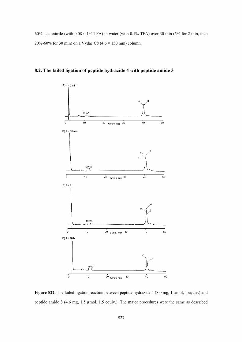
8.2. The failed ligation of peptide hydrazide 4 with peptide amide 3
Figure S22. The failed ligation reaction between peptide hydrazide 4 (8.0 mg, 1 mol, 1 equiv.) and
peptide amide 3 (4.6 mg, 1.5 mol, 1.5 equiv.). The major procedures were the same as described
S28
above. The ligation was monitored by analytical RP-HPLC chromatograms (= 214 nm). Time
points: 0 min, 60 min, 9 h and 16 h after the addition of peptide amide 3. The peak marked with 4’
corresponds to the peptide thioester generated from peptide hydrazide 4. The peak marked with 4’’
corresponds to the hydrolysis product of 4’. Peptides 3, 4’ and 4’’ had almost the same retention
time. HPLC conditions: a linear gradient of 20-60% acetonitrile (with 0.08-0.1% TFA) in water
(with 0.1% TFA) over 50 min (5% for 2 min, then 20% for 20 min, then 20%-60% for 30 min) on a
Vydac C8 (4.6 × 150 mm) column.
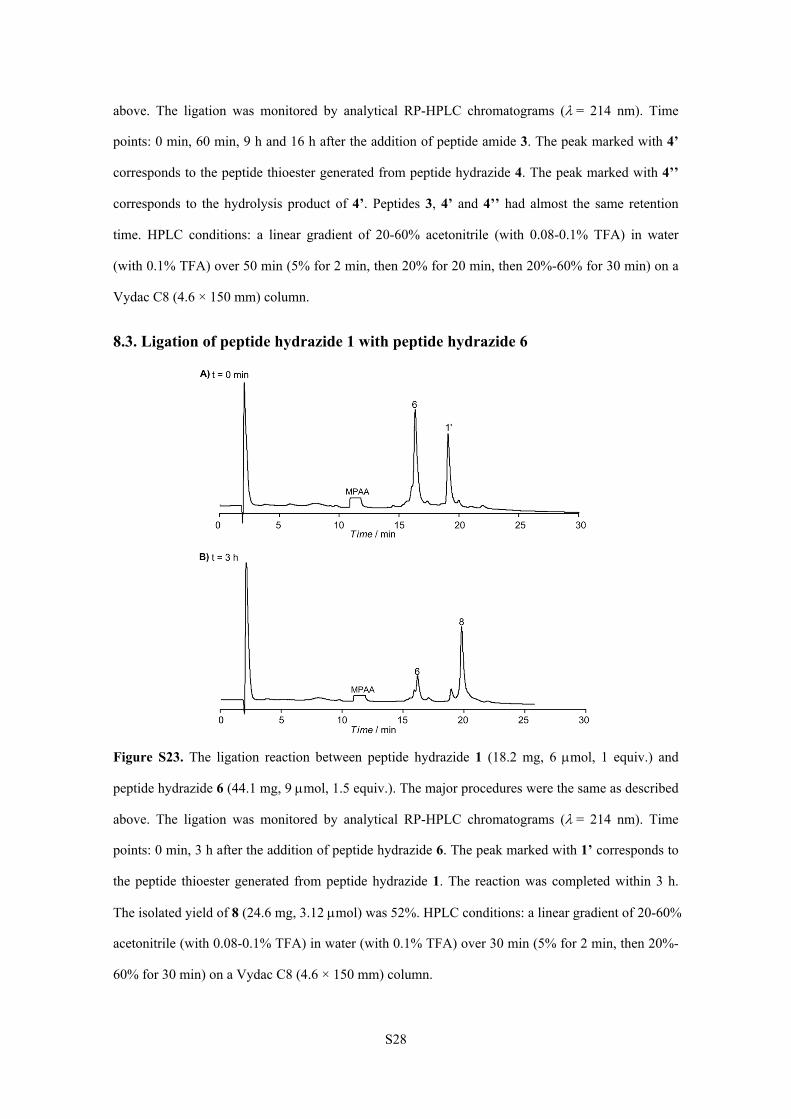
8.3. Ligation of peptide hydrazide 1 with peptide hydrazide 6
Figure S23. The ligation reaction between peptide hydrazide 1 (18.2 mg, 6 mol, 1 equiv.) and
peptide hydrazide 6 (44.1 mg, 9 mol, 1.5 equiv.). The major procedures were the same as described
above. The ligation was monitored by analytical RP-HPLC chromatograms (= 214 nm). Time
points: 0 min, 3 h after the addition of peptide hydrazide 6. The peak marked with 1’ corresponds to
the peptide thioester generated from peptide hydrazide 1. The reaction was completed within 3 h.
The isolated yield of 8 (24.6 mg, 3.12 mol) was 52%. HPLC conditions: a linear gradient of 20-60%
acetonitrile (with 0.08-0.1% TFA) in water (with 0.1% TFA) over 30 min (5% for 2 min, then 20%-
60% for 30 min) on a Vydac C8 (4.6 × 150 mm) column.
S29
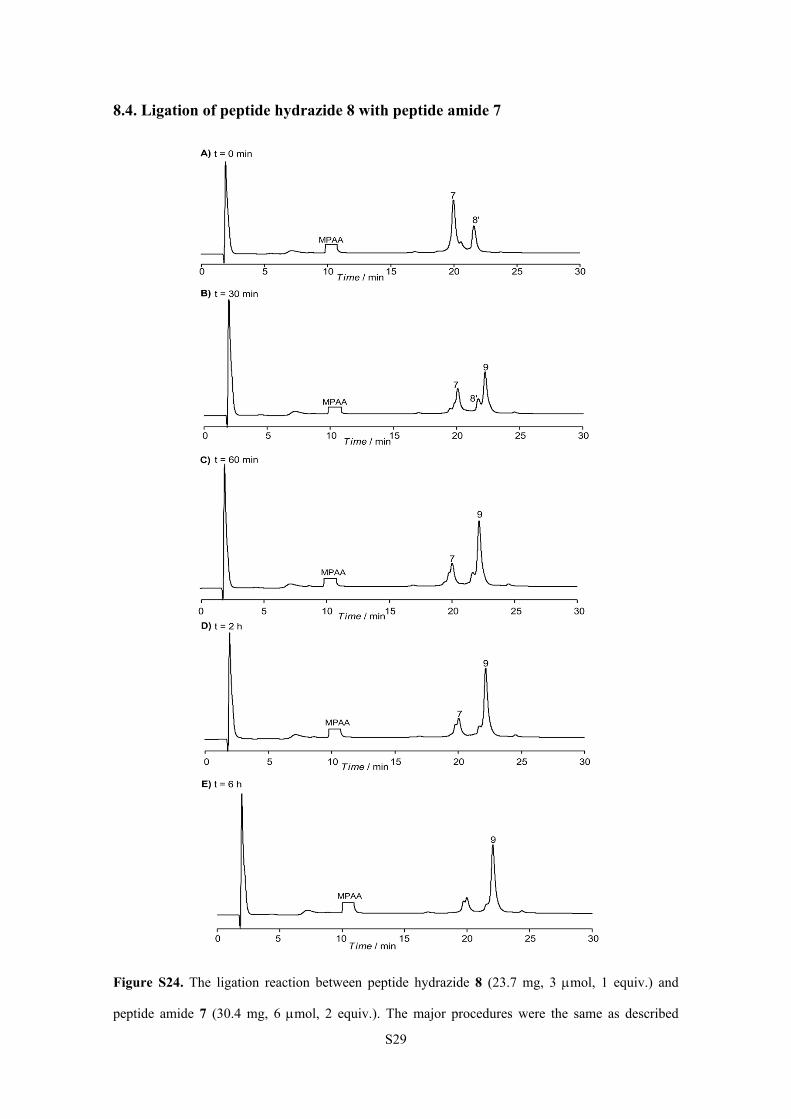
8.4. Ligation of peptide hydrazide 8 with peptide amide 7
Figure S24. The ligation reaction between peptide hydrazide 8 (23.7 mg, 3 mol, 1 equiv.) and
peptide amide 7 (30.4 mg, 6 mol, 2 equiv.). The major procedures were the same as described
S30
above. The ligation was monitored by analytical RP-HPLC chromatograms (= 214 nm). Time
points: 0 min, 30 min, 60 min, 2 h and 6 h after the addition of peptide amide 7. The peak marked
with 8’ corresponds to the peptide thioester generated from peptide hydrazide 8. The reaction was
completed within 6 h. The isolated yield of 9 (16.7 mg, 1.29 mol) was 43%. HPLC conditions: a
linear gradient of 20-60% acetonitrile (with 0.08-0.1% TFA) in water (with 0.1% TFA) over 30 min
(5% for 2 min, then 20%-60% for 30 min) on a Vydac C8 (4.6 × 150 mm) column.
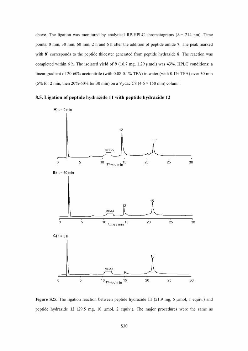
8.5. Ligation of peptide hydrazide 11 with peptide hydrazide 12
Figure S25. The ligation reaction between peptide hydrazide 11 (21.9 mg, 5 mol, 1 equiv.) and
peptide hydrazide 12 (29.5 mg, 10 mol, 2 equiv.). The major procedures were the same as
S31
described above. The ligation was monitored by analytical RP-HPLC chromatograms (= 214 nm).
Time points: 0 min, 60 min and 5 h after the addition of peptide hydrazide 12. The peak marked with
11’ corresponds to the peptide thioester generated from peptide hydrazide 11. The reaction was
completed within 5 h. The isolated yield of 15 (15.7 mg, 2.15 mol) was 43%. HPLC conditions: a
linear gradient of 20-60% acetonitrile (with 0.08-0.1% TFA) in water (with 0.1% TFA) over 30 min
(5% for 2 min, then 20%-60% for 30 min) on a Vydac C8 (4.6 × 150 mm) column.
8.6. Ligation of peptide hydrazide 16 with peptide amide 13
The ligation reaction between peptide hydrazide 16 (11.1 mg, 1.5 mol, 1 equiv.) and peptide
amide 13 (16.2 mg, 3 mol, 2 equiv.). The major procedures were the same as described above. The
ligation was monitored by analytical RP-HPLC chromatograms (= 214 nm) (Figure 3 in main text).
Time points: 0 min, 60 min and 6 h after the addition of peptide amide 13. The peak marked with 16’
corresponds to the peptide thioester generated from peptide hydrazide 16. The reaction was
completed within 6 h. The isolated yield of 17 (7.9 mg, 0.62 mol) was 41%. HPLC conditions: a
linear gradient of 20-60% acetonitrile (with 0.08-0.1% TFA) in water (with 0.1% TFA) over 30 min
(5% for 2 min, then 20%-60% for 30 min) on a Vydac C8 (4.6 × 150 mm) column.
S32
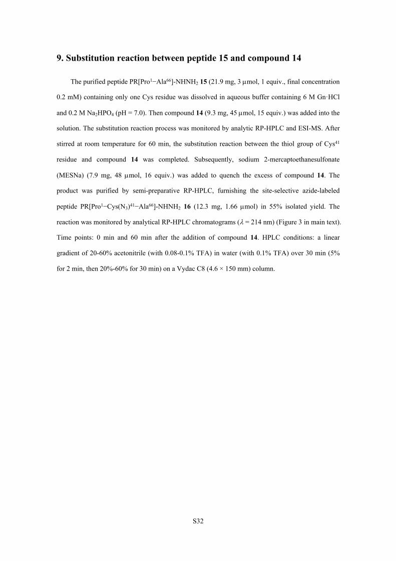
9. Substitution reaction between peptide 15 and compound 14
The purified peptide PR[Pro1−Ala66]-NHNH2 15 (21.9 mg, 3 mol, 1 equiv., final concentration
0.2 mM) containing only one Cys residue was dissolved in aqueous buffer containing 6 M Gn·HCl
and 0.2 M Na2HPO4 (pH = 7.0). Then compound 14 (9.3 mg, 45 mol, 15 equiv.) was added into the
solution. The substitution reaction process was monitored by analytic RP-HPLC and ESI-MS. After
stirred at room temperature for 60 min, the substitution reaction between the thiol group of Cys41
residue and compound 14 was completed. Subsequently, sodium 2-mercaptoethanesulfonate
(MESNa) (7.9 mg, 48 mol, 16 equiv.) was added to quench the excess of compound 14. The
product was purified by semi-preparative RP-HPLC, furnishing the site-selective azide-labeled
peptide PR[Pro1−Cys(N3)41−Ala66]-NHNH2 16 (12.3 mg, 1.66 mol) in 55% isolated yield. The
reaction was monitored by analytical RP-HPLC chromatograms (= 214 nm) (Figure 3 in main text).
Time points: 0 min and 60 min after the addition of compound 14. HPLC conditions: a linear
gradient of 20-60% acetonitrile (with 0.08-0.1% TFA) in water (with 0.1% TFA) over 30 min (5%
for 2 min, then 20%-60% for 30 min) on a Vydac C8 (4.6 × 150 mm) column.
S33
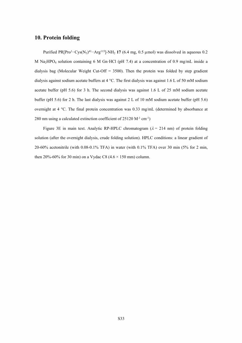
10. Protein folding
Purified PR[Pro1−Cys(N3)41−Arg115]-NH2 17 (6.4 mg, 0.5 mol) was dissolved in aqueous 0.2
M Na2HPO4 solution containing 6 M Gn·HCl (pH 7.4) at a concentration of 0.9 mg/mL inside a
dialysis bag (Molecular Weight Cut-Off = 3500). Then the protein was folded by step gradient
dialysis against sodium acetate buffers at 4 °C. The first dialysis was against 1.6 L of 50 mM sodium
acetate buffer (pH 5.6) for 3 h. The second dialysis was against 1.6 L of 25 mM sodium acetate
buffer (pH 5.6) for 2 h. The last dialysis was against 2 L of 10 mM sodium acetate buffer (pH 5.6)
overnight at 4 °C. The final protein concentration was 0.33 mg/mL (determined by absorbance at
280 nm using a calculated extinction coefficient of 25120 M-1 cm-1)
Figure 3E in main text. Analytic RP-HPLC chromatogram (= 214 nm) of protein folding
solution (after the overnight dialysis, crude folding solution). HPLC conditions: a linear gradient of
20-60% acetonitrile (with 0.08-0.1% TFA) in water (with 0.1% TFA) over 30 min (5% for 2 min,
then 20%-60% for 30 min) on a Vydac C8 (4.6 × 150 mm) column.
S34
11. Substrate hydrolysis by synthetic azide-labeled HIV-1 protease
The synthetic substrate peptide 19 (0.2 mg, 0.2 mol) was dissolved in 1 mL of 50 mM NaOAc,
0.2 M NaCl buffer with 1% (v/v) DMSO at pH 5.6. Then, 5 L solution of folded HIV-1 protease
(1.65 g, 0.152 nmol) was added into the solution. The mixture was then incubated at 37 ºC. The
hydrolysis process was monitored by analytic RP-HPLC (= 214 nm) and ESI-MS. RP-HPLC
conditions: a linear gradient of 5-70% acetonitrile (with 0.08-0.1% TFA) in water (with 0.1% TFA)
over 30 min on a Vydac C8 (4.6 × 150 mm) column.
S35
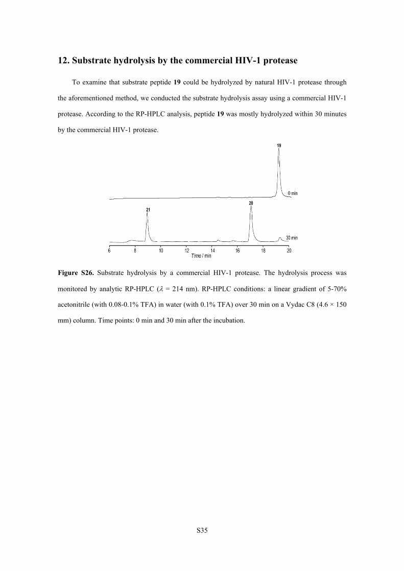
12. Substrate hydrolysis by the commercial HIV-1 protease
To examine that substrate peptide 19 could be hydrolyzed by natural HIV-1 protease through
the aforementioned method, we conducted the substrate hydrolysis assay using a commercial HIV-1
protease. According to the RP-HPLC analysis, peptide 19 was mostly hydrolyzed within 30 minutes
by the commercial HIV-1 protease.
Figure S26. Substrate hydrolysis by a commercial HIV-1 protease. The hydrolysis process was
monitored by analytic RP-HPLC ( = 214 nm). RP-HPLC conditions: a linear gradient of 5-70%
acetonitrile (with 0.08-0.1% TFA) in water (with 0.1% TFA) over 30 min on a Vydac C8 (4.6 × 150
mm) column. Time points: 0 min and 30 min after the incubation.
S36
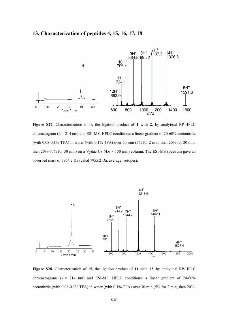
13. Characterization of peptides 4, 15, 16, 17, 18
Figure S27. Characterization of 4, the ligation product of 1 with 2, by analytical RP-HPLC
chromatogram (= 214 nm) and ESI-MS. HPLC conditions: a linear gradient of 20-60% acetonitrile
(with 0.08-0.1% TFA) in water (with 0.1% TFA) over 50 min (5% for 2 min, then 20% for 20 min,
then 20%-60% for 30 min) on a Vydac C8 (4.6 × 150 mm) column. The ESI-MS spectrum gave an
observed mass of 7954.2 Da (calcd 7955.2 Da, average isotopes).
Figure S28. Characterization of 15, the ligation product of 11 with 12, by analytical RP-HPLC
chromatograms (= 214 nm) and ESI-MS. HPLC conditions: a linear gradient of 20-60%
acetonitrile (with 0.08-0.1% TFA) in water (with 0.1% TFA) over 30 min (5% for 2 min, then 20%-
S37
60% for 30 min) on a Vydac C8 (4.6 × 150 mm) column. The ESI-MS spectrum gave an observed
mass of 7305.8 Da (calcd 7305.5 Da, average isotopes).
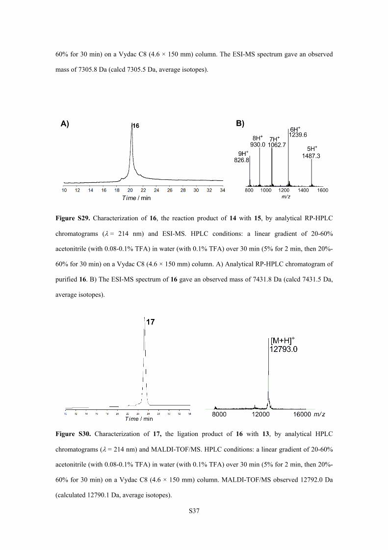
Figure S29. Characterization of 16, the reaction product of 14 with 15, by analytical RP-HPLC
chromatograms (= 214 nm) and ESI-MS. HPLC conditions: a linear gradient of 20-60%
acetonitrile (with 0.08-0.1% TFA) in water (with 0.1% TFA) over 30 min (5% for 2 min, then 20%-
60% for 30 min) on a Vydac C8 (4.6 × 150 mm) column. A) Analytical RP-HPLC chromatogram of
purified 16. B) The ESI-MS spectrum of 16 gave an observed mass of 7431.8 Da (calcd 7431.5 Da,
average isotopes).
Figure S30. Characterization of 17, the ligation product of 16 with 13, by analytical HPLC
chromatograms (= 214 nm) and MALDI-TOF/MS. HPLC conditions: a linear gradient of 20-60%
acetonitrile (with 0.08-0.1% TFA) in water (with 0.1% TFA) over 30 min (5% for 2 min, then 20%-
60% for 30 min) on a Vydac C8 (4.6 × 150 mm) column. MALDI-TOF/MS observed 12792.0 Da
(calculated 12790.1 Da, average isotopes).
S38
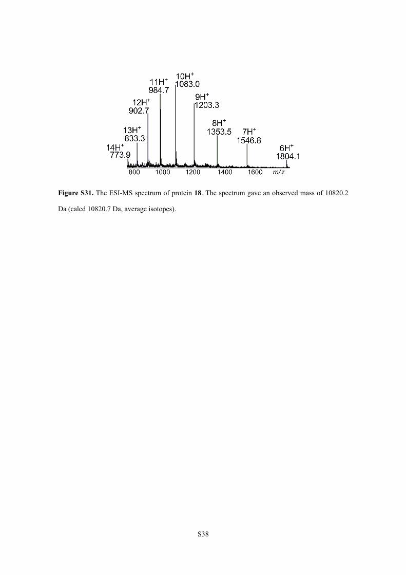
Figure S31. The ESI-MS spectrum of protein 18. The spectrum gave an observed mass of 10820.2
Da (calcd 10820.7 Da, average isotopes).
S39
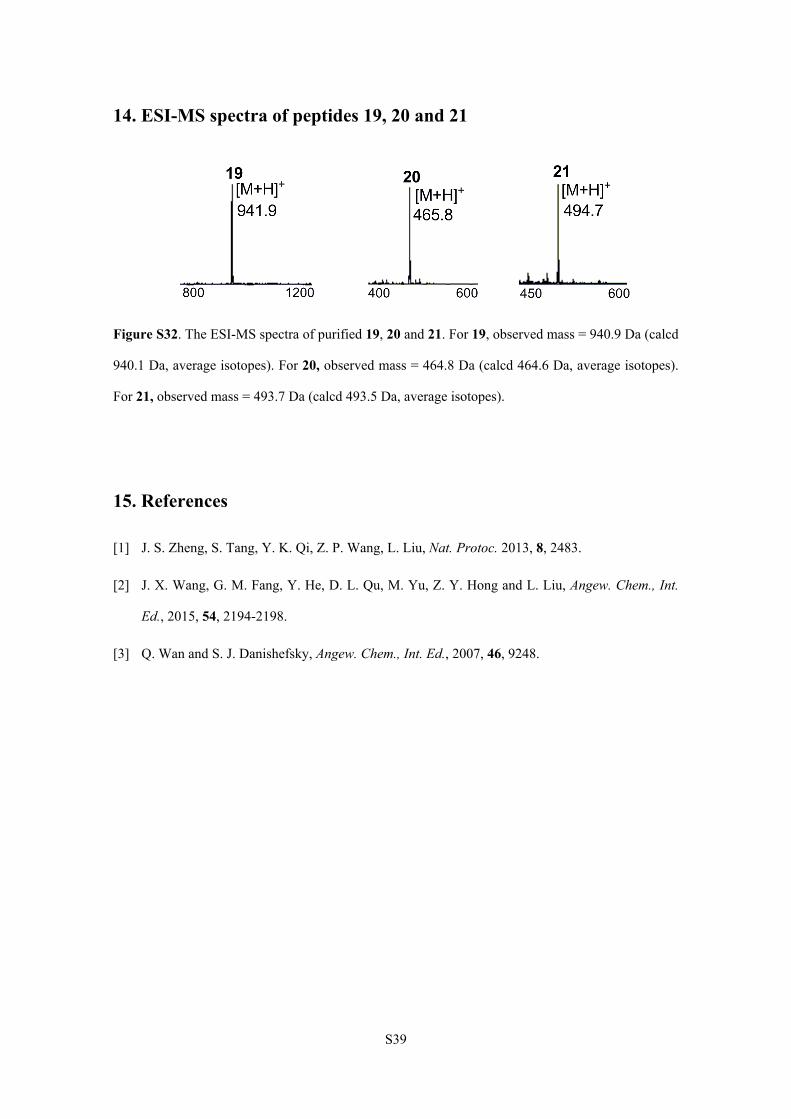
14. ESI-MS spectra of peptides 19, 20 and 21
Figure S32. The ESI-MS spectra of purified 19, 20 and 21. For 19, observed mass = 940.9 Da (calcd
940.1 Da, average isotopes). For 20, observed mass = 464.8 Da (calcd 464.6 Da, average isotopes).
For 21, observed mass = 493.7 Da (calcd 493.5 Da, average isotopes).
15. References
[1] J. S. Zheng, S. Tang, Y. K. Qi, Z. P. Wang, L. Liu, Nat. Protoc. 2013, 8, 2483.
[2] J. X. Wang, G. M. Fang, Y. He, D. L. Qu, M. Yu, Z. Y. Hong and L. Liu, Angew. Chem., Int.
Ed., 2015, 54, 2194-2198.
[3] Q. Wan and S. J. Danishefsky, Angew. Chem., Int. Ed., 2007, 46, 9248.