Fmoc Peptide Solid Phase Synthesis
371
-
Upload
lorenzosanv -
Category
Documents
-
view
1.657 -
download
6
Transcript of Fmoc Peptide Solid Phase Synthesis


Fmoc solid phase peptide synthesis

The Practical Approach Series
SERIES EDITOR
B. D. HAMESDepartment of Biochemistry and Molecular Biology
University of Leeds, Leeds LS2 9JT, UK
See also the Practical Approach web site at http://www.oup.co.uk/PAS* indicates new and forthcoming titles
Affinity ChromatographyAffinity SeparationsAnaerobic MicrobiologyAnimal Cell Culture
(2nd edition)Animal Virus PathogenesisAntibodies I and IIAntibody EngineeringAntisense TechnologyApoptosisApplied Microbial PhysiologyBasic Cell CultureBehavioural NeuroscienceBioenergeticsBiological Data AnalysisBiomechanics - MaterialsBiomechanics - Structures and
SystemsBiosensorsC ElegansCarbohydrate Analysis
(2nd edition)Cell-Cell InteractionsThe Cell Cycle
if Cell Growth, Differentiationand Senescence
* Cell SeparationCellular CalciumCellular Interactions in
DevelopmentCellular NeurobiologyChromatin
if Chromosome StructuralAnalysis
Clinical ImmunologyComplement
* Crystallization of NucleicAcids and Proteins(2nd edition)
Cytokines (2nd edition)The CytoskeletonCytoskeleton: signalling and
cell regulationDiagnostic Molecular
Pathology I and IIDNA and Protein Sequence
AnalysisDNA Cloning 1: Core
Techniques (2nd edition)

DNA Cloning 2: ExpressionSystems (2nd edition)
DNA Cloning 3: ComplexGenomes (2nd edition)
DNA Cloning 4: MammalianSystems (2nd edition)
DNA MicroarraysDNA VirusesDrosophila (2nd edition)Electron Microscopy in
BiologyElectron Microscopy in
Molecular BiologyElectrophysiologyEnzyme AssaysEpithelial Cell CultureEssential Developmental
BiologyEssential Molecular Biology I
and IIEukaryotic DNA ReplicationExperimental NeuroanatomyExtracellular MatrixFmoc solid phase peptide
synthesisFlow Cytometry (2nd edition)Free RadicalsGel Electrophoresis of Nucleic
Acids (2nd edition)Gel Electrophoresis of Proteins
(3rd edition)Gene Probes 1 and 2
* Gene Targeting (2nd edition)Gene TranscriptionGenome MappingGlycobiologyGrowth Factors and Receptors
Haemopoiesis* High Resolution
ChromotographyHistocompatibility TestingHIV Volumes 1 and 2
* HPLC of Macromolecules(2nd edition)
Human Cytogenetics I and II(2nd edition)
Human Genetic DiseaseAnalysis
* Image Processing and Analysisif Immobilized Biomolecules in
AnalysisImmunochemistry 1Immunochemistry 2ImmunocytochemistryImmunodiagnostics
if In Situ Hybridization(2nd edition)
lodinated Density GradientMedia
Ion Channels* Light Microscopy (2nd edition)
Lipid Modification ofProteins
Lipoprotein AnalysisLiposomes
* Lymphocytes (2nd edition)Mammalian Cell
BiotechnologyMedical ParasitologyMedical VirologyMHC Volumes 1 and 2Molecular Genetic Analysis of
Populations (2nd edition)Molecular Genetics of Yeast

Molecular Imaging inNeuroscience
Molecular Plant Pathology Iand II
Molecular VirologyMonitoring Neuronal Activity
if Mouse Genetics andTransgenics
Mutagenicity TestingMutation DetectionNeural Cell CultureNeural TransplantationNeurochemistry (2nd edition)Neuronal Cell LinesNMR of Biological
MacromoleculesNon-isotopic Methods in
Molecular BiologyNucleic Acid Hybridisation
if Nuclear ReceptorsOligonucleotides and
AnaloguesOligonucleotide SynthesisPCR 1PCR 2
*PCR 3:PCR In SituHybridization
Peptide AntigensPhotosynthesis: Energy
Trans ductionPlant Cell BiologyPlant Cell Culture (2nd edition)Plant Molecular BiologyPlasmids (2nd edition)
PlateletsPostimplantation Mammalian
EmbryosPost-translational ProcessingPreparative CentrifugationProtein Blotting
if Protein ExpressionProtein Engineering
* Protein Localization byFluorescence Microscopy
if Protein Phosphorylation(2nd edition)
Protein PurificationApplications
Protein Purification MethodsProtein SequencingProtein Structure
(2nd edition)Protein Structure PredictionProtein TargetingProteolytic EnzymesPulsed Field Gel
ElectrophoresisRNA Processing I and IIRNA-Protein InteractionsSignalling by InositidesSignal Transduction
(2nd edition)Subcellular FractionationSignal Transduction
if Transcription Factors(2nd edition)
Tumour Immunobiologyif Virus Culture

Fmoc solid phasepeptide synthesis
A Practical Approach
Edited by
WENG C. CHANSchool of Pharmaceutical Sciences,
University of Nottingham, University Park,Nottingham NG7 2RD
and
PETER D. WHITECN Biosciences, Padge Road, Beeston,
Nottingham NG9 2JR
OXFORDUNIVERSITY PRESS

OXFORDUNIVERSITY PRESS
Great Clarendon Street, Oxford OX2 6DPOxford University Press is a department of the University of Oxford
and furthers the University's aim of excellence in research, scholarship,and education by publishing worldwide in
Oxford New YorkAthens Auckland Bangkok Bogota Buenos Aires Calcutta
Cape Town Chennai Dar es Salaam Delhi Florence Hong Kong IstanbulKarachi Kuala Lumpur Madrid Melbourne Mexico City Mumbai
Nairobi Paris Sao Paulo Singapore Taipei Tokyo Toronto Warsawand associated companies in Berlin Ibadan
Oxford is a registered trade mark of Oxford University Press
Published in the United Statesby Oxford University Press Inc., New York
© Oxford University Press, 2000
All rights reserved. No part of this publication may be reproduced,stored in a retrieval system, or transmitted, in any form or by any means,
without the prior permission in writing of Oxford University Press.Within the UK, exceptions are allowed in respect of any fair dealing for the
purpose of research or private study, or criticism or review, as permittedunder the Copyright, Designs and Patents Act, 1988, or in the case
of reprographic reproduction in accordance with the terms of licencesissued by the Copyright Licensing Agency. Enquiries concerning
reproduction outside those terms and in other countries should besent to the Rights Department, Oxford University Press,
at the address above.
This book is sold subject to the condition that it shall not, by wayof trade or otherwise, be lent, re-sold, hired out, or otherwise circulatedwithout the publisher's prior consent in any form of binding or cover
other than that in which it is published and without a similar conditionincluding this condition being imposed on the subsequent purchaser
Users of books in the Practical Approach Series are advised that prudentlaboratory safety procedures should be followed at all times. Oxford
University Press makes no representation, express or implied, in respect ofthe accuracy of the material set forth in books in this series and cannotaccept any legal responsibility or liability for any errors or omissions
that may be made.
A catalogue record for this book is available from the British Library
Library of Congress Cataloging in Publication DataFmoc solid phase peptide synthesis : a practical approach / edited by
Weng C. Chan and Peter D. White.(The practical approach series ; 222)
Includes bibliographical references and index.1. Peptides-Synthesis. 2. Solid-phase synthesis. I. Chan, Weng
C. II. White, Peter D. III. Series.QD431.25.S93F66 2000 547'.756-dc21 99-41250
ISBN 0 19 963 725 3 (Hbk)0 19 963 724 5 (Pbk)
Typeset by Footnote Graphics,Warminster, Wilts
Printed in Great Britain by Information Press Ltd,Eynsham, Oxford
Preface
This volume, like others in the Practical Approach Series, is intended to be pri-marily a practical guide to the subject, and as such, no attempt has been madeby contributing authors to provide comprehensive reviews of the literature. Asthe title suggests, this work is devoted entirely to the Fmoc/tBu approach of solidphase peptide synthesis. This is not intended as any slight on the Merrifieldtechnique; it was felt best to restrict the scope of this volume, in view of thelimited space available, the number of similar works covering the Merrifieldtechnique already in print, and the numerous innovations made in theFmoc/tBu method over the last decade.
In the years since the publication of Atherton and Sheppard's seminalvolume in this series on Fmoc/tBu solid phase peptide synthesis, the techniquehas matured considerably to become the standard approach for the routineproduction of peptides. The problems outstanding at the time of publication ofthis earlier work have now, for the most part, been solved. As a result, innova-tors in the field have been able to focus their efforts on developing newmethodologies and chemistry for the synthesis of more complex structures.The focus of this new volume is therefore much broader, and covers not onlythe essential procedures for the production of linear peptides but also moreadvanced techniques for the preparation of cyclic, side-chain modified, phos-pho- and glycopeptides. Many other methods also deserving attention havebeen included: convergent peptide synthesis; peptide-protein conjugation;chemoselective ligation; and chemoselective purification. The difficult prepa-ration of cysteine- and methionine-containing peptides is also covered, as wellas methods for overcoming aggregation during peptide chain assembly.
Many of the techniques developed for the production of large arrays ofpeptides by parallel synthesis, such as T-bag, SPOT, and PIN synthesis, havenaturally been included. Finally, a survey of available automated instrumenta-tion has also been provided.
W.C.C.P.D.W.

This page intentionally left blank

Contents
List of Contributors xviiAbbreviations xxi
1. Introduction — a retrospective viewpoint 1R. C. Sheppard
2. Basic principles 9Peter D. White and Weng C. Chan
1. The solid phase principle 9
2. Merrifield SPPS 113. Fmoc/tBu SPPS 11
Resins 13Linkers 15Side-chain protecting groups 26First residue attachment 26Coupling step 27N-Fmoc deprotection reaction 27Aggregation 30Enantiomerization 31Side reactions 32Cleavage reaction 36Limitations 36
References 37
3. Basic procedures 41Weng C, Chan and Peter D. White
1. Introduction 41
2. Manual synthesis 41
3. Resin handling 41
4. Resin functionalization 44
5. Attachment of the first residue 44Hydroxymethyl-based resins 44Trityl-based linkers 50Aminomethyl-based linkers 51

Contents
6. Fmoc removal 51
7. Coupling methods 52DIC/HOBt 53Active esters 54Symmetrical anhydride 55Aminium/phosphonium activation methods 57Acid fluorides 59
8. Assembly of the peptide chain 60
9. Analytical procedures 61Resin tests 61Bromophenol blue monitoring 62Fmoc determination 62HPLC analysis 63
10. TFA-mediated cleavage 64Preparing the resin for cleavage 64Cleavage reactions releasing fully deprotected peptides 65Peptide isolation 70Monitoring the cleavage reaction 72Release of fully protected peptides from hyper-acid labile supports
with l% TFA 73
References 74
4. Preparation and handling of peptidescontaining methionine and cysteine 77Fernando Albericio, loana Annis, Miriam Royo, and George Barany
1. Introduction 77
2. Methionine 78
3. Cysteine 81Cysteine protection 81Solid phase synthesis of cysteine-containing peptides 87Formation of disulphides 91
References 109
5. Difficult peptides 115Martin Quibell and Tony Johnson
1. Introduction 115
2. Difficult peptides—an overview 115Background 115Identifying the effects of aggregation 117Effect of resins, solvents, and additives on aggregation 118

Contents
3. Predicting difficult peptide sequences 119
4. The N-(2-hydroxy-4-methoxybenzyl) (Hmb) backbone amideprotecting group 122
Development and preparation of (Hmb)amino acid derivatives 122Incorporation of N-(2-hydroxy-4-methoxybenzyl)amino acid
residues 125Site selection for Hmb backbone protection 127Improved difficult peptide syntheses through Hmb backbone
protection 128Use of Hmb protection to increase solution solubility 131
References 132
6. Synthesis of modified peptides 137Sarah L. Mellor, Donald A. Wellings, Jean-Alain Fehrentz,Marielle Paris, Jean Martinez, Nicholas J. Ede,Andrew M. Bray, David J. Evans, and G. B. Bloomberg
1. C-terminal modifications 137Peptidyl N-alkyl amides 137Use of the 4-hydroxymethylbenzoic acid linkage agent for the
synthesis of C-terminal modified peptides 141Peptide hydroxamic acids 149Peptide aldehydes by solid phase synthesis 153Synthesis of C-terminal peptide aldehydes on the Multipin™
system using the oxazolidine linker 161
2. N-terminal modifications 166Biotinylation 167Reductive alkylation 168
3. Side-chain modifications 169Introduction 169Strategy design in the synthesis of atypical peptides 169
References 178
7. Phosphopeptide synthesis 183Peter D. White
1. Introduction 183
2. Building block approach 183Introduction 183Practical considerations 184Illustrative examples 185
3. Global phosphorylation 187Introduction 187
xi

Contents
General protocol for post-synthetic phosphorylation 189Illustrative examples 190Thiophosphorylated peptides 191
4. Analysis of phosphopeptides 192HPLC analysis 192Mass spectroscopy 192
References 193
8. Glycopeptide synthesis 195Jan Kihlberg
1. Introduction 195
2. Strategic considerations in glycopeptide synthesis 197
3. Formation of glycosidic linkages to amino acids 198
4. Choosing protective groups for glycosylated amino acids 200Protection of the a-amino group 200Protection of the a-carboxyl group 201Protection of the carbohydrate hydroxyl groups 201Suitable linkers and resins 202
5. Preparation of glycosylated amino acids 203
6. Synthesis of a glycopeptide from HIV gp 120 209
References 211
9. Convergent peptide synthesis 215Kleomenis Barlos and Dimitrios Gatos
1. Introduction 215Strategy in convergent synthesis 215
2. Solid phase synthesis of protected peptide fragments 216Fragment selection 216Synthesis of protected peptide fragments 216
3. Solid phase fragment condensation 220Esterification of the C-terminal fragment on 2-chlorotrityl chloride
resin 220Activation and condensation of protected peptide fragments 221
4. Phase and direction change 223Fragment condensation in solution 223Two-directional synthesis. Attachment of fragments on resins of
the trityl-type through an amino acid side chain functional group 225
xii

Contents
5. Deprotection, purification, and purity determination ofthe synthetic peptides 227
References 227
10. Methods of preparing peptide—carrierconjugates 229Jan W. Drijfhout and Peter Hoogerhout
1. Introduction 229
2. Homobifunctional cross-linking 229
3. Heterobifunctional cross-linking 231Conjugation of thiol-containing peptides to proteins 231Conjugation of peptides to thiolated carriers 234
4. MAP-core constructs as peptide-carriers 236Sequential synthesis of MAPs 237Synthesis of MAPs by fragment condensation 237
References 239
11. Chemoselective and orthogonal ligationtechniques 243James P. Tarn and Y.-A. Lu
1. Introduction 243
2. Chemoselective non-amide ligation 244Thiol chemistry 245Carbonyl chemistry 247
3. Orthogonal amide ligation 252Carbonyl chemistry 252Thiol chemistry 258
References 262
12. Purification of large peptides usingChemoselective tags 265Paolo Mascagni
1. Introduction 265
2. Purification of large polypeptides using Fmoc-basedchromatographic probes 266
The concept of selective and reversible labelling 266
xiii

Contents
3. Capping of unreacted polypeptide chains in SPPS 268
4. RP-HPLC using lipophilic chromatographic probes 269
5. Affinity chromatography using biotinylatedchromatographic probes 274
References 276
13. Instrumentation for automated solid phasepeptide synthesis 277Linda E. Cammish and Steven A. Kates
1. Introduction 277
2. Batchwise peptide synthesis 279PE Biosystems Model 433A peptide synthesis system 280Protein Technologies, Inc., SONATA/Pilot™ peptide synthesizer 283Protein Technologies Inc., Model PS3™ peptide synthesizer 283Advanced ChemTech Model 90 peptide synthesizer 284Advanced ChemTech Model 400 production-scale synthesizer 286ABIMED EPS 221 synthesizer 287
3. Continuous-flow peptide synthesis 287PE Biosystems Pioneer™ peptide synthesis system 289
4. Multiple peptide synthesis systems 291PE Biosystems Pioneer MPS option 291Protein Technologies, Inc., SYMPHONY/Multiplex™ peptide
synthesizer 292Advanced ChemTech Models 348, 396, and 357 bimolecular
synthesizers 293ABIMED AMS 422 multiple peptide synthesizer 294ABIMED ASP 222 Auto-Spot Robot 296ZINSSER ANALYTIC SMPS 350 multiple peptide synthesizer 297ZINSSER ANALYTIC SOPHAS solid phase synthesizer 298SHIMADZU PSSM-8 peptide synthesizer 298
5. Conclusions 299
References 300
14. Manual multiple synthesis methods 303B. Dorner, J. M. Ostresh, R. A. Houghten, Ronald Frank,Andrea Tiepold, John E. Fox, Andrew M. Bray,Nicholas J. Ede, Ian W. James, and Geoffrey Wickham
1. Simultaneous multiple peptide synthesis-the T-bag method 303Introduction 303The T-bag method 303
xiv

Contents
2. Multiple peptide synthesis with the SPOT-technique 305Introduction 305Synthesis of peptide SPOT-arrays 308Applications of peptide SPOT-arrays 313
3. Manual multipeptide synthesis in block arrays 314Hardware 314Chemistry 315Software 317
4. Synthesis of peptides by the Multipin™ method 319Introduction 319Linkers 320Peptide synthesis 321
References 325
Appendices 329Al Equipment and reagents for peptide synthesis 329
A2 List of suppliers 331
A3 Useful tables 337
Index 341
XV

This page intentionally left blank

Contributors
F. ALBERICIODepartment of Organic Chemistry, University of Barcelona, Marti i Franques1-11, Barcelona E-08028, Spain.
I. ANNISUnion Carbide Corporation, PO Box 610, Bound Brook, NJ 08805, USA.
G. BARANYDepartment of Chemistry, University of Minnesota, 207 Pleasant St SE,Minneapolis, MN 55455-0431, USA.
K. BARLOSDepartment of Chemistry, University of Patras, 26 110 Patras, Greece.
G. BLOOMBERGDepartment of Biochemistry, University of Bristol, Medical School, BristolBS8 1ID, UK.
A. M. BRAYChiron Mimotopes Pty. Ltd., 11 Duerdin Street, Clayton, Victoria 3168,Australia.
L. E. CAMMISHMettler-Toledo Myriad Ltd., 2 Saxon Way, Melbourn, Royston, HertfordshireSG8 6DN, UK.
W. C. CHANSchool of Pharmaceutical Sciences, University of Nottingham, UniversityPark, Nottingham NG7 2RD, UK.
B. DORNERCalbiochem-Novabiochem AG, Weidenmattweg 4, CH-4448 Laufelfingen,Switzerland.
J. W. DRLFFHOUTDepartment of Immunohaematology and Blood Bank, Leiden UniversityMedical Center, PO Box 9600, 2300 RC Leiden, The Netherlands.
N. J. EDEChiron Mimotopes Pty. Ltd., 11 Duerdin Street, Clayton, Victoria 3168,Australia.
D. J. EVANSAvecia LSM, Gadbrook Park, Northwich, Cheshire CW9 7RA, UK.

Contributors
J.-A. FEHRENTZLaboratoire de Chimie et Pharmacologie de Molecules d'Interet Biologique,Associe au CNRS, Faculte de Pharmacie, 15 av. C. Flahault, 34060 Montpellier,France.
J. E. FOXAlta Bioscience, School of Biochemistry, University of Birmingham, PO Box363, Edgbaston, Birmingham B15 2TT, UK.
R. FRANKAG Molecular Recognition, Gesellschaft fur Biotechnologische ForschungmbH, Mascheroder Weg 1, D-38124 Braunschweig, Germany.
D. GATOSDepartment of Chemistry, University of Patras, 26 110 Patras, Greece.
P. HOOGERHOUTRIVM—National Institute of Public Health and Environment, Laboratory forVaccine Research, PO Box 1, 3720 BA Bilthoven, The Netherlands.
R. A. HOUGHTENTorrey Pines Institute for Molecular Studies, 3550 General Atomics Court,San Diego, CA 92121, USA.
I. w. JAMESChiron Mimotopes Pty. Ltd., 11 Duerdin Street, Clayton, Victoria 3168,Australia.
T. JOHNSONPeptide Therapeutics Ltd., Peterhouse Technology Park, 100 Fulbourn Road,Cambridge CB1 9PT, UK.
S. A. KATESPE Biosystems, 500 Old Connecticut Path, Framingham, MA 01701, USA.
J. KIHLBERGDepartment of Organic Chemistry, Umea University, SE-901 87 Umea, Sweden.
Y.-A. LUDepartment of Microbiology and Immunology, School of Medicine, Vander-bilt University, A-5119 Med. Ctr North, Nashville, TN 37232-2363, USA.
J.-A. MARTINEZLaboratoire de Chimie et Pharmacologie de Molecules d'Interet Biologique,Associe au CNRS, Faculte de Pharmacie, 15 av. C. Flahault, 34060 Montpellier,France.
P. MASCAGNIDirector of Research, Italfarmaco Research Centre, Via dei lavoratori 54,20092 Cinisello Balsamo, Milano, Italy.
xviii

Contributors
S. L. MELLORDepartment of Chemistry, Bedson Building, University of Newcastle uponTyne, Newcastle upon Tyne NE1 7RU, UK.
J. M. OSTRESHTorrey Pines Institute for Molecular Studies, 3550 General Atomics Court,San Diego, CA 92121, USA.
M. PARISLaboratoire de Chimie et Pharmacologie de Molecules d'Interet Biologique,Associe au CNRS, Faculte de Pharmacie, 15 av. C. Flahault, 34060 Montpellier,France.
M. QUIBELLPeptide Therapeutics Ltd., Peterhouse Technology Park, 100 Fulbourn Road,Cambridge CB1 9PT, UK.
M. ROYODepartment of Organic Chemistry, University of Barcelona, Marti i Franques1-11, Barcelona E-08028, Spain.
R. C. SHEPPARD15 Kinnaird Way, Cambridge CB1 4SN, UK.
J. P. TAMDepartment of Microbiology and Immunology, School of Medicine, Vander-bilt University, A-5119 Med. Ctr North, Nashville, TN 37232-2363, USA.
A. TIEPOLDAG Molecular Recognition, Gesellschaft fur Biotechnologische ForschungmbH, Mascheroder Weg 1, D-38124 Braunschweig, Germany.
D. A. WELLINGSAvecia LSM, Gadbrook Park, Northwich, Cheshire CW9 7RA, UK.
p. D. WHITECN Biosciences (UK) Ltd., Boulevard Industrial Park, Padge Road, Beeston,Nottingham NG9 2JR, UK.
G. WICKHAMChiron Mimotopes Pty. Ltd., 11 Duerdin Street, Clayton, Victoria 3168,Australia.
xix

This page intentionally left blank

Abbreviations
AbuAcAcClAcOHAcmAcNAc2OACP1-AdaAllAllocBoc
Boc-N=N-BocBOP
BpocBrAcBrAc-ONSuBSABumBzlCbzCltCLTR2C1-ZCpnlOCpn60CysDBUDCBDCCDCEDCMDdeDdiv
DhbtOHDIC
2-aminobutyric acidacetylacetyl chlorideacetic acidacetamidomethylacetonitrileacetic anhydrideacyl carrier protein1-adamantylallylallyloxycarbonyltert-butoxycarbonyldi-tert-butyl dicarbonatebis(tert-butyl)azodicarboxylatebenzotriazol-l-yloxytris(dimethylamino)phosphoniumhexafluorophosphatebiphenylisopropoxycarbonylbromoacetylN-succinimidyl bromoacetatebovine serum albumintert-butoxymethylbenzylcarbobenzyloxy2-chlorotrityl2-chlorotrityl resin2-chlorobenzyloxycarbonylchaperonin 10 kDachaperonin 60 kDacysteinel,8-diazabicyclo[5.4.0]undec-7-ene2,6-dichlorobenzoyl chloride1,3-dicyclohexylcarbodiimidedichloroethanedichloromethanel-(4,4-dimethyl-2,6-dioxocyclohexylidene)ethyll-(4,4-dimethyl-2,6-dioxocyclohexylidene)-3-methylbutyl(in some literature, this is also abbreviated to ivDde)3,4-dihydro-3-hydroxy-4-oxo-l,2,3-benzotriazine1,3-diisopropylcarbodiimide

Abbreviations
DIPEA AVV-diisopropylethylamineDMA N,N-dimethylacetamideDmab 4-[N-(l-(4,4-dimethyl-2,6-dioxocyclohexylidene)-3-
methylbutyl)amino]benzylDMAP 4-(N,N-diinethylamino)pyridineDmb 2,4-dimethoxybenzylDMF N,N-dimethylformamideDMS dimethyl sulphideDMSO dimethyl sulphoxideDTNP 2,2'-dithiobis(5-nitropyridine)DTT 1,4-dithiothreitolEDT 1,2-ethanedithiolEDTA ethylenediamine tetraacetic acidEMS ethylmethyl sulphideEtOAc ethyl acetateFmoc 9-fluorenylmethoxycarbonylFmoc-Cl 9-fluorenylmethoxychloroformateGdm guanidineGSH reduced glutathioneGSSG oxidized glutathioneHAL 5-(4-hydroxymethyl-3,5-dimethoxyphenoxy)valerylHATU N-[(dimethylamino)-lH-l,2,3-triazolo[4,5-6]pyridin-l-
ylmethylene]-N-methylmethanaminiumhexafluorophosphate N-oxide
HBTU N-[(lH-benzotriazol-l-yl)(dimethylammo)methylene]-N-methylmethanaminium hexafluorophosphate N-oxide
HEMA poly(hydroxyethyl methacrylate)HFIP hexafluoroisopropanolHmb 2-hydroxy-4-methoxybenzylHMBA 4-hydroxymethylbenzoylHMPA 4-hydroxymethylphenoxyacetylHMPB 4-hydroxymethylphenoxybutyrylHOAt l-hydroxy-7-azabenzotriazoleHOBt 1-hydroxybenzotriazoleKLH keyhole limpet haemocyaninLCD liquid crystal displayLC-MS liquid chromatography-mass spectrometryMAP multiple antigen peptideMBHA 4-methylbenzhydrylamineMeb 4-methylbenzylMelm 1-methylimidazoleMet methionineMHPA 4-hydroxy-3-methoxyphenoxyacetylMmt 4-methoxytrityl
xxii

Abbreviations
Mob 4-methoxybenzylMPS multiple peptide synthesisMSNT l-(mesitylene-2-sulphonyl)-3-nitro-lH-l ,2,4-triazoleMtr 4-methoxy-2,3,6-trimethylbenzenesulphonylMtt 4-methyltritylNMA N-methylmercaptoacetamideNMM N-methylmorpholineNMP N-methylpyrrolidoneNTN N-terminal nucleophileNpys 3-nitro-2-pyridinesulphenylPAH penicillin G acylasePAL 5-(4-aminomethyl-3,5-dimethoxyphenoxy)valerylPAM 4-hydroxymethylphenylacetamidomethyl polystyrenePbf 2,2,4,6,7-pentamethyldihydrobenzofuran-5-sulphonylPBS phosphate-buffered salinePDP 3-(2-pyridyldithiol)propylPfp 2,3,4,5,6-pentafluorophenylPEGA polyethylene glycol polyacrylamide copolymerPEG-PS polyethylene glycol-polystyrenePEO-PS polyethylene oxide-polystyrenePhacm phenylacetamidomethylPic 4-picolylPip phenylisopropylPmc 2,2,5,7,8-pentamethylchroman-6-sulphonylPS polystyrenePyAOP 7-azabenzotriazol-l-yloxytris(pyrrolidino)phosphonium
hexafluorophosphatePyBOP benzotriazol-1 -yloxy tris(pyrrolidino)phosphonium
hexafluorophosphatePyr 2-pyridinesulphenylRP-HPLC reversed-phase high performance liquid chromatographyRT room temperatureSAMA S-acetylmercaptoacetylScm methoxycarbonylsulphenylSDS sodium dodecylsulphateSnm S-(N-methyl-N-phenylcarbamoyl)sulphenylSPDP N-succinimidyl 3-(2-pyridyldithiol)propionateSPPS solid-phase peptide synthesistBu fert-butylTbdms tert-butyldimethylsilylTBAF tetrabutylammonium fluorideTBDPS tert-butyldiphenylsilylTBTU N-[(lH-benzotriazol-l-yl)(dimethylamino)methylene]-N-
methylmethanaminium tetrafluoroborate N-oxide
xxiii

Abbreviations
TCEP tris(carboxyethyl)phosphineTDO 2,3-dihydro-2,5-diphenyl-4-hydroxy-3-oxothiophen-l,l-
dioxideTEA triethylaraineTES triethylsilaneTFA trifluoroacetic acidTFE trifluoroethanolTFFH tetramethylfluoroformamidinium hexafluorophosphateTFMSA trifluoromethanesulphonic acidTHF tetrahydrofuranTIS triisopropylsilaneTLC thin-layer chromatographyTmob 2,4,6-trimethoxybenzylTMS trimethylsilylTMSBr trimethylsilyl bromideTmse trimethylsilylethylTNBS 2,4,6-trinitrobenzenesulphonic acidTrt tritylTTd tetanus toxoidXan xanthen-9-ylZ(2-Cl)-OSu N-(2-chlorobenzyloxycarbonyl)succinimide
XXIV

Introduction—a retrospectiveviewpointR. C. SHEPPARD
The Chemical Society publication Annual Reports on the Progress of Chemistryfor 1963 attempted to inform readers of all the highly significant advances inall the major fields of pure chemistry during that year. Fortunately, the sectionon peptide chemistry (1) drew attention to a paper by R. B. Merrifield whichhad just been published in the Journal of the American Chemical Society (2):
A novel approach to peptide synthesis has been the use of a chloromethylated poly-styrene polymer as an insoluble but porous solid phase on which the coupling reactionsare carried out. Attachment to the polymer constitutes protection of the carboxylgroup (as a modified benzyl ester), and the peptide is lengthened from its amino-endby successive carbodiimide couplings. The method has been applied to the synthesis ofa tetrapeptide, but incomplete reactions lead to the accumulation of by products.Further development of this interesting method is awaited.
I remember thinking at the time that in this paper we had possibly seen boththe beginning and the end of the interesting new technique of solid phasepeptide synthesis. To many organic chemists, the described result was thatanticipated—difficulty in bringing heterogeneous reactions to completion re-sulting in impure products. Both this and purification problems were expectedto worsen as the chain length was increased beyond Merrifield's tetrapeptidelimit. In fact, I probably had at the time an inadequate appreciation of the dif-ference between truly heterogeneous or surface reactions and those in thesolvated gel phase. The latter approaches much more closely the solution situa-tion. However, the new technique also flouted many of the basic principles ofcontemporary organic synthesis which required rigorous isolation, purification,and characterization regimes following each synthetic step. In Merrifield's newtechnique, isolation consisted simply of washing the solid resin, there was noother purification of the products of each reaction, and little or no character-ization of resin-bound intermediates was attempted. The first two of these areof course the important characteristics which give the method its speed andsimplicity and contribute to its efficiency. Small wonder, though, that in manyminds there was doubt about the future of the new technique.
1
R. C. Sheppard
Before the 1963 volume of Annual Reports was even published the situationhad changed dramatically. A second paper from Merrifield appeared in thesame journal (3) and was noted rather cryptically:
Added in proof: The method has now been developed further and used in a synthesisof bradykinin.
By any criterion this was an outstanding synthesis. Improvements in thechemistry were described which enabled the nonapeptide sequence ofbradykinin to be assembled in four days and isolated, purified, and fullycharacterized in a further five. The overall yield of highly pure, fully activematerial was 68%. These results greatly exceeded any which could beachieved by contemporary solution methods and triggered an explosion ofactivity in solid phase peptide synthesis.
Merrifield's development was well timed, coming in a period when naturalpeptide hormones, antibiotics, and other biologically active peptides werebeing isolated apace and subjected to vigorous structural and functionalstudy. The new technique was adopted with enthusiasm by biochemists,pharmacologists, and others who saw in solid phase peptide synthesis ananswer to their pressing need for synthetic analogues of the new naturalpeptides. Some 10 years later, Meienhofer in his important 1973 review (4)*was already able to list more than 500 published solid phase syntheses. Suchan output would have required commitment of prodigiously greater resourceshad it been achieved by contemporary solution methods. Application of solidphase peptide synthesis has continued to grow vigorously and expanded withequal success into the very important field of oligonucleotide synthesis.
Of course, not all of the early synthetic products were obtained withoutdifficulty or in a satisfactory state of purity. Chemical problems, limitations inavailable purification procedures, and the absence of reliable analyticaltechniques applicable to the solid phase must have led to many failures, somegoing unrecognized. Peptide chemists in many laboratories had been active insolid phase synthesis during this time, but real understanding of the systemwas slow to emerge in a period when the emphasis was on applications. Of themany variants in detail suggested (4) during this early period, few achievedany widespread popularity. Indeed in some laboratories the 1964 Merrifieldtechnique is still practised today with only minor changes.
The chemical problems associated with solid phase peptide synthesis arenow better understood. Some, for example difficulties caused by vigorousreaction conditions and differential lability of temporary and permanentprotecting groups, are associated with the particular chemical implementationof the method. Others, notably the need for near quantitative conversion at
*In this Introduction, references not given specifically are contained, prior to 1972, inMeienhofer's review (4), prior to 1989 in Solid Phase Peptide Synthesis. A Practical Approach (5),and prior to 1993 in the European Peptide Symposium Josef Rudinger Lecture (6). Reference 5gives a general survey of the development of Fmoc-based solid phase synthesis up to 1989.

1 : Introduction — a retrospective viewpoint
every stage and the effect of solvation and the nature of the polymer supporton internal structure and reactivity, are intrinsic to the solid phase principleitself. Solvation effects within the solid support have proved to be highlysignificant. Initially, solid phase synthesis was widely thought to provide acomplete solution to the solubility problems which had beset solution phasepeptide synthesis for the previous two decades. In fact, solubility problems arenot completely avoided, they simply reappear in different form (see later).
Merrifield's unproved technique (3) (Figure 1) made use of protecting groupsbased predominantly on benzyl and t-butyl derivatives. Selective cleavage ofthe terminal t-butoxycarbonyl-amino derivative is required at every cycle ofamino acid addition. Both this temporary protecting group and the morepermanent benzyl derivatives used for side chain and carboxy terminal pro-tection (peptide-resin linkage) are acid labile, and selectivity in cleavagecannot be absolute. Concomitant loss of some small proportion of side chainprotecting groups and of the peptide-resin linkage must also occur at everycycle. The former results in liberation of reactive functional groups in thepeptide sequence with potential for side reactions and impure final products;the latter in progressive loss of peptide from the polymer support withlowering of final yield and appearance of undesired functionality on the resin.It is a credit to the conditions devised for the Merrifield technique that bothside reactions are usually held to acceptable levels.
A further inevitable consequence is that the more permanent benzyl de-rivatives will require substantially stronger acidic conditions for their eventualcleavage. Vigorous reagents such as liquid hydrogen fluoride are commonlyused. Not all peptide sequences are entirely stable to such reagents, and assynthetic targets have become more and more ambitious, destructive sidereactions have become potentially more serious. Liquid hydrogen fluoride isalso a particularly unpleasant and hazardous reagent requiring specialequipment for its safe handling.
Over the years, these considerations have concerned many chemists.Indeed Merrifield himself must have been early concerned about the need formild reaction conditions. His original technique (2) employed the very stablebenzyl-nitrobenzyl a-amino-protecting group-resin linkage combination,soon to be replaced in his bradykinin paper (3) by the more labile t-butyl/
Figure 1. The Merrifield strategy illustrated for the synthesis of a dipeptide. Typicalcleavage reagents for the various protecting groups are shown.

R. C. Sheppard
benzyl system. Other relevant suggestions during the early period (4) centredaround development of more acid-labile a-amino-protecting groups. N-Trityl,o-nitrophenylsulphenyl, a,a-dimethyl-3,5-dimethoxybenzyloxycarbonyl, andbiphenylisopropoxycarbonyl (Bpoc) were among those explored with perhapsthe last being the most promising. In combination with a more acid-labile p-alkoxybenzyl ester peptide-resin linkage, Bpoc groups provided an altern-ative system (7) with milder reaction conditions, but it failed to displace oreven seriously compete with the by now well-established Merrifield tech-nique. Probably difficulties of compatible side chain protection and the lack ofcommercially available amino acid derivatives were significant factors at thetime. The conservatism shown by many practitioners of solid phase peptidesynthesis has been strongly evident throughout its history.
Development work on solid phase peptide synthesis began in our labora-tory in Cambridge in 1972. Recognizing the need for accelerated methods ofsynthesis in a biology laboratory, we hoped to devise a new system whichwould avoid or at least ameliorate some of the difficulties mentioned above.As a first step, my colleague Eric Atherton successfully developed a new polarsolid support compatible with polar reaction media which took into accountthe special solvation requirements of both solid phase peptide (8) and oligo-nucleotide (9) synthesis. For the test sequences studied it had advantages overthe customary apolar polystyrene resin. In peptide synthesis it was usedinitially with a similar combination of acid-labile protecting groups to those ofthe standard Merrifield technique. As the new resin was applied to longer andmore complex sequences, however, we felt that the substantial exposure toacidic regents involved in both the repeated cleavage of t-butoxycarbonyl(Boc) groups and in the final detachment reaction might now be a limitingfactor. Furthermore, I could not fail to notice that the rigorous safety pre-cautions in handling liquid hydrogen fluoride in the laboratory which wereinitially practised so assiduously seemed to become progressively less im-portant. This was a matter of great concern, especially as I had recentlyencountered a peptide chemist friend at the European Peptide Symposiumwith a serious and long-lasting hydrogen fluoride burn on his wrist.
An obvious next step was to rearrange the overall protecting group strategyso that the exposure to acidic reagents was reduced. Eric Atherton had earliershown in a trial assembly of bradykinin that basic reagents could be used tocleave peptides resin-bound through a base-labile p-carboxybenzyl alcohollinking agent. Alternative assignment of base-lability to the N-terminal pro-tecting group would allow t-butyl or other very acid-labile groups to be usedfor side chain and C-terminal (resin linkage) protection. This could eliminatecompletely treatment with liquid hydrogen fluoride or other hyperacidicreagents and provide a chemically much milder overall system.
A range of base-labile N-terminal protecting groups was thereforeinvestigated for application in solid phase peptide synthesis (5). Carpino's9-fluorenylmethoxycarbonyl (Fmoc) group (10) soon proved to be quite

1: Introduction—a retrospective viewpoint
exceptionally suitable. Carpino had proposed this amino-protecting group in1972 but it had attracted little attention. The reason it had not found favour insolution chemistry is easy to see. Most of the established amino-protectinggroups are cleaved with the formation of inert, often volatile or otherwiseeasily eliminated co-products. Fluorenylmethoxycarbonyl derivatives arecleaved by organic bases with the initial formation of non-volatile andreactive dibenzofulvene with the potential for addition or polymerizationreactions. This problem is largely avoided in solid phase synthesis wheresoluble co-products are eliminated by simple washing. In continuous flowsolid phase synthesis (see below), soluble co-products are removed fromcontact with the resin almost as soon as they are formed, reducing further thepossibility of undesired side reactions.
We learned later that Hans Meienhofer and his colleagues at Hoffman-laRoche in Nutley, New Jersey, were following a similar line of development inpolystyrene-based solid phase series. The results from the two laboratorieswere published nearly simultaneously (11, 12).
Fmoc-based chemistry (Figure 2) provided an efficient alternative to theMerrifield technique. The full range of Fmoc-amino acids were readily pre-pared using fluorenylmethyl chloroformate (5), although care was required intheir purification. Some laboratories reported early difficulties now recog-nized as due to impure reagents. An early improvement was the use hydroxy-succinimide esters in the preparation of Fmoc-amino acids (13). Chemicalcompanies were quick to recognize their potential and the full range of simpleand side chain protected amino acid derivatives soon became commerciallyavailable. This was an important factor encouraging widespread adoption ofthe method. Pentafluorophenyl esters and, later, esters of dihydrooxo-benzotriazine provided convenient storable reagents suitable for use inautomatic synthesizers (5).
Our enthusiasm for the Fmoc-based solid phase procedure was finally con-firmed in 1993 when an independent comparative evaluation of the Boc andFmoc procedures was carried out simultaneously in a number of laboratories.
Figure 2. The Fmoc strategy illustrated for the synthesis of a dipeptide. A variety ofcoupling reagents (carbodiimides, pentafluorophenyl esters, aminium salts) may beused.

R. C. Sheppard
Compelling evidence was obtained for the benefit of the milder reactionconditions provided by the Fmoc method (14).
Introduction of the Fmoc procedure brought with it another advantagewhich has had far-reaching effects on the practise of the method. Fluorenederivatives have strong ultraviolet absorption in an accessible part of thespectrum. This property provided the basis for a simple method of analyticalcontrol in solid phase synthesis. Take-up of fluorene derivatives from solutiononto the solid phase and vice versa could now be easily followed spectro-metrically. It encouraged the development of the continuous flow method ofsynthesis in which reagents were pumped continuously through a stationary,physically supported resin bed (5). Inclusion of a UV cell within the flowingreagent stream provided a complete and immediate record of the acylationand deprotection steps of the synthesis as they were occurring. At its simplestit provided important reassurance to the chemist that all was progressingnormally or enabled operator or synthesizer errors to be corrected in goodtime. At a higher level it enabled the construction of fully automatic syn-thesizers in which progression from one step to the next was controlled bycomputer interpretation of the analytical data. This is in contrast to earliersolid phase techniques which were commonly operated completely blind.
The ultraviolet absorption of fluorene derivatives also encouraged moredetailed analytical studies of solid phase reactions. In particular, it allowedcareful examination of the so-called 'difficult sequence' problem (6, 15). Forsome peptide sequences, sudden, sometimes catastrophic reduction in therates of both deprotection and coupling reactions are observed as the resin-bound chains lengthen. This is believed to be a consequence of association ofthe peptide chains within the resin matrix, i.e. to inadequate solvation.Frequently synthesis cannot be completed satisfactorily because of these verylow reaction rates. 'Difficult sequences' are encountered in using both Bocand Fmoc chemistry, although in the latter the generally lower solubility(solvation) of Fmoc derivatives may also contribute. They are minimized butnot eliminated through the use of more powerfully solvating media of thedimethylformamide or dimethylsulphoxide types. Inclusion of tertiary amidebonds within the peptide sequence as with proline residues reduces theoccurrence of 'difficult sequences', suggesting that interchain hydrogenbonding may be an important factor.
A spectrometric study of the effect of amino acid composition, side chainstructure and protecting groups, and solvent on the rate of Fmoc-cleavagereactions provided much further information regarding 'difficult sequences'(15). In particular, it established that tertiary amide bonds need be inserted inthe peptide chain only at about every sixth residue to eliminate interchainassociation, making the use of temporary alkyl or aryl N-substituents apractical possibility. Massive steric hindrance is usually encountered duringacylation of, for example, N-benzyl peptide resins (as in I, R1 = R2 = H), but asubstitution pattern in the aromatic ring (I, R1 = OH; R2 = OMe) was estab-

1: Introduction—a retrospective viewpoint
lished which provided both an acid-labile, easily removed protecting groupand a facile mechanistic pathway for the acylation reaction (16). Coupling ofthe incoming Fmoc-amino acid now occurs at the relatively unhindered ortho-hydroxy group with internal base catalysis provided by the nearby secondaryamino function. An intramolecular O —» N shift then completed the couplingreaction. This last migration step was not as rapid as we had anticipated,probably due to crowding in the cyclic intermediate, but it provided a veryworkable system which offers promise of a complete solution to the 'difficultsequences' problem. Equally importantly, it also solved the critical solubilityproblem which has for so long prevented the development of efficient andgeneral solid phase fragment condensation procedures. Fmoc-based solidphase synthesis can now be used for the preparation of freely soluble, easilypurified protected peptide fragments which can be efficiently assembled on asolid support into synthetic proteins (17).
Fmoc-based solid phased peptide synthesis is now firmly establishedalongside the Merrifield technique. It has proved both efficient and versatile,providing stepwise (single residue addition) and fragment assembly pro-cedures, analytical and monitoring techniques, and solutions to some of thepreviously limiting chemical problems. It encouraged the development ofcontinuous flow methods and the construction of sophisticated automaticpeptide synthesizing equipment with analytical feedback control. It has beenapplied to the synthesis of modified peptides including phosphorylated andglycosylated derivatives, to the multiple synthesis of peptides, and incombinatorial techniques, and generally offers promise as the method ofchoice for the synthesis of peptides.
References1. Sheppard, R. C. (1963). Ann, Rep., 60, 448.2. Merrifield, R. B. (1963). J. Amer. Chem. Soc., 85, 2149.3. Merrifield, R. B. (1964). J. Amer. Chem. Soc., 86, 304.4. Meienhofer, H. (1973). Progr. Hormone Research, 2, 46.5. Atherton, E. and Sheppard, R. C. (1989). Solid phase peptide synthesis. A practical
approach. IRL Press, Oxford.6. Sheppard, R. C. (1995). In Peptides 1994. Proceedings of the 23rd European
Peptide Symposium, Braga, Portugal, p. 3. ESCOM, Leiden.7. Wang, S. S. and Merrifield, R. B. (1969). J. Amer. Chem. Soc., 91, 6488.

R. C. Sheppard
8. Atherton, E., Clive, D. L. J., and Sheppard, R. C. (1975). J. Amer. Chem. Soc., 97,6584.
9. Gait, M. J. and Sheppard, R. C. (1976). J. Amer. Chem. Soc., 98, 8514.10. Carpino, L. A. and Han, G. Y. (1972). J. Org. Chem., 37, 3404.11. Chang, C.-D. and Meienhofer, J. (1978). Int. J. Peptide Prot. Res., 11, 246.12. Atherton, E., Fox, H., Harkiss, D., Logan, C. J., Sheppard, R. C., and Williams, B.
J. (1978). J. Chem. Soc., Chem. Comm., 537.13. Sigler, G. F., Fuller, W. D., Chaturvedi, N. C., Goodman, M., and Verlander, M.
(1983). Biopolymers, 22, 2157.14. Fields, G. B., Carr, S. A., Marshak, D. R., Smith, A. J., Stults, J. T., Williams, L. C.,
Williams, K. R., and Young, J. D. (1993). In Techniques in protein chemistry IV(ed. R. H. Angeletti), p. 229. Academic Press, New York.
15. Bedford, J., Hyde, C., Johnson, T., Wen, J. J., Owen, D., Quibell, M., andSheppard, R. C. (1992). Int. J. Peptide Prot. Res., 40, 300.
16. Johnson, T., Quibell, M., and Sheppard, R. C. (1995). J. Peptide Sci., 1, 11.17. Quibell, M., Packman, L. C., and Johnson, T. (1995). J. Amer. Chem. Soc., 117,
11656.

Basic principlesPETER D. WHITE and WENG C. CHAN
1. The solid phase principleConstruction of a peptide chain on an insoluble solid support has obviousbenefits: separation of the intermediate peptides from soluble reagents andsolvents can be effected simply by filtration and washing with consequentsavings in time and labour over the corresponding operations in solution syn-thesis; many of the operations are amenable to automation; excess reagentscan be employed to help to drive reactions to completion; and physical lossescan be minimized as the peptide remains attached to the support throughoutthe synthesis. This approach does, however, have its attendant limitations. By-products arising from either incomplete reactions, side reactions, or impurereagents will accumulate on the resin during chain assembly and contaminatethe final product. The effects on product purity of achieving less than 100%chemical efficiency in every step are illustrated dramatically in Table 1. Thishas serious implications with regard to product purification as the impuritiesgenerated will, by their nature, be very similar to the desired peptide andtherefore extremely difficult to remove. Furthermore, the analytical techniquesemployed for following the progress of reactions in solution are generally notapplicable, and recourse must generally be made to simple qualitative colourtests to detect the presence of residual amines on the solid phase.
The principles of solid phase synthesis are illustrated in Figure 1. The C-terminal amino acid residue of the target peptide is attached to an insolublesupport via its carboxyl group. Any functional groups in amino acid side chainsmust be masked with permanent protecting groups that are not affected by thereactions conditions employed during peptide chain assembly. The temporaryprotecting group masking the a-amino group during the initial resin loading isremoved. An excess of the second amino acid is introduced, with the carboxygroup of this amino acid being activated for amide bond formation throughgeneration of an activated ester or by reaction with a coupling reagent. Aftercoupling, excess reagents are removed by washing and the protecting groupremoved from the N-terminus of the dipeptide, prior to addition of the thirdamino acid residue. This process is repeated until the desired peptide
2

Peter D. White and Weng C. Chan
Table 1. Effects of accumulated
No.
1020304050
of reactions
Overall yields
errors on final product yields
Yield of each reaction (%)
100 99 95100 90 60100 81 36100 74 21100 67 13100 61 8
90351241<1
sequence is assembled. In a final step, the peptide is released from the supportand the side-chain protecting groups removed. Generally, side-chainprotecting groups and resin linkage are chosen such that protecting groups areremoved and the assembled peptide released under the same conditions.
For a general account of peptide synthesis and many of the techniques
Figure 1. The solid phase peptide synthesis (SPPS) principle.
10

2: Basic Principles
described later in this volume, the reader's attention is drawn to the recentlypublished book by Williams, Albericio and Giralt (1).
2. Merrifield SPPSThe principal features of the Merrifield technique (2-4), as it is now practised,are illustrated in Figure 2. The C-terminal amino acid is anchored to thesupport through formation of a benzyl ester with hydroxymethylphenylace-tamidomethyl polystyrene (PAM resin). (The original support, chloromethylpolystyrene, is now rarely utilized, as its use can give rise to side-reactions,especially during the synthesis of longer peptides, owing to the limited acidstability of peptide-benzyl ester linkage.) The tert-butoxycarbonyl (Boc)group is used for temporary protection of the a-amino group. Removal of thisgroup is usually effected with neat trifluoroacetic (TFA) or TFA in dichloro-methane (DCM). The resulting trifluoroacetate is neutralized prior to couplingwith diisopropylethylamine (DIPEA) in DCM, or neutralized in situ duringthe coupling reaction. Coupling was originally carried out by activation of theincoming amino acid with dicyclohexylcarbodiimide (DCC) in DCM, butnowadays the use of pre-formed amino acid symmetrical anhydrides orbenzotriazolyl esters in DMF or N-methylpyrrolidone (NMP) is favoured. Formasking of the side-chains of trifunctional amino acids, a range of benzyl-based protecting groups have been developed, chemically fine-tuned to suitthe requirements of particular functional groups by substitution of the benzylring with appropriate electron donating or withdrawing groups. Release ofthe peptide from the resin and removal of the side-chain protecting groups isusually effected with anhydrous hydrogen fluoride.
With improvements in the quality of base-resin material and Boc-protectedamino acids and the introduction of improved HF cleavage protocols, theMerrifield method has developed over the years into an extremely powerfultool, which in skilled hands has enabled the remarkably efficient synthesis of anumber of large peptides and small proteins (2). However, the need to usehighly toxic liquid HF in a special polytetrafluoroethylene (PTFE)-linedapparatus has generally deterred most newcomers to the field from taking upthis method, with the unfortunate result that the number of practitioners ofthe Merrifield method is gradually dwindling.
3. Fmoc/tBu SPPSUnlike the Merrifield approach (Figure 2) which utilizes a regime ofgraduated acidolysis to achieve selectivity in the removal of temporary andpermanent protection, the Fmoc/tBu method (5) is based on an orthogonalprotecting group strategy, using the base-labile N-Fmoc group for protectionof the a-amino group and acid-labile side-chain protecting groups and resin-
11

Peter D. White and Weng C. Chan
Repeat
Figure 2. Merrifield SPPS.
linkage agents. Since removal of temporary and permanent protection iseffected by completely different chemical mechanisms, side-chain protectinggroups and linkage agents can be employed that are removed underconsiderably milder conditions than those used in the Merrifield method. Inpractice, t-butyl- and trityl-based side-chain protection and alkoxybenzyl-based linkers are used as they can be removed with TFA. This reagent is anexcellent solvent for peptides, can be used in standard glass laboratoryglassware, and being volatile is readily removed by evaporation. Indeed, it isthis convenience of the cleavage reaction and the ease with which the method
12

2: Basic Principles
Figure 3. Fmoc SPPS.
can be adapted to multiple peptide synthesis that are undoubtedly the reasonsfor the popularity of the Fmoc/tBu approach.
The salient features of the Fmoc/tBu approach are summarized in Figure 3.The C-terminal residue is anchored to a TFA-labile linkage agent. The side-chain functionalities are protected with TFA-labile protecting groups. Thetemporary N-Fmoc protecting group is removed with 20% piperidine inDMF. Coupling is typically carried out in DMF or NMP with pre-formedactive esters or using activation reagents that generate in situ benzotriazolylesters. Cleavage of the peptide from the resin and global side-chaindeprotection is effected with 95% TFA. The background and development ofthe Fmoc approach to SPPS has been the subject of a number of excellentreviews (6, 7).
3.1 ResinsFor the preparation of more than 50 umol of peptide, synthesis is normallycarried out on beaded resins. Two practical procedures are in common usage,
13

Peter D. White and Weng C. Chan
Reagents in
Solvents in
Waste
(a)
Figure 4. Batchwise (a) and continuous-flow (b) synthesis.
known as batchwise and continuous-flow, which differ principally in themethod employed for washing of the resin between synthetic steps (Figure 4).In the batchwise process, the peptidyl resin is contained within a frittedreaction vessel, and reagents are added portionwise through the top of thevessel and removed by the appropriate application of positive nitrogenpressure or vacuum (Chapter 13, Section 2). In continuous-flow synthesis, theresin is packed into a column and washing is achieved by pumping solventthrough the resin bed (see Chapter 13, Section 3).
For batchwise synthesis, the base matrix used is almost invariably 1%divinylbenzene cross-linked polystyrene (PS). It is relatively inexpensiveto produce, swells in the solvents most commonly used in peptide syn-thesis, namely DCM, DMF, and NMP, and can be readily functionalizedusing the Friedel-Crafts reaction with chloromethyl, aminomethyl, andbenzhydrylamino groups.
Polystyrene-based resins can also be used in continuous-flow synthesis,provided the beads are co-packed with glass beads and/or low flow rates areused. However, this arrangement is not entirely satisfactory, and the use ofone of the supports especially manufactured for this purpose is preferred asthey are designed to withstand the pressures generated in pumped-flowsystems. The first commercially available continuous-flow supports consistedof a dimethylacrylamide carrier polymer contained within the pores of a rigidkieselguhr (Macrosorb®, NovaSyn K®) or polystyrene (Polyhipe®) matrix.These materials were somewhat friable and had a tendency to degrade overthe course of long assemblies. Furthermore, being prepared from irregular-shaped particles, the beads did not always pack evenly, which led to problems
14
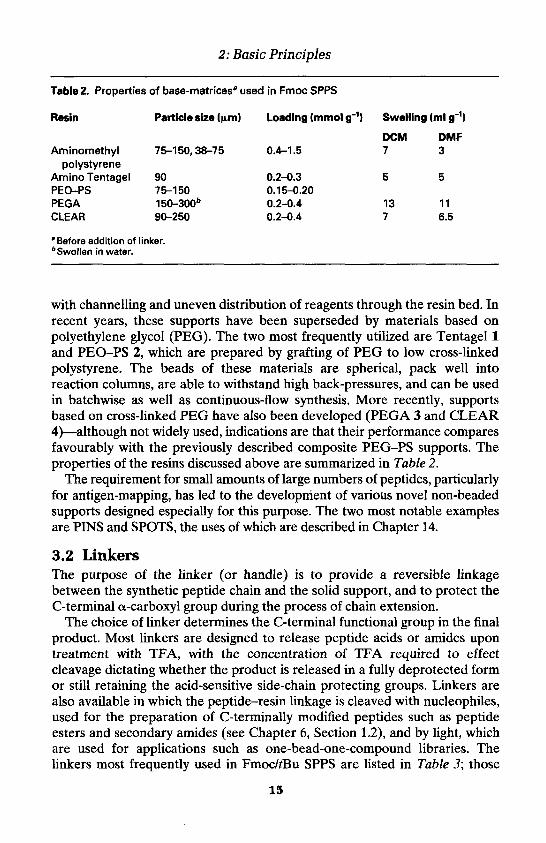
2: Basic Principles
Table 2. Properties of base-matricesa used in Fmoc SPPS
Resin Particle size (um) Loading (mmol g-1) Swelling (ml g-1)
DCM DMFAminomethyl 75-150,38-75 0.4-1.5 7 3
polystyreneAminoTentagel 90 0.2-0.3 5 5PEO-PS 75-150 0.15-0.20PEGA 150-300b 0.2-0.4 13 11CLEAR 90-250 0.2-0.4 7 6.5
" Before addition of linker.bSwollen in water.
with channelling and uneven distribution of reagents through the resin bed. Inrecent years, these supports have been superseded by materials based onpolyethylene glycol (PEG). The two most frequently utilized are Tentagel 1and PEO-PS 2, which are prepared by grafting of PEG to low cross-linkedpolystyrene. The beads of these materials are spherical, pack well intoreaction columns, are able to withstand high back-pressures, and can be usedin batchwise as well as continuous-flow synthesis. More recently, supportsbased on cross-linked PEG have also been developed (PEGA 3 and CLEAR4)—although not widely used, indications are that their performance comparesfavourably with the previously described composite PEG-PS supports. Theproperties of the resins discussed above are summarized in Table 2.
The requirement for small amounts of large numbers of peptides, particularlyfor antigen-mapping, has led to the development of various novel non-beadedsupports designed especially for this purpose. The two most notable examplesare PINS and SPOTS, the uses of which are described in Chapter 14.
3.2 LinkersThe purpose of the linker (or handle) is to provide a reversible linkagebetween the synthetic peptide chain and the solid support, and to protect theC-terminal a-carboxyl group during the process of chain extension.
The choice of linker determines the C-terminal functional group in the finalproduct. Most linkers are designed to release peptide acids or amides upontreatment with TFA, with the concentration of TFA required to effectcleavage dictating whether the product is released in a fully deprotected formor still retaining the acid-sensitive side-chain protecting groups. Linkers arealso available in which the peptide-resin linkage is cleaved with nucleophiles,used for the preparation of C-terminally modified peptides such as peptideesters and secondary amides (see Chapter 6, Section 1.2), and by light, whichare used for applications such as one-bead-one-compound libraries. Thelinkers most frequently used in Fmoc/tBu SPPS are listed in Table 3; those
15

Peter D. White and Weng C. Chan
16

2: Basic Principles
Table 3. Linker resins commonly used for the synthesis of peptide acids and peptideamides
17

Peter D. White and Weng C. Chan
TableS. Continued
Linker resins
18

2: Basic Principles
Table 3. Continued
Linker resins Cleavage conditions Refs
Nucleophilic displacement to C-terminal modified peptides (Base-cleavable linkers)
19

Table 4. Common side-chain protecting groups used in Fmoc/tBu solid phase peptide synthesis


Table 4. Continued


Table 4. Continued


Peter D. White and Weng C. Chan
recommended for the routine synthesis of peptide acids and carboxamides arehighlighted.
3.3 Side-chain protecting groupsMore than half of the amino acids commonly encountered in proteins haveside-chains that contain reactive functional groups. In solid phase synthesis, itis usual for all of these potentially reactive groups to be masked because ofthe rather harsh conditions employed and the need to achieve the highestlevel of efficiency in all chemical reactions. For routine synthesis, protectinggroups that are removed with TFA are usually employed as this allows thepeptide to be globally deprotected at the same time as it is cleaved or releasedfrom the support. Furthermore, a wide range of groups is also available whichcan be selectively removed on the solid phase, thus enabling the selectivemodification of side-chains of individual residues within the peptide chain.These find application in the synthesis of cyclic peptides, phosphopeptides,and biotinylated peptides (see Chapter 6). Table 4 lists the most commonlyused side-chain protecting groups, together with the conditions required fortheir removal; those recommended for routine use are highlighted.
3.4 First residue attachmentThe first step in the process of solid phase peptide synthesis is the attaching,or loading, of the resin linker with the C-terminal amino acid. The satisfactoryexecution of this process is particular important because, first, the extent ofthis reaction will determine the yield of the final product and, second, sites onthe resin not reacted in this initial process can potentially be acylated insubsequent cycles, leading to the generation of related C-terminally truncatedby-products. In the case of resins in which the anchorage point is a hydroxylgroup, this process is often accompanied by enantiomerization, owing to theharshness of the conditions applied to effect this esterification. The problem ismost serious with histidine and cysteine, and for these residues the use oftrityl-based resins is recommended as these are loaded under conditionswhich do not cause loss of chiral integrity (Chapter 3, Section 5.2 and Chapter4, Section 3.2.1).
Peptides containing proline or N-alkylated amino acids in the C-terminaldipeptide sequence present special problems because of the ease with whichthese dipeptides cyclize to give the corresponding diketopiperazine (Figure5). This not only results in a reduction in yield of the desired product, but mayalso lead to the generation of truncated sequences through subsequentacylation of the regenerated starting resin. This side-reaction is mostproblematic with supports in which the dipeptide in anchored by a benzylester, and for this reason resins in which attachment is via a more hinderedtrityl ester should be used (Chapter 3, Section 5.2).
26

2: Basic Principles
Figure 5. Diketopiperazine formation.
3.5 Coupling stepMost methods of amide bond formation involve chemical activation of thecarboxy component. Those commonly employed in organic synthesis aregenerally regarded as too harsh to be used in peptide synthesis, leading to theformation of over-activated intermediates, which are unselective in theirreactions and consequently prone to side-reactions. Peptide chemists havetherefore sought milder activating methods, mostly based on the formation ofactive esters, pre-formed or generated in situ.
Those in most frequent use are listed in Table 5, together with cross-reference to the relevant protocols in later chapters.
3.6 N-Fmoc deprotection reactionIn solid phase synthesis, removal of the N-Fmoc group is usually achieved bytreatment with 20-50% v/v piperidine in DMF. The mechanism of the Fmoc-deprotection reaction is shown in Figure 6. The key step is initialdeprotonation of the fluorene ring to generate the aromatic cyclopentadiene-type intermediate. This rapidly eliminates to form dibenzofulvene, which isscavenged by piperidine to afford the adduct 5. The products of thedeprotection reaction absorb UV strongly, offering potential for monitoringof this reaction. In continuous-flow peptide synthesizers this is achieved byfollowing the change in optical density of the column effluent with time. Thecurve shown in Figure 7a was obtained at 304 nm using a 0.1 mm path-lengthquartz flow-cell and is typical of that obtained during a deprotection reactionthat follows normal reaction kinetics. For such reactions (providing flow rateand column volume are kept constant), the progress of peptide chainassembly can be assessed by comparing the area under consecutive curves.With sluggish deprotection reactions (Figure 7b), such as those encounteredwith aggregated sequences, the area under the deprotection is reduced andcan no longer be meaningfully compared with that of the previous cycle.
In synthesizers operating in the batchwise mode of synthesis, monitoring ofthe deprotection reaction is carried out by taking aliquots and measuring thechange over time of the optical density or conductivity of the sample.
Whilst deprotection with piperidine is effective in most cases, it has beenshown that for long peptides incomplete Fmoc deprotection can occur even in
27

Table 5. Coupling methods used in Fmoc/tBu SPPS
Coupling reagent
DIC (or DCC)
DIC (or DCC)
PyBOP(orCO TBTU, HBTU)
HATU
TFFH
Additive Active species
Symmetricalanhydride
HOBt Benzotriazolylester
HOBt Benzotriazolylester
9-Azabenzotriazolylester
Acid fluoride
Conditions
Fmoc-amino acid/DIC (2:1)in DCM
Fmoc-amino acid/DIC/HOBt (1:1:1) in DMF
Fmoc-amino acid/PyBOP/HOBt/DIPEA (1:1:1:2) inDMF
Fmoc-amino acid/HATU/DIPEA (1:1:2) in DMF
Fmoc-amino acid/TFFH/DIPEA (1:1:2) in DMF
Comments
Anhydride, generatedin DCM, but used inDMF. Wastes 1 eq. ofamino acid derivative.
Activation in DMF is slow
Most popular couplingmethod. Activation processextremely fast
Excellent method fordifficult couplings
Particularly useful forcoupling N-alkyl and a-
Further details
Chapter 3,Section 7.3
Chapter 3,Section 7.1
Chapter 3Section 7.4
Chapter 3,Section 7.4
Chapter 3:Section 7.5
substituted residues
DIC, diisopropylcarbodiimide; DCC, dicyclohexylcarbodiimide; HOBt, 1-hydroxybenzotriazole; PyBOP, benzotriazol-1-yloxytris(pyrrolidino)phosphoniumhexafluorophosphate; TBTU, N-((1H-benzotriazol-1-yl)(dimethylamino)methylene]-/\/-methylmethanaminium tetrafluoroborate; HBTU, N-[(1W-benzotriazol-1-yl)(dimethylamino)methylene]-N-methylmethanaminium hexafluorophosphate N-oxide; HATU, N-[(dimethylamino)-1H-1,2,3-triazolo[4,5-D]pyridin-1-ylrnethyl-enel-W-methylmethanaminium hexafluorophosphate N-oxide; TFFH, tetramethylfluoroformamidinium hexafluorophosphate; DIPEA, diisopropylethylamine.

2: Basic Principles
Figure 7. Fmoc deprotection profiles obtained from continuous-flow synthesis. (a) TypicalFmoc deprotection profile; (b) extended deprotection indicative of aggregation.
the presence of high concentrations of piperidine. In these cases, it isadvisable to increase the time required for deprotection or to use a strongerbase such as l,8-diazabicyclo[5.4.0]undec-7-ene (DBU) (70). This tertiarybase appears to be a very good alternative to piperidine since it causes rapiddeprotection, less enantiomerization of resin-bound C-terminal Cys(Trt), and
29

Peter D. White and Weng C. Chan
reduces the extent of broadening of UV Fmoc-deprotection peaks. In batchsynthesis, it is advisable when using DBU to also add 2% piperidine to thedeprotection mixture in order to scavenge the dibenzofulvene produced onFmoc removal, and thus prevent alkylation of resin amino groups (71).
3.7 AggregationWith the improvements in linker, protecting group, and cleavage strategiesmade over the last decades, the cause of failure in Fmoc SPPS is now mostlikely to be aggregation. Growing peptide chains built up on a resin matrixcan form secondary structures or aggregates either with other peptide chainsor with the polymer support. This causes lower reaction rates and thereforelow coupling yields. Shrinking of the resin matrix indicates aggregation inbatch synthesis. In continuous flow synthesis, it is detected by flattening andbroadening of the deprotection profile. The driving force for this intra- andinterchain association is thought to be hydrogen bonding and hydrophobicforces. This association leads to incomplete solvation of the peptide-resincomplex, sudden shrinkage of the gel matrix, and reduced reagent penetra-tion, ultimately resulting in failure of either the acylation or deprotectionreaction or both. A detailed discussion on identifying and overcoming theeffects of aggregation is given in Chapter 5.
Aggregation can occur from as early as the fifth residue coupled (72). Thetendency for aggregation depends on the nature of the peptide and side-chainprotecting groups, with sequences containing a high proportion of Ala, Val,Ile, Asn, Gln residues showing particular propensity for this effect.
The insertion of a proline or N-alkyl amino acid residue into a sequence isknown to disrupt formation of p-sheets and other secondary structuresthought to be responsible for the aggregation (73). The effects can be longrange, with the outset of aggregation often being postponed for as many as sixresidues, or eliminated altogether. Recently, two independent approacheshave been developed which exploit this principle. The approach by Sheppardand co-workers utilizes temporary N-2-hydroxy-4-methoxybenzyl (Hmb)protection of the peptide backbone amide and is discussed in detailed inChapter 5.
Mutter's method involves reversible conversion of serine or threonineresidues into a pseudoproline residue, via formation of an oxazolidinedipeptide (74, 75). The pseudoproline is introduced as a dipeptide unit,overcoming the problems normally associated with acylation of secondaryamino acids. This has the added advantage of extending the peptide chain bytwo residues in one step. As the pseudoproline unit is stable toAcOH/trifluoroethanol/DCM, peptides prepared on extremely acid-labileresins, such as 2-chlorotrityl chloride, can be isolated with the pseudoprolinemoiety still in place. This can be advantageous when preparing peptides foruse in fragment condensation reactions or when dealing with intractable
30

Figure 8. Ring-opening of oxazolidine dipeptides.
sequences, since peptides containing pseudoproline residues often exhibitmarkedly improved solubility properties. Regeneration of the Thr or Serresidue from the oxazolidine can be effected by treatment with TFA in thenormal manner (Figure 8).
3.8 EnantiomerizationOf the 20 amino acids that are commonly found in proteins, with theexception of glycine, all have a chiral centre of L-configuration at their a-carbon atoms, and two, isoleucine and threonine, also have a chiral centre intheir side-chains. The biological properties of proteins and peptides arecritically dependent on the configuration of the backbone chiral centres, somaintaining the integrity of these centres is of paramount importance inpeptide synthesis.
In all 19 optically active amino acids, one of the substituents on the ot-carbon is a potentially acidic hydrogen atom. Removal and subsequentreattachment of this proton represents a potential mechanism forenantiomerization of this chiral centre. Chiral integrity is particularly at riskduring coupling as the acidity of this proton is greatly enhanced when thecarboxy group is activated. In practice, direct enolization does not appears tobe an important mechanism for enantiomerization except for amino acidssuch as phenylglycine, which offer an additional mode of enol stabilization.
The other mechanism by which enantiomerization occurs involvesdeprotonation and ring opening of an oxazolone intermediate, which isgenerated by attack on the activated carboxy group of the adjacent amidebond (Figure 9). Oxazolone formation occurs easily with carboxy-activatedpeptides, but can also occur with N-urethane-protected amino acids if thecarboxy group is strongly activated. Oxazolones derived from urethane-protected amino acids are quite resistant to deprotonation and, in general,enantiomerization is rarely encountered in stepwise synthesis, except with thespecial cases of histidine and cysteine, and during attachment of the C-terminal residue to hydroxy-functionalized resins (Chapter 3, Section 5). Inpractice enantiomerization via oxazolone formation is mainly associated withthe fragment condensation approach as this inevitably involves carboxyactivation of peptide fragments. For this reason, fragments are normally
31
2: Basic Principles

Figure 9. Enantiomerization via oxazolone formation.
joined at glycine, which is achiral, or proline, which is resistant to oxazoloneformation.
Enantiomerization of cysteine can occur during coupling and duringpiperidine-mediated removal of N™-Fmoc, when the cysteine residue isanchored to the resin via an ester bond, and may involve a p-elimination-typemechanism; these problems are discussed in Chapter 4. With histidine, it is theproximity of the basic Tr-nitrogen of the imidazole side-chain to the a-hydrogen is thought to account for the facile enantiomerization of carboxy-activated histidine derivatives. This is not normally a problem during routinechain extension using Fmoc-His(Trt) derivatives, but may become an issuewith aggregated sequences where slow reactions are encountered. In suchcases, the use of derivatives in which the ir-nitrogen is blocked, such as Fmoc-His(Bum)-OH (58), is recommended.
3.9 Side reactionsNewcomers to solid phase peptide synthesis reading any review on the subjectmay be forgiven for believing that the technique is fraught with difficultiesand that it is virtually impossible to prepare any peptide without encounteringmajor side-reactions. Side-reactions do certainly occur, but most are welldocumented and can be generally avoided by careful planning of the synthesisand by the appropriate selection of protecting groups and resin linker.
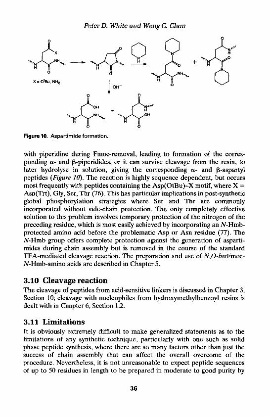
The side-reactions that can occur during chain assembly are listed in Table6; those associated with the cleavage reaction are given in Chapter 3, Table 5.Aspartimide formation requires special mention as this is the side-reactionmost likely to be encountered in routine synthesis, the others being normallyonly observed if the recommendations given in this and subsequent chaptersare not followed. The reaction involves attack of the nitrogen attached to thea-carboxy group of aspartic acid or asparagine on the side-chain ester oramide group respectively, resulting in formation of a five-membered imide.This intermediate can suffer a number of fates: it can undergo ring opening
32
Peter D. White and Weng C. Chan

Table 6. Side-reactions that can occur during chain assembly
Residue Occurrence Structurelr formed affected
Comments
Ornithine formation. Occurs when protonation or acyl-based groups are used for protection of the guanidine side-chain
8-Lactam formation. Results from attack of the N8-atom on the activated carboxy group. Most problematic with Arg PSAs
Cyanoalanine formation. Occurs during the activation of Asn when the side-chain carboxamide is not protected
Deamidation. Can occur in aqueous media, particularly under basic conditions, leading to formation of a- and p-aspartyl peptides. Most problematic with peptides containing Asn-Gly and Asn-Ser
Possible action
Prevented through the use of sulphonyl- based protection, such as Pbf and Pmc
Addition of HOBt inhibits lactam formation. Slow reactions may need repeating with fresh reagents
Use pre-formed active esters, such as Fmoc-Asn-OPfp, or use a side-chain protected derivative, such as Fmoc- Asn(Trt)-OH
Avoid prolonged exposure

Table 6. Continued
Residue Occurrence Structure/s formed Comments affected
ASP S Aspartimide formation. Can result in formation of a- and P-piperidides p-aspartyl peptides. Most problematic with Asp(OtBu)-X (X = Gly, Asn(Trt), Ser, Thr, Arg(Pmc) sequences
The Asp-Pro amide bond is slowly cleaved in acidic aqueous media
Enantiomerization
Enantiomerization. Occurs during attachment of Cys to hydroxy- functionalized resins and subsequent Fmoc removal
Piperidinylalanine formation. Can occur with Cys attached to hydroxy-functionalized resins
Possible action
Incorporate a N-Hmb-protected amino acid prior to addition of Asp
Avoid prolonged exposure
Use a pre-formed OPfp or DIC/HOBt activation for addition of Cys (Chapter 4, Section 3.2.2) Use a trityl-based resin (Chapter 4, Section 3.2.1)
Does not occur with Cys attached to amino-functionalized resins (Chapter 4, Section 3.2.3)
Pyroglutamate formation. May occur during N-Fmoc removal with peptides containing N-terminal Glu, side-chain protected with benzyl-based groups
Keep deprotection time to a minimum

Pyroglutamate formation. Occurs in acidic aqueous media if side-chain not protected
Avoid the preparation of peptides containing Wterminal Gln
Dipeptide formation. Can occur during loading of Gly to hydroxy- functionalized resins if symmetrical anhydride is used
Enantiomerization. Can occur during attachment of His to hydroxy- functionalized resins
Load resin using OPfp ester/DMAP, or the dichlorobenzoyl chloride method (Chapter 3, Section 5.1)
Use trityl-based resins
Oxidation. Can occur during all manipulations involving Met- containing peptides
Handle Met peptides and peptidyl-resins under inert atmosphere. Reduce sulphoxide (Chapter 4, Section 2)
Diketopiperazine formation. Results in loss of C-terminal dipeptide from resin. May also result in formation of truncated sequences missing C-terminal dipeptide
For peptides acids, use trityl-based resins. For other resins, keep deprotection time to aminimum.
C, when located at C-terminus; N, when located at N-terminus; A, occurs during coupling; S, may occur during chain-assembly; P, post-synthetic modification. PSA, pre-formed symmetrical anhydride.
Peter D. White and Weng C. Chan
Figure 10. Aspartimide formation.
with piperidine during Fmoc-removal, leading to formation of the corres-ponding a- and p-piperidides, or it can survive cleavage from the resin, tolater hydrolyse in solution, giving the corresponding a- and p-aspartylpeptides (Figure 10). The reaction is highly sequence dependent, but occursmost frequently with peptides containing the Asp(OtBu)-X motif, where X =Asn(Trt), Gly, Ser, Thr (76). This has particular implications in post-syntheticglobal phosphorylation strategies where Ser and Thr are commonlyincorporated without side-chain protection. The only completely effectivesolution to this problem involves temporary protection of the nitrogen of thepreceding residue, which is most easily achieved by incorporating an Af-Hmb-protected amino acid before the problematic Asp or Asn residue (77). TheW-Hmb group offers complete protection against the generation of asparti-mides during chain assembly but is removed in the course of the standardTFA-mediated cleavage reaction. The preparation and use of N,O-bisFmoc-Af-Hmb-amino acids are described in Chapter 5.
3.10 Cleavage reactionThe cleavage of peptides from acid-sensitive linkers is discussed in Chapter 3,Section 10; cleavage with nucleophiles from hydroxymethylbenzoyl resins isdealt with in Chapter 6, Section 1.2.
3.11 LimitationsIt is obviously extremely difficult to make generalized statements as to thelimitations of any synthetic technique, particularly with one such as solidphase peptide synthesis, where there are so many factors other than just thesuccess of chain assembly that can affect the overall overcome of theprocedure. Nevertheless, it is not unreasonable to expect peptide sequencesof up to 50 residues in length to be prepared in moderate to good purity by
36

2: Basic Principles
the standard methods, subject to the important caveat that aggregation duringchain assembly did not occur.
However, as the chain length increases, separation of the target peptidefrom the accumulated by-products becomes increasingly difficult. Withparticularly long peptides, the small and almost undetectable levels ofimpurities in protected amino acid derivatives and solvents, together withobvious by-products arising from incomplete Fmoc-removal and couplingreactions, begin to have an ever-increasing impact on the outcome of thesynthesis. For these reasons, methods of chemoselective ligation, a techniquewhereby smaller, more manageable peptides are coupled together in aqueousmedia, and chemoselective purification, a method whereby the target peptidecan be chemically tagged to assist purification, have been increasinglyexploited for the production of large peptides and small proteins by chemicalsynthesis. These powerful techniques are dealt with in detail in Chapters 11and 12.
References1. Lloyd-Williams, P., Albericio, F., and Giralt, E. (1997). Chemical approaches to
the synthesis of peptide and proteins (Series ed. C. W. Rees). CRC Press, NewYork.
2. Kent, S. B. H. (1988). Ann. Rev. Biochem., 57, 957.3. Barany, G., Kneib-Cordnier, N., and Mullen, D. G. (1987). Int. J. Peptide Protein
Res., 1987, 705.4. Stewart, J. M. and Young, J. D. (ed.) (1984). Solid phase peptide synthesis (2nd
edn). Pierce Chemical Company, Rockford.5. Atherton, E., Fox, H., Harkiss, D., and Sheppard, R. C. (1978). J. Chem. Soc.,
Chem. Commun., 539.6. Fields, G. B. and Noble, R. L. (1990). Int. J. Peptide Protein Res., 35, 161.7. Grant, G. (ed.) (1992). Synthetic peptides. W. H. Freeman & Co., New York.8. Wang, S.-S. (1973). J. Am. Chem. Soc., 95, 1328.9. Sheppard, R. C. and Williams, B. J. (1982). Int. J. Peptide Protein Res., 20, 451.
10. Rink, H. (1987). Tetrahedron Lett., 28, 3787.11. Florsheimer, A. and Riniker, B. (1991). Peptides 1990: Proceedings of the 21st
European Peptide Symposium (ed. E. Giralt and D. Andreu), p. 131. ESCOM,Lieden.
12. Barlos, K., Gatos, D., Kallitsis, J., Papaphotiu, G., Sotiriu, P., Wenqing, Y., andSchafer, W. (1989). Tetrahedron Lett., 30, 3943.
13. Barlos, K., Gatos, D., Kapolos, S., Papaphotiu, G., Schafer, W., and Wenqing, Y.(1989). Tetrahedron Lett., 30, 3947.
14. Barlos, K., Chatzi, O., Gatos, D., and Stavropoulos, G. (1991). Int. J. PeptideProtein Res., 37, 513.
15. Bollhagen, R., Schmiedberger, M., Barlos, K., and Grell, E. (1994). J. Chem. Soc.,Chem. Commun., 2559.
16. Mergler, M., Nyfeler, R., Tanner, R., Gosteli, J., and Grogg, P. (1988).Tetrahedron Lett., 29, 4009.
37

Peter D. White and Weng C. Chan
17. Albericio, F. and Barany, G. (1991). Tetrahedron Lett., 32, 1015.18. Bayer, E., Clausen, N., Goldammer, C., Henkel, B., Rapp, W., and Zhang, L.
(1994). In Peptides: chemistry, structure and biology (ed. R. S. Hodges and J. A.Smith), p. 156. ESCOM, Leiden.
19. Bernatowicz, M. S., Daniels, S. B., and K6ster, H. (1989). Tetrahedron Lett., 30,4645.
20. Albericio, F., Kneib-Cordonier, N., Biancalana, S., Gera, L., Masada, R. I.,Hudson, D., and Barany, G. (1990). J. Org. Chem., 55, 3730.
21. Sieber, P. (1987). Tetrahedron Lett., 28, 2107.22. Chan, W. C., White, P. D., Beythien, J., and Steinauer, R. (1995). J. Chem. Soc.,
Chem. Commun., 589.23. Han, Y., Bontems, S. L., Hegges, P., Munson, M. C., Minor, C. A., Kates, S. A.,
Albericio, F., and Barany, G. (1996). J. Org. Chem., 61, 6326.24. Bray, A. M., Valerio, R. M., and Maeji, N. J. (1993). Tetrahedron Lett., 34, 4411.25. Bray, A. M., Jhingran, A.G., Valerio, R. M., and Maeji, N. J. (1994). J. Org.
Chem., 59, 2197.26. Atherton, E., Logan, C. J., and Sheppard, R. C. (1981). J. Chem. Soc., Perkin
Trans. 7, 538.27. Nicolas, E., Clemente, J., Perello, M., Albericio, F., Pedroso, E., and Giralt, E.
(1992). Tetrahedron Lett., 33, 2183.28. Backes, B. J., Virgilio, A. A., and Ellman, J. A. (1996). J Am. Chem. Soc., 118,
3055.29. Hammer, R. P., Albericio, F., Gera, I., and Barany, G. (1990). Int. J. Peptide
Protein Res., 35, 31.30. Holmes, C. P. and Jones, D. G. (1995). J. Org. Chem., 60, 2318.31. Atherton, E., Sheppard, R. C., and Wade, J. D. (1983). J. Chem. Soc., Chem.
Commun., 1060.32. Fujino, M., Wakimsu, M., and Kitadu, C. (1981). Chem. Pharm. Bull, 29, 2825.33. Ramage, R. and Green, J. (1987). Tetrahedron Lett, 28, 2287.34. Ramage, R., Green, J., and Blake, A. J. (1991). Tetrahedron, 47, 6353.35. Carpino, L. A., Shroff, H., Triolo, S. A., Mansour, M. E., Wenschuch, H., and
Albericio, F. (1993). Tetrahedron Lett., 34, 7829.36. Schwyzer, R. and Dietrich, H. (1961). Helv. Chim. Acta, 44, 2003.37. Okada, Y., Iguchi, S., and Kawasaki, K. (1987). J. Chem. Soc., Chem. Commun.,
1532.38. Okada, Y. and Iguchi, S. (1988). J. Chem. Soc., Perkin Trans. 1, 2129.39. Trzeciak, A. and Bannwarth, W. (1992). Tetrahedron Lett., 33, 4557.40. Kates, S. A., Sole, N. A., Johnson, C. R., Hudson, D., Barany, G., and Albericio,
F. (1993). Tetrahedron Lett., 34, 1549.41. Dessolin, M., Guillerez, M.-G., Thieriet, N., Guibe, F., and Loffet, A. (1995).
Tetrahedron Lett., 36, 5741.42. Chan, W. C., Bycroft, B. W., Evans, D. J., and White, P. D. (1995). J. Chem. Soc.,
Chem. Commun., 2209.43. Yue, C. W., Thierry, J., and Potier, P. (1993). Tetrahedron Lett., 34, 323.44. Sieber, P. and Riniker, B. (1991). Tetrahedron Lett., 32, 739.45. Wegand, F., Steglich, W., Bjarnason, J., Ahktar, R., and Chytil, N. (1968). Chem.
Ber., 101, 3623.46. Wegand, F., Steglich, W., and Bjarnason, J. (1968). Chem. Ber., 101, 3642.
38

2: Basic Principles
47. Friede, M., Denery, S., Neimark, J., Kieffer, S., Gausepohl, H., and Briand, J. P.(1992). Peptide Res., 5, 145.
48. Hiskey, R. G., Mizoguchi, T., and Igeta, H. (1966). J. Org. Chem., 31, 1188.49. Veber, D. F., Milkowski, J. D., Varga, S. L., Denkewalter, R. G., and Hirschmann,
R. (1972). J. Am. Chem. Soc., 94, 5456.50. Kamber, B. (1971). Helv. Chim. Acta, 54, 927.51. Fujii, N., Okata, A., Funakoshi, S., Bessho, K., and Yajima, H. (1987). J. Chem.
Soc., Chem. Commun., 163.52. Weber, U. and Hartter, P. (1970). Hoppe-Seyler's Zeitschrift fur Physiologische
Chemie, 351, 1384.53. McCurdy, S. N. (1989). Peptide Res., 2, 147.54. Munson, M. C., Garcia-Echeverria, C, Albericio, F., and Barany, G. (1992). J.
Org. Chem., 57, 3013.55. Barlos, K., Gatos, D., Hatzi, O., Koch, N., and Koutsogianni, S. (1996). Int. J.
Peptide Protein Res., 47, 148.56. Sieber, P. and Riniker, B. (1987). Tetrahedron Lett., 28, 6031.57. Yamashiro, D., Blake, J., and Li, C. H. (1972). J. Am. Chem. Soc., 94, 2855.58. Colombo, R., Colombo, F., and Jones, J. H. (1984). J. Chem. Soc., Chem.
Commun., 292.59. Chang, C. D., Wakai, M., Ahmed, M., Meienhofer, J., Lundell, E. D., and Huang,
J. D. (1980). Int. J. Peptide Protein Res., 15, 59.60. Thieriet, N., Alsina, J., Giralt, E., Guibe, F., and Albericio, F. (1997). Tetrahedron
Lett., 38, 7275.61. Bycroft, B. W., Chan, W. C., Chhabra, S. R., and Hone, N. D. (1993). J. Chem.
Soc., Chem. Commun., 778.62. Chhabra, S. R., Hothi, B., Evans, D. J., White, P. D., Bycroft, B. W., and Chan, W.
C. (1998). Tetrahedron Lett., 39, 1603.63. Aletras, A., Barlos, K., Gatos, D., Koutsogianni, S., and Mamos, P. (1995). Int. J.
Peptide Protein Res., 45, 488.64. Beyerman, H. C. and Bontekoe, J. S. (1961). Proc. Chem. Soc., 249.65. Callahan, F. M., Anderson, G. W., Paul, R., and Zimmermann, J. E. (1963). J. Am.
Chem. Soc., 85, 201.66. Barlos, K., Gatos, D., Koutsogianni, S., Schafer, W., Stavropoulos, G. and Yao, W.
Q. (1991). Tetrahedron Lett., 32, 471.67. Barlos, K., Gatos, D., and Koutsogianni, S. (1998). J. Peptide Res., 51, 194.68. Fischer, P. M. (1992). Tetrahedron Lett., 33, 7605.69. White, P. (1992). Peptides: chemistry and biology (ed. J. A. Smith and J. E. Rivier),
p. 537. ESCOM, Leiden.70. Wade, J. D., Bedford, J., Sheppard, R. C., and Tregear, G. W. (1991). Peptide Res.,
4, 194.71. Kates, S. A., Nuria, A., Sole, M., Beyermann, M., Barany, G., and Albericio, F.
(1996). Peptide Res., 9, 106.72. Bedford, J., Hyde, C., Johnson, T., Jun, W., Owen, D., Quibell, M., and Sheppard,
R. C. (1992). Int. J. Peptide Protein Res., 40, 300.73. Toniolo, C., Bonora, G. M., Mutter, M., and Pillai, V. N. R. (1981). Makromol.
Chem., 182, 2007.74. Mutter, M., Nefzi, A, Sato, T., Sun, X., Wahl, F., and Wohr, T. (1995). Peptide
Res., 8, 145.
39

Peter D. White and Weng C. Chan
75. Wohr, T., Wahl, F., Nefzi, A., Rohwedder, B., Sato, T., Sun, X., and Mutter, M.(1996). J. Am. Chem. Soc., 118, 9218.
76. Lauer, J. L., Fields, C. G., and Fields, G. B. (1994). Lett. Pept. Sci, 1, 197.77. Quibell, M., Owen, D., Packman, L. C, and Johnson, T. (1994). J. Chem. Soc.,
Chem. Commun., 2343.
40

Basic proceduresWENG C. CHAN and PETER D. WHITE
1. IntroductionA number of excellent descriptions of the techniques related to peptide chainassembly have already been published (1-3). These processes are also de-scribed in the operator manuals supplied by the peptide synthesis instrumentmanufacturers. Accordingly, the treatment of the subject presented here hasbeen kept brief in order to provide more space in this volume for those topicsnot covered in detail in other publications of this type.
The protocols have been written as they would be carried out using amanual peptide synthesis vessel. Whilst it is appreciated that most scientistspreparing peptides will be using automated peptide synthesizers, it is notpossible, given the wide variation in operating procedures, to describe howsuch methods may be applied to individual instruments. Particular emphasishas been given here to those operations which are typically carried out off-instrument, such as first residue attachment and peptide-resin cleavage.
2. Manual synthesisThe operations described in this chapter can be carried out in a purpose-builtpeptide synthesis vessel or in a sintered glass funnel fitted with a three-waystopcock (Figure 1). The operation of the system is extremely simple: solventsare added from the top of the vessel, ensuring any resin adhering to the sidesis rinsed down into the resin bed; the resin bed is agitated by setting the tap toposition 1 to allow flow of nitrogen to the reaction vessel; solvents and re-agents are removed by setting the tap to position 2 to connect the vessel to thevacuum. The use of such vessels has previously been described in detail (4).
3. Resin handlingPeptide synthesis resins are extremely fragile and the beads, if wronglyhandled, can easily fracture, leading to the generation of fines which can blockreaction vessel filter-frits and solvent lines.
3

WengC. Chan and Peter D. White
Position 2
Figure 1. Manual peptide synthesis bubbler vessel.
It is particularly important that the correct method is used for mixing the resinand soluble reactants. Polystyrene-based supports are best agitated bybubbling an inert gas through the resin bed, or by shaking or vortexing thereaction vessel. Whilst all of these approaches are employed in commercialsynthesizers, gas-bubbling and shaking are the most appropriate for use inmanual synthesis. PEG-PS and PEGA resins once swollen are sufficientlyrigid to be packed in columns and washed in a continuous-flow fashion bypumping liquid through the resin bed. They can also be agitated by gas-bubbling and shaking. Agitation of any resin with a magnetic stirrer is notrecommended as the action results in attrition of the resin particles.
All resins must be swollen with an appropriate solvent before use, with theexception of PEGA resins which are supplied as a slurry in methanol or water.Underivatized polystyrene resins do not swell particularly well in dimethyl-formamide (DMF), hence the resin must be first swollen in dichloromethane(DCM). In contrast, PEG-PS resins can be packed dry into the reaction vesseland be swollen directly with DMF. In continuous-flow instruments, pumpingthe solvent up the reaction column is an effective way to remove air bubblestrapped in the resin bed.
42
3: Basic procedures
Resins are normally washed between reaction steps with DMF as it is anexcellent solvent both for protected amino acid derivatives and for mostreagents used in peptide synthesis.
At the end of the synthesis or before long-term storage, resins must beshrunk and dried to remove all traces of solvent from within the resin particlesas described in Protocol 1C. Many resins, when dry, adhere strongly to glassand plastic surfaces, making it difficult to remove the last traces of thematerial from the reaction vessel or filter funnel. Any resin remaining afterremoval of the bulk of the material can be washed with DMF down into thebottom corner of the filter-frit, washed with DCM, being careful not to spreadthe resin back up the sides of the vessel, excess DCM removed, and the dampresin removed with a spatula.
PEGA resins must always be handled in the swollen state as the resin beadshave no structure when dry. Following chain assembly the resin can bewashed successively with DMF and DCM, excess DCM removed, and theresin transferred to the cleavage reactor as a damp cake.
Protocol 1. Basic resin-handling procedures
A. Swelling (Polystyrene-based resins)
1. Place the dry resin in a peptide synthesis reactor (Figure 1).2. Add sufficient DCM to cover resin with three-times bed volume.3. Shake reaction vessel gently to remove air bubbles and form a sus-
pension of resin.4. Leave for 30 min.5. Apply vacuum to remove DCM.
B. Washing1. Add three bed volumes of solvent.2. Agitate for 1 min.3. Apply vacuum to drain resin.4. Repeat step 1 four times.
C. Drying-down resin1. Wash resin successively (as described in B) with either: DMF, DCM,
MeOH, and/or hexane (polystyrene-based resins); or DMF, isopropanol,hexane (loaded 2-chlorotrityl resins); or DMF, DCM, ether (PEG-PSresins).
2. Air-dry the resin by application of the vacuum for 10 min.3. Transfer resin to a sample tube.4. Dry in vacuo for 18 h.
43

Weng C. Chan and Peter D. White
4. Resin functionalizationAttachment of linkage agents bearing carboxyl functional groups to amino-functionalized resins can be effected using any of the standard peptidecoupling methods described in Section 7. However, for linkers containinghydroxyl groups, such as hydroxymethylphenoxyacetyl (HMPA) and hydro-methylbenzoyl (HMBA), the use of coupling methods which do not involvethe addition of a tertiary base are recommended if self-acylation is to beavoided. An unusual ammo acid such as norleucine inserted between the resinand the linker can serve as an internal standard, enabling attachment andcleavage yields to be easily determined by amino acid analysis following acidhydrolysis. A protocol for loading amino-functionalized resin with HMBAlinker is provided in Chapter 6, Section 1.2.2.
Polystyrene, PEG-PS, and PEGA-based resins pre-functionalized with allthe standard linkers required for the production of peptide acids, peptidesamides, and protected peptide fragments are commercially available.
5. Attachment of the first residueAnchoring of the first residue to the synthesis support is one of the mostcritical steps in peptide synthesis; an injudicious combination of linker,loading method, or amino acid derivative can have a highly deleterious effectson the purity of the final product, and the use of poorly loaded resins is simplyjust a waste of expensive reagents.
The properties of the various linker-derivatized resins used in Fmoc SPPShave already been discussed and were summarized in Chapter 2, Table 2. Forthe purpose of selecting the appropriate procedure for attachment of the firstresidue, these can be conveniently separated into three categories on the basisof their reactive functional groups (hydroxymethyl-, trityl chloride-, andaminomethyl-based resins); the mostly commonly used are listed in Table 1.
5.1 Hydroxymethyl-based resinsThe protocols described in this section are applicable to all linker-derivatizedresins in which loading involves esterification of the first amino acid to a linkerhydroxyl group. The ease of esterification with Fmoc-amino acids increases inthe order
Formation of ester bonds is considerably more difficult than that of amidebonds; harsher conditions than those normally used for chain elongation mustbe employed which, unless carried out under controlled conditions, can leadto low substitution, enantiomerization, and dipeptide formation. To ensuresatisfactory results when using these protocols it is important that thefollowing precautions are observed:(a) All reagents and glassware must be dried thoroughly before use as the
presence of moisture can severely affect loading efficiencies.
44

3: Basic procedures
(b) Amino acids containhig water of recrystallization should be dried either bydissolving in ethyl acetate, drying the solution over anhydrous magnesiumsulphate, and isolating by evaporation; or by repeated evaporation fromdioxane.
(c) Reactions should be conducted using the minimum volumes of solvent, toensure maximum reagent concentration.
(d) For the synthesis of peptide acids containing C-terminal cysteine, histi-dine, proline, methionine and tryptophan residues, it is better to usetrityl-based resins, such as 2-chlorotrityl resin; for details see Section 5.2.Alternative protocols for attachment of Cys are also provided in Chapter4, Section 3.2.1.
45
Table 1. Commonly used resins for SPPS

WengC. Chan and Peter D. White
(e) Peptides containing C-terminal glutamine and asparagine residues arebest prepared using amide-forming resins such as 9 and 10 loaded with a-f-butyl esters of glutamic or aspartic acid.
(f) If aminium or phosphonium coupling methods are to be used for sub-sequent chain extension, unreacted resin-bound hydroxyl groups shouldbe capped, following attachment of the first residue, to prevent growth ofdeletion sequences from these sites.
(g) After each reaction, the loading efficiency should be checked using themethod described in Protocol 14 and, if necessary, the reaction repeatedusing fresh reagents.
5.1.1 Symmetrical anhydride methodThis approach is the most commonly used method for esterification ofhydroxyl-functionalized resins (Figure 2). The reaction is typically carried outin DMF, using 5 eq of anhydride and 0.1-1 eq of 4-dimethylaminopyridine(DMAP) (5), and is usually > 70% complete within 1 h. In cases of difficulty,the esterification should be repeated with fresh reagents rather than extend-ing the reaction time. With arginine, this reaction may need to be repeated asmany as three times to achieve acceptable loading, owing to rapid conversationof the symmetrical anhydride to the unreactive 8-lactam.
12
Figure 2. Symmetrical anhydride method for first residue attachment.
Owing to the basic character of DMAP, some enantiomerization anddipeptide formation can be expected; the amount depends on the quantityused, the duration of the reaction, and the nature of the amino acid.Enantiomerization is particularly problematic with cysteine and histidine (6).For most other amino acids, however, the levels of enantiomerization can begenerally controlled to within acceptable limits by keeping the amount ofDMAP used to a minimum. Dipeptide formation can be a problem with thismethod, particularly during the loading of glycine (7).
Protocol 2. Attachment to alcohol-based resins usingsymmetrical anhydrides
Reagents• Fmoc-amino acid derivatives • Dry, acid-free DCM. DIC • DMF• Hydroxyl-functionalized peptide synthesis resin « DMAP
46

3: Basic procedures
Method
1. Place the resin in a dry reaction vessel.
2. Swell and wash with DMF as described in Protocol 1.
3. Prepare the appropriate Fmoc-amino acid anhydride (5 eq)a accordingto Protocol 9. Dissolve in minimum volume of DMF and add to resin. Ifnecessary add extra DMF to ensure complete coverage of the resinbed.
4. Dissolve DMAP (0.1 eq)a in DMF, and add this solution to the resin/amino acid mixture. Stopper the vessel and allow the mixture toagitate at room temperature for 1 h.
5. Wash the resin with DMF (5 times).
6. Transfer 10 mg of resin with a wide-mouth Pasteur pipette to a smallsintered glass funnel. Wash and dry-down as described in Protocol 1C.Determine the extent of loading using Protocol 14. If the loading isbelow 70%, repeat steps 2-5.
7. Add benzoic anhydride (5 eq)a and pyridine (1 eq)a in DMF, and agitatefor 30 min.
8. Wash and shrink-down the resin as described in Protocol 1C.
" Relative to resin substitution level (loading).
5.1.2 Dichlorobenzoyl chloride methodThis method, although requiring longer reaction times than the symmetricalmethod, results in products virtually free of impurities arising from enantio-merization and dipeptide formation (6). It involves formation of a mixedanhydride between an Fmoc-amino acid derivative and 2,6-dichlorobenzoylchloride (DCB) in DMF/pyridine (Figure 3). This anhydride effects esterifica-tion of resin-bound hydroxyl groups in yields of typically > 70% in 18 h.Levels of D-isomer and dipeptide formation for most amino acids are typicallyless than 1%, with S-acetamidomethyl cysteine giving the most enantio-merization (6.5% D-isomer) of the amino acids tested (8).
Repeating this reaction with fresh reagents appears to result in littleimprovement in yield. Sites not acylated by Fmoc-amino acid are apparentlycapped, presumably with 2,6-benzoyl groups. For this reason, it is notessential to cap following this reaction.
Figure 3. DCB method for first residue attachment.
47

Weng C. Chan and Peter D. White
Protocol 3. DCB method
Reagents• Fmoc-amino acid derivatives • DMF• DCB • Pyridine• Hydroxyl-functionalized peptide synthesis resin
Method
1. Place the resin in a dry reaction vessel.
2. Swell and wash with DMF, as described in Protocol 1. Add extra DMF ifnecessary to ensure complete coverage of the resin.
3. Add the appropriate Fmoc-amino acid (5 eq)a, followed by pyridine(8.25 eq)a. Gently agitate until the amino acid has dissolved com-pletely.
4. Add DCB (5 eq)a and agitate the mixture gently for 18 h.
5. Wash and shrink-down the resin as described in Protocol 1C.
aRelative to resin loading.
5.1.3 MSNT/Melm methodOriginally developed for DNA synthesis, this method has in recent years beenincreasingly used in peptide synthesis for the esterification of protected aminoacids to solid supports (9). It has proved particularly effective in loadingFmoc-amino acids to the relatively unreactive hydroxyl group of HMBA-derivatized resins (10) and in situations where enantiomerization is a problem(11). The reaction involves treating the protected amino acid in DCM withl-(mesitylene-2-sulphonyl)-3-nitro-7//-l,2,4-triazole (MSNT) 13 and 1-methyl-imidazole (Melm) (Figure 4). The process is extremely moisture sensitive andso it is essential that all glassware and reagents are dried thoroughly beforeuse.
48

3: Basic procedures
Figure 4. MSNT/Melm method for first residue attachment.
Protocol 4. MSNT/Melm method
Reagents• Fmoc-amino acid derivatives • Dry, acid-free DCM• MSNT . Melm. DryTHF . DMF• Hydroxyl-functionalized peptide synthesis resin
Method
1. Place the resin in a dry reaction vessel.
2. Swell and wash with DCM as described in Protocol 1. Add sufficientDCM to cover resin and flush vessel with nitrogen. Seal with aseptum.
3. Weigh the appropriate Fmoc-amino acid (5 eq) into a dry, round-bottomed flask, equipped with a magnetic stirrer. Add dry DCM todissolve the amino acid derivative (approximately 3 ml/mmol). Oneor two drops of THF can be added to aid complete dissolution.
4. Add Melm (3.75 eq) followed by MSNT (5 eq). Flush flask withnitrogen and seal with a septum, then stir the mixture until the MSNThas dissolved.
5. Using a syringe, transfer the amino acid solution to the vesselcontaining the resin.
6. Allow the mixture to stand at room temperature for 1 h, with gentleagitation.
7. Wash with DCM (5 times) and DMF (5 times).
8. Transfer 10 mg of resin with a wide-mouthed Pasteur pipette to asmall, sintered glass funnel. Wash and dry-down as described inProtocol 1C. Determine the extent of loading using Protocol 14. If theloading is below 70%, repeat steps 2-7.
9. Add benzoic anhydride (5 eq) and pyridine (1 eq) in DMF and agitategently for 30 min.
10. Wash and dry-down the resin as described in Protocol 1C.
49

Weng C. Chan and Peter D. White
5.2 Trityl-based linkersTwo trityl-based supports are presently in common use for the production ofpeptide acids by Fmoc SPPS: 2-chlorotrityl chloride resin 7 (12) and 4-(chloro(diphenyl)methyl)benzoyl resin 8 (13).
These resins are extremely moisture sensitive, and so it is essential that allreagents and glassware should be dried thoroughly before use. 2-Chlorotritylchloride resin can be stored desiccated at room temperature; 4-(chloro-(diphenyl)methyl)benzoyl resins should be generated from the precursortrityl alcohol immediately before use as described in Protocol 5. The chemicalcharacteristics of 7 and 8 are identical.
2-Chlorotrityl chloride resin is normally supplied with a displaceablechlorine content of 1.0-1.6 mmol/g. For the purposes of peptide synthesis,this substitution can sometimes be too high and can be reduced by treating theresin with a sub-stoichiometric amount of amino acid derivative and thencapping unreacted sites with MeOH.
Since coupling to trityl chloride-type resins does not involve activation ofthe incoming amino acid derivative the process is free from enantiomerizationand dipeptide formation (14), making these supports ideal tools for thepreparation of peptides containing C-terminal histidine or cysteine residues.Their use is also recommended for the synthesis of peptides containing C-terminal proline (15), methionine, or tryptophan residues (14). Proline-containing dipeptide attached to standard benzyl alcohol-based supports,such as 1 and 6, can undergo diketopiperazine formation with concommitantloss of peptide chains during Fmoc group removal. Methionine and typtophanresidues can be alkylated by the cations generated from a benzyl-based linkerduring the TFA-mediated cleavage reaction, leading to reattachment of thepeptide to the solid support with a corresponding reduction in yield. Theextent of both of these side-reactions appears to be significantly reduced whentrityl-based resins are used, a fact which has been attributed to the bulk of thelinker group.
Protocol 5. Chlorination of trityl alcohol resins
Reagents• Dry toluene • Freshly distilled acetyl chloride (AcCI)• 4-(Hydroxy(diphenyl)methyl)benzoyl • Dry DCM
aminomethyl resin
Method
1. Place the resin in a sintered glass funnel.
2. Swell and wash with DCM as described in Protocol 1.
3. Wash with toluene (5 times).
50

3: Basic procedures
4. Transfer resin as a damp cake to a round-bottomed flask. Addsufficient toluene to cover resin bed.
5. Add AcCI (1 ml/g resin). Fit flask with reflux condenser and CaCI2 guardtube.
6. Heat mixture at 60°C for 3 h.
7. Allow to cool to room temperature.
8. Slurry resin with toluene into a dry reaction vessel and wash withDCM.
9. Load immediately as described in Chapter 9, Protocol 1.
5.3 Aminomethyl-based linkersFor the addition of the first amino acid to supports derivatized with peptideamide-forming linkers, such as trialkoxybenzhydrylamine 9, trialkoxybenzyl-amine 10, and aminoxanthenyl 11 resins, any of the standard peptide couplingmethods described in Section 7 can be used. Since basic reagents such as 4-dimethylaminopyridine are not required to effect this transformation, there isno risk of enantiomerization or dipeptide formation. Many resins of this typeare supplied Fmoc-protected and these should be treated with piperidine priorto the addition of the first amino acid. With automated peptide synthesizers,Fmoc removal and loading with the C-terminal residue can be programmed tooccur as part of the normal coupling cycle.
The addition of the first residue to dimethoxybenzhydrylamine-based resinscan be sluggish, particularly the p-branched amino acids, such as valine andisoleucine. Repeating the coupling after 1 h with fresh reagents generallyleads to complete resin derivatization.
Coupling through the side-chains of the a-t-butyl esters of glutamatic acidor aspartic acid to amide-forming linkages provides a facile route to C-terminal glutamine- and asparagine-containing peptides (16, 17).
6. Fmoc removalRemoval of the Fmoc group from the N-terminus of the resin-bound peptidechain is normally achieved by treating the peptidyl resin with 20-50%piperidine in DMF. The reaction is typically complete within 4-10 min,depending on the nature of the peptide being synthesized. With peptides con-taining aspartic acid and asparagine, inclusion of 0.1 M HOBt in thedeprotection mixture has been found to be partially effective in suppressingaspartimide formation (18).
In the case of sequences which have become aggregated the standard treat-ment with piperidine in DMF may not always be effective. Even extendingthe time of deprotection reaction considerably does not always result in com-
51

Weng C. Chan and Peter D. White
plete removal of Fmoc groups; in such cases, the use of the non-nucleophilicbase l,8-diazabicyclo[5.4.0]undec-7-ene (DBU) is recommended (19). It isnormally employed in combination with piperidine as a solution in DMF(20)—piperidine is included to scavenge the dibenzofulvene that is formed onFmoc removal, thus preventing alkylating of the newly liberated amino group.The efficacy of this reagent has been demonstrated in the synthesis of thenotoriously difficult polyalanine sequence, H-(Ala)10-Val-OH (20). However,care should be exercised with peptides containing aspartic acid andasparagine residues as it has been found to promote aspartimide formation(18, 21).
Protocol 6. Manual removal of Fmoc groups
Reagents• 20% Piperidine in DMF (v/v) or DBU/piperidine/DMF (1:1:48 v/v)
Method
1. Place the peptidyl resin in a sintered glass funnel or manual SPPSreaction vessel.
2. If the resin is dry, swell the resin as described in Protocol 1A.
3. Wash resin with DMF (5 times).
4. Add deprotection reagent to cover resin.
5. Agitate gently for 2 min. Drain off reagent.
6. Repeat steps 4-5 twice.
7. Wash resin with DMF (5 times).
7. Coupling methodsThe stepwise introduction of Na-protected amino acids in solid phase syn-thesis normally involves in situ carboxy activation of the incoming amino acidor the use of pre-formed activated amino acid derivatives. In order to drivethe acylation to completion an excess of activated amino acid derivative isutilized, typically 2-10 times the resin functionality. The excess used dependson the nature of the activated species and on the resin loading and voidvolume of the reaction vessel employed. The most important consideration isto maintain as high an effective concentration of reagents as possible. Insmall-scale synthesis and with low-functionality resins, large excesses mayneed to be used as the dead volume of such systems tends to be quite high;conversely with resins of high functionality lower excesses can be utilized.
The coupling time required to effect complete acylation depends on the
52

3: Basic procedures
nature of the active species and the peptide sequence being assembled and onthe concentration of the reagents and resin functionality. For highly reactivespecies, such as those generated in situ using activating reagents like TFFH14(22), PyBOP 15 (23), TBTU 16 (24), and HATU 17 (25), 30 min is usuallysufficient. However, when coupling together hindered residues, such as (3-branched amino acids or N-substituted amino acids, or when preparing pep-tides that have become aggregated, reaction time may need to be increasedconsiderably. In manual synthesis, it is standard practice to check the com-pleteness of the coupling reaction before proceeding to the next step in thesynthesis cycle. For this purpose a number of sensitive tests have beendeveloped to detect the presence of residual amine on the solid support(Protocol 13).
With peptides that have become aggregated it is frequently not possible toobtain complete reaction, even after carrying out multiple acylation reactions.In such cases it is often necessary to repeat the synthesis, incorporating anHmb-protected residue (Chapter 5) or an oxazolidine-dipeptide (26-28) twoto four residues before the region prone to aggregation.
The order of efficiency of the methods described below is: OPfp esters/HOBt < DIC/HOBt < HBTU « PyBOP < HATU. The reader is directed toa recent discussion on coupling reagents by Albericio and Carpino (29).
7.1 DIC/HOBtThe reaction of an Fmoc-amino acid with an equivalent amount of DIC andHOBt results in formation of the corresponding OBt ester 18. The reaction isfastest in non-polar solvents like DCM (30) but also works well in DMF,provided the mixture is allowed to pre-activate before being added to the
53

Weng C. Chan and Peter D. White
resin. The use of DIG is preferred to that of dicyclohexylcarbodiimide (DCC)because the urea generated by the former is soluble in DMF. This procedureis also effective for the generation of OAt esters 19 (25).
Protocol 7. Synthesis of OXt esters
Reagents• Fmoc-amino acid derivatives • HOXt (HOBt or HOAt)• DIC • DMF
Method
1. Place the appropriate Fmoc-amino acid (5 eq) and HOXt (5 eq)a in a dryround-bottomed flask or sample vial, equipped with a magnetic stirrer.
2. Add the minimum of DMF to dissolve.
3. Add DIC dropwise (5 eq).
4. Stir the mixture for 20 min and add the solution to the N-deblockedpeptidyl resin.
5. Agitate resin gently for 1 h.
6. Transfer 10 mg of resin with a wide-mouthed Pasteur pipette to asmall, sintered glass funnel. Wash and dry-down as described inProtocol 1C. Perform Kaiser or TNBS test (Protocol 13). If positive,repeat steps 5 and 6.
7. If resin still gives a positive colour test after 4 h, wash resin with DMF(5 times) and repeat coupling reaction with fresh reagents.
8. Collect the derivatized resin as outlined previously.
aRelative to resin functionality.
7.2 Active estersOf the various active esters that have been exploited in solution phase peptidesynthesis, only the pentafluorophenyl esters (OPfp) 20 have found wideapplication in Fmoc/rBu solid phase synthesis. They are efficient acylationagents, particularly when used in conjunction with an equivalent of HOBt,
54

3: Basic procedures
and their use is generally regarded to be free from side-reactions (31).Derivatives are commercially available for most proteinous amino acids.
Protocol 8. OPfp esters
Reagents• Fmoc-amino acid pentafluorophenyl ester • HOBt. DMF
Method
1. Weigh the appropriate Fmoc-amino acid pentafluorophenyl ester(5 eq)a and HOBt (5 eq) in a dried sample vial.
2. Add the minimum volume of DMF to dissolve.
3. Add the solution to the N-protected peptidyl resin.
4. Agitate resin gently for 1 h.
5. Transfer 10 mg of resin with a wide-mouthed Pasteur pipette to asmall, sintered glass funnel. Wash and dry-down as described inProtocol 1C. Perform Kaiser or TNBS test (Protocol 73). If positive,repeat steps 4 and 5.
6. If resin still gives a positive colour test after 4 h, wash resin with DMF(5 times) and repeat coupling reaction with fresh reagents.
7. Collect the derivatized resin as outlined previously.
aRelative to resin functionality.
7.3 Symmetrical anhydrideReaction of an Fmoc-amino acid with 0.5 eq of a carbodiimide in DCMgenerates the corresponding symmetrical anhydride 12 (Figure 5). If thesymmetrical anhydride is to be used directly in solid phase synthesis, DICshould be used as the dehydrating reagent.
Some Fmoc-amino acids are not readily soluble in DCM and will require theaddition of DMF to assist complete dissolution. Many Fmoc-amino anhydridesare also poorly soluble in DCM and will precipitate from the reaction mixtureas they are formed. However, this is not a problem, since they re-dissolve in
55

Weng C. Chan and Peter D. White
Figure 5. Formation of symmetrical anhydride.
DMF following removal of DCM by evaporation. This procedure is notappropriate for activation of asparagine residues with unprotected amideside-chains as this can lead to cyanoalanine formation (32).
Protocol 9. Symmetrical anhydrides
Reagents• Fmoc-amino acid derivatives • Dry, acid-free DCM. DIC . DMF
Method
1. Place the appropriate Fmoc-amino acid (10 eq)a in a dry, round-bottomed flask, equipped with a magnetic stirrer.
2. Add dry DCM to dissolve the amino acid derivative, using approxi-mately 3 ml/mmol. One or two drops of DMF may be needed to aidcomplete dissolution.
3. Add a solution of DIC (5 eq)a in dry DCM to the amino acid solution.
4. Stir the mixture for 10 min at 0°C, keeping the reaction mixture free ofmoisture with a calcium chloride drying tube. If a gelatinous solid(symmetrical anhydride) precipitates, add dry DMF dropwise toredissolve. Continue stirring for a further 10 min.
5. Remove the DCM by evaporation under reduced pressure using arotary evaporator.
6. Redissolve the residue in the minimum volume of DMF, and add thesolution to the N-deprotected peptidyl resin.
7. Agitate resin gently for 1 h.
8. Transfer 10 mg of resin with a wide-mouthed Pasteur pipette to asmall, sintered-glass funnel. Wash and dry-down as described inProtocol 1C. Perform Kaiser or TNBS test (Protocol 73). If positive,repeat steps 7-8.
9. If resin still gives a positive colour test after 4 h, wash resin with DMF(5 times) and repeat coupling reaction with fresh reagents.
'Relative to resin functionality.
56

3: Basic procedures
7.4 Aminium/phosphonium activation methodsIn recent years, aminium- (until recently referred to as uronium) andphosphonium-based derivatives have become the preferred tools for in situcarboxyl activation. The most popular reagents PyBOP 15 (23), TBTU 16(24), and HBTU 21 (24, 33) smoothly convert Fmoc-amino acids, in thepresence of a tertiary base, to their corresponding OBt esters (Figure 6).Recently, analogous derivatives HATU 17 (25) and PyAOP 22 (34) whichgenerate OAt esters have become commercially available. These are used inan identical manner but have been shown to give superior results in terms ofboth coupling efficiency and suppression of enantiomerization (34-37).
The reactivities of analogous aminium and phosphonium derivatives areessentially equivalent. However, aminium-based reagents can, if used in-appropriately, cause capping of resin-bound amino groups through formationof N-terminally guanidinated peptides 23 (38). This side-reaction occurs most
57

Weng C. Chan and Peter D. White
frequently during on-resin cyclization reactions where, by the very nature ofthe reaction, exposure of the amino component to the activator cannot beavoided. Hence for this application, phosphonium derivatives such as PyBOPor PyAOP should be used.
On automated peptide synthesizers, the activation reaction is generallycarried out by dispensing solutions of reagents to vials containing the appro-priate amino acid derivatives. TBTU is preferred to PyBOP for this applicationas solutions of TBTU in DMF have greater stability.
Protocol 10. TBTU/PyBOP activation
Reagents• Fmoc-amino acid derivatives • TBTU or PyBOP• HOBt . DIPEA. DMF
Method1. Weigh out into a dry glass vial Fmoc-amino acida (5 eq)b, HOBtc (5 eq),
and PyBOP (5 eq) or TBTUd (4.9 eq).2. Add a minimum amount of DMF to effect total dissolution.3. Add DIPEA (10 eq) and mix thoroughly.4. Add the solution immediately to the N-deblocked peptidyl resin.5. Agitate resin gently for 1 h.6. Transfer 10 mg of resin with a wide-mouthed Pasteur pipette to a
small, sintered glass funnel. Wash and dry-down as described inProtocol 1C. Perform Kaiser or TNBS test (Protocol 73). If positive,repeat steps 5-6.
7. If resin still gives a positive colour test after 4 h, wash resin with DMF(5 times) and repeat coupling reaction with fresh reagents.
aConsiderable enantiomerization occurs during aminium- or phosphonium-mediated couplingof Fmoc-protected cysteine derivatives (39, 40). Cysteine is best introduced using theappropriate symmetrical anhydride or OPfp ester.b Relative to resin functionality.c HOBt may be omitted, although this may increase the risk of enantiomerization of susceptibleamino acids.dlt is advisable to use a slight deficiency of aminium reagent to ensure that no unreactedmaterial remains in the reaction mixture.
Protocol 11. HATU activation
Reagents• Fmoc-amino acid derivatives • DIPEA. HATU • DMF
58

3: Basic procedures
Method
1. Weigh out into a dry glass vial Fmoc-amino acid8 (5 eq)6 and HATU(4.9 eq)c.
2. Add a minimum amount of DMF to effect total dissolution.
3. Add DIPEA (10 eq) and mix thoroughly.
4. Add the solution immediately to the N-deprotected peptidyl resin.
5. Agitate resin gently for 30 min.
6. Transfer 10 mg of resin with a wide-mouthed Pasteur pipette to asmall, sintered glass funnel. Wash and dry-down as described inProtocol 1C. Perform Kaiser or TNBS test (Protocol 13). If positive,repeat steps 5-6.
7. If resin still gives a positive colour test after 4 h, wash resin with DMF(5 times) and repeat coupling reaction with fresh reagents.
aConsiderable enantiomerization occurs during aminium- or phosphonium-mediated couplingof Fmoc-protected cysteine derivatives (39, 40). Cysteine is best introduced using theappropriate symmetrical anhydride or OPfp ester.b Relative to resin functionality.clt is advisable to use a slight deficiency of aminium reagent to ensure that no unreactedmaterial remains in the reaction mixture.
7.5 Acid fluoridesAcid fluorides are less reactive than acid chlorides (41) but, nevertheless, areextremely powerful acylating agents. Fmoc-amino fluorides are stable in thepresence of tertiary amines, unlike the corresponding acid chlorides which arerapidly converted to less reactive oxazolones (42), making them idealreagents for peptide synthesis (43). They can be generated and isolated usingreagents such as TFFH (44), diethylaminosulphur trifluoride (DAST) (45), orcyanuric fluoride (46). However, the most convenient strategy is to generatethe acid fluoride in situ using TFFH 14 as a coupling reagent (47). Thereaction is carried out in DMF under conditions compatible with normalprotocols used for solid phase synthesis, by reacting the protected amino acidderivative with TFFH in the presence of 2 eq of DIPEA.
Fmoc-amino acid fluorides have proved to be extremely effective tools forthe preparation of pep tides containing contiguous N-alkyl and a,a-disubstituted amino acid residues (43, 47, 48).
Protocol 12. TFFH activation
Reagents• Fmoc-amino acid • DIPEA. TFFH . DMF
59

Weng C. Chan and Peter D. White
Protocol 12. ContinuedMethod1. Weigh out into a dry glass vial Fmoc-amino acid (5 eq) and TFFH
(4.9 eq).
2. Add a minimum amount of DMF to effect total dissolution.
3. Add DIPEA (10 eq) and mix thoroughly.
4. Add the solution immediately to the N-deblocked peptidyl resin.
5. Agitate resin gently for 1 h.
6. Transfer 10 mg of resin with a wide-mouthed Pasteur pipette to asmall, sintered glass funnel. Wash and dry-down as described inProtocol 1C. Perform Kaiser or TNBS test (Protocol 13). If positive,repeat steps 5-6.
7. If resin still gives a positive colour test after 4 h, wash resin with DMF(5 times) and repeat coupling reaction with fresh reagents.
8. Assembly of the peptide chainThe addition of each successive amino acid in the peptide chain involvescarrying out a combination of the protocols described above. A typicalprocedure for effecting addition of one amino acid is given in Table 2. DMF isusually employed as the wash solvent, although NMP or DMA can also beused. If the synthesis has to be interrupted, it is best to halt the synthesis afterthe DMF wash step following removal of the N-terminal Fmoc group. Fmoc-protected peptidyl resins have a tendency to lose Fmoc on storage, generatingdibenzofulvene, which is able to react with and effectively block the newlyexposed N-terminal amino group.
Table 2. Typical reaction cycle for addition of one amino acid residue, starting fromwashed, Fmoc-protected resin
Procedure
Deprotect 1. Add 20% piperidine, agitate for 2 minand drain
Wash 2. Wash resin with DMF
Acylation 3. Add activated amino acid derivativedissolved in DMF to resin4. Agitate gently
Test 5. Remove resin sample for colour test6. Continue agitation if necessary
Wash 7. Wash resin with DMF
Time(min)
6
1
30
Repeat Protocol
6, steps 4-6
5
7-12
60

3: Basic procedures
The exceptions are peptides containing an N-terminal glutamine residuewithout side-chain protection and polymer-bound dipeptides—these shouldbe stored Fmoc-protected, in order to prevent intramolecular cyclization. Fora period of a few days, the peptidyl resin can be stored swollen in DMF at5°C. For longer periods, the resin should be stored in the dried state; suchresins should be re-swollen in DMF for 18 h before use.
9. Analytical procedures9.1 Resin testsThe ninhydrin test, devised by Kaiser (49), is the most widely used qualitativetest for the presence or absence of free amino groups (Protocol 13A). The testis simple and rapid, although it should be noted that some deprotected aminoacids do not show the expected dark blue colour typical of free primary aminogroups (50) (e.g. serine, asparagine, aspartic acid) and that proline, being asecondary amino acid, does not yield a positive reaction. Furthermore,occasionally false negative results are observed, particularly with stronglyaggregated sequences. Other methods such as picric acid monitoring (51) orthe TNBS test (52) (Protocol 13B) are also available. For proline, thechloranil test (53) is recommended (Protocol 13C).
Protocol 13. Qualitative amine tests
A. Kaiser test
Reagents• 5% Ninhydrin in ethanol (w/v) • KCN in pyridine (2 ml 0.001 M KCN in 98 ml• 80% Phenol in ethanol (w/v) pyridine)
Method
1. Sample a few resin beads and wash several times with ethanol.
2. Transfer to a small glass tube and add 2 drops of each of the solutionsabove.
3. Mix well and heat to 120°C for 4-6 min.
4. The presence of resin-bound free amine is indicated by blue resinbeads.
B. TNBS test
Reagents• 5% DIPEA in DMF (v/v) • 1% TNBS in water (w/v)
Method
1. Sample a few resin beads and wash several times with ethanol.
61

Weng C. Chan and Peter D. White
Protocol 13. Continued2. Put the sample on a microscope slide, add 1 drop of each solution.
3. Watch the sample under the microscope and look for changes in colour.3
4. The test is positive when the resin beads turn yellow or red within 10min and negative when the beads remain colourless.
C. Chloranil test
Reagents• 2% p-Chloranil in DMF (w/v) . 2% Acetaldehyde in DMF (v/v)
Method
1. Add 1 drop of acetaldehyde solution and 1 drop of p-chloranil solutionto a few mg of resin beads placed on a microscope slide.
2. Allow to stand at room temperature for 10 min.
3. Blue stained resin beads indicate the presence of amines.
a It may be more convenient to perform this procedure by placing the resin beads in a 2 mlignition tube, adding 3 drops of each reagents, and leaving the suspension for 10 min. Decantoff the supernatant, re-suspend the resin beads in HPLC-grade acetone or DMF, and look forcoloration of the beads.
9.2 Bromophenol blue monitoringBromophenol blue monitoring is a non-destructive technique for followingthe progress of coupling reactions (54). Bromophenol is an indicator whichchanges from blue to yellow on protonation. It binds to unacylated resin-bound amino groups, staining the resin beads blue. The test is carried out bysimply adding 3 drops of 1 % bromophenol in DMA to the coupling reaction.As the reaction proceeds the blue coloration fades to yellow, indicatingcomplete acylation.
9.3 Fmoc determinationThe method described below provides a quick and easy means of obtainingthe substitution of any Fmoc-functionalized resin. The theoretical substitutionof a peptidyl resin can be calculated from the substitution of the base resinusing Equation 1.
A = theoretical substitution (mmol/g);B = substitution of starting resin (mmol/g);M = molecular weight of target peptide, plus all protecting groups;X = 18 for hydroxymethyl; 36 for trityl chloride; 17 for aminomethyl-basedresins, but 239 if resin is initially protected with Fmoc.
62
3: Basic procedures
Protocol 14. Estimation of level of first residue attachment
Equipment and reagents• Absorption spectrophotometer • 20% Piperidine in DMF• Fmoc-amino acid resin
Method1. Take 3 x 10 mm matched silica UV cells.2. Weigh dry Fmoc-amino acid resin (approx. 1 umol with respect to
Fmoc) into two of the cells.3. Dispense freshly prepared 20% piperidine in DMF (3.00 ml) into each
of the three UV cells.4. Agitate the resin suspension with the aid of a Pasteur pipette for 5-10
min.a
5. Place the reference cell containing only the 20% piperidine solutioninto a spectrophotometer and zero at 290 nm.
6. Place, in turn, the cells containing the settled resin into the spectro-photometer and read the absorbance at 290 nm. Take an average ofthese two values.
7. Calculate the loading using Equation 2.Loading (mmol/g) = (Abssampie) / (mg of sample x 1.75)b [2]
• For polystyrene-based resins that have high substitution levels (> 0.4 mmol/g) and have beenextensively dried in vacuo, leave the suspension (with occasional agitation) for 2-3 h.bBased on E = 5253 M-1 cm-1
9.4 HPLC analysisReverse-phase HPLC is the standard analytical tool used for the control ofthe purity of synthetic peptides. Analysis is most commonly performed onreversed-phase HPLC columns, consisting of 3-5 um, 100-300 A pore size C8or C18 silica; the larger pore material is generally favoured for the analysis oflarger peptides (> 30 residues). Samples are eluted with a gradient formedbetween water and acetonitrile. An ion-pairing reagent is added to bothsolvents to improve resolution and selectivity. The most popular buffersystems are listed in Table 3.
For all new peptides, it is good practice to use an exploratory gradient start-ing at 5% acetonitrile, rising to 90% over a period of 20-40 min, depending onthe particle size of silica used and the length and diameter of the column.Once the chromatographic behaviour of the peptide has been established,shorter or shallower gradients can be used for more detailed analysis.Monitoring is normally performed at 210-230 nm.
When preparing samples for analysis, it is essential that the peptide is
63

Weng C. Chan and Peter D. White
Table 3. HPLC systems
Starting buffer Developing buffer
0.1% TFA in water 0.1% TFA in 90/10 acetonitrile/water (v/v)
0.01 M Ammonium 0.01 M Ammonium acetate in 90/10 acetonitrile/water (v/v)acetate in water
0.05 M Triethylammonium 0.05 M Triethylammonium phosphate (pH 2.25)/acetonitrile;phosphate (pH 2.25) in 2:3 (v/v)water
completely dissolved, otherwise an unrepresentative result may be obtained.Basic peptides will generally dissolve in dilute aqueous acetic acid. For acidicpeptides, addition of a drop of ammonia will generally effect dissolution.Mixtures of acetonitrile and water are excellent solvents for synthetic pep-tides, although the acetonitrile concentration must be kept to a minimum orpeak splitting can occur. DMF can also be used in small amounts, providedthat the elution gradient is maintained at 5-10% acetonitrile until the DMFhas been fully eluted. Some peptides dissolve in denaturants such asguanidine hydrochloride or urea. For extremely intractable sequences,trifluoroacetic acid can be employed but, if used at high concentrations, it candissolve the silica at the top of the column, leading to peak broadening andsplitting.
Liquid chromatography-mass spectrometry (LC-MS) is an extremelypower tool for the analysis of peptides, providing not only information on thepurity of the product but also conformation of structures. The system typicallyconsists of a microbore HPLC system coupled to an electrospray mass spec-trometer. Using such a system the composition of a crude peptide mixture canbe quickly determined and by-products identified, enabling syntheticprotocols to be rapidly optimized.
Further, detailed information on HPLC and other techniques that areapplied for the analysis of synthetic peptides has been reviewed exten-sively(55, 56).
10. TFA-mediated cleavageThis section describes the procedures used for cleavage of peptides from acid-sensitive linkages. The protocols for the release of peptides from base-sensitive linkages are given in Chapter 6.
10.1 Preparing the resin for cleavageBefore the cleavage reaction can be performed, it is essential that the peptidylresin is thoroughly washed to remove all traces of DMF, as described inProtocol 1C.
64

3: Basic procedures
10.2 Cleavage reactions releasing fully deprotected peptidesOwing to the variability in the behaviour of different peptide resins, it isrecommended that a preliminary small-scale cleavage of peptide-resin using20-30 mg sample be carried out to determine the optimum cleavage con-ditions, such as the choice of scavenger(s) and length of reaction. This willenable the extent of cleavage (e.g. by quantitative analysis of the referenceamino acid attached to the linker, where appropriate) and the quality of thecrude cleaved peptide (by LC-MS) to be determined.
10.2.1 TFA cleavageRelease of peptides anchored to alkoxybenzyl alcohol and trialkoxybenzyl-amine linkages is generally achieved by treating the peptidyl-resin with 95%TFA for 1-3 h. Under these conditions, t-butyl-based protecting groups, Pmcand Pbf from arginine, and Trt groups from asparagine, glutamine, histidine,and cysteine are also cleaved, resulting in the formation of the fully side-deprotected peptide. During this process highly reactive cationic species aregenerated from the protecting groups and resin-linkers which can, unlesstrapped, react with and modify those amino acids containing electron-richfunctional groups: tyrosine, tryptophan, methionine, and cysteine. For thisreason various nucleophilic reagents, known as scavengers, are added to theTFA to quench these ions.
The most frequently used scavenger is water, which is moderately effectiveat scavenging t-butyl cations and the products of the cleavage ofarylsulphonyl-based protecting groups. 1,2-Ethanedithiol (EDT) is the bestscavenger for t-butyl cations and, like water, also offers some protection tounprotected tryptophan against sulphonation. It also assists in the removal ofthe trityl protecting group from cysteine. Suppression of methionine oxida-tion can be effected by addition of ethylmethyl sulphide, ethanedithiol, orthioanisole. The last reagent is also known to accelerate removal of Mtr, Pmc,and Pbf from arginine residues; however, care should be exercised in its use asthere is some evidence to suggest that it can cause partial removal of Acm, t-butylthio, or t-butyl protecting groups from cysteine residues (57).
Phenol is thought to offer some protection to Tyr and Trp residues (58).Trialkylsilanes, such as triisopropylsilane (TIS) and triethylsilane (TES), havebeen shown to be effective, non-odorous substitutes for EDT (59), particularlyfor peptides containing Arg(Pmc)/Arg(Pbf) and Trp(Boc) (60, 61). Thesereagents are also very efficient at quenching highly stabilized cations liberatedon cleavage of Trt (59), Tmob (61, 62), and the Rink amide linker, and hencetheir use is strongly recommended when these moieties are present.
A number of universal cleavage mixtures have been advocated, the mostpopular of which are Reagent K (TFA/thioanisole/water/phenol/EDT82.5:5:5:5:2.5 v/v) and Reagent R (TFA/thioanisole/anisole/EDT 90:5:3:2 v/v)(58). However, with recent developments in protecting group chemistry, in
65

Weng C. Chan and Peter D. White
Table 4. Cleavage cocktails
Cocktail Time
Peptides containing all amino acids except Arg(Mtr) TFA/TIS/water 1.5-3hCys(Trt), Met, and unprotected Trp 95:2.5:2.5 (v/v!Peptides containing all amino acids except Arg(Mtr) TFA/TIS/water/EDT 1.5-3and/or unprotected Trp 94:1:2.5:2.5 (v/v)Allpeptides TFA/thioanisole/water/ 1.5-18 ha
phenol/EDT82.5:5:5:5:2.5 (v/v)
" Depending on number of Arg(Mtr) residues present.
particular the introduction of Trp(Boc) and Arg(Pbf/Pmc), the use of suchcomplex mixtures containing toxic and odorous reagents are no longernecessary, except in certain circumstances. In general, the cleavage cocktailslisted in Table 4 will give excellent results in most cases. In those instanceswhere difficulties are encountered, Table 5 can help in the identification of theproblem and in the location of a possible solution.
Protocol 15. General TFA cleavage
Method
1. Place dry resin in a flask and add cleavage reagent (Table 4).
2. Flush flask with N2, stopper and leave to stand at room temperaturewith occasional swirling for 1.5-18 h, depending on sequence (Table 4).
3. Remove the resin by filtration under reduced pressure through asintered glass funnel. Wash the resin twice with clean TFA.a
4. Combine filtrates and isolate the peptide as described in Protocol 17.
aThe expended resin should not be discarded but retained, in case it should prove necessaryto repeat the cleavage reaction.
10.2.2 TMSBr cleavageTrimethylsilyl bromide (TMSBr) in TFA (63) cleanly removes all acid-labileprotecting groups used in Fmoc/tBu SPPS. The reaction is extremely rapid,with the complete deprotection of a peptide containing four Mtr protectedarginine residues having been achieved within 15 min (64). The use of thisreagent also suppresses the formation of sulphonated by-products arisingfrom inter- and intra-molecular transfer of sulphonyl-based protecting groupsfrom arginine to the indole ring of tryptophan.
66

Table 5. Side reactions encountered during cleavage reactions
Mass Residue affected difference
-7 1 CYS
Possible explanation
Loss of Acm
Reduction of the indole ring of Trp
+ I 6
+44
+56
+56
Met
Trp(Boc)
Cys, Met, Tyr, Trp
Sulphoxide formation. A small amount of sulphoxide formation is not uncommon (-5%)
Incomplete decarboxylation of Trp(C0,H) intermediate
Possible remedies
Repeat cleavage omitting thioanisole from cleavage cocktail Omit silane scavengers from the cleavage cocktail and substitute for EDT
Use peroxide-free ether for peptide precipitation and include EDT or EMS in cleavage cocktail. Ensure Met used is free of sulphoxides. Reduce sulphoxides using method in Chapter 4, Protocol 1
Lyophilize peptide from 0.1% Ad3H aq. Repeat as necessary
Incomplete removal of t-Bu groups Re-treat peptide with cleavage cocktail, monitoring reaction by HPLC
t-Butylation Increase EDT content of cleavage cocktail. Resynthesize peptide using Trt- protected derivatives for introduction of Ser, Thr, and Tyr, and Fmoc-Trp(Boc) for addition of Trp

Table 5. Continued
Mass Residue affected difference
+172 Trp
+212
+212
+222
+242
Arg(Mtr) and Trp
H3C
nla
Possible explanation
Dithioketal formation. Associated with the prolonged exposure of Trp or EDT- containing cleavage cocktails, usually observed during the removal of Mtr protection from Arg
Incomplete removal of Mtr.
Modification of Trp by Mtr. Identified by characteristic UV spectrum (Figure 7). Extending the cleavage reaction has no effect on product composition
Omitted to remove Fmoc group prior to cleavage reaction
Incomplete removal of Trt
Possible remedies
Repeat cleavage, monitoring reaction by HPLC to ensure minimum reaction time. Repeat cleavage using TMSBr in TFA. Resynthesize peptide using Fmoc- Trp(Boc)
Re-treat peptide with cleavage cocktail, monitoring reaction by HPLC. Repeat cleavage using TMSBr in TFA. Resynthesize peptide using Fmoc- Arg(Pbf). Repeat cleavage using TMSBr in TFA. Resynthesize peptide using Fmoc- Arg(Pbf1 and Fmoc- Trp(Boc1.
Treat peptide with 20% piperidine in DMF for 20 min. Evaporate and triturate with ether
Re-treat peptide with cleavage cocktail containing EDT and TIS. Precipitate product directly with ether, without evaporation of TFA. Repeat process until no Trt remains

+242 N-terminal Asn(Trt) Incomplete removal of Trt
+254/266 Arg ( Pbf I/( Pmc) Incomplete removal of Pbf/Pmc
+2541266 Arg(Pbf)/(Pmc) and Trp Modification of Trp by PbfPmc. Identified by characteristic UV spectrum1 Extending the cleavage reaction has no effect on product composition
Re-treat peptide with cleavage cocktail, monitoring reaction by HPLC. Resynthesize peptide using Fmoc- Asn(Mtt)
Re-treat peptide with cleavage cocktail, monitoring reaction by HPLC Repeat cleavage using TMSBr in TFA. Resynthesize peptide using Fmoc- Trp(Boc1.

Weng C. Chan and Peter D. White
Protocol 16. TMSBr cleavage
Reagents• TFA • m-Cresol• EDT • Thioanisole« TMSBr • Peptidyl resin
Method
1. Place dry resin in a flask and add m-cresol (0.25 ml/g resin), EDT(1.25 ml/g resin), thioanisole (2.35 ml), and TFA (18 ml/g resin).
2. Add peptide resin and cool to 0°C.
3. Flush flask with N2 and quickly add TMSBr (3.3 ml/g resin).
4. Flush flask again with N2 and stopper.
5. Leave stand for 15 min with occasional agitation.
6. Remove the resin by filtration under reduced pressure through asintered glass funnel. Wash the resin twice with clean TFA.
7. Combine filtrates and isolate the peptide as described in Protocol 17.
10.3 Peptide isolationFollowing the cleavage reaction, the peptide is usually precipitated by theaddition of cold diethyl or t-butyl methyl ether. The precipitation can be donedirectly from the TFA solution, or following evaporation of the TFA andvolatile scavengers. The latter approach generally gives better yields, par-ticularly of small peptides, but can lead to incomplete deprotection of S-tritylcysteine residues due to the reversibility of the reaction.
Since it is beyond the scope of this book to give a detailed descriptions ofthe techniques employed for the purification of synthetic peptides, readers arerecommended to refer to the following reviews (55, 65).
Protocol 17. Peptide isolation
Equipment and reagents• Rotary evaporator equipped with CO2/ • Peroxide-free diethyl ether or t-butyl
acetone cold finger and oil pump protected methyl etherby a soda-lime trap
A. Quick method
1. Transfer the cleavage mixture to an appropriate sized round-bottomedflask.
2. Evaporate the TFA and scavenger mixture to a glassy film.a
70

3: Basic procedures
3. Add cold ether slowly (equal to volume of original solution), beingcareful not to disturb the film of peptide adhering to the walls of theflask.
4. Wash the peptide film by gently swirling the ether around the flask.
5. Decant the etherb and repeat the washing process four times.
6. Allow the peptide to air-dry.
7. Dissolve in a suitable aqueous buffer and lyophilize.
B. Filtration
1. Carry out steps 1-3 of Protocol 17A, or add the TFA solution (i.e.cleavage mixture) directly to a 10-fold volume of cold ether.
2. Suspend the peptide in the ether solution by triturating the sides of theflask with a spatula.
3. Filter the precipitated peptide through a hardened filter paper in aHirsch funnel under light vacuum.
4. Wash the precipitate further with cold ether.b
5. Dissolve the peptide in a suitable aqueous buffer and lyophilize.
C. Centrifugation
1. Carry out steps 1-3 of Protocol 17A, or add the TFA solution directlyto a 10-fold volume of cold ether.
2. Suspend the peptide in the ether solution by triturating the sides ofthe flask with a spatula.
3. Transfer the suspension to a centrifuge tube and seal.
4. Place the tube inside a larger tube and seal.
5. Centrifuge for 10 min using a bench-top instrument. It is essentialthat a spark-proof centrifuge is used for this purpose.
6. Carefully decant the ether from the tube.b
7. Add fresh ether, seal and shake the tube to re-suspend the peptide.
8. Centrifuge.
9. Repeat steps 6-8 four times.
10. Dissolve the pellet in a suitable aqueous buffer and lyophilize.
aFor small-scale multiple synthesis the evaporation step is best carried out in a multi-sampleconcentrator such as a Genevac*.bThe ether washings should be retained until the yield of product has been established. If apoor yield is obtained, the washings should be evaporated in vacuo to dryness and theprecipitation procedure repeated.
71

Weng C. Chan and Peter D. White
10.4 Monitoring the cleavage reactionIt is often helpful to follow the progress of the cleavage reaction, particularlyif the peptide of interest contains residues that are difficult to deprotect, suchas Arg(Mtr), or it is suspected that side-reactions are occurring during thisprocess. This is done by removing a small portion of the solution, evaporatingthe TFA and isolating the peptide using one of the methods given in Protocol17. The peptide can then be analysed by LC-MS or by HPLC equipped withdiode-array detection. The latter method is a particularly valuable tool foridentifying modifications to tryptophan residues that cannot be unequivocallyassigned by MS (Figure 7). If such post-column detection methods are un-available, the following rules of thumb may prove helpful in the interpretationof HPLC elution profiles:
1. Partially protected and modified peptides generally elute later than thecorresponding unmodified peptide; the greater the number of protectinggroups remaining, the more strongly it is retained on the reversed-phaseHPLC column.
2. Partially protected and modified peptides usually can be distinguished byperforming a time course study; the proportion of the former will decreaseduring the course of the reaction, whereas the converse is true for the latter.
3. Methionine sulphoxide-containing peptides invariably elute earlier thanthe corresponding unoxidized material.
Figure 7. UV spectra of some commonly encountered Trp modifications.
72

3: Basic procedures
10.5 Release of fully protected peptides from hyper-acidlabile supports with 1% TFA
Peptides attached to resins 3, 4, 5, 7, 8, and 11 can be released under con-ditions that leave most standard side-chain protecting groups intact. Thesesupports are therefore valuable tools for the preparation of fully protectedpeptides for use in fragment condensation or cyclization strategies. Peptiderelease is normally effected by repetitive treatment with 1% TFA in DCM;for resins 5, 7, and 8 this can also be achieved under even milder conditionsusing TFE in DCM, as described in Chapter 9, Protocol 3.
Careful experimentation is essential if premature loss of side-chain protect-ing groups is to be avoided. Ideally, the cleavage should be carried out in asealed manual peptide synthesis reaction vessel, and the filtration effected byapplying nitrogen pressure rather than by the use of a vacuum, to preventevaporation of the highly volatile DCM. For peptides containing methionineor tryptophan, 1% EDT should be added to the cleavage mixture to preventreattachment of the peptide; this is not essential with trityl-based resins. If thepeptide contains a C-terminal tryptophan residue, the use of Trp(Boc) isstrongly recommended (66).
The purification of protected peptide fragments is described in Chapter 9,Protocol 4.
Protocol 18. Cleavage with dilute TFA solution
Reagents• 1% TFA in DCM • 10% Pyridine in MeOH
Method
1. Pre-swell the peptidyl-resin (1 mmol) with DCM in a manual peptidesynthesis vessel, and then wash with DCM (3 times).
2. Drain off excess DCM.
3. Add the TFA solution (10 ml), stopper the funnel and agitate gentlyfor 2 min.
4. Remove stopper and replace with quick-fit adaptor connected to alow-pressure nitrogen supply.
5. Filter solution by applying nitrogen pressure into a flask containingpyridine solution (2 ml).
6. Repeat steps 3-5 up to 10 times.
7. Wash the residual protected peptide from the resin with DCM andMeOH.
8. Check filtrates by TLCa or HPLC.
73
Weng C. Chan and Peter D. White
Protocol 18. Continued
9. Combine product-containing filtrates and evaporate under reducedpressure to 5% of the volume.
10. Add water (40 ml) to the residue and cool mixture with ice to aidprecipitation of the product.
11. Filter the precipitated peptide through a hardened filter paper in aHirsch funnel under light vacuum.
12. Wash product consecutively with water (3 times), 5% NaHC03 aq.(2 times), water (3 times), 0.05 M KHS04 (2 times) and water (6 times).
13. Dry the peptide sample in a desiccator in vacua over KOH pellets.
"TLC Analysis of protected peptide fragments is described in Chapter 9, Protocol 2. Typically,the maximal concentration of peptide may be contained in the first wash or in one of the laterwashes, depending on the buffering capacity of the amide bonds and other functional groupspresent.
References1. Atherton, E. and Sheppard, R. C. (1989). In Solid phase peptide synthesis: a
practical approach (Series ed. D. Rickwood and B. D. Hames). IRL Press, Oxford.2. Stewart, J. M. and Young, J. D. (ed.) (1984). Solid phase peptide synthesis, 2nd
edn. Pierce Chemical Company, Rockford.3. Wellings, D. A. and Atherton, E. (1997). In Methods in enzymology (ed. G. B.
Fields), Vol. 289, p. 44. Academic Press, New York.4. Atherton, E. and Sheppard, R. C. (1989). In Solid phase peptide synthesis:
a practical approach (Series ed. D. Rickwood and B. D. Hames), pp. 137-139. IRLPress, Oxford.
5. Atherton, E. and Sheppard, R. C. (1989). In Solid phase peptide synthesis:a practical approach (Series ed. D. Rickwood and B. D. Hames), p. 134. IRLPress, Oxford.
6. Sieber, P. (1987). Tetrahedron Lett., 28, 6147.7. Pedroso, E., Grandas, A., Saralalegui, M. A., Giralt, E., Granier, C., and van
Rietschoten, J. (1982). Int. J. Peptide Protein Res., 15, 301.8. Pharmacia LKB Biochrom, Biological Chemistry Report 89:3.9. Blankemeyer, B., Nimtz, M., and Frank, R. (1990). Tetrahedron Lett., 31,1701.
10. Meldal, M., Svendsen, L., Juliano, M. A., Del Nery, E., and Scharfstein, J. (1998).J. Peptide Sci., 4, 83.
11. Harth-Fritschy, E. and Cantacuzene, D. (1997). J. Pept. Res., 50, 415.12. Barlos, K., Gatos, D., Kallitsis, K., Papaphotiu, G., Sotiriu, P., Wenqing, Y., and
Schafer, W. (1989). Tetrahedron Lett., 30, 3943,13. Bayer, E., Clausen, N., Goldammer, C., Henkel, B., Rapp, W., and Zhang, L.
(1994). In Peptides: chemistry, structure and biology (ed. R. S. Hodges and J. A.Smith), p. 156. ESCOM, Lieden.
14. Barlos, K., Chatzi, O., Gatos, D., and Stavropoulos, G. (1991). Int. J. PeptideProtein Res., 37, 513.
74

3: Basic procedures
15. Steinauer, R., and White, P. (1994). In Innovations and perspectives in solid phasesynthesis, 3rd International Symposium (ed. R. Epton), p. 689. MayflowerWorldwide Ltd., Birmingham.
16. Breipohl, G., Knoll, J., and Stiiber, W. (1990). Int. J. Peptide Protein Res., 35, 281.17. Albericio, F., van Abel, R., and Barany, G. (1990). Int. J. Peptide Protein Res., 35,
284.18. Lauer, J. L., Fields, C. G., and Fields, G. B. (1994). Lett. Pept. Sci., 1, 197.19. Wade, J. D., Bedford, J., Sheppard, R. C., and Tregear, G. W. (1991). Pept. Res., 4,
194.20. Kates, S. A., Nuria, A., Sole, M., Beyermann, M., Barany, G., and Albericio, F.
(1996). Pept. Res., 9, 106.21. Kitas, E., Knorr, R., Trzeciak, A., and Bannwarth, W. (1991) Helv. Chim. Acta, 74,
1314.22. Carpino, L. A. and El-Faham, A. (1995). J. Am. Chem. Soc., 117, 5401.23. Coste, J., Le-Nguyen, D., and Castro, B. (1990). Tetrahedron Lett., 31, 205.24. Knorr, R., Trzeciak, A., Bannwarth, W., and Gillessen, D. (1989). Tetrahedron
Lett., 30, 1927.25. Carpino, L. A. (1993). /. Am. Chem. Soc., 115, 4397.26. Mutter, M., Nefzi, A., Sato, T., Sun, X., Wahl, F., and Wohr, T. (1995). Pept. Res.,
8, 145.27. Wohr, T., Wahl, F., Nefzi, A., Rohwedder, B., Sato, T., Sun, X., and Mutter, M.
(1996). /. Am. Chem. Soc., 118, 9218.28. White, P., Bloomberg, G., and Munns, M. (1998). Poster 19 presented at the 25th
European Peptide Symposium, Budapest.29. Albericio, F. and Carpino, L. A. (1997). In Methods in enzymology (ed. G. B.
Fields), Vol. 289, p. 104. Academic Press, London.30. Rich, D. H. and Singh, J. (1979). In The peptides (ed. E. Gross and J.
Meinenhofer), Vol. 1, p. 241. Academic Press, New York.31. Atherton, E., Holder, J., Meldal, M., Sheppard, R. C., and Valerio, R. M. (1988).
J. Chem. Soc., Perkin Trans. 1, 2887.32. Mosjov, S., Mitchell, A. R., and Merrifield, R. B. (1980). J. Org. Chem., 45, 555.33. Fields, C. G., Lloyd, D. H., Macdonald, R. L., Otteson, K. M., and Noble, R. L.
(1991). Pept. Res., 4, 95.34. Albericio, F., Cases, M., Alsina, J., Triolo, S. A., Carpino, L. A., and Kates, S. A.
(1997) Tetrahedron Lett., 38, 4853.35. Carpino, L. A., El-Faham., A., Minor, C., and Albericio, F. (1994). J. Chem. Soc.,
Chem. Commun., 201.36. Carpino, L. A. and El-Faham, A. (1994). J. Org. Chem., 59, 695.37. Carpino, L. A., El-Faham, A., and Albericio, F. (1994). Tetrahedron Lett., 35,
2279.38. Gausepolh, H., Pieles, H., and Frank, R. W. (1992). In Peptides: chemistry,
structure, and biology (ed. J. A. Smith and J. E. Rivier), p. 523. ESCOM, Lieden.39. Kaiser, T., Nicholson, G. J., Kohlbau, H. J., and Voelter, W. (1996). Tetrahedron
Left., 37, 1187.40. Han, Y. X., Albericio, F., and Barany, G. (1997). J. Org. Chem., 62, 4307.41. Carpino, L. A., lonescu, D., and El-Faham, A. (1998). Tetrahedron Lett., 39, 241.42. Carpino, L. A., Chao, H. G., Beyermann, M., and Bienert, M. (1991). J. Org.
Chem., 56,2635.
75

Weng C. Chan and Peter D. White
43. Carpino, L. A., Beyermann, M., Wenschuh, H., and Bienert, M. (1996). Acc.Chem. Res., 29, 268.
44. Carpino, L. A. and El-Faham, A. (1995). /. Am. Chem. Soc., 117, 5401.45. Kaduk, C., Wenschuh, H., Beyermann, M., Forner, K., and Carpino, L. A. (1996).
Lett. Pept. Sci., 2, 285.46. Bertho, J. N., Loffet, A., Pinel, C., Reuther, F., and Sennyey, G. (1991).
Tetrahedron Lett, 32, 1303.47. Triolo, S. A., lonescu, D., Wenschuh, H., Sole, N., El-Faham, A., Carpino, L. A.,
and Kates, S. A. (1998). In Peptides 1996 (ed. R. Epton and R. Ramage), p. 839.Mayflower Scientific Ltd., Birmingham.
48. Wenschuh, H., Beyermann, M., Winter, R., Bienert, M., lonescu, D., and Carpino,L. A. (1996). Tetrahedron Lett., 37, 5483.
49. Kaiser, E., Colescott, R. L., Bossinger, C. D., and Cook, P. I. (1970). Anal.Biochem., 34, 595.
50. Fontenot, J. D. (1991). Pept. Res., 4,19.51. Oded, A. and Houghten, R. A. (1990). Pept. Res., 3, 42.52. Hancock, W. S. and Battersby, J. E. (1976). Anal. Biochem., 71, 260.53. Vojkovsky, T. (1995). Pept. Res., 8, 236.54. Krchndk, V., Vdgner, J., and Lebl., M. (1988). Int. J. Peptide Protein Res., 32, 415.55. Dunn, B. M. and Pennington, M. W. (1994). In Peptide analysis protocols (ed. J.
Walker). Humana Press, Totowa.56. G. B. Fields (ed.) (1997). Methods in enzymology (Series ed. J. N. Abelson and M.
I. Simon), Vol. 289. Academic Press, New York.57. Atherton, E., Sheppard, R. C., and Ward, P. (1985). J. Chem. Soc., Perkin Trans.
1, 2065.58. King, D. S., Fields, C. G., and Fields, G. B. (1990). Int. J. Peptide Protein Res., 36,
255.59. Pearson, D., Blanchette, M., Baker, M. L., and Guidon, C. A. (1989). Tetrahedron
Lett., 30, 2739.60. White, P. (1993). Novabiochem Innovations, 2/93.61. Fields, C. G. and Fields, G. B. (1993). Tetrahedron Lett., 34, 6661.62. Sole, N. A. and Barany, G. (1992). /. Org. Chem., 57, 5399.63. Funakoshi, S., Murayama, E., Guo, L., Fujii, N., and Yajima, H. (1988). /. Chem.
Soc., Chem. Commun., 382.64. Penrose, A. and White, P. (1989). Poster 487 presented at the 19th meeting of the
Federation of European Biochemical Societies, Rome.65. Atherton, E. and Sheppard, R. C. (1989). In Solid phase peptide synthesis: a
practical approach (Series ed. D. Rickwood and B. D. Hames), p. 156. IRL Press,Oxford.
66. Kamber, B. and Riniker, B. (1992). In Peptides: chemistry, structure, and biology(Series ed. J. A. Smith and J. E. Rivier), p. 525. ESCOM, Lieden.
76

Preparation and handling ofpeptides containing methionine and
cysteineFERNANDO ALBERICIO, IOANA ANNIS, MIRIAM ROYO, and
GEORGE BARANY
1. IntroductionAmong the genetically encoded amino acid residues, methionine (Met) andcysteine (Cys) are special because they each contain an atom of sulphur. Thepresent chapter describes how these residues are incorporated into peptidesin the context of an Fmoc/tBu solid-phase synthesis strategy, as well as furtherconsiderations once the synthetic peptide is released from the support. Ofadded interest, some manipulations of Cys are advantageously performed atthe level of the assembled peptide-resin, prior to cleavage. Many of theaspects discussed here also carry over to the preparation of peptides using aBoc/Bzl strategy.
The major problems associated with management of Met reflect thesusceptibility of the thioether to alkylation and oxidation (1-3). One of themerits of the Fmoc/tBu strategy, in contrast to Boc/Bzl, is that in the formerstrategy Met is usually introduced without recourse to a protecting group forthe thioether side-chain. As documented in this chapter, a proper under-standing of acidolytic cleavage conditions and the availability of selectiveprocedures to reverse any inadvertent oxidation are likely to lead to success inobtaining homogeneous peptides containing Met.
Management of Cys provides additional significant challenges (4-8). Forsome targets, Cys is required with its side-chain in the free thiol form, whereasfor other targets, an even number of Cys residues pair with each other viadisulphide linkage(s) to provide cystine residue(s). Disulphide bridges play animportant role in the folding and structural stabilization of many naturalpeptides and proteins, and their artificial introduction into natural or designedpeptides is a useful approach to improve biological activities/specificities andstabilities. Furthermore, use of a disulphide bridge is a preferred method toconjugate peptides to protein carriers for increasing the response in immuno-
4

Fernando Albericio et al.
logical studies, to link two separate chains for developing discontinuousepitopes, and to generate active site models. This chapter describes Cysprotecting groups, how they are removed to provide either free thiols ordisulphides directly, and various strategies and practical considerations tominimize side reactions and maximize formation of the desired products.
2. MethionineThe thioether side-chain of Met is subject to alkylation and oxidation sidereactions, either during the synthetic process or during subsequent handlingof the Met-containing peptide. Alkylation of Met by carbocations proceedsrapidly under acidic conditions to give relatively stable sulphonium salts (9);in Fmoc/tBu synthesis, the risk is during the final acidolytic deprotection/cleavage step. Suitable scavengers must then be used that compete withpeptide alkylation, and hence serve to minimize by-product formation. Any ofa variety of cocktails developed for complete removal of side-chain protectinggroups in the Fmoc/tBu strategy are also satisfactory for preventing Metalkylation; these include Reagent K (TFA-phenol-H2O-thioanisole-1,2-ethanedithiol (82.5:5:5:5:2.5), ref. 10), Reagent R (TFA-thioanisole-1,2-ethanedithiol-anisole (90:5:3:2), ref. 11), and Reagent B (TFA-phenol-H2O-triisopropylsilane (88:5:5:2), ref. 12). As has been shown for peptidessynthesized by Boc/Bzl chemistry, where the risk is present at each stepduring the repetitive acidolytic N^-Boc removal, the tert-butylation of Met isultimately reversible (9). Thus, crude peptides that include as components theunwanted tert-butyl sulphonium salts may be heated at 50-55°C for 24 h,either in the solid form or in 1 M aqueous AcOH solution, and then submittedto chromatographic purification (9).
Complete oxidation of Met to its sulphone takes place only under severeconditions, and has not been reported as a problem for synthetic peptides.However, for some sequences, partial oxidation to methionine sulphoxide[Met(O)] can occur relatively easily upon prolonged exposure to air (13).Such oxidations are possible upon handling/storage of the peptide-resin, orduring work-up of the peptide once it has been cleaved from the resin; theyare minimal when operations are conducted under an inert atmosphere. How-ever, the conditions under which photolabile linkers are cleaved lead toalmost quantitative Met(O) formation (14, 15). Finally, Met is susceptible tomodification (along with Trp, and to a lesser extent Tyr) when peptide-resinsor free peptides are treated with oxidizing reagents, for example, with the goalto prepare disulphide bridges (discussed later in Section 3.3).
While the sulphoxide can be reduced at a later stage (see following), itsmere presence via partial oxidation of the thioether may complicate handlingand analysis of the peptide. Sulphoxide moieties are chiral, and thus peptidesamples can be resolved into at least three components, the unoxidizedthioether and the two sulphoxide diastereoisomers (15).
78

4: Preparation and handling of peptides containing Met and Cys
Cases do arise when protection of Met is indicated even in concert withFmoc chemistry, and this is achieved by intentional incorporation of a Met(O)residue (16). In fact, the corresponding course is followed widely in Boc/Bzlchemistry, where the use of Met(O) is recommended to prevent alkylationside reactions (1). The standard 'high' conditions for final cleavage of side-chain protecting groups in Boc chemistry, involving strong acids such as HF ortrifluoromethylsulphonic acid (TFMSA), are such as to leave the sulphoxideunaffected, but the parent thioether is conveniently regenerated when strongacids are used in the presence of a high concentration of (CH3)2S, the firststage of recommended 'low/high' cleavage methods (17). Fmoc-Met(O)-OHhas been recommended in the preparation of protected peptides in con-vergent strategies, not only to avoid unwanted side reactions, but also due tothe beneficial effect of the extra polarity from the sulphoxide moiety for thepurification steps. In addition, some synthetic strategies directed at peptideswhich will contain disulphide moieties and Met together may rely on oxidationmethodologies to create the sulphur-sulphur bridges that are incompatiblewith unprotected Met; in these cases, Met(O) should be used.
When Met(O) residues are present in a peptide, either inadvertently due topartial oxidation, or because the corresponding oxidized building blocks wereused during synthesis, a separate step must be carried out to reduce these tothe thioethers. Several methods have been described for reduction ofMet(O)-containing peptides under relatively mild conditions. When these areapplied to disulphide-containing peptides, there is often a risk that already-formed bridges could be partially reduced and/or scrambled. A method usingtrimethylsilyl bromide (TMSBr) and 1,2-ethanedithiol (EDT) under anhydrousconditions is particularly suitable for the reduction of large numbers of pep-tides, for example as prepared by multiple synthesis and combinatorial librarytechniques, although it is not compatible with disulphides (18). Reduction issuccessful with TFA as solvent, meaning that Met(O) reduction can be con-ducted simultaneously with the final cleavage of peptide from support (18).Moreover, no side reactions have been observed for Trp- and Tyr-containingpeptides. A different strategy defers the conversion until a later stage of theoverall process, that is after work-up of the peptide is complete and it hasbeen dissolved in an aqueous milieu. Among numerous reducing agents thatwere tested, N-methylmercaptoacetamide (NMA) proved to have the bestkinetic parameters (19). Even so, complete reaction with NMA takes morethan a day, and the reagent is apparently not compatible with the presence ofdisulphides (19). A further method, based on the redox system TiCl4/NaI,gives rapid reduction to the thioether (20, 21). However, reaction conditionsare difficult to control, and with extended time disulphide bonds are reducedand Trp residues are unstable. Finally, reduction by NH4I-(CH3)2S in TFA iscompatible with the presence of disulphide bridges, as well as with His andTyr (6, 16, 22-25). When the treatment was carried out on a peptide with asingle free thiol, the corresponding peptide homodimer formed (25). Trp
79

Fernando Albericio et al.
residues may dimerize through 2-indolylindoline derivatives, but this sidereaction can be minimized due to the presence of (CH3)2S, and by carefulcontrol of reaction times (25).
Protocol 1. Reduction of Met(O)-containing peptides
Equipment and reagents• Glass-stoppered flask• Met(0)-containing peptide• Anhydrous solvent: CH2CI2, MeCN, DMF,or TFA• Diethyl ether• NMA
CCI4
Screw-cap centrifuge tubesAcOHEDTTMSBrtert-Butyl alcohol(CH3)2SNH4I
A. TMSBr-EDT reduction (18)
Reduction of individual peptides1. Dissolve the peptide (ca. 20 mg = 20 mM) in a suitable anhydrous
solvent (10-20 ml; CH2CI2, MeCN, DMF, or TFA can be used; in somecases, for improving solubility, 5% (v/v) of AcOH can be added) in aglass-stoppered flask.
2. Add EDT (15.7 ul/ml solvent; final concentration 0.2 M) followed byTMSBr (13 ul/ml solvent; final concentration 0.1 M) to the above stirredsolution. A fluffy colourless precipitate forms after a short time.
3. After 15 min at 25°C, remove the solvent in vacuo.4. Add chilled diethyl ether to the remaining residue.5. Centrifuge the suspension of the precipitated reduced peptide, wash
twice with diethyl ether.6. Dissolve the pellet and lyophilize from tert-butyl alcohol-H2O (4:1).
Simultaneous reduction of multiple peptides1. Place the peptides (1-2 mg = 1-2 mM) in screw-cap centrifuge tubes
and dissolve in TFA (1 ml/mg peptide).2. Add EDT (15.7 ul/ml TFA) followed by TMSBr (13 ul/ml TFA).3. Close the tubes tightly.4. After 15 min at 25°C, add chilled diethyl ether (8 ml/tube).5. Centrifuge and wash the precipitated peptides with diethyl ether.
Simultaneous reductive cleavage of multiple peptides from the polymericsupport1. Subject the peptide-resins (50 mg) to the usual protocols for the
cleavage of peptides, with the exception that the cleavage cocktails(0.5 ml) must omit H2O as a scavenger.
80

4: Preparation and handling of peptides containing Met and Cys
2. Add EDT (8 ul) and TMSBr (6.5 ul) to the mixture 15 min before the endof the cleavage time.
3. Isolate the peptide as usual.
B. N-Methylmercaptoacetamide reduction (19)
1. Dissolve the peptides in 10% aqueous AcOH, to a concentration of1-10 mg/ml (= 1-10 mM).
2. Add NMA (10 eq).
3. Flush the mixture with N2 and leave for 12-36 h at 35-40°C.
4. Lyophilize the solution, and purify the peptide by HPLC.
C. NH4I-(CH3)2S reduction (16, 24, 25)
1. Dissolve the peptide in TFA to a final concentration of ~ 2 mg/ml(= 2 mM).
2. Cool the solution to 0°C.
3. Add (CH3)2S (~ 30 eq) and NH4I (~ 30 eq), and vigorously stir theresultant suspension for 3 h.
4. Quench the reaction by adding 4 volumes each of H2O and CCI4.
5. Wash the aqueous phase with CCI4 (3 x).
6. Evaporate in vacuo volatile components from the aqueous solution,and lyophilize the solution.
3. Cysteine3.1 Cysteine protectionThe cysteine protecting groups that are most useful for an Fmoc/tBu strategyare listed in Table 1 (see also Chapter 2, Table 4), together with indications ofconditions under which they are stable, as well as preferred methods toremove them. In contrast to most other side-chain protecting groups, wherecleavage provides the parent functionality, the removal of protecting groupsfrom cysteine can yield the free thiol, thiolate metal salt (mercaptide), ordisulphide, depending on the reagent(s) applied and the precise reaction con-ditions. The availability of several cleavage methods becomes useful indesigning regioselective routes to peptides with multiple disulphide bridges(discussed later, in Section 3.3.5). The Fmoc synthesis of peptides with N-terminal Cys involves some special considerations which are coveredelsewhere in this chapter (Section 3.3.4).
For many applications, the most convenient protecting groups are thosethat are removed concurrently with all other side-chain protecting groups andsimultaneous cleavage from the support. Given that the last step in the
81

Fernando Albericio et al.
Fmoc/tBu strategy uses nearly neat TFA, together with appropriate scavengers,suitable Cys protecting groups include S-9H-xanthen-9-yl (5-Xan) (26), 5-4-methoxytriphenylmethyl (5-Mmt) (27), 5-2,4,6-trimethoxybenzyl (5-Tmob)(28), and S-triphenylmethyl (5-Trt) (29) (ordered by decreasing relative rateof cleavage). Convenient deprotection/cleavage cocktails include Reagent K(10), Reagent R (11), and Reagent B (12). With relatively few exceptions, thedeblocked Cys residues are obtained in the predominantly free thiol form as aresult of these treatments. The more labile of these protecting groups havethe additional advantage that their removal can be carried out selectivelywhile the peptide is still anchored to acid-labile handle supports. Thus, 5-Xanand 5-Tmob can be cleaved in the presence of the same scavengers, but atsubstantially reduced TFA concentration, e.g. 1 and 7%, respectively, that donot cleave common anchors such as PAC and PAL (26, 28, 30). This approachprovides thiol substrates for resin-bound oxidation to disulphides. Finally, the5-Xan, 5-Tmob, S-Mmt, and 5-Trt groups can all be oxidized directly todisulphides with appropriate reagents (Section 3.3.3).
The TFA-stable protecting groups, 5-4-methoxybenzyl (5-Mob) and 5-4-methylbenzyl (5-Meb), used normally in the Boc/Bzl strategy, can also be ofvalue in Fmoc strategies (1, 5, 7, 8). The aforementioned protecting groupswill survive many of the manipulations involving other Cys residues that areprotected with more labile groups, and hence can provide an extra level ofselectivity in regioselective schemes. Ultimately, removal of 5-Mob or 5-Mebwith HF-anisole (9:1) or HF-p-cresol (9:1) at 0°C will liberate additional thiolgroups ready for chemical manipulation. Alternatively, 5-Mob can beremoved with CH3SiCl3-Ph(SO)Ph-TFA (31) to furnish a disulphide directly.In a similar approach, the properties of the 5-tert-butyl (5-tBu) group can beexploited: S-fBu is stable to TFA, and even to short treatment with HF-m-cresol at 0°C, although S-tBu is removed with neat HF at 20 °C, and at 0°Cwith HF in the presence of anisole and/or sulphur-containing scavengerssuch as thiophenol and thioanisole (5, 32-34). Moreover, S-tBu is similarlyconverted to disulphides with CH3SiCl3-Ph(SO)Ph-TFA (31).
The 5-acetamidomethyl (5-Acm) (35) group and its phenyl analogue 5-phenylacetamidomethyl (5-Phacm) (36, 37) are essentially stable to acidconditions; their removal is mediated by certain metals or electrophilic re-agents in properly chosen solvents. Treatment of Cys(Acm)-containingpeptides with either thallium(III) trifluoroacetate [Tl(tfa)3] in TFA (23) orwith iodine in AcOH-H2O mixtures (38, 39) leads directly to the correspond-ing peptides with a disulphide bond. Alternatively, 5-Acm removal occursupon reaction with either mercuric acetate [Hg(OAc)2] in pH 4.0 buffer (35)or with a TFA solution of silver trifluoromethanesulphonate in the presenceof anisole (40, 41). For each of these latter procedures, the initial mercaptideis next treated with excess either hydrogen sulphide, B-mercaptoethanol, ordithiothreitol to remove the metal and provide the free thiol. All of theaforementioned transformations can be carried out both in solution and in the
82

Table 1. Cysteine protecting groups compatible with Fmoc/tBu solid-phase peptide synthesis a
Lability
Moderate acid
Protecting group
Xan
Tmob
Mmt
Trt
Structure Stability
Base,Nucleophiles
Base,Nucleophiles
Base,Nucleophiles
Base,Nucleophiles
Removal conditions
Dilute TFA/scavengers,I2.TI(III)
Dilute TFA/scavengers,I2,TI(III)
Dilute TFA/scavengers,Hg(ll),Ag(l),l2,TI(lll),RSCI
Dilute TFA/scavengers,Hg(ll),Ag(l),l2,TI(lll),RSCI

Table 1. Continued
Lability
Concentrated acid
Metal ions
Protecting group
Mob
Meb
tBu
Acm
Phacm
Structure Stability
TFA, Base,RSCI, I2
TFA, Ag(l), Base,RSCI
HF(0°C), Base,Ag(l), I2
HF(0°C),TFA, Base
HF, Base
Removal conditions
HF(0°C),TI(lll),Hg(ll),Ag(l),Ph(SO)Ph-CH3SiCI3
HF(0°C),TI(III),Ph(SO)Ph-CH3SiCI3
HF(20°C), Hg(ll), NpsCI,Ph(SO)Ph-CH3SiCI3
Hg(ll),Ag(l), I2,TI(III),RSCI,Ph(SO)Ph-CH3SiCI3
Hg(ll),TI(lll),l2,Penicillin amidohydrolase

Reducing agents
" Adapted from similar table in ref. 8. The protecting group structure is drawn to include the sulphur (shown in bold) of the cysteine that is being protected.Unless indicated otherwise, conditions or reagents outlined under 'Removal' are intended for quantitative removal and/or oxidative cleavage; the products fromthese reactions may be free thiols, mercaptides, or disulphides. Acid-promoted deprotections must be carried out in the presence of appropriate scavengers thatprevent reattachment of the protecting carbocations. For metal-mediated removals, the counterion and solvent are sometimes critical. Also, when metal ions areused as deprotecting agents, treatment with excess [J-mercaptoethanol or hydrogen sulphide is necessary to generate the free thiol. Sometimes metal ions bindvery tightly and are difficult to remove completely from the peptide. For further details on all aspects of this table, as well as literature citation, see text.
85

Fernando Albericio et al.
solid-phase mode (5, 7, 8). An advantage of the latter is that removal ofnoxious reagents and by-products can be achieved by simple filtration andwashing.
In the context of developing regioselective routes for the preparation ofmultiple-cystine peptides, the use of S-Phacm is particularly promising.S-Phacm is removed selectively, even in the presence of Acm groups, bymeans of penicillin G acylase (PAH) from Escherichia coll (EC 3.5.1.11), anenzyme with P1 specificity for the phenylacetyl residue (36). A convenientway to facilitate work-up involves the use of enzyme that is immobilized oneither a dry fibre or acrylic beads. Under the enzymatic cleavage conditions(pH 7.8, 35°C), the unstable 5-aminomethyl intermediates decompose to thethiols, but these undergo further oxidation to form disulphides (i.e. an intra-molecular cyclic product if the product has two appropriately positioned Cysresidues or a symmetrical homodimer if a single Cys) (37).
Method
1. Dissolve the peptide in either Et3N-AcOH (0.02 M) or NH4OAc (0.05 M)pH 7.8 aqueous buffers at 35°C, to obtain a final concentration of0.2 mg/ml (= 0.2 mM).
2. Add PAH immobilized on acrylic beads (Eupergit-PcA; Rohm Pharma,Weiterstad, Germany) (1 EU/umol of Phacm), and monitor the reactionby HPLC.
3. On completion of the reaction (usually 24 h), add ~ 3 vol. AcOH-H2O(9:1).
4. Filter the mixture through a disposable pipette with a glass wool plug,and lyophilize the filtrate.
5. Purify the peptide using preparative HPLC.a
aYields of 80-90% have been reported.
Mixed disulphides are usually precluded as cysteine protecting groups inthe Fmoc/tBu strategy, because such functions are relatively unstable on ex-posure to piperidine. However, the S-tert-butylmercapto (S-SfBu) group (42)is sufficiently hindered to be compatible; its removal is achieved by treatmentwith either B-mercaptoethanol, dithiothreitol (DTT), tri-n-butylphosphine(Bu3P), or other reducing agents (5, 6).
86
Protocol 2. Enzymatic deprotection (37)
Reagents• Penicillin G acylase (PAH) immobilized
on acrylic beads (Eupergit-PcA; RohmPharma)
• Aqueous buffer: Et3N-AcOH or NH4OAc. AcOH-H2O (9:1)

4: Preparation and handling of peptides containing Met and Cys
The S-4-picolyJ group has been used only rarely in solid-phase synthesis,but it might become useful for the synthesis of large peptides because itspresence could confer improved solubility (43, 44). Removal is achievedeither by electrolytic reduction or (preferably) by means of Zn-AcOH (44).
3.2 Solid-phase synthesis of cysteine-containing peptides3.2.1 C-terminal cysteine peptide acidsRacemization during the esterification of protected amino acids ontohydroxymethyl functionalities of substituted benzyl anchor-resins is a well-documented problem in solid-phase peptide synthesis (1-3, 45). This sidereaction is exacerbated when couplings are mediated by carbodiimides in thepresence of the catalyst 4-dimethylaminopyridine (DMAP), especially in thecase of cysteine (46-48). Standard methods provide as much as 10-40% of D-Cys. Safer ways to anchor C-terminal Cys involve the use of halornelhylfunctionalized resins, such as the 2-chlorotrityl chloride resin (48, 49), the4-(bromomethyl)phenoxyacetamide resin (similar to Wang resin) (50), andthe bromo-SASRIN resin (51, 52) (Figure 1). With these resins, the esterifica-lion step is a nucleophilic displacement of the halogen group by the carboxy-late of the protected amino acid derivative, and racemization levels aresignificantly lower (0.3-2.5%, depending on the .S-protecting group; .S-Trtrelatively more prone to racemization) (53).
Figure 1. Functionalized resins used for the solid-phase synthesis of peptide acids with C-terminal cysteine,
Protocol 3. Esterification of Fmoc-Cys(Trt,-OH onto 2-chlorotritylchloride resina (49, 53)
Reagents2-Chlomtrityl chloride resinFmoc-Cvs(Trt)-OHMeOH
• Piperidine
CH2Cl2. DIPEA. DMF. Isopropanol
Method1. Wash the 2-chlorotrityl chloride resin (typical loading 1.0 mmol/g) with
CH2CI2.
87

Fernando Albericio et al.
Protocol 3. Continued2. Add a solution of Fmoc-Cys(Trt)-OH (3 eq with respect to Cl groups on
resin) and DIPEA (3.3 eq) in a minimal volume of CH2CI2.
3. Stir the mixture for 5 min at 25°C.
4. Add a further 6.6 eq DIPEA and stir the mixture for 2 h.
5. Terminate the reaction by adding MeOH (1 ul/1 umol of startingchloride).
6. After 10 min stirring, filter the Fmoc-Cys(Trt)-CITrt resin, and subjectthe filtered resin to the following further washings and treatments:DMF, CH2CI2, DMF, piperidine-CHzCI DMF (1:10:10, 10 min), piperidine-DMF (1:4, 15 min),b DMF, isopropanol, DMF, isopropanol, and MeOH.
7. Dry the resin in vacuo.
aAlso refer to Chapter 9, Section 2.2.1.bAs a general practice, amino acyl esters to the 2-chlorotrityl chloride resin are best storedwith the free amino group, after Fmoc removal.
When carrying out esterification of Fmoc-protected cysteine derivatives tohydroxymethyl functionalized resins, two methods that avoid the use ofDMAP appear promising to reduce the level of racemization. One involvesthe use of 2,3-dihydro-2,5-diphenyl-4-hydroxy-3-oxo-thiophen-l, l-dioxide(TDO) esters (54, 55), and the other applies PyAOP in the presence ofDIPEA at low temperature (56).
Protocol 4. Incorporation of Fmoc-Cys(Trt)-OH ontohydroxymethyl functionalized resins
Reagents• Fmoc-Cys(Trt)-TDO ester » DIPEA. DMF • PyAOP. Fmoc-Cys(Trt)-OH . CCI4
A. Use of TDO esters (54, 55)
1. Add a solution of Fmoc-Cys(Trt)-TDO ester (3 eq with respect to hydroxylfunction) and DIPEA (3 eq) in minimal amount of DMF to the resin.
2. Gently shake the suspension for 2 h at 25°C.3. Filter off the red solution and wash the resin thoroughly with DMF.
B. Use of PyAOP (56)
1. Add a solution of Fmoc-Cys(Trt)-OH (5 eq with respect to hydroxylfunction) and DIPEA (10 eq) in the minimal volume of DMF to the resinat -20°C (dry ice-CCI4 bath).
88

4: Preparation and handling of peptides containing Met and Cys
2. After 5 min, add solid PyAOP (5 eq).
3. Stir the reaction mixture for 2 h at -20°C.
4. Remove the cooling bath and stir the suspension for an additional 2 hat 25°C.
5. Filter the suspension and wash the resin thoroughly with DMF.
Even if C-terminal Cys is safely anchored as an ester, further epimcrizationmay occur upon chain assembly, specifically during repetitive Na-Fmocremovals promoted by the base piperidine (47). With p-alkoxybenzyl esteranchoring, the degree of epimerization was found to vary with theS-protecting group in the order StBu (34% after 16 h of treatment withpiperidine-DMF, 1:4) > Trt (24%) >> Acm (11%), tBu (9%). However,racemization was not detected when starting with 2-chlorotrityl chloride resin,perhaps due to steric hindrance from the resin (48). Another effective strategy,confirmed experimentally to circumvent racemization in the synthesis ofC-terminal Cys peptide acids, involves side-chain anchoring of Fmoc-Cys tBuester via its free thiol to a 5-(9-hydroxyxanthen-2-oxy)valcric acid handle,which then becomes attached to amino-funclionali/cd supports by standardcoupling methods (57) (Figure 2). Finally, there is no racemization when theC-terminal Cys is anchored as an amide to PAL and related supports (30, 47).
3.2.2 Chain assembly using protected cysteine derivativesIt has recently become appreciated that cysteine derivatives can undergo sub-stantial levels of racemization specifically at the step where they are in-corporated (58-60). For example, standard protocols for coupling mediatedby the recently popularized phosphonium and aminium (uronium) salts(BOP, PyAOP, HBTU, HATU) (61) typically involve 5-min pre-activationtimes and are conducted in the presence of suitable additives, such as HOBtor HOAt, plus a tertiary amine base, such as DIPEA or NMM. Under suchconditions, the levels of racemization in a model peptide were in the range5-33% (60). However, these levels were in general reduced by a factor of six-or sevenfold by avoiding the pre-activation step (60). Additional strategies to
Figure 2. Side-chain anchoring strategy for solid-phase synthesis of peptide acids withC-terminal cysleine.
89
Fernando Albericio et al.
reduce racemization involved change to a hindered and/or weaker base suchas 2,4,6-trimethylpyridine (TMP, collidine) or 2,6-di-tert-butyl-4-(dimethyl-amino)pyridine [DB(DMAP)] (62); twofold reduction in the amount of base;and change in solvent from neat DMF to the less polar CH2C12-DMF (1:1)(60, 63).
Coupling methods for the safe incorporation of cysteine with minimalracemization (< 1% per step) include aminium or phosphonium salts/HOBtor HOAt/TMP or DB(DMAP) (1:1:1) without pre-activation in CH2C12-DMF (1:1); DIC/HOBt (or HOAt) (1:1) with 5-min pre-activation (avoidingthe pre-activation step notably increase racemization in this case); andpreformed OPfp esters in CH2C12-DMF (1:1) (60, 63).
3.2.3 Side reactionsThe most important family of side reactions, namely potential loss of con-figuration at various stages of solid-phase anchoring and chain assembly, hasbeen mentioned already (Sections 3.2.1 and 3.2.2). Other problems should beconsidered as well. Oxidation and alkylation of the thioether of S-protectedCys is always a possibility, but such processes appear to be less severe in thecase of Cys than with Met. A number of side reactions are likely to besequence dependent; some of these involve modifications of other residues byspecies released during deprotection and/or oxidation.
When C-terminal cysteine peptides are synthesized, a 3-(l-piperidinyl)-alanine residue can form at the C-terminus (64). This side reaction might beexplained by a base-catalysed B-elimination of the protected sulphur of theside-chain, to give a dehydroalanine intermediate which is trapped by anucleophilic addition of the deprotection base piperidine. Occurrence of theside reaction is diagnosed by mass spectrometry, i.e. a peak of 51 Da higher inmass for peptides synthesized with Trt, and 20 Da lower for those synthesizedwith Acm. The amount of undesired by-product depends on the polymericsupport, the side-chain protecting group, and the linker used to anchorcysteine to the resin. Although not recognized at the time, this side reactionexplains a failed literature synthesis (65) of a peptide acid that started withC-terminal Fmoc-Cys(StBu)-Wang-PS resin. In a systematic study (64), agreater amount of 3-(l-piperidinyl)alanine adduct was detected with PS resinscompared with PEG-PS supports, with S-Acm protection compared withS-Trt, and with C-terminal anchoring to Wang-type resins compared withPAL. In contrast, the side reaction is negligible when C-terminal peptideamides are prepared starting with Fmoc-Cys(Trt)-OH.
Although the 5-Acm group has been reported to be completely stable toprolonged TFA treatment, and even to anhydrous HF, loss of Acm can occurduring final TFA acidolysis (66, 67). This loss can result in premature disul-phide formation, as well as transfer of the Acm group to suitably positionedacceptors such as a Tyr residue (alkylation ortho to the phenolic function)(67). Reagent K, which includes phenol among the scavengers in its recipe,
90

4: Preparation and handling of peptides containing Met and Cys
applied in a volume to ensure high peptide dilution once cleavage has occurred,has been reported to minimize these side reactions (67). Similarly, alkylationof the carboxamide group of Asn was reported during the treatment of thetripeptide Boc-Asn-Cys(Acm)-Pro-OBzl with HC1 in diglyme (68), and thehydroxyl groups of Ser and Thr were modified on removal of the S-Acmgroup with metal ions reagents Tl(tfa)3 and Hg(OAc)2 (69). The latter sidereaction was circumvented by adding a trivalent alcohol, i.e. glycerol, as ascavenger.
3.3 Formation of disulphides3.3.1 General strategies and potential problemsOnce the linear sequence of a cysteine-containing peptide has been assembledsatisfactorily, additional steps are needed should the desired synthetic prod-uct include one or more disulphide bridge(s). Further considerations applydepending on whether the cysteine residues involved are on the same chain oron different chains, i.e. intramolecular or intermolecular bonds, respectively(the preparation of two-chain parallel symmetrical dimers bridged by twodisulphides is a special case, and different from the corresponding antiparalleldimers). Intramolecular oxidations are often plagued by the unwanted form-ation of dimers and higher oligomers, a problem which can be mitigated butnot necessarily eliminated by working under high-dilution conditions. Afurther option, often useful, involves disulphide formation from resin-boundprecursors, taking advantage of the pseudo-dilution phenomenon which canfavour intramolecular cyclization (1, 70, 71).
For many peptides with a single intramolecular disulphide, the most con-venient way to create the needed sulphur-sulphur bridges needed is byoxidation of the corresponding linear precursors with two free sulphydryls(Scheme 1, Approach A). Deprotection/oxidation of the precursor with thesulphydryls protected (Scheme 1, Approach B) can be applied as well.Peptides with four, six, or eight cysteine residues present a statistical obstacle,since they can, in principle, give rise to 3, 15, and 105 different intramolecularisomers. Nevertheless, random co-oxidation (generalization of Approach A)under highly optimized conditions can often be successful to produce a majorregioisomer with the naturally occurring pairing, since the polypeptide chainsin their reduced poly(thiol) form may fold into native-like conformations thatfavour the proper alignments of disulphides. Alternatively, regioselectiveroutes aimed at multiple intramolecular disulphide peptides (and inter-molecular parallel symmetrical dimers) are readily contemplated; these extendthe pairwise co-oxidation approaches with at least two families of selectivelyremovable S-protecting groups. The challenge is to avoid scrambling sidereactions, that is the breaking of already formed disulphide bridges withresultant production of non-desired isomers.
The preparation of peptides with intermolecular disulphides also requires
91

Fernando Albericio et al.
Scheme 1. Synthetic approaches to the formation of disulphide bridges. The residues tobe linked are either on the same (intramolecular reactions; dotted line indicatesintervening residues) chain or different (intermolecular reaction; dotted line indicatesdiscontinuity) chains. X1 and X2 designate S-protecting groups that are stable to chainassembly conditions. X2 and X2* may or may not be the same, depending on thechemistry chosen for the directed method. Reactions can be carried out either on-resin orin solution; this facet and additional considerations are described more fully in the text.
the use of two different S-protecting groups, together with chemistry for'directed' disulphide formation (Scheme 1, Approach C). While one mightcontemplate a simple co-oxidation of the two chains in either the free thiol orthe S-protected form, such strategies are of limited reliability because of thestatistical formation of both unwanted homodimers. In the directed approach,one cysteine is differentially activated (either directly due to the structure ofan appropriate S-protecting group, or after selective deprotection and con-version to an activated form); a subsequent displacement reaction from asecond cysteine, which has been separately deblocked, gives the disulphidebond. Conditions must be found for minimal disproportionation, that is con-version of the desired unsymmetrical intermolecular heterodimers to sym-metrical homodimer species. Directed approaches are also sometimesbeneficial for intramolecular cyclizations.
3.3.2 From free thiol precursorsThe simplest method for the preparation of disulphide-containing peptides inconcert with an Fmoc/tBu synthesis strategy is represented by Approach A inScheme 1. On completion of linear chain assembly using the same TFA-labileprotecting group (i.e. 5-Xan, S-Tmob, 5-Mmt, or SVTrt) for all Cys residues,the final acidolytic cleavage/deprotection generates the precursor with free
92

4: Preparation and handling of peptides containing Met and Cys
sulphydryl groups. A variety of oxidation procedures can then be investi-gated, and it should be stressed that the precise experimental conditions, suchas pH, ionic strength, organic co-solvent, temperature, time, and concentra-tion, are often critical (1-8, 72). High dilution is recommended in order toavoid physical aggregation and the formation of dimers, oligomers, or intract-able polymers. However, the formation of such intermolecular covalentlybridged by-products cannot always be avoided and 'recycling' of non-mono-meric species through alternation of reduction/re-oxidation steps is also quitedifficult to achieve in practice (5, 73).
For many peptide targets, it is generally helpful to pre-treat the crudematerial with appropriate reducing agents, such as DTT or B-mercapto-ethanol. Tertiary phosphines, particularly the water-soluble tris(2-carboxy-ethyl)phosphine (TCEP) (74), can also be used for this purpose and they havethe added advantage of being effective at somewhat acidic pH. Regardless ofthe reduction protocol followed, the resultant poly(thiol) linear precursor isthen purified, at acidic pH, to minimize unwanted premature inter- orintramolecular disulphide formation, and to ensure that only discrete linearmonomeric species, free of oligomeric or disulphide-mispaired contaminants,will be subjected to the oxidation/folding process.
Protocol 5. Pretreatment to reduce undesired disulphide bridges
Reagents• Peptide. 0.1 M NH4HCO3
. DTT
. 10%AcOH(aq)
• Concentrated HCI(aq)• Buffer. 0.1 M Tris-HCI, 6 M guanidine-
hydrochloride (Gdm-HCI), and 1 mM EDTA
A. Reduction with DTT (75-77)
1. Dissolve the crude peptide in 0.1 M NH4HC03, pH 7.9-8.0, to obtain afinal concentration of 1-3 mg/ml (= 1-3 mM).
2. Add DTT (5 eq relative to the number of desired thiol groups), andflush the mixture with N2 and leave standing for 2-6 h at 25°C.a
3. Add 10% AcOH(aq) to adjust the reaction mixture to pH 2-3, andlyophilize the mixture.
4. Remove the excess DTT and other low molecular weight componentsby gel filtration.
B. Reduction with DTT under denaturing conditions (78)
1. Dissolve the crude peptide, to a concentration of 1-10 mg/ml (= 1-10mM), in a buffer b containing 0.1 M Tris-HCI, 6 M guanidine hydro-chloride (Gdm-HCI), and 1 mM EDTA, pH 8.5-8.7.
93

Fernando Albericio et al.
Protocol 5. Continued2. Prepare a 0.8 M stock solution of DTT using the above buffer solution.
3. Add to the reaction mixture a portion of the DDT stock solution, toachieve a final DTT concentration of 0.03-0.1 M (20-50 eq relative tothe number of thiol groups).
4. Flush the reaction mixture and leave the mixture under N2 at 25°C.a
5. Reaction times are found to vary from 3 to 16 h.
6. On completion of reaction, add concentrated HCI(aq), to adjust themixture to pH 2-3.
7. Carry out semi-preparative HPLC to purify the peptide.c
' Can be carried out at higher temperatures when needed.bDegas and flush the buffer with argon.c Sometimes, the reduced peptide is prone to rapid re-oxidation, not necessarily to the desiredproducts. In these cases, it is recommended to carry out a rapid (< 2 min) desalting by passingthe reduced peptide through a PD-10 column (Sephadex G-25M, Pharmacia), eluted with thebuffer chosen for the disulphide formation/folding process.
Bisulphide bridge formation from free sulphydryl precursors may also becarried out while the peptide is retained on the solid support (5, 8, 30, 65, 71,79-81). This approach has several advantages, including elimination of prob-lems of peptide solubility, and facile removal of oxidizing agents and solventsby filtration. In addition, the already discussed pseudo-dilution phenomenonthat applies to solid-phase reactions favours intramolecular disulphide forma-tion over intermolecular side reactions. Alternatively, chains with a single freesulphydryl can be subjected to on-resin intermolecular dimerization. In all ofthese cases, Cys residues which are protected by 5-Xan, S-Tmob, S-Mmt, orS-StBu are first selectively deblocked by mild acid, or by thiolysis (Section3.1). On-resin oxidation is then carried out (sometimes in the presence of asecond family of paired orthogonally S-protected Cys residues) and, at anappropriate stage, the peptide is released from the support by cleavagecocktails that do not affect the formed disulphide bridge(s).
3.3.2.1 Air oxidationThe most convenient oxidation is carried out in the presence of atmosphericoxygen, at high dilution, and generally under slightly alkaline conditions(1-8). This widely used approach may be subject to one or more of thefollowing limitations: (a) dimerization or worse, despite precautions to carryout reactions at low concentrations of substrate; (b) inadequate solubility forbasic or hydrophobic peptides; (c) very long duration (up to 5 days) some-times required for complete reaction; (d) difficulty in controlling oxidation,because the rate depends on trace amounts of metal ions; and (e)accumulation of side products due to oxidation of Met residues.
94

4: Preparation and handling of peptides containing Met and Cys
3.3.2.2 Oxidation in the presence of redox buffersWhen poly(thiol) precursors are oxidized in the presence of mixtures of lowmolecular weight disulphides and thiols, overall rates and yields are oftenbetter than in the case of straightforward air oxidation. This is because themechanism changes from direct oxidation (free radical intermediates) to thiol-disulphide exchange (thiolate intermediate), which facilitates the reshufflingof incorrect disulphides to the natural ones. Mixtures of oxidized and reducedglutathione, cysteine, cysteamine, or (J-mercaptoethanol are commonly used.As before, high dilution of the peptide or protein substrate is necessary tomaximize yields of the desired intramolecular disulphide-bridged species, andavoid formation of oligomers (5, 73).
95
Protocol 6. Air oxidation
Reagents• Peptide (free thiol form)• Peptide-resin• Et3N
. Buffera
. NMP
A. Oxidation in solution (1, 5, 8, 80, 81)1. Dissolve the peptide (free thiol form) in an appropriate buffera at a
concentration of 0.01-0.10 mg/ml (= 0.01-0.1 mM).b
2. Stir the solution in an open atmosphere, or with oxygen bubblingthrough it.
3. Monitoring of reactions should be carried out by HPLC. Reaction timesare found to vary from a few hours to a few days.
B. On-resin oxidation (71, 79, 80, 81)1. Suspend the peptide-resin in a solution of Et3N (0.02-0.175 M, 2-10 eq)
in W-methylpyrrolidone (NMP).2. Gently bubble air or oxygen through the suspension for 5-36 h at
25°C.C
aBuffers used for air oxidation typically have pH values of 6.5-8.5, with the rate of oxidationhigher with increasing pH (assuming that the substrate is soluble at the pH value chosen). Themost widely used aqueous buffers are 0.1-0.2 M Tris-HCI or Tris-acetate, pH 7.7-8.7; 0.01 Mphosphate buffers, pH 7-8; 0.2 M ammonium acetate, pH 6-7; and 0.01 M ammoniumbicarbonate, pH 8. For the oxidation of certain peptides, the use of organic co-solvents(methanol, acetonitrile, dioxane, TFE) and the addition of tertiary amines (NMM, Et3N, orDIPEA) is recommended (72, 80, 81). Gdm-HCI (2-8 M) is sometimes added to aid in solubility,and to increase the conformational flexibility of peptides—this is reported to result in overallimprovements of the rates and yields of air oxidations. Addition of CuCI2 (0.1-1 uM) has alsobeen reported to improve certain oxidations,b Concentrations as low as 1 uM and as high as 1 mM have been reported.cOther polar solvents that swell the resin can also be used. Both intra- and intermoleculardisulphides have been made in this way.

Fernando Albericio et al.
Protocol 7. Use of redox buffers (5, 8, 73, 76, 81-84)
Reagents• Poly(thiol) peptide• Gdm-HCI or urea• Aqueous buffers
Method
1. Prepare a buffer of 0.1-0.2 M Tris-HCI, pH 7.7-8.7, containing 1 mMEDTA, reduced (1-10 mM) and oxidized (0.1-1 mM) glutathione.aTypically, the optimal molar ratio of the reduced to oxidizedcompound is 10:1, but ratios of up to 1:1 have been reported.
2. Dissolve the previously reduced and purified poly(thiol) peptide in theabove redox buffer to a concentration of 0.05-0.1 mg/ml (= 0.05-0.1 mM).
3. In cases where formation of physical aggregates occur, add a non-denaturing chaotropic (1-2 M Gdm-HCI or urea).
4. Allow the oxidation to proceed at 25-35°C, and monitor the progressof the oxidation by HPLC; 16 h to 2 days are common reactiontimes. b
5. Concentrate the oxidized peptide by lyophilization and purify by gelfiltration on a Sephadex G-10 or G-25 column, using aqueous buffersat acidic pH.
aCysteine/cystine, cysteamine/cystamine, p-mercaptoethanol/2-hydroxyethyl disulphide,reduced/oxidized dithiothreitol can be use as alternatives.bIf intractable precipitates form during this procedure, an alternative folding/oxidation isrecommended in which the peptide is oxidized against a series of redox buffers with a slow pHgradient from 2.2 to 8 (85, 86).
3.3.2.3 Dimethylsulphoxide-mediated oxidationAddition of dimethylsulphoxide (DMSO) is often useful for promotingoxidation of free thiols to disulphides (87, 88). The oxidant is miscible withwater, so concentrations of up to 20% (v/v) can be used to promote fasterreactions. Oxidation occurs over an extended pH range (pH 3-8), meaningthat conditions can be found under which the substrates that undergooxidation have improved solubility characteristics. Side reactions involvingoxidation of sensitive side-chains (Met, Trp, or Tyr) have not been observed.Sometimes, complete removal of DMSO presents problems (30).
96
Redox buffer: Tris-HCI, EDTA, reduced andoxidized glutathione

4: Preparation and handling of peptides containing Met and Cys
3.3.2.4 Potassium ferricyanide-mediated oxidationPotassium ferricyanide is a relatively mild inorganic oxidizing reagent that isused widely for the conversion of bis(thiol) to disulphides (5, 6, 8). SinceK3Fe(CN)6 is slightly light sensitive, reactions are best conducted in the dark.Oxidation side products are possible when Met or Trp residues are present inthe substrate (89, 90).
Protocol 9. K3Fe(CN)6 mediated oxidation
Protocol 8. DMSO mediated oxidation
Reagents• Crude peptide. (NH4)2C03
. TFA• Phosphate buffer
. AcOH• DMSO. CH3CN
A. Oxidation under slightly acidic pH conditions (81, 88)
1. Dissolve the crude peptide in AcOH and water (as required).
2. Dilute the solution to obtain a final peptide concentration of 0.5-1.6mg/ml (= 0.5-1.6 mM), and a final concentration of 5% AcOH in water.
3. Adjust the pH to 6 with (NH4)2CO3.
4. Add DMSO (10-20% by volume), and allow the oxidation to proceedfor 5-24 h at 25°C (monitor by HPLC).
5. Dilute the final reaction mixture twofold with 0.05% aq. TFA-5%aqueous CH3CN.
6. Load the solution onto a preparative reversed-phase HPLC column forDMSO removal and product purification.
B. Oxidation under slightly basic pH conditions (81, 88)
1. Dissolve the crude peptide in 0.01 M phosphate buffer, pH 7.5, toobtain a final concentration of ~ 1 mg/ml (= 1 mM).
2. Add DMSO (1% by volume) and leave the reaction mixture at 25°C
3. Monitor the reaction by HPLC, and after completion (~ 3-7 h),lyophilize the mixture.
Reagents• Peptide (free thiol form). K3Fe(CN)6
• Peptide-resin. CH2CI2
• Buffer. AcOH. DMF
97

Fernando Albericio et al.
Protocol 9. Continued
A. Oxidation in solution (91, 92)
1. Dissolve the peptide (free thiol form) to a concentration of 0.1-1 mg/ml(= 0.1-1 mM), in a suitable buffer (acidic or basic, depending on thesolubility characteristics of the peptide).
2. Slowly add the above peptide solution to a 0.01 M aqueous K3Fe(CN)6
solution, under nitrogen, at 25°C. a,b
3. On completion of reaction, adjust the pH to 5 with 50% aqueous AcOH.
4. Filter the solution, first through Celite and then (under mild suction)through a weakly basic anion exchange column (AG-3) to removeferro- and ferricyanide ions.
5. Wash the AG-3 column with water, and apply the filtrate, along withthe washes, to a weakly acidic cation exchange column or a Sephadexcolumn equilibrated with 50% aqueous AcOH.
6. Elute to obtain the peptide. Yields of 20-50% purified product aretypically obtained.
B. On-resin oxidation (65, 81)
1. Swell the peptide-resin in DMF for 1 h.
2. Drain off the DMF.
3. Add a homogeneous solution of 0.1-0.5 M K3Fe(CN)6 in H20-DMF (1:1to 1:10). c
4. Gently agitate the suspension overnight at 25°C in the dark.
5. After 12-24 h reaction, wash the peptide-resin several times with H20,DMF, and CH2CI2.
6. Dry the oxidized peptide-resin in vacuo, and cleave the peptide fromthe resin with an acid cocktail of choice that excludes thiol scavengers.
•The amount of oxidant used should be in 20% excess over theory, and the pH of the reactionsolution should be kept constant at 6.8-7.0 by controlled addition of 10% aqueous ammonia.Addition times vary between 6 and 24 h, with purer products noted upon slower addition. Forthe formation of intermolecular homodimers, the peptide thiol solution must be moreconcentrated (> 1 mg/ml = 1 mM)), and addition is carried out inversely, i.e. oxidizing solutionadded to peptide solution.bFor peptides containing Trp and/or Met, which have side-chains susceptible to oxidation inthe presence of excess K3Fe(CN)6, Misicka and Hruby (90) proposed a modification of theabove procedure: add simultaneously the peptide solution and the oxidant solution (sameconcentrations as above), very slowly and at the same rate, to a reaction flask. This modi-fication allows for peptide and ferricyanide to be at the equimolar highly-dilute concentrationbelieved to allow for optimal intramolecular cyclization without side reactions.cThe overall amount of K3Fe(CN)6 used is calculated to be 10-20 eq with respect to the amountof SH groups on the peptide-resin, and the higher concentration of oxidant in aqueous solutionrequires correspondingly less of the DMF co-solvent.
98

4: Preparation and handling of peptides containing Met and Cys
3.3.2.5 Oxidation mediated by solid-phase Ellman's reagentEllman's reagent, 5,5'-dithiobis(2-nitrobenzoic acid) (developed originally inthe context of an assay for measuring free thiol concentration underphysiological conditions (93)), when bound through two sites to a suitablesolid-support (PEG-PS or modified Sephadex) is an effective, mild oxidizingreagent that promotes the formation of intramolecular disulphide bridges (94)(Figure 3). This approach presents several advantages, including circum-vention of potential problems due to solubility characteristics of substrates aswell as reagent, the chance to carry out reactions under mild conditions con-ducive to product formation and to drive reactions to completion with excessreagent, the ready separation of reagent and concomitant isolation of productby simple filtration, and the recovery of reagent suitable for regeneration andreuse. In addition, the pseudo-dilution principle for polymer-supported re-actions favours intramolecular reactions, so the level of dimcr formation isreduced and often negligible. The rates and yields arc dependent on thesubstrate's intrinsic propensity to be oxidized, as well as pH, resin loading,and the solid support The main side reaction involves covalent adsorption of
Figure 3. Intramoecular disulphide formation mediated by solid-phase Ellman's reagents.
99

Fernando Albericio et al.
the substrate to the support through intermolecular disulphide bridges; thisdiminishes the yield but does not affect the purity of the desired intra-molecular disulphide which is isolated. Moreover, the thiol substrate can berecovered by reduction, and recycled through the oxidation process.
Protocol 10. Oxidation mediated by solid-phase Ellman's reagent(94)
Equipment and reagents• Plastic syringe fined with a polypropylene
frit• Rotary shaker• Peptide (free thiol form). Buffer-CH3CN-CH3OH
. CH2CI2
. DMF• Solid-phase Ellman's reagent: 5,5'-dithiobis
(2-nitrobenzoic acid) (DTNB)-derivatisedsolid support
Method
1. Dissolve the peptide (free thiol form), to a concentration of 0.1-1mg/ml (= 0.1-1 mM), in a suitable aqueous buffer pH 2.7-7.0 -CH3CN-CH3OH (2:1:1).
2. Separately, weigh the solid-phase reagent (e.g. ~50 mg/ml peptidesolution, at 0.2 mmol/g corresponding to 15-fold excess based onDTNB functions) into a plastic syringe fitted with a polypropylenefrit.
3. When the parent solid support is PEG-PS, swell the resin in CH2CI2,wash with DMF follow by CH2CI2, and drain. In the case of Sephadex,only DMF washes are necessary, prior to draining.
4. Plug syringe tip using a small septum or a plastic lock cap.
5. Add the solution of peptide substrate to the syringe already con-taining the solid-phase Ellman's reagent.
6. Gently stir magnetically or agitate the reaction mixture on a rotaryshaker at 25°C, and monitor the reaction by taking aliquots from theliquid phase for HPLC analysis.
7. Reaction times are found to vary from 0.5 h to 30 h depending on thebuffer pH, solid-support, and ease of oxidation, with significantlyfaster rates in more basic pH, and on PEG-PS.
8. On completion of oxidation, as judged by disappearance of thereduced substrate peak in the HPLC trace, drain the liquid phase intoa vial using positive air pressure.
9. Wash (3 x) the residual resin with the buffer, pH 2.7-7.0 - CH3CN-CH3OH (2:1:1).
10. Combine the washes with the drained liquid phase.
100

4: Preparation and handling of peptides containing Met and Cys
11. Lyophilize to give the oxidized peptide. If the peptide in the free thiolform was pure, no additional purification of the oxidized peptide isrequired.
3.3.3 From protected thiol precursorsDisulphide bridges can also be formed directly from certain S-protectedcysteine residues, without the intermediacy of free bis(thiol) intermediatesthat might undergo unwanted premature oxidation (Scheme 1, Approach B).Both of the cysteine residues to be paired are protected with the same kind ofprotecting group, and the approach can be generalized in experiments aimedat the regioselective construction of multiple disulphide bridges.
Suitable oxidative reagents include iodine (38), Tl(tfa)3 (23), and alkyltri-chlorosilanes-sulphoxide (31). The deprotection/oxidation step can be carriedout either in solution, once the free peptide has been cleaved from the resin,or while the protected peptide is still on the resin. In a variation on the theme,silver-mediated deprotection can be followed immediately by a mild oxidizingenvironment, e.g. acidic DMSO (40, 41, 95). As specified in the subsectionsthat follow, caution is required when peptide substrates contain sensitiveresidues, especially Met and Trp (38, 96).
3.3.3.1 Iodine oxidationIodine-mediated oxidation has been applied in conjunction with S-Xan, S-Tmob, S-Mmt, S-Trt, and 5-Acm protection (5, 8, 26-28, 38). Optimalsolvents include AcOH-H2O (4:1), MeOH-H2O (4:1), and DMF-H2O (4:1)(39). The main caution with iodine is to avoid over-oxidation of the thiolfunctionality to the corresponding sulphonic acid, as well as to prevent orminimize modification of other sensitive amino acid side-chains (Tyr, Met,Trp) (5, 8, 38). It is advisable to monitor reactions, e.g. by HPLC; oncecomplete oxidation is verified, quenching should be carried out so as to lessenthe extent of any possible side reactions. Other oxidizing agents, such ascyanogen iodide (97) or JV-iodosuccinimide (98), can be used.
Protocol 11. Iodine oxidation
Reagents• Peptide. AcOH• Iodine. CCI4
• Peptide-resin (protected with S-Acm, S-Xan, S-Tmob, or S-Trt)
. DMF
A. Oxidation in solution (38, 39, 81, 99)
1. Dissolve the peptide in AcOH-H2O (4:1), to a final concentration ofabout 2 mg/ml (= 2 mM).
101

Fernando Albericio et al.
Protocol 11. Continued2. Add iodine (10 eq) to the above solution in one portion.3. Stir the reaction mixture for 10 min to 1 h at 25°C.4. On complete oxidation, revealed by HPLC monitoring, quench the
reaction by diluting to twice the volume with H2O, and extract theiodine with CCI4 (5-6 times, equal volume each time).
5. Lyophilize the aqueous phase, and purify the product by preparativeHPLC.
B. On-resin oxidation (26, 81)1. Swell the peptide-resin (protected with S-Acm, S-Xan, S-Tmob, or
S-Trt) in DMF.2. Add iodine (10-20 eq) to the suspension.3. Gently stir the mixture at 25°C for 1-4 h.4. Wash, drain, and dry in vacuo the oxidized peptide-resin. Oxidation
efficiency by this procedure is in the range 60-80%.
3.3.3.2 Thallium(III) trifluoroacetate oxidationThallium(III) trifluoroacetate [Tl(tfa)3] is a mild oxidant that sometimes givesbetter yields and purities of desired disulphide products, with respect tomethods using I2 (23). TFA is the solvent of choice for reasons both ofsolubilization and chemistry, but DMF is an acceptable solvent for on-resinoxidations in conjunction with TFA-labile anchoring linkages (71). His andTyr survive exposure to Tl(tfa)3, but Met and Trp are compatible only if pro-tected (23, 96). Thus, the only way that the use of Tl for disulphide formationin the presence of Trp can be reconciled with the Fmoc/fBu strategy is whenTrp is protected with Nin-Boc (100) and solid-phase oxidation is applied (theBoc group is removed during TFA cleavage, so oxidation cannot be carriedout in solution; also the Nin-For (1, 5-7, 101) group favoured in conjunctionwith Boc/Bzl chemistry is lost under the conditions of Na-Fmoc removal).When starting with bis(5-Acm) sequences, only a slight excess of Tl(tfa)3
should be used. Bis(S-Trt) sequences cannot be oxidized at all with thisreagent, while bis(Tmob) sequences tolerate a range of reagent excess (28).The major limitation of this reagent is its high toxicity. As with other metals,thallium can be difficult to remove entirely from sulphur-containing peptides.
Protocol 12. Tl(tfa)3 oxidation
Reagents• Peptide (S-Acm or S-Tmob)
Tl(tfa)3
• Bis(S-Acm) peptide-resin. DMF
• TFA• Anisole• Diethyl ether• CH2CI2
102

4: Preparation and handling of peptides containing Met and Cys
A. Oxidation in solution (30)
1. Dissolve the peptide in TFA-anisole (19:1), to a final concentration ofabout 1.1 mg/ml (= 1.1 mM).
2. Chill the solution to 0°C.
3. AddTI(tfa)3(1.2eq) and stir the resultant mixture for 5-18 h at 4°C.
4. Concentrate the reaction mixture, and add diethyl ether (—3.5 ml foreach umol peptide) to precipitate the crude product.
5. Triturate the product with diethyl ether for 2 min, followed by centri-fugation and removal of the ether by decantation.
6. Repeat the trituration/centrifugation cycle three times, to ensure thecomplete removal of thallium salts. Yields of 35-50% of crude cyclicpeptide have been reported.
B. On-resin oxidation (30, 71)
1. Swell the bis(S-Acm) peptide-resin in DMF.
2. Drain, and add Tl(tfa)3 (1.5-2 eq) in DMF-anisole (19:1, -0.35 ml/25 mgresin).
3. After 2-18 h at 0°C, wash the resin with DMF and CH2CI2 to remove theexcess Tl reagent. Yields of up to 80-95% have been reported.
3.3.3.3 Alkyltrichlorosilane-sulphoxide oxidationMixtures of alkyltrichlorosilanes and sulphoxides can cleave severalS-protecting groups (e.g. S-Acm, S-tBu, S-Mob, S-Meb) resulting in the directformation of disulphides (31, 102-106). The reactions are fast, and are re-ported to be compatible with pre-existing disulphides formed in an ortho-gonal scheme. Depending on the protecting groups present in the substrate,certain chlorosilane-sulphoxide combinations are more effective than others;the most generally effective milieus apply diphenylsulphoxide [Ph(SO)Ph]with either CH3SiCl3 or SiCl4 (102). The method is compatible with mostamino acid side-chains, although not with Trp, because the side-chain indolecan become fully chlorinated under the oxidizing conditions (77).
Protocol 13. Alkyltrichlorosilane-sulphoxide oxidation (33,77,102,104-106)
Reagents• Peptide-resin (S-Acm, S-tBu, S-Mob, or
S-Meb protected).TFA
. CH3SiCI3
. Ph(SO)Ph
• Anisole. NH4F• Diethyl ether. AcOH
103
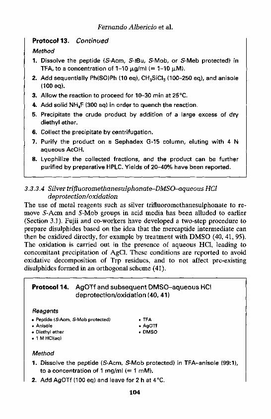
Fernando Albericio et al.
Protocol 13. Continued
Method
1. Dissolve the peptide (S-Acm, S-tBu, S-Mob, or S-Meb protected) inTFA, to a concentration of 1-10 (ug/ml (= 1-10 uM).
2. Add sequentially Ph(SO)Ph (10 eq), CH3SiCI3 (100-250 eq), and anisole(100 eq).
3. Allow the reaction to proceed for 10-30 min at 25°C.
4. Add solid NH4F (300 eq) in order to quench the reaction.
5. Precipitate the crude product by addition of a large excess of drydiethyl ether.
6. Collect the precipitate by centrifugation.
7. Purify the product on a Sephadex G-15 column, eluting with 4 Naqueous AcOH.
8. Lyophilize the collected fractions, and the product can be furtherpurified by preparative HPLC. Yields of 20-40% have been reported.
3.3.3.4 Silver trifluoromethanesulphonate-DMSO-aqueous HCldeprotection/oxidation
The use of metal reagents such as silver trifluoromethanesulphonate to re-move S-Acm and S-Mob groups in acid media has been alluded to earlier(Section 3.1). Fujii and co-workers have developed a two-step procedure toprepare disulphides based on the idea that the mercaptide intermediate canthen be oxidized directly, for example by treatment with DMSO (40, 41, 95).The oxidation is carried out in the presence of aqueous HCl, leading toconcomitant precipitation of AgCl. These conditions are reported to avoidoxidative decomposition of Trp residues, and to not affect pre-existingdisulphides formed in an orthogonal scheme (41).
Protocol 14. AgOTf and subsequent DMSO-aqueous HCldeprotection/oxidation (40, 41)
Reagents• Peptide (S-Acm, S-Mob protected) • TFA. Anisole . AgOTf• Diethyl ether • DMSO• 1 M HCI(aq)
Method
1. Dissolve the peptide (S-Acm, S-Mob protected) in TFA-anisole (99:1),to a concentration of 1 mg/ml (= 1 mM).
2. Add AgOTf (100 eq) and leave for 2 h at 4°C.
104

4: Preparation and handling of peptides 'containing Met and Cys
3. Add ice-cold diethyl ether (15 volumes) and collect the resultingprecipitate.
4. Wash the precipitated product with ice-cold diethyl ether (3 times).
5. Dissolve the peptide product in DMSO-1 M HCI(aq) (1:1), to achieve afinal peptide concentration of 0.1 mM.
6. Allow the oxidation reaction to proceed for 7 h at 25°C.
7. Remove the AgCI precipitate by filtration.
8. Dilute the filtrate with H2O and apply to preparative reversed-phaseHPLC to purify the product. Yields by this procedure are in the range50-60%.
3.3.4 Directed disulphide formationDirected methods of disulphide bridge formation (Scheme 1, Approach C)offer control whenever two different peptide chains must be linked (1, 4, 5, 8,107). (The applications of directed methods for intramolecular disulphideformation can be gleaned from the original publications (32, 108-110) and arenot covered here.) One chain will have a Cys residue with a free thiol,prepared by methods already described (Section 3.1), whereas in the otherchain the Cys residue needs to be blocked by a suitably activated form, suchas S-3-nitro-2-pyridinesulphenyl (S-Npys) (111-113), S-2-pyridinesulphenyl
(S-Pyr) (33, 3 4 , 114), S-methoxycarbonylsulphenyl (S-Scm) (115, 116), o r S - ( N - m e t h y l - N - p h e n y l c a r b a m o y l ) - s u l p h e n y l (5-Snm) (110, 116). This latter
need immediately raises a problem in the context of the Fmoc/tBu strategy forchain assembly, because the mentioned dual-purpose S-protecting/activatinggroups are labile to bases such as piperidine used for repetitive Na-Fmocremoval. Thus, if the plan is to incorporate a protected Cys residue withoutfurther transformations, it must be the N-terminal residue (fortunately, aconvenient position when the goal is conjugation to a protein carrier). Boc-Cys(Npys)-OH (111, 112) and Boc-Cys(Snm)-OH (110, 116) can be used inthis way.
Generation of internal activated Cys residues, as needed for directedmethods of disulphide formation, involves multi-stage procedures whereby adifferent S-protecting group is used to incorporate the Cys into the linearsequence. Accordingly, 5-Trt, S-Acm, S-Tmob, or 5-Xan protected peptide-resins, as well as peptides in solution containing those same S-protecting groupor a free thiol, can be treated with appropriate sulphenyl chlorides as follows:(1) methoxycarbonylsulphenyl chloride in chloroform and/or methanol willgive 5-Scm (107, 115, 116); (2) consecutive reactions in CH2C12 with chloro-carbonylsulphenyl chloride, followed by N-methylaniline, will give 5-Snm(110, 116); (3) Pyr-Cl and Npys-Cl in acetic acid will give respectively S-Pyr(117) and 5-Npys (111, 112, 118) (Figure 4). Sulphenyl chlorides are highlyreactive electrophilic reagents which are sometimes difficult to manipulate;
105

Fernando Albericio et al.
Figure 4. Dual S-protecting/activating groups for cysteine residues.
they will cause irreversible modification of Trp, and it is not possible toachieve selective activation with multiple Cys. An attractive and mildalternative is to prepare the peptide where the Cys is obtained in its free thiolform, and then carry out reaction with a reagent such as 2,2'-dithiobispyridine(34, 114, 119, 120) or 2,2'-dithiobis(5-nitropyridine) (DTNP) (121) to giverespectively 5-Pyr and S-Npys. Reaction with DTNP is particularly fast,requiring less than a minute between pH 3.5 and 6.5, and proceeding at anacceptable rate in aqueous AcOH (121). The aforementioned transform-ations are usually achieved with a purified peptide, but in some cases, thearomatic disulphides have been used to quench the thiols directly as they areliberated in an appropriate S-deprotective step (34, 114). Another reagentwhich can be used to activate a Cys-containing peptide preparatory to directeddisulphide formation is bis(tert-butyl)azodicarboxylate (Boc-N=N-Boc), whichis used to generate an isolable sulphenyl hydrazide intermediate (122, 123)(Figure 5).
Directed reactions to form disulphide bridges, applying the various acti-vated forms already described, are often rapid, facile, and readily driven tocompletion due to the electronic characteristics of the leaving group. Asdescribed in the leading references, reactions can be conducted in solution oron-resin, in aqueous or organic milieus, and under acidic or mildly basicconditions.
Figure 5. Reagents used for the activation of cysteine residues.
Protocol 15. Directed disulphide formation
Reagents• Peptide (free thiol form). AcOH• Diethyl ether. DTT• Acetate-buffered saline
2,2'-Dithiobis(5-nitropyridine) (DTNP)TFACHCI3Keyhole limpet haemocyanin (KLH)NaOH
106

4: Preparation and handling of peptides containing Met and Cys
n-Propanol-0.1 M Tris • Tri-n-butylphosphine2,2'-Dithiopyridine • MeCN-0.1 M Tris10% aq. sodium carbonate solution • Boc-N=N-BocAr-satu rated DMF • Diethyl etherNH4C03
A. Disulphide formation through activation with DTNP (121) andconjugation to a carrier protein (113)
1. Dissolve DTNP (3-5 eq) in a minimal amount of AcOH-H2O (3:1).
2. Add the peptide with the free thiol function in one portion withvigorous stirring.
3. After 4-6 h reaction, add AcOH (3 volumes) and lyophilize.
4. Extract the resultant solid with 0.1% aqueous TFA, sonicate, centri-fuge, and finally lyophilize the supernatant to obtain a powder.
5. Wash with diethyl ether-CHCI3 (7:3) to remove residual organicreagents and by-products.
6. Separately, partially reduce the carrier protein (keyhole limpethaemocyanin, KLH) by adding DTT (1.16 mg) to a solution of KLH(7.5 mg) in acetate-buffered saline (ABS; 0.1 M sodium acetate, 0.1 Msodium chloride, pH 4.5; 0.5 ml).
7. Allow the mixture to react for 1 h.
8. Purify on a Sephadex G-25 column (1 x 19 cm) by eluting with ABS,and UV monitoring at 220 nm.
9. Pool the protein fractions (3-4 ml).
10. Add to the protein solution, the Cys(Npys)-peptide (5-10 mg,lyophilized powder) from step 5.
11. Adjust the pH to 5.0 with 1 M NaOH.
12. After stirring overnight, adjust the solution to pH 7.0 by addition of1 M aqueous NaOH, and stir for another 3 h.
13. Dialyse the solution against 10 mM NH4CO3 (3 x 200 ml). Thepeptide:protein ratios are determined from acid hydrolysates of theconjugates.
B. Disulphide formation through activation with 2,2'-dithiopyridine (114)
1. Dissolve the peptide with the free thiol function in n-propanol-0.1 MTris pH 8 (1:1).
2. Add tri-n-butylphosphine (1.1 eq) and allow reaction to proceed for16 h under Ar atmosphere.
3. Add a solution of 2,2'-dithiopyridine (40 eq) in n-propanol-0.1 M TrispH8(1:1).
4. Stir the resultant mixture for 10 min.
107

Fernando Albericio et al.
Protocol 15. Continued
5. Purify by chromatography on Sephadex G-10, eluting with 0.1 M AcOHto give the activated peptide.
6. Dissolve the second peptide, containing a free thiol, in AcOH-H20(1:9).
7. Add to a solution of the activated peptide (from step 5) in MeCN-0.1 MTrispH8(1:1).
8. Adjust the pH to 6 with 10% aqueous sodium carbonate solution.
9. After 30 min, concentrate the reaction mixture and purify the productdisulphide by preparative HPLC.
C. Disulphide formation through activation with Boc-N=N-Boc (122)
1. Add dropwise a solution of a peptide (with a free thiol function) in Ar-saturated DMF to a solution of Boc-N=N-Boc (1 eq) in Ar-saturatedDMF, under vigorous stirring.
2. Allow the reaction to proceed for 12 h at 25°C,
3. Remove the bulk of the solvent by evaporation in vacuo.
4. Triturate the residue with diethyl ether.
5. Collect the precipitate, wash with diethyl ether, and dry in vacuo.
6. Dissolve the above peptide product (containing an activated thiol;1 eq) in Ar-saturated DMF.
7. Add dropwise the activated peptide solution to a solution of peptide(with a free thiol function; 1.4 eq) in Ar-saturated DMF, with stirring at25 °C.
8. After 24 h, repeat steps 3-5 to obtain a precipitate and purify further bySephadex chromatography.
3.3.5 Regioselective formation of disulphidesRegioselective formation of disulphides can be tackled by applying someof the already described methods (1-8). The general approach, when thedisulphides are intramolecular, involves graduated deprotection (often ortho-gonal) and/or co-oxidations of pairwise cysteine residues, as specified by theoriginal protection scheme. If an intermolecular disulphide is part of thetarget, a directed method is used to link two chains. Although these principlesare reasonably self-evident, their implementation in practice retains a sub-stantial measure of art and empiricism. Selectivities in the Cys deprotectionchemistries, compatibilities with sensitive residues, stabilities of already-formed disulphides to subsequent transformations, and solubility consider-ations are among the factors that influence success. For more complicatedtargets, the yields (if reported at all) tend to be quite low. The following para-
108

4: Preparation and handling of peptides containing Met and Cys
graphs provide a brief overview, but readers interested in more informationshould consult the aforementioned general reviews and original literaturecontributions cited therein.
For the preparation by the Fmoc/tBu strategy of peptides containing twodisulphides, the best combination appears to be an acid-labile protectinggroup (S-Trt, S-Tmob, or S-Xan) for two of the Cys, and S-Acm for theremaining two Cys (5, 30, 81). The formation of the first disulphide bridgeoccurs either in solution or on-resin after selective acidolysis has given abis(thiol), bis(Acm) intermediate. In a subsequent step, deprotection/oxidation of Acm is carried out to form the second bridge. An alternativetwo-dimensional orthogonal strategy uses acid-stable, thiolysable S-StBu toprotect the two Cys residues that are joined first, and again 5-Acm for theremaining two Cys (66, 122). In addition, the S-Phacm/S-Acm combinationhas been demonstrated for a simple parallel cystine dimer (37).
The extra selectivity to prepare peptides containing three disulphides canbe provided by incorporation of more acid-stable groups such as 5-Mob or 5-tBu. Deprotection/oxidation upon treatment with alkyltrichlorosilanes-sulphoxides, or related procedures, may give the last disulphide; while strongacid, for example HF at 0°C or TFMSA at 25°C in the presence of carefullychosen scavengers, provides a free thiol which may be manipulated further infavourable cases (34, 80, 124).
It may be adequate to adopt an intermediate partially regioselective strategywith the goal to obtain peptides with a multiple disulphide array. The idea isto use one class of protecting groups to control the pairing of one particularset of Cys residues, and the second class to block all of the remaining Cys.Thus, one might hope that establishment of the initial disulphide bridge mightbias the oxidation process that creates the subsequent bridges. Literatureprecedents are available when this was done in concert with chain assemblyby either Boc/Bzl (125, 126) or Fmoc/tBu (127) chemistry.
References1. Barany, G. and Merrifield, R. B. (1979). In The peptides—analysis, synthesis,
biology (ed. E. Gross and J. Meienhofer), Vol. 2, pp. 1-284. Academic Press, NewYork.
2. Fields, G. B., Tian, Z., and Barany, G. (1992). In Synthetic peptides: a user's guide(ed. Grant, G. A.), pp. 259-345. W. H. Freeman, New York.
3. Lloyd-Williams, P., Albericio, F., and Giralt, E. (1997). Chemical approaches tothe synthesis of peptides and proteins. CRC, Boca Raton, Florida.
4. Bullesbach, E. E. (1992). Kontakte (Darmstadt), 1, 21.5. Andreu, D., Albericio, F., Sole, N. A., Munson, M. C, Ferrer, M., and Barany, G.
(1994). In Methods in molecular biology, Vol. 35: Peptide synthesis protocols (ed.M. W. Pennington and B. M. Dunn), pp. 91-169. Humana Press, Totowa, NJ.
6. Kiso, Y. and Yajima, H. (1995). In Peptides—synthesis, structures, and applications(ed. B. Gutte), pp. 39-91. Academic Press, San Diego.
109

Fernando Albericio et al.
7. Moroder, L., Besse, D., Musiol, H. J., Rudolph-Bohner, S., and Siedler, F. (1996).Biopolymers, 40, 207.
8. Annis, I., Hargittai, B., and Barany, G. (1997). Methods in Enzymology, 289, 198.9. Noble, R. L., Yamashiro, D., and Li, C. H. (1976). J. Am. Chem. Soc., 98, 2324,
and references cited therein.10. King, D. S., Fields, C. G., and Fields, G. B. (1990). Int. J. Pept. Prot. Res., 36,
255.11. Albericio, F., Kneib-Cordonier, N., Biancalana, S., Gera, L., Masada, R. L,
Hudson, D., and Barany, G. (1990). J. Org. Chem., 55, 3730.12. Sole, N. A. and Barany, G. (1992). J. Org. Chem., 57, 5399.13. Hofmann, K, Haas, W., Smithers, M. J., Wells, R. D., Wolman, Y., Yanaihara, N.,
and Zanetti, G. (1965). J. Am. Chem. Soc., 87, 620.14. Hammer, R. P., Albericio, F., Gera, L., and Barany, G. (1990). Int. J. Pept. Prot.
Res., 36,31.15. Lloyd-Williams, P., Albericio, F., and Giralt, E. (1991). Int. J. Pept. Prot. Res., 37,
58.16. Ferrer, T., Nicolas, E., and Giralt, E. (1999). Lett. Pept. Sci., 6, 165.17. Tarn, J. P. and Merrifield, R. B. (1987). In The Peptides (ed. S. Udenfriend and J.
Meienhofer), Vol. 9, pp. 185-248, Academic Press, New York.18. Beck, W. and Jung, G. (1994). Lett. Pept. Sci., 1, 31.19. Houghten, R. A. and Li, C. H. (1979). Anal. Biochem., 98, 3646.20. Mapelli, C. and Leftheris, K. (1992). In Peptides 1992. Proceedings of the Twenty-
Second European Peptide Symposium (ed. C. H. Schneider and A. N. Eberle),pp. 207-208. Escom, Leiden, The Netherlands.
21. Pennington, M. W. and Byrnes, M. (1995). Peptide Research, 8, 39.22. Izeboud, E., and Beyerman, H. C. (1978). Recueil des Travaux Chimiques des
Pays-Bas, 97, 1.23. Fujii, N., Otaka, A., Funakoshi, S., Bessho, K., Watanabe, T., Akaji, K., and
Yajima, H. (1987). Chem. Pharm. Bull, 35, 2339, and references cited therein.24. Nicolas, E., Vilaseca, M., and Giralt, E. (1995). Tetrahedron, 51, 5701.25. Vilaseca, M., Nicolas, E., Capdevila, F., and Giralt. E. (1998). Tetrahedron, 54,
15273.26. Han, Y. and Barany, G. (1997). J. Org. Chem., 62, 3841.27. Barlos, K., Gatos, D., Chatzi, O., Koch, N., and Koutsogianni, S. (1996). Int.
J. Pept. Prot. Res., 47, 148.28. Munson, M. C., Garcia-Echeverria, C., Albericio, F., and Barany, G. (1992).
J. Org. Chem., 57, 3013.29. Zervas, L. and Theodoporopoulos, D. M. (1956). J. Am. Chem. Soc., 78, 1359.30. Munson, M. C. and Barany, G. (1993). J. Am. Chem. Soc., 115, 10203.31. Akaji, K., Tatsumi, T., Yoshida, M., Kimura, T., Fujiwara, Y., and Kiso, Y. (1991).
J. Chem. Soc., Chem. Commun., 167, and references cited therein.32. Ploux, O., Chassaing, G., and Marquet, A. (1987). Int. J. Pept. Prot. Res., 29, 162.33. Akaji, K., Fujino, K., Tatsumi, T., and Kiso, Y. (1992). Tetrahedron Lett., 33, 1073,
and references cited therein.34. Maruyama, K., Nagata, K., Tanaka, M., Nagasawa, H., Isogai, A., Ishizaki, H., and
Suzuki, A. (1992). J. Prot. Chem., 11, 1.35. Veber, D. F., Milkowski, J. D., Varga, S. L., Denkewalter, R. G., and Hirschmann,
R. (1972). J. Am. Chem. Soc., 94, 5456.
110

4: Preparation and handling of peptides containing Met and Cys
36. Hermann, P. and Schillings, T. (1993). In Peptides 1992: Proceedings of theTwenty-Second European Peptide Symposium (ed. C. H. Schneider and A. N.Eberle), pp. 411-412. Escom, Leiden, The Netherlands, and references citedtherein.
37. Royo, M., Alsina, J., Giralt, E., Slomczynska, U., and Albericio, F. (1995). /.Chem. Soc., Perkin Trans. 1, 1095.
38. Kamber, B., Hartmann, A., Eisler, K., Riniker, B., Rink, H., Sieber, P., and Rittel,W. (1980). Helv. Chim. Acta, 63, 899.
39. Ruiz-Gayo, M., Albericio, F., Royo, M., Garcia Echeverria, C., Pedroso, E., Pons,M., and Giralt, E. (1989). Anales de Quimica, 85C, 116.
40. Tamamura, H., Otaka, A., Nakamura, J., Okubo, K., Koide, T., Ikeda, K., Ibuka,T., and Fujii, N. (1995). Int. J. Pept. Prot. Res., 45, 312.
41. Tamamura, H., Matsumoto, F., Sakano, K., Ibuka, T., and Fujii, N. (1998). Chem.Commun., 151.
42. Weber, U. and Hartter, P. (1970). Hoppe-Seyler's Zeitschrift fur PhysiologischeChemie, 351, 1384.
43. Gosden, A., MacRae, R., and Young, G. T. (1977). /. Chem. Res. (Synopsis), 22.44. Ramage, R. (1999). Personal communication.45. Grandas, A., Jorba, X., Giralt, E., and Pedroso, E. (1989). Int. J. Pept. Prot. Res.,
33, 386.46. Sieber, P. (1987). Tetrahedron Lett., 28, 6147.47. Atherton, E., Hardy, P. M., Harris, D. E., and Mattews, B. H. (1991). In Peptides
1990: Proceedings of the Twenty-First European Peptide Symposium (ed. E. Giraltand D. Andreu), pp. 243-244. Escom, Leiden, The Netherlands.
48. Fujiwara, Y., Akajii, K., and Kiso, Y. (1994). Chem. Pharm. Bull, 42, 724.49. Barlos, K., Chatzi, O., Gatos, D., and Stavropoulos, G.. (1991). Int. J. Pept. Prot.
Res., 37, 513.50. Bernatowicz, M. S., Kearney, T., Neves, R. S., and Koster, H. (1989). Tetrahedron
Lett., 30, 4341.51. Mergler, M., Nyfeler, R., Tanner, R., Gosteli, J., and Grogg, P. (1988).
Tetrahedron Lett., 29, 4005.52. Mergler, M., Gosteli, J., Nyfeler, R., Tanner, R., and Grogg, P. (1989). In Peptides
1988: Proceedings of the 20th European Peptide Symposium (ed. G. Jung and E.Bayer), pp. 133-135. Walter de Gruyter, New York.
53. Chiva, C., Vilaseca, M., Giralt, E., and Albericio, F. (1999). J. Pept. Sci., S, 131.54. Kirstgen, R., Sheppard, R. C., and Steglich, W. (1987). /. Chem. Soc., Chem.
Commun., 1870.55. Kirstgen, R., Olbrich, A., Rehwinkel, H., and Steglich, W. (1988). Justus Liebigs
Annalen der Chemie, 437.56. Kates, S. A., Diekmann, E., El-Faham, A., Ionescu, D., McGuinness, B. F., Triolo,
S. A., Albericio, F., and Carpino, L.A. (1996). In Techniques in protein chemistryVII (ed. D. R. Marshak), pp. 515-523. Academic Press, New York.
57. Han, Y., Vagner, J., and Barany, G. (1996). In Innovation and perspectives in solidphase synthesis and combinatorial libraries: peptides, proteins and nucleic acids,small molecule organic chemical diversity (ed. R. Epton) , pp. 385-388. MayflowerWorldwide Ltd., Birmingham, UK.
58. Musiol, H. J., Siedler, F., Quarzago, D., and Moroder, L. (1994). Biopolymer, 34,1553.
111

Fernando Albericio et al.
59. Kaiser, T., Nicholson, G., Kohlbau, H. J., and Voelter, W. (1996). TetrahedronLett., 37, 1187.
60. Han, Y., Albericio, F., and Barany, G. (1997). J. Org. Chem., 62, 4307.61. Albericio, F. and Carpino, L. A. (1997). Methods in Enzymology, 289, 104.62. Carpino, L. A., Ionescu, D., and El-Faham, A. (1996). J. Org. Chem., 61, 2460.63. Angell, Y. M., Han, Y., Albericio, F., and Barany, G. (1998). In Peptides—
chemistry and biology. Proceedings of the Fifteen American Peptide Symposium(ed. J. P. Tam and P. T. P. Kaumaya), pp. 339-340. Kluwer-ESCOM, Dordrecht,The Netherlands.
64. Lukszo, J., Patterson, D., Albericio, F., and Kates, S. A. (1996). Lett. Pept. Sci., 3,157.
65. Eritja, R., Ziehler-Martin, J. P., Walker, P. A., Lee, T. D., Legesse, K., Albericio,F., and Kaplan, B. E. (1987). Tetrahedron, 43, 2675.
66. Atherton, E., Sheppard, R. C., and Ward, P. (1985). J. Chem. Soc., Perkin Trans.1, 2065.
67. Engebretsen, M., Agner, E., Sandosham, J., and Fischer, P. M. (1997). Int. J. Pept.Prot. Res., 49, 341.
68. Mendelson, W. L., Tickner, A. M., Holmes, M. M., and Lantos, I. (1990). Int. J.Pept. Prot. Res., 35, 249.
69. Lamthanh, H., Roumestand, C., Deprun, C., and Menez, A. (1993). Int. J. Pept.Prot. Res., 41, 85.
70. Mazur, S. and Jayalekshmy, P. (1978). /. Am. Chem. Soc., 101, 677.71. Albericio, F., Hammer, R. P., Garcia-Echevarria, C., Molins, M. A., Chang, J. L.,
Munson, M. C., Pons, M., Giralt, E., and Barany, G. (1991). Int. J. Pept. Prot. Res.,37, 402.
72. Royo, M., Contreras, M. A., Giralt, E., Albericio, F., and Pons, M. (1998). /. Am.Chem. Soc., 120, 6639.
73. Jaenicke, R. and Rudolph, R. (1989). In Protein structure: a practical approach(ed. T. E. Creighton), pp. 191-223. IRL Press, Oxford, and references cited therein.
74. Burns, J. A., Butler, J. C., Moran, J., and Whitesides, G. M. (1991). J. Org. Chem.,56, 2648.
75. Cleland, W. W. (1964). Biochemistry, 3, 480.76. Hantgan, R. R., Hammes, G. G., and Scheraga, H. A. (1974). Biochemistry, 13,
3421.77. Akaji, K., Tatsumi, T., Yoshida, M., Kimura, T., Fujiwara, Y., and Kiso, Y. (1992).
J. Am. Chem. Soc., 114, 4137.78. Ferrer, M., Woodward, C., and Barany, G. (1992). Int. J. Pept. Prot. Res., 40, 194.79. Munson, M. C., Lebl, M., Slaninova, J., and Barany, G. (1993). Peptide Research,
6, 155.80. Kellenberger, C., Hietter, H., and Luu, B. (1995). Peptide Research, 8, 321.81. Barany, G., Angell, Y. M., Annis, I., Chen, L., Gross, C. M., and Hargittai B.
(1998). In Proceedings of the Fifth International Symposium on Solid-PhaseSynthesis and Combinatorial Chemical Libraries (ed. R. Epton), pp. 85-88.Mayflower Scientific Ltd., UK.
82. Karim Ahmed, A., Schaffer, S. W., and Wetlaufer D. B. (1975). J. Biol. Chem.,250, 8477.
83. Rothwarf, D. M. and Scheraga, H. A. (1993). Biochemistry, 32, 2680.84. Ruoppolo, M. and Freedman, R. B. (1995). Biochemistry, 34, 9380.
112

4: Preparation and handling of peptides containing Met and Cys
85. Sabatier, J.-M., Darbon, H., Fourquet, P., Rochat, H., and Van Rietschoten, J.(1987). Int. J. Pept. Prot. Res., 30, 125.
86. Kuhelj, R., Dolinar, M., Pungercar, J., and Turk, V. (1995). Eur. J. Biochem., 229,533.
87. Wallace, T. J. (1964). J. Am. Chem. Soc., 86, 2018.88. Tam, J. P., Wu, C.-R, Liu, W., and Zhang, J.-W. (1991). J. Am. Chem. Soc., 113,
6657.89. Sieber, P., Eisler, K., Kamber, B., Riniker, B., Rittel, W., Marki, F., and De
Gasparo, M. (1978). Hoppe-Seyler's Zeitschrift fur Physiologische Chemie, 359,113.
90. Misicka, A. and Hruby, V. J. (1994). Polish J. Chem., 68, 893.91. Rivier, J., Kaiser, R., and Galyean, R. (1978). Biopolymers, 17, 1927, and
references cited therein.92. Gray, W. R., Luque, F. A., Galyean, R., Atherton, E., Sheppard, R. C, Stone, B.
L., Reyes, A., Alford, J., McIntosh, M., Olivera, B. M., Cruz, L. J., and Rivier, J.(1984). Biochemistry, 23, 2796.
93. Ellman, G. L. (1959). Archives of Biochemistry and Biophysics, 82, 70.94. Annis, I., Chen, L., and Barany, G. (1998). J. Am. Chem. Soc., 120, 7226.95. Tamamura, H., Otaka, A., Nakamura, J., Okubo, K., Koide, T., Ikeda, K., and
Fujii, N. (1993). J. Chem. Soc., Chem. Commun., 283.96. Edwards, W. B., Fields, C. G., Anderson, C. J., Pajeau, T. S., Welch, M. J., and
Fields, G. B. (1994). J. Med. Chem., 37, 3749.97. Bishop, P. and Chmielewski, J. (1992). Tetrahedron Lett., 33, 6263.98. Shih, H. (1993). J. Org. Chem., 58, 3003.99. Chen, L., Bauerova, H., Slaninova, J., and Barany, G. (1996). Peptide Research,
9, 114.100. White, P. (1992). In Peptides: chemistry, and biology. Proceedings of the 12th
American Peptide Symposium (ed. J. A. Smith and J. E. Rivier), pp. 537-538,Escom, Leiden, The Netherlands.
101. Previero, A., Antonia, M., Previero, C., and Cavadore, J. C. (1976). BiochimicaBiophysica Acta, 147, 453.
102. Koide, T., Otaka, A., Suzuki, H., and Fujii, N. (1991). Synthetic Letters, 345.103. Akaji, K., Nishiuchi, H., and Kiso, Y. (1995). Tetrahedron Lett., 36, 1875.104. Otaka, A., Koide, T., Shide, A., and Fujii, N. (1991). Tetrahedron Lett., 32,
1223.105. Koide, T., Otaka, A., and Fujii, N. (1993). Chem. Pharm. Bull., 41, 1030.106. Adeva, A., Camarero, J. A., Giralt, E., and Andreu, D. (1995). Tetrahedron Lett.,
36, 3885.107. Brois, S. J., Pilot, J. F., and Barnum, H. W. (1970). J. Am. Chem. Soc., 92, 7629.108. Mott, A. W., Somczynska, U., and Barany, G (1986). In Forum peptides le Cap
d'Agde 1984 (ed. B. Castro and J. Martinez), pp. 321. Les Impressions Dohr,Nancy, France.
109. Ridge, R. J., Matsueda, G. R., Haber, E., and Matsueda, R. (1982). Int. J. Pept.Prot. Res., 19, 490.
110. Chen, L., Zoulikova, L, Slaninova, J., and Barany, G. (1997). J. Med. Chem., 40,864.
111. Matsueda, R., Kimura, T., Kaiser, E.T., and Matsueda, G. R. (1981). ChemistryLetters, 6, 737.
113

Fernando Albericio et al.
112. Bernatowicz, M. S., Matsueda, R., and Matsueda, G. R. (1986). Int. J. Pept. Prot.Res., 28, 107.
113. Albericio, F., Andreu, D., Giralt, E., Navalpotro, C., Pedroso, E., Ponsati, B.,and Ruiz-Gayo, M. (1989). Int. J. Pept. Prot. Res., 34, 124.
114. Ruiz-Gayo, M., Royo, M., Fernandez, I., Albericio, F., Giralt, E., and Pons, M.(1993). J. Org. Chem., 58. 6319.
115. Kamber, B. (1973). Helv. Chim. Acta, 56, 1370.116. Schroll A. L., and Barany, G. (1989). J. Org. Chem., 54, 244, and references cited
therein.117. Castell, J. V. and Tun-Kyi, A. (1979). Helv. Chim. Acta, 62, 2507.118. Simmonds, R. G., Tupper, D. E., and Harris, J. R. (1994). Int. J. Pept. Prot. Res.,
43, 363.119. Carlsson, J., Drevin, H., and Axen, R. (1978). Biochemistry J., 173, 723.120. Mant, C. T., Kondejewski, L. H., Cachia, P. J., Monera, O. D., and Hodges, R. S.
(1996). Methods in Enzymology, 289, 426.121. Rabanal, F., DeGrado, W. F., and Dutton, P. L. (1996). Tetrahedron Lett., 37,
1347.122. Wunsch, E., Moroder, L., Gohring-Romani, S., Musiol, H.-J., Gohring, W., and
Bovermann, G. (1988). Int. J. Pept. Prot. Res., 32, 368.123. Wunsch, E. and Romani, S. (1982). Hoppe-Seyler's Zeitschrift fur Physiologische
Chemie, 363, 449.124. Bullesbach, E. E. and Schwabe, C. (1991). J. Biol. Chem., 266, 754.125. Tarn, J. P. and Lu, Y.-A. (1997). Tetrahedron Lett., 38, 5599.126. Shimonishi, Y., Hidaka, Y., Koizumi, M., Hane, M., Aimoto, S., Takeda, T.,
Miwatani, T., and Takeda, Y. (1987). FEBS Lett., 215, 165.127. Spetzler, J. C., Rao, C., and Tam, J. P. (1994). Int. J. Pept. Prot. Res., 43, 351.
114
Difficult peptidesMARTIN QUIBELL and TONY JOHNSON
1. IntroductionSolid phase peptide synthesis (SPPS), first proposed by R. B. Merrifield in1962 (1), has evolved over three decades into a tremendously powerfulmethod for the preparation of peptides and small proteins (2, 3). An absoluteprerequisite for successful syntheses in all solid phase schemes is thatreactions which accumulate solid supported products, and by the very natureof the technique contaminating by-products, must proceed cleanly andefficiently. During the earlier years of SPPS this optimal situation was notalways achieved, primarily due to contaminated reagents and ill-definedpolymers in combination with poorly flexible protection strategies (4, 5). Asthe methods of SPPS gained popularity and more widespread application,reagents and protection strategies were improved and refined (6, 7). How-ever, reports of notable successful syntheses were accompanied by thenunexplained failures, which have since been collectively termed 'difficultpeptides' (8, 9). This chapter describes how an intrinsic understanding behindthe occurrence of 'difficult peptides' has accumulated, leading to a generalsynthetic solution—the utilization of a backbone amide protection strategy.
2. Difficult peptides—an overview2.1 BackgroundWithin a few years of the introduction of SPPS, it was recognized that theassembly of some peptide sequences posed a special synthetic problem. Themain feature evident during these syntheses was a sudden decrease in reactionkinetics, leading to incomplete amino-acylation by activated amino acidresidues. The unreacted sites were readily detected by the Kaiser test for freeamine (10); however, couplings showed no significant improvement evenupon repeated or prolonged reaction. Efficient reactions are known to occurwithin a fully solvated peptide-polymer matrix, where reagent penetration israpid and unhindered (11). This optimal situation no longer exists during theassembly of a difficult peptide, where the normally accessible solid phase
5

Martin Quibell and Tony Johnson
reaction matrix becomes partially inaccessible during assembly. This situationarises suddenly, typically 6-12 residues into the synthesis, and may then persistfor a number of cycles before easing, or in extreme cases remain throughoutthe completion of the assembly. The crude products are particularly poor ifslower coupling B-branched residues (isoleucine, valine, threonine) are intro-duced after the onset of synthetic difficulties. The principles underlying theoccurrence of difficult peptide sequences have for many years been the focusof intense debate and research (8, 9, 12). An intrinsic feature of the numerousideas proposed is that aggregation occurs resulting in poor solvation withinthe peptide-polymer matrix.
In a fully solvated system, the reactive peptide amino termini are wellseparated and readily accessible, along with polymer chains and cross-linksthat are extended and mobile. This is eloquently represented in Figure la,along with scenarios in which partial collapse or aggregation could occurleading to hindrance of the amino termini. In scenario b, the once solvatedpeptide chains have self-associated, possibly through intramolecular hydro-gen bonding, minimizing interaction between the reaction medium and theamino termini and limiting the accessibility of reagents. Scenario c depictspoor solvation of the polymer backbone, again hindering reagent access to thepeptide chains. Scenario d depicts peptide chain association, but now viainterchain as opposed to intrachain bonding as in b. This effectively producesa more highly cross-linked matrix that is less mobile or accessible than shownin the optimal situation a.
During the early 1970s it was suggested that the occurrence of difficult pep-tides possibly arose due to an incompatibility between the apolar polystyreneresins, polar peptide chains, and apolar reaction media, such as dichloro-methane, used for SPPS as depicted in Figure 1b or 1c (13). This prompted theinvestigation of alternative polymer supports and reaction media to the
Figure 1. Scenarios depicting the possible solvation or aggregation states of a polymer-bound peptide sequence, (a) Peptide and polymer chains (bold) are fully solvated. (b)Peptide chains are intramolecularly aggregated whilst polymer is solvated. (c) Peptidechains are well solvated whilst the polymer backbone is poorly solvated. (d) Peptidechains are intermolecularly aggregated whilst polymer is solvated, effectively increasingthe overall level of cross-linking in the matrix. (Reproduced from ref. 7 with permission.)
116

5: Difficult peptides
original Merrifield system. Since peptides are polyamides, a major advance incompatibility was the introduction by Sheppard and co-workers (14) of apolyamide-based polymer support along with the use of dipolar aprotic amidesolvents. These new protocols enabled the successful preparation of the deca-peptide sequence from the acyl carrier protein (ACP)-(65-74), which was notprepared readily by standard solid phase techniques (15, 16). However, thisbreakthrough in the synthesis of difficult peptides did not provide the com-plete solution since some sequences still remained prone to aggregation (17).It is now accepted that a major contributing factor to the phenomenon ofdifficult peptides originates in the intrinsic properties of the peptide sequenceitself. A wealth of physical evidence has been accumulated (see later) to showthat aggregation primarily occurs through the self-association of polymer-bound peptide sequences by intermolecular hydrogen bonding as depicted inFigure ld. Thus, a general solution to difficult peptide syntheses requires in-hibition of the root cause of the problem, intermolecular backbone hydrogenbonding.
2.2 Identifying the effects of aggregationFollowing the introduction of polyamide supports, Sheppard and co-workersadvocated a milder orthogonal Fmoc/f-butyl chemistry for SPPS in place ofthe somewhat harsh Boc/benzyl protocols of the original Merrifield scheme(18, 19). This new chemistry, along with the advent of a flow stable physicallysupported polyamide gel, led to the widely adopted continuous flow Fmoc-polyamide technique for SPPS (20-22). This proved to be a pivotal con-tribution in the understanding of the difficult peptide phenomenon throughthe real-time spectrophotometric monitoring of a fluorene derived chromo-phore, released at each Na-Fmoc deprotection cycle, into the solvent stream.
The unhindered release of fluorene derivatives from a fully solvatedpeptide-polymer matrix results in a characteristic 'bell-shaped' elution profile(20) (Figure 2a). Syntheses that exhibit such profiles throughout their assemblyare generally of an excellent crude quality. In the case of difficult peptidesequences, the slowing of reaction kinetics at the onset of aggregation isreadily observed as a broadening of the release of fluorene derivatives fromthe reaction matrix (Figure 26). In severe instances, the monitored release of
Figure 2. Typical Fmoc deprotection profiles (measured at 275 nm) observed duringcontinuous flow SPPS. (a) Unhindered release of fluorene chromophore from a fullysolvated peptide-polymer matrix, (b) Release from a partially aggregated peptide-polymer. (c) Release from a fully aggregated peptide-polymer matrix.
117

Martin Quibell and Tony Johnson
fluorene shows a dramatically flattened profile, indicating slow and possiblyincomplete removal of the Na-Fmoc group (Figure 2c), occasionally seen asFmoc-terminated products in the crude peptide (23).
Numerous non-invasive methods for the identification of difficult peptidesequences have been described, each adding to an overall understanding ofthe phenomenon. A 'swellographic' method, which is based on the actualphysical bed volume of a swollen gel peptide-resin matrix (24-27) providescompelling visual evidence for aggregation. All the popular polymeric gelsupports used for peptide synthesis absorb organic solvents and swell to manytimes their dry bed volume. As a resin-bound peptide sequence is extended,the swollen bed volume of the matrix increases (12). However, in thesynthesis of a difficult peptide sequence, the swollen bed volume suddenlydecreases by up to 50%, reflecting the increased cross-linking introducedthrough intermolecular hydrogen bonding between the peptide chains.
The fundamental physiochemical properties of difficult peptide sequenceshave been investigated using infrared (28-31) and NMR analysis of polymer-bound peptide sequences. Infrared measurement of peptide-polymer gelmatrices clearly show the formation of B-secondary structure on transition ofa fully solvated to an aggregated sequence. On aggregation, clear bands at1220, 1240, and 1300 cm-1 in the amide III region and 1665 cm-1 in the amide Iregion, characteristic of a B-sheet secondary structure, are observed. A similareffect has been described by Narita et al. (32) correlating protected peptidesolubility in solution with structure. With Fmoc/polyamide SPPS techniques,the ortho-substituted phenyl ring of residual Fmoc may be observed at 1025cm-1 on deprotection of a difficult peptide sequence (29). Mapelli andSwerdloff (33) have described the 13C NMR of a gel polymer-bound ACP-(65-74), showing a broadening of the NMR line widths upon aggregation.
2.3 Effect of resins, solvents, and additives on aggregationNumerous modified SPPS protocols have been suggested to improve thequality of difficult peptide syntheses. A common theme among the majority ofthese protocols is an attempt to limit the extent of B-sheet secondary structureformation by altering the solvation properties of the polymer, the polymer-bound protected peptide, and/or the surrounding reaction media.
The last decade has seen a resurgence in the development of new polymersupports, which exhibit improved solvation during the synthesis of difficultpeptides. A graft polymer of polyethylene glycol-polystyrene (PEG-PS) hasshown impressive improvements in the synthesis of a difficult polyalaninesequence when compared to a polystyrene-based support (34). Meldal (35)has described the use of a polyethylene glycol-polydimethylacrylamide(PEGA) based polymer in an improved preparation of ACP-(65-74). Animportant factor to consider in the performance of any polymer support is theoverall functional loading and the distribution of that loading, highlighted in
118

5: Difficult peptides
an investigation by Tarn and Lu (27). The use of low-substitution resins(typically 0.1-0.4 mmol/g) may be advantageous since a high density ofpeptide chains may mimic an increased degree of cross-linking and poorsolvation. However, Tam and Lu conclude that many such commercial resinsmay not be evenly functionalized and can contain areas of highly clusteredfunctionality resulting in very poor solvation.
Alternatively, attempts have been made to improve the solvation prop-erties of the reaction media, rather than those of the polymer support alone.Virtually all SPPS, regardless of the type of polymer support, is now per-formed in dipolar aprotic amide solvents originally suggested by Sheppardand co-workers (14). Additionally, numerous additives that may facilitate thedisruption of secondary structure formation through the potential eliminationof peptide-chain intermolecular hydrogen bonds have been investigated. Theaddition of chaotropic salts such as potassium thiocyanate (36) or lithiumchloride (37), typically at a concentration of 0.4 M, or detergent (38) at 1%v/v has been suggested. Improved solvation has been described upon theaddition of trifluoroethanol (39), hexafluoroisopropanol (40), N-methyl-pyrrolidinone (41), and dimethylsulphoxide (42) to reaction media.
Each of the aforementioned modifications has undoubtedly provided im-proved crude products in a number of such syntheses. However, improvedsolvation of the polymer-peptide/reaction media, although extremelydesirable, only aims to limit the effects and extent of B-sheet secondarystructure formation. The potential for intermolecular backbone hydrogenbonding, the fundamental cause of the phenomenon, still exists.
3. Predicting difficult peptide sequencesIn order to effectively assembly difficult peptides, more complex protocolsaimed at the disruption of secondary structure formation need to be intro-duced prior to the onset of synthetic difficulties. Ideally, this could beachieved if a predictive method describing where and when B-structureformation will occur, based simply on examination of the primary sequence,was available. Unfortunately, there is no complete solution available to date,although a number of groups have developed reasonably successful guide-lines. Current predictive methods are derived from four main investigations:
1. Statistical analysis of data gathered during the SPPS of hundreds ofpeptides (43-46);
2. Ab initio calculations based upon Chou and Fasman parameters (47) fromknown protein structures (28);
3. Correlation of protected peptide solubility in solution with structure (32,48, 49);
4. The effects of host-guest substitutions within a given difficult peptideframework (50, 51).
119

Martin Quibell and Tony Johnson
A statistical analysis method has been used to ascribe an aggregationparameter for each amino acid (44, 45) based on the change in swollen volumeof a peptidyl-resin on couplings in 87 unrelated sequences. Amino acids suchas proline, arginine, and histidine were rarely present towards the carboxyterminus of sequences that showed subsequent aggregation and were accord-ingly given low indices. Conversely, amino acids such as alanine, isoleucine,methionine, and lysine were often present and went on to give difficultpeptide problems. The method predicts difficult peptide syntheses withreasonable success, through a run of high aggregation parameter residues inthe 6-9 carboxy-terminal residue region of an assembly. The predictivemethods of Van Woerkom and Van Nispen (43) are based on the analysis of696 couplings from 88 sequences. Residues that are difficult to couple with,and the effects of chain length on these couplings, are described.
Narita et al. (48) developed a predictive method which can potentiallyidentify soluble protected peptide intermediates for solution-based fragmentcondensations. A fully solvated peptide-polymer is considered to contain a'soluble' peptide chain, whilst an aggregated matrix contains a partiallysoluble or 'insoluble' chain (12). Thus, the findings of Narita et al. might beused to predict solid phase difficult peptide syntheses if one considers theaddition of each amino acid during assembly as the formation of a newprotected segment and access that segment accordingly. The single moststriking conclusion from the work of Narita et al. was the onset of an un-ordered structure in solution of a segment containing an appropriatelypositioned tertiary amide bond. Such segments showed substantially im-proved solubility (32, 49).
The host-guest substitution investigations of Sheppard and co-workers werebased on the introduction of guest residues into a host sequence (I) with astrong tendency towards aggregation, followed by continuous flow SPPSmonitoring of the Na-Fmoc fluorene-derived chromophore released at eachcycle (51). After introduction of the two guest residues, the syntheses werecontinued by addition of alanine residues until aggregation was observedthrough broad Fmoc deprotection profiles.
Figures 3a-d show the deprotection profile peak heights as a function of chainlength and guest residue and a number of conclusions were clearly apparent.The hydrophobic guest residues alanine, valine, isoleucine, leucine, andphenylalanine showed a strong tendency to aggregate on the addition of onlyone or two more alanine residues (Figure 3a). A similar result based onaggregation parameters was obtained by Krchnak et al. (44, 45). The side-chain protecting group of trifunctional residues, e.g. ranging from the bulkyhydrophobic trityl to the relatively polar acetamidomethyl for a series ofcysteine derivatives, showed little effect on the propensity for aggregation
120

5: Difficult peptides
Figure 3. Deprotection peak heights as a function of host residue (-X-X-) and chain lengthin the SPPS of the known difficult peptide test sequence H-(Ala)n-X-X-(Ala)3-Val-poly-dimethylacrylamide (I). (a) General hydrophobic residues. (b) Effect of side-chain pro-tection of cysteine. (c) Effect of trityl protection of side-chain carboxamide of glutamineand asparagine. (d) Effect of arginine and tertiary amino acids proline and sarcosine.(Reproduced from ref. 51 with permission from Munksgaard International Publishers Ltd.,Copenhagen.)
(Figure 3b). An exception was the side-chain carboxamide protection ofglutamine and possibly asparagine by trityl, which showed a beneficial effectin delaying aggregation (Figure 3c). The introduction of arginine delayed theonset of aggregation significantly, as did the introduction of histidine (datanot shown) (Figure 3d). The most striking result was the introduction of thetertiary amide residue such as proline or sarcosine which abolished the pro-pensity for aggregation until a further six alanine residues were added (Figure3d), this being the minimum length at which polyalanine itself aggregated(Figure 3a).
The host-guest results, along with the segment solubility results of Narita etal. (48, 49) and Blackmeer et al (52), suggested that the difficult peptideproblem might be eliminated by the introduction of a reversibly N-alkylatedamino acid residue prior to aggregation.
121

Martin Quibell and Tony Johnson
4. The N-(2-hydroxy-4-methoxybenzyl) (Hmb)backbone amide-protecting group
4.1 Development and preparation of (Hmb)amino acidderivatives
Numerous factors required consideration during the development of asecondary amide protecting group suitable for use in Fmoc/t-butyl SPPS.Complete stability to the repetitive acylation and piperidine-mediated Na-Fmoc deprotection cycles was essential. Additionally, a group readily removedupon the acidolytic side-chain deprotection and C-terminal peptide-handlecleavage, mediated by trifluoroacetic acid, was desired. These requirementspointed to N-aralkyl-substituted amino acid residues and a range of methyl-and methoxy-substituted benzyl, furfuryl, and thienyl derivatives were pre-pared which exhibited the desired chemical properties (53). Unfortunately,following incorporation into a peptide chain, such derivatives proved ex-tremely difficult to N-acylate quantitatively. This obstacle precludes thegeneral use of N-aralkyl derivatives in SPPS. However, the problem of slowN-acylation was largely overcome by the introduction of a 2-hydroxylsubstituent into the 4-methoxybenzyl system (see Figure 4). Acylation of theN-(2-hydroxy-4-methoxybenzyl) residue is achieved through an internal base-
Figure 4. Scheme for the incorporation of N,O-bis(Fmoc)-N-(Hmb)-protected pentafluoro-phenyl esters (a) into a peptide chain. Key hydrogen-bonded tautomer (c) catalysesesterification to (e) which undergoes O -N acyl transfer.
122
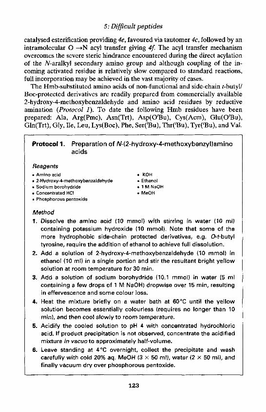
5: Difficult peptides
catalysed esterification providing 4e, favoured via tautomer 4c, followed by anintramolecular O —N acyl transfer giving 4f. The acyl transfer mechanismovercomes the severe steric hindrance encountered during the direct acylationof the N-aralkyl secondary amino group and although coupling of the in-coming activated residue is relatively slow compared to standard reactions,full incorporation may be achieved in the vast majority of cases.
The Hmb-substituted amino acids of non-functional and side-chain t-butyl/Boc-protected derivatives are readily prepared from commercially available2-hydroxy-4-methoxybenzaldehyde and amino acid residues by reductiveamination (Protocol 1). To date the following Hmb residues have beenprepared: Ala, Arg(Pmc), Asn(Trt), Asp(O'Bu), Cys(Acm), Glu(OtBu),Gln(Trt), Gly, Ile, Leu, Lys(Boc), Phe, Ser(tBu), Thr(tBu), Tyr(tBu), and Val.
Protocol 1. Preparation of N-(2-hydroxy-4-methoxybenzyl)aminoacids
Reagents• Amino acid • KOH• 2-Hydroxy-4-methoxybenzaldehyde • Ethanol• Sodium borohydride • 1 M NaOH• Concentrated HCI • MeOH• Phosphorous pentoxide
Method1. Dissolve the amino acid (10 mmol) with stirring in water (10 ml)
containing potassium hydroxide (10 mmol). Note that some of themore hydrophobic side-chain protected derivatives, e.g. 0-t-butyltyrosine, require the addition of ethanol to achieve full dissolution.
2. Add a solution of 2-hydroxy-4-methoxybenzaldehyde (10 mmol) inethanol (10 ml) in a single portion and stir the resultant bright yellowsolution at room temperature for 30 min.
3. Add a solution of sodium borohydride (10.1 mmol) in water (5 mlcontaining a few drops of 1 M NaOH) dropwise over 15 min, resultingin effervescence and some colour loss.
4. Heat the mixture briefly on a water bath at 60°C until the yellowsolution becomes essentially colourless (requires no longer than 10min), and then cool slowly to room temperature.
5. Acidify the cooled solution to pH 4 with concentrated hydrochloricacid. If product precipitation is not observed, concentrate the acidifiedmixture in vacuoto approximately half-volume.
6. Leave standing at 4°C overnight, collect the precipitate and washcarefully with cold 20% aq. MeOH (3 x 50 ml), water (2 x 50 ml), andfinally vacuum dry over phosphorous pentoxide.
123

Martin Quibell and Tony Johnson
Protocol 2. Preparation of N,O-bis(Fmoc)-N-(2-hydroxy-4-methoxybenzyDamino acids
Reagents• N-(2-Hydroxy-4-methoxybenzyl)amino acid • Sodium carbonate decahydrate• 1,4-Dioxane • Fmoc-CI. Diethyl ether . 0.5 M HCI• Na2S04 • MeOH. CHCI3
Method
1. Add the N-(2-hydroxy-4-methoxybenzyl)amino acid (5 mmol) to an ice-cold stirred solution of sodium carbonate decahydrate (16 mmol, 3.2excess) in water (50 ml). Add 1,4-dioxane (30-50 ml) to ensuredissolution.
2. Add dropwise a solution of Fmoc-CI (10.5 mmol, 2.1 excess) in dioxane(10 ml) over 1 h.
3. After a further 1-2 h reaction, monitored by reverse-phase HPLC, theoriginal clear solution should thicken with the precipitated product.
4. Quench the reaction mixture with diethyl ether (100 ml), and extractthe aqueous layer with further ether (2 x 100 ml).a
5. Wash the combined ethereal extracts with 0.5 M HCI (2 x 100 ml), dryover Na2SCv and evaporate in vacuoto an oily solid.
6. Dry the solid in vacuo overnight.
7. Purify the crude derivative using Kieselgel 60 (Merck) (20 g silica/gcrude product), initially packed and sample loaded in CHCI3. Elute thecolumn with CHCI3 until the UV positive material no longer elutes.
8. Gradually change the elution solvent up to 5% MeOH in CHCI3.b
9. Pool the desired fractions and evaporate in vacuoto a crystalline solid.Isolated yields are generally around 20% based on the starting(Hmb)amino acid.
aThese ethereal layers contain the sodium salt of the desired bis(Fmoc) product.bIn general the bis(Fmoc) derivatives elute at Rf = 0.3 (5% MeOH in CHCI3).
A key point to note in the synthesis of the N,0-bis(Fmoc)-N-(2-hydroxy-4-methoxy benzyl)amino acid derivatives is the balance of water to dioxane inthe reaction mixture. Improved yields are obtained if the sodium salt of thedesired product precipitates in the reaction mixture on its formation, since theFmoc group present on the 2-hydroxyl as a carbonate is removed slowly byaq. Na2CO3. Thus, if the proportion of dioxane present in the reactionmixture is sufficient to keep the desired product is solution, then pre-
124

5: Difficult peptides
dominately the monoFmoc species are isolated. The bisFmoc acid derivativesof (Hmb)Ala, (Hmb)Asp(OtBu), (Hmb)Gly, (Hmb)Leu, (Hmb)Lys(Boc),(Hmb)Phe, and (Hmb)Val along with a selection of the pentafluorophenylesters are available from Novabiochem.
Protocol 3. Preparation of N,O-bis(Fmoc)-N-(2-hydroxy-4-methoxybenzyDamino acid pentafluorophenyl esters
Reagents• N,O-bis(Fmoc)-N-(2-hydroxy-4-methoxy • THF
benzyllamino acid • Pentafluorophenol. DCC . CHCI3
Method
1. Dissolve N, O-bis(Fmoc)-N-(2-hydroxy-4-methoxybenzyl)amino acid(10 mmol) and pentafluorophenol (11 mmol) in dry THF (40 ml) withstirring and ice-cooling.
2. Add dropwise a solution of DCC (10.5 mmol) in dry THF (10 ml) over5 min, leave at 0°C for 30 min.
3. Stir overnight at room temperature.
4. Filter off the precipitated dicyclohexylurea, wash with THF (2 x 10 ml),and discard.
5. Evaporate in vacuothe combined THF solution to give an oily solid.
6. Purify the crude ester using Kieselgel 60 (Merck) (10 g silica/g crudeproduct) packed with CHCI3 and load the sample in CHCI3. Elute thecolumn with CHCI3 until UV positive material no longer elutes.
7. Combine the appropriate fractions and evaporated to dryness invacuo. Triturate the residual material with hexane.
8. Collect the purified ester by filtration and dry in vacuo. Isolated yieldsare typically > 90% based on starting acid.
4.2 Incorporation of N-(2-hydroxy-4-methoxybenzyl)aminoacid residues
The incorporation of (Hmb)amino acid residues is conveniently achievedthrough 1-hydroxybenzotriazole activation of the N,O-bis(Fmoc)-N-(Hmb)-protected pentafluorophenyl esters (Figure 4a). A number of groups haverecently described the successful use of the Na-Fmoc-A^-(Hmb)-amino acidderivatives (54, 55) in the synthesis of difficult peptides. However, we havepreviously found that the monoFmoc derivatives of residues other thanglycine or alanine couple slowly to resin-bound peptide chains (56). The slowcoupling of these species is attributed to the formation of a relatively low
125

Martin Quibell and Tony Johnson
Table 1. (Fmoc-AA-)2O/DCM coupling times (in hours) to terminal (Hmb)AA...resin a,b
Incoming residue N-terminal (Hmb)amino acid residue
GlyAlaLeuAsp(OtBu)PheVal
Gly
0.250.250.5111
Ala
0.250.25124- C
Leu
0.51346-_ C
Asp(OtBu)
124824_ C
Phe
1252424_ C
Gln(Trt)
34242424_ C
Val
35242424-c
aConditions: 10 equiv. of anhydride in a minimum of dichloromethane. Note that some anhydridesare not very soluble, e.g. Phe, Cys(Acm), to which a maximum of 10% v/v DMF may be added to aidsolubility. Acylations still proceed adequately if the anhydride remains gelatinous.bReadily monitored by the Kaiser amine test (10).cSlow reactions; not reaching completion even upon prolonged coupling.
reactivity 4,5-dihydro-8-methoxy-l,4-benzoxazepin-2(3H)-one, a seven-membered lactone, upon activation of the monoFmoc derivative (54, 57). Ingeneral, we have found that the fully protected, pentafluorophenyl esteractivated derivatives give superior results. As standard, the N,O-bis(Fmoc)-N-(Hmb)-protected pentafluorophenyl esters are coupled at a threefoldexcess. Even the most hindered additions, e.g. Fmoc-N-(FmocHmb)Ile-OPfp/HOBt onto H-Ile—PepsynKAR, proceed quantitatively with an over-night reaction.
Quantitative N-acylation of the terminal Hmb residue, even with the aid ofthe acyl transfer mechanism, requires special attention. As illustrated inFigure 4, an initial base-catalysed esterification occurs which is favouredthrough tautomer 4c. Optimal acylation occurs via symmetric anhydride (7)(Protocol 4), urethane-N-carboxy anhydride (58), or pre-formed acid fluoride(59) in the apolar aprotic solvent dichloromethane, conditions which preservethe crucial hydrogen-bonded structure 4c. Typical acylation times are detailedin Table 1.
Protocol 4. General preparation of symmetric anhydrides forHmb residue acylation
Equipment and reagents• 50 ml Falcon tube• Fmoc-amino acid derivative. DIC. Resin-bound N-terminal (Hmb)amino acid
. DCM
. DMF
. MeOH
. Ether
Method1. Dissolve the Fmoc-amino acid derivative (20 eq excess to resin
loading) in DCM (5 ml/mmol amino acid) with stirring and ice-cooling
126

5: Difficult peptides
in a 50 ml Falcon tube. If the amino acid appears particularly insoluble,then add DMF (500 ul) to aid dissolution. Note that some residues willstill appear only partially soluble, e.g. Phe, Ala, Cys(Acm).
2. Add DIC (10 eq) in DCM (1 ml) over a few minutes—initially themixture generally becomes much more soluble.
3. Stir the mixture at 0°C for 30 min.
4. Add the resin-bound N-terminal (Hmb)amino acid to the anhydridesolution.a
5. Either dilute the reaction volume with additional DCM or evaporatebriefly in vacuo until the resin mixture is just covered. Seal the Falcontube to minimize solvent evaporation and leave the reaction mixturefor an appropriate length of time.b
6. Collect the resin by filtration and wash the resin with DMF (5 vol.)followed by methanol (2 vol.) and ether (2 vol.).
aA number of anhydrides appear fairly insoluble, but experience has shown that acylationdoes occur.bRefer to Table 1.
4.3 Site selection for Hmb backbone protectionThe choice of site for Hmb protection is based on the application of a set ofstandard considerations of the difficult peptide being synthesized:
1. Protection must be implemented prior to aggregation to be effective.
2. Backbone protection need only be introduced a maximum of each sixthresidue (51, 60, 61). As the synthesis extends this becomes more flexible.Thus for a 20 residue sequence containing no proline residues, a maximumof 2 or 3 Hmb residues are required located approximately at residues 5-8and 12-15.
3. The amide bond to be protected needs to be compatible with Hmbchemistry. The main restriction here is that incoming B-branched residues(Val, Ile, Thr) do not couple quantitatively to Hmb residues other thanglycine. All other combinations may be protected.
4. Aspartyl bonds prone to base-catalysed transformations, e.g. Asp(OtBu)-Asn or Asp(OtBu)-Gly, require protection (62).
5. Given a choice of position for Hmb protection, dependent on the desiredsequence, the chemically easiest bond to synthesize is protected (seefollowing example).
The details of synthetic improvements upon the introduction of Hmb pro-tection into a wide range of difficult peptides have been published, illustratingthe decisions (55, 60, 61, 63-66).
127

Martin Quibell and Tony Johnson
4.4 Improved difficult peptide syntheses through Hmbbackbone protection
The following examples illustrate the dramatic improvement obtained in diffi-cult peptide syntheses on the introduction of only one or two judiciouslyplaced Hmb residues and exemplifies the decision processes undertaken whenchoosing the optimal backbone amide bond for protection.
4.4.1 Example 1. An HIV-related octadecapeptide sequenceThe syntheses were performed on 0.1 mmol/g Pepsyn KAR resins (Nova-biochem) under standard conditions (7) using a Crystal® automatedsynthesizer (Novabiochem). The 18-mer target (II) which constituted the C-terminal section of a larger 44-residue target was originally prepared withoutside-chain protection for Asn3,5 or Gin11 and differential protection of thecysteine residues as Cys4(Trt) and Cys13(Acm).
H-Leu1-Ile-Asn-Cys(Trt)-Asn5-Thr(tBu)-Ser(tBu)-Val8-Ile-Thr(tBu)-Gln11
Ala-Cys(Acm)-Pro-Lys(Boc)-Val-SerCBu)-Phe18-Pepsyn KAR (II)
Na-Fmoc deprotection monitoring showed a dramatic broadening at Val8,which is situated six residues from the B-structure inhibiting tertiary residuePro14. Subsequent additions of the remaining seven residues were extremelyslow, especially Thr(tBu)6 and Cys(Trt)4, and could not be driven to com-pletion even on repeated couplings (as judged by the Kaiser test; ref. 10). Theresultant crude product was of a poor quality containing only 33% (by HPLCintegration) of the desired material along with numerous impurities(Figure 5a).
The synthesis was repeated using trityl side-chain protection of the Gln11
carboxamide (cf. results illustrated in Figure 3c). The broadening of de-protection profiles was inhibited by three additional residues from Val8 toAsn5. However, synthetic difficulties persisted throughout the remainder ofthe assembly, with only a modest improvement in the final crude product.
With the above observed delay of aggregation onset by the introduction ofGln(Trt)11, the addition of a single appropriately placed Hmb residue waspredicted to inhibit aggregation throughout the remainder of the assembly.Since the point of Hmb introduction needs to be prior to aggregationdifficulties (4.3 rule 1), this defined residues Asn5—Thr(tBu)10 as possibilities,taking into account the presence of Pro14. Since the use of an incoming B-branched residue onto an Hmb residue (other than Gly) is not quantitative(4.3 rule 3), the possibilities are limited to Cys(Trt)4 onto (Hmb)Asn5 orSer(tBu)7 onto (Hmb)Val8. The optimal point of introduction for backboneprotection was chosen as Val8 (4.3 rule 5) which was predicted to delayaggregation for at least a further six residues which would complete themajority of the assembly. (Hmb)Val8 was coupled via 3 eq Fmoc-N-(FmocHmb)Val-OPfp/HOBt onto H-Ile9—Pepsyn KAR in 6 h. This was
128
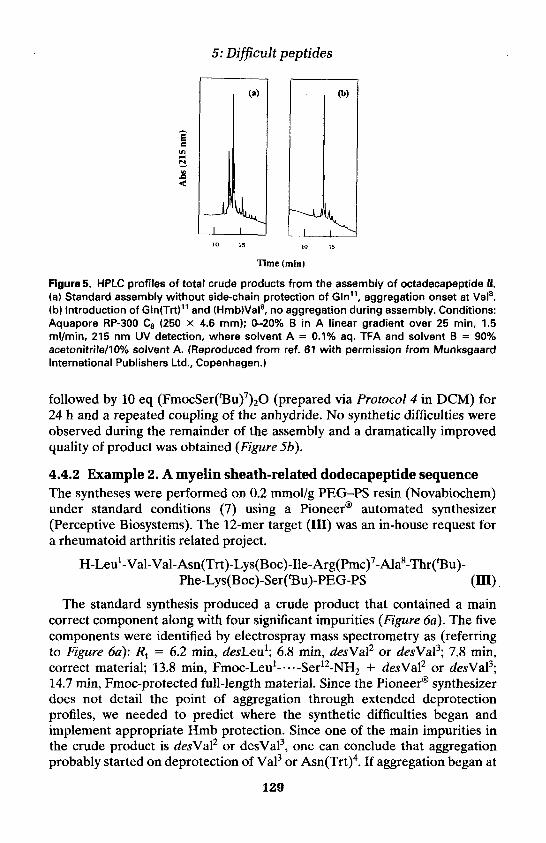
5: Difficult peptides
Figure 5. HPLC profiles of total crude products from the assembly of octadecapeptide II.(a) Standard assembly without side-chain protection of Gln11, aggregation onset at Val8.(b) Introduction of Gln(Trt)11 and (Hmb)Val8, no aggregation during assembly. Conditions:Aquapore RP-300 C8 (250 X 4.6 mm); 0-20% B in A linear gradient over 25 min, 1.5ml/min, 215 nm UV detection, where solvent A = 0.1% aq. TFA and solvent B = 90%acetonitrile/10% solvent A. (Reproduced from ref. 61 with permission from MunksgaardInternational Publishers Ltd., Copenhagen.)
followed by 10 eq (FmocSer(tBu)7)2O (prepared via Protocol 4 in DCM) for24 h and a repeated coupling of the anhydride. No synthetic difficulties wereobserved during the remainder of the assembly and a dramatically improvedquality of product was obtained (Figure 56).
4.4.2 Example 2. A myelin sheath-related dodecapeptide sequenceThe syntheses were performed on 0.2 mmol/g PEG-PS resin (Novabiochem)under standard conditions (7) using a Pioneer® automated synthesizer(Perceptive Biosystems). The 12-mer target (III) was an in-house request fora rheumatoid arthritis related project.
The standard synthesis produced a crude product that contained a maincorrect component along with four significant impurities (Figure 6a). The fivecomponents were identified by electrospray mass spectrometry as (referringto Figure 6a): Rt = 6.2 min, desLeu1; 6.8 min, desVal2 or desVal3; 7.8 min,correct material; 13.8 min, Fmoc-Leu1 -...-Ser12-NH2 + desVal2 or desVal3;14.7 min, Fmoc-protected full-length material. Since the Pioneer® synthesizerdoes not detail the point of aggregation through extended deprotectionprofiles, we needed to predict where the synthetic difficulties began andimplement appropriate Hmb protection. Since one of the main impurities inthe crude product is desVal2 or desVal3, one can conclude that aggregationprobably started on deprotection of Val3 or Asn(Trt)4. If aggregation began at
129

Martin Quibell and Tony Johnson
Figure 6. HPLC profiles of total crude products from the assembly of decadecapeptide III.(a) Standard assembly without backbone protection. (b) Introduction of (Hmb)Ala8, noaggregation during assembly. Conditions: Phenomenex C4 (250 x 4.6 mm); 0-90% B in Alinear gradient over 25 min, 1.5ml/min, 215 nm UV detection, where solvent A = 0.1% aq.TFA and solvent B = 90% acetonitrile/10% solvent A.
Lys(Boc)5, then one would expect to see a significant desAsn4 product. Thislater position for aggregation onset, at residue 3 or 4, is consistent with theinhibiting effect exhibited by Arg(Pmc)7 (cf. Figure 4d, Arg(Pmc) behaves ina similar manner to Arg(Mtr)). Thus Hmb protection needs to be im-plemented between Lys(Boc)5 and Ala8 in order to be effective for theremainder of the synthesis. The three available options are betweenAsn(Trt)4 and Lys(Boc)5, Lys(Boc)5 and Ile6, or Arg(Pmc)7 and Ala8. Each ofthese options is reasonable, thus the decision point is simply based on thebond with the least steric constraints and the easiest (and therefore leastprecious) Hmb residue to prepare.
The assembly was repeated using (Hmb)Ala8, coupled via 3 eq Fmoc-N-(FmocHmb)Ala-OPfp/HOBt onto H-Thr(tBu)9—PEG-PS in 4 h. This wasfollowed by 10 eq FmocArg(Pmc)7)2O (prepared via Protocol 4 in DCM) for12 h. No synthetic difficulties were observed during the remainder of theassembly and a substantially improved quality of product was obtained(Figure 6b).
4.4.3 Example 3. Acyl carrier protein, ACP-(65-74)This decapeptide sequence (IV) is a classic difficult peptide synthesis that hasbeen used to assess the performance of new coupling procedures (8),additives (67), resin supports (35), backbone protection (60), etc.
130
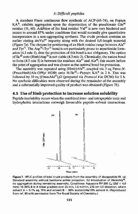
5: Difficult peptides
A standard Fmoc continuous flow synthesis of ACP-(65-74), on PepsynKAR, exhibits aggregation upon the deprotection of the penultimate Gln66
residue (51, 60). Addition of the final residue Val65 is now very hindered andoccurs to around 85% under conditions that would normally give quantitativeincorporation in a non-aggregating synthesis. The crude product contains anearlier eluting desVal65 impurity along with the desired full-length material(Figure 7a). The choices for positioning of an Hmb residue range between Ala67
and Tyr71. The Asp70-Tyr71 bond is not particularly prone to aspartimide form-ation (4.3 rule 4), thus the protection of this bond is not obligatory. The optionof Ile69 onto (Hmb)Asp70 is not viable (4.3 rule 3). Chemically, the easiest bondto form (4.3 rule 5) is between the residues Ala67 and Ala68; this occurs beforethe point of aggregation and was chosen as the optimal bond for protection.
The assembly was repeated using (Hmb)Ala68, coupled via 3 eq Fmoc-N-(FmocHmb)Ala-OPfp/ HOBt onto H-Ile69—Pepsyn KAR in 2 h. This wasfollowed by 10 eq (FmocAla67)2O (prepared via Protocol 4 in DCM) for 1 h.No synthetic difficulties were observed during the remainder of the assemblyand a substantially improved quality of product was obtained (Figure 7b).
4.5 Use of Hmb protection to increase solution solubilityPeptide insolubility occurs when the combined inter- and intrapeptide ionic andhydrophobic interactions outweigh favourable peptide-solvent interactions.
Figure 7. HPLC profiles of total crude products from the assembly of decapeptide IV. (a)Standard assembly without backbone amide protection, (b) Introduction of (Hmb)Ala68,no aggregation during remaining assembly. Conditions: Aquapore RP-300 C8 (250 x 4.6mm); 10-50% B in A linear gradient over 25 min, 1.5 ml/min, 215 nm UV detection, wheresolvent A = 0.1% aq. TFA and solvent B = 90% acetonitrile/10% solvent A. (Reproducedfrom ref. 60 with permission from The Royal Society of Chemistry.)
131

Martin Quibell and Tony Johnson
As described in the preceding discussions, when this situation arises within aresin-bound peptide chain, aggregation occurs. When this occurs in solution,individual peptide chains associate leading to precipitation, the majorinteraction being interchain hydrogen bonding, mediated primarily via thesecondary amide bonds along the peptide backbone. As detailed earlier,Narita and co-workers (32, 48, 49, 68) have shown that these precipitates arean ordered B-structure by FT-IR. We have shown through light microscopyand X-ray diffraction that the hydrophobic 3-amyloid-(34-42) C-terminalforms ordered precipitates in solution (69).
The key to solubilizing such sequences is the elimination of ordered B-structures. Narita et al. (49) and Blaakmeer et al. (52) have described thechain dissociating and powerful solubilizing effects gained through protectionof an amide bond in solution with the 2,4-dimethoxybenzyl (Dmob)-protecting group. Retention of the structurally similar Hmb amide-protectinggroup shows the same solubilizing effect on an otherwise free peptide insolution. However, the Hmb group was initially designed to be labile to tri-fluoroacetic acid. Thus, an Fmoc/t-butyl protection-based solid phasesynthesis cannot be directly cleaved to the free peptide whilst still retainingHmb protection. This may, however, be achieved by attenuation of the acidlability of Hmb, in a fully reversible manner, by simple acetylation of the 2-hydroxyl functionality (70).
This protocol was successfully applied in the preparation and purification ofthe poorly soluble protein 3-amyloid-(1-43) (63). The sequence was assembledwith Hmb protection of Gly25'29'33'37 and Phe20 to eliminate the potential forresin-bound aggregation. Simple acetylation of the Hmb 2-hydroxyl moieties,mediated by acetic anhydride and diisopropylethylamine in DMF, andsubsequent TFA cleavage produced the crude intermediate B-amyloid-[l-43,(AcHmb)Phe20, (AcHmb)Gly25,29,33,37]. This backbone-protected intermediateexhibited excellent solubility at > 150 mg/ml in a mixture of 0.1% aq. TFA/acetonitrile (1:1) allowing straightforward purification by standard reverse-phase HPLC techniques (63). The equivalent sequence without backbone pro-tection is very poorly soluble in aqueous acetonitrile solutions and extremelydifficult to purify by standard HPLC techniques. Following purification, theAcHmb-protected intermediate was treated with 5% hydrazine-hydrate inDMF to de-O-acetylate, restoring the Hmb group and TFA lability.
The retention of Hmb backbone protection on fully protected peptidesegments in solution also gives extraordinarily soluble materials (71, 72).Backbone-protected segments are readily purified by standard reverse-phaseHPLC and subsequently couple smoothly and cleanly in either DMF or DCMdue to the highly concentrated coupling solutions prepared.
References1. Merrifield, R. B. (1962). Fed. Proc. Fed. Am. Soc. Exp. Biol, 21, 412.2. Sheppard, R. C. (1988). Chem. Br., 557, and references cited therein.
132

5: Difficult peptides
3. Fields, G. B. and Noble, R. L. (1990). Int. J. Peptide Protein Res., 35, 161.4. Kent, S. B. H. (1984). In Peptides: structure and function (ed. V. J. Hruby and
D. H. Rich), p. 99. Pierce Chemical Co., Rockford, IL.5. Kent, S. B. H. and Merrifield, R. B. (1983). Int. J. Pept. Prot. Res., 22, 57.6. Gross, E. and Meienhofer, J. (ed.) (1981). The peptides, analysis, synthesis,
biology, Vol. 3. Academic Press, New York.7. Atherton, E. and Sheppard, R. C. (1989). In Solid phase peptide synthesis: a
practical approach, p. 1. Oxford University Press, UK.8. Kent, S. B. H. (1985). In Peptides, structure and function; Proceedings of the 9th
American Peptide Symposium (ed. C. M. Deber., V. J. Hruby, and K. D. Kopple),p. 407. Pierce Chemical Co., Rockford, IL.
9. Atherton, E, and Sheppard, R. C. (1985). In Peptides, structure and function;Proceedings of the 9th American Peptide Symposium (ed. C. M. Deber., V. J.Hruby, and K. D. Kopple), p. 415. Pierce Chemical Co., Rockford, IL.
10. Kaiser, E., Colescott, R. L., Bossinger, C. D., and Cook, P. I. (1970). Analyt.Biochem., 34, 595.
11. Merrifield, R. B. and Littau, V. (1969). In Peptides 1968, p. 179. North-HollandPublishing Co., Amsterdam.
12. Kent, S. B. H., Alewood, D., Alewood, P., Baca, M., Jones, A., and Schnollzer, M.(1992). In Innovations and perspectives in solid phase synthesis; 2nd InternationalSymposium (ed. R. Epton), p. 1. Intercept Ltd., Andover, UK.
13. Sheppard, R. C. (1973). In Peptides 1971; Proceedings of the 7th European PeptideSymposium (ed. H. Nesvadba), p. 111, North-Holland Publishing Co., Amsterdam.
14. Atherton, E., Clive, D. L. J., and Sheppard, R. C. (1975). /. Am. Chem. Soc., 97,6584.
15. Hancock, W. S., Prescott, D. J., Vagelos, P. R., and Marshall, G. R. (1973). J. Org.Chem., 38, 774.
16. Atherton, E. and Sheppard, R. C. (1974). In Peptides 1974 (ed. W. Wolmer),p. 123. Wiley, New York.
17. Nguyen, O. and Sheppard, R. C. (1989). In Proceedings of the 20th EuropeanPeptide Symposium (ed. G. Jung), p. 151. Walter de Gruyter, Berlin, Germany.
18. Atherton, E., Fox, H., Harkiss, D., Logan, C. J., Sheppard, R. C., and Williams, B.J. (1978). J. Chem. Soc., Chem. Commun., 537.
19. Atherton, E., Logan, C. J., and Sheppard, R. C. (1981). J. Chem. Soc., Perkin.Trans. 1, 538.
20. Dryland, A. and Sheppard, R. C. (1986). /. Chem. Soc.. Perkin Trans 1,125.21. Atherton, E. and Sheppard, R. C. (1986). J. Chem. Soc.. Chem. Commun., 165.22. Dryland, A. and Sheppard, R. C. (1988). Tetrahedron, 44, 859.23. Due Larsen, B. and Holm, A. (1994). Int. J. Pept. Prot. Res., 43, 1.24. Rodionov, I. L., Baru, M. B., and Ivanov, V. T. (1992). In Innovations and
perspectives in solid phase synthesis; 2nd International Symposium (ed. R. Epton),p. 449. Intercept Ltd., Andover, UK.
25. Rodionov, I. L., Baru, M. B., and Ivanov, V. T. (1992). In Innovations andperspectives in solid phase synthesis, 2nd International Symposium (ed. R. Epton),p. 455. Intercept Ltd., Andover, UK.
26. Beyermann, M., Klose, A., Wenschuh, H., and Bienert, M. (1993). In Peptides1992; Proceedings of the 22nd European Peptide Symposium (ed. C. H. Schneiderand A. N. Eberle), p. 263. Escom, Leiden, Netherlands.
133

Martin Quibell and Tony Johnson
27. Tam, J. P. and Lu, Y.-A. (1995). /. Am. Chem. Soc., 117, 12058.28. De L. Milton, R. C., Milton, S. C. F., and Adams, P. A. (1990). J. Am. Chem. Soc.,
112, 6039.29. Due Larsen, B., Holm, A., Christensen, D. H., Werner, F., and Faurskov, N.
(1992). In Innovations and perspectives in solid phase synthesis; 2nd InternationalSymposium (ed. R. Epton), p. 363. Intercept Ltd., Andover, UK.
30. Toniolo, C., Bonara, G. M., Mutter, M., and Maser, F. (1983). /. Chem. Soc.,Chem. Commun., 1298, and references cited therein.
31. Narita, M., Honda, S., Umeyama, H., and Ogura, T. (1988). Bull. Chem. Soc. Jpn.,61, 1201.
32. Narita, M., Ishikawa, K., Nakano, H., and Isokawa, S. (1984). Int. J. Pept. Prot.Res., 24, 14.
33. Mapelli, C. and Swerdloff, M. D. (1991). In Peptides 1990; Proceedings of the 21stEuropean Peptide Symposium (ed. E. Giralt and D. Andreu), p. 316. Escom,Leiden, Netherlands.
34. Barany, G., Sole, N. A., Van Abel, R. J., Albericio, F., and Selsted, M. E. (1992).In Innovations and perspectives in solid phase synthesis; 2nd InternationalSymposium (ed. R. Epton), p. 29. Intercept Ltd., Andover, UK.
35. Meldal, M. (1992). Tetrahedron Lett. 33, 3077.36. Klis, W. A. and Stewart, J. M. (1990). In Peptides: chemistry, structure and biology;
Proceedings of the 11th American Peptide Symposium (ed. J. E. Rivier and G. R.Marshall), p. 904. Escom, Leiden, Netherlands.
37. Thaler, A., Seebach, D., and Cardinaux, F. (1991). Helv. Chim. Acta., 74, 628.38. Zhang, L., Goldammer, C., Henkel, B., Zuhl, F., Jung, G., and Bayer, E. (1994). In
Innovations and perspectives in solid phase synthesis; 3rd International Symposium(ed. R. Epton), p. 711. Mayflower Worldwide Ltd., Birmingham, UK.
39. Yamashiro, D., Blake, J., and Li, C. H. (1976). Tetrahedron Lett., 18, 1469.40. Milton, S. C. F. and De L. Milton, R. C. (1990). Int. J. Pept. Prot. Res., 36, 193.41. Fields, G. B. and Fields, C. G. (1991). /. Am. Chem. Soc., 113, 4202.42. Hyde, C. B., Johnson, T., and Sheppard, R. C. (1992). /. Chem. Soc.. Chem.
Commun., 1573.43. Van Woerkom, W. J. and Van Nispen, J. W. (1991). Int. J. Pept. Prot. Res., 38, 103.44. Krchnak, V., Flegelova, Z., and Vagner, J. (1993). Int. J. Pept. Prot. Res., 42,
450.45. Krchnak, V. and Vagner, J. (1992). In Innovations and perspectives in solid phase
synthesis; 2nd International Symposium (ed. R. Epton), p. 419. Intercept Ltd.,Andover, UK.
46. Steinschneider, A. (1994). Int. J. Pept. Prot. Res., 44, 49.47. Chou, P. Y. and Fasman, G. D. (1978). In Advances in enzymology (ed. A.
Meister), Vol. 47, p. 45. Wiley, New York.48. Narita, M., Ishikawa, K., Chen, J.-Y., and Kim, Y. (1984). Int. J. Pept. Prot. Res.,
24, 580.49. Narita, M., Fukunaga, T., Wakabayashi, A., Ishikawa, K. and Nakano, H. (1984).
Int. J. Pept. Prot. Res., 24, 306.50. Merrifield, R. B. (1988). Paper presented at the EURCHEM Conference on New
trends in peptide synthesis, Port Camargue, France.51. Bedford, J., Hyde, C., Johnson, T., Jun, W., Owen, D., Quibell, M., and Sheppard,
R. C. (1992). Int. J. Pept. Prot. Res., 40, 300.
134

5: Difficult peptides
52. Blaakmeer, J., Tijsse-Klasen, T., and Tesser, G. I. (1991). Int. J. Pept. Prot. Res.,37, 556.
53. Johnson, T., Quibell, M., and Sheppard, R. C. (1995). J. Pept. Sci., 1, 11.54. Nicolas, E., Pujades, M., Bacardit, J., Giralt, E., and Albericio, F. (1997).
Tetrahedron Lett., 38, 2317.55. Zeng, W., Regamey, P.-O., Rose, K., Wang, Y., and Bayer, E. (1997). Int. J. Pept.
Prot. Res., 49, 273.56. Quibell, M. and Johnson, T. (1993). Unpublished observations.57. Offer, J., Johnson, T., and Quibell, M. (1997). Tetrahedron Lett., 38, 9047.58. Fuller, W. D., Cohen, M. P., Shabankarech, M., Blair, R. K., Goodman, M., and
Naider, F. R. (1990). J. Am. Chem. Soc., 112, 7414.59. Kaduk, C., Wenschuh, H., Beyermann, M., Forner, K., Carpino, L. A., and
Bienert, M. (1995). Lett. Pept. Sci., 2, 285.60. Johnson, T. J., Quibell, M., Owen, D., and Sheppard, R. C. (1993). J. Chem. Soc.,
Chem. Commun., 4, 369.61. Hyde, C., Johnson, T. J., Owen, D., Quibell, M., and Sheppard, R. C. (1994). Int. J.
Pept. Prot. Res., 43, 431.62. Quibell, M., Owen, D., Packman, L. C., and Johnson, T. J. (1994). J. Chem. Soc.,
Chem. Commun., 2343.63. Quibell, M., Turnell, W. G., and Johnson, T. (1995). J. Chem. Soc., Perkin Trans.
7, 2019.64. Johnson, T., Packman, L. C., Hyde, C. B., Owen, D., and Quibell, M. (1995). J.
Chem. Soc,. Perkin Trans. 1, 719.65. Simmonds, R. G. (1996). Int. J. Pept. Prot. Res., 47, 36.66. Packman, L. C., Quibell, M., and Johnson, T. (1994). Pept. Res., 7(3), 125.67. Hyde, C., Johnson, T., and Sheppard, R. C. (1992). /. Chem. Soc., Chem.
Commun., 21, 1573.68. Narita, M., Ogura, T., Sato, K., and Honda, S. (1986). Bull. Chem. Soc. Jpn., 59,
2433.69. Quibell, M., Johnson, T., and Turnell, W. G. (1994). Biomed. Pept. Proteins
Nucleic Acids, 1, 3.70. Quibell, M., Turnell, W. G., and Johnson, T. (1994). Tetrahedron Lett., 35, 2237.71. Quibell, M., Packman, L. C., and Johnson, T. (1995). J. Am. Chem. Soc., 117,
11656.72. Quibell, M., Packman, L. C., and Johnson, T. (1996). J. Chem. Soc., Perkin Trans.
1, 1227.
135

This page intentionally left blank

Synthesis of modified peptidesSARAH L. MELLOR, DONALD A. WELLINGS,
JEAN-ALAIN FEHRENTZ, MARIELLE PARIS, JEAN MARTINEZ,NICHOLAS J. EDE, ANDREW M. BRAY, DAVID J. EVANS
and G. B. BLOOMBERG
1. C-terminal modifications1.1 Peptidyl N-alkyl amidesSeveral important hormones such as oxytocin, secretin, and LHRH are knownto be peptidyl amides (1-3). In addition to these, other peptidyl amides suchas indolicidin and the protegrins have been shown to exhibit potent anti-microbial activity (4, 5). The in vivo production of such compounds is viaendogenous enzymatic cleavage of propeptides (6), making their synthesis bygenetic engineering notoriously difficult. Furthermore, to facilitate the survivalof synthetic peptidyl amides in vivo, an obvious defence against the action ofcarboxypeptidases is the N-alkylation of the carboxylic amide terminus. Suchsecondary amides would be expected to exhibit vastly different solubility andtransport properties to primary amides, thus their chemical synthesis is ofimmense importance.
1.1.1 Synthesis of peptidyl N-alkyl amidesThe solid phase synthesis of peptidyl amides and peptidyl N-alkyl amides iscentred around two main strategies:
1. Ammonolysis/aminolysis of resin-bound esters (Figure la; see Section 1.2).2. Use of resin-bound primary amines, which may in turn be chemically
modified to generate novel secondary amine functionalized linkers for thesynthesis of peptidyl N-alkyl amides (Figure 1b).
1.1.2 Use of 9-(fluorenylmethoxycarbonylamino)xanthen-3-yloxymethyl polystyrene and its chemical modification toresin-bound secondary amines
Early examples of the use of resin-bound amines for the solid synthesis ofpeptidyl amides involve the use of linkers such as benzhydrylamine or benzyl-amine. Following peptide assembly, these linkers require highly acidic (e.g.
6

Figure 1. (a) Ammonolysis/aminolysis of resin-bound esters. (b) Synthesis of peptidylamides and peptidyl N-alkyl amides using amine-based linkers.
HF) mediated cleavage, and hence simultaneous removal of acid-labile side-chain protection groups occurs and may cause problems.
Systematic modifications of these linker-resins by substitution with electron-donating substituents have resulted in the generation of numerous linker-resinswith increased acid sensitivity. Notably, the 4-(2',4'-dimethoxyphenylamino-methyl)phenoxy derivatized (Rink) resin 1 (7), which is cleavable by 95% v/vTFA, and the 5-(2-fluorenylmethoxycarbonylaminornethyl-3,5-dimethoxy)-phenoxyvaleric acid (PAL) linker 2 (8) acidolysed by 75% v/v TFA.
Greater acid lability has been achieved using the xanthenyl derivatizedresin, 9-(fluorenylmethoxycarbonylamino)xanthen-3-yloxymethyl polystyrene(Sieber amide) resin 3 (9,10), which besides being cleavable by 1% v/v TFA,holds the added advantage of readily undergoing reductive N-alkylation toafford resin-bound secondary amines for the synthesis of peptidyl N-alkyl
138

6: Synthesis of modified peptides
amides (11). In addition to the method detailed below (Protocol 1), a numberof alternative approaches have recently been reported (12-14).
Protocol 1.
Reagents
Synthesis and use of N-alkylated 9-aminoxanthenyl(Sieber) resin
9-(Fmoc-amino)xanthen-3-yloxymethylpolystyrene (Sieber amide) resinDCM20% v/v Piperidine in DMFDMFGlacial acetic acidTrimethylorthoformate
AldehydeSodium borohydrideMethanolN-Fmoc-amino acidHOAtHATUDIPEA
A. Synthesis of N-alkylated 9-aminoxanthenyl (Sieber) resin
Prior to undertaking the reaction, ensure that all glassware is oven-dried.
1. Pre-swell the 9-(Fmoc-amino)xanthen-3-yloxymethyl polystyrene(Sieber amide) resin (0.05 mmol, ca. 100 mg) in DCM for 1 h in apeptide synthesizer column.
2. Treat the resin with 20% v/v piperidine in DMF (10 min at 2.5 ml min-1
or 4 x 5 min batchwise).
3. Wash the resin with DMF (15 min at 2.5 ml min-1).
4. Remove any excess DMF from the resin, and ensure that the resin issuspended in 0.75 ml of DMF.
5. Add the required aldehyde (0.25 mmol) followed by glacial acetic acid(15 ul, giving a 2% v/v solution in DMF) and allow the reaction toproceed for 45 min with occasional agitation.
6. Wash the resin briefly with DMF (2 min at 2.5 ml min-1).
139

Sarah L. Mellor et al.
Protocol 1. Continued7. Repeat steps 4 and 5, or repeat step 4 and then add the required
aldehyde (0.25 mmol) followed by trimethylorthoformate (0.20 ml).
8. After a further 75-min reaction time with occasional agitation, washthe resin with DMF (5 min at 2.5 ml min-1).
9. Remove any excess DMF, and ensure that the resin is suspended in1.50 ml of DMF.
10. Add sodium borohydride (8 mg, 0.20 mmol), followed immediatelyby 0.20 ml of methanol.
11. Add further amounts of sodium borohydride (12 mg, 0.30 mmol)portionwise over a period of 30 min.
12. Gently agitate (by stirring) the reaction mixture for a further 90 min.
13. Thoroughly wash the resin with DMF (20 min at 2.5 ml min-1).
B. Acylation of N-alkylated 9-aminoxanthenyl (Sieber) resin
The synthesized 9-(N-alkyl)aminoxanthenyl resin is then acylated with anN-Fmoc-protected amino acid.
1. Remove DMF by applying a positive pressure to the resin-bedcontained in the column.
2. In a dry 5-ml round-bottomed flask or an appropriate sample-bottle, dis-solve the N-Fmoc-amino acid (0.25 mmol) in DMF (0.8 ml), and add tothe solution HOAt (0.1 mmol), HATU (0.25 mmol), and DIPEA (0.5 mmol).
3. Leave the resultant solution for 2 min, and then add to the resin.
4. Gently agitate the suspension for 18 h.
5. Wash the resin with DMF (15 min at 2.5 ml min-1).
6. The derivatized resin may be used immediately for peptide synthesisusing standard Fmoc/tBu proceduresa or collected using a Buchnerfunnel, and washed with dichloromethane (20 ml) and hexane (10 ml)before being dried in vacuo for storage.
aFollowing peptide assembly, the protected peptidyl N-alkyl amide may be cleaved from thelinker-resin by treatment with 1-2% TFA/0.1% triethylsilane in DCM (5 x 2 min, 2-3 ml for eachtreatment). If a fully deprotected peptidyl N-alkyl amide is required, it is recommended that atwo-step procedure is applied: (a) cleave the peptide from the solid support by exposure to 5%TFA/0.1% triethylsilane in DCM (10 ml) for 10 min, filter under gravity to remove resin, washwith 5% TFA in DCM (20 ml), and evaporate in vacuo the filtrate to dryness; (b) treat the residuematerial with a standard 90% TFA cocktail for complete acidolysis of side-chain protectinggroups.
Numerous resin-bound alkyl amines have been generated using thereductive alkylation technique. Their subsequent acylation with Fmoc-phenylalanine has been used to assess the effectiveness of the reaction. Using
140

6: Synthesis of modified peptides
Table 1. N-acylation efficiencies and estimated product puritiesfor N-Fmoc-phenylalanyl-N-alkyl aminoxanthen-3-yloxymethylpolystyrene resin
Alkyl group
3-Phenylpropyl3,3-DimethylbutylPropyl3-CyclohexylpropylisoButylBenzyl
% Efficiency
949999969955
% Purity
929898949898
Table 2. Reaction efficiencies and estimated product purities foracylation of N-isobutylaminoxanthen-3-yloxymethyl polystyreneresin
Amino acid
Fmoc-Asp(OtBu)-OHFmoc-Ser(OtBu)-OHFmoc-Pro-OHFmoc-Val-OH
% Efficiency
100886750
% Purity
92979494
'Fmoc-substitution' as a measure of reaction efficiency and RP-HPLC as anestimate of purity, it was established that the reaction generally generatesproducts of high purity. However, the acylation efficiency, although generallygood, is decreased markedly if the resin-bound alkyl amine is particularlysterically hindered (Table 1). Likewise, comparing the acylation of resin-boundisobutylamine using a variety of N-Fmoc-amino acids, it was also observed thatacylation efficiency decreases as the acylating species becomes more bulky(Table 2). These considerations must therefore be part of any syntheticstrategies for the solid phase construction of peptidyl N-alkyl amides.
1.2 Use of the 4-hydroxymethylbenzoic acid linkage agentfor the synthesis of C-terminal modified peptides
1.2.1 IntroductionSince the original concept for the use of linkage agents coupled separately topolyacrylamide resin was proposed by Sheppard and co-workers (15-17) ithas become commonplace to use custom-designed moieties to allow C-terminal modifications during cleavage of assembled peptides from the solidsupport. 4-Hydroxymethylbenzoic acid 4 is one of the most versatile of thelinkage agents described by Atherton et al. (18) due mainly to its susceptibilityto nucleophilic attack combined with an inertness to strong acids. Theacylated linkage agent is cleaved readily with ammonia (19) and was therefore
141

Sarah L. Mellor et al.
originally used for the preparation of peptide amides. The flexibility is en-hanced by the ability to remove side-chain protection before or after cleavageof the peptide from the resin, hence side-chain protected peptide amides canbe prepared for subsequent fragment assembly. In a complimentary fashion,the linkage can be cleaved with hydrazine to produce hydrazides for sub-sequent activation as azides, for coupling in fragment assembly. The benzylester functionality is stable to piperidine used for removal of the Fmoc group,making the linkage agent equally applicable to Boc or Fmoc chemistry.
The use of 4-hydroxymethylbenzoic acid for routine preparation of peptideamides has been somewhat superseded by acid-labile linkage agents thatliberate the amide with simultaneous cleavage of the peptide from the resin(7, 20). For preparation of peptide amides on a large scale, however, 4-hydroxymethylbenzoic acid is still favoured economically.
In more recent years, the need to create molecular diversity for peptidelibraries has resulted in somewhat of a renaissance for the 4-hydroxy-methylbenzoic acid linkage agent. The ability to cleave the peptide-to-resinester with many nucleophiles including hydroxide, alkoxides, and primaryamines has added to the library workers arsenal.
However, 4-hydroxymethylbenzoic acid is not without its drawbacks as alinkage agent. Peptides containing C-terminal proline are particularly proneto diketopiperazine formation (21) due to cleavage of the dipeptide from theresin by its own internal nucleophile. This can often be overcome by insertionof a 'Boc' amino acid as the second amino acid in the chain. Acidolyticremoval of the Boc group affords fugitive protection of the amine as a salt,which can be neutralized in situ during the next amino acid coupling. Thereactivity of other sites in the peptide towards nucleophiles should not beoverlooked when assessing the applicability of the linker to assembly of aspecific peptide (e.g. rearrangement of aspartic acid residues to aspartimide isnot uncommon).
1.2.2 Coupling 4-hydroxymethylbenzoic acid to the solid supportThe linkage agent can be coupled to amino-functionalized resin as thepreformed trichlorophenyl ester using the protocols described by Athertonand Sheppard (22) or activated in situ using diisopropylcarbodiimide and 1-hydroxybenzotriazole as described in Protocol 2. Although 4-hydroxymethyl-benzoic acid can be used with many amino-functionalized supports, it is bestused in conjunction with polyacrylamide where the solvation properties of theresin allow more facile recovery of the peptide from the pores of the supportfollowing cleavage. As with most linkage agents, it is recommended that an
142
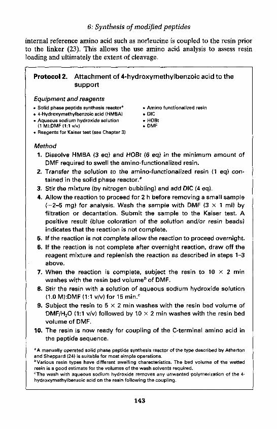
6: Synthesis of modified peptides
internal reference amino acid such as norleucine is coupled to the resin priorto the linker (23). This allows the use amino acid analysis to assess resinloading and ultimately the extent of cleavage.
Protocol 2. Attachment of 4-hydroxymethylbenzoic acid to thesupport
Equipment and reagents• Solid phase peptide synthesis reactora • Amino functionalized resin• 4-Hydroxymethylbenzoic acid (HMBA) . DIC• Aqueous sodium hydroxide solution • HOBt
(1 M):DMF(1:1 v/v) . DMF• Reagents for Kaiser test (see Chapter 3)
Method1. Dissolve HMBA (3 eq) and HOBt (6 eq) in the minimum amount of
DMF required to swell the amino-functionalized resin.2. Transfer the solution to the amino-functionalized resin (1 eq) con-
tained in the solid phase reactor.a3. Stir the mixture (by nitrogen bubbling) and add DIC (4 eq).4. Allow the reaction to proceed for 2 h before removing a small sample
(-2-5 mg) for analysis. Wash the sample with DMF (3 x 1 ml) byfiltration or decantation. Submit the sample to the Kaiser test. Apositive result (blue coloration of the solution and/or resin beads)indicates that the reaction is not complete.
5. If the reaction is not complete allow the reaction to proceed overnight.6. If the reaction is not complete after overnight reaction, draw off the
reagent mixture and replenish the reaction as described in steps 1-3above.
7. When the reaction is complete, subject the resin to 10 x 2 minwashes with the resin bed volumeb of DMF.
8. Stir the resin with a solution of aqueous sodium hydroxide solution(1.0 M):DMF (1:1 v/v) for 15 min.c
9. Subject the resin to 5 x 2 min washes with the resin bed volume ofDMF/H2O (1:1 v/v) followed by 10 x 2 min washes with the resin bedvolume of DMF.
10. The resin is now ready for coupling of the C-terminal amino acid inthe peptide sequence.
aA manually operated solid phase peptide synthesis reactor of the type described by Athertonand Sheppard (24) is suitable for most simple operations.bVarious resin types have different swelling characteristics. The bed volume of the wettedresin is a good estimate for the volumes of the wash solvents required.cThe wash with aqueous sodium hydroxide removes any unwanted polymerization of the 4-hydroxymethylbenzoic acid on the resin following the coupling.
143

Sarah L. Mellor et al.
1.2.3 Coupling the C-terminal amino acid to 4-hydroxymethylbenzoyl resin
Precautions should be taken to avoid racemization of the C-terminal aminoacid during the acylation reaction. Coupling with the carboxylate salt of thefirst amino acid to the halogenated linker is now popularly employed andmany other methods have also been described to reduce enantiomerformation.
The procedure described in these protocols, using DIC for activation anda catalytic amount of the hypernucleophile 4-dimethylaminopyridine(DMAP) for ester bond formation, reduces racemization down to amountsthat are not detectable if the conditions described in Protocol 3 are strictlyadhered to.
Protocol 3. Coupling the C-terminal amino acid to 4-hydroxymethylbenzoyl resin
Equipment and reagents• Solid phase peptide synthesis reactora • DIC• 4-hydroxymethylbenzoyl resin • DMAP• Fmoc-amino acid (Fmoc-AA-OH) • DMF• Acetic anhydride (Ac20)
Method
1. Place the 4-hydroxymethylbenzoyl resin (1 eq) in the solid phasereactor, followed by the Fmoc-AA-OH to be coupled (3 eq).
2. Add sufficient DMF to make the resin just mobile to nitrogenagitation.
3. Add DIC (4 eq), following this with the dropwise addition of a solutionof DMAP dissolved in DMF (0.1 eq, ca. 50 mmol/l).
4. Allow the reaction to proceed for 60 min, draw off the reactionsolution and subject the resin to 2 X 2 min washes with the resin bedvolume of DMF.
5. Recouple the amino acid as in steps 1-3.
6. Subject the resin to 5 x 2 min washes with the resin bed volumeb ofDMF.
7. To acetylate any remaining hydroxyl functions, add sufficient DMF tomake the swollen resin just mobile to nitrogen agitation.
8. Add Ac2O (6 eq) and DMAP (0.1 eq).
9. After 1 h draw off the reaction solution and wash the resin with 10resin bed volumes of DMF.
144

6: Synthesis of modified peptides
10. Use the Fmoc-amino acid derivatized resin for peptide assembly, orwash with DCM (20 ml) and methanol (10 ml), and dry in vacuo for 2 hprior to storage.
aA manually operated solid phase peptide synthesis reactor of the type described by Athertonand Sheppard (24) is suitable for most simple operations.bVarious resin types have different swelling characteristics. The bed volume of the wettedresin is a good estimate for the volumes of the wash solvents required.
1.2.4 Side-chain deprotection following peptide-resin assemblyStandard procedures for Fmoc-based peptide chain elongation are describedelsewhere in this text (Chapter 3). To produce the required peptide as theunprotected molecule, side-chain deprotection is best carried out prior tocleavage of the peptide from the resin. Selection of the appropriate mixture oftrifluoroacetic acid and scavengers for side-chain deprotection is described inChapter 3, Section 10.
Protocol 4. Side-chain deprotection of the peptide-resin
Equipment and reagents• Solid phase peptide synthesis reactora • TFA• Trifluoroacetic acid/scavenger cocktail • DCM
(Deprotection mixture) • DMF• Diethyl ether
Method1. Place the peptidyl resin in the solid phase reactor and add approxi-
mately twice the swollen resin bed volume of deprotection mixture.2. Agitate the mixture and allow the reaction to proceed for the required
amount of time.b
3. Draw off the deprotection mixture and subject the resin to 3 x 2 minwashes with the resin bed volume of TFA.
4. Subject the resin to 2 min washes with the resin bed volume of DCM(3 times) and Et2O (3 times).
5. Air dry the side-chain deprotected peptidyl resin overnight prior tocleavage.
a A manually operated solid phase peptide synthesis reactor of the type described by Athertonand Sheppard (24) is suitable for most simple operations.bThe reaction time and deprotection mixture is described in Chapter 3, Section 10.
1.2.5 Cleavage of peptide from the resin using gaseous ammoniaThe cleavage procedure for removal of the peptide from the resin as theamide will differ slightly depending on the nature of the solid support.
145

Sarah L. Mellor et al.
Figure 2. Pressure vessel layout.
Polystyrene-based matrices require wetting with an organic solvent to allowcomplete cleavage and often require washing with organic solvents such asDMF to completely extract the peptide from the resin. Peptides can becleaved from polyacrylamide based supports in the gas phase due to the easeof penetration of the pores of the peptide-resin matrix with ammonia. Theadvantage of this approach is that the peptide can often be washed off theresin using water or aqueous solvents. The procedure described in Protocol 5is equally applicable to other gaseous primary amines such as methylamineand ethylamine.
The reaction should be carried out in a suitable pressure vessel with asimilar layout to that described in Figure 2. A key feature of this equipment isthe pressure release valve to prevent over-pressurizing the vessel.
Protocol 5. Cleavage of peptide from the resin with gaseousammonia
Equipment and reagents• Stainless steel pressure vessel" • Dry-Ice• Peptide-resin for cleavage • Water• Ammonia cylinder • DMF
Method1. Place the peptide-resin in the pressure vessel within a suitable
container (e.g. crystallizing dish).2. After ensuring that the ammonia valve is closed, open the vacuum
valve and evacuate the vessel.b
3. Close off the vacuum valve, open the ammonia valve and open theammonia supply.
146

6: Synthesis of modified peptides
4. Cool the base of the vessel in dry-ice for 20-30 min to allow ammoniato condense in the pressure vessel.
5. Close off the ammonia valve and ammonia supply.6. Remove the vessel from the dry-ice and allow the reaction to proceed
overnight at room temperature.
7. Cool the base of the vessel in dry-ice for 20-30 min to allow ammoniato condense in the base of the pressure vessel.
8. Purge the ammonia from the system by passing nitrogen into thevessel through one valve and scrubbing the exhaust via the othervalve.
9. Remove the mixture of peptide and resin from the pressure vesseland transfer to a glass sinter.
10. Subject the resin to 3 x 5 min washes of an appropriate solventc forthe peptide.
11. Recover the peptide from the solvent by standard procedures.
aA simple pressure vessel with a 100 psi pressure release valve is suitable. These can bepurchased from Parr Ltd.b A high vacuum is not required.cFor protected peptides DMF is often suitable. Aqueous MeCN is suitable for most deprotectedpeptides.
1.2.6 Cleavage of peptide from the resin using hydrazinePeptide hydrazides are readily converted to the corresponding acylazides bythe action of alkyl nitrites and under appropriate conditions these activatedspecies can be among the least susceptible to racemization. Cleavage of thepeptide from the resin as the hydrazide is most useful with protectedmolecules, which can subsequently be used in fragment condensation toassemble longer peptides.
Protocol 6. Cleavage of peptide from the resin with hydrazinehydrate
Equipment and reagents• Solid phase peptide synthesis reactora • Peptide-resin for cleavage• Rotary evaporator • Hydrazine hydrate. DMF . Water• Diethyl ether
Method
1. Place the peptide-resin in a solid phase peptide synthesis reactor.a
2. Add the minimum amount of DMF required to make the resin mobileto N2 agitation.
147
Sarah L. Mellor et al.
Protocol 6. Continued
3. Add hydrazine hydrate (equivalent to 5% v/v of the DMF added above)and allow the reaction to proceed for 15 min.
4. Filter off the hydrazine hydrate solution which now contains thepeptidyl hydrazide and subject the resin to 3 x 2 min washes with theresin bed volumeb of DMF.
5. Reduce the combined hydrazine hydrate solution and DMF washes toan oil by rotary evaporation.
6. Precipitate protected peptidyl hydrazide by addition of water, or in thecase of side-chain deprotected molecules precipitate by addition ofEt20.
7. Recover the peptide by filtration and dry under vacuum.
aA manually operated solid phase peptide synthesis reactor of the type described by Athertonand Sheppard (24) is suitable for most simple operations.bVarious resin types have different swelling characteristics. The bed volume of the wettedresin is a good estimate for the volumes of the wash solvents required.
1.2.7 Cleavage of peptide from the resin by trans-esterification withalcohols
Cleavage of the peptide from the resin by trans-esterification can be achievedreadily with most alcohols. The resultant C-terminal-protected molecules canbe used in fragment condensation to assemble longer peptides. The protocoldescribed here involves cleavage with methanol in the presence of a tertiarybase. The methyl ester produced can subsequently be removed bysaponification.
Protocol 7. Cleavage of peptide from the resin by trans-esterification with methanol
Equipment and reagents• Solid phase peptide synthesis reactora • Peptide-resin for cleavage• Rotary evaporator • Methanol• DMF • Water• Diethyl ether • DIPEA
Method
1. Place the peptide-resin in a solid phase peptide synthesis reactor.a
2. Add the minimum amount of DMF/MeOH (1:1 v/v) required to makethe resin mobile to N2 agitation.
148

6: Synthesis of modified peptides
3. Add DIPEA (equivalent to 10% v/v of the DMF/MeOH added above) andallow the reaction to proceed for 1 h.
4. Filter off the DMF/MeOH solution that now contains the peptide methylester and subject the resin to 3 x 2 min washes with the resin bedvolumeb of DMF.
5. Reduce the combined DMF/MeOH solution and DMF washes to an oilby rotary evaporation.
6. Precipitate protected peptides by addition of water, or in the case ofside-chain deprotected molecules precipitate by addition of Et2O.
7. Recover the peptide by filtration and dry under vacuum.
aA manually operated solid phase peptide synthesis reactor of the type described by Athertonand Sheppard (24) is suitable for most simple operations.bVarious resin types have different swelling characteristics. The bed volume of the wettedresin is a good estimate for the volumes of the wash solvents required.
1.3 Peptide hydroxamic acids1.3.1 IntroductionPeptide hydroxamates or hydroxamic acids have been shown to be potentenzyme inhibitors. Their target enzymes, the matrix metalloproteinases(MMPs), are a family of endopeptidases with a catalytic domain that houses azinc atom as a coenzyme. The ability of the hydroxamate functionality tochelate such metal ions (25) and thus disrupt coenzyme activity is believed tobe the reason for inhibitory activity.
A wide variety of matrix metalloproteinases have been shown to be sus-ceptible to inhibition by a number of specific peptide hydroxamate basedinhibitors, for example foroxymithine (Figure 3d) has been shown to inhibitangiotensin-converting enzyme (26), and propioxatin A (Figure 3b) acts againstenkephalinase B (27), an enzyme which hydrolyses the body's naturallyoccurring analgesic agents. Furthermore, matlystatin B (Figure 4) inhibits
Figure 3. (a) Foroxymithine and (b) propioxatin A.
149

Sarah L. Mellor et al.
type IV collagenases (28), recently found to be secreted in large amounts bymetastatic tumour cells. Thus, inhibition of such enzymes has importantconsequences for the relief of pain and the control of a variety of life-threatening diseases, ensuring that peptide hydroxamates play a pivotal rolein therapeutic strategies.
1.3.2 Synthesis of peptide hydroxamatesMuch of the research carried out on peptide hydroxamates has involved theirisolation from micro-organisms or biological tissues, but to isolate sufficientquantities of pure material for biological testing by this method can involvemanipulating large volumes of solvent and can be difficult and time-consuming.
Specific examples of the total synthesis of a number of peptide hydroxa-mates in solution have been reported, for example matlystatin B (28) (Figure 4),but a more generic method involves the reaction of peptidyl succinimide esterwith hydroxylamine hydrochloride (Figure 5).
Solid phase strategyThe solid phase synthesis of peptide hydroxamates encompasses two mainstrategies. The first, the transamination of a resin-bound peptidyl ester usinghydroxylamine hydrochloride, is restricted to a limited number of examples,e.g. tepoxalin (29) (Figure 6). The second strategy involves attaching an N-protected hydroxylamine to the resin via the hydroxyl functionality,deprotecting and using the new hydroxylamine linker as a handle for peptidesynthesis (Figure 7).
Figure 4. Retro-synthetic analysis of matlystatin B.
Figure 5. Generation of a peptidyl hydroxamic acid from a succinimide ester.
150

6: Synthesis of modified peptides
Figure 6. Solid phase synthesis of tepoxalin.
Figure 7. Strategy for the generation of resin-bound hydroxylamine.
Two different N-protected hydroxylamines have been used to accomplishthe synthesis of these novel hydroxylamine-functionalized resins. N-Hydroxy-phthalimide has been employed in two ways: first, as a nucleophile to displaceeither a resin-bound chloride ion (30) or a resin-bound mesylate ion (31)(Figure 8a); and second, to generate the active phosphonium species requiredto perform a Mitsunobu reaction on a hydroxyl-functionalized resin (32)(Figure 8b). In both cases, the resin-bound N-hydroxyphthalimide ester that isgenerated is subsequently treated with hydrazine to afford resin-boundhydroxylamine.
To negate the need to use the harsh and potentially hazardous deprotectionreagent hydrazine, a superior alternative, which is particularly compatible foruse in Fmoc/tBu solid phase synthesis, is N-Fmoc-hydroxylamine, which canbe used to effect a nucleophilic displacement reaction of 2-chlorotritylchloride resin (33) (Figure 9). The resultant N-Fmoc-aminooxy-2-chlorotritylpolystyrene resin can be used for the synthesis of peptide hydroxamates asdescribed in Protocol 8.
Figure 8. Synthesis of a resin-bound N-hydroxyphthalimide ester via (a) displacement ofa mesylate or chloride ion or (b) a Mitsunobu reaction, followed by hydrazinolysis togenerate a resin-bound hydroxylamine.
151

Sarah L. Mellor et al.
Figure 9. Synthesis of N-Fmoc-aminooxy-2-chlorotrityl polystyrene resin.
Protocol 8. N-Fmoc-aminooxy-2-chlorotrityl polystyrene resin forthe synthesis of peptide hydroxamic acids
Reagents• N-Fmoc-aminooxy-2-chlorotrityl polystyrene resin • DCM. Piperidine in DMF (20% v/v) • DMF
Fmoc-amino acid • HOAt. HATU . DIPEA
Method1. Place N-Fmoc-aminooxy-2-chlorotrityl polystyrene resin (0.05 mmol)
in a peptide synthesizer column, and pre-swell in DCM for 24 h.2. Treat the resin with 20% v/v piperidine in DMF (10 min at 2.5 ml min-1,
or using batchwise treatment), followed by DMF (15 min at 2.5 mlmin-1), after which excess DMF is expelled from the column.
3. In a dry 5-ml round-bottomed flask or an appropriate glass samplebottle, dissolve the Fmoc-amino acid (0.4 mmol) in 1.0 ml DMF, andadd HOAt (0.4 mmol), HATU (0.4 mmol), and DIPEA (0.8 mmol).a
4. Allow the activation to proceed for 1 min.5. Add the reaction mixture to the resin.6. Gently agitate the reaction suspension for 24 h at room temperature.7. Wash the resin with DMF (10 min at 2.5 ml min-1).8. Use the Fmoc-amino acid derivatized aminooxy-2-chlorotrityl poly-
styrene for peptide assembly, using standard Fmoc/tBu procedures.9. Following peptide assembly, collect the peptide-resin in a Buchner
funnel, wash with DCM (20 ml) and hexane (10 ml), and dry in vacuofor 1 h.
10. Treat the peptide-resin with standard 90% TFA cocktail.11. Recover the peptide hydroxamic acid using standard procedures.
aThe HOAt/HATU-mediated acylation used for the addition of the first amino acid has beenseen to generate good reproducible results, for example with Fmoc-phenylalanine the 'Fmoc-substitution' of the resultant A/-Fmoc-phenylalanyl-aminooxy-2-chlorotrityl polystyrene resinindicated a 94% reaction efficiency, compared with the 100% efficiency obtained when usingthe corresponding amino acid fluoride.
152

6: Synthesis of modified peptides
1.4 Peptide aldehydes by solid phase synthesis1.4.1 IntroductionPreparation of peptide aldehydes has been reported in numerous papersbecause these compounds present two main interests. First, they can be usedfor further chemistry (formation of reduced bond, Wittig reactions, ligation,etc.), and second, they have been found to be potential inhibitors of severalclasses of enzymes such as serine proteases (34, 35), prohormone convertases(36), cysteinyl proteases (34, 37, 38), and aspartyl proteases (39, 40). These in-hibitory properties result from the tetrahedral hydrated C-terminus aldehydicfunction, which mimics the transition state of the substrate hydrolysis.
Various methods for the solution synthesis of peptide aldehydes have beendescribed: (i) the corresponding peptide alcohol can be oxidized to thealdehyde (41, 42)—if the desired sequence does not contain other alcoholfunctions; (ii) reduction of the corresponding methyl esters using diiso-butylaluminium hydride (43); (iii) arginal analogue syntheses, important instudies of their anticoagulant, antithrombotic properties, have been reported,including the use of 8-lactam (35, 44, 45) and semicarbazone derivatives (34).A particular synthesis of an inhibitor of interleukin-lp converting enzyme wasdescribed by Chapman (37), in which the C-terminus aldehydic residue of thiscompound is an aspartyl residue: the aspartyl aldehyde moiety was protectedas the corresponding O-benzylacylal which could be coupled and thenhydrogenolysed to afford the desired compound. Moreover, a recent reportmentioned the use of thiazolidines as aldehyde precursors (46). Until recently,only one publication (47) concerned solid phase synthesis, this method beingderived from a solution preparation of aldehydic peptides (48).
We have focused our attention on the solid phase synthesis of such com-pounds and described our results here. Alternative routes for the preparationof peptide aldehydes and side-chain protected peptide aldehydes in solid phasesynthesis are described. Three new linkers that are stable under classical Fmocor Boc strategies have been developed to obtain the peptide aldehyde from thesolid support. One of these linkers was conceptualized on the basis of theWeinreb amide (49) and the other on the basis of phenolic esters (50). Bothstrategies required the reduction with hydrides of the peptide-linker-resin to re-lease the peptidic aldehydic function. The use of these two different approacheswas demonstrated by the synthesis of N-protected a-amino-aldehydes andpeptide aldehydes. The third approach used the ozonolysis reaction for thegeneration of the desired aldehyde. This concept requires a linker incorporat-ing a double bond in the a-position of the asymmetric carbon of the C-terminalresidue that will be cleaved by ozone to produce the carbonyl function.
1.4.2 Weinreb amide-based linkerAmong all the described preparations of N-protected amino aldehydes andpeptide aldehydes, the reduction of Weinreb amides is used most widely. This
153

Sarah L. Mellor et al.
method has been applied successfully to the synthesis of N-protected a-amino-aldehydes and peptide aldehydes using Z, Boc and Fmoc chemistry.On the basis of this stable intermediate, it was reasonable to apply thisstrategy in the solid phase synthesis of peptide aldehydes. This was done bythe synthesis of a linker (51) that incorporates the methoxyamino func-tionality. This linker, N-(tert-butoxycarbonyl)-Af-methoxy-3-aminopropionicacid, was coupled to a solid support (i.e. MBHA resin), allowing after Boc-deprotection peptide elongation by either classical Boc or Fmoc strategy onthe solid support.
This methodology was validated by the synthesis of Boc-Ala-H and Boc-Phe-H; these compounds were purified on a silica gel column according to theprocedure described by Ho and Ngu (52) to prevent racemization duringpurification. Their optical purities were in accordance with those reported inthe literature (53).
The peptide aldehydes were then synthesized on the solid support asoutlined in Figure 10. The aldehydic tripeptide Boc-Phe-Val-Ala-H wassynthesized as a control peptide for comparison of the methods and for theepimerization study. Dipeptides Z-Val-Phe-H and Z-Val-Ala-H were de-scribed as potential inhibitors of aspartic proteases specifically as HIVprotease inhibitors (39), Ac-Leu-Leu-Lys-H or Ac-Leu-Val-Lys-H werereported as leupeptin analogues (34). The peptide aldehydes were releasedfrom the resin by treatment with AlLiH4 followed by hydrolysis. Thetripeptide aldehydes Boc-Leu-Leu-Lys(2Cl-Z)-H and Ac-Leu-Val-Lys(2Cl-Z)-H were obtained according to the same procedure. As described pre-viously (54), owing to the presence of several amide functions, the equivalentamount of LiAlH4 has to be increased with the length of the peptide.
In all reactions, no over-reduction was observed, confirming the formationof the stable metal-chelate intermediate described by Nahm and Weinreb(49). Furthermore, during the reduction, no aldehyde derivative could be
Figure 10. Synthesis of peptide aldehydes via the Weinreb amide linker.
154

6: Synthesis of modified peptides
detected in the supernatant of the reaction mixture before quenching. Afterhydrolysis, peptide aldehydes were detected by TLC. The crude productswere studied by reverse-phase HPLC and by 1H NMR. Examination of the 1HNMR spectra revealed the presence of a single aldehydic proton signal,indicating the absence of epimerization and the presence of some traceimpurities (55).
Protocol 9. Preparation of peptide aldehydes by reduction of apolymer-supported Weinreb amide
Reagents• Boc-peptide-Weinreb amide resin,a dried thoroughly • DCM• Argon • Anhydrous DMF. LiAIH4 . 1 M aqueous KHS04
• Saturated aqueous NaHC03 • Saturated aqueous NaCI
Method
1. Place peptidyl resina (0.6 mmol) in a round-bottomed flask equippedwith a magnetic stirrer, and add sufficient THF to cover. Flush flaskwith argon, seal, and place in an ice-bath.
2. Weigh LiAIH4 (0.114 g, 3 mmol) into a dry sample tube. Quickly openflask and wash the LiAIH4 into the reaction mixture with dry THF. Flushflask with argon, seal, and stir for 40 min.
3. Add 1 M aq. KHSO4 dropwise to reaction mixture until no more gas isevolved.
4. Filter the mixture through a sintered glass funnel, collecting thefiltrates.
5. Wash the resin with DCM (2 times).
6. Dilute the filtrate with DCM and wash, using a separating funnel, with1 M aq. KHS04 (3 times), saturated aq. NaHCO3 (3 times), and saturatedaq. NaCI (3 times).
7. Dry the organic layer over anhydrous Na2SO4.
8. Filter solution and evaporate using a rotary evaporator.
9. If necessary,b purify the peptide on a column packed with silica-geleluted with AcOEt/hexane (7:3 v/v) containing 0.1% pyridine.
aThe peptidyl resin is prepared by standard Fmoc solid phase procedures on 3-(N-methoxyaminolpropionamido functionalized resin. 3-(N-Methoxyamino)propionamidomethylpolystyrene is available from Novabiochem. The N-terminal amino acid residue can beintroduced by direct coupling of the appropriate Boc-protected amino acid or by capping theterminal residue with Boc2O.b Purification can lead to epimerization of chiral centre at the aldehyde residue.
155

Sarah L. Mellor et al.
1.4.3 Phenyl ester based linkerZlatoidsky (50) recently described the synthesis of N-protected a-aminoaldehydes via reduction of the corresponding phenyl esters by lithium tri(tert-butoxy)aluminium hydride. Since 4-hydroxybenzoic acid is commerciallyavailable, we decided to explore this reaction in the solid phase strategy.
The linker was simply obtained by condensation of 4-hydroxybenzoic acidwith N-protected amino acid carboxyanhydrides (UNCAs) (56) leading to thecorresponding N-protected amino-esters. These compounds were then directlyanchored to an amino-functionalized resin. Classical peptide elongation on asolid support could then be performed (Figure 11).
N-protected a-amino-aldehydes were synthesized in the solid phase asdescribed by Zlatoidsky (50) by reduction with AlLiH(OtBu)3 of thecorresponding N-protected amino phenyl esters linked to the support. In allour attempts, we have obtained a mixture of the aldehyde and the alcohol. Inour hands, reduction in a solution of N-protected amino phenyl esters led tothe aldehyde as a major compound, but also to the corresponding alcohol as aminor side-product.
In addition, several peptide aldehydes were synthesized by reduction of thepeptide-linked-resin moiety (55). Both the presence of the peptide aldehydeand alcohol were observed. This phenomenon can be explained by the over-reduction of the corresponding aldehyde in the presence of excess hydride.Hence, this strategy has to be used carefully for the synthesis of peptidealdehydes.
:
Figure 11. Synthesis of peptide aldehydes via the phenyl ester linker.
156

6: Synthesis of modified peptides
1.4.4 Epimerization of the carbon in position a to the aldehydicfunction
The stereo-configuration of the C-terminal residue is important for thebiological activity of peptide aldehydes. As described earlier (54), thealdehydic signal in 1H NMR studies is a very good indicator of the possibleepimerization of the C-terminal residue in aldehydic peptides containingthree or more residues. It should be noted that no effective method ofpurification of such peptide aldehydes without racemization is known.
Indeed, we observed epimerization of the a-carbon by 1H NMR spectro-scopy for the model peptide aldehyde Boc-Phe-Val-Ala-H purified either byflash chromatography on silica gel as described above (52) (0.1 % pyridine aseluant) or by reversed-phase HPLC. These spectra revealed two aldehydicproton peaks (in CDC13) indicating that some epimerization had occurredduring purification. These two signals, corresponding to the two diastereo-isomers LLL and LLD (55), could be observed in CDC13 but not in [2H6]-DMSO. This unique observation could explain why no papers describing theRP-HPLC purification of peptide aldehydes mentioned a loss of opticalpurity.
1.4.5 Solid phase synthesis by ozonolysisAs indicated above, the purity of the crude is very important for the prep-aration of peptide aldehydes, since no effective purification procedure ispossible without epimerization of the C-terminus residue. We found that thecleanest approach for these syntheses was the treatment by ozone of anethylenic compound linked to a solid support. It produced the ozonide thatwas treated to yield the corresponding aldehyde. Recently, Sylvain et al. (57)demonstrated the stability of the Merrifield resin towards ozonolysis. Theyreported the use of ozone as a versatile reagent for the generation ofaldehydes, carboxylic acids, and alcohols on a solid support. We have usedozonolysis to generate peptide aldehydes on cleavage from the solid support,a strategy that Frechet and Schuerch described for the solid phase synthesis ofoligosaccharides (58). The synthesis of peptide aldehydes based on the use ofan a,B-unsaturated y-amino acid as a linker to the solid support is sum-marized in Figure 12. The N-protected a,B-unsaturated y-amino acid wassynthesized by a Wittig reaction between the carboethoxymethylene triphenyl-phosphorane and the N-protected a-amino aldehyde (49, 53), followed bysaponification to yield the corresponding ethylenic compound, which can beanchored to the solid support (59). After removal of the N-protecting group,elongation by classical methods of solid phase peptide synthesis (Boc or Fmocstrategies) was possible.
To validate this methodology, several N-protected peptide aldehydes weresynthesized by this method. Three different resins were used to check theircompatibility with this strategy: a Merrifield resin with an ester linkage, the
157

Sarah L. Mellor et al.
Figure 12. Synthesis of peptide aldehydes by ozonolysis of the linker.
MBHA, and (Met)Expansin (60) resins with an amide linkage. The modeltripeptide aldehyde Boc-Phe-Val-Ala-H was synthesized on the three resins.These peptides were checked by RP-HPLC and studied by 1H NMR in CDC13
without purification. The results showed that peptide aldehydes with highpurity could be obtained using this strategy with no detectable trace ofracemization (within the limit of sensitivity of 1H NMR).
An aspartyl-containing peptide aldehyde has also been synthesized usingthe same approach. Indeed, most of the previously described syntheses ofpeptide aldehydes are incompatible with the presence of protected Asp orGlu residues. We have shown in this work that benzyl ester derivatives ofaspartic acid can be used to obtain aspartyl-containing peptide aldehydes bythis methodology Protocol (10-12).
Protocol 10. Preparation of Boc-amino acid aldehyde
ReagentsBoc-amino acid N,N-methoxymethylamide • Anhydrous THF
. LiAIH4 . Ether• 1 M aqueous KHS04 • Saturated aqueous NaCI• Saturated aqueous NaHCO3
Method
1. Place Boc-amino acid N,N-methoxymethylamide (23.3 mmol) in around-bottomed flask equipped with a magnetic stirrer.
2. Add THF (100 ml) and place in an ice-bath.
3. Add LiAIH4 (1.10 g, 29 mmol) portionwise, allowing evolution of gas tosubside between additions.
4. After 40 min, check the reaction by TLC. If starting material isconsumed, add 1 M aq. KHSO4 (100 ml).
5. Remove the THF on a rotary evaporator.
158

6: Synthesis of modified peptides
6. Redissolve the residue in diethyl ether or ethyl acetate, and washorganic phase in a separating funnel with 1 M aq. KHS04 (3 times),saturated aq. NaHCO3 (3 times), and saturated aq. NaCI (3 times).
7. Dry the organic layer over anhydrous Na2SO4. Filter solution andevaporate using a rotary evaporator. The residue is used withoutfurther purification.
Protocol 11. Preparation of a,B-unsaturated y-ammo acid linker
Reagents• Boc-amino acid aldehyde• Anhydrous toluene• Ethyl acetate/hexane (1:1 v/v)• 1 M aqueous NaOH• 1 M aqueous KHSO4
• Saturated aqueous NaCI. DCM. DIPEA/DMF(1:9 v/v)• Aminomethyl polystyrene
Carboethoxymethylene triphenylphosphoraneAnhydrous Na2SO4
Silica-gelEthanolSaturated aqueous NaHCO3
TFA/DCM (1:1 v/v)DIPEADMF
Method
1. Place Boc-amino acid aldehyde (23.2 mmol) and carboethoxy-methylene triphenylphosphorane (8.88 g, 25.5 mmol) in a round-bottomed flask equipped with a magnetic stirrer.
2. Add toluene (100 ml) and stir to dissolve.
3. Heat mixture in an oil bath for 1 h at 80°C.
4. Remove the toluene on a rotary evaporator and dissolve the residuein ether (200 ml).
5. Wash the organic phase with water (3 times) and dry over Na2SO4.
6. Remove solvent on a rotary evaporator.
7. Purify the residue on a column of silica-gel eluted with ethylacetate/hexane (1:1 v/v).
8. Add to the resulting oil, 1 M aq. NaOH (14 ml) and EtOH (100 ml).
9. Stir for 5 h to effect the saponification of the ester.
10. Remove the ethanol on a rotary evaporator.
11. Wash the aqueous solution in a separating funnel with ether (3times). Acidify with 1 M aq. KHSO4 and the extract the product fromthe aqueous solution with ethyl acetate (5 times).
12. Wash organic phase with 1 M aq. KHS04 (3 times), saturated aq.NaHC03 (3 times) and saturated aq. NaCI (3 times).
13. Dry the organic layer over anhydrous Na2SO4.
159

Sarah L. Mellor et al.
Protocol 11. Continued14. Filter solution and evaporate using a rotary evaporator.
15. Couple the resulting linker to aminomethyl polystyrene using standardcoupling methods.
16. Treat the resultant resin with TFA/DCM mixture for 30 min to removethe Boc group.
17. Wash the resin with DCM (80 ml), DIPEA in DMF (80 ml), and DMF(3 x 80 ml).
18. Assemble the peptide using standard methods.
Protocol 12. Typical ozonolysis of the peptide linked via an a,(B-unsaturated y-amino acid linker to the resin
Reagents• Peptidyl resin • DCM• Argon • Thiourea (400 mg, 5.25 mmol) dissolved• Ozone generator in MeOH/DCM (1:2 v/v)
Method
1. Place the peptidyl resin (0.3 mmol) in a two-necked round-bottomedflask equipped with a magnetic stirrer and a gas bubbler tube.
2. Add DCM (10 ml) to cover resin.
3. Cool flask to-80°C.
4. Bubble a stream of ozone through the solution.
5. Once the reaction mixture turns blue, continue ozone addition for afurther 5 min.
6. Bubble argon through the solution for 10 min to remove ozone.
7. Allow the reaction mixture to warm to room temperature.
8. Add thiourea solution and stir for 10 min.
9. Isolate the peptide aldehyde according to steps 4-9, Protocol 9.
1.4.6 SummaryWe have designed, constructed, and tested three different linkers for thesynthesis of aldehydic peptides on a solid support. Two strategies avoided thepreliminary synthesis of the C-terminal amino-aldehyde and are based on thereduction by hydrides of the linker to generate the desired aldehydic function.As demonstrated, these syntheses were devoid of epimerization but require apurification step that is always a source of some epimerization of the Cabearing the aldehyde. These new linkers can be used for further chemical
160
6: Synthesis of modified peptides
synthesis on a solid support and for the preparation of larger syntheticpseudopeptides as shown by the solid phase preparation of a hexapeptidewith a reduced peptide bond Ac-Leu-Leu-LysY[CH2NH]Phe-Asp-Ala-NH2(55).
The third strategy, the ozonolysis procedure, allowed the preparation ofseveral peptide aldehydes on three different resins (Merrifield resin, MBHA,(Met)Expansin). Furthermore, this methodology could be used in a sequencecontaining aspartyl or glutamyl residues. However, the presence in thesequence of functional groups sensitive to ozonolysis should be avoided.According to the treatment of the ozonide intermediate, this strategy couldalso lead to partially protected peptides with a free C-terminal carboxylicacid, which could be useful for peptide synthesis by fragment condensation,or to C-terminal peptide alcohols. On the other hand, this methodology couldbe applied to organic reactions on a solid support in which aldehydes (orcarboxylic acids or alcohols) are the final targets.
More recently, several papers reporting the solid phase synthesis of peptidealdehydes have been published. Dinh and Amstrong (61) outline the use ofWeinreb-type amides on solid support for the synthesis of ketones andaldehydes; Ede and Bray (62) report a new linker based on the oxazolidinemoiety and use this method with Multipin™ technology; Galeotti et al. (63)apply their thiazolidinyl linker to a solid support. The great interest in thesealdehydic compounds has also been demonstrated by the fact that theWeinreb amide resin is now commercially available from Bachem andNovabiochem.
1.5 Synthesis of C-terminal peptide aldehydes on theMultipin™ system using the oxazolidine linker
1.5.1 IntroductionPeptide aldehydes have played a vital role in the early structure-functionstudies of proteolytic enzyme mechanisms (64). The solid phase syntheses ofpeptide aldehydes via the oxazolidine linker (62) is one of a number ofreported methods, including the semicarbazone (47), the Weinreb amide (55),the ozonolysis (65), the thiazolidine (63), and the backbone amide linkermethod (66).
The oxazolidine linker satisfies the following criteria for a general linker forthe immobilization of aldehyde functionality: (i) it is low cost and easy toconstruct, (ii) it is chemically stable (long shelf life), (iii) it does not requirepre-formation (i.e. the aldehyde couples directly to the solid phase), (iv)aldehyde attachment is generic, and (v) it cleaves under mild conditions whichleave no residue upon evaporation. As shown in Figure 13, the protectedamino acid aldehyde is attached to the solid phase by condensation with athreonine residue 1 to form a support-bound oxazolidine system 2. Theoxazolidine 2 is stable to non-aqueous acid and to base, and as the secondary
161

Sarah L. Mellor et al.
Figure 13. Synthesis of leupeptin, Ac-Leu-Leu-Arg-H.
amine is difficult to acylate, it can tolerate subsequent peptide synthesis.Being stable to non-aqueous acid, TFA may be used to remove acid-labileprotecting groups prior to release of the aldehyde function (i.e. cleavage). Thepeptide aldehyde is cleaved using aqueous TFA (0.1%) containingacetonitrile as a co-solvent.
This section describes the derivatization of SynPhase™ crowns withthreonine, followed by oxazolidine formation with the arginine aldehydederivative Fmoc-Arg(Pbf)-H (53) to give 2, which is used to prepare thepeptide aldehyde leupeptin 4 (Figure 13). Leupeptin is a naturally occurringmicrobial peptide aldehyde derived from the culture filtrates of differentstreptomyces. Acetylation of the peptide aldehyde with acetic anhydride isperformed at 4°C for 5 min to avoid acetylation of the oxazolidine secondaryamine. Earlier studies with the oxazolidine linker showed that cleavagecannot be obtained if the oxazolidine secondary amine is acetylated (62).Following side-chain deprotection, 4 is cleaved from the crowns using 0.1%TFA in 60% v/v acetonitrile/water. Although a tripeptide is prepared in thisexample, this linking strategy can be used for generic peptide aldehyde andsmall molecule synthesis.
1.5.2 Peptide synthesisThe protocols presented here describe the synthesis of a single peptidealdehyde. Synthesis using the Multipin™ method can be performed in avariety of vessels depending on the scale of synthesis. Sealable polypropylene
162

6: Synthesis of modified peptides
tubes and glass vials are suitable for small-scale syntheses, where smallnumbers of target compounds are prepared in parallel. Larger-volume vesselsor round-bottomed flasks are suitable for larger-scale syntheses, whereas the8 X 12 array format is convenient when larger numbers of target peptidealdehydes are prepared. The Multipin™ method (67) is an effective, low cost,simultaneous multiple peptide synthesis technology. Synthesis can be per-formed by following the procedures provided in the manuals of commerciallyavailable Multipin™ kits. It is also advisable to consult a reference work on pep-tide synthesis (68, 69; see Chapters 2 and 3) for supplementary information.
Protocol 13. Preparation of support-bound a-hydroxymethylamino acid (threonine) aldehyde linker (1)
General equipment and reagents required for Protocols 13-16• Solvent-resistant pipettes (e.g. Labsystems • Deprotection reagent: 20% v/v piperidine in
and Brand) and tips DMF grade• Solvents: DMF, MeOH. . All reagents should be of analar or higher
Equipment and reagents• SynPhase SP-MD-I-NOF crowns (methacrylic • N-(9-fluorenylmethoxycarbonyl-L-threonine
acid/dimethylacrylamide grafted crowns, I- (Fmoc-Thr-OH)series (loading of 8-10 umol/crown), Fmoc- • DICprotected • HOBt
Method
This example describes the preparation of 10 SynPhase crowns coupledwith threonine.
1. Place 10 SynPhase crowns in a 20 ml glass vial.
2. Add 20% piperidine/DMF (10 ml) and stand for 20 min.
3. Drain the reaction vial of solution and add DMF (10 ml) to wash thecrowns. Stand for 5 min.
4. Repeat step 3 two more times.
5. Dissolve Fmoc-Thr-OH (341 mg, 1.0 mmol) and 1-hydroxybenzo-triazole (153 mg, 1.0 mmol) in DMF (10 ml) to give a 0.1 M solution.Add DIC (152 ul, 1.0 mmol). Mix thoroughly and stand for 2 min.
6. Add the activated amino acid solution to the crown and stand for 2 h.
7. Repeat steps 3 and 4.
8. Add 20% piperidine/DMF (10 ml) to the drained crowns and stand for20 min.
9. Repeat steps 3 and 4.
10. Wash the crowns in methanol (10 ml) twice for 5 min, then air-dry.
163

Sarah L. Mellor et al.
Protocol 14. Attachment of Fmoc-Arg(Pbf)-H to a-hydroxymethylamino acid (threonine) aldehyde linker crowns togive 2
Equipment and reagents• SynPhase crowns (SP-MD-I-NOF, 10) de- . N-Fmoc-AP-2,2,4,6,7-pentamethyldihydro-
rivatized with deprotected threonine as benzofuran-5-sulphonyl-arginine aldehydeoutlined in Protocol 13 prepared by literature method (53) see
• Reaction solvent: 1% diisopropylethyl- Protocol 10amine in methanol • Teflon-capped glass vials (20 ml, Wheaton,
• DPIEA Millville, NJ)
Method
This example describes the preparation of 10 crowns derivatized withFmoc-Arg(Pbf)-H.
1. Place threonine-derivatized SynPhase crowns (10) in a 20 ml Teflon-capped glass vial.
2. Dissolve Fmoc-Arg(Pbf)-H (633 mg, 1.0 mmol) in reaction solvent(10 ml) to give a 0.1 M solution. Mix thoroughly.
3. Add the aldehyde solution to the crowns and heat at 60°C (in an ovenor heating block) for 2 h.
4. Drain the reaction vial of solution and add methanol (10 ml) to washthe crowns. Stand for 5 min.
5. Repeat step 4 twice and then allow crowns to air-dry.
Protocol 15. Synthesis of peptide aldehyde Ac-Leu-Leu-Arg-H (3)
Equipment and reagents• SynPhase crowns (2) derivatized with • DIC
Fmoc-Arg(Pbf)-H from Protocol 14 • DIPEA• N-Fluorenylmethoxycarbonyl-L-leucine • HOBt
(Fmoc-Leu-OH) . DCM
Method
1. Place two SynPhase crowns derivatized with Fmoc-Arg(Pbf)-H (2) intoa 5 ml glass vial.
2. Add 20% piperidine/DMF (5 ml) and stand for 20 min.
3. Drain the reaction vial of solution and add DMF (5 ml) to wash thecrowns. Stand for 5 min.
164

6: Synthesis of modified peptides
4. Repeat step 3 two more times.
5. Dissolve Fmoc-Leu-OH (177 mg, 0.5 mmol) and HOBt (80 mg,0.52 mmol) in DMF (5 ml). Add DIC (78 (ul, 0.5 mmol). Mix thoroughlyand stand for 2 min.
6. Add the activated amino acid solution (0.1 M solution) to the crownsand stand for 2 h at 20°C.
7. Repeat steps 3 and 4.
8. Couple the second leucine residue by repeating steps 2-6.
9. Remove the Fmoc-protecting group by repeating steps 2-4.
10. To acetylate the N-terminus,a dissolve acetic anhydride (150 ul, 2.6(umol) and DIPEA (20 ul, 0.1 umol) in DMF (4.8 ml). Mix thoroughlyand cool the acetylation mixture in an ice-bath.
11. Add to the crowns and leave for 5 min.
12. Wash the crowns with DMF (2 x 5 ml) in quick succession (10 secwashes).
13. Repeat steps 3 and 4.
14. Wash crowns with DCM (2 x 5 ml, 5 min each) and air-dry (30 min).
' It is important to acetylate for only 5 min, since longer acetylation times at room temperatureresult in partial (up to 30%) acetylation of the oxazolidine secondary amine. Earlier results (62)have shown that if the oxazolidine is acetylated, no cleavage of the oxazolidine to yield peptidealdehyde occurs.
Protocol 16. Side-chain deprotection and cleavage to producepeptide aldehyde (4)
Equipment and reagents• TFA (Warning: corrosive and toxic) • DCM• Cleavage solution: acetonitrile/water/TFA • Teflon-capped glass vials (5 ml, Wheaton,
(60:40:0.1 v/v) Millville, NJ)
Method
1. Side-chain deprotect the peptide aldehyde by immersion of the crown(3) in TFA for 1 h at room temperature.a The volume of side-chaindeprotection solution used must cover the crown, typically 1 ml for anl-series crown in a polypropylene tube.
2. Remove the crown from the TFA solution and wash with TFA (1 ml,1 min) and DCM (1 ml, 2 x 1 min).
165

Sarah L. Mellor et al.
3. Remove the crown from the DCM and allow to air-dry (2 min).
4. Place the crown in a 5 ml Teflon-capped glass vial.
5. Add cleavage solution (2 ml) and cap the vial.
6. Heat the solution to 60°C for 30 min to cleave the peptide aldehyde.
7. Remove the crown from the cleavage solution and discard.
8. Analyse and lyophilizethe solution (supernatant) immediately.
"Shorter times (40 min) can be used for other side-chain protecting groups such as t-butylesters and ethers.
1.5.3 Peptide aldehyde analysisIn aqueous solutions, peptide aldehydes exist as an equilibrium state betweenthe aldehyde (A) and hydrated forms (B). In the example presented here(Figure 14), leupeptin, the C-terminal arginal, can also cyclize to form theaminol (C). The same equilibrium states are seen in the reverse-phase HPLCchromatogram (three broad peaks). The electrospray mass spectrum showsonly the [M + H]+ (A and C) and [M + H + 18 (H2O)]+ (B) peaks.
Figure 14. Equilibrium state between peptide aldehyde (A), hydrated form (B), and cyclicaminol (C) in aqueous solution.
2. N-terminal modificationsChemical modification of the N-terminus of a peptide is often necessary toaccomplish a variety of objectives. First, it can be useful as a device forsimplifying the synthesis of difficult sequences; second, it can assist thepurification of the synthesized peptide; third, it can provide a useful 'tag' bywhich to identify the peptide. Finally, peptides bearing a chemically modifiedN-terminus are not recognized by aminopeptidases and therefore exhibit alonger half-life in vivo. Examples of N-terminus modifications are: formyl-ation using 2,4,5-trichlorophenyl formate; acetylation using acetic anhydride;tert-butoxycarbonylation using di-tert-butyl dicarbonate; and pyroglutamyl-ation using 2,3,4,5,6-pentachlorophenyl pyroglutamate.
166

6: Synthesis of modified peptides
2.1 BiotinylationThe purpose of biotinylating the N-terminus of a peptide is rooted in the highaffinity that biotin exhibits towards avidin. This strong binding has beenexploited in several ways, for example as a means of immobilizing moleculesonto surfaces for biophysical studies (70), and also to assist in peptidepurification whereby a peptide which is biotinylated at the N-terminus ispassed through an avidin agarose column (71). Unbound impurities are theneluted before the biotinylated peptide is removed from the column. Theprocedure outlined in Protocol 17 gives a general method for attachingbiotin to the N-terminus of a peptide. One disadvantage of this is thatthe biotin cannot subsequently be removed. To this end, reagents thatgenerate a transient biotin 'tag', such as 2-biotinyldimedone, have beendeveloped (72).
Protocol 17. Biotinylation
Reagents• Biotin • DIG. DMF . HOAt
Method1. Following solid phase peptide assembly (0.05 mmol scale), and
removal of the final Na-amine protecting group, expel DMF from theresin bed.
2. Dissolve biotin (122 mg, 0.5 mmol) in DMF (5 ml)a and add DIC (78 ul,0.5 mmol).
3. Stir the solution for 10 min at room temperature.
4. Add HOAt (70 mg, 0.5 mmol) to the solution.
5. Add the mixture to the peptide-resin and stir gently for 24 h at roomtemperature.
6. Wash the derivatized peptide-resin with DMF (e.g. 10 min at 2.5 mlmin-1) and perform an amine test (see Chapter 3).
7. If the amine test is positive, repeat steps 1-4. If the test is negative,collect the modified peptide-resin, wash with DCM (15 ml), MeOH(10 ml), and dry in vacuo.
8. The material is now readily for cleavage of N-terminal modifiedpeptide.
aThe excessive volume of solvent used here is necessary because of biotin's low solubility inDMF.
167

Sarah L. Mellor et al.
2.2 Reductive alkylationReductive alkylation at the both the N- and the C-terminus has been reportedfor a wide variety of purposes. As a tool for increasing molecular diversity incombinatorial chemistry, its objective is to increase the peptide half-life invivo. However, choosing a suitable alkyl/aryl group to be attached to the N-terminus can also have a wide-reaching application for improving peptide-coupling reactions. The 2-hydroxy-4-methoxybenzyl (Hmb) group developedby Sheppard and co-workers (73) provides a molecular framework that isideally set up for an intramolecular acyl transfer. Its incorporation into'difficult sequences' has been shown to completely suppress aspartimideformation (74). The resultant tertiary amides are not susceptible to hydrogen-bond formation and hence secondary structure formation is suppressed,preventing peptide aggregation during synthesis. The advantages outlinedabove ultimately result in cleaner and easier to purify peptides, and a typicalprocedure for any reductive alkylation is outlined in Protocol 18 (75, 76).
Protocol 18. Reductive alkylation
Reagents• Glacial acetic acid • Sodium borohydride• Aldehyde, e.g. 2-hydroxy-4-methoxybenzaldehyde • DMF• Trimethylorthoformate • Methanol• DCM • Hexane
Method
1. Following solid phase peptide assembly (0.05 mmol scale), and afterremoval of the final Na-amine protecting group, remove excess DMFfrom the resin bed.
2. Ensure that the peptide-resin is suspended in 0.75 ml DMF.
3. Add 2-hydroxy-4-methoxybenzaldehyde (39 mg, 0.25 mmol), followedby glacial acetic acid (15 ul, giving 2% v/v in DMF).
4. Gently agitate or stir the reaction mixture for 1 h.
5. Wash the resin briefly with DMF (2 min at 2.5 ml min-1).
6. Repeat steps 1 and 2.
7. Add 2-hydroxy-4-methoxybenzaldehyde (39 mg, 0.25 mmol), followedby trimethylorthoformate (0.15 ml).
8. Gently agitate the reaction suspension for 75 min.
9. Wash the resin with DMF (5 min at 2.5 ml min-1).
10. Remove any excess DMF, and ensure that the resin is suspended in1.5 ml DMF.
168

6: Synthesis of modified peptides
11. Add sodium borohydride (8 mg, 0.20 mmol), immediately followedby 0.20 ml of methanol.
12. Add further amounts of sodium borohydride (12 mg, 0.30 mmol)portionwise over a period of 30 min.
13. Stir the reaction mixture gently for a further 90 min.14. Wash the resin thoroughly with DMF (20 min at 2.5 ml min-1).15. Collect the modified peptide-resin, and wash with DCM (15 ml),
MeOH (10 ml), DCM (20 ml), and hexane (10 ml), and dry in vacuo.16. The sample is now readily for further chemical transformations, or for
TFA-mediated cleavage.
3. Side-chain modifications3.1 IntroductionThe chemistry utilized within Fmoc/fBu solid phase peptide synthesis (SPPS)offers a defined, facile approach to the generation of peptide and peptide-based entities in respectable yields. However, the construction of complex,atypical peptides is limited within the current parameters of this approach.Regiospecific chemical modification of carboxylic acid or amino functionalgroups within a resin-bound construct requires en route a highly selectiveprotection/deprotection strategy that must fit within the existing restraints ofdifferential Fmoc/tBu protection. Selective removal of a semi-permanentprotecting group with a unique reagent provides an additional orthogonallevel to the synthetic strategy. The versatility afforded by this approach is ofconsiderable interest for the generation of complex modified peptides.
3.2 Strategy design in the synthesis of atypical peptidesRecently, there has been a concerted effort to develop protecting groups thatutilize a different mechanism of cleavage to achieve selectivity, yet are com-patible within the rigid confines of the Fmoc/tBu approach. Ideally, regio-specific removal of semi-permanent protection with a unique reagent willafford the desired additional level of orthogonality to the synthetic strategy.However, these protecting groups must fulfil a number of necessary criteria tobe considered suitable for Fmoc SPPS. Introduction of these moieties mustproceed in a facile manner with no loss of chiral integrity of the Fmoc-aminoacid derivative. The protecting group must also remain chemically inertthroughout the synthesis, yet be removed in a facile manner to liberate theappropriate functional group.
3.2.1 Branched peptidesAs the lysine residue is bifunctional, both the Na- and NE-amino groupsrequire differential protection for effective peptide elongation. Commonly, anacid-labile semi-permanent protecting group, e.g. Boc, is used in conjunction
169

Sarah L. Mellor et al.
with the temporary Na-Fmoc protecting group. Hence, Ne-Boc removal isachieved during the universal cleavage/deprotection step, which accompaniesliberation of the peptidic material from the solid support. If, however, bothamino groups are protected as bis(Fmoc), deprotection would afford abifunctional template, thus increasing the resin substitution twofold. Eachsubsequent coupling would proceed in parallel on both the Na- and Ne-aminofunctionalities, affording a branched peptide. This procedure is suitable forthe generation of mono-epitopic branched entities. However, in order togenerate a branched peptide composed of two individual sequences, ortho-gonal protecting groups are required for the two amino groups. There exist anumber of such orthogonal protecting groups suitable for this purpose, eachwith specific advantages/limitations over other candidates.
3.2.2 Orthogonal amine-protecting groups for the solid phasesynthesis of branched peptides
The orthogonal amino approach may also be exploited in the synthesis of di-epitopic peptide constructs. In this approach two separate antigenic sequencescan be assembled. Suitably protected lysine derivatives, such as Fmoc-Lys(Ddiv)-OH (77), Ddiv-Lys(Fmoc)-OH, Fmoc-Lys(Alloc)-OH, and Alloc-Lys(Fmoc)-OH, allow the assembly of a single sequence after Fmocdeprotection - chemical structures of these protecting groups are detailed inChapters 2 and 3. Selective removal of the orthogonal protection thus allows theassembly of another sequence in a similar manner. These di-epitopic constructsare of potential use in inducing cytotoxic T-lymphocyte response as they allowthe incorporation of B- and T-cell epitopes within the same macromolecule.
Although the procedure outlined in Protocol 19 may be applied usingFmoc-Lys(Dde)-OH (78, 79) instead of Fmoc-Lys(Ddiv)-OH, this is notrecommended as: (i) the N-Dde group is relatively more susceptible toremoval on either prolonged or repeated treatment with 20% piperidine inDMF (77, 79); (ii) N-Dde, in the presence of free amines, is susceptible tomigration via intra- or intermolecular transamination pathways (80).
Protocol 19. Automated assembly of a tetravalent di-epitopicpeptide construct
Reagents• Fmoc-PAL-PEG polystyrene Fmoc-B-Ala-OH« Fmoc-Lys(Fmoc)-OH « Fmoc-Lys(Ddiv)-OH• Hydrazine monohydrate
Method1. Pre-swell a sample of Fmoc-PAL-PEG PS (0.5 g, 0.1 mmol) in DMF for
1 h in a reaction column.2. Treat the resin with 20% v/v piperidine in DMF for 10 min (continuous
flow rate 2.5 ml min-1).
170

6: Synthesis of modified peptides
3. Wash the resin with DMF (10 min, 3.0 ml min-1).4. Expel any excess DMF from the resin bed.5. Prepare a solution of Fmoc-pAla-OH (0.8 mmol), TBTU (0.8 mmol),
HOBt (0.4 mmol) in DMF (2.5 ml), and add DIPEA (1.6 mmol). Leavethe mixture for 2 min.
6. Add the mixture to the resin bed.7. Gently agitate the reaction suspension for 2 h.8. Repeat step 3, and then steps 2-4.9. Prepare a solution of Fmoc-Lys(Fmoc)-OH (0.4 mmol), TBTU (0.4
mmol), HOBt (0.2 mmol) in DMF (1.5 ml), and add DIPEA (0.8 mmol).Leave the mixture for 2 min.
10. Repeat steps 6-8.a
11. Prepare a solution of Fmoc-Lys(Ddiv)-OH (0.8 mmol), TBTU (0.8mmol), HOBt (0.4 mmol) in DMF (2.5 ml), and add DIPEA (1.6 mmol).Leave the mixture for 2 min.
12. Repeat steps 6-8.b
13. Perform stepwise assembly of the first antigenic sequence usingstandard Fmoc/fBu procedures, using a mixture of Fmoc-amino acid(0.4 mmol), TBTU (0.4 mmol), HOBt (0.2 mmol), and DIPEA (0.8mmol).
14. Following completion of peptide assembly, repeat step 3, then steps2-4.
15. Treat the peptide-resin with Boc20 (1.0 mmol) in DMF (1.5 ml) for 2 h.16. Wash the peptide-resin with DMF (10 min, 3.0 ml min-1).17. Treat the resin, by a continuous flow method, with 2% v/v NH2NH2
monohydrate in DMF for 30 min at 3.0 ml min-1.c18. Wash the peptide-resin with DMF (10 min, 3.0 ml min-1).19. Perform stepwise assembly of the first antigenic sequence using
standard Fmoc/tBu procedures, using a mixture of Fmoc-amino acid(0.4 mmol), TBTU (0.4 mmol), HOBt (0.2 mmol), and DIPEA (0.8 mmol).a
20. Repeat steps 2-3.21. Collect the peptide-resin construct, wash with DCM (30 ml) and
hexane (10ml), and dry in vacuo.22. The peptide construct can be cleaved from the solid support using
standard TFA acidolytic mixtures.
aThere are now 0.2 mmol of reactive amine sites on the solid support.bThis results in 0.1 mmol of reactive amine sites (a-amino groups) on the solid support.' Selective deprotection of N-Ddiv is effected, which results in 0.1 mmol of reactive amine sites(e-amino groups) on the solid support.dDue to possible steric effect, it is recommended that a reaction time of 18 h is applied for thecoupling of the initial C-terminal amino acid residue.
171

Sarah L. Mellor et al.
3.2.3 Solid phase synthesis of cyclic peptidesLike their linear counterparts, cyclic peptides are key biomolecules inunderstanding structure-activity relationships. Furthermore, cyclic peptideshave been shown to display an increased resistance to enzymatic degradation,thus enhancing their desirability as therapeutic targets.
Conventional synthesis of lactam-bridged cyclic peptides have in the pastutilized the efficient assembly of the linear sequence by SPPS and uponcleavage from the solid support, cyclization of the semi-protected peptide isthen carried out in solution. This method requires a dilute medium tominimize dimerization as a result of intermolecular condensation of the linearsequences (81). As a consequence, the reaction rate and yield are drasticallyreduced.
In contrast, on-resin cyclization requires the selective removal of both thedesired amino and the carboxylic acid protecting groups in the presence of allother semi-permanent protection. Activation of the carboxyl group andsubsequent intramolecular aminolysis allows the formation of a lactam-bridged cyclic peptide. Subsequently, cleavage/deprotection of the peptidefrom the solid support affords the fully deprotected cyclic peptide. Thus, theon-resin approach has distinct advantages over the solution phase technique.This method permits the use of an excess of activation reagents, and theremoval of these reagents and by-products is achieved by simple filtration,followed by washing the resin with copious solvent (DMF). Common resin-monitoring techniques can be used to monitor the efficiency of this reaction.In addition, the solid support has a direct pseudo-dilution effect on thecyclization reaction. However, on-resin cyclization requires the use of side-chain protecting groups for the Asp/Glu and Lys/Orn/Dpr residues that areorthogonal to other side-chain protecting groups.
Orthogonal carboxyl-protecting groupProtection of the carboxyl of Asp/Glu by 2,4-dimethoxybenzyl (Dmb) estersprovides an approach for head-to-tail cyclic peptide formation (82). The Dmbester can be removed selectively by 1% v/v TFA in DCM in the presence oftBu- and sulphonyl-based side-chain protection within minutes. However, thetechnique is limited as these conditions are sufficient to remove the side-chaintrityl protection of Cys and His residues. Minor incidental loss of tBu duringthis procedure has also been reported (83).
A truly orthogonal approach avoids these impediments, allowing removalwithout incidental loss of protection. Of the suitable candidates threecarboxyl-protecting groups in particular offer a facile, generic route.Trimethylsilylethyl (OTmse) ester protection (84) has been used successfullyfor the synthesis of cyclic peptides on TFA-labile resin (85). Selective rapidremoval is accomplished over 20 min using tetrabutylammonium fluoride(TBAF) in DMF. These favourably non-acidic conditions eliminate the
172

6: Synthesis of modified peptides
potential for premature loss of semi-permanent protection during removal.Significantly, these conditions are also sufficient to remove N-Fmoc. Thisallows the simultaneous removal of both the terminal N-amino and carboxyprotection by a single reaction.
The allyl ester protection offers a similar approach (86-88) to that ofOTmse esters. However, its removal requires the use of a complex cleavagemixture; typically Pd(PPh3)-DMSO-THF-0.5 M aqueous HCl-4-methyl-morpholine over 2 h (89). Care must be taken as the deprotection mixture ishazardous and air sensitive. It should be noted that Protocols 20 and 21outlined below can be applied for the on-resin deprotection of the N-Allocgroup.
Protocol 20. Selective on-resin removal of allyl ester protectinggroup
Reagents• Tetrakistriphenylphosphine palladium• 0.5 M aqueous HCI• 4-Methylmorpholine• 0.5% Diethyldithiocarbamic acid sodium
salt in DMF (w/v)
DMSOTHF5% DIPEA in DMF (v/v)DCMDIPEA
Method
1. Dry all glassware overnight at 90°C.
2. Transfer the peptidyl resin into a dried round-bottomed flask anddesiccate for 1 h at 40°C.
3. Seal the flask with an appropriate rubber septum.
4. Purge the vessel with a gentle stream of Ar, using a hypodermicneedle as the release valve.
5. Weigh Pd(PPh3)4 (1.0 eq to resin substitution) into a similar dry round-bottomed flask.
6. Add a mixture of DMSO-THF-0.5 M aqueous HCI-4-methylmorpho-line (2:2:1:0.5 eq, 5 ml per 100 mg of resin), and dissolve the catalystby Ar agitation.
7. When a solution is obtained, seal the flask with a rubber septum.
8. Transfer the solution obtained in step 7 to the flask containing theresin (prepared in steps 2-4) using a canular or a gas-tight syringe(purged with Ar).
9. Gently swirl the suspension intermittently over a 3 h duration at 25°C.
10. Remove the solution by filtration, and wash with 5% v/v DIPEA inDMF (5 x twice resin bed volume) followed by 0.5 % sodium diethyl-dithiocarbamate in DMF (5 x twice resin bed volume).
173

Sarah L. Mellor et al.
Protocol 20. Continued11. Wash the resin further with DCM (5 x 10 ml) and diethyl ether (5 x
5ml).
12. Dry resin in vacuo for 2 h.
Protocol 21. Alternative on-resin palladium-mediatedallyloxycarbonyl and allyl deprotectionsa
Reagents• Tetrakistriphenylphosphine palladium • DCM• 5,5-Dimethylcyclohexane-1,3-dione • DIPEA. DCM/THF(1:1 v/v) . DMF. 0.5% DIPEA in DMF (v/v) . Diethyl ether• 0.5% Diethyldithiocarbamic acid sodium salt • THF
in DMF (w/v)
Method1. Place the peptidyl resin (200 mg) in a peptide synthesis vessel.2. Swell the resin as described in Chapter 3, Protocol 1A.3. Add tetrakistriphenylphosphine palladium (1 eq) and 5,5-dimethyl-
cyclohexane-1,3-dione (10 eq) dissolved in degassed DCM/THF (2 ml).4. Flush with argon, seal, and wrap in silver foil to exclude light.5. Agitate vigorously for 2 h.6. Remove the solution by filtration and wash the resin with DIPEA/DMF
(5 times), followed by diethyldithiocarbamic acid sodium salt in DMF(10 x 10 ml) to remove the palladium residues.
7. Wash the resin further with DCM (5 X 10 ml) and diethyl ether (5 X5ml).
8. Dry resin in vacuo for 2 h.
a Protocol supplied by Professor Mark Bradley, University of Southampton.
The 4-[N-(l-(4,4-dimethyl-2,6-dioxocyclohexylidene)-3-methylbutyl)amino]-benzyl (ODmab) esters display complete stability to 20% v/v piperidine inDMF and acidolysis (TFA), yet are readily removed with 2% v/v hydrazinemonohydrate in DMF within minutes. However, it should be noted that thisdeprotection condition is sufficient to remove the N-Fmoc protecting group.Nevertheless, the ODmab ester has distinct advantages over OTmse and allylesters, since its removal is facile (90, 91) and avoids extensive post-columnmanipulations. In a manner similar to the allyl ester technique (92), ODmabester removal can be automated for convenience. It should, however, be
174

6: Synthesis of modified peptides
noted that hydrazinolysis of Dde/ODmab-based protecting groups in thepresence of allyl/Alloc protecting groups could lead to significant reduction ofthe allyl moiety. This undesired side-reaction can be prevented by theaddition of allyl alcohol as a scavenger in the deprotection reagent (93). Thus,in such circumstances, the recommended hydrazinolysis reagent is: 5% v/vhydrazine monohydrate, 3% v/v allyl alcohol in DMF or 10% v/v hydrazinemonohydrate, plus 200 eq allyl alcohol in DMF.
Protocol 22. Selective on-resin removal of N-Dde(Ddiv)/ODmab-based protection
Reagents• 2% v/v Hydrazine monohydrate in DMF • DMF
(MUST be freshly prepared and used within 24 h) • DIPEA. 10% v/v DIPEA in DMF . DCM
A. Procedure for manual removal
1. Suspend the peptidyl resin in DMF in a sintered glass funnel (twiceresin bed volume) and allow to swell for a minimum of 1 h. If thepeptidyl resin contains >15 amino acid residues, extend this swellingtime to >6 h.
2. Remove DMF by suction or positive pressure.
3. Wash the resin with DMF (3 x twice resin bed volume).
4. Add the solution of 2% v/v hydrazine monohydrate in DMF (twiceresin bed volume) to the resin and agitate for 5 min.
5. Remove the deprotection reagent by suction.
6. Repeat step 4, but extend the time to 7 min.
7. Remove the deprotection reagent by suction, and wash the resin withDMF (10 X twice resin bed volume).
8. Wash the partially deprotected peptide-resin with 10% v/v DIPEA inDMF (5 x twice resin bed volume).
9. Wash the resin further with DCM (5 x 10 ml) and diethyl ether (5 x5ml).
10. Collect, and dry the resin in vacuo for 2 h.
B. Procedure for continuous-flow removal
1. Suspend the peptidyl resin in DMF within a reaction column (twiceresin bed volume) and allow to swell for a minimum of 1 h. If thepeptidyl resin contains >15 amino acid residues, extend this swellingtime to >6 h.
175

Sarah L. Mellor et al.
Protocol 22. Continued
2. Prepare a solution of 2% hydrazine monohydrate in DMF. Using amanual function, purge the reagent flow-line for 20 min at 2.5 mlmin-1.
3. Connect the reaction column (containing the peptide-resin), and treatthe resin with 2% hydrazine monohydrate in DMF at 2.5 ml min-1 for10 min.
4. Monitor the progress of the deprotection using a flow-cell spectro-photometric method at 266 nm.
5. Wash the resin with DMF (12 min, 2.5 ml min-1), followed by 10% v/vDIPEA in DMF (12 min, 2.5 ml min-1).
6. Collect the resin, and wash with DCM (5 x 10 ml) and hexane (2 x5ml).
7. Dry the partially deprotected peptide-resin in vacuo for 2 h.
3.2.4 Activation procedure for on-resin lactam formationOn-resin lactam (amide) formation or cyclization can be achieved via HOBtactive esters using a number of reagents. Carbodiimide and phosphoniumactivation have all been used for ring closure with minimal loss of chiralintegrity of the activated amino acid residue. The choice of reagent andauxiliary nucleophile (i.e. HOBt, HOAt) is at the discretion of the operator(see Chapters 2 and 3). However, it is worth noting that successful peptidecyclization is, to an extent, sequence dependent. Nominal ring closure is notuncommon and is due in part to the spatial orientation of the peptide back-bone and steric hindrance.
Protocol 23. Facile cyclization on-resin: synthesis of cycliclactam-bridged peptides
Reagents• Peptide-resin that is partially deprotected, • DIC
i.e. contains a free carboxylic acid and • HOAtamine functionalities • DCM
• DMF • Methanol
Method
1. Suspend the peptidyl resin in DMF (twice resin bed volume), and allowto swell for 6-18 h.
2. Remove DMF from the resin bed, and add fresh DMF.
3. Prepare a solution of DIC (1.5 equivalent to free carboxylic acid) andHOAt (1.0 eq) in DMF (1 ml per 0.1 mmol of DIC).
176
6: Synthesis of modified peptides
4. Add the solution to the resin.
5. Agitate the reaction suspension for 18 h at room temperature, andmonitor the progress of the cyclization reaction by resin test (e.g.Kaiser, TNBS, etc.; see Chapter 3, Protocol 13).
6. Wash the resin with DMF (5 x twice resin bed volume).
7. Collect the resin in a sinter funnel, and wash with DCM (5 x 20 ml) andmethanol (2 x 10 ml).
8. Dry the cyclized peptide-resin in vacuo for 2 h.
3.2.5 On-resin removal of trityl-protecting groupsThe side-chain protecting groups of Cys(Mmt) (94), Lys(Mtt) (95), Ser(Trt)(96), Thr(Trt) (96), and Tyr(2-Clt) can be removed with 1% v/v TFA inDCM. As these conditions leave most standard protecting groups andpeptide-resin linkages unaffected, this enables the side-chain functionality ofthese residues to be selectively unmasked whilst the peptide is attached to thesolid phase. This capability allows the modification of individual amino acidresidues in the peptide chain and has been exploited in the preparation ofphosphopeptides (97) and lysine-branched peptides (95), and in theconstruction of templates for combinatorial chemistry (98).
With the exception of lysine, the process of trityl group removal from theside-chains of these amino acids is an equilibrium. Consequently, in order topush the equilibrium in the desired direction, a cation scavenger such astriisopropylsilane (TIS) or 1,2-ethanedithiol (EDT) is added to the cleavagemixture, or the reaction is conducted in a continuous-flow manner. With thelatter approach, the progress of the reaction can be monitored at 400-500 nmand by following the decrease in optical density with time of the reactioneffluent.
Protocol 24. Removal of trityl on solid phase
Reagents• Dry acid-free DCM . TFA/DCM/TIS (1:94:5 v/v)
Method
1. Place the peptidyl resin in a sintered glass funnel or manual peptidesynthesis vessel.
2. Wash resin with DCM (5 X twice resin bed volume).
3. Remove excess DCM by applying nitrogen pressure.
4. Add TFA mixture (3 times resin bed volume) and allow to stand for2 min.
177

Sarah L. Mellor et al.
Protocol 24. Continued
5. Remove reagent by applying nitrogen pressure.
6. Repeat step 4 three times.
7. Check completeness of reaction by treating a small sample of resinwith TFA/DCM (1:1).a
8. Repeat step 4 until a colourless solution is obtained.
9. Wash resin with DCM (5 x twice resin bed volume).
10. Collect the partially deprotected peptide-resin, and dry in vacuo for30 min.
aThis test is not appropriate if the peptide contains other trityl-protected amino acids.
References1. du Vigneaud, V., Ressler, C., Swann, J. M., Roberts, C. W., Katsoyannis, P. G.,
and Gordon, S. (1953). 7. Am. Chem. Soc., 75, 4879.2. Mutt, V., Magnusson, S., Jorpes, J. E., and Dahl, E. (1965). Biochemistry, 4, 2358.3. Matsuo, H., Baba, Y., Nair, R. M. G., and Schally, A. V. (1971). Biochem.
Biophys. Res. Commun., 43, 1334.4. Selsted, M. E., Novotny, M. J., Morris, W. L., Tang, Y.-Q., Smith, W., and Cullor,
J. S. (1992). 7. Biol. Chem., 267, 4292.5. Mirgorodskaya, O. A., Shevchenko, A. A., Abdalla, K. O. M. A., Chernusevich, L
V., Egorov, T. A., Musaliamov, A. X., Kokryakov, V. N., and Shamova, O. V.(1993). FEBS Lett., 330, 339.
6. Bradbury, A. F. and Smyth, D. G. (1983). Biochem. Biophys. Res. Commun., 112,372.
7. Rink, H. (1987). Tetrahedron Lett., 28, 3787.8. Albercio, F., Kneib-Cordonier, N., Biancalana, S., Gera, L., Masada, R. I.,
Hudson, D., and Barany, G. (1990). 7. Org. Chem., 55, 3730.9. Sieber, P. (1987). Tetrahedron Lett., 28, 2107.
10. Chan, W. C., White, P. D., Beythien, J., and Steinauer, R. (1995). J. Chem. Soc.,Chem. Commun., 589.
11. Chan, W. C. and Mellor, S. L. (1995). 7. Chem. Soc., Chem. Commun., 1474.12. Garigipati, R. S. (1997). Tetrahedron Lett., 38, 6807.13. Brown, D. S., Revill, J. M., and Shute, R. E. (1998). Tetrahedron Lett., 39, 8533.14. Brill, W. K.-D., Schmidt, E, and Tommasi, R. A. (1998). Syn. Lett., 906.15. Atherton E, Clive, D. L. J., and Sheppard, R. C. (1975). 7. Am. Chem. Soc., 97,
6584.16. Atherton, E., Gait, M. J., Sheppard, R. C., and Williams, B. J. (1979). Biorg.
Chem., 8, 351.17. Arshady R., Atherton, E., Clive, D. J. L., and Sheppard, R. C. (1981). 7. Chem.
Soc., Perkin 1, 529.18. Atherton, E., Logan, C. J., and Sheppard, R. C. (1981). 7. Chem. Soc., Perkin 1,
538.
178

6: Synthesis of modified peptides
19. Brown, E., Sheppard, R. C, and Williams, B. J. (1980). J. Chem. Soc., Chem.Commun., 1093.
20. Atherton, E. and Woodhouse, D. P. (1987). Zeneca CRB (unpublished work).21. Pedroso, E., Grandas, A., de las Heras, X., Eritja, R., and Giralt, E. (1986).
Tetrahedron Lett., 27,243.22. Atherton, E. and Sheppard, R. C. (ed.) (1989). Solid phase peptide synthesis: a
practical approach, p. 70. IRL Press, Oxford.23. Atherton, E. and Sheppard, R. C. (ed.) (1989). Solid phase peptide synthesis: a
practical approach, p. 132. IRL Press, Oxford.24. Atherton, E. and Sheppard, R. C. (ed.) (1989). Solid phase peptide synthesis: a
practical approach, p. 89. IRL Press, Oxford.25. Grams, F., Reinemer, P., Powers, J. C., Kleine, T., Pieper, M., Tschesche, H.,
Huber, H., and Bode, W. (1995). Eur. J. Biochem., 228,830.26. Umezawa, H., Aoyagi, T., Ogawa, K., Obata, T., linuma, H., Naganawa, H.,
Hamada, M., and Takeuchi, T. (1985). J. Antibiotics, 38,1813.27. Inaoka, Y., Takahashi, S., and Kinoshita, T. (1986). J. Antibiotics, 39,1378.28. Tamaki, K., Ogita, T., Tanazawa, K., and Sugimura, Y. (1993). Tetrahedron Lett.,
34,683.29. Cody, D. R., DeWitt, S. H. H., Hodges, J. C., Moos, W. H., Pavia, M. R., Roth,
B. D, Schroeder, M. C., and Stankovic, C. J. (1994). US Patent 5,324,483.30. Prasad, V. V. K., Warnes, P. A., and Lieberman, S. (1983). J. Steroid Biochem., 18,
257.31. Richter, L. S. and Desai, M. C. (1997). Tetrahedron Lett., 38, 321.32. Floyd, C. D., Lewis, C. N., Patel, S. R., and Whittaker, M. (1996). Tetrahedron
Lett., 37, 8045.33. Mellor, S. L., McGuire, C., and Chan, W. C. (1997). Tetrahedron Lett., 38,3311.34. McConnell, R. M., York, J. L., Frizzel, D., and Ezell, C. (1993). J. Med. Chem., 36,
1084.35. Bajusz, S., Szell, E., Badgy, D., Barabas, E., Horvath, G., Dioszegi, M., Fittler, Z.,
Szabo, G., Juhasz, A., Tomori, E., and Szilagyi, G. (1990). /. Med. Chem., 33,1729.36. Basak, A., Jean, F., Seidah, N. G., arid Lazure, C. (1994). Int. J. Peptide Protein
Res., 44,253.37. Chapman, K. T. (1992). Bioorg. Med. Chem. Lett., 2,613.38. Graybill, T. L., Dolle, R. E., Helaszek, C. T., Miller, R. E., and Ator, M. A. (1994).
Int. J. Peptide Protein Res., 44,173.39. Sarrubi, E., Seneci, P. F., and Angelastro, M. R. (1992). FEBS Lett., 319,253.40. Fehrentz, J. A., Heitz, A., Castro, B., Cazaubon, C., and Nisato, D. (1984). FEBS
Lett., 167,273.41. Kawamura, K., Kondo, S., Maeda, K., and Umezawa, H. (1969). Chem. Pharm.
Bull.,11,1902.42. Woo, J. T., Sigeizumi, S., Yamaguchi, K., Sugimoto, K., Kobori, T., Tsuji, T., and
Kondo, K. (1995). BioMed. Chem. Lett., 5,1501.43. McConnel, R. M., Barnes, G. E., Hoyng, C. F., and Gunn, J. M. (1990). J. Med.
Chem., 33, 86.44. Balasubramanian, N., St Laurent, D. R., Federici, M. E., Meanwell, N. A., Wright,
J. J., Schumacher, W. A., and Seiler, S. M. (1993). J. Med. Chem., 36,300.45. Schuman, R. T., Rothenberger, R. B., Campbell, C. S., Smith, G. F., Gifford-
Moore, D. S., and Gesellchen, P. D. (1993). J. Med. Chem., 36, 314.
179

Sarah L. Mellor et al.
46. Galeotti, N., Plagnes, E., and Jouin, P. (1997). Tetrahedron Lett., 38,2459.47. Murphy, A. M., Dagnino, R., Vallar, P. L., Trippe, A. J., Sherman, S. L., Lumpkin,
R. H., Tamura, S. Y., and Webb, T. R. (1992). /. Am. Chem. Soc., 114,3156.48. Dagnino, R. and Webb, T. R. (1994). Tetrahedron Lett., 35,2125.49. Nahm, S. and Weinreb, S. (1981). Tetrahedron Lett., 22, 3518.50. Zlatoidsky P. (1994). Helvetica Chimica Acta, 77,150.51. Fehrentz, J. A., Paris, M., Heitz, A., Velek, J., Liu, C. F., Winternitz, F., and
Martinez J. (1995). Tetrahedron Lett., 36, 7871.52. Ho, P. T. and Ngu, K. (1993). J. Org. Chem., 58,2313.53. Fehrentz, J. A. and Castro, B. (1983). Synthesis, 676.54. Fehrentz, J. A., Heitz, A., and Castro, B. (1985). Int. J. Peptide Protein Res., 26,
236.55. Fehrentz, J. A., Paris, M., Heitz, A., Velek, J., Winternitz, F., and Martinez, J.
(1997). J. Org. Chem., 62,6792.56. Fuller, W. D., Cohen, M. P., Shabankareh, M., and Blair, R. K. (1990). J. Am.
Chem. Soc., 112,7414.57. Sylvain, C., Wagner, A., and Mioskowski, C. (1997). Tetrahedron Lett., 38,1043.58. Frechet, J. M. and Schuerch, C. (1971). /. Am. Chem. Soc., 93,492.59. Pothion, C, Paris, M., Heitz, A., Rocheblave, L., Rouch, F., Fehrentz, J. A., and
Martinez, J. (1997). Tetrahedron Lett., 38,7749.60. Baleux, F., Daunis, J., and Jacquier, R. (1984). Tetrahedron Lett., 25,5893.61. Dinh, T. Q. and Amstrong, R. W. (1996). Tetrahedron Lett., 37,1161.62. Ede, N. J. and Bray, A. M. (1997). Tetrahedron Lett., 38, 7119.63. Galeotti, N., Giraud, M., and Jouin, P. (1997). Letters in Peptide Science, 4,437.64. Otto, H.-H. and Schirmeister, T. (1997). Chem. Rev., 97,133.65. Paris, M., Heitz, A., Guerlavais, V., Cristau, M., Fehrentz, J.-A., and Martinez, J.
(1998). Tetrahedron Lett., 39,7287.66. Jensen, K. J., Alsina, J., Songster, M. F., Vgner, J., Albericio, F., and Barany, G.
(1998). J. Am. Chem. Soc., 120,5441.67. Valeric, R. M., Bray, A. M., and Maeji, N. J. (1994). Int. J. Peptide Protein. Res.,
44,158.68. Atherton, E. and Sheppard, R. C. (ed.) (1989). Solid phase peptide synthesis: a
practical approach. IRL Press, Oxford.69. Bodanszky, M. and Trost, B. (ed.) (1993). Principles of peptides synthesis.
Springer-Verlag, NY.70. Roberts, C. J., Davies, M. C., Tendler, S. J. B., Williams, P. M., Davies, J.,
Dawkes, A. C., Yearwood, C. D. L., and Edwards, J. C. (1996). Ultramicroscopy,62,149.
71. Lobl, T. J., Deibel, M. R, Jr., and Yem, A. W. (1988). Anal. Biochem., 170,502.72. Kellam, B., Chan, W. C., Chhabra, S. R., and Bycroft, B. W. (1997). Tetrahedron
Lett., 38, 5391.73. Johnson, T., Quibell, M., Owen, D., and Sheppard, R. C. (1993). J. Chem. Soc.,
Chem. Commun., 369.74. Quibell, M., Owen, D., Packman, L. C., and Johnson, T. (1994). J. Chem. Soc.,
Chem. Commun., 2343.75. Ede, N. J., How Ang, K., James, I. W., and Bray, A. M. (1996). Tetrahedron Lett.,
37, 9097.76. Chan, W. C. (1996). Unpublished data.
180

6: Synthesis of modified peptides
77. Chhabra, S. R., Hothi, B., Evans, D. J., White, P. D., Bycroft, B. W., and Chan, W.C. (1997). Tetrahedron Lett., 39,1603.
78. Bycroft, B. W., Chan, W. C., Chhabra, S. R., Teesdale-Spittle, P. H., and Hardy, P.M. (1993). J. Chem. Soc., Chem. Commun.,776.
79. Bycroft, B. W, Chan, W. C., Chhabra, S. R., and Hone, N. D. (1993). /. Chem.Soc., Chem. Commun., 778.
80. Augustyns, K., Kraas, W., and Jung, G. (1998). /. Peptide Res., 51,127.81. Ziegler, K., Eberle, H., and Ohlinger, H. (1933). Liebigs Ann. Chem., 504,95.82. McMurray, J. (1991). Tetrahedron Lett., 32,7679.83. McMurray, J. and Lewis, C. A. (1993). Tetrahedron Lett., 34, 8059.84. Sieber, P. (1977). Helv. Chim. Acta, 60,2711.85. Marlowe, C. (1993). Bioorg. Med. Chem. Lett., 3,437.86. Trzeciak, A. and Bannwarth, W. (1992). Tetrahedron Lett., 33,4557.87. Kates, S. A., Sole, N. A., Johnson, C. R., Hudson, D., Barany, G., and Albericio,
F. (1993). Tetrahedron Lett., 34,1549.88. Alsina, J., Rabanal, F., Giralt, E., and Albericio, F. (1994). Tetrahedron Lett., 35,
9633.89. Bloomberg, G. B., Askin, D., Gargaro, A. R., and Tanner, M. J. A. (1993).
Tetrahedron Lett., 34,4709.90. Chan, W. C., Bycroft, B. W., Evans, D. J., and White, P. D. (1995). J. Chem. Soc.,
Chem. Commun., 2209.91. Chan, W. C., Bycroft, B. W., Evans, D. J., and White, P. D. (1997). Unpublished
data.92. Kates, S. A., Daniels, S. B., and Albericio, F. (1993). Anal. Biochem., 212,303.93. Rohwedder, B., Mutti, Y., Dumy, P., and Mutter, M. (1998). Tetrahedron Lett., 39,
1175.94. Barlos, K., Gates, D., Hatzi, O., Koch, N., and Koutsogianno, S. (1996). Int. J.
Peptide Protein Res., 47,148.95. Aletras, A., Barlos, K., Gatos, D., Koutsogianni, S., and Mamos, P. (1995). Int. J.
Peptide Protein Res., 45,488.96. Barlos, K., Gatos, D., Koutsogianni, S., Schafer, W., Stavropoulos, G. and
Wenging, Y. (1991). Tetrahedron Lett., 32,471.97. Pullen, N., Brown, N. G., Sharma, R. P., and Akhtar, M. (1993). Biochemistry, 32,
3958.98. Eichler, J., Lucka, A. W., and Houghten, R. A. (1996). In Innovations and
perspectives in solid phase synthesis and combinatorial libraries (ed. R. Epton),p. 201. Mayflower Scientific Ltd., Birmingham, UK.
181

This page intentionally left blank
Phosphopeptide synthesisPETER D. WHITE
1. IntroductionProtein phosphorylation mediated by protein kinases is the principal mech-anism by which eukaryotic cellular processes are modulated by externalphysiological stimuli (1). Phosphopeptides are essential tools for the study ofthis process, serving as model substrates for phosphatases, as antigens for theproduction of antibodies against phosphorylated proteins, and as referencecompounds for determining their physical parameters. The development ofmethods for the production of phosphopeptides has consequently attractedconsiderable interest over the last few years, and these endeavours haveyielded reliable procedures which have now made their synthesis routine.
There are two strategies used currently for the preparation of phosphopep-tides: the building block approach, in which pre-formed protected phospho-amino acids are incorporated during the course of chain assembly, and theglobal phosphorylation method, which involves post-synthetic phosphoryla-tion of serine, threonine, or tyrosine side-chain hydroxyl groups on the solidsupport.
2. Building block approach2.1 IntroductionThe building block procedure is certainly the more straightforward of the twoapproaches and has now become, owing to the availability of suitably pro-tected phosphoamino acids, the standard method for the routine productionof phosphopeptides.
For the side-chain protection of phosphotyrosine in Fmoc/fBu-based solidphase synthesis, methyl (2), benzyl (3, 4), t-butyl (5, 6), dialkylamino (7), andsilyl (8) groups have been employed. Of these, benzyl is most useful as it is themost convenient to introduce and is rapidly removed during the TFA-mediated acidolysis step. Only the mono-benzyl ester, Fmoc-Tyr(PO(OBzl)-OH)-OH 1 (9, 10), is available commercially; the dibenzyl ester offers no
7

Peter D. White
practical benefit as it undergoes mono-debenzylation in the course of thepiperidine-mediated Fmoc deprotection reaction (11).
Also available commercially is Fmoc-Tyr(PO3H2)-OH 2 (12). This de-rivative, despite having no phosphate protection, appears to work well,particularly in the synthesis of small- to medium-sized phosphopeptides;although formation of the pyrophosphate 3 can be a problem in peptidescontaining adjacent Tyr(PO3H2) residues (13,14).
Phosphate triesters of serine and threonine are not compatible withFmoc/fBu chemistry as they undergo B-elimination when treated withpiperidine, resulting in the formation of dehydroalanine and dehydoamino-butyric acid, respectively (Figure 1). For this reason, it was long believed thatthe building block approach could not be used for preparation of peptidescontaining these amino acids. Fortunately, in 1994, Wakamiya et al. (15)demonstrated that by using a mono-protected derivative, such as Fmoc-Ser(PO(OBzl)OH)-OH 4, this side-reaction could be almost totally sup-pressed. The corresponding threonine derivative has now also been described(9,16), and a growing body of literature has been accumulated attesting to theutility of these derivatives (17-20).
2.2 Practical considerationsThe following considerations apply to the synthesis of phosphopeptides usingFmoc-Ser(PO(OBzl)OH)-OH, Fmoc-Thr(PO(OBzl)OH)-OH, Fmoc-Tyr-(PO(OBzl)OH)-OH, and Fmoc-Tyr(PO3H2)-OH.1. Incorporation of these derivatives into the peptide chain is best effected
using either OBt or OAt esters, generated from the corresponding uronium
184

Figure 1. Formation of dehydroamino acids by ^-elimination.
(aminium) or phosphonium activating reagent as described in Chapter 3,Protocol 11. Addition of the residue following the phosphoamino acid canbe sluggish, so it is advisable that the completeness of this reaction bechecked using either the TNBS or the Kaiser test (Chapter 3, Protocol 14).Particular problems can occur when assembling strings of consecutivephosphoamino acids; in these cases, repeat coupling might be necessary.
2. Fmoc group removal can be effected with piperidine in DMF in the normalmanner, as described in Chapter 3, Protocol 7. To the author's knowledge,the stability of the mono-benzyl phosphoserine and phosphothreoninederivatives to DBU has not been determined.
3. The side-chain benzyl group is normally removed with 95% TFA in 1-3 h,although peptides containing multiple benzyl-protected phosphoamino acidresidues may require longer. Trialkylsilane containing cleavage mixtures,such as TFA/TIS/water (95:2.5:2.5), give excellent results (Chapter 3,Protocol 16).
4. Introduction of a phosphoamino acid can profoundly affect the ease ofassembly of a given sequence. Peptides that can be efficiently assembledusing unphosphorylated building blocks are frequently difficult to preparein the phosphorylated form. In these cases, post-synthetic phosphorylationoften proves to be a superior method.
2.3 Illustrative examples2.3.1 Synthesis of H-Ala-Pro-Gly-Asp-Ala-Tyr(PO3H2)-Gly-Pro-Lys-
Leu-Ala-NH2 using Fmoc-Tyr(PO3Hz)-OHH-Ala-Pro-Gly-Asp(O'Bu)-Ala-Tyr(PO3H2)-Gly-Pro-Lys(Boc)-Leu-Ala-NovaSyn® KR was assembled using standard Fmoc SPPS protocols. Allacylation reactions were carried out using a fivefold excess of Fmoc-amino
185

Peter D. White
acid activated with 1 eq of PyBOP® in the presence of HOBt (1 eq) andDIPEA (2 eq). A coupling time of 30 min was used throughout, with theexception of the C-terminal residue which was double coupled for 45 min.
Cleavage of the peptide from the resin and side-chain deprotection werecarried out by treatment of the peptidyl resin with TFA/TIS/water (90:5:5) for2.5 h. The crude peptide was analysed by HPLC (Figure 2d) and plasmadesorption-mass spectrometry (PD-MS) [expected MH+ 1139.2, found1139.1].
2.3.2 Synthesis of H-Gly-Asp-Phe-Tyr(PO3H2)-Glu-Ile-Pro-Glu-Glu-Ser(PO3H2)-Leu-NH2 using Fmoc-Ser/Tyr(PO(OBzl)OH)-OH
H-Gly-Asp(O'Bu)-Phe-Tyr(PO(OBzl)OH)-Glu(OrBu)-Ile-Pro-Glu(0(Bu)-Glu(O'Bu)-Ser(PO(OBzl)OH)-Leu-NovaSyn® TGR was assembled usingstandard Fmoc SPPS protocols. All acylation reactions were carried out usinga 10-fold excess of Fmoc-amino acid activated with TBTU (1 eq) in thepresence of HOBt (1 eq) and DIPEA (2 eq). A coupling time of 1 h was usedthroughout. Cleavage of the peptide from the resin and side-chaindeprotection were effected as above. The crude peptide was analysed byHPLC (Figure 2b) and electrospray-mass spectrometry (ES-MS) [expectedMH-1456.2, found 1456.1].
2.3.3 Synthesis of H-Tyr-Glu-Ser(PO3H2)-Leu-Ser(PO3H2)-Ser-Ser-Glu-NH2 using Fmoc-Ser(PO(OBzl)OH)-OH
H-Tyr('Bu)-Glu(OtBu)-Ser(PO(OBzl)OH)-Leu-Ser(PO(OBzl)OH)-Ser('Bu)-Ser('Bu)-Glu(OtBu)-NovaSyn® TGR was prepared as described inSection 2.3.2. The crude peptide was analysed by HPLC (Figure 2c) and ESI[expected MH~ 1058.9, found 1058.0].
Figure2. HPLC of total crude phosphopeptides. (a) H-Ala-Pro-Gly-Asp-Ala-Tyr(PO3H2)-Gly-Pro-Lys-Leu-Ala-NH2. (b) H-Gly-Asp-Phe-Tyr(PO3H2)-Glu-lle-Pro-Glu-Glu-Ser(PO3H2)-Leu-NH2. (c) H-Tyr-Glu-Ser(PO3H2)-Leu-Ser(PO3H2)-Ser-Ser-Glu-NH2. Conditions for (a):column TSK-120T 5 n,m; eluant A, 0.1% aq. TFA, eluant B, 90% acetonitrile/10%water/0.1% TFA; gradient 5% B for 5 min then to 100% in 50 min; flow rate 1 ml/min;Conditions for (b) and (c): column Shandon Tris C3; eluant A, 0.06% aq. TFA, eluant B,90% acetonitrile/10% water/0.06% TFA; gradient 5% B for 5 min then to 100% in 40 min;flow rate 1 ml/min. Optical density was measured at 214 nm.
186

7; Phosphopeptide synthesis
3. Global phosphorylation3.1 IntroductionWith the introduction of the mono-benzyl derivatives of serine and threonine,the global phosphorylation methodology has become the technique of secondchoice, only being used in cases where the building block approach fails togive satisfactory results. It does, however, have the advantage of furnishingboth phosphorylated and unphosphorylated peptides in the same synthesis,which can be useful when comparing the biological activity of both forms.
Assembly of the resin-bound peptide can be effected using standard Fmocprotocols. The residue at the phosphorylation site can be incorporated eitherwithout side-chain protection or with a side-chain protecting group, such astrityl (21) or t-butyldimethylsilyl (22), which can be selectively removed onthe solid phase immediately before the phosphorylation reaction. When un-protected serine or threonine is incorporated, coupling of subsequent residuesis best achieved using carbodiimide/HOBt activation chemistry: uronium- andphosphonium-type reagents can potentially mediate acylation of unprotectedhydroxyl side chains (23). This precaution is not required when the peptidecontains unprotected tyrosine residues, as any phenol esters generated will bedestroyed during the subsequent Fmoc-deprotection cycle.
The N-terminal residue should be incorporated as an N-Boc derivative, orthe N-terminal amino group can be capped by treating the resin with di-t-butyl-dicarbonate (5 eq) in DMF for 30 min. The former method is preferredwhen the peptide contains residues with unprotected side-chain hydroxylfunctionalities.
Functionalization of the free resin-bound hydroxyl has been carried outusing phosphoramidites (3, 4, 24, 25), phosphochloridates (26, 27), and H-phosphonates (28). The most widely used approach - with phosphoramidites- is presented in this chapter.
The most useful phosphoramidites described to date are the di-f-butyl (29)and dibenzyl derivatives (30, 31), as their respective protecting groups areremoved during the course of the normal TFA cleavage reaction. The utilityof both these reagents in the global phosphorylation of Tyr, Ser, and Thrresidues is now thoroughly established (3,4,24, 32-35).
Phosphorylation using phosphoramidites is a two-step procedure (Figure 3):the resin-bound hydroxyl group is first converted to a phosphite triester byreaction with the appropriate phosphoramidite in the presence of tetrazoleand, in the second step, this phosphorous(III) intermediate is oxidized to thedesired phosphate.
For oxidation of the intermediate phosphite ester, m-chloroperbenzoic acid(24), aqueous iodine (3) and f-butyl hydroperoxide (4,25,33) have been used.The use of anhydrous t-butyl hydroperoxide gives the best results as thisreagent does not appear to cause appreciable oxidation of methionine (4),tryptophan, or cysteine residues (36).
187

Figure 4. H-Phosphonate formation.
188
Figure 3. Post-synthetic phosphorylation.
Peter D. White

7: Phosphopeptide synthesis
Benzyl-protected amidites generally give better results than f-butylamidites: in the presence of the weakly acidic tetrazole, di-f-butyl phosphiteesters have a tendency to lose a t-butyl group, leading to formation of thecorresponding H-phosphonate 5 (12, 37) (Figure 4). Fortunately, this by-product can be converted to the desired phosphate by treatment with iodinein pyridine (38).
3.2 General protocol for post-synthetic phosphorylationPhosphoramidites are both moisture- and air-sensitive, hence Protocol 1 mustbe carried out using only thoroughly dried reagents and glassware under a dryinert atmosphere of argon or oxygen-free nitrogen. All transfer of reagentsshould be carried out using gas-tight syringes flushed with argon, or withdouble-ended needles.
The use of DMF is preferred to acetonitrile in the phosphinylation step,since tetrazole is far more soluble in this solvent, enabling the reaction to beconducted at a much higher concentration. Anhydrous DMF is commerciallyavailable stored under nitrogen in bottles fitted with a septum.
Only resublimed DNA-grade tetrazole should be used; this is availablefrom Sigma in septum-sealed, argon-filled vials containing 300 mg of material,sufficient for carrying out a 0.1-mmol-scale reaction. Dibenzyl-./VJV-diisopropylphosphoramidite is available in argon-filled one-shot vials fromNovabiochem.
Protocol 1. General protocol for phosphorylation
Equipment and reagents• A regulated cylinder of argon, equipped • Anhydrous DMF
with an outlet line tipped with a narrow- . Argon-filled balloon attached to luer ofgauge needle hypodermic needle
. Dibenzyl-fV,N-diisopropylphosphoramidite . Resublimed DNA-grade tetrazole (300 mg,(500 mg, 1.44 mmol) 4.28 mmol)
• 3 M f-butyl hydroperoxide in isooctane • Peptidyl resin.• 5 ml Gas-tight syringe tipped with a needle • 10 ml manual peptide synthesis vessel
Method
1. Place the peptidyl resin (0.1 mmol) in the peptide synthesis vesseland dry overnight at 40°C in vacuo.
2. Seal the reaction vessel with a rubber septum.
3. Open the tap and flush the vessel with a stream of argon delivered viaa needle inserted through the septum. Close tap.
4. Insert needle of argon-filled balloon and the syringe needle into theseptum of the DMF bottle and fill the syringe with 5 ml of solvent.Remove syringe and balloon.
189

Peter D. White
Protocol 1. Continued5. Remove the metal insert from the top of a tetrazole vial. Insert syringe
needle and balloon needle into the vial septum. Slowly add DMF tovial, allowing the displaced argon to fill the syringe betweenadditions. Shake vial until contents dissolved.
6. Fill syringe with 4.2 ml of tetrazole solution. Remove syringe andballoon.
7. Remove the metal insert from the top of a vial containing dibenzyl-A/,N/-diisopropylphosphoramidite and insert syringe and balloonneedles into septum. Add tetrazole solution and shake vial to mixcontents.
8. Fill syringe with amidite/tetrazole solution.
9. Insert syringe and balloon needles into septum of the vesselcontaining the resin.
10. Add amidite/tetrazole solution. Remove syringe.
11. Gently agitate reaction vessel for 1 h.
12. Open the tap and allow reagents to drain away.
13. Close tap, add fresh DMF using the syringe, agitate, and open tap andallow to drain. Repeat this washing procedure three times. Close tap.
14. Add 3 M f-butyl hydroperoxide in isooctane to cover resin. Gentlyagitate for 30 min.
15. Wash and dry the resin as described in Chapter 3, Protocol 1C.
3.3 Illustrative examples3.3.1 Synthesis of H-Phe-Phe-Lys-Asn-Ile-Val-Thr-Pro-Arg-
Thr(PO3H2)-Pro-Pro-Pro-Ser-GIn-Gly-Lys-NH2 using Fmoc-Thr-OH
Boc-Phe-Phe-Lys(Boc)-Asn-Ile-Val-Thr-Pro-Arg(Pmc)-Thr-Pro-Pro-Pro-Ser(tBu)-Gln-Gly-Lys(Boc)-NovaSyn KR resin was prepared on a NovSynCrystal® peptide synthesizer. Boc-Phe-OH and Fmoc-Thr-OH were intro-duced using 5 eq of OBt ester, prepared by in situ reaction between theprotected amino acid, HOBt, and DIG. All other residues were coupled usinga fivefold excess of Fmoc OPfp ester in the presence of 1 eq of HOBt.
Phosphorylation of the resin was carried out as described in Protocol 1,except that dibenzyl-A^W-diethylphosphoramidite was used.
The phosphorylated peptide was cleaved from the resin using TFA/thioanisole/phenol/ethanedithiol/water (82.5:5:5:2.5:5) for 2 h. The crudepeptide was analysed by HPLC (Figure 5d) and PD-MS [expected MH+
1994.5, found 1994.5].
190

7: Phosphopeptide synthesis
3.3.2 Synthesis H-Gly-Asp-Phe-Glu-Glu-Ile-Pro-Glu-Glu-Thr(PO3H2)-Leu-NH2 using Fmoc-Tyr(Trt)-OH
Boc-Gly-Asp(OtBu)-Phe-Glu(O'Bu)-Glu(O'Bu)-Ile-Pro-Glu(OrBu)-Glu(O'Bu)-Thr(Trt)-Leu-Rink amide resin was prepared on an ABI 431A®peptide synthesizer. All acylation reactions were carried out for 1 h using a10-fold excess of Fmoc-amino acids activated with TBTU (1 eq) in thepresence of DIPEA (2 eq) and HOBt (1 eq). The N-terminal Gly residue wasintroduced as the Boc derivative. The Trt group was removed from the side-chain of Thr using Protocol 8, Chapter 9.
The resin-bound peptide was then phosphinylated using di-benzyl-N.N-diisopropylphosphoramidite/tetrazole and oxidized with t-butyl hydroper-oxide in isooctane as described in Protocol 1.
The phosphorylated peptide was cleaved from the resin using TFA/TIS/water (95:2.5:2.5) for 2 h, and analysed by HPLC (Figure 5b) and ES-MS[expected MH+ 1381.3, found 1381.3].
3.4 Thiophosphorylated peptidesOxidation of the intermediate phosphite triester can also be effected usingelemental sulphur in CS2 (39), tetraethylthiuram disulphide in CH3CN (39),dibenzoyl tetrasulphide (40), and dithiasuccinimide (41), to yield the corres-ponding resin-bound thiophosphorylated peptide.
For cleavage and side-chain deprotection of thiophosphopeptidyl resins,the use of TFA/thiophenol/water/EDT/TIS (91.5:2.5:2.5:2.5:1 v/v) is recom-mended.
Figure 5. HPLC of total crude phosphopeptides. (a) H-Phe-Phe-Lys-Asn-lle-Val-Thr-Pro-Arg-Thr(PO3H2)-Pro-Pro-Pro-Ser-Gln-Gly-Lys-NH2. Conditions: column Beckman C18;eluant A, 0.1% aq. TFA, eluant B, 0.1% TFA in acetonitrile; gradient 0% to 100% B in 40min; flow rate 1 ml/min. (b) H-Gly-Asp-Phe-Glu-Glu-lle-Pro-Glu-Glu-Thr(PO3H2)-Leu-NH2.Conditions: column TSK-120T; eluant A, 0.1% aq. TFA, eluant B, 90% acetonitrile/10%water/0.1% TFA; gradient 5% B for 5 min then to 100% in 50 min; flow rate 1 ml/min.Optical density was measured at 214 nm.
191

Peter D. White
Care should be taken to avoid prolonged exposure of thiophosphoryl pep-tides to acidic aqueous conditions as this can result in loss of the thiophos-phate group (39, 40). Purification of these peptides is best achieved byreverse-phase HPLC using a gradient of CH3CN in 0.1 M triethylammoniumacetate (pH 7.5) (39).
Protocol 2. Thiophosphorylation
Equipment and reagents• A regulated cylinder of argon, equipped • Argon-filled balloon attached to luer of
with an outlet line tipped with a narrow- hypodermic needlegauge needle . 15% tetraethylthiuram in acetonitrile
.5ml Gas-tight syringe tipped with a needle . 10 ml manual peptide synthesis vessel• Dry acetonitrile
Method
1. Phosphinylate the resin as described in steps 1-13 of Protocol 1.
2. Wash the resin with acetonitrile in the manner described in Protocol 1,step 13.
3. Add tetraethylthiuram in acetonitrile (3 ml) using a syringe.
4. Gently agitate for 2 h.
5. Wash and dry the resin as described in Chapter 3, Protocol 1C.
4. Analysis of phosphopeptides4.1 HPLC analysisAs a general rule, phosphopeptides elute earlier in TFA-containing buffersfrom reverse-phase supports than the corresponding unphosphorylated and//-phosphonate peptides. In situations where these are poorly resolved, betterseparations can often be achieved by using ammonium acetate or triethyl-ammonium acetate buffers at pH 7.0.
Diode array detection is a useful tool for identification of phosphotyrosine-containing peptides as phosphorylation of the tyrosine phenolic hydroxylcauses a hypsochromic shift in the UV spectra from 275 to 266 nm (41).
4.2 Mass spectroscopyPhosphopeptides have a tendency to fragment during electrospray andplasma-desorption mass spectrometry: phosphoserine- and threonine-containing peptides p-eliminate with loss of 98 mass units, corresponding tocleavage of H3PO4; phosphotyrosine-containing peptides fragment with lossof 174 mass units, due to cleavage of phenylphosphate (32).
192

7: Phosphopeptide synthesis
In positive ion mode, phosphopeptides form strong sodium and potassiumadducts, which often give more intense signals than the parent ion. Peptidescontaining multiple phosphorylation sites often give better spectra if recordedin negative ion mode.
References1. Cohen, P. (1982). Nature, 296,613.2. Kitas, E. A., Perich, J. W., Wade, J. D., Johns, R. B., and Tregear, G. W. (1989).
Tetrahedron Lett., 30,6229.3. Kitas, E. A., Knorr, R., Trzeciak, A., and Bannwarth, W. (1991). Helv. Chim.
Acta, 74,1314.4. Andrews, D. M., Kitchin, J., and Scale, P. W. (1991). Int. J. Peptide Protein Res.,
38,469.5. Perich, J. W. and Reynolds, E. C. (1991). Int. J. Peptide Protein Res., 37,572.6. Perich, J. W., Ruzzene, M., Pinna, L. A., and Reynolds, E. C. (1994). Int. J. Peptide
Protein Res., 43,39.7. Chao, H.-G., Leiting, B., Reiss, P. D., Burkhardt, A. L., Klimas, C. E., Bolen, J. B.,
and Matsueda, G. R. (1995). J. Org. Chem., 60,7710.8. Chao, H. G., Bernatowicz, M. S., Reiss, P. D., and Matsueda, G. R. (1994). J. Org.
Chem., 59,6681.9. White, P. and Beythien, J. (1996). In Innovations and perspectives in solid phase
synthesis and combinatorial libraries: 4th International Symposium (ed. R. Epton),p. 557. Mayflower Scientific Ltd., Birmingham.
10. Handa, B. K. and Hobbs, C. J. (1998). J. Pept. Sci., 4,138.11. Kitas, E. A., Wade, J. D., Johns, R. B., Perich, J. W., and Tregear, G. W. (1991). J.
Chem. Soc., Chem. Commun., 338.12. Ottinger, E. A., Shekels, L. L., Bernlohr, D. A., and Barany, G. (1993).
Biochemistry, 32,4354.13. Ottinger, E. A., Xu, Q. H., and Barany, G. (1996). Pept. Res., 9,223.14. Garcia Echeverria, C. (1995). Lett. Pept. ScL, 2,93.15. Wakamiya, T., Saruta, K., Yasuoka, J., and Kusumoto, S. (1994). Chem. Lett.,
1099.16. Vorherr, T. and Bannwarth, W. (1995). Bioorg. Med. Chem. Lett., S, 2661.17. Gururaja, T. L. and Levine, M. J. (1996). Pept. Res., 9,283.18. John, M., Briand, J.-P., and Schnarr, M. (1996). Pept. Res., 9,71.19. Wakamiya, T., Togashi, R., Nishida, T., Saruta, K., Yasuoka, J., Kusumoto, S.,
Aimoto, S., Kumagaye, K. Y., Nakajima, K., and Nagata, K. (1997). Bioorg. Med.Chem., 5,135.
20. Shapiro, G., Buchler, D., Dalvit, C., Fernandez, M, D., Gomezlor, B., Pombovillar,E., Stauss, U., and Swoboda, R. (1996). Bioorg. Med. Chem. Lett., 6,409.
21. Pullen, N., Brown, N. G., Sharma, R. P., and Akhtar, M. (1993). Biochemistry, 32,3958.
22. Shapiro, G., Swoboda. R., and Stauss, U. (1994). Tetrahedron Lett., 35,869.23. Yon, M., White, P., and Groome, N. (1994). In Innovations and perspectives in
solid phase synthesis: 3rd International Symposium (ed. R. Epton), p. 707.Mayflower Worldwide Ltd., Birmingham.
193

Peter D. White
24. Perich, J. W., Nguyen, D. L., and Reynolds, E. C. (1991). Tetrahedron Lett., 32,4033.
25. de Bont, H. B. A., van Boom, J. H., and Liskamp, R. M. J. (1990). TetrahedronLett., 31,249.
26. Otvos, L., Jr., Elekes, L, and Lee, V. M.-Y. (1989). Int. J. Peptide Protein Res., 34,129.
27. Hormozdiari, P. and Gani, D. (1996). Tetrahedron Lett., 37, 8227.28. Larsson, E. and Liming, B. (1994). Tetrahedron Lett., 35, 2737.29. Perich, J. W. and Johns, R. B. (1988). Tetrahedron Lett., 29,2369.30. de Bont, H. B. A., Veeneman, G. H., van Boom, J. H., and Liskamp, R. M. J.
(1990). Reel Trav. Chim. Pays-Bas, 109,27.31. Bannwarth, W. and Trzeciak, A. (1987). Helv. Chim. Acta, 70,175.32. Hoffmann, R., Wachs, W. O., Berger, R. G, Kalbitzer, H. R., Waidelich, D.,
Bayer, E., Wagnerredeker, W., and Zeppezauer, M. (1995). Int. J. Peptide ProteinRes., 45,26.
33. Staerkaer, G., Jakobsen, M. H., Olsen, C. E., and Holm, A. (1991). TetrahedronLett., 32,5389.
34. Perich, J. W. (1996). Lett. Pept. Scl, 3,127.35. Perich, J. W., Meggio, F., and Pinna, L. A. (1996). Bioorg. Med. Chem., 4,143.36. White, P. (1994). Unpublished work.37. Xu, Q. H., Ottinger, E. A., Sole, N. A., and Barany, G. (1997). Lett. Pept. Sci., 3,
333.38. Perich, J. W. (1998). Lett. Pept. Sci., 5,49.39. Kitas, E., Kiing, E., and Bannwarth, W. (1994). Int. J. Peptide Protein Res., 43,146.40. Tegge, W. (1994). Int. J. Peptide Protein Res., 43, 448.41. Xu, Q., Musier-Forsyth, K., Hammer, R. P., and Barany, G. (1996). In Peptides:
chemistry, structure and biology (ed. P. T. P. Kaumaya and R. P. Hodges), p. 123.Mayflower Scientific Ltd., Birmingham.
194

8
Glycopeptide synthesisJAN KIHLBERG
1. IntroductionMost eukaryotic proteins, some bacterial and many viral proteins carrystructurally diverse carbohydrates that are covalently attached through N- orO-glycosidic bonds to the side chains of asparagine, serine, threonine,hydroxylysine, tyrosine, and hydroxyproline (reviewed in ref. 1). In nature,N-linked glycoproteins are assembled by post-translational, enzymatic attach-ment of a common oligosaccharide having the composition GlcMangGlcNAc2to the side chain of asparagine (reviewed in ref. 2). This saccharide is thenmodified enzymatically, thereby giving structural variation to the part remotefrom the protein. However, AMinked glycoproteins have a common penta-saccharide core (Mana3(Mana6)Manp4GlcNAc(34GlcNAc) in which thechitobiose moiety (GlcNAcB4GlcNAc) is bound to asparagine (1) (Figure 1).By contrast, O-linked glycoproteins are built up by sequential attachment ofmonosaccharides by different enzymes to hydroxylated amino acids in theprotein, and therefore no common core is formed (2). Thus, W-acetyl-a-D-galactosamine attached to serine (2) and threonine (3) is found in mucinsecreted from epithelial cells. B-D-Xylosyl serine (4) is found in many proteo-glycans, whereas B-D-galactosyl hydroxylysine (5) is common in collagenfound in connective tissue. O-L-Fucosyl residues linked to serine (6) andthreonine (7) are found in fibrinolytic and coagulation proteins. N-Acetyl-3-D-glucosamine attached to serine (8) and threonine (9) occurs frequently inglycoproteins located in the nucleus and cytoplasm, whereas glycoproteinsproduced by yeast have a-D-mannosyl residues linked to serine (10) andthreonine (11). Larger structures are usually formed by attachment ofadditional saccharides to the O-linked 2-4 when found in glycoproteins.Structures 5,10, and 11 can also carry additional monosaccharides.
In recent years numerous glycoproteins have been isolated and character-ized, but the roles for the protein-bound carbohydrates have only just begunto be unravelled (1). It is now well established that glycosylation affects boththe physiochemical properties and the biological functions of a glycoprotein.For instance, glycosylation has been found to influence uptake, distribution,

fan Kihlberg
Figure 1. Examples of important carbohydrate-amino acid linkages found in N- and O-linked glycoproteins.
excretion, and proteolytic stability. It is also known to have important roles incommunication between cells and in attachment of bacteria and viruses to thehost.
Efforts to understand the role of glycosylation of proteins, or to developglycopeptides as tools in drug discovery and drug design, have led to sub-stantial progress in development of methodology for the synthesis ofglycopeptides during the last decades. This chapter attempts to summarizecurrent knowledge on the use of glycosylated amino acids as building blocksin solid phase synthesis of glycopeptides based on the 9-fluorenylmethoxy-carbonyl (Fmoc) protective group strategy.
First, some general strategic considerations will be given. Then methods forstereoselective attachment of carbohydrates to amino acids are discussed ingeneral terms, but the reader is referred to other sources to obtain moreextensive information concerning assembly of oligosaccharides (reviewed inref. 3). In solid phase synthesis of both peptides and glycopeptides it is essentialto obtain quantitative removal of protective groups without degradation ofthe product at the end of the synthetic sequence. The additional structural
196

8: Glycopeptide synthesis
complexity and the chemical properties of glycopeptides make the choice ofprotective groups more demanding and this will therefore be discussed insome detail together with conditions which allow deprotection under mildconditions. Synthetic procedures that provide glycosylated amino acidscorresponding to some of the structures shown in Figure 1 have been includedat the end of the chapter in an attempt to exemplify the most efficientmethods available at present. An example of the use of one of these buildingblocks in solid phase synthesis is also provided.
2. Strategic considerations in glycopeptide synthesisIn principal, glycopeptides can be prepared either by attachment of the carbo-hydrate moiety to the target peptide or by using glycosylated amino acids asbuilding blocks in stepwise synthesis. Condensation between a glycosyl amineand a peptide containing an aspartic or glutamic acid residue has been accom-plished in syntheses of N-linked glycopeptides (4-6). However, glycosyla-mines can undergo anomerization (7), resulting in the formation of undesireda-glycosides, which might be more difficult to remove in the case of glyco-peptides than for glycosylated amino acids. Direct O-glycosylation of serineand threonine residues in peptides has met with only limited success in mostcases (8-10), mainly due to the low solubility of peptides under conditionsused for glycoside synthesis. Recently, some encouraging progress was madein the glycosylation of serine residues in simple model peptides attached to asolid phase, but it remains to be seen if the approach is compatible with morecomplex peptides (11). At present the use of glycosylated amino acids asbuilding blocks in stepwise assembly is therefore more reliable and efficientfor synthesis of both N- and O-linked glycopeptides than attempts to attachcarbohydrates directly to a peptide.
As for peptides, yields obtained in solution synthesis of glycopeptides areoften only modest and isolation of intermediates makes the approachcumbersome when performed on a small scale. Recent efforts have thereforemainly focused on solid phase synthesis, which today constitutes the mostreliable and efficient method for preparation of glycopeptides (12-15).Consequently, efficient synthetic routes to glycosylated amino acids are ofcentral importance for the preparation of glycopeptides.
It might be expected that the carbohydrate moiety of a glycosylated aminoacid would impose steric hindrance in stepwise glycopeptide synthesis,thereby reducing coupling efficiency and hampering further elongation of thepeptide chain. However, an overview of glycopeptides prepared to date revealthat no such problems exist. This conclusion is independent of both the aminoacid and the carbohydrate moiety, as revealed by the fact that amino acidswhich carry oligosaccharides as large as undecasaccharides have been foundto couple as well as ordinary, non-glycosylated amino acids (16). Even though
197

Jan Kihlberg
any method for activation and coupling used in synthesis of peptides shouldalso be suitable for a glycosylated amino acid, two methods have been usedpredominantly—the N,N'-diisopropylcarbodiimide/l-hydroxybenzotriazolemethod and the pentafluorophenyl (OPfp) ester method (12,13).
The preparation of glycosylated amino acids requires considerable effort,so the smallest possible amount should be used in each coupling. A 1-1.5-foldexcess of the activated glycoamino acid compared to the capacity (sub-stitution) of the peptide-resin has been shown to be sufficient to obtaincomplete and fast coupling. Importantly, even an equivalent amount of theglycosylated amino acid has been used without a serious decrease in theoverall yield (17, 18). When a minimal amount of glycosylated amino acid isbeing coupled, monitoring of couplings becomes important. This can beperformed visually or in a spectrophotometer using either 3,4-dihydro-3-hydroxy-4-oxo-l,2,3-benzotriazine (DhbtOH) (19) or bromophenol blue (20)as indicator of unreacted amino groups on the solid phase.
3. Formation of glycosidic linkages to amino acidsMethods that allow stereoselective formation of glycosidic bonds betweencarbohydrates and amino acids are crucial in the synthesis of glycopeptides.The equatorial, B-glycosidic bond between N-acetyl-D-glucosamine andasparagine (1, Figure 1) found in N-linked glycoproteins is preferably formedby acylation of a glycosyl amine by the side chain carboxyl group of asparticacid, using standard methods for peptide synthesis (Figure 2) (21). Therequired glycosyl amines are available by reduction of glycosyl azides or bytreatment of unprotected carbohydrates with ammonium hydrogen carbon-ate. For 1,2-trans-O-linked glycosylated amino acids (4,5,10, and 11, Figure 7)the glycosidic bond is most commonly established under the influence of a par-ticipating acetyl or benzoyl group at O-2 of the carbohydrate (cf. Figure 3d).
Figure 2. /V-linked glycopeptides can be obtained by acylation of glycosyl amines,prepared either by reduction of glycosyl azides or directly from reducing sugars.
198

8: Glycopeptide synthesis
Figures. For O-linked glycopeptides an equatorial, (B-glycosidic bond is usually estab-lished using a participating acyl group at O-2 (a). However, the presence of an acetamidogroup at C-2, as in N-acetylglucosamine or N-acetylgalactosamine, results in formation ofan oxazoline, a reaction which is avoided if, instead, N-2 carries either a trichloro-ethoxycarbonyl, an allyloxycarbonyl, or a dithiasuccinoyl group (b). A non-participatinggroup, such as the azido group or an O-benzyl group, is employed when axial a-glycosides are to be prepared and the substituent at C-2 is equatorial (c).
However, in attempted preparations of 1,2-trans-linked O-glycosides of N-acetyl-D-glucosamine (8 and 9, Figure 1), the presence of the N-acetylaminogroup at C-2 results in the predominant formation of a 1,2-oxazoline, aproblem which can be avoided if the amino group is protected with either atrichloroethoxycarbonyl, an allyloxycarbonyl, or a dithiasuccinoyl group(Figure 3b).
199

Jan Kihlberg
Axial, ct-O-glycosides (2, 3, 6, and 7, Figure 1) are usually formed in thepresence of a non-participating group such as an azido group or an O-benzylether at C-2 of the saccharide (Figure 3c). In the formation of 0-linkedglycosides the carbohydrate is activated by Lewis acid-promoted abstractionof a leaving group from the anomeric carbon (C-l), but formation of a-glycosides relies on equilibration of the leaving group from the thermo-dynamically favoured axial to the more reactive equatorial orientation (3).
4. Choosing protective groups for glycosylated aminoacids
When choosing protective groups for glycosylated amino acids, the additionalcomplexity of glycopeptides compared to ordinary peptides must be takeninto consideration. Importantly, glycosidic bonds are labile towards strongacids and glycopeptides might undergo side-reactions, such as 3-eliminationof glycosylated serine or threonine residues and epimerization of peptidestereocentres on treatment with a strong base (Figure 4).
Figure 4. All O-glycosides are cleaved by strong acids. In addition, O-linked derivatives ofserine and threonine can undergo base-catalysed p-elimination.
4.1 Protection of the a-amino groupIn the terf-butoxycarbonyl (Boc) strategy for solid phase peptide synthesis,cleavage from the solid phase and side chain deprotection is performed withstrong acids, e.g. hydrogen fluoride. Since treatment of a glycopeptide with astrong acid results in partial, or in most cases complete, cleavage of glycosidicbonds, the Boc approach is less suitable for solid phase glycopeptide syn-thesis. The 9-fluorenylmethoxycarbonyl (Fmoc) group (22), which is removedby a weak base, e.g. piperidine, morpholine, or l,8-diazabicyclo[5.4.0]undec-7-ene (DBU), provides a more versatile alternative. In the Fmoc strategycleavage from the solid phase and simultaneous deprotection of amino acidside chains is usually carried out with the moderately strong trifluoroaceticacid (TFA). As discussed in Section 4.3, the glycosidic bonds of most sac-charides are stable towards TFA even though it may sometimes be necessaryto use acyl protective groups to obtain sufficient stability.
Since glycosides of serine and threonine can undergo p-elimination on
200

8: Glycopeptide synthesis
treatment with base, morpholine has been advocated for Fmoc removal (23,24) in synthesis of glycopeptides. It has, however, been shown that the morebasic piperidine, which is routinely used for Fmoc cleavage in synthesis ofpeptides, does not causes p-elimination of 0-linked glycopeptides. The use ofpiperidine is in fact preferable to morpholine (25). Numerous syntheticexamples have now established that the Fmoc protective group protocol issufficiently mild to allow efficient and reliable solid phase assembly ofglycopeptides.
4.2 Protection of the a-carboxyl groupBuilding blocks for use in solid phase glycopeptide synthesis have beenprepared predominantly by glycosylation of Fmoc amino acids having allyl,benzyl, phenacyl, or tert-butyl protected carboxyl groups (13, 21). Each ofthese esters can be removed selectively in the presence of the Fmoc group bypalladium-catalysed allyl transfer, hydrogenolysis, zinc reduction, or TFA-treatment, respectively. After deprotection, the liberated carboxyl group isactivated, usually by the N,N'-diisopropylcarbodiimide/l-hydroxybenzotriazolemethod.
An attractive alternative takes advantage of the fact that Fmoc amino acidpentafluorophenyl esters, which are in general use as acylating agents inpeptide synthesis, are stable enough to survive glycosylation and subsequentpurification (13). Protective group manipulations are thus minimized, sincethe pentafluorophenyl ester plays a dual role, first as a protecting group andthen as an acylating reagent. Protective group manipulations can be furtherdecreased by direct O-glycosylation of Fmoc amino acids having unprotectedcarboxyl groups (26). This method has been used to prepare numerous 1,2-£rans-0-glycosylated Fmoc amino acids and, recently, 1,2-cw-linked glycosidesof fucose (27).
4.3 Protection of the carbohydrate hydroxyl groupsAcetyl and benzoyl protective groups have often been used for protection ofthe carbohydrate hydroxyl groups in glycosylated amino acids. In view oftheir electron-withdrawing nature, they have been advocated to stabilize O-glycosidic bonds during the acid-catalysed cleavage from the solid phase,Several investigations have established, however, that 0-glycosidic linkagesof common monosaccharides, such as glucose, galactose, mannose, N-acetyl-glucosamine, and JV-acetylgalactosamine, survive treatment with TFA for alimited period of time (<2 h) in the absence of acyl protection of the hydroxylgroups (6, 28, 29). However, prolonged treatment with acid, or in some casesaddition of nucleophiles such as water, may result in decomposition of theoligosaccharide moiety.
Interestingly, glycopeptides containing the 6-deoxysugar L-fucose, whichundergoes acid-catalysed hydrolysis only 5-6 times faster than the
201

fan Kihlberg
corresponding non-deoxygenated monosaccharide galactose, have beenshown to require stabilizing acyl protective groups during cleavage with TFA(30, 31). Although acyl protection of carbohydrate hydroxyl groups is notalways necessary, it could be considered as a suitable precaution to avoiddegradation during cleavage from the solid phase. Furthermore, in manycases the synthetic route to the glycosylated amino acid automaticallyprovides acyl protective groups.
It has been emphasized that side-reactions such as (^-elimination andepimerization of peptide stereocentres could be encountered during base-mediated removal of O-acyl protective groups from glycopeptides (23). Thisconcern appears to be unwarranted for acetyl groups, since neither (3-elimination nor epimerization were encountered when an 0-linked modelglycopeptide was treated under conditions which are in common use fordeacetylation of glycopeptides (i.e. hydrazine hydrate in methanol, saturatedmethanolic ammonia, or dilute sodium methoxide) (32). However, benzoylgroups require more drastic conditions for their removal, which resulted inslow B-elimination and epimerization, suggesting that it might be desirable toavoid benzoyl protection of glycopeptides.
Acid-labile protective groups have recently been introduced as an altern-ative to acyl protection for glycosylated amino acids. Trimethylsilyl (TMS)protective groups have been employed for 1-aminoalditols and mono-heptasaccharides linked in a non-natural way to asparagine (33,34). Recently,the more stable tert-butyldimethylsilyl (TBDMS), tert-butyldiphenylsilyl(TBDPS), p-methoxybenzyl, and isopropylidene groups have been used insyntheses of both N- and O-linked building blocks which correspond toglycosylated amino acids 1, 3, and 5 (17, 35-37). When used in glycopeptidesynthesis, these protective groups were rapidly and completely removedsimultaneously with TFA-mediated cleavage from the solid phase, therebyeliminating the need for separate deprotection of the carbohydrate moiety.
Benzyl ethers are commonly used as protective groups in oligosaccharidesynthesis and they have recently begun to find use also in solid phase glyco-peptide synthesis (38, 39). However, benzyl ethers are usually removed byhydrogenolysis, which is incompatible with glycopeptides containing cysteine,methionine, and to some extent tryptophan (40). Moreover, TFA-catalysedcleavage of O-benzylated glycopeptides from the solid phase was found toresult in the formation of several, partially de-O-benzylated products, therebycomplicating the final debenzylation and purification. Interestingly, a recentreport suggests that this problem may be circumvented by performing thedeprotection in two acid-catalysed steps (41).
4.4 Suitable linkers and resinsAs revealed in the preceding sections, glycopeptides are conveniently syn-thesized on the solid phase under conditions identical to those employed for
202

8: Glycopeptide synthesis
synthesis of peptides by the Fmoc/fBu strategy. Consequently, linkers andresins used in Fmoc peptide synthesis can also be used without problems forsynthesis of glycopeptides. Glycopeptides having a C-terminal carboxylic acidhave therefore usually been prepared attached directly to a 4-alkoxybenzylalcohol resin (42) or via a 4-hydroxymethylphenoxyacetic acid-type linker(43). The Rink linker, p-[a-(fluoren-9-ylmethoxyformamido)-2,4-dimethoxy-benzyljphenoxyacetic acid, is suitable for synthesis of C-terminal amides (44).Recently, an allylic linker termed HYCRON, which allowed Pd(0)-catalysedcleavage under almost neutral conditions, was described (45). This linker issuitable for synthesis of glycopeptides which do not withstand the basic andacidic conditions normally employed in the Fmoc-strategy.
5. Preparation of glycosylated amino acidsAccess to glycosylated amino acids for use as building blocks in solid phasesynthesis is the key to success in the preparation of glycopeptides. However,only a few glycosylated amino acids carrying protective groups suitable fordirect use in solid phase synthesis are commercially available at present. Thus,Na-(9-fluorenylmethoxycarbonyl)-Ar*-(2-acetamido-3,4,6-tri-0-acetyl-2-deoxy-p-D-glucopyranosyl)-L-asparagine, ^-(Q-fluorenylmethoxycarbonyl)-3-O-(2-acetamido-3,4,6-tri-0-acetyl-2-deoxy-a-D-galactopyranosyl)-L-serine,and ]Va-(9-fluorenylmethoxycarbonyl)-3-O-(2-acetamido-3,4,6-tri-O-acetyl-2-deoxy-a-D-galactopyranosyl)-L-threonine can be purchased from BachemFeinchemikalien AG, Bubendorf, Switzerland. The glycosylated asparaginecorresponds to the most important saccharide-amino acid linkage found in N-linked glycoproteins, whereas the two 0-linked building blocks represent theTn-antigen, a tumour-associated antigen that is extended with furthersaccharides in a large number of glycoproteins produced by non-malignantcells. In order to allow access to some commonly occurring glycosylatedamino acids, which are not commercially available, synthetic procedures are

Jan Kihlberg
given in the following protocols. Protocol 1 describes attachment of thedisaccharide chitobiose to asparagine, but it has also been applied to a largenumber of other oligosaccharides ranging in size up to undecasaccharides(16). The other four protocols present syntheses of O-linked glycosylatedamino acids found in the nucleus and cytoplasm (Protocol 2), proteoglycans(Protocol 3), fibrinolytic and coagulation proteins (Protocol 4), and finally inglycoproteins produced by yeast cells (Protocol 5).
Protocol 1. Preparation of Na-(9-fluorenylmethoxycarbonyl)-/Vy-[2-acetamido-4-O-(2-acetamido-3,4,6-tri-O-acetyl-2-deoxy-3-D-glucopyranosyl)-3,6-di-0-acetyl-2-deoxy-p-D-glucopyranosyl]-L-asparagine(16) (16) (Scheme 1)
A. Na-(9-Fluorenylmethoxycarbonyl)-L-aspartic acid B-3,4-dihydro-4-oxobenzotriazin-3-yl ester a-tert-butyl ester (13)
Dicyclohexylcarbodiimide (DCC, 376 mg, 1.82 mmol) was added to asolution of A/"-(9-fluorenylmethoxycarbonyl)-L-aspartic acid tert-butylester (12, 750 mg, 1.82 mmol) in freshly distilled THF (10 ml) at -35°C.After 10 min DhbtOH (298 mg, 1.82 mmol) was added and the mixturewas stirred for a further 2 h before being allowed to attain room tempera-ture. Then the mixture was filtered through Celite and concentrated. Theresidue was dissolved in THF (7.5 ml), filtered again through Celite andconcentrated. Flash column chromatography on SiO2 (heptane/EtOAc,4:1) gave 13 (890 mg, 88%).
1NMR (CDCI3) 8 4.65 (ddd, 1H, H-a), 3.32 (m, 2H, H-0), 1.46 (s, 9H, (Bu);13C NMR (CDCI3) 8 51.2 (C-a), 35.0 (C-0), 28.2 (CMe3).
B. 2-Acetamido-4-O-(2-acetamido-2-deoxy-$-D-glucopyranosyl)-2-deoxy-fi-D-glucopyranosyl amine (15)
NH4HCO3 (s) was added to 2-acetamido-4-O-(2-acetamido-2-deoxy-|3-D-glucopyranosyl)-2-deoxy-p-D-glucopyranose (14, 200 mg, 470 p.mol,Sigma, Missouri, USA) at 45°C in DMSO (50 ml) until a saturated solutionwas obtained. Portions of NH4HC03 (s) were added frequently to themixture during 30 h to ensure saturation.
Conversion of 14 into 15 was monitored by 1H NMR spectroscopy inDMSO-d6. When conversion was complete the solution was lyophilized togive crude 15 which was used directly in the next step.
C. Na-(9-Fluorenylmethoxycarbonyl)-W-[2-acetamido-4-O-(2-acetamido-3,4,6-tri-O-acetyl-2-deoxy-p-D-glucopyranosyl)-3,6-di-O-acetyl-2-deoxy-p-D-glucopyranosyl]-L-asparagine (16).
204
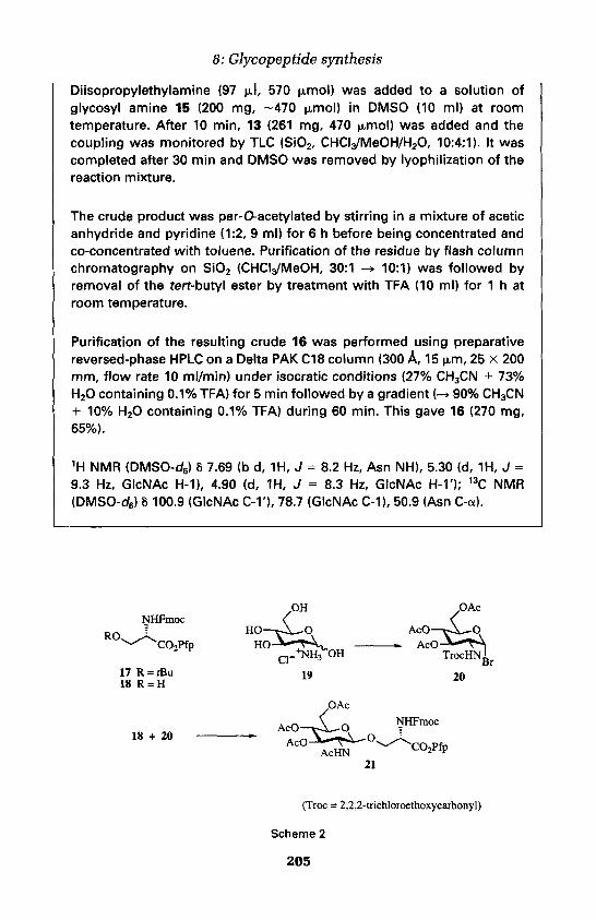
8: Glycopeptide synthesis
Diisopropylethylamine (97 IJL!, 570 (xmol) was added to a solution ofglycosyl amine 15 (200 mg, -470 |o.mol) in DMSO (10 ml) at roomtemperature. After 10 min, 13 (261 mg, 470 ^mol) was added and thecoupling was monitored by TLC (Si02, CHCl3/MeOH/H2O, 10:4:1). It wascompleted after 30 min and DMSO was removed by lyophilization of thereaction mixture.
The crude product was per-O-acetylated by stirring in a mixture of aceticanhydride and pyridine (1:2, 9 ml) for 6 h before being concentrated andco-concentrated with toluene. Purification of the residue by flash columnchromatography on SiO2 (CHCIa/MeOH, 30:1 -» 10:1) was followed byremoval of the fert-butyl ester by treatment with TFA (10 ml) for 1 h atroom temperature.
Purification of the resulting crude 16 was performed using preparativereversed-phase HPLC on a Delta PAK CIS column (300 A, 15 p.m, 25 x 200mm, flow rate 10 ml/min) under isocratic conditions (27% CH3CN + 73%H2O containing 0.1% TFA) for 5 min followed by a gradient (-»90% CH3CN+ 10% H2O containing 0.1% TFA) during 60 min. This gave 16 (270 mg,65%).
1H NMR (DMSO-d6) 8 7.69 (b d, 1H, J = 8.2 Hz, Asn NH), 5.30 (d, 1H, J =9.3 Hz, GlcNAc H-1), 4.90 (d, 1H, J = 8.3 Hz, GlcNAc H-V); 13C NMR(DMSO-cC6) 8 100.9 (GlcNAc C-1'), 78.7 (GlcNAc C-1), 50.9 (Asn C-a).

fan Kihlberg
Protocol 2. Preparation of A^-O-fluorenylmethoxycarbonyD-S-O-(2-acetamido-3,4,6-tri-O-acetyl-2-deoxy-p-D-glucopyranosyl)-L-serine pentafluorophenyl ester (21) (46) (Scheme 2)
A. Na-(9-Fluorenylmethoxycarbonyl)-L-serine pentafluorophenyl ester (18)
TFA (20 ml) was added at room temperature to /V-(9-fluorenyl-methoxycarbonyl)-3-O-(terf-butyl)-L-serine pentafluorophenyl ester (17,1.14 g, 2.06 mmol). The solution was concentrated in vacuo after 30 minand the residue was twice co-concentrated with toluene to give 18 (0.96 g,94%); m.p. 139-140°C (crystallized from diethyl ether).
B. 3,4,6-Tri-O-acetyl-2-deoxy-2-(2,2,2-trichloroethoxycarbonylamino)-oi-D-glucopyranosyl bromide (21)
2,2,2-Trichloroethoxycarbonyl chloride (1.00 g, 4.72 mmol) was added toa solution of 2-amino-2-deoxy-D-glucopyranose hydrochloride (0.68 g,3.16 mmol) and NaHCO3 (0.68 g, 8.1 mmol) in water (10 ml) at 0°C. After2 h the mixture was allowed to attain room temperature and was thenstirred overnight. After filtration, the colourless precipitate was washedwith water and diethyl ether and then recrystallized from EtOH to give 2-deoxy-2-(2,2,2-trichloroethoxycarbonylamino)-D-glucopyranose (0.98 g,87%); m.p. 183-184°C (dec.); [a]D
19 +50° (c 0.84, MeOH).
2-Deoxy-2-(2,2,2-trichloroethoxycarbonylamino)-D-glucopyranose (0.70 g,1.92 mmol) was treated overnight with a mixture of acetic anhydride andpyridine (1:1, 20 ml). The solution was concentrated and co-concentratedtwice with toluene. The crude, syrupy tetraacetate was dissolved in dryCH2CI2 (200 ml), which was saturated with dry hydrogen bromide at 0°Cand then left in a sealed flask at room temperature for 18 h. Concentrationgave a syrup which was dried over P2O5 and NaOH under vacuum to give1.40 g of crude 20 which was used in the next step without furtherpurification.
C. Na-(9-Fluorenylmethoxycarbonyl)-3-O-(2-acetamido-3,4,6-tri-O-acetyl-2-deoxy-p-D-glucopyranosyl)-L-serine pentafluorophenyl ester (21) (46)
A solution of bromide 20 (1.00 g, 1.84 mmol) in CH2CI2 (8 ml)-was addeddropwise to 18 (816 mg, 1.66 mmol), silver trifluoromethanesulphonate(0.52 g, 2.02 mmol) and 3 A molecular sieves in CH2CI2 (10 ml) at -40°C inthe dark. After 2 h the solution was allowed to attain room temperatureand was then filtered through Celite and concentrated.
Flash column chromatography of the residue on Si02 (heptane/EtOAc,3:1) gave Aa-(9-fluorenylmethoxycarbonyl)-3-O-[2-(2,2,2-trichloroethoxy-
206

8: Glypopeptide synthesis
carbonylamino)-3,4,6-tri-O-acetyl-2-deoxy-(}-D-glucopyranosyl]-L-serinepentafluorophenyl ester (1.35 g, 85%). This was dissolved in a mixture ofTHF, acetic anhydride, and AcOH (40 ml, 3:2:1), and zinc dust (3.3 g, 325mesh) was added with stirring. After 2 h the stirred mixture was filteredthrough Celite and the filter cake was washed several times with distilledTHF. The filtrate was concentrated and the residue was purified by flashcolumn chromatography on dried Si02 (heptane/EtOAc, 1:2) to give 21(0.871 g, 75%).
m.p. 207°C (crystallized from diethyl ether); [a]D2S-10° (c 1.0, CHCI3); 'H
NMR (CDCI3) 5 4.91 (ddd, 1H, J = 2.7, 3.6 and 8.6 Hz, Ser H-a), 4.89 (d, 1H,J = 8.3 Hz, GlcNAc H-1), 1.93 (s, 1H, GlcNAc Ac); 13C NMR (CDCI3) 8 100.6(GlcNAc C-1), 54.3 (Ser C-a), 23.2 (GlcNAc, COMe).
Protocol 3. Preparation of Aa-(9-fluorenylmethoxycarbonyl)-3-O-(2,3,4-tri-Oacetyl-(3-D-xylopyranosyl)-L-serinepentafluorophenyl ester (23) (47) (Scheme 3)
Boron trifluoride etherate (120 |xl, 0.94 mmol) was added to a solution of/V-Fmoc-L-serine pentafluorophenyl ester (18, 186 mg, 377 (xmol) and1,2,3,4-tetra-Oacetyl-p-D-xylopyranose (22, 100 mg, 314 (xmol, Sigma,Missouri, USA) in dry CH2CI2 (2.0 ml).
After 1.5 h at room temperature the solution was diluted with CH2CI2(8 ml) and washed with saturated aqueous NaHC03 (10 ml). The aqueousphase was extracted with CH2CI2(2 x 10 ml) and the combined organicsolution was dried over sodium sulphate, filtered, and concentrated. Theresidue was purified twice by flash column chromatography on driedSiO2 (first heptane/EtOAc, 3:2 -> 1:1, then toluene/CH3CN, 10:1) to give 23(139 mg, 60%).
1H NMR (CDCI3) 8 5.16 (t, 1H, J = 7.9 Hz, Xyl H-3), 4.59 (d, 1H, J = 6.1 Hz,Xyl H-1), 3.90 (dd, 1H, J = 10.4 and 3.4 Hz, Ser H-p); 13C NMR (CDCI3) 8100.5 (Xyl C-1).
207

Jan Kihlberg
Protocol 4. Preparation of /Va-(9-fluorenylmethoxycarbonyl)-3-O-(2,3,4-tri-O-acetyl-a-L-fucopyranosyl)-L-serine(26) (48) (Scheme 4)
Boron trifluoride etherate (110 ul, 0.90 mmol) was added to a solution of1,2,3,4-tetra-O-acetyl-L-fucopyranose (24, 50 mg, 150 jtmol, Sigma,Missouri, USA) and /V-Fmoc-L-serine (25, 59 mg, 180 umol) in dry CH3CN(3 ml). After 4 h the solution was diluted with CH2CI2 (10 ml) and washedwith water (10 ml). The aqueous phase was extracted with CH2CI2 (2 x. 10ml) and the combined organic phase was dried with Na2SO4, filtered, andconcentrated. The crude product was purified in 30-40 mg portions bypreparative reversed-phase HPLC on a Kromasil C8 column (100 A, 5 um,20 x 250 mm, flow rate 10 ml/min) under isocratic conditions(CH3CN/H20, 1:1+0.1% TFA) to give 26 (39 mg, 44%).
[a]D20-46° (cO.58, CHCI3); 'H NMR (CD3OD) 8 4.98-4.88 (m, 2H, Fuc H-1,2),
3.91 (dd, 1H, J = 5.5 and 9.5 Hz, Ser H-0), 3.68 (dd, 1H, J = 3.0 and 9.5 Hz,Ser H-B'), 0.95 (d, 3H, J = 6.5 Hz, Fuc H-6); 13C NMR (CDCI3) 8 97.3 (Fuc C-1), 56.8 (Ser C-a), 16.1 (Fuc C-6).
Protocol 5. Preparation of /VM9-fluorenylmethoxycarbonyl)-3-O-(2,3,4,6-tetra-O-acetyl-a-D-mannopyfanosyl)-L-threonine (29) (26) (Scheme 5)
Boron trifluoride etherate (154 ul, 1.23 mmol) was added to a solution of1,2,3,4,6-penta-O-acetyl-a-D-mannopyranose (27, 53.1 mg, 136 (xmol,
208

8: Glycopeptide synthesis
Sigma, Missouri, USA) and NA-Fmoc-L-threonine (28, 56.1 mg, 164 umol)in dry CH2CI2(2.5 ml) under a nitrogen atmosphere. The resulting solutionwas stirred at room temperature for 20 h, then diluted with CH2CI2 (8 ml),washed with 1 M aqueous HCI (1 ml) and water (1 ml), dried over Na2SO4,filtered, and concentrated. The residue was purified by preparativereversed-phase HPLC on a Kromasil C8 column (100 A, 5 M,UM, 20 x 250mm, flow rate 10 ml/min) under isocratic conditions (CH3CN/H20, 1:1 +0.1% TFA) to give 29 (38.5 mg, 42%).
[a]D20 +41° (cO.67, CHCI3);
1H NMR (CDCI3) 5 4.94 (b s, 1H, Man H-1), 4.54(b d, 1H, J = 9.0 Hz, Thr H-a), 1.32 (d, 3H, J = 6.4 Hz, Thr H-y); 13C NMR(CDCI3) 8 98.7 (Man C-1, JCH = 172 Hz), 17.9 (Thr C-y).
6. Synthesis of a glycopeptide from HIV gp120This section describes the synthesis of glycopeptide 31, which corresponds toresidues 312-327 of of 120 from the HIV-3B isolate (Scheme 6) (25). Sincegp 120 has been reported to carry 2-acetamido-2-deoxy-a-D-galactopyranosylmoieties, this monosaccharide was attached to a threonine in 31 whichconstitutes a potential O-glycosylation site. In addition, a cysteine residue wasincorporated at the C-terminus in order to allow conjugation to carrierproteins in attempts to elicit an immune response to 31.
Glycopeptide 31 was prepared on the solid phase under standard conditionsused in Fmoc synthesis, but some problems were encountered in the synthesiswhich reflect the more complex structure of glycopeptides compared toordinary peptides. In a first attempt morpholine was used for Fmoc-removalafter incorporation of the O-acetylated glycosylated building block 30 toavoid potential p-elimination (23) of the O-linked glycan. It was found thatuse of morpholine led to slow and incomplete removal of the Fmoc group ofthe following amino acids and after cleavage from the solid phase 31contained several by-products that were difficult to remove by preparativereversed-phase HPLC.
When the synthesis was repeated using the stronger base piperidine, whichis usually employed for synthesis of peptides, fast and complete Fmocremoval was observed and crude 31 of high purity was then obtained. No B-elimination could be detected with piperidine, an observation that has laterbeen confirmed in syntheses of a large number of O-linked glycopeptides. Itwas also found that removal of the O-acetyl protective groups from thecarbohydrate moiety had to be performed before cleavage from the solidphase in order to avoid cysteine-induced degradation of the glycopeptideunder the basic conditions used for de-O-acetylation. As pointed out above,the glycosidic bonds of most of the common saccharides are stable duringtreatment with TFA. This was also the case for 31, which could be isolated in
209

Jan Kihlberg
Scheme 6
an overall yield of 45% after deacetylation with saturated methanolicammonia and subsequent cleavage from the solid phase with TFA.
Protocol 6. Preparation of N-acetyl-glycyl-L-arginyl-L-alanyl-L-phenylalanyl-L-valyl-O-(2-acetamido-2-deoxy-a-D-galactopyranosyl)-L-threonyl-L-isoleucyl-glycyl-L-lysyl-L-isoleucyl-glycyl-L-asparaginyl-L-methionyl-L-arginyl-L-glutaminyl-L-alanyl-L-cysteine amide (31)(25) (Scheme 6)
Glycopeptide 31 was synthesized in a custom-made, fully automaticcontinuous-flow peptide synthesizer constructed essentially as describedelsewhere (19). A resin consisting of a cross-linked polystyrene backbonegrafted with poly(ethylene glycol) chains (TentaGel™, Rapp Polymer,Germany) and functionalized with the Rink amide linker (p-[a-(fluoren-9-ylmethoxyformamido)-2,4-dimethoxybenzyl]phenoxyacetic acid, Nova-biochem, Switzerland) was used for the syntheses. DMF was distilledbefore use.
In the synthesis of glycopeptide 31, 400 mg (70 umol) of linker-derivatizedresin was used. The NA-Fmoc-amino acids, which had standard side-chainprotective groups, and the glycosylated amino acid 30 were coupled tothe peptide-resin as 1-benzotriazolyl (HOBt) esters. These were preparedin the synthesizer by reaction of the Na-Fmoc amino acid (0.28 mmol), 1-hydroxybenzotriazole (0.42 mmol), and 1,3-diisopropylcarbodiimide (0.27mmol) in DMF (1.3 ml). The glycosylated amino acid 30 (0.17 mmol) wasactivated in the same way. After 45 min bromophenol blue in DMF (0.15mM, 0.35 ml) was added to the solution of the activated amino acid by thesynthesizer, and the solution was recirculated through the columncontaining the resin. The acylation was monitored (20) using theabsorbance of bromophenol blue at 600 nm, and the peptide-resin wasautomatically washed with DMF after 1 h or when monitoring revealedthe coupling to be complete. NA-Fmoc deprotection of the peptide resinwas performed by a flow of 20% piperidine in DMF (2 ml/min) through the
210

8: Glycopeptide synthesis
column for 12.5-17.5 min, and was monitored (49) using the absorbanceof the dibenzofulvene-piperidine adduct at 350 nm. After removal of theN-terminal Fmoc group the peptide-resin was again washedautomatically with DMF and then acetylated with acetic anhydride (0.8ml) in the synthesizer.
After completion of the synthesis, the resin was removed from thesynthesizer and washed with dichloromethane ( 5 x 5 ml) and dried undervacuum. Deacetylation of the carbohydrate moiety was performed on theresin (200 mg, 22 ixmol) using saturated methanolic ammonia (10 ml).The glycopeptide was then cleaved from the resin and the amino acidside chains were deprotected, by treatment with trifluoroacetic acid/water/thioanisole/ethanedithiol (87.5:5:5:2.5 v/v, 20 ml) for 2 h, followedby filtration. Acetic acid (20 ml) was added to the filtrate, the solution wasconcentrated, and acetic acid (20 ml) was added again, followed byconcentration. The residue was triturated with diethyl ether (10 ml) whichgave a solid, crude glycopeptide that was dissolved in acetic acid/water(1:4, 25 ml) and freeze-dried.
Purification was performed by preparative reversed-phase HPLC on aKromasil C8 column (100 A, 5 n-m, 20 x 250 mm, flow rate 11 ml/min)under isocratic conditions (21% CH3CN in H20 + 0.1% TFA) to give 31(30 mg, 69% peptide content, 45% overall yield). FABMS, amino acidanalysis, and 1H NMR data have been reported (25).
References1. Sharon, N. and Lis, H. (1997). In Glycosdences: status and perspectives (ed. H.-J.
Gabius and S. Gabius), p. 133. Chapman & Hall, Weinheim.2. Brockhausen, I. and Schachter, H. (1997). In Glycosdences: status and perspectives
(ed. H.-J. Gabius and S. Gabius), p. 79. Chapman & Hall, Weinheim.3. Veeneman, G. H. (1998). In Carbohydrate chemistry (ed. G.-J. Boons), p. 98.
Blackie Academic and Professional, London.4. Cohen-Anisfeld, S. T. and Lansbury Jr, P. T. (1993). J. Am. Chem. Soc., 115,
10531.5. Vetter, D., Tumelty, D., Singh, S. K., and Gallop, M. A. (1995). Angew. Chem.,
Int.Ed.Engl.,34,60.6. Offer, J., Quibell, M., and Johnson, T. (1996). J. Chem. Soc., Perkin Trans. 1,175.7. Paul, B. and Korytnyk, W. (1978). Carbohydr. Res., 67,457.8. Hollosi, M., Kollat, E., Laczko, I., Medzihradszky, K. F., Thurin, J., and Otvos Jr,
L. (1991). Tetrahedron Lett., 32,1531.9. Andrews, D. M. and Scale, P. W. (1993). Int. J. Peptide Protein Res., 42,165.
10. Paulsen, H., Schleyer, A., Mathieux, N., Meldal, M., and Bock, K. (1997). J. Chem.Soc., Perkin Trans. 1, 281.
211
Jan Kihlberg
11. Schleyer, A., Meldal, M., Manat, R., Paulsen, H., and Bock, K. (1997). Angew.Chem., Int. Ed. Engl, 36,1976.
12. Norberg, T., Liming, B., and Tejbrant, J. (1994). Methods Enzymol, 247,87.13. Meldal, M. (1994). In Neoglycoconjugates: preparation and applications (ed. Y. C.
Lee and R. T. Lee), p. 145. Academic Press, San Diego.14. Kihlberg, J. and Elofsson, M. (1997). Curr. Med. Chem., 4,79.15. Arsequell, G. and Valencia, G. (1997). Tetrahedron: Asymmetry, 8,2839.16. Meinjohanns, E., Meldal, M., Paulsen, H., Dwek, R. A., and Bock, K. (1998). J.
Chem. Soc., Perkin Trans. 1, 549.17. Elofsson, M., Salvador, L. A., and Kihlberg, J. (1997). Tetrahedron, 53, 369.18. Jansson, A. M., Jensen, K. J., Meldal, M., Lomako, J., Lomako, W. M., Olsen, C.
E., and Bock, K. (1996). J. Chem. Soc., Perkin Trans. 1,1001.19. Cameron, L. R., Holder, J. L., Meldal, M., and Sheppard, R. C. (1988). /. Chem.
Soc., Perkin Trans. 1, 2895.20. Flegel, M. and Sheppard, R. C. (1990). J. Chem. Soc., Chem. Commun., 536.21. Garg, H. G., von dem Bruch, K., and Kunz, H. (1994). Adv. Carbohydr. Chem.
Biochem.,50,277.22. Carpino, L. A. and Han, G. Y. (1972). J. Org. Chem., 37,3404.23. Kunz, H. (1987). Angew. Chem., Int. Ed. Engl, 26,294.24. Kunz, H. and Brill, W. K.-D. (1992). Trends Glycosci. Glycotechnol., 4,71.25. Vuljanic, T., Bergquist, K.-E., Clausen, H., Roy, S., and Kihlberg, J. (1996).
Tetrahedron, 52, 7983.26. Salvador, L. A., Elofsson, M., and Kihlberg, J. (1995). Tetrahedron, 51,5643.27. Kihlberg, J., Elofsson, M., and Salvador, L. A. (1997). Methods Enzymol., 289,
221.28. Urge, L., Otvos Jr, L., Lang, E., Wroblewski, K., Laczko, I., and Hollosi, M.
(1992). Carbohydr. Res., 235,83.29. Urge, L., Jackson, D. C., Gorbics, L., Wroblewski, K., Graczyk, G., and Otvos Jr,
L. (1994). Tetrahedron, 50,2373.30. Unverzagt, C. and Kunz, H. (1994). Bioorg. Med. Chem., 2,1189.31. Peters, S., Lowary, T. L., Hindsgaul, O., Meldal, M., and Bock, K. (1995). J. Chem.
Soc., Perkin Trans. 1,3017.32. Sjolin, P., Elofsson, M., and Kihlberg, J. (1996). J. Org. Chem., 61, 560.33. Christiansen-Brams, I., Meldal, M., and Bock, K. (1993). Tetrahedron Lett., 34,
3315.34. Christiansen-Brams, I., Jansson, A. M., Meldal, M., Breddam, K., and Bock, K.
(1994). Bioorg. Med. Chem., 2,1153.35. Broddefalk, J., Backhand, J., Almqvist, F., Johansson, M., Holmdahl, R., and
Kihlberg, J. (1998). /. Am. Chem. Soc., 120, 7676.36. Broddefalk, J., Bergquist, K.-E., and Kihlberg, J. (1998). Tetrahedron, 54,12047.37. Holm, B., Linse, S, and Kihlberg, J. (1998). Tetrahedron, 54,11995.38. Nakahara, Y., Nakahara, Y., and Ogawa, T. (1996). Carbohydr. Res., 292, 71.39. Guo, Z.-W., Nakahara, Y., Nakahara, Y., and Ogawa, T. (1997). Angew. Chem.,
Int. Ed. Engl, 36,1464.40. Bodanszky, M. and Martinez, J. (1981). Synthesis, 333.41. Nakahara, Y., Nakahara, Y., Ito, Y., and Ogawa, T. (1997). Tetrahedron Lett., 38,
7211.42. Wang, S.-S. (1973). J. Am. Chem. Soc., 94,1328.
212

8: Glycopeptide synthesis
43. Sheppard, R. C. and Williams, B. J. (1982). Int. J. Peptide Protein Res., 20,451.44. Rink, H. (1987). Tetrahedron Lett., 28,3787.45. Seitz, O. and Kunz, H. (1997). J. Org. Chem., 62,813.46. Meinjohanns, E., Meldal, M., and Bock, K. (1995). Tetrahedron Lett., 36, 9205.47. Sjolin, P. and Kihlberg, J., in preparation.48. Elofsson, M., Roy, S., Salvador, L. A., and Kihlberg, J. (1996). Tetrahedron Lett.,
37,7645.49. Dryland, A. and Sheppard, R. C. (1986). J. Chem. Soc., Perkin Trans. 1,125.
213

This page intentionally left blank

Convergent peptide synthesisKLEOMENIS BARLOS and DIMITRIOS GATOS
1. IntroductionBesides the classical step-by-step synthesis, the convergent solid phase peptidesynthesis (CSPPS) was developed for the preparation of complex and difficultpeptides and small proteins. According to this method, suitably protectedpeptide fragments spanning the entire peptide sequence and prepared on thesolid phase are condensed, either on a solid support or in solution, to thetarget peptide. Convergent synthesis is reviewed in recent publications (1-3).In this chapter, full experimental details are given for the preparation ofcomplex peptides by applying convergent techniques, using 2-chlorotritylchloride resin (CLTR) and Fmoc-amino acids.
1.1 Strategy in convergent synthesisIn the step-by-step peptide chain elongation the resin-bound C-terminalamino acid is reacted sequentially with suitably protected and activated aminoacids. The peptide is thus elongated steadily towards the N-terminal direction.This is advantageous over the opposite direction where the elongation isperformed from the N- to the C-terminus, because in the second case thegrowing peptide is activated at the C-terminal amino acid, which leads to itsextensive racemization. This limits considerably the synthetic possibilities ofthe method. In convergent synthesis, no directional restrictions exist and thechain elongation can be performed with equal possibility to be successful toany direction. Figure 1 describes schematically the C- to N-terminal synthesiswhich is the most studied to date. The strategies where the synthesis be-gins from a central fragment and the peptide chain is extended to both C- and
Figure 1. CSPPS with C-to N-terminal chain extension.
9

Kleomenis Balos and Dimitrios Gatos
N-terminal directions and from the N-terminal towards the C-terminal can beconsidered, at the present time, to be in its infancy.
2. Solid phase synthesis of protected peptidefragments
2.1 Fragment selectionIn general, protected peptide fragments of any length can be used in thecondensation reaction, if they are of satisfactory purity and solubility.Usually, fragments of up to 15 amino acids in length are used, because of theirsimpler purification by RP-HPLC compared with the longer peptides. Thesolubility of protected peptide acids is independent of their length.
The selection of the correct fragments is very important for the success ofconvergent synthesis. It is helpful to analyse all available structural infor-mation, determined or calculated, for the target peptide. Peptide regionswhere p-turns are known to occur are readily identified as 'difficult' sequencesduring their synthesis. The peptide fragments should be selected to containone part of the p -turn region at the C-terminus of the C-component and thesecond part at the N-terminus of the N-component. Contrary to othermethods, including the step-by-step approach, the CSPPS of rat atriopeptin,using protected fragments selected to cut the peptide p-turn region, giveexcellent results (4). Pro-residues contained in the peptide chain impede theformation of P-turns. Pro-rich peptides, such as the 115 amino acid residuescontaining antigen of core mucin (5), can be synthesized very effectively.Similar p-turn interrupting effects can be achieved by the incorporation in thecorresponding sequence, if present, of Ser or Thr as their acetonidederivatives or by the use of backbone-amide protection.
Owing to the lack of valuable information in the literature, we performedexperiments to determine the suitability of the various amino acids as C- andN-terminal amino acids of the protected fragments. We found that in terms ofracemization and condensation rates, Gly, Pro, Lys(Boc), and Glu(tBu) arepreferred as the C-terminal amino acid and Gly, Ala, Asp(tBu), Glu(tBu),Lys(Boc), and Ser(rBu) as the N-terminal. In general, the condensationefficiency depends more on the nature of the resin-bound N-component thanon that of the C-component.
2.2 Synthesis of protected peptide fragments2.2.1 Esterification of the first amino acid on 2-chlorotrityl chloride
resinThe esterification of Fmoc-amino acids and peptides to CLTR proceedsrapidly, in high yield, and without racemization (Protocol 1). Nevertheless,the amino acid-resin bond is extremely sensitive until the Fmoc-function has
216

9: Convergent peptide synthesis
been removed. This must be performed immediately after the esterification,or substantial cleavage of the amino acid from the resin occurs.
Protocol 1. Esterification of amino acids on 2-chlorotrityl chlorideresin
Equipment and reagents• Solid phase reactor • 2-Chlorotrityl chloride resin• Fmoc-amino acid « DIPEA. DCM • MeOH• DMF • Piperidine• Hexane • Isopropanol
Method1. In a solid phase reactor, suspend 1 g 2-chlorotrityl chloride resin
(1.0-2.0 mmol chloride/g resin) in 8 ml DCM, shake for 5 min and filterthe resin.
2. Add to the resin a solution of 2 mmol Fmoc-amino acid and 5 mmolDIPEA dissolved in 8 ml DCM. Shake for 30 min at room temperatureand filter the resin.
3. Wash the resin with DMF ( 2 x 2 min).4. Add to the resin 10 ml of a mixture of DCM/MeOH/DIPEA (80:15:5),
shake for 10 min and filter. Repeat the operation once.5. Wash the resin with 3 x 10 ml DMF and filter.6. Add 10 ml 25% piperidine in DMF, shake for 3 min and filter. Repeat for
20 min.7. Wash the resin with 10 ml DMF ( 6 x 2 min), 6 ml isopropanol ( 3 x 5
min), and 4 x 6 ml hexane, filter, and dry by suction in air for 15 min.Dry the resin for 24 h in vacuo.
8. Store at 4°C.
2.2.2 Peptide chain elongationStarting from the resin-bound amino acid, the first coupling must be per-formed under mildly basic or neutral conditions. Hence, dehydrating agentssuch as TBTU/HOBt/DIPEA (1:1:2) or pre-formed benzotriazolyl esters ofthe Fmoc-amino acids give no side products during the first coupling. In situcoupling using Fmoc-amino acid, HOBt and DCC or DIC can cause theformation of 0.5-2.5% tripeptide. The sequence of this tripeptide containstwo residues of the resin-bound amino acid. This indicates that the initiallyformed dipeptide is cleaved from the resin under these conditions and reactswith the resin-bound amino acid to yield the tripeptide. All subsequentcouplings can be performed by any coupling procedure used in Fmoc-chemistry, without the formation of side products.
217

Kleomenis Balos and Dimitrios Gatos
Protocol 2. Monitoring solid phase synthesis by TLC and RP-HPLC
Equipment and reagents• Eppendorf vial • TFE• Peptide resin ester • DCM« Elution mixture: chloroform/MeOH/AcOH • DMSO
or toluene/AcOH/MeOH
Method1. Place 1 mg of well-washed peptide resin ester in an Eppendorf vial.2. Add 5 drops of TFE/DCM (2:8) and shake for 5 min.3. Apply a spot from the resulting mixture on Kieselgel 60 for TLC
analysis. Develop the chromatogram using chloroform/MeOH/AcOH(90:8:2), toluene/AcOH/MeOH (70:15:15), or similar mixtures for theelution. Visualize the chromatogram at 254 nm.
4. Add 10 drops of DMSO to the resin in the vial, and centrifuge. Use 5 ulfrom the supernatant peptide solution for the HPLC analysis.
2.2.3 Cleavage of protected fragments from the resinProtected peptides can be cleaved selectively from the CLTR by variousmethods, in the presence of side chain protection of the tert-butyl type. Byfollowing Protocol 2, the purity of the resin-bound peptide is generally >97%.The peptide, cleaved by treatment with TFE/DCM (2:8) (Protocol 3) andobtained in 70-80% yield, can be subjected to the fragment condensationreactions without further purification. Depending on the individual peptidesequence, His(Trt) can be detritylated by this reagent in 1-4% yield. Fast,selective, and quantitative cleavage occurs using AcOH/TFE/DCM (1:2:7),HFIP/DCM (3:7), and 1% TFA in DCM. AcOH contained in the cleavagemixture is very difficult to remove by drying under vacuum. Reprecipitationof the fragment from TFE/water or DMF/water is necessary for its completeremoval. Otherwise, extensive acetylation of the resin-bound N-componentoccurs during the fragment condensation. HFIP-containing mixtures detrity-late to high extent His(Trt) and Ser(Trt) contained in the peptide sequenceand must be avoided in such cases.
Protocol 3. Cleavage of protected peptide fragments from 2-chlorotrityl resin
Equipment and reagents• Round-bottomed flask • TFE• Rotary evaporator • DCM• Peptide-resin • Hexane• Ether
218

9: Convergent peptide synthesis
Method1. Place 1 g of peptide-resin in a round-bottomed flask, add 15 ml of the
TFE/DCM (2:8) cleavage solution, and stir magnetically for 45 min atroom temperature.
2. Filter the resin and wash it twice with 10 ml of the cleavage solution.3. Concentrate the combined filtrates on a rotary evaporator.4. Add 20 ml cold ether and 10 ml hexane to the residue.5. Isolate the resulting solid by centrifugation and wash with ether (4 x
40 ml).6. Dry the protected peptide for 6 h in vacua and store at -20 °C.
2.2.4 Purification of protected fragmentsThe most effective procedure to purify protected peptide fragments is RP-HPLC (Protocol 4). Gradient conditions, which start with a high waterconcentration of the eluant lead in most cases to the peptide precipitation atthe beginning of the elution. Therefore, it is advantageous to start the elutionwith a high MeCN content and to perform the elution under isocratic con-ditions using RP materials of 40-80 u,m. Fragments of medium polarity underthese conditions show the best elution properties. Therefore, it is in manycases favourable to use an appropriate protection scheme to obtain fragmentsof medium polarity. In some cases it is better, for example, to apply un-protected Asn or Gin to increase the polarity of the fragments. Selectiveremoval of a side chain protecting group of a hydroxy amino acid is alsopossible for achieving medium polarity. In this case the fragment is preparedcontaining Ser(Trt), Thr(Trt), or Tyr(Clt) at the required position. The pro-tecting groups of these amino acids can be removed easily and simultaneouslyduring the cleavage of the protected peptides from the resin (Protocol 8).
Protocol 4. Purification of protected fragments by RP-HPLC
Equipment and reagents• Lobar column, size C, prepacked
with Lichroprep RP-8, 40-63 n.m• Injector, equipped with a 2.5 ml sample loop• Crude protected peptide. DMSO
Metering pumpUV detector, detection at 300 nmFraction collectorTFAMeCN
Method1. Inject a peptide sample for HPLC analysis using a C8 column of size 4
x 125 mm; elute using a gradient of 50% MeCN in 0.5% TFA-water to100% MeCN in 0.5% TFA-water for 30 min; flow rate 1 ml/min, detectionat 265 nm. Determine the MeCN concentration of the peptide elution.
2. Equilibrate the Lobar column for 60 min with water and MeCN equal to
219

Kleomenis Balos and Dimitrios Gatos
Protocol 4. Continued
the determined MeCN concentration of the elution of the peptide inthe analytical run plus 10%; flow rate 7 ml/min.
3. Dissolve 400 mg of the crude protected peptide in 2 ml DMSO.4. Filter through a 45 n,m membrane.5. Inject the clear peptide solution on the Lobar column and elute under
isocratic conditions with the mixture used for the equilibration of thecolumn. In most cases the protected peptide will elute in the 90-150min fractions.
6. Dilute the collected fractions containing the pure peptide with an equalvolume of water and lyophilize.
7. Dry the protected peptide for 24 h in vacua and store at -20°C.
3. Solid phase fragment condensation3.1 Esterification of the C-terminal fragment on
2-chlorotrityl chloride resinThe loading of the starting resin in the C-terminal fragment (Protocol 5) mustbe chosen so that at the end of the condensation reactions the content in theprotected peptide is not higher than 50% of the peptide-resin matrix. Inaddition, peptide chain aggregation will occur in high loaded resins. Like theother fragments, the resin-bound C-terminal fragment should be of the highestpossible purity. This can be achieved by purification and reattachment ontothe resin. The size of the C-terminal fragment must be as long as possible.Experiments in our laboratory show that long protected fragments esterifyfirst the more advantageous positions in the outside region of the resin bead.Thus, amino acids and small fragments reach these positions easily and reactmore readily with the free amino function. By contrast, if small fragments oramino acids are used for the first esterification on the resin, they are able toesterify the more 'difficult' positions deeper inside the resin bead due to theirfast movement. This will cause slower rates during the condensation reactionsof the longer fragments, which move slower inside the resin beads.
Protocol 5. Attachment of the C-terminal fragment onto 2-chlorotrityl resin
Equipment and reagents• Round-bottomed flask• Eppendorf vial• Peptide. DMF• Piperidine• Hexane
220
2-Chlorotrityl chloride resinDCMDIPEAMeOHIsopropanol

9: Convergent peptide synthesis
Method
1. Place 1 g of 2-chlorotrityl chloride resin in a round-bottomed flask andswell the resin using the minimum amount of DCM for 5 min.
2. Add 4 mmol DIPEA to the resin.
3. In the minimum amount of DMF, dissolve 1.5 M excess over thecalculated quantity of the protected fragment that is required toachieve the desired loading. Add the peptide solution to the resin.
4. Immediately transfer 20 ul of the resin suspension into an Eppendorfvial containing 100 nl DMF, and centrifuge. Use 5 ul of the supernatantsolution for HPLC analysis and determine the peak area, whichcorresponds to the initial fragment concentration.
5. Repeat step 4 every 30 min with the remaining peptide in solution.
6. Filter the resin.
7. Wash the resin with DMF ( 6 X 3 min), DCM/MeOH/DIPEA (80:15:5; 2 X15 min), and DMF (3 x 50 ml).
8. Treat the resin with 5% piperidine in DMF for 5 min and with 25%piperidine for 20 min.
9. Wash the resin with DMF (3 x 30 ml), DMSO (4 x 30 ml), isopropanol(3 x 30 ml), and hexane (3 x 30 ml), dry by a 15 min suction in air andfor 4 h under vacuum. Store the resin at -20°C.
3.2 Activation and condensation of protected peptidefragments
The rate of the condensation of protected fragments with the resin-bound N-component increases with the concentration of the fragment. Therefore,fragment solutions of the highest possible concentration should be applied.The solvents and condensing reagents used for the long-lasting fragmentcondensation play a much more important role for the product purity and thesuppression of racemization compared to the fast amino acid coupling. Eventreatment of various resin-bound N-components, in the absence of the C-component (N-terminal fragment), with condensing reagents in DMF,DMAC, and N-methylpyrrolidone for 10 h at room temperature leads to theformation of several by-products. Similarly, treatment of various C-com-ponents with the same solvents leads to the partial removal of the W-Fmocgroup of the fragments. Such by-products are not formed if DMSO is used assolvent.
Modern coupling agents such as BOP, HBTU, TBTU, and HATU are wellsuited for the racemization-free coupling of amino acids. These reagents weretested in various solvents during the condensation of the resin-boundprothymosin a (ProTa) 95-109 fragment with Fmoc-ProTa(87-94)-OH whichcontains Glu(tBu) as the C-terminal amino acid (Figure 2). The 70-90%
221

Kleomenis Balos and Dimitrios Gatos
Fmoc-Lys(Boc)-Arg(Pmc)-Ala-Ala-Qlu(tBu)-Asp(tBu)-A8p(tBu)-Glu(Bu)-OH
H-Asp((Bu)-Asp(tBu)-Asp(tBu)-Val-Asp(tBu)-Thr(tBu)-Lys(Boc)-Lys(Boc)-Gln(Trt)-Lys(Boc)-Thr(tBu)-Asp(Su)-Glu(Su)-Asp(Bu)-Asp((Bu)-0-Q
Figure2. Fmoc-ProTa(87-94)-OH and the resin-bound ProTa(95-109) used for theevaluation of racemization suppressing conditions in solid phase fragment condensation.
racemization determined by HPLC was independent of the solvent used. Bycontrast, in condensations where DCC/HOBt, DIC/HOBt, and DIC/DhbtOHwere used as dehydrating agents, racemization in DMSO was <1%. Con-densations in solvents of the amide type gave much higher racemization. Theuse of DCM, even as co-solvent of DMSO, also led to high racemization.DhbtOH as additive gave low racemization but the condensations resulted inthe formation of unidentified side-products. The conclusions are:
1. DMSO is the best solvent in terms of solvating properties, purity ofproducts, and suppression of racemization.
2. DCC/HOBt and DIC/HOBt are the best condensing agents.3. The C-component must be applied in the highest possible concentration.4. To obtain products of high purity and suppress racemization in long-lasting
condensations, it is better to perform a double coupling after 6 h con-densation than to leave the condensation to proceed for a long time.Otherwise, the remaining peptide fragment when left in its activated forma long time period undergoes extensive racemization, even if DMSO andracemization suppressing agents are used.
Protocol 6. Solid phase fragment condensation
Reagents• Protected peptide fragment. DMSO• Peptide-resin. DMF• Hexane
HOBtDICDIPEAIsopropanolPiperidine
Method1. Dissolve the protected fragment (3-5-fold excess over the resin bound
N-component) and 2.0 eq HOBt in the highest possible concentrationin DMSO.
2. Add 2.0 eq DIC.
3. Add to this solution the dry resin without pre-swelling and shake for6h.
4. Check the condensation reaction by using the Kaiser test, TLC, and
222

9: Convergent peptide synthesis
HPLC. SDS-PAGE (silver staining) can also be used after totaldeprotection of the peptide.
5. Wash the resin and repeat the condensation if the Kaiser test ispositive.
6. Wash the resin with DMSO (6 X).
7. Add to the resin a 0.5 M solution of a 7-fold excess of acetic anhydrideand DIPEA in DMSO, and shake for 1 h at RT.
8. Wash the resin with DMF (6 x), isopropanol (3 x), and hexane (3 x),dry with a 15 min suction in air. Store the resin at -20°C.
9. For the Fmoc-removal, treat the resin with 5% piperidine in DMF for5 min and with 25% piperidine for 20 min, wash the resin with DMF(4 x), DMSO (3 x), isopropanol (4 x), and hexane (3 x), and use it, assoon as possible, for the next condensation. If the Fmoc-removal is notcomplete, repeat the piperidine treatment. In very difficult cases, treatwith 20% diethylamine in DMF for 25 min.
With the exception of the two final condensations, after every condensationthe remaining unreacted resin-bound amino groups should be acetylated.After the final condensation, acetylation should not be performed because thelipophilicity of the peptide increases and the acetylated smaller peptide canhave a similar elution time to the required target peptide. This can make theseparation and purification steps more difficult.
4. Phase and direction change4.1 Fragment condensation in solutionNotwithstanding the selection of peptide fragments, the protecting groups,and the methods used, some steps of the CSPPS can prove to be very difficult.In such cases, the condensation reactions can be performed in solution. Thisprocedure is often advantageous from an economical point of view, since theC- and N-components are used in equimolar amounts. The required N-component is prepared by the esterification with 2-chlorotrityl chloride insolution of a protected peptide prepared previously either by the step-by-stepor fragment condensation on CLTR (Figure 3). If a peptide amide is required,
Figures. Change in the peptide synthesis phase.
223

Kleomenis Balos and Dimitrios Gatos
a suitable amide linker is used or the fragment is converted to the corres-ponding amide by treatment with DIC/HOBt.NH3 in DMSO.
Protocol 7. Synthesis of 2-chlorotrityl esters of protectedfragments
Reagents• Protected peptide fragment • DMF• DIPEA • 2-Chlorotrityl chloride. DCM . Ether. DMSO • KOH pellets• Piperidlne • THF
Method1. Dissolve the protected fragment in the highest possible concentration
in DMF and add 10 eq DIPEA.
2. Prepare a 1 M solution of 5 eq, with respect to the protectedfragment, of 2-chlorotrityl chloride in DCM.
3. Add the chloride solution dropwise to the stirred peptide solution andcontinue stirring for 6-12 h at RT. Check for completion of theesterification by TLC.
4. Remove DCM in the rotary evaporator.5. Precipitate the protected fragment by the addition of ether to the
residual DMF solution.
6. Filter or centrifuge the protected fragment and wash with ether (5 x).7. Dry the fragment for 6 h in vacuo over potassium hydroxide pellets.
8. Dissolve the protected fragment in 4% piperidine in DMF/THF (50:50)and stir the solution at RT for 2 h. Check for completion of the Fmoc-removal by TLC and HPLC.
9. Remove THF in the rotary evaporator and add ether to the remainingsolution until precipitation of the protected peptide occurs.
10. Filter the precipitated peptide, wash with ether (8 x), and dry undervacuum.
The 2-chlorotrityl peptide ester obtained after fragment condensation insolution can be selectively deprotected at the C-terminal carboxy group in thepresence of tBu-based protecting groups. This is achieved by a 45-mintreatment of the protected peptide with TFE/DCM (30:70), followed by thereattachment of the fragment onto the 2-chlorotrityl chloride resin (Protocol5). The convergent synthesis can then be continued by the sequential con-densation of the next fragments on the solid phase. Fragment condensationon a solid support gives, in general, crude peptides of higher purity than that
224

9; Convergent peptide synthesis
of the corresponding condensation in solution. Therefore, the reattachment ofthe product on a suitable resin should be performed as soon as possible.
4.2 Two-directional synthesis. Attachment of fragments onresins of the trityl-type through an amino acid side chainfunctional group
An alternative approach to that of changing the phase of the synthesis toovercome difficulties encountered in CSPPS is to begin the synthesis from acentral part of the peptide. The starting peptide is synthesized, as usual, on 2-chlorotrityl resin using Fmoc-amino acids. One of the amino acids, specificallyHis, Cys, Lys, or Ser, contained in the sequence is introduced using His(Mmt),Cys(Mmt), Lys(Mtt), or Ser(Trt), respectively. All other amino acid side-chain protecting groups are of the fBu-type. These Mmt, Mtt, and Trt side-chain protecting groups are extremely acid sensitive and can be removedselectively by treatment with 1% TFA in DCM in the presence of His(Trt),Cys(Trt), or the tBu-type protection (Protocol 8). The fragments obtained,selectively deprotected at an amino acid side-chain, can be reattached on asuitable trityl-type resin (Figure 4). Instead of 2-chlorotrityl chloride resin, the4-methoxytrityl chloride resin for the attachment through the His or Cys sidechain and the trityl chloride resin for the attachment via the Lys or Serresidues are strongly recommended. Since the ester bonds of peptides to thesetrityl resins are extremely acid, the esterification reaction is reversible underthese conditions; thus, protection of the C-terminal carboxy group in thepeptide fragment is not necessary.
Protocol 8. Selective deprotection of His(Mmt)-, Cys(Mmt)-,Ser(Trt)-, Lys(Mtt)-, Thr(Trt)-, and Tyr(Clt)-containingpeptides
Reagents• Crude protected peptide • TFA. DCM/TES (95:5) . MeOH• Ether
Method1. Add to 250 mg of crude protected peptide (obtained using Protocol 3)
40 ml of 1% TFA in DCMA"ES (95:5) and stir for 1 h at RT.2. Check for completion of the deprotection by using TLC and HPLC.3. Add 5 ml MeOH to the resulting mixture.4. Concentrate the solution on a rotary evaporator.5. Precipitate the peptide by addition of ether.6. Centrifuge the peptide, wash with ether (5 x), and dry for 12 h in vacuo.
225

Kleomenis Balos and Dimitrios Gates
Figure 4. Attachment of protected peptides, through an amino acid side chain functionalgroup, on resins of the trityl type.
In order to extend the peptide chain towards the C-terminus, the resin-boundfragment obtained contains a free carboxy group which can be activated bytreating with DIC/HOBt and condensed with excess of a suitably protected N-component (Figure 5). In addition, the free carboxy group of side chainattached peptides can be esterified by 2-chlorotrityl chloride by using a verysimilar procedure to that described in Protocol 9, or can be converted byDIC/HOBt»NH3 to the corresponding resin-bound peptide amide. Thesynthesis is then continued towards the N-terminal of the required peptide.Side chain attached fragments are very useful for the insertion of bifunctionalcompounds, such as linear and cyclic diamines, into peptide chains.
Figure 5. Peptide chain extension to both C- and N-terminal directions.
Protocol 9. Attachment of protected fragments through the sidechains of His, Cys, Lys, and Ser on resins of the trityltype
Equipment and reagents• Round-bottomed flask • DCM. Trityl chloride resin • DIPEA. DMF . MeOH• TFE • Isopropanol• Hexane
Method
1. Place 1 g of 4-methoxyltrityl chloride resin (for attachment through theHis or Cys side chain) or trityl chloride resin (for Lys or Ser) in a round-bottomed flask and swell it using the minimum amount of DCM for5 min.
2. Add 4 mmol DIPEA to the resin.
226
9: Convergent peptide synthesis
3. Dissolve in the minimum amount of DMF, a twofold molar excess overthe calculated quantity of the protected fragment that is required forachieving the required loading.
4. Add the peptide solution to the resin and stir for 5 h at RT.6. Filter the resin and wash with DMF ( 6 x 3 min), DCM/MeOH/DIPEA
(80:15:5; 3x15 min), and DCM (3 X).7. Wash the resin with TFE/DCM (20:80; 3 x 3 min), DMF (3 X), iPrOH (3
x), and hexane (3 x ) and dry the resin by suction in air (15 min) andthen in vacua for 6 h.
5. Deprotection, purification, and puritydetermination of the synthetic peptides
Several procedures are described in the literature for minimizing sidereactions that take place during the peptide deprotection. In the case ofconvergently synthesized large peptides, a three-step deprotection seems togive the best results. The protected peptide is cleaved from the trityl resinwith the Aa-Fmoc group intact. Then the peptide is treated with 65% TFA inDCM/EDT (95:5) for 1 h at RT. After evaporation of the solvent andprecipitation by the addition of ether, the crude partially deprotected peptideis treated for 4 h with TFA/water/EDT (90:5:5) to remove remaining protect-ing groups and especially the Pmc or Pbf groups used for the protection ofArg. The mixture is then concentrated in vacua, precipitated, and washedwith ether. The Aa-Fmoc-peptide, deprotected on the side chains, is thensubjected to a first purification step by RP-HPLC. From the partially purifiedpeptide obtained, the Fmoc group is cleaved by treatment with 5% piperidinesolution in water for 1-2 h. The mixture is then acidified with 10% acetic acidand extracted with diethyl ether to remove the Fmoc-deprotection products.After lyophilization, the totally deprotected peptide is purified by RP-HPLCusing, if possible, isocratic elution conditions.
To establish the correct structure of the synthetic peptide, amino acidanalysis, mass spectrometric analysis, and tryptic peptide mapping, includingsequencing of the tryptic fragments, are necessary. The peptide purity is bestdetermined by HPLC and CE. Importantly, small peaks observed after atryptic digestion eluting close to the expected tryptic fragments on RP-HPLCcorrespond usually to by-products of the synthesis. Their absence is a veryimportant indication of the high purity of the synthetically obtained peptideor protein.
References1. Lloyd-Williams, P., Albericio, F., and Giralt, E. (1997). Chemical approaches to the
synthesis of peptides and proteins. CRC Press, Boca Raton, New York.
227
Kleomenis Balos and Dimitrios Gatos
2. Lloyd-Williams, P., Albericio, F., and Giralt, E. (1993). Tetrahedron, 49,11065.3. Benz, H. (1994). Synthesis, 337.4. Gatos, D., Patrianakou, S, Hatzi, O., and Barlos, K. (1997). Lett. Pept. Sci., 4,177.5. Krambovitis, E., Hatzidakis, G., and Barlos, K. (1998). J. Biol. Chem.,273,10874.
228

10
Methods of preparingpeptide-carrier conjugatesJAN W. DRIJFHOUT and PETER HOOGERHOUT
1. IntroductionFor many applications, peptides should be conjugated to carriers. An im-portant example is the conjugation of peptides to proteins, carbohydrates, orlipids for immunological studies. Further examples are the preparation ofpeptide affinity media and the conjugation of peptides to suitable coatingcompounds on surfaces for enzyme-linked immunosorbent assay and plasmaresonance.
The most important consideration in designing conjugates is that thepeptide part of the conjugate must retain its biological activity. This meansthat the site of activity of the peptide must not be involved in the conjugationreaction. However, the active part of the peptide is often not precisely known,which complicates the design of a proper conjugation strategy. In addition, ifthe peptide is a fragment of a larger biologically active protein, the smallpeptide will frequently not adopt the conformation (for instance, a loopstructure) of the corresponding sequence in the native protein. This mightalso abolish desired biological properties, such as the possibility to inducefunctional antibodies recognizing the native protein. In that case, appropriateartificial conformational restrictions should be introduced into the peptide - ifpossible.
From the numerous conjugation methods available (1), only a few aredescribed in this chapter. One example concerns the application of thehomobifunctional cross-linker glutaraldehyde. Heterobifunctional cross-linking is illustrated by coupling of thiol-containing peptides or carriers tosulphydryl-reactive carriers or peptides, respectively.
2. Homobifunctional cross-linkingA general and easy method of conjugating peptides to proteins is to make useof homobifunctional cross-linkers. An example of a homobifunctional cross-

Jan Wouter Drijfhout and Peter Hoogerhout
linker is glutaraldehyde (1, 2) (see Protocol 1). This bis-aldehyde reacts withamine groups at neutral or basic pH to yield enamines, which can be reducedoptionally to amines with sodium cyanoborohydride. The peptide and theprotein react selectively via their N-terminus and/or lysine side chains to givenot only a very complex mixture of products—peptide-peptide, peptide-protein, and protein-protein conjugates—but also large constructs containingpeptide and protein. Typically, several peptide molecules per proteinmolecule are coupled. In view of the complex reaction pathway, the batch-to-batch reproducibility of the conjugate is difficult to control. If the peptidecontains a lysine in the active part, it is recommended to extend the peptideN- or C-terminally with some additional lysines during synthesis in order toprovide more amine groups for conjugation.
Numerous homobifunctional cross-linkers are available commercially (e.g.from Pierce), including disuccinimidyl derivatives (3, 4), water-solublesulphodisuccinimidyl derivatives (5, 6), and diimido esters (7-9). Alsoavailable are cross-linkers containing bonds that can be cleaved by reduction(10, 11), hydroxylamine (12, 13), base (14), oxidation (15, 16), etc., whichmakes it possible to liberate the peptide from the conjugate for analyticalpurposes.
Protocol 1. Conjugation of a peptide to bovine serum albuminwith glutaraldehyde
Equipment, buffers, and reagents• Disposable gel filtration columns packed • Phosphate-buffered saline (PBS), i.e. 10
with Sephadex G-25 M (PD-10® columns, mM sodium phosphate in physiological saltPharmacia) (pH 7.2)
• 0.1 M sodium phosphate buffer (pH 8) • Glutaraldehyde, 25% w/w solution in water8
• Bovine serum albumin, BSA (Sigma)
Method
1. Dissolve 3 mg of, for example, a 15-mer peptide and 5 mg BSA in1.4 ml of 0.1 M sodium phosphate buffer (pH 8).
2. Dilute 5 pj of the stock solution of 25% glutaraldehyde with 500 uJ PBS.
3. Add 100 (ul of the freshly prepared diluted glutaraldehyde solution tothe peptide/BSA solution at t = 0, 30, 60, and 90 min and mix well.After the last addition, leave the reaction mixture to stand for a further2h.b
4. Equilibrate a PD-10® column with at least 25 ml of PBS/water (1:1, v/v).Start the equilibration approximately 30 min before the end of theconjugation reaction.
5. Apply the reaction mixture (1.8 ml) to the PD-10® column and discardthe eluent.
230

10: Methods of preparing peptide-carrier conjugates
6. Apply 0.7 ml of PBS/water (1:1) to the column and again discard theeluent.
7. Place a collection tube under the column and apply 3.2 ml ofPBS/water (1:1) to the column.
8. Lyophilize the collected column effluent and store the material at-20°C until use.
9. Reconstitute the lyophilized conjugate with 1.6 ml of water. The solutionobtained contains about 1 mg/ml of conjugated peptide in PBS.
"Glutaraldehyde-containing solutions and reaction mixtures should be kept closed to avoid airoxidation.b During the conjugation reaction positive charges of the protein are removed which makes theprotein less soluble. Depending on the nature of the peptide to be conjugated, precipitationmay occur.
3. Heterobifunctional cross-linkingAnother approach towards conjugation is the use of cross-linkers containingtwo different reactive groups. Compared with homobifunctional cross-linkers,heterobifunctional linkers offer the possibility to prepare better-defined con-jugates, since peptide-peptide and carrier-carrier conjugation can be prevented.Convenient procedures are based on couplings of thiols to sulphydryl-reactivegroups.
3.1 Conjugation of thiol-containing peptides to proteinsCysteine-containing peptides can be conjugated to sulphydryl-reactiveproteins, provided that the cysteine is not essential for the biological activity.If the peptide is devoid of Cys, this amino acid can be added N- or C-terminally during synthesis. Depending on the linker attached to the solidphase, C-terminal incorporation is not recommended to avoid possibleracemization of Cys (17).
Useful sulphydryl-reactive groups on the protein are maleimide, pyridyl-dithio, and bromoacetyl. Maleimide-activated keyhole limpet haemocyanin,bovine serum albumin, and ovalbumin are available commercially. The degreeof functionalization can be determined indirectly (18) by incubation of a smallsample of the maleimide-modified protein with an excess of cysteamine,followed by addition of dipyridyldisulphide which re-acts with the remainingcysteamine. The amount of thiopyridone formed can be determined by UVmeasurement. Conjugation of a Cys-peptide to maleimide-activated proteinsproceeds through addition of the thiol to the carbon-carbon double bond ofthe maleimide group (Figure 1, pathway a). The reaction is fast and cantherefore be performed at a pH as low as 5, which decreases the rate ofundesired (air) oxidation of the Cys-peptide to disulphide dimers. The molar
231

Jan Wouter Drijfhout and Peter Hoogerhout
peptide-protein ratio in the conjugate can be estimated by determination ofresidual maleimide groups—if present—as described above. Finally, it shouldbe noted that the stability of maleimide-conjugates is optimal at about pH 6(19).
The active ester Af-succinimidyl 3-(2-pyridyldithio)propionate (SPDP) isthe classical reagent to modify proteins with pyridyldithiopropyl (PDF)groups (20). If the protein to be modified contains many free Cys-residues thiscreates the risk of carrier-carrier conjugation. In this case it is possible toblock the free thiols with iodo- or bromoacetic acid (or amide) beforereaction with SPDP (or to use a different conjugation method, as described inSection 3.2).
The number of linkers introduced can be determined by incubation of asmall sample of the modified protein with an excess of a thiol, for instancedithiothreitol, and analysis of the amount of 2-pyridinethione which isreleased (see above). Likewise, the formation of a disulphide conjugate froma Cys-peptide and a pyridyldithio-protein (Figure 1, pathway b, R3 = H) canbe monitored by UV measurement. Since the amount of 2-pyridinethioneformed is proportional to the amount of peptide coupled, the peptide-proteinratio is obtained immediately. This is advantageously if the peptide-proteinratio is important for application of the conjugate. The optimal reactionconditions (i.e. the amounts of SPDP or peptide to be used for conjugation) toobtain a desired peptide-protein ratio can be established readily due to theconvenient method of analysis. However, the (in vivo) lability of thedisulphide bond may be disadvantageous in further studies of the conjugate.
N-Succinimidyl bromoacetate (BrAc-ONSu) is the reagent of choice forbromoacetylation of proteins (21). The incorporation of bromoacetyl (BrAc)groups can be determined indirectly by measurement of the amount of freeprimary amino groups with 2,4,6-trinitrobenzenesulphonic acid before andafter the modification (22). Cys-peptides couple to BrAc-proteins to give verystable thioether conjugates (Figure 1, pathway c). The peptide-protein ratiocan be determined by amino acid analysis by quantitation of the amount of S-carboxycysteine (21,23) after hydrolysis of a sample of the conjugate.
Instead of using Cys-containing peptides it is convenient to use S-acetylmercaptoacetyl (SAMA) peptides for conjugation purposes. UnlikeCys, SAMA is not sensitive towards (air) oxidation during purification of thepeptide. SAMA can be introduced into deprotected peptides in solution byreaction with N-succinimidyl S-acetylmercaptoacetate (18, 24). Selectiveintroduction of SAMA can be achieved at the end of solid phase synthesis byN-terminal modification (Chapter 6) of the side-chain protected and resin-bound peptide with pentafluorophenyl S-acetylmercaptoacetate (SAMA-OPfp) and 1-hydroxybenzotriazole (25). The thiol group of SAMA-peptidescan be liberated by reaction with hydroxylamine. This S-deacetylation can beperformed in the conjugation reaction mixture (25, 26), i.e. in the presence ofthe sulphydryl-reactive protein (Protocol 2).
232
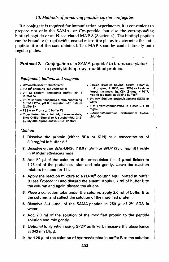
10: Methods of preparing peptide-carrier conjugates
If a conjugate is required for immunization experiments, it is convenient toprepare not only the SAMA- or Cys-peptide, but also the correspondingbiotinyl-peptide or an N-acetylated MAP-8 (Section 4). The biotinyl-peptidecan be bound to (strept)avidin-coated microtitre plates to determine the anti-peptide titre of the sera obtained. The MAP-8 can be coated directly ontoregular plates.
Protocol 2. Conjugation of a SAMA-peptideato bromoacetylatedor pyridyldithiopropyl-modified proteins
Equipment, buffers, and reagents
• UV/visible spectrophotometer• PD-10* columns (see Protocol 1)• 0.1 M sodium phosphate buffer, pH 8
(buffer A)• 0.1 M sodium phosphate buffer, containing
5 mM EOT A, pH 6, deaerated with helium(buffer B)
• PBS (see Protocol 1, buffer C)• Cross-linker: N-succinimidyl bromoacetate,
BrAc-ONSu (Sigma) or N-succinimidyl 3-(2-pyridyldithiolpropionate, SPDP (Pierce)
• Carrier protein: bovine serum albumin,BSA (Sigma, A 7030, min 98%) or keyholelimpet haemocyanin, KLH (Sigma, H 7017,lyophilized from stabilizing buffer)6
• 2% w/v Sodium dodecylsulphate (SDS) inwater
• 2 M Hydroxylamine-HCI in buffer B (140mg/ml)
• 2-Aminoethanethiol (cysteamine) hydro-chloride
Method
1. Dissolve the protein (either BSA or KLH) at a concentration of3.0 mg/ml in buffer A.c
2. Dissolve either BrAc-ONSu (18.9 mg/ml) or SPDP (25.0 mg/ml) freshlyin N-dimethylacetamide.
3. Add 50 uJ of the solution of the cross-linker (i.e. 4 umol linker) to1.75 ml of the protein solution and mix gently. Leave the reactionmixture to stand for 1 h.
4. Apply the reaction mixture to a PD-10® column equilibrated in bufferB (see Protocol 1) and discard the eluent. Apply 0.7 ml of buffer B tothe column and again discard the eluent.
5. Place a collection tube under the column, apply 3.0 ml of buffer B tothe column, and collect the solution of the modified protein.
6. Dissolve 3-4 u.mol of the SAMA-peptide in 250 ul of 2% SDS inwater.
7. Add 2.0 ml of the solution of the modified protein to the peptidesolution and mix gently.
8. Optional (only when using SPDP as linker): measure the absorbanceat 343 nm (A343).
9. Add 25 uJ of the solution of hydroxylamine in buffer B to the solution
233
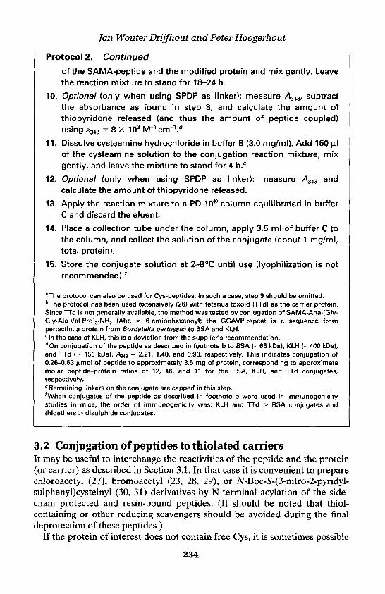
Jan Wouter Drijfhout and Peter Hoogerhout
Protocol 2. Continued
of the SAMA-peptide and the modified protein and mix gently. Leavethe reaction mixture to stand for 18-24 h.
10. Optional (only when using SPDP as linker): measure A343, subtractthe absorbance as found in step 8, and calculate the amount ofthiopyridone released (and thus the amount of peptide coupled)using e343 = 8 x 10³ M1 cm-1.D
11. Dissolve cysteamine hydrochloride in buffer B (3.0 mg/ml). Add 150 uJof the cysteamine solution to the conjugation reaction mixture, mixgently, and leave the mixture to stand for 4 h.e
12. Optional (only when using SPDP as linker): measure A343 andcalculate the amount of thiopyridone released.
13. Apply the reaction mixture to a PD-10® column equilibrated in bufferC and discard the eluent.
14. Place a collection tube under the column, apply 3.5 ml of buffer C tothe column, and collect the solution of the conjugate (about 1 mg/ml,total protein).
15. Store the conjugate solution at 2-8 °C until use (lyophilization is notrecommended)F
AThe protocol can also be used for Cys-peptides. In such a case, step 9 should be omitted.BThe protocol has been used extensively (26) with tetanus toxoid (TTd) as the carrier protein.Since TTd is not generally available, the method was tested by conjugation of SAMA-Aha-[Gly-Gly-Ala-Val-Pro]3-NH2 (Aha = 6-aminohexanoyl; the GGAVP-repeat is a sequence frompertactin, a protein from Bordetella pertussis) to BSA and KLH.cln the case of KLH, this is a deviation from the supplier's recommendation.dOn conjugation of the peptide as described in footnote b to BSA (~ 65 kDa), KLH (~ 400 kDa),and TTd (~ 150 kDa), A343 = 2.21, 1.40, and 0.93, respectively. This indicates conjugation of0.26-0.63 (umol of peptide to approximately 3.5 mg of protein, corresponding to approximatemolar peptide-protein ratios of 12, 46, and 11 for the BSA, KLH, and TTd conjugates,respectively.6Remaining linkers on the conjugate are capped in this step.FWhen conjugates of the peptide as described in footnote b were used in immunogenicitystudies in mice, the order of immunogenicity was: KLH and TTd > BSA conjugates andthioethers > disulphide conjugates.
3.2 Conjugation of peptides to thiolated carriersIt may be useful to interchange the reactivities of the peptide and the protein(or carrier) as described in Section 3.1. In that case it is convenient to preparechloroacetyl (27), bromoacetyl (23, 28, 29), or Af-Boc-5-(3-nitro-2-pyridyl-sulphenyl)cysteinyl (30, 31) derivatives by N-terminal acylation of the side-chain protected and resin-bound peptides. (It should be noted that thiol-containing or other reducing scavengers should be avoided during the finaldeprotection of these peptides.)
If the protein of interest does not contain free Cys, it is sometimes possible
234
10: Methods of preparing peptide-carrier conjugates
c
Figure 1. Conjugation of thiols to maleimides (pathway a); 2-pyridyldithio derivatives(pathway b, R3 = H); 3-nitro-2-pyridyldithio derivatives (pathway b, R3 = N02), e.g. a Boc-S-(3-nitro-2-pyridylsulphenyl)cysteinyl-peptide; or bromoacetyl derivatives (pathway c).
to generate thiols by disulphide reduction (if unfolding of the protein isallowed). In most cases modification of the protein is required. Directthiolation of proteins can be achieved by reaction with iminothiolane (32) or(Af-acyl) homocysteine thiolactone (33). Protected thiol groups can beintroduced with N-succinimidyl 5-acetylmercaptoacetate (24), SPDP (20), orcystamine and l-ethyl-3-(3-dimethylaminopropyl) carbodiimide (34). Thedegree of thiolation can be determined by colorimetric assay with 5,5'-dithiobis(2-nitrobenzoic acid) (35, 36).
Protocol 3 provides an example of immobilization of Npys-peptide tothiopropyl-Sepharose (37).
Protocol 3. Immobilization of an Npys-peptide to thiopropyl-Sepharose
Equipment, buffers, and reagents• Thiopropyl-Sepharose 6B (Pharmacia)• Polypropylene or glass column (30 x 4
mm) with filter• 0.1 M sodium phosphate buffer, pH 8
(buffer A)
• 0.1 M sodium phosphate buffer, containing10 mM DTT, pH 8 (buffer B)
• 3% v/v acetic acid in water (buffer C)
235
Jan Wouter Drijfhout and Peter Hoogerhout
Protocol 3. Continued
Method
1. Swell 50 mg thiopropyl-Sepharose 6B (containing about 3.75protected thiol groups) in 2.0 ml of buffer A for 1 h.
2. Apply the suspension to the column and wash with 5 ml of buffer A toobtain a column bed of about 180 ul (1.5 cm in height).
3. Wash the column with 10 ml of buffer B during 30 min to remove the2-thiopyridyl groups. An A343 of 0.6 for a 5-fold diluted (with buffer A)effluent will indicate the presence of 3.75 (unol thiol groups on thecolumn.
4. Wash the column with 10 ml of buffer A.
5. Dissolve 6 umol of an N-terminal Cys(Npys)-peptide in 1 ml of bufferA.
6. Add 4 X 250 (uJ of the peptide solution to the column at t = 0, 10, 20,and 30 min. The occurrence of an intense yellow colour indicates thatthe conjugation is proceeding. During the fourth addition no colourshould develop which indicates complete coupling of all thiol groupson the column during the first three additions.
7. Wash the column with 10 ml of buffer A.
8. Optional: wash the column with 10 ml of buffer C in order to acidify theresin before storage.
9. Optional: dilute the resin 10 times with Sepharose 4B to increase thevolume for convenient column volumes during chromatography.
4. MAP-core constructs as peptide-carriersAs well as by conjugation to a macromolecule, such as a protein, peptides canbe presented as a larger construct in the form of a multiple antigen peptide(MAP). In this type of construct various copies of the peptide are attached toa small core structure (38). The construct, which is fully synthetic, is alsoknown as a dendrimer (39). An MAP might contain one particular peptide(homo-MAP) or two (or even more) distinct peptides (hetero-MAP). MAPscan be prepared by direct sequential synthesis or by fragment condensation ofmodified peptides and an activated MAP core construct.
HPLC purification of MAPs can be complicated by the fact that thesestructures might show broad signals when applied to reversed-phase columns.This might be caused by slow molecular motions of the large constructs duringchromatography. In addition, mass spectrometric analysis of MAPs is oftentroublesome.
236

10: Methods of preparing peptide-carrier conjugates
4.1 Sequential synthesis of MAPsMAPs (40, 41) can be constructed by straightforward peptide synthesis. At acertain stage in the synthesis, acylation is performed with Fmoc-Lys(Fmoc)-OH. After deprotection, two new amino groups are available for coupling ofthe next amino acid, yielding a double peptide chain. This process can berepeated several times, yielding MAPs with 2-16 peptide copies. To produceMAPs containing multiple copies of two different peptides, coupling can beperformed with, for example, Fmoc-Lys(Dde)-OH (42). After synthesis ofmultiple copies of the first (N-acetylated) peptide, the semi-orthogonal Ddeprotection is removed. Then synthesis of multiple copies of the secondpeptide is performed (43).
Especially when MAP-8 or MAP-16 constructs are made, space within thepeptide-resin might become limited. It is therefore advisable to use resinswith a low loading (e.g. 0.05-0.2 meq/g). The purity of directly preparedMAPs is always lower compared to that of the corresponding peptide,because in the case of a side reaction in one of the branches, the otherbranches are also part of a failure sequence.
4.2 Synthesis of MAPs by fragment condensationApart from direct sequential MAP-synthesis, it is also possible to prepareMAPs by fragment condensation (44, 45). The core to which the peptides willbe attached and the peptide(s) themselves are synthesized and purifiedseparately. Subsequently, multiple copies of a peptide are conjugated to thecore. The core and the peptides should be modified to allow for controlledconjugation. This can be performed in several ways, for example by con-jugation of a thiol-containing peptide to a bromoacetylated core, yielding astable thioether MAP. An example is given of the synthesis of an MAP-8 inwhich the peptide molecules are attached to the core (Figure 2) by disulphidelinkages (Figure 1, pathway b, R3 = NO2). This type of MAP can be checkedby reduction which liberates the thiopeptide from the core. The core de-scribed contains eight glutamic acid residues to increase the solubility and a
Figure 2. Structure of the MAP core construct obtained according to Protocol 4 after step 8.
237

Jan Wouter Drijftiout and Peter Hoogerhout
4-aminobutyric acid residue which allows for determination of peptide/coreratio after amino acid analysis (44).
Protocol 4. Preparation of an MAP-8 containing disulphide-linkedpeptide molecules
Equipment, buffers, and reagents• Wang (4-hydroxymethylphenoxymethyl poly-
styrene) resin, loading 0.092 mmol/gDimethylacetamide (DMA)8
DCMDiethyl etherFmoc-4-aminobutyric acidDCCDMAPPiperidineFmoc-Lys(Fmoc)-OHFmoc-GluCBu)-OH1-Hydroxybenzotriazole (HOBt)
• Pentafluorophenyl S-acetylmercaptoacetate(SAMA-OPfp)
• Trifluoroacetic acid (TFA)• 0.2 M Sodium phosphate buffer, pH 6.5
(buffer A)• 0.1 M Sodium phosphate buffer, containing
10 mM DTT, pH 8 (buffer B)• Hydroxylamine-HCI in water (70 g/l, 1 M)• 10% v/v Acetic acid in water (buffer C)• 0.05 M Acetic acid in water (buffer D)• Sephadex G-25® superfine
Method
1. Dissolve Fmoc-4-aminobutyric acid (324 mg, 1 mmol) in dichloro-methane (20 ml) and add solid DCC (103 mg, 0.5 mmol) to the stirredsolution.
2. Filter after 40 min and evaporate the solvent under vacuum.
3. Immediately dissolve the residue in 3 ml dry DMA,3 add 500 mgWang resin (loading 0.092 mmol/g OH) (46), add DMAP (24 mg,0.2 mmol), and shake over a period of 2 h.
4. Collect and wash the resin with 30 ml of DMA.
5. Remove the Fmoc-protection with 20% v/v piperidine in DMA andperform coupling cycles by applying one of the protocols fromChapter 3 in which the following amino acid derivatives are used:cycle 1: 0.2 mmol Fmoc-Lys(Fmoc)-OH; cycle 2: 0.3 mmol Fmoc-Lys(Fmoc)-OH; cycle 3: 0.4 mmol Fmoc-Lys(Fmoc)-OH; cycle 4:1.0 mmol Fmoc-GluCBuHDH.
6. Remove the Fmoc-protection with 20% piperidine in DMA, wash theresin with 50 ml DMA, and perform a coupling cycle with SAMA-OPfp(300 mg, 1 mmol) in the presence of HOBt (135 mg, 1 mmol) during2h.b
7. Wash the resin with DMA (50 ml) and MeOH (50 ml) and dry in vacuofor 1 h.
8. Treat 350 mg of this resin with TFA/water (95:5 v/v, 10 ml) for 2 h.
9. Filter off the resin, wash the resin with TFA (5 ml) and evaporate thecombined filtrates under vacuum to about 3 ml.
238

10: Methods of preparing peptide-carrier conjugates
10. Precipitate with 30 ml diethyl ether and wash the precipitate twicewith 20 ml diethyl ether and dry.
11. Dissolve the crude material (MAP core) in 20 ml buffer C andlyophilize.
12. Synthesize an N-terminal Cys(Npys)-peptide.13. Prepare a solution of 1 umol MAP core, obtained in step 11 (con-
taining 8 (umol S-Ac), in 2 ml of buffer A (solution A).14. Prepare a solution of 16 umol Npys-peptide in 1.6 ml of buffer A
(solution B).15. Add solution B to solution A and then add 100 ul hydroxylamine'HCI
solution (100 umol) and stir in a closed reactor (preferably undernitrogen) for 4 h.
16. Equilibrate a column (150 x 1 cm) of Sephadex G-25® superfine withbuffer D, apply the reaction mixture to the column, and elute withbuffer D. The yellow band which elutes after the MAP contains 3-nitro-2-thiopyridone (47). The amount of 3-nitro-2-thiopyridoneliberated can be measured using e329 = 7.3 x 10³ M-1 cm-1 in buffer D.
17. Optional: an aliquot of the MAP-8 can be reduced with buffer B. TheHPLC trace of the reaction product should contain signals from thethiopeptide and the deacetylated core. These products should co-elute with the compound obtained from treatment of the Npys-peptide with buffer B and the compound obtained from treatment ofthe core with hydroxylamine, respectively.
18. Optional: the MAP-8 obtained can be analysed by amino acid analysis,from which the peptide content of the MAP can be calculated as theratio of the amount of amino acids from the peptide (except Glu andLys) versus the amount of 4-aminobutyric acid in the hydrolysate.
" A/,W-dimethylformamide or /V-methylpyrrolidinone can also be used"For introduction of S-acetylmercapto groups, the use of active esters in the presence of HOBtis preferred since the S-acetyl protection is very labile to base.
References1. Hermanson, G. T. (1996). Bioconjugate techniques. Academic Press, San Diego.2. Pfaff, E, Mussgay, M., Bohm, H. O., Schulz, G. E., and Schaller, H. (1982). EMBO
J., 1,869.3. Wood, C. L., and O'Dorisio, M. S. (1985). J. Biol. Chem., 260,1243.4. Tsudo, M., Kozak, R. W., Goldman, C. K., and Waldman, T. A. (1987). Proc. Natl.
Acad. Sci. USA, 84,4215.5. Staros, J. V. (1982). Biochemistry, 21,3950.6. D'Souza, S. E., Ginsberg, M. H., Lam, S. C. T., and Plow, E. F. (1988). J. Biol.
Chem., 263, 3943.
239

Jan Wouter Drijfhout and Peter Hoogerhout
7. Hartman, F. C, and Wold, F. (1966). J. Am. Chem. Soc., 88,3890.8. Hartman, F. C., and Wold, F. (1967). Biochemistry, 6,2439.9. O'Keeffe, E. T., Mordick, T., and Bell, J. E. (1980). Biochemistry, 19,4962.
10. Lomant, A. J., and Fairbanks, G. (1976). J. Mol. Biol., 104,243.11. loannides, C. G., Itoh, K, Fox, F. E., Pahwa, R., Good, R. A., and Platsoucas,
C. D. (1987). Proc. Natl. Acad. Sci. USA, 84,4244.12. Moenner, M., Chevallier, B., Badet, J., and Barritault, D. (1986). Proc. Natl. Acad.
Sci. USA 'S3,5024.13. Abdella, P. M., Smith, P. K., and Royer, G. P. (1979). Biochem. Biophys. Res.
Commun., 87, 734.14. Zarling, D. A., Watson, A., and Bach, F. H. (1980). J. Immunol., 124, 913.15. Smith, R. J, Capaldi, R. A., Muchmore, D., and Dahlquist, F. (1978).
Biochemistry, 17,3719.16. Bragg, P. D., and Hou, C. (1980). Eur. J. Biochem., 106,495.17. Kaiser, T., Nicholson, G. J., Kohlbau, H. J., and Voelter, W. (1996). Tetrahedron
Lett., 37,1187.18. Van Bree, J. B. M. M., van Nispen, J. W., Verhoef, J. C., and Breimer, D. D.
(1991). /. Pharmac. Sci., 80,46.19. Kitagawa, T., Shimozono, T., Aikawa, T., Yoshida, T., and Nishimura, H. (1981).
Chem. Pharm. Bull., 29,1130.20. Carlsson, J., Drevin, H., and Axen (1978). Biochem. J., 173,723.21. Bernatowicz, M. S., and Matsueda, G. R. (1986). Anal. Biochem., 155, 95.22. Habeeb, A. F. S. A. (1966). Anal. Biochem., 14, 328.23. Kolodny, N., and Robey, F. A. (1990). Anal. Biochem., 187,136.24. Duncan, R. J. S., Weston, P. D., and Wrigglesworth, R. (1983). Anal. Biochem.,
132,68.25. Drijfhout, J. W., Bloemhoff, W., Poolman, J. T., and Hoogerhout, P. (1990). Anal.
Biochem., 187, 349.26. Brugghe, H. F., Timmermans, H. A. M., van Unen, L. M. A., ten Hove, G. J., van
de Werken, G., Poolman, J. T., and Hoogerhout, P. (1994). Int. J. Peptide ProteinRes., 43,166.
27. Lindner, W., and Robey, F. A. (1987). Int. J. Peptide Protein Res., 30,794.28. Robey, F. A, and Fields, F. A. (1989). Anal. Biochem., 177, 373.29. Robey, F. A. (1992). In Current protocols in immunology (ed. Coico, R.). Vol 2,
unit 9.5, John Wiley & Sons, Inc.30. Drijfhout, J. W., Perdijk, E. W., Weijer, W. J, and Bloemhoff, W. 1988). Int. J.
Peptide Protein Res., 32,161.31. Ponsati, B., Giralt, E., and Andreu, D. (1989). Anal. Biochem., 181,389.32. Ghosh, S. S., Kao, P. M., McCue, A. W., and Chapelle, H. L. (1990). Bioconjugate
Chem., 1,71.33. Leanza, W. J., Chupak, L. S., Tolman, R. L., and Marburg, S. (1992). Bioconjugate
Chem., 3, 514.34. Lin, C., Mihal, K. A., and Kruger, R. J. (1990). Biochim. Biophys. Acta, 1038,382.35. Ellman, G. L. (1959). Arch. Biochem. Biophys., 82,70.36. Riddles, P. W., Blakeley, R. L., and Zerner, B. (1979). Anal. Biochem., 94,75.37. Welling, G. W., Geurts, T., Van Gorkum, J., Damhof, R. A., Drijfhout, J. W.,. Bloemhoff, W., and Welling-Wester, S. (1990). J. Chromatogr., 512,337.38. Tarn, J. P. (1996). J. Immunol. Method, 196,17.
240
10: Methods of preparing peptide-carrier conjugates
39. Spetzler, J. C., and Tarn, J. P. (1996). Pept. Res., 9,290.40. Tarn, J. P., and Zavala, F. (1989). J. Immunol. Meth., 124,53.41. Tarn, J. P., Clavijo, P., Lu, Y. A., Nussenzweig, V., Nussenzweig, R., and Zavala,
F. (1990). J. Exp. Med., 171,299.42. Bycroft, B. W., Chan, W. C., Chhabra, S. R. and Hone, N. D. (1993) J. Chem. Soc.,
Chem. Commun., 778.43. Chai, S. H., Clavijo, P, Tarn, J. P., and Zavala, F. (1992). J. Immunol., 149,
2385.44. Drijfhout, J. W., and Bloemhoff, W. (1991). Int. J. Peptide Protein Res., 37,27.45. Lu, Y. A., Clavijo, P., Galantino, M., Shen, Z. Y., Liu, Y., and Tarn, J. P. (1991).
Mol. Immunol., 28,623.46. Wang, S. S. (1973). J. Am. Chem. Soc., 95,1328.47. Matsueda, R., and Aiba, K. (1978). Chem. Lett., 951.
241

This page intentionally left blank

11
Chemoselective and orthogonalligation techniques
JAMES P. TAM and Y.-A. LU
1. IntroductionPeptide synthesis through segment ligation of unprotected peptides (totalsynthesis) and peptides to proteins (semi-synthesis) in aqueous solution isappealingly simple and efficient because protection and activation steps are notrequired. In addition, this method offers the potential to access a diverse groupof macromolecules such as circular proteins, branched peptides, and proteinconjugates which are difficult to obtain through conventional approaches usingprotecting group strategies. Furthermore, the use of unprotected peptidesegments overcomes the problem of solubility encountered in the conventionalapproach to the synthesis of large peptides or proteins in solution.
Conceptually, ligation can be approached two ways. In the first approach, anon-amide bond is formed between two peptide segments through a pair ofmutually reactive functional groups (Figure 1A). Typical methods of non-amide ligation include oxime, hydrazone, and thiazolidine as the couplinglinkages. This type of reaction is traditionally referred to as chemoselectiveligation. Non-amide ligation is characteristically flexible in joining two seg-ments that result in amino-to-amino end (3a), carboxyl-to-amine (3b) or end-to-side chain (3c) structures (Figure 1). This flexibility permits synthesis ofprotein mimetics and branched peptide dendrimers.
In the second approach, an amide bond is formed through a two-step reactionsequence involving four functional moieties, two nucleophiles and twoelectrophiles in the reaction centre (Figure IB). This reaction is usually usedfor end-to-end coupling between the Ca-moiety of one peptide segment andthe NMerminus of another peptide segment resulting in a peptide-backboneproduct. Similar to the non-amide chemoselective ligation, the first step inorthogonal ligation is a capture reaction by a pair of mutually reactive groups.In general, two nucleophiles, a weak-base nucleophile on the side chain andan ot-amino, are located at the N-terminus as an N-terminal nucleophile 5(NTN). The two electrophiles, usually an O-glycol-aldehyde or an 5-ester 4,are located at the C-terminus of the another peptide segment. The initial non-amide capture of two segments through the side chain NTN with the O- or
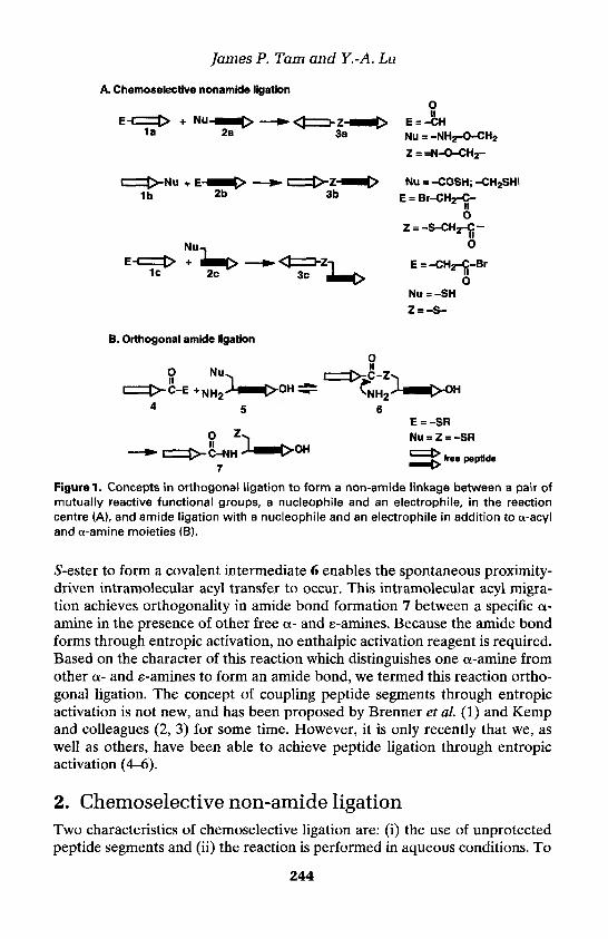
James P. Tam and Y.-A. Lu
A. Chemoselective nonamide ligation
Figure 1. Concepts in orthogonal ligation to form a non-amide linkage between a pair ofmutually reactive functional groups, a nucleophile and an electrophile, in the reactioncentre (A), and amide ligation with a nucleophile and an electrophile in addition to a-acyland a-amine moieties (B).
S-ester to form a covalent intermediate 6 enables the spontaneous proximity-driven intramolecular acyl transfer to occur. This intramolecular acyl migra-tion achieves orthogonality in amide bond formation 7 between a specific a-amine in the presence of other free a- and e-amines. Because the amide bondforms through entropic activation, no enthalpic activation reagent is required.Based on the character of this reaction which distinguishes one a-amine fromother a- and e-amines to form an amide bond, we termed this reaction ortho-gonal ligation. The concept of coupling peptide segments through entropicactivation is not new, and has been proposed by Brenner et al. (1) and Kempand colleagues (2, 3) for some time. However, it is only recently that we, aswell as others, have been able to achieve peptide ligation through entropicactivation (4-6).
2. Chemoselective non-amide ligationTwo characteristics of chemoselective ligation are: (i) the use of unprotectedpeptide segments and (ii) the reaction is performed in aqueous conditions. To
244

Figure 2. Non-amide orthogonal ligation.
achieve these chemistries, a reactive pair consisting of a nucleophile and anelectrophile is placed on the peptides during solid phase synthesis. Usually,the nucleophile is a weak base which has either a pKa significantly lower thanthe a- or e-amines or a nucleophilicity much stronger than the a-amine, so thatthe ligation can be selective in aqueous buffered solution at pH ~ 7. Applic-able weak bases include alkyl thiol, acyl thiol, 1,2-aminothiol (N-terminalcysteine), acylhydrazine, and arylhydrazine (Figure 2). Reactive electrophilesinclude haloacetyl, activated unsymmetrical disulphide, maleimide, oraldehyde. Chemoselectivity is achieved when these mutually reactive groupsare brought together in aqueous solution with the weak base as the solenucleophile to react with the electrophile. Protection of other functionalgroups on the peptides therefore becomes unnecessary.
2.1 Thiol chemistryThiol chemistry exploits the extraordinary reactivity of sulphhydryls in alkyl-ation with x-halocarbonyls, in addition to conjugated olefins and sulphur-sulphur exchange with disulphides. In practice, in thiol alkylation and additionto conjugated olefins, disulphides have frequently been used for chemo-selective ligation in both peptide synthesis and semi-synthesis because of thestability of the products.
2.1.1 ThioalkylationThiols are stronger nucleophiles than amines or alcohols in aqueous bufferedsolutions at neutral to mildly basic pH. Thus, our laboratory has exploitedthioalkylation for chemoselective segment ligation using unprotected peptides
245
11: Chemoselective and orthogonal ligation techniques
James P. Tarn and Y.-A. Lu
of a C-terminal segment of TGFa (7). Subsequently, we used a segment ofgp!20-(312-329) of type I human immunodeficiency virus (HIV-I) (8) in 1991to form branched peptides or protein mimetics. In both cases, thioalkylationof a chloroacetyl group incorporated on the lysine core matrix of a peptidedendrimer yielded a multiple antigen peptide system (MAPs) withunambiguous structures as determined by mass spectrometric analysis.Similarly, ligation of two unprotected peptides in aqueous condition wasachieved by another group to form peptides and proteins containing a non-amide bond at the coupling site (9).
Thioalkylation is popular in protein chemistry as a means of attachingligands, peptides, or reporter groups and cross-linking reagents because thethiol groups in proteins are easy to access. Thus, thioalkylation is also a con-venient method for the chemoselective ligation of peptides. The reactive thioland haloacetyl groups can be added to the synthetic peptides during stepwisesolid phase synthesis. Both alkyl (RCH2SH) and acyl (RCOSH) thiols havebeen used. Alkyl thiols, generally derived from cysteine, can be placed at anyposition, whereas acyl thiol in the form of thiocarboxylic acid is limited atpresent to the C-terminus. The haloacetyl moiety, whose C-X bond is acti-vated by the acetyl group, can be attached to the N-terminus or the side chainof lysine positioned anywhere in the sequence. The chloroacetyl group isstable to HF cleavage conditions in the absence of thiol scavengers. The rateof thioalkylation to give thioethers is pH dependent and increases as the pHbecomes more basic. Because side reactions, such as oxidation of thiol todisulphide as well as hydrolysis of the haloacetyl group, occur significantly atmore alkaline pH, thioalkylation is commonly performed at pH 7.5-8. Tominimize disulphide formation, Defoort et al. (10) have used phosphine as areducing agent with significant success. We have found that S-S oxidation canbe minimized by using a small amount of phosphine prior to the reaction andincluding EDTA during the reaction (11,12).
2.1.2 Thiol-disulphide exchangeThiol-disulphide exchanges to form unsymmetrical disulphide bonds havebeen exploited extensively both in protein and peptide chemistry. King et al.(13) have used an aromatic thiol, the 4-dithiopyridyl group, as a cysteinyl-activating group to cross-link proteins via intermolecular disulphide bonds.Similarly, other aromatic thiols such as 2-thiopyridyl and nitropyridylsulphenyl (Npys) have been used in peptide synthesis to form unsymmetricaldisulphides. These and other similar methods can be applied to the ligation ofunprotected peptides.
2.1.3 Thiol additionA thioether can also be formed by adding a cysteine thiol to an activateddouble bond of a maleimido group. This method is popular for cross-linkingproteins with reporter groups, and Kitagawa and Aikawa (14) have shown
246

11: Chemoselective and orthogonal ligation techniques
that insulin-containing maleimido groups can be coupled to glycoproteins.The reaction is specific and is usually carried out in aqueous solution with anoptimum pH around 7, although lysine and histidine also react slowly with themaleimido group at this pH. Thus, long reaction times should be avoided.Furthermore, hydrolysis of maleimides to non-reactive maleic acid occursabove pH 8. This method is convenient because N-alkyl or N-aryl maleimidegroups are available commercially either as free carboxylic acids or activeesters such as Af-hydroxysuccinimides, which can be incorporated as a pre-formed unit in solid phase synthesis. The maleimido group on lysine andphenylalanine has been shown to be stable to trifluoroacetic acid for 3 h, andis fully compatible with Fmoc chemistry when the maleimido group is addedlast in the sequence of peptide assembly.
2.2 Carbonyl chemistryAldehydes and ketones represent a group of electrophiles that are not presentin peptides but are exceptionally reactive electrophiles exploited for chemo-selective ligation. In aqueous conditions, particularly at acidic pH, carbonylsreact with amines to form unstable imines. To obtain stable products for select-ive exploitation, weak bases are used, particularly those with a strong tendencyfor a condensation reaction with aldehydes. In general, the desirable weakbases, except cysteine, do not occur naturally in amino acid sequences, andseveral types of weak bases for ligation to aldehydes have been developed forthis purpose. The first type consists of conjugated amines whose basicities arelowered by neighbouring electron-withdrawing groups that also stabilizeimino products. These include hydroxylamine to give oxime, and substitutedhydrazines, such as acylhydrazines and phenylhydrazines, to give hydrazone.The second type contains the 1,2-disubstituted pattern which forms a cycliccompound in an addition reaction to the imino intermediates. This includesderivatives of 1,2-aminoethanethiol and 1,2-aminoethanol such as those foundin N-terminal cysteine and threonine. These 1,2-disubstituted weak bases reactwith aldehyde to form a proline-like ring such as thiazolidine from cysteineand oxazolidine from threonine.
2.2.1 OximeAlthough hydroxylamine-aldehyde chemistry was used by Erlanger et al. (15)to form oximes and later by Pochon et al. (16) for site-specific conjugation toprotein, this weak base was first used for peptide synthesis by Rose (17), andlater by Tuchscherer (18) and Shao and Tarn (11). The hydroxylamine com-ponent for oxime formation is usually introduced using the commerciallyavailable aminooxyacetic acid, NH2OCH2COOH, which can also be used as aprotected unit for incorporation at any position in the peptide sequenceduring the solid phase synthesis. Aminooxyacetyl-containing peptides arestable to HF and TFA cleavage conditions and can be ligated to aldehydes togive an oxime linkage. Oxime linkages have been used for preparing end-to-
247

James P. Tarn and Y.-A. Lu
side chain cyclized peptides and peptide dendrimers (11, 19). Such intra-molecular cyclization by oxime formation is highly chemoselective andflexible, since the reactive weak base-aldehyde pair can be placed in differentconfigurations, allowing side chain-to-side chain, end-to-end, and end-to-sidechain cyclization (11).
Typically, a serine residue (a masked aldehyde precursor) is coupled to alysyl side chain and the aminooxyacetyl group is used as the weak base at theN-terminus. Under mildly acidic conditions, this weak base is the only re-active nucleophile present. All other functional groups on the amino acid sidechains, including the NE-amine of lysine, are protonated or form reversibleSchiff s bases.
2.2.2 HydrazoneLigation by hydrazone formation is probably one of the oldest methods inprotein and carbohydrate chemistry (20). In general, acyl- and aryl-sub-stituted hydrazines have been used successfully. Offord, Rose and colleagues(21-23) have applied acylhydrazine-aldehyde chemistry for site-specific con-jugation to proteins, protein semi-synthesis, and backbone engineering. Touse hydrazide-aldehyde chemistry to ligate a peptide on the N-terminus of asecond peptide, Shao and Tarn (11) have prepared Boc-monohydrazide succinicacid, which allows facile introduction to the a-amine and the side chain oflysine at any position. Thus, unprotected peptides containing the hydrazidesuccinyl group are used to ligate to the aldehyde-containing peptide withgreat efficiency. Similarly, 4-Boc-inonohydrazinobenzoic (Hob) acid has beendeveloped as a derivative for analogous modification strategies during peptidesynthesis (12). Similar to the hydroxylamine derivative, Hob-peptides arestable to the usual cleavage conditions involving TFA or HF. The progress ofthe phenylhydrazone reaction can be conveniently monitored with UV at 340nm (12). The preparation of a peptide dendrimer through hydrazone ligationis described in the following protocols.
Protocol 1. Preparation of glyoxylyl tetravalent lysine corepeptide
Reagents• H-Ala-OCH3-HCI • Benzotriazol-l-yl-oxy-tris(dimethylamino). Methyl dimethoxyacetate phosphonium hexafluorophosphate (BOP). Boc-Lys(Boc)-OH • Silica gel, 130-270 mesh, 60 A• N,N-Diisopropylethylamine (DIPEA)
A. Preparation of Boc-Lys(Boc)-Ala-OCH3
1. Cool in an ice-bath a solution (DMF 12 ml:DCM 6 ml) of H-Ala-OCH3-HCI (1.4 g, 10 mmol) and Boc-Lys(Boc)-OH (3.46 g, 10 mmol)cooled in ice-bath.
248

11: Chemoselective and orthogonal ligation techniques
2. Add BOP (4.42 g, 10 mmol) and DIPEA (2.84 g, 22 mmol).
3. Stir the reaction mixture for 30 min at 0°C and then at roomtemperature for 20 h.
4. Remove DMF and DCM in vacua.
5. Add ethyl acetate (50 ml) to residue.
6. Extract organic phase with saturated NaCI(aq) (2 x 50 ml), 2% w/vKHSO4(aq) (2 x 50 ml), 5% w/v NaHCO3(aq) (3 x 50 ml), and water (3 x50 ml).
7. Dry organic phase using anhydrous Na2S04.
8. Concentrate organic phase to dryness.
9. Recrystallize from ethyl acetate/hexane give dipeptide Boc-Lys(Boc)-Ala-OCH3. YiekJ 4.37 g
(91%). MS: calc. MH+ 479.6. C24H37N3O7 req. C, 60.11; H, 7.78; N, 8.76.
B. Preparation ofBoc-Lys(Boc)-Lys[Boc-Lys(Boc)]-Ala-OCH3
1. Add 50% v/v TFA in DCM (20 ml) to dipeptide (A) (0.96 g, 2 mmol),and stir at room temperature for 20 min.
2. Remove TFA/DCM in vacuo.
3. Wash residue with diethyl ether (4 x 10 ml) and dissolve in DMF(20 ml).
4. Add DIPEA (1.55 g, 12 mmol) and Boc-Lys(Boc)-OH (1.44 g, 4.2 mmol)to above DMF solution cooled to 0°C.
5. Add BOP (1.89 g, 4.2 mmol), stir for 30 min at 0°C then for 24 h atroom temperature.
6. Remove DMF and DCM in vacuo.
7. Add ethyl acetate (50 ml) to residue.
8. Wash organic phase with saturated NaCI(aq) (2 x 50 ml), 2% w/vKHS04(aq) (2 x 50 ml), 5% w/v NaHC03(aq) (3 X 50 ml), and water (3X 50 ml).
9. Dry organic phase using anhydrous Na2SO4.
10. Concentrate in vacuo organic phase to dryness.
11. Recrystallize from ethyl acetate/hexane to yield Boc-Lys(Boc)-Lys[Boc-Lys(Boc)]-Ala-OCH3. Yield 1.65 g (92%). MS: calc. M+ + Na+
911. C42H77N7O13 req. C, 56.8; H, 8.74; N, 11.04.
C. Preparation of <CH3O)2CHCOOH
1. Add 0.5 M NaOH(aq) (10.5 ml, 5.25 mmol) to a stirred solution ofmethyl dimethoxyacetate (671 mg, 5 mmol) in methanol (5 ml).
2. Stir for 2 h, monitor completion of reaction by TLC.
249

James P. Tarn and Y.-A. Lu
3. Remove methanol by evaporation in vacua, and dilute to 25 ml withwater.
4. Wash aqueous solution with ethyl acetate (3 x 10 ml).
5. Concentrate to 10 ml by evaporation in vacua, lyophilize to dryness.
D. Branched tetrapeptide (CH3O)2CHCO-Lys((CH3O)2CHCO)-Lys[(CH3O)2CHCO-Lys-((CH3O)2CHCO)]-Ala-CI-l3
1. Add 50% v/v TFA in DCM (20 ml) to (B) (0.89 g, 1 mmol), and stir for20 min.
2. Remove TFA/DCM, wash residue with ether (3 X 20 ml) and dissolvein DMF (10 ml).
3. Add powder (C) (5 mmol), BOP (1.86 g, 4.2 mmol), and DIPEA (0.65 g,5 mmol) to above DMF solution. Stir at room temperature for 20 h.
4. Remove DMF by evaporation in vacuo, add ethyl acetate (10 ml) toresidue, stand at 4°C overnight. The product precipitates.
5. Filter to collect product and dry in vacuo.
6. Take the ethyl acetate supernatant from step 4 and evaporated todryness in vacuo.
7. Dissolve dried product in water (3 ml).
8. Add glacial acetic acid (4 drops), filter out precipitate (HOBt).
9. Concentrate filtrate to dryness in vacuo.
10. Triturate residue with diethyl ether (4 X 10 ml) to give a whitepowder.
11. Combine products from steps 4 and 10, and purify on silica gel (40 g)column (elute with CHCIs/EtOAc/MeOH, 60:25:15). Yield 680 mg(76%). MS: calc. M+ + Na+ 919.0. C38H69N7017 req. C, 50.94; H, 7.66; N,10.94.
E. Preparation of CHO-CO-Lys(CHO-CO)-Lys[CHO-CO-Lys(CHO-CO)]-Ala-OH
1. Add 0.1 M NaOH(aq) (1.8 ml, 0.18 mmol) to a solution (10 ml water,2 ml MeOH) of branched tetrapeptide (D) (134.4 mg, 0.15 mmol), andstir for 2 h at room temperature.
2. Remove MeOH.
3. Add 0.2 M HCI(aq) (0.25 ml) to remaining aqueous solution.
4. Lyophilize to give white powder.
5. Dissolve powder in water (1.5 ml).
6. Add concentrated HCI (5 ml) to 500 ul of the solution prepared in step5 (50 umol) and stir for 3 h at room temperature.
250

11: Chemoselective and orthogonal ligation techniques
7. Concentrate to dryness in vacua on water bath at 35°C.
8. Purify residue by RP-HPLC to afford the glyoxylyl core peptide(22.5 mg, 64%). MS: calc. M+ 698.7.
Protocol 2. Synthesis of peptide dendrimer through hydrazoneligation
Reagents. Peptide NH2NHCOCH2CH2CO-[VMEYKARR- . CHO-CO-Lys(CHO-CO)-LyslCHO-CO-
KRAAIHVMLALA] (synthesized by Fmoc/f- Lys(CHO-CO)]-Ala-OH (from Protocol 1)Bu chemistry) . 0.2 M AcONa/AcOH buffer, pH 5.7
• Dimethylsulphoxide (DMSO)
Method
1. Add DMSO (800 ul) to a mixture of NH2NHCOCH2CH2CO-[peptide] (in400 u| water, 2 umol), CHO-CO-Lys(CHO-CO)-Lys[CHO-CO-Lys(CHO-CO)]-Ala-OH (in 40 ul water, 0.2 n,mol), and acetate buffer (400 ul).
2. Adjust pH to 5.7.
3. Incubate at room temperature for 5 h.
4. Purify ligation product by RP-HPLC.
5. Adjust the pH of HPLC fractions collected to 5.5.
6. Lyophilize to give the required product (contains sodium trifluoro-acetate).
2.2.3 ThiazolidineThe reaction of aldehydes with N-terminal cysteine to give thiazolidine haslong been known, but only recently has this reaction been exploited forchemoselective segment ligation (5, 11,12, 24, 25). Thiazolidine and oxazoli-dine are thia- and oxaproline analogues. This method affords a heterocyclicpseudoproline at the ligation site, and may be useful in imparting con-formational constraint to the peptides. Unlike thiol chemistry, thiazolidinering formation requires both a thiol and an amine in a 1,2-substitutedrelationship, making the reaction highly specific. Thiazolidine ring formationcan be performed at pH 2-8, the reaction rate accelerating with increasingpH. Since the thiol group is readily oxidized at high pH, pH 4.5-5.4 generallyprovides efficient rates without side reactions. Thiazolidine ligation is gen-erally more practical than oxazolidine ligation since this reaction undergoesrapid ring-chain tautomerization. Furthermore, thiazolidine is >104 timesmore stable than the oxazolidine and prefers ring formation, whereasoxazolidine generally prefers an open chain imine form and must be acylatedto give the stable oxazolidine ring.
251

fames P. Tarn and Y.-A. Lu
A. Oxidation
CH2OH
HQ-CH2
NHyCH-
B. Llgation
ft HS-i
free peptide
Figures. Thiazolidine formation in ligation site by two steps. (A) Oxidation of a C-terminal 1,2-propanediol peptide or an N-terminal serinyl peptide by NalO4 to form a C-terminal or an N-terminal peptide aldehyde, respectively. (B) An aldehyde ligated with an/V-cysteinyl peptide to form a thiazolidine peptide with different orientations of thethiazolidine ring.
The peptide aldehyde can be prepared in several ways. Oxidation of an N-terminal serinyl peptide or oxidation of a C-terminal 1,2-propanediol peptideyields the N-terminal or C-terminal peptide aldehyde, respectively (seeSection 3.1.2), to give ligation products with different orientations of theirthiazolidine ring (Figure 3). Typically, use of water-miscible organic solvents,such as DMF, or reactions performed at 37 °C significantly accelerate theligation reaction (11).
3. Orthogonal amide ligationOrthogonal ligation provides an amide bond at the coupling site. Thus, unlikechemoselective ligation, it can produce a peptide with an all-a-amide back-bone and can be considered as a strategy for convergent peptide synthesis. Toachieve an amide ligation reaction, four functional groups in the reactioncentre are required to accommodate a capture reaction between a nucleophileand an electrophile followed by an acyl migration to form an amide bond.Two general methods based on thiol and carbonyl chemistries have beendeveloped to ligate unprotected peptide segments to form the peptide.
3.1 Carbonyl chemistry3.1.1 Thiazolidine ligation for pseudoprolineIn thiazolidine ligation, the reaction is performed between a glycolaldehydepeptide ester and an Na-cysteinyl peptide. The capture step is based on
252

1 1 : Chemoselective and orthogonal ligation techniques
n HS
_ ft ,CZt>-c-NH-<
Figure 4. Pseudoproline formation at the ligation side by a C-terminal aldehyde peptidereacting with a 1,2-aminothiol peptide at acidic pH. The reactive carboxyl and aminotermini are positioned in close proximity for spontaneous peptide bond formationthrough an entropy-driven intramolecular O-> /Vacyl transfer.
thiazolidine (Thz) ligation (see Section 2.2.3) (5, 26, 27), and the ortho-gonality of this reaction is based on the specific condensation between analdehyde and the 1,2-aminothiol at acidic pH in the presence of otherunprotected and reactive functional groups (Figure 4). Consequently, thethiazolidine formation brings the a-amine of one peptide segment close to thea-carboxyl moiety of another peptide segment, thus permitting peptide bondformation through an entropy-driven intramolecular O —» N acyl transfer. Apseudoproline residue is formed at the ligation site and can be viewed as aproline surrogate (2-hydroxymethyl-3-thiaproline).
3.1.2 Preparation of peptide aldehydesSince peptide aldehydes are involved in both chemoselective and orthogonalligations, the syntheses of peptide aldehydes involving either Boc or Fmocchemistry are described in greater detail. Although aldehydes can be obtainedby many organic transformations, two methods for preparing N-terminalpeptide aldehydes and five methods for C-terminal peptide aldehydes havebeen developed specifically for peptide ligation (Figure 5).
The first method exploits the popular NaIO4 oxidation of N-terminal Ser,Thr, or Cys to give an a-oxoacyl group (28, 29). Free pep tides containingthese N-terminal amino acids can serve as aldehyde precursors prior to theligation. Periodate oxidation of a 2-aminoalcohol such as Ser is facile andabout 1000 times faster than diol oxidation (21). Hence, oxidation of an N-Ser-containing peptide could be accomplished within a few minutes at neutralpH. Oxidation of Trp or other sensitive groups within the peptide sequencecan be minimized by adding a large excess of methionine as scavenger.Oxidation of C-terminal Cys to give the corresponding aldehyde requires alonger reaction time (25). In practice, transformation of a terminal 1,2-aminoalcohol (Ser, Thr) or 1,2-aminothiol (Cys) to an aldehyde is convenientand highly compatible with the overall scheme of peptide synthesis, as bothBoc and Fmoc chemistry can be used to generate the peptide aldehydeprecursor.
Another method based on the same principle has been developed for the
253

Figures. The methods for preparation of peptide aldehydes. Methods 1 and 2 prepare anN-terminal aldehyde peptide by oxidation of an N-terminal Ser-peptide or hydrolysis of adimethoxyacetate-peptide. Methods 3-7 prepare C-terminal aldehyde peptides. Methods3 and 4 are known as the 'n + V method. The peptide alkyl ester or alkyl thioester isobtained from different types of resins; then a masked amino acid glycodiol ester isintroduced by enzymatic synthesis (3a, 3b) or by a chemical method (4); finally, thepeptide is treated with TFA to give the aldehyde peptide. In methods 5 and 6, aldehydepeptides are obtained from oxidation of a peptide glycol diol ester. In method 7,treatment of an N-protected peptide thioester resin with Pd(0) and Et3SiH gives the cleavedC-terminal peptide aldehyde.
preparation of peptide dendrimers (11). Methyl dimethoxyacetate can becoupled to an amino group such as the branched lysine peptide, and acidhydrolysis will give the N-terminal aldehyde (Figure 5, method 2). Thismethod is more suitable for Fmoc than Boc chemistry.
The third and fourth methods developed by Liu and Tarn (5, 26) and Liu
254
James P. Tam and Y.-A. Lu

11: Chemoselective and orthogonal ligation techniques
and co-workers (27) are known as the 'n + 1' method, in which an amino acidcontaining a masked aldehyde is added to the C-terminus of a purifiedpeptide segment. This method bypasses problems associated with aldehydeinstability during TFA cleavage or NaIO4 oxidation of large peptidesegments and allows the synthesis of peptides by Boc chemistry. Bothmethods involve the solid phase synthesis of a peptide containing either analkyl ester or alkyl thioester at the C-terminus. The masked amino acidglycolaldehyde ester is then introduced by enzymatic synthesis (Figure 5,method 3) or a chemical method (Figure 5, method 4). In the enzymaticsynthesis, the glycolaldehyde component, amino acid dimethoxyethyl esterprepared from its Z-protected form by hydrogenolysis, is incorporated ontothe C-terminus of the peptide segment through the trypsin-catalysed,kinetically controlled aminolysis of the peptide ester bond (30), usuallyconducted in a solution containing a high content of water-miscible organicsolvent such as DMF/H2O (60:40 v/v).
The enzymatic synthesis is fast and is completed in 15 min. After puri-fication, the peptide obtained, with an acetal-protected aldehyde ester at itsC-terminus, is treated with 95% TFA/H2O for 5 min to release the aldehydefunction.
The chemical method via Ag+ ions is more versatile because it overcomesthe limitation imposed by substrate specificity of an enzymatic reaction. Inthis 'n + 1' coupling, formation of a peptide bond involves a large un-protected peptide and a small functionalized amino acid. An excess of anamino acid containing a masked glycoaldehyde is added to the peptide ester.The selectivity and efficiency of this coupling is driven by the Ag+ ionmediated activation which distinguishes the CMhioester from other sidechain unprotected carboxylic groups and by an overwhelming excess of thesmall amino acid derivative of the masked glycol. The protected aldehyde canthen be unmasked by mild acidic conditions in the presence of the othercomponent bearing the weak base to complete the ligation reaction.
The next three methods are on-resin derivatization based on linkers witha masked aldehyde moiety. The method developed by Botti et al. (31)(Figure 5, method 5) involves a new resin to yield a C-terminal peptideglyceric ester. After assembling the peptide sequence by Fmoc chemistry onthis resin, cleavage gives a glyceric ester diol which is converted to theglycoaldehyde by oxidation with NaIO4 at pH 5. The rate of diol oxidationincreases as the pH falls below 5. Thus, at pH 2 the oxidation is completewithin a few minutes. At pH 6-7 the rate of oxidation is slow, and requiresseveral hours at pH 7. It should be noted that when the oxidation isperformed below pH 5, the Met residues oxidize to Met(O), even in thepresence of a large excess of methionine as a scavenger. This side reactioncan be avoided at neutral pH. The cyclic acetal resin is prepared as describedin the following protocols.
255

fames P. Tarn and Y.-A. Lu
Protocol 3. Preparation of the cyclic acetal resin
Reagents• Chloromethyl polystyrene • DMSO• Ethylene glycol dimethyl ether • Glycol• 1,4-Dioxane • Hydroxylamine hydrochloride
Method1. Add sodium carbonate (0.2 g) to a suspension of chloromethyl poly-
styrene resin (0.5 g, 0.35 mmol) in DMSO (3 ml). Stir for 7 h at 155°C.This gives the benzaldehyde resin.
2. Wash resin with water, methanol, DMF, DCM, and diethyl ether (30 mleach).
3. Dry resin in vacuo.4. FTIR analysis of benzaldehyde resin shows the typical carbonyl
absorption at 1700 cm-1.5. Optional analysis of resin. Treat the resin (10 mg) with a large excess
of hydroxylamine in pyridine for 6 h at 95°C. Wash resin and dry. FTIRanalysis shows no carbonyl absorption at 1700 cnr1 and microanalysisshows 0.82% nitrogen, indicating a loading of aldehyde of 0.59 mmol/g.
6. Mix and stir the benzaldehyde resin (1 g, 0.59 mmol) with anhydrousethylene glycol dimethyl ether (10 ml), glycol (1 g), and p-toluene-sulphonic acid (10 mg) for 24 h at 90°C.
7. Wash resin with 3% sodium carbonate(aq)/dioxane (1:1 v/v), water,dioxane, DMF, methanol, DCM, and diethyl ether (50 ml each).
8. Dry resin in vacuo.
Protocol 4. Preparation of Fmoc-Gly-OCH2-cyclic acetal resin
Reagents• Fmoc-Gly-OH • 1,4-Dioxane• 4-Dimethylaminopyridine (DMAP) • W-Hydroxybenzotriazole (HOBt)• W./V-Dicyclohexylcarbodiimide (DCC)
Method1. Stir a mixture of Fmoc-Gly-OH (1.66 g, 5.58 mmol), DCC (0.63 g,
3 mmol), and HOBt (44 mg, 0.28 mmol) in DMF (10 ml) for 20 min.2. Add the solution prepared in step 1 to glycerol-acetal resin (1 g,
prepared by Protocol 3) in DMF (5 ml) and stir for 18 h.3. Wash resin with dioxane, DMF, methanol, DCM, and diethyl ether
(50 ml).
256

11: Chemoselective and orthogonal ligation techniques
4. Dry resin in vacua.
5. Analyse the resin by microanalysis (typically 0.71% of nitrogen, corres-ponding to a substitution of 0.5 mmol/g) or by Fmoc-substitutiondetermination (see Chapter 3).
Method 6 (Figure 5) developed by Zhang et al. (32) is based on Fmoc-chemistry synthesis of a C-terminal diol peptide on a diol-derivatized 2-chlorotrityl resin. Oxidation of the free diol peptide gives the peptidealdehyde. A detailed procedure for the preparation of a diol trityl resin andC-terminal peptide aldehyde is given in the following protocols.
Protocol 5. Preparation of N-Fmoc-3-amino-1,2-propanediolderivatized 2-chlorotrityl polystyrene
Reagents• Fluorenylmethyl chloroformate • 3-Amino-1,2-propanediol• Triethylamine • 2-Chlorotrityl chloride polystyrene resin• DMAP • W,/V-Dimethylacetamide• r-Butyl-2,2,2-trichloroacetimidate
Method
1. Add fluorenylmethyl chloroformate (5.2 g, 20 mmol) to a solution of3-amino-1,2-propanediol (1.8 g, 20 mmol) and triethylamine (3.1 ml,22 mmol) in 100 ml of water/1,4-dioxane (1:3 v/v) at 4°C.
2. Stir for 18 h.
3. Remove solvent by evaporation in vacuo.
4. Add ethyl acetate (10 ml) to the residue and crystallize to give 5.6 g(89% yield) of 3-(9-fluorenylmethoxycarbonyl)-amino-1,2-propane-diol. m.p. 133-134°C. MS: calc. MH+ 314.
5. Add 3-(9-fluorenylmethoxycarbonyl)-amino-1,2-propanediol (1.25 g,4 mmol) to 2-chlorotrityl chloride resin (1 g, 1 mmol) and DMAP(0.5 g, 2 mmol) in dry N,N-dimethylacetamide (10 ml).
6. Gently agitate the mixture for 24 h.
7. Wash resin with DMF, methanol, and DCM (30 ml each).
8. Dry resin in vacuo.
9. Analysis of resin: take 5-10 mg resin for determination of resin Fmoc-substitution (0.8 mmol/g).
10. Add f-butyl-2,2,2-trichloroacetimidate (200 ul, 1.07 mmol) in DCM(25 ml) and shake for 30 min to block the free hydroxy group of thederivatized 2-chlorotrityl polystyrene.
257

fames P. Tarn and Y.-A. Lu
Protocol 6. Preparation of C-termina! peptide aldehyde,AAVALLPAVLLALLA-NHCH2CHO
Reagents• C-terminal 1,2-propanediol peptide: • Sodium periodate
AAVALLPAVLLALLA-NHCH2CH(OH)CH2OH,synthesized by Fmoc chemistry on Fmoc-3-amino-1,2-diol derivatized 2-chlorotritylresin (prepared by Protocol 6)
Method
1. Add Nal04 (20 mM, 0.6 ml in water) solution to the peptide
AAVALLPAVLLALLA-NHCH2CH(OH)CH2OH (30 mg, 0.02 mmol)solution in 33% acetic acid(aq) (6 ml).
2. Run RP-HPLC to determine the reaction after 15 min.
3. Purify the reaction mixture by using RP-HPLC.
The final method (Figure 5, method 7) is an on-resin Pd(0)-catalysedreduction of thioesters. Treatment of N-protected peptide thioester resin withPd(0) in the presence of Et3SiH gives the cleaved C-terminal peptide aldehyde.The reaction is performed typically under N2 in THF or DCM using a 20-30-fold excess of Et3SiH at 4°C to avoid racemization. The catalyst is obtained byin situ reduction of Pd(OAc)2. The yield ranges from 80 to 89% (33).
3.2 Thiol chemistry3.2.1 Thioester ligation for Cys and MetThioester ligation involves two segments, each containing a thioester and aCys that results in the production of a cysteine residue at the ligation site. Inthis approach, the first step is formation of a covalent thioester-linked inter-mediate by nucleophilic attack of the thiol side chain of an W-cysteinepeptide on a thioester segment. An intramolecular 5 —»N acyl transfer to forman amide bond then completes the ligation reaction (Figure 6).
There are two approaches in thioester ligation. Our laboratory (4) uses apre-derivatized thioester resin that permits the generation of a thioesterdirectly, following solid phase synthesis. It should be noted that the requiredpeptidyl thioester segment is best prepared using Merrifield Boc-chemistry.The ligation reaction is performed under strongly reductive conditions and anexcess of the thiol-component (cysteinyl-peptide) is used. Based on the rates,yields, and product distributions of the thiol-thioester exchange reaction atvarious pHs ranging from 5.6 to 7.6, an increase of 1.5- to 2-fold productpurity with each pH unit is observed. Three groups of by-products areobserved: Na,S-diacyl peptides; Aa-acyl, disulphide conjugate peptides; and
258

11: Chemoselective and orthogonal ligation techniques
Figures. Thiol-thioester exchange to form an amide bond. Nucleophilic attack takesplace by the side chain thiol functionality in a cysteinyl peptide at a thioester segment toform an intramolecular thioester, which undergoes intramolecular S ->/V acyl transfer toform an amide bond.
the hydrolysed peptidyl thioester. At pH 5.6, the desired ligation reaction isslowest and hydrolysis of the peptidyl thioester greatest (78%). At pH 7.6,hydrolysis occurs at a slow rate, to yield 56% of the desired ligation product.
Collectively, these results support the use of basic pH to inhibit hydrolysisof the peptidyl thioester, and the strong reducing environment of a small thioland trialkylphosphine to prevent AAS-diacyl by-products and to reducedisulphide formation. Using these optimized conditions, peptides rangingfrom 9 to 88 amino acids with 60-88% yield were prepared. The methods forpreparation of the thioester resin and ligation are described below.
The second approach is reported by Kent et al. (34) and differs only by theuse of a peptidyl thiocarboxylic acid as precursor and the peptidyl thioester isobtained via thioalkylation or disulphide exchange. This method requires anextra step but the ligation mechanism is similar to that described above.
Protocol 7. Preparation of Boc-Gly-thioester resin
Reagents• 4-Methylbenzhydrylamine polystyrene resin
(MBHA resin)• 3-Mercaptopropionic acid• N,N-Diisopropylcarbodiimide (DIG)• Triphenytphosphine
Boc-Gly-OHHOBtAcetyl chlorideCysteine methyl ester-hydrochlorideDIPEA
Method1. Swell MBHA resin (10 g, 5.4 mmol) in DCM (200 ml) for 10 min, in an
appropriate SPPS reaction vessel.2. Filter the resin.3. Add a solution of 3-mercaptopropionic acid (3.44 g, 32.4 mmol) in
70 ml DMF to resin, and shake for 3 min.4. Add a solution of HOBt (4.38 g, 32.4 mmol) in 70 ml DMF to reaction
vessel, and mix for 3 min.5. Add a solution of DIG (5.07 ml, 32.4 mmol) in 60 ml DCM, and mix
gently for 120 min.
259
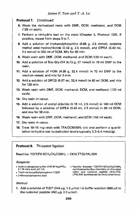
fames P. Tarn and Y.-A. Lu
Protocol 7. Continued6. Wash the derivatized resin with DMF, DCM, methanol, and DCM
(120 ml each).
7. Perform a ninhydrin test on the resin (Chapter 3, Protocol 13A). Ifpositive, repeat from steps 3 to 7.
8. Add a solution of triphenylphosphine (0.65 g, 2.5 mmol), cysteinemethyl ester-hydrochloride (0.43 g, 2.5 mmol), and DIPEA (0.43 ml,2.5 mmol) in 200 ml of DCM. Mix for 60 min.
9. Wash resin with DMF, DCM, methanol, and DCM (120 ml each).
10. Add a solution of Boc-Gly-OH (4.73 g, 27 mmol) in 70 ml DMF to theresin.
11. Add a solution of HOBt (4.38 g, 32.4 mmol) in 70 ml DMF to thereaction vessel, and mix for 3 min.
12. Add a solution of DIPCD (5.07 ml, 32.4 mmol) in 60 ml DCM, and mixfor 120 min.
13. Wash resin with DMF, DCM, methanol, DCM, and methanol (120 mleach).
14. Dry resin in vacua.
15. Add a solution of acetyl chloride (0.18 ml, 2.5 mmol) in 100 ml DCMfollowed by a solution of DIPEA (0.43 ml, 2.5 mmol) in 50 ml DCM,and mix for 30 min.
16. Wash resin with DMF, DCM, methanol, and DCM (150 ml each).
17. Dry resin in vacua.
18. Treat 10-15 mg resin with TFA/DCM(50% v/v) and perform a quanti-tative ninhydrin test (substitution levels typically 0.3-0.4 mmol/g).
Protocol 8. Thioester ligation
Reaction: TQFCFH-SCH2CH2CONH2 + CKVLTTGLPALISW
Reagents• Sodium phosphate buffer (0.02 M Na2HP04- . Peptidyl thioester TQFCFH-SCH2CH2CONH2
0.1 M citric acid, pH 7.6) (synthesized by Boc-chemistry on thio-ester. Tris(2-carboxyethyl)phosphine (TCEP) resin) and cysteinyl peptide CKVLTTG-. 3-Mercaptopropionic acid LPALISW (synthesized by Fmoc-chemistry)
Method
1. Add a solution of TCEP (344 ug, 1.2 (umol) in buffer solution (600 ul) tothe cysteinyl peptide (450 ug, 0.3
260

11: Chemoselective and orthogonal ligation techniques
2. Add the cysteinyl peptide solution to the peptidyl thioester (94 (ug,0.1 (umol).
3. Run analytical RP-HPLC at set time-intervals, e.g. 0 min, 30 min, 1 h,and 3 h.
4. Purify the ligated peptide TQFCFHCKVLTTGLPALISW by RP-HPLC.
Thioesters can also be used for intramolecular ligation for the synthesis ofend-to-end cyclic peptides (35, 36). The linear peptide precursor shouldcontain both an N-terminal cysteine residue and a C-terminal thioester.Under previously described conditions, intramolecular thiol-exchange pro-duces a thiolactone, which then undergoes S -» N migration to yield an end-to-end cyclic peptide.
Thioester ligation to yield a methionine residue can be achieved between aC-terminal thioester peptide and an N-terminal homocysteinyl peptide (37).The ligation reaction results in the formation of a homocysteine residue,which is readily S-methylated with excess methyl p-nitrobenzenesulphonate,to yield a methionine residue at the ligation site, or another alkylating reagentto form unusual amino acid residues.
3.2.2 Perthioester ligation for CysSimilar to thioester ligation, perthioester ligation produces Cys at the ligationsite (Figure 7). Two segments containing, respectively, a thiocarboxylic acidand a cysteinyl derivative are involved. However, in this case, the intra-molecular acyl transfer reaction involves an acyl disulphide functionality (38).The method features specific capture of a peptide segment containing the N-terminal thiol-activated Cys residue by the C"-thiocarboxylic acid of thesecond segment. The mixed acyl disulphide perthioester then undergoes arapid intramolecular S —» N acyl transfer via a six-membered ring inter-mediate. Thiolytic reduction of the resulting hydrodisulphide (S-SH) givesthe native Cys residue at the ligation site.
This approach has been used for the synthesis of a 32-residue peptide (38)from two purified segments, specifically the acyl segment (15 residues)containing a thiocarboxylic acid and the amino segment containing an JV0
Cys(Npys). The reaction was carried out at pH 2 in aqueous acetonitrile, andthe segment concentration was 10-15 |xM. During the course of the ligationreaction, release of a yellow coloration (due to released Npys-H) wasobserved, indicating the occurrence of the capture reaction by sulphur-sulphur exchange. Finally, the pH was adjusted to 6, and the reaction mixturetreated with 1,4-dithiothreitol (DTT) to afford the desired product.
The high efficiency of the first capture step is attributed to the Npys-activated sulphide, and to the super-nucleophilicity and low pKa of thethiocarboxylic acid compared to a normal alkyl thiol. The acyl transfer was
261

fames P. Tam and Y.-A. Lu
Figure 7. General scheme of perthioester ligation. The capture of an activated thiol sidechain of an N-terminal Cys residue of the amino segment by the CMhiocarboxylic acid ofthe acyl segment results in the formation a mixed acyl disulphide which undergoes arapid intramolecular S -»N acyl transfer through a six-membered ring intermediate.Thiolytic reduction of the resulting hydrodisulphide (S-SH) using DTT gives the nativeCys residue at the ligation site.
rapid and 90% complete within 5 min, even at pH < 4. The efficiency of thisacyl transfer step is attributed to the activated acyl disulphide and to the closeproximity of the C"-acyl and AA-amine in the six-membered ring inter-mediate. This method of orthogonal segment ligation followed by disulphidereduction is a one-pot process, which does not require the isolation of inter-mediates. Its high efficiency and simplicity make this strategy an attractiveapproach for the synthesis of large peptides and proteins.
3.2.3 Imidazjple ligation for HisThe imidazole group in the histidine side chain is a well-known weak base andcan participate in acyl transfer reactions in a number of enzymatic process.Thus, the NTN of His has also been exploited for orthogonal ligation to giveHis at the ligation site. In this strategy, one segment contains the AA-histidineand the second acyl segment contains a C-terminal thiocarboxylic acid orthioester. Orthogonal ligation occurs to yield an Xaa-His bond when asuitable thiophilic promoter, such as an aryl disulphide or Ag+ ion, is added toactivate the acyl segment. This leads to A™ —» Na acyl transfer, resulting in apeptide bond formation (39). This method has been used for the ligation ofhistidine-containing peptides such as human calcitonin and parathyroidhormone. A drawback in this strategy is that the reactive acyl imidazoleintermediate is particularly prone to hydrolysis.
References1. Brenner, M., Zimmerman, J. P., Wehrmiiller, J., Quitt, P., and Photaki, I. (1955).
Experientia, 11, 397.2. Kemp, D. S. (1981). Biopolymers, 20,1793.3. Fotouhi, N., Galakatos, N., and Kemp, D.S. (1989). /. Org. Chem., 54,2803.
262

11: Chemoselective and orthogonal ligation techniques
4. Tarn, J. P, Lu, Y.-A., Liu, C. F., and Shao, J. (1995). J. Proc. Natl. Acad. Sci. USA,92,12485.
5. Liu, C. F. and Tarn, J. P. (1994). Proc. Natl. Acad. Sci. USA, 91,6584.6. Schnolzer, M. and Kent, S. B. H. (1992). Science, 256,221.7. Lu, Y.-A., Clavijo, P., Galantino, M., Shen, Z.-Y., Liu, W., and Tarn, J. P. (1991).
Molecular Immunology, 28, 623.8. Defoort, J. P., Nardelli, B., Huang, W., Ho, D. D., and Tarn, J. P. (1992). Proc.
Natl. Acad. Sci. USA, 89,3879.9. Muir, T. W., Williams, M. J., Ginsberg, M. H., and Kent, S. B. H. (1994).
Biochemistry, 33,7701.10. Defoort, J. P., Nardelli, B., Huang, W., and Tarn, J. P. (1992). Int. J. Peptide
Protein Res., 40,214.11. Shao, J. and Tarn, J. P. (1995). /. Am. Chem. Soc., 117,3893.12. Spetzler, J. C. and Tarn, J. P. (1995). Int. J. Pept. Protein Res., 45,78.13. King, T. P., Li', Y., and Kochoumian, L. (1978). Biochemistry, 171,499.14. Kitagawa, T. and Aikawa, T. (1976). J. Biochem. (Tokyo), 79,233.15. Erlanger, B. F., Borek, F., Beiser, S. M., and Lieberman, S. J. (1957). Biol. Chem.,
288,713.16. Pochon, S., Buchegger, F., Pelegrin, A., Mach, J.-P., Offord, R. E., Ryser, J. E.,
and Rose, K. (1989). Int. J. Cancer, 43,1188.17. Rose, K. (1994). J. Am. Chem. Soc., 116, 30.18. Tuchscherer, G. (1993). Tetrahedron Lett., 34,8419.19. Pallin, T. D. and Tarn, J. P. (1995). J. Chem. Soc., Chem. Commun., 2021.20. Bergbreiter, D. E. and Moinongan, M. (1991). In Comprehensive organic synthesis
and efficiency in modern organic chemistry (ed. B. M. Trost and J. Flemming), Vol2, p. 503, Pergamon, New York.
21. Gaertner, H. F., Rose, K., Cotton, R., Timms, D., Gamble, R., and Offord, R. E.(1992). Bioconjugate Chem., 3, 262.
22. Gaertner, H.F., Offord, R. E., Cotton, R., Timms, D., Camble, R., and Rose, K.(1994). J. Biol. Chem., 269, 7224.
23. Fisch, I., Kunzi, G., Rose, K., and Offord, R. E. (1992). Bioconjugate Chem., 3,147.24. Rao, C. and Tarn, J. P. (1994). J. Am. Chem. Soc., 116,6975.25. Tarn, J. P., Rao, C., Liu, C. F., and Shao, J. (1995). Int. J. Pept. Protein Res., 45,209.26. Liu, C. F. and Tarn, J. P. (1994). J. Am. Chem. Soc., 116,4149.27. Liu, C. F., Rao, C., and Tarn, J. P. (1996). J. Am. Chem. Soc., 118,307.28. Clamp, R. and Hough, L. (1965). Biochem. J., 94,17.29. Dixon, H. B. F. and Fields, R. (1972). In Methods in enzymology (ed. C. H. W.
Hirs and S. N. Timasheff), Vol. 25, p. 409, Academic Press, New York.30. Barbas III, C. F., Matos, J. R., West, J. B., and Wong, C. H. (1988). J. Am. Chem.
Soc., 110,5162.31. Botti, P., Pallin, T. D., and Tarn, J. P. (1996). J. Am. Chem. Soc., 118,10018.32. Zhang, L., Torgerson, T. R., Liu, X. Y., Timmons, S., Colosia, A. D., Hawiger, J.,
and Tarn, J. P. (1998). Proc. Natl. Acad. Sci. USA, 95, 9184.33. Wyslouch-Cieszynska, A. and Tarn, J. P. (1999). Frontiers of peptides science:
Proceedings of the 15th APS, (ed. J. P. Tarn and P. T. P. Kaumaya), p. 263. KluwerAcademic Publishers, Dordrecht.
34. Dawson, P. E., Muir, T. W., Clark-Lewis, I., and Kent, S. B. H. (1994). Science,266,776.
263

35. Zhang, L. and Tarn, J. P. (1997). /. Am. Chem. Soc., 119,2363.36. Tarn, J. P. and Lu, Y.-A. (1997). Tetrahedron Lett., 38,5599.37. Tarn, J. P. and Yu, Q. (1998). Biopolymer, 46, 319.38. Liu, C. F., Rao, C., and Tarn, J. P. (1996). Tetrahedron Lett., 37, 933.39. Zhang, L. and Tarn, J. P. (1997). Tetrahedron Lett., 38,3.

12
Purification of large peptides usingchemoselective tags
PAOLO MASCAGNI
1. IntroductionIn solid phase peptide synthesis (SPPS), deletion sequences are generated ateach addition of amino acid due to non-quantitative coupling reactions. Theirconcentration increases exponentially with the length of the peptide chain,and after many cycles not only do they represent a large proportion of thecrude preparation, but they can also exhibit physicochemical characteristicssimilar to the target sequence. Thus, these deletion-sequence contaminantspresent major problems for removal, or even detection.
In general, purification of synthetic peptides by conventional chromato-graphy is based on hydrophobicity differences (using RP-HPLC) and chargedifferences (using ion-exchange chromatography). For short sequences, theuse of one or both techniques is in general sufficient to obtain a product withhigh purity. However, on increasing the number of amino acid residues, thepeptide secondary and progressively tertiary and quaternary structures beginto play an important role and the conformation of the largest peptides candecisively affect their retention behaviour. Furthermore, very closely relatedimpurities such as deletion sequences lacking one or few residues can bechromatographically indistinguishable from the target sequence. Therefore,purification of large synthetic peptides is a complex and time-consuming taskthat requires the use of several separation techniques with the inevitabledramatic reduction in yields of the final material.
Permanent termination (capping) of unreacted chains using a large excessof an acylating agent after each coupling step prevents the formation ofdeletion sequences and generates N-truncated peptides. However, even underthese more favourable conditions, separation of the target sequence fromchromatographically similar N-capped polypeptides requires extensivepurification.
If the target sequence could be specifically and transiently labelled so thatthe resulting product were selectively recognized by a specific stationaryphase, then separation from impurities should be facilitated. This chapter

Paolo Mascagni
deals with such an approach and in particular with the purification of largepolypeptides, assembled by solid phase strategy, using lipophilic and biotin-based 9-fluorenylmethoxycarbonyl (Fmoc) chromatographic probes.
2. Purification of large polypeptides using Fmoc-based chromatographic probes
2.1 The concept of selective and reversible labellingAssuming that the formation of deletion sequences is prevented by cappingunreacted chains (see Protocol 1), a reciprocal strategy can be applied thatinvolves functional protection of all polymer-supported peptide chains thatare still growing, with a specially chosen affinity reagent or chromatographicprobe. In this way, the desired chains are distinguished from all by-productsthat have been irreversibly terminated either by capping or as a result ofchemical side reactions. The chromatographic probe contains an affinitygroup and a reversible linker (ref. 1 and references cited therein), which arestable under the conditions used to deprotect and cleave the peptide from theresin support. Affinity-labelled peptides are separated from terminatedpeptides which lack the affinity group by selective binding to a suitablestationary phase. The affinity label is then selectively removed using reagentswhich do not harm the peptide integrity.
The basic steps of this strategy are shown in Scheme 1. It should be em-phasized that impurities generated by side reactions occurring either duringsynthesis or during deprotection and that affect both the target sequence andcapped peptides are not removed by this technique.
The first example of purification by selective labelling was described in 1968for a peptide that was synthesized in solution (2). In 1976, Merrifield and co-workers (3) applied this concept to SPPS and demonstrated that peptidesbearing an intentionally introduced Cys-Met N-terminal dipeptide can bepurified by absorption onto an organometallic immobilized matrix. The nativesequence lacking Cys-Met was recovered through a CNBr-mediated Met-Xspecific cleavage.
Following these pioneering studies, several chromatographic probes havebeen proposed as a result of intentional design or modification of otherprocedures (for a review on the subject see refs 1 and 4).
2.1.1 Fmoc-based chromatographic probesThe Fmoc group is ideally suited as a template molecule to develop chroma-tographic probes, because its urethane link with the amino acid N" is stable tothe harsh, acidic conditions used to cleave peptides from the resin support,but it is labile under mild basic conditions that do not harm the polypeptidechain (5). Furthermore, because the probe is added to the peptide chain at theend of synthesis, Fmoc-probes can aid the purification of peptides synthesized
266

12: Purification of large peptides using chemioselective tags

Paolo Mascagni
using either of the two most widely used chemistry, i.e. Fmoc-chemistry and t-butoxycarbonyl (Boc)-chemistry.
The first Fmoc-based chromatographic probe was described in 1978 byMerrifield (6) and contained negative charges provided by a sulpho groupat position 2 of the fluorenyl moiety. The strongly acidic peptides thusderivatized were separated from unlabelled peptides by ion-exchangechromatography.
Another Fmoc-based probe, recently developed, contains benzo-fusedanalogues of the Fmoc molecule. It can be used to purify peptides by eitherporous graphitized carbon chromatography (7) or RP-HPLC (8).
In 1989, we proposed the 4-CO2H-Fmoc derivative 1 (9) as a probe for thepurification of large synthetic peptides (10). The advantage of 1 over otherFmoc-based molecules is in its carboxylic function which can be derivatizedwith groups possessing different chromatographic characteristics. For instance,addition of a lipophilic tail to 1 will generate a probe for RP-HPLC, whereasa polyionic tail or biotin will convert 1 in a probe for ion-exchange chroma-tography and affinity chromatography, respectively (9-14).
The usefulness of lipophilic probes in RP-HPLC purification was demon-strated with the synthesis of several proteins made by either an Fmoc strategyor the alternative Boc chemistry (9-16).
3. Capping of unreacted polypeptide chains in SPPSThe classical capping reagent, acetic anhydride, has been shown to be ex-tremely successful for the synthesis of relatively short peptides, but itsapplication to longer polypeptides has not been well documented. Further-more, in our hands the use of acetic anhydride in conjunction with an auto-mated peptide synthesizer has not been altogether satisfactory (17). Althougha systematic study to identify the reasons for this poor performance was notcarried out, several pieces of evidence indicated that the quality and stabilityof the acetic anhydride solutions used for capping are contributory factors.We therefore searched for solid reagents which could be weighed outaccurately in small amounts and dissolved in the required solvent just prior totheir use.
From this screening activity, we identified Ar-(2-chlorobenzyloxycarbonyl)-succinimide (Z(2-Cl)-OSu) as sufficiently reactive so that completion of acapping reaction requires only a few minutes (17). Furthermore, Z(2-Cl)-OSuand the terminating Z(2-C1) group are stable to the reagents used in both Bocand Fmoc chemistries.
In general, the capping reaction is carried out using a large excess of Z(2-Cl)-OSu (fourfold excess of all peptide chains or 400-fold excess assuming99% coupling) and in the presence of an organic base to catalyse the reaction.It should be noted that a small amount of unreacted chains might persist evenafter repetitive capping reactions. This phenomenon, which is common to all
268

12: Purification of large peptides using chemioselective tags
capping reagents, is probably due to inaccessibility to solvent of those peptidechains buried in the bulk of the resin-peptide matrix.
Protocol 1. Capping of unreacted polypeptide chains
Reagents• 0.284 g (4-fold excess") Z(2CI)-OSu in 2 ml N,N-diisopropylethylamine (DIPEA) in
of N-methylpyrrolidone (NMP)-dichloro- NMP. If not available, NMP can be sub-methane (DCM) (1:1 v/v) plus 1 ml of 2 M stituted with dimethylformamide (DMF)
Method
1. Prepare a fresh solution of Z(2CI)-OSu.
2. At the end of the coupling reaction, condition the peptidyl-resin with amixture of NMP/DCM (1:1, v/v) in preparation for the capping step.Remove the solvent but avoid drying the peptidyl-resin completely asit should be kept moist.
3. Add the Z(2CI)-OSu solution to the peptidyl-resin and vortex for 5min.b The reaction can be followed by the standard ninhydrin test orother similar colorimetric methods.
4. Remove the reaction solution by filtration and wash the productthoroughly with NMP/DCM (1:1, v/v). The peptidyl-resin is now readyfor the next coupling cycle.c
"The quantities given in the example refer to a concentration of amino groups present on theresin of 0.25 meq.6 If other methods of mixing are used, the reaction may take slightly longer.The capping cycle can be carried out in an automated fashion because it requires the samesolvents and reagents used for chain assembly.
4. RP-HPLC using lipophilic chromatographicprobes
In a comparative study, several hydrophobic probes differing in the type ofsubstitution at position 4 of 1 were prepared (12). Parameters evaluatedincluded stability of the probe during coupling and acid deprotection, RP-HPLC separation between derivatized and underivatized molecules, and timerequired for probe removal. Probe 2 offered the best compromise between allthese parameters; it is easily converted to an activated form (e.g. 2a) by usingthe same chemistry as for activation of the Fmoc molecule. If the reactionbetween 2a and the peptidyl-resin is carried out immediately after completionof chain assembly, the base-catalysed reaction requires 2-3 h. Longer reactiontimes might be necessary if derivatization is not carried out on freshly pre-pared polypeptides. In this case it is advisable to store the peptidyl-resin in an
269

Paolo Mascagni
inert solvent (NMP or DMF) at low temperature prior to use. The degree ofcoupling should be monitored using standard colorimetric methods (e.g. theninhydrin reaction) and the reaction repeated with a freshly prepared solutionof activated probe if coupling is not satisfactory.
The probe and the probe-peptide link are stable to acids and in the pres-ence of the most widely used scavengers. However, both scavengers and acidsmust be of high quality if side reactions at the Fmoc group and the peptidechain are to be avoided. In general, the solubility profile of large polypeptidesis not significantly altered by the presence of lipophilic probe 2.
The extent of HPLC separation between derivatized and underivatizedpeptides depends on the length of the polypeptide chain, the type of station-ary phase used for separation (e.g. C4-C18), and the elution conditions. Whenusing CIS columns the difference in retention time between the two speciesvaries from about 10 min for peptides of 40-60 residues to about 5 min in thecase of larger sequences. A typical elution profile for a 100-residue polypep-tide is shown in Figure 1. As the length of the chain increases, a progressivereduction in the separation between labelled and unlabelled chains isobserved. Unpublished results showed that beyond about 150 residues, theretention times of a probe-peptide adduct and underivatized chains are verysimilar. However, for sequences up to 120-130 residues, the method issequence independent and can therefore be standardized.
After semi-preparative RP-HPLC, homogeneous fractions are pooled,lyophilized, and redissolved in an aqueous solvent. In the case of sparinglysoluble polypeptides, denaturing solutions (e.g. 6 M guanidinium-HCl) can beused which do not affect the subsequent reaction of probe removal. This iscarried out by the addition of an organic base (e.g. NEt3, see Protocol 4) whichcatalyses the B-elimination typical for the Fmoc protection giving the corres-ponding dibenzofulvene derivative and free polypeptide (Scheme 1). A finalsemi-preparative RP-HPLC separates the latter from the probe by-product.
The homogeneity and yields of the final preparation depend strongly onthe length of the polypeptide chain and on the conditions used for bothsynthesis and cleavage from the resin support. Under optimized conditions,
Figure 1. Purification of rat CpnIO (101 residues) using Fmoc probe 2. (A) AnalyticalRP-HPLC (C4 medium) of crude underivatized rat CpnIO. (B) Addition of lipophilic probe 2increases the retention time of the protein (labelled 2) thus facilitating purification fromunderivatized truncated sequences (labelled 1). (C) Purified protein derivatized with 2. (D)Purified protein after treatment with 5% aqueous TEA to remove 2. (E) ESI-MS of purifiedrat Cpn10. (f) Deconvoluted mass spectrum for purified rat Cpn10. The calculated massof target product is 10770.57 Da (average). The found mass is 10771.0. (G) RP-HPLC (C4medium, gradient TFA-water into 100% TFA-AcCN, 60 min) of purified rat Cpn10. Theinsert shows the expanded peak. (H) CZE of purified rat Cpn10. The concentration of themajor peak (protein in its native, heptameric state) is 84%. Separate size-exclusionchromatography experiments showed that the majority of the flanking peaks correspondto protein with correct sequence but having an aggregation state different from the majorpeak.
270

12: Purification of large peptides using chemioselective tags
271

Paolo Mascagni
medium-sized peptides (40-60 residues) are obtained in yields rangingbetween 20 and 40% and purity is greater than about 95%. For largersequences (e.g. 100 residues), the purity is 85-95% as judged by chroma-tography, mass spectrometry, and capillary zone electrophoresis (CZE) (seeFigure 1 for example).
In a comparative study we found that yields of a 100 amino acid-longprotein purified with probe 2 are 5-15% and about 10-times those from aconventional multistep purification scheme (13). Under optimized conditions,the entire process of derivatization, cleavage, semi-preparative purification,fraction analysis, probe removal, and further semi-preparative chromato-graphy requires about one week using standard laboratory equipment.
Protocol 2. Attachment of lipophilic probe 2 to the N-terminus ofthe peptidyl-resin
Reagents• 4-Dodecylaminocarbonyl-fluoren-9-ylmethyl succinimidyl carbonate 2a (enquire with author
for availability)
Method
1. After chain assembly, remove the N-terminal protecting group as usual,then allow the peptidyl-resin to swell in DCM. If peptide synthesis isperformed via the Boc strategy, after removal of the N-terminalprotecting group, neutralize the peptidyl-resin with brief (1-2 min)washings with a 5% solution of DIPEA in DCM.
2. Dissolve the activated chromatographic probe 2a (4 eq of resinsubstitution3) in sufficient trifluoroethanol (TFE)-DCM (1:3, v/v) toobtain a 0.2 M solution. Add a catalytic amount of DIPEA (5-10 ul),then transfer the solution to the peptidyl-resin.
3. Vortex the mixture in the reaction vessel for 2 h.b Monitor theefficiency of the incorporation reaction by the quantitative ninhydrinmethod or other standard colorimetric methods. If the reaction has notgone to completion, continue vortexing for a further 2 h. Wash thepeptidyl-resin with DCM and dry under vacuum in preparation for acidcleavage.
aAssume that all coupling reactions have gone to completion and that the resin substitutionvalue at the nth cycle is the same as that at the first cycle.aIf other means of agitation are used, the reaction may take longer.
272

12: Purification of large peptides using chemioselective tags
Protocol 3. Semi-preparative RP-HPLC
Equipment and reagents• HPLC apparatus with UV detector • Solvent A: H2O-2% AcCN-0.1% TFA• RP-HPLC semi-preparative column: station- • Solvent B: AcCN-0.08% TFA
ary phase, CIS, dimensions, 10 x 250 mm
Method
1. Connect the semi-preparative C18 column to the HPLC apparatus andcondition it with solvent A.
2. Dissolve crude derivatized polypeptide (50-70 mg) in sufficient solventA to obtain a clear solution. Submit an aliquot to analytical RP-HPLCand apply the bulk of it to the top of the C18 column.
3. Equilibrate with solvent A for 15 min and then apply the gradient.3 Ifmonitoring is at 214 nm (peptide bond) two large envelopes will bedetected.6 Monitoring at 300 nm will show only the second, moreretained envelope, which contains the probe-derivatized polypeptide.
4. Collect 2-3 ml fractions throughout the second envelope0 and submitthem to analytical RP-HPLC.
5. Combine homogeneous fractions and lyophilize.
"Elution conditions: gradient 0-32% B in 91 min, 32-50% B in 90 min, 50-80% B in 30 min; flowrate 2.5 ml/min."Under these conditions most large polypeptide impurities elute as a broad envelope afterabout 90-100 min and are followed by another envelope containing the polypeptide-probeadduct.clf separation of the two envelopes is not seen because of high peptide loading, fractionsshould be collected throughout the combined envelopes.
Protocol 4. Probe removal
Equipment and reagents• HPLC instrument and C18 RP semi- • Triethylamine (TEA)
preparative column (10 x 250 mm)
Method
1. Dissolve purified probe-polypeptide adduct in either H2O or a suitablebuffer3 to a final concentration of approximately 1 mg/ml.
2. While stirring, add small aliquots of TEA to a final concentration of 5%v/v. Stir the solution for 30 minbat RT.
3. Quench the reaction by adding acid (e.g. TFA) to pH 2-4.
273

Paolo Mascagni
Protocol 4. Continued
4. Apply the solution to a CIS column and repeat the same semi-preparative purification as described in Protocol 3.c
" In the case of poorly soluble polypeptides, denaturing solutions such as 6 M guanidinium-HCIcan be used.'Progression of the reaction can be monitored by HPLC using a short RP column which allowsfor rapid analysis."Owing to its highly hydrophobic character the probe released from the polypeptide chain ishighly retained on the column.
5. Affinity chromatography using biotinylatedchromatographic probes
The concept of selective labelling can be applied to biotinylated peptides andtheir purification by avidin/agarose affinity chromatography. This approach isas effective as that based on lipophilic probes and the choice of methoddepends on the subsequent applications of the purified polypeptide. Thus, forthe biologist the presence of a biotinylated group on a protein at a specificlocation offers a number of interesting possibilities for bioassays, antibodygeneration, fluorescent microscopy, cytochemistry/immunochemistry, andindirect affinity chromatography.
The first attempts at purifying chemically synthesized proteins using avidin-based affinity chromatography involved covalent attachment of biotin to theN-terminal residue of the peptidyl-resin (18, 19). The method, althougheffective, gives peptides which are permanently derivatized with biotin. Areversible linker was described in 1993 (20). The probe, 2-[.N-biotinyl amino-ethylsulphonyljethyl p-nitrophenyl carbonate, was used to purify peptides ofdifferent lengths, i.e. magainin-2, h-GRF, and transducin -y-subunit, althoughit was less effective for the longer sequences (20).
Our first Fmoc-based probe contained biotin attached directly to the fluorenylring. This, however, gave unsatisfactory binding to avidin/agarose presumablybecause of steric hindrance caused by the bulky Fmoc group. Subsequent probestherefore contained a spacer between the 4-CO group and biotin. In particular,by using molecule 3 highly homogeneous synthetic rat CpnlO (101 residues) wasobtained in 10% yield (14). Furthermore, the same avidin-biotinylated proteincomplex was used for the isolation of the natural ligand (e.g. tetradecamericCpn60, MW ~ 840 000) of rat CpnlO from a crude cell lysate (21).
The procedure for coupling 3a to the peptidyl-resin is similar to that forprobe 2. After acid cleavage, a solution of the crude mixture is applieddirectly to an avidin-based affinity column and elution with buffers removesall underivatized chains. The peptide-containing column can be treated withan organic base to release free peptide (Protocol 5) or used directly for bind-ing studies or affinity purification.
274

12: Purification of large peptides using chemioselective tags
Protocol 5. Purification of peptides using biotinylated-probe 3
Equipment and reagents• 4-[5-(Biotinylamino)pentylaminocarbonyl] • Disposable polypropylene columns (2.5 x 1
fluoren-9-ylmethyl succinimidyl carbonate cm)3b (enquire with author for availability) . Sodium acetate buffer (0.2 M, pH 4)
• Immobilized monomeric avidin gel (Pierce)
Method1. At the end of polypeptide chain assembly, take enough peptidyl-resin
so as to have about 3 jjimol protected peptide3 and remove the Na-protection from the terminal residue as usual. Wash the peptidyl-resin with DCM. If chain assembly is performed with Boc chemistry,neutralize (2 min) the freshly deprotected peptidyl-resin with 5%DIPEA in DCM and then wash with DCM.
2. Dissolve 20.7 mg (30 u,mol) of 4-[5-(biotinylamino)pentylamino-carbonyl]fluoren-9-ylmethyl succinimidyl carbonate <3b) in 200 mlDCM/NMP (3:1, v/v) and add a catalytic amount (5-10 ul) of DIPEA.
3. Transfer the solution to the reaction vessel containing the N-deprotected peptidyl-resin and vortex at room temperature overnight.
4. Wash the peptidyl-resin with DCM-NMP (3:1, v/v) first, then with DCMonly and dry.
5. Cleave the peptide from the resin as usual and lyophilize the crudepeptide from the appropriate solvent.
6. Load immobilized avidin into the disposable column and equilibratewith the sodium acetate buffer for 1 h.
7. Dissolve crude lyophilized peptide in sufficient sodium acetate bufferto obtain a clear solution. Incubate the peptide solution with theimmobilized avidin for 30 min with occasional mixing. Remove analiquot of the supernatant and determine by RP-HPLC whether all thebiotinylated-polypeptide has been bound. If underivatized peptide isstill present in the solution continue the incubation for a further 30 min.
8. Wash the resin with sodium acetate buffer and then water. To removethe peptide from the biotinylated probe-avidin complex add a 5%solution of TEA in water to the resin and leave for 15-30 min withoccasional mixing.
9. Collect the solution and neutralize it with 10% AcOH.10. Desalt the purified protein either by RP-HPLC or dialysis.
aWork out the number of ^moles of peptide by using the resin substitution value and byassuming that all coupling reactions have gone to completion. In this way the resinsubstitution value at the nth cycle is the same as that at the first cycle.
275

Paolo Mascagni
References1. Barany, G. and Merrifield, F. B. (1980). In Peptides, analysis, synthesis and biology
(ed. E. Gross and J. Meienhofer), Vol. 2, p. 163. Academic Press, London.2. Gamble, R., Garner, R., and Young G. T. (1968). Nature, 217, 247.3. Krieger, D. E., Erickson, B. W., and Merrifield, R. B. (1976). Proc. Natl Acad. Sci.
USA, 73,3160.4. Mascagni, P., Ball, H. L., and Bertolini, G. (1997). Anal. Chim. Acta, 352,375.5. Carpino, L. A. and Han, G. Y. (1973). J. Am. Chem. Soc., 92,5748.6. Merrifield, R. B. and Bach, A. E. (1978). J. Org. Chem., 43, 25.7. Ramage, R. and Raphy, G. (1992). Tetrahedron Lett., 33,385.8. Brown, A. R., Irving, S. L., Ramage, R., and Raphy, G. (1995). Tetrahedron, 51,
11815.9. Mutter, M. and Belief, D. (1984). Helv. Chim. Acta, 67, 2009.
10. Ball, H. L., Grecian, C, Kent, S. B. H., and Mascagni, P. (1990). In Peptides,chemistry, structure and biology (ed. J. E. Rivier and G. R. Marshall), p. 435.Escom, Leiden.
11. Ball, H. L. and Mascagni, P. (1992). Int. J. Peptide Protein Res, 40,370.12. Ball, H. L., Bertolini, G., Levi, S.. and Mascagni, P. (1994). J. Chromatogr., 686,
73.13. Ball, H. L. and Mascagni, P. (1996). Int. J. Peptide Protein Res., 48,31.14. Ball, H. L., Bertolini, G., and Mascagni, P. (1995). /. Pept. Sci., 1, 288.15. Fossati, G., Lucietto, P., Giuliani, P., Coates, A. R. M., Harding, S., Colfen, H.,
Legname, G., Chan, E., Zaliani, A., and Mascagni, P. (1995). J. Biol. Chem., 270,26159.
16. Ball, H. L., Chan, A. W. E., Gibbons, W. A., Coates, A. R. M., and Mascagni, P.(1996). J. Pept. Sci., 3,168.
17. Ball, H. L. and Mascagni, P. (1995). Lett. Pept. Sci., 2,49.18. Lobl, T. J., Deibel Jr, M. R., and Yem, A. W. (1988). Anal. Biochem., 170, 502.19. Tomasselli, A. G., Bannow, C. A., Deibel Jr, M. R., Hui, J. O., Zurcher-Neely, H.
A., Reardon, I. M., Smith, C. W., and Heinrikson, R. (1992). J. Biol. Chem., 267,10232.
20. Funakoshi, S., Fukuda, H., and Fujii, N. (1993). J. Chromatogr., 638, 21.21. Ball, H. L. and Mascagni, P. (1996). J. Pept. Sci., 3,252.
276

Instrumentation for automated solidphase peptide synthesisLINDA E. CAMMISH and STEVEN A. KATES
1. IntroductionThe concept of solid phase peptide synthesis introduced by Merrifield in 1963(1) involves elongating a peptide chain on a polymeric support via a two-steprepetitive process: removal of the N°-protecting group and coupling of thenext incoming amino acid. A second feature of the solid phase technique isthat reagents are added in large excesses which can be removed by simplefiltration and washing. Since these operations occur in a single reaction vessel,the entire process is amenable to automation. Essential requirements for afully automatic synthesizer include a set of solvent and reagent reservoirs, aswell as a suitable reaction vessel to contain the solid support and enablemixing with solvents and reagents. Additionally, a system is required forselection of specific solvents and reagents with accurate measurement fordelivery to and removal from the reaction vessel, and a programmer tofacilitate these automatic operations is necessary.
The current commercially available instruments offer a variety of featuresin terms of their scale (15 mg to 5 kg of resin), chemical compatibility with 9-fluorenylmethyloxycarbonyl/terf-butyl (Fmoc/iBu) and tert-butyloxycarbonyl/benzyl (Boc/Bzl)-based methods, software (reaction monitoring and feedbackcontrol), and flexibility (additional washing and multiple activation strategies).In addition, certain instruments are better suited for the synthesis of morecomplex peptides such as cyclic, phosphorylated, and glycosylated sequenceswhile others possess the ability to assemble a large number of peptidesequences. The selection of an instrument is dependent on the requirementsand demands of an individual laboratory. This chapter will describe thefeatures of the currently available systems.
As the field of solid phase synthesis evolved, manufacturers designed systemsbased on the synergy between chemistry and engineering. A key componentto an instrument is the handling of amino acids and their subsequent activa-tion to couple to a polymeric support. The goal of an automated system is toduplicate conditions that provide stability to reactive species that might
13

Linda E. Cammish and Steven A. Kates
decompose. Standard protocols for automated synthesis incorporate carbo-diimide, phosphonium, and aminium/uronium reagents, preformed activeesters, and acid fluorides. For further details on coupling methods, seeChapter 3. A second issue related to coupling chemistry is the time requiredto dissolve an amino acid and store this solution.
From an automation perspective, preformed active esters and acid fluoridesare the simplest reagents to incorporate into a system (for reviews see refs 2and 3). Preferably, solid derivatives are placed in tubes and dissolved at pointof use in N,N-dimethylformamide (DMF) or Af-methylpyrrolidone (NMP).For pentafluorophenyl esters, residues are dissolved typically in a solution of1-hydroxybenzotriazole (HOBt) in DMF at a concentration specific to anindividual instrument. Preformed solutions of activated amino acid esters orfluorides (4) are not stable for long periods of time and are not recommended.
A^TV'-dicyclohexylcarbodiimide (DCC) (5) and N,N-diisopropylcarbo-diimide (DIG) (6) in conjunction with an Na-protected amino acid are widelyused coupling methods for Boc- and Fmoc-based methods, respectively, withthe latter forming a more soluble urea by-product in DMF. To prevent manyundesired side reactions and to improve coupling efficiencies with carbo-diimide chemistry, HOBt (7) and, more recently, l-hydroxy-7-azabenzo-triazole (HOAt) (8) were introduced into the reaction mixture. Typicalprotocols for this activation strategy include: (i) a preformed solution of DCCor DIC adding to an Na-protected amino acid and HOBt or HOAt (referredto as HOXt), followed by dissolution and activation; (ii) a preformed solutionof DCC or DIC and a preformed solution of HOXt adding to an Na-protectedamino acid, followed by dissolution and activation; (iii) a preformed solutionof DCC or DIC adding to a solution of an Na-protected amino acid for on-linedissolution and activation. Prior to delivering an activated amino acid to apeptidyl-resin, preactivation might occur in a vial that contained the solidamino acid residue or the activated solution can be transferred to a designatedvessel.
Phosphonium benzotriazolyl Af-oxytrisdimethylaminophosphonium hexa-fluorophosphate (BOP) (9), benzotriazol-l-yloxytris(pyrrolidino)phosphoniumhexafluorophosphate (PyBOP) (10), and 7-azabenzotriazol-l-yloxytris(pyr-rolidino)phosphonium hexafluorophosphate (PyAOP) (11), and aminium saltsof N- [(1H-benzotriazol-1 -yl) (dimethylamino)methylene] -TV-rnethylmethana-minium hexafluorophosphate W-oxide (HBTU) (12), ,N-[(l//-benzotriazol-l-yl)(dimethylamino)methylene]-Af-methylmethanaminium tetrafluoroborate(TBTU) (13), and Ar-[(dimethylamino)-lH-l,2,3-triazolo[4,5-6]pyridin-l-ylmethylene]-.N-methylmethan aminium hexafluorophosphate W-oxide (HATU)(14) are used in conjunction with Na-protected amino acids and a tertiarybase. Recently, it was reported that the use of HOXt in HXTU- and PyXOP-mediated couplings did not significantly increase the yield and purity of asequence (15). Since there are at least three components required for anefficient acylation, protocols are different than those used with carbodiimide
278

13: Instrumentation for automated solid-phase peptide synthesis
and active ester/fluoride methods to accommodate the additional reagents.An Na-protected amino acid, aminium/phosphonium salt, and HOXt (ifdesired) are placed in a tube and treated with a solution of base in DMF orNMP followed by activation. Alternatively, separate solutions of activatorand base are added to the Na-protected amino acid, followed by activation. Insome instances separate solutions of activator and base are added to analiquot of a predissolved Na-protected amino acid solution, followed byactivation. For optimal results, the tertiary base should be stored as a solutionin a vessel separate from the aminium/phosphonium salt and amino acid.
Since both expense and time are associated with performing a stepwisesynthesis, monitoring this process is a critical step to automation. Therepetitive nature of chain elongation, in conjunction with exposing a primaryamine, provides an opportunity to analyse the progression of the synthesis.Qualitative monitoring can be accomplished by removing an aliquot of thepeptidyl resin from the reaction vessel and performing a ninhydrin (16) testfor free amino and imino groups. Other methods for monitoring aminesinclude picric acid (17), 4,4'-dimethoxytritylchloride (18), and bromophenolblue (19). Quinoline yellow has been incorporated as a dye to monitoracylation reactions in continuous-flow instrumentation using counteriondistribution monitoring techniques (20). With Fmoc chemistry, on removal ofthe Na-amino Fmoc protecting group during piperidine treatment, the by-products, dibenzofulvene-piperidine adduct, and piperidine carbamate saltcan be monitored by ultraviolet (UV) (21-23) and conductivity (24-26)detectors, respectively. These monitoring techniques should only be used as aguide to assist in the determination of the quality of the peptide, difficulty ofsynthesis, and performance of the instrument. In addition, since these tech-niques are not quantitative they cannot be used to calculate couplingefficiencies. Amino acid analysis (27) and Edman degradation (28-30) are theappropriate methods to determine the sequence and composition of apeptide.
2. Batchwise peptide synthesisIn automated batch peptide synthesis instruments, the reaction vessel is de-signed to enable the addition and removal of solvents and reagents through afilter via application of gas pressure or vacuum. Agitation of the solid supportis achieved by shaking, vortexing, inverting the reaction vessel, or bubbling aninert gas from the bottom of the reaction vessel. Solvent delivery and the flowrate are generally accomplished by application of an external gas pressure. Inthis instance, to ensure accurate solvent flow rates, a volume calibration isrequired so that the external gas pressure is set to facilitate a specific flow rate.In some systems, solvent flow is achieved by motor driven syringe delivery. Atypical wash protocol for a batch instrument includes the delivery of a solventto the reaction chamber, agitation for a designated time, solvent removal, and
279
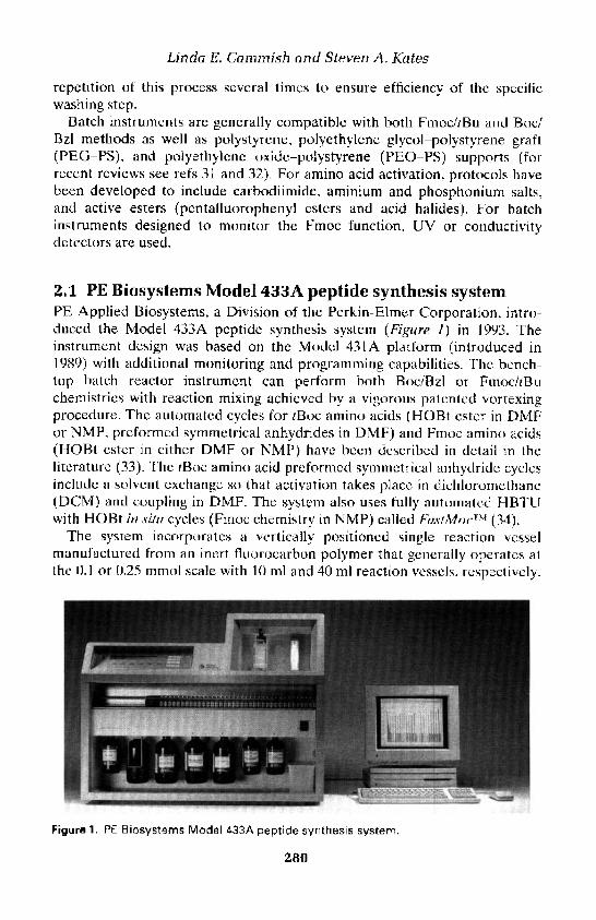
Linda E. Cammish and Steven A. Kates
repetition of this process several limes to ensure efficiency of the specificwashing step.
Batch instruments are generally compatible with both Fmoc/tBu and Boc/Bzl methods as well as polystyrene, polyethylene glycol-polystyrene graft(PEG-PS), and polyethylene oxide-polystyrene (PEO-PS) supports (forrecent reviews see refs 31 and 32). For amino acid activation, protocols havebeen developed to include carbodiimide, aminium and phosphonium salts,and active esters (pentafluorophenyl esters and acid halides). For hatchinstruments designed to monitor the Fmoc function, UV or conductivitydetectors are used.
2.1 PE Biosystems Model 433A peptide synthesis systemPE Applied Biosystems, a Division of the Perkin-Elmer Corporation, intro-duced the Model 433A peptide synthesis system (Figure I) in 1993. Theinstrument design was based on the Model 431A platform (introduced in1989) with additional monitoring and programming capabilities. The bench-top batch reactor instrument can perform both Boc/Bzl or Fmoc/tBuchemistries with reaction mixing achieved by a vigorous patented vortexingprocedure. The automated cycles for ;Boc amino acids (HOBt ester in DMFor NMP, preformed symmetrical anhydrides in DMF) and Fmoc amino acids(HOBt ester in either DMF or NMP) have been described in detail in theliterature (33). The rBoc amino acid preformed symmetrical anhydride cyclesinclude a solvent exchange so that activation takes place in dichloromethane(DCM) and coupling in DMF. The system also uses fully automated HBTUwith HOBt in situ cycles (Fmoc chemistry in NMP) called" FtistMoc™ (34).
The system incorporates a vertically positioned single reaction vesselmanufactured from an inert fluorocarbon polymer that generally operates atthe 0.1 or 0.25 mmol scale with 10 ml and 40 ml reaction vessels, respectively.
Figure 1. PE Biosystems Model 433A peptide synthesis system.
280

13: Instrumentation for automated solid-phase peptide synthesis
A range of reaction vessels are available to enable synthesis scales rangingfrom 0.005 to 1.0 mmol (3 ml reaction vessel for 0.005, 0.010, and 0.025 mmol;10 ml reaction vessel for <0.1 mmol; 40 ml reaction vessel for <0.25 mmol; 55ml reaction vessel for <1.0 mmol). Different sizes of reaction vessels arerequired to enable resin swelling and efficiency of washing steps. Mixing inthe reaction vessel occurs by vortex agitation that allows for efficientresin-fluid interaction.
Amino acid derivatives are dry packed in cartridges provided by the manu-facturer. Pre-weighed cartridges are available commercially. The cartridgesare placed in the desired sequence in a guideway that can hold a maximum of50 cartridges in a single loading. The cartridges pass through a cartridge guideand bar-code reader prior to amino acid dissolution to enable correctpositioning and verification of the amino acid sequence, respectively.
Amino acid derivatives are dissolved via a pressure-driven needle-basedassembly that punctures the amino acid cartridge septum and delivers solventsfor dissolution and gas for mixing. The amino acid solution is then transferredvia the needle-based assembly to the preactivation vessel where conversion ofthe amino acid residue to its corresponding activated derivative takes place.Since the activation process is generally carried out in a preactivation vesselseparated from the reaction vessel, the formation of an activated Fmoc or tBocamino acid species occurs immediately prior to the coupling reaction.Subsequent to completion of the activation of an amino acid residue, thesolution is transferred to the reaction vessel via gas pressure delivery. Anyprecipitated by-product is filtered during this step and left behind in thepreactivation vessel where it is dissolved and washed out to waste with asolution of methanol (MeOH) and DCM. Alternatively, according to theactivation method used, the solutions of the amino acid derivative and thecoupling reagent can be delivered directly to the reaction vessel, bypassing thepreactivation vessel. Following the coupling reaction and wash of thepolymeric support, a resin sampler can be programmed to automaticallyremove aliquots of the resin for ninhydrin or amino acid analysis. In thisinstance an alternative resin sampling reaction vessel must be used.
Two different types of activation are generally used on the Model 433A,namely HBTU or HOBt-DCC. Activation with HBTU is also referred to asFastMoc chemistry. In the activation step of the 0.25 mmol and 0.10 mmolFastMoc cycles, 1.0 mmol of dry protected amino acid in the cartridge isdissolved in solutions of HBTU-HOBt and N,N-diisopropylethylamine(DIPEA) in DMF with additional NMP added. The FastMoc 1.0 mmol cyclesuse three 1.0 mmol cartridges, or a total of 3.0 mmol of Fmoc amino acid. Theactivated Fmoc amino acid is formed rapidly and the solution transferreddirectly to the reaction vessel. All washing steps are performed with NMP andFmoc deprotection is achieved by treatment with piperidine-DMF (1:4).Cycle times are 24, 45, and 70 min for the 0.1, 0.25, and 1.0 mmol scales,respectively, with times potentially increasing when feedback monitoring is
281

Linda E. Cammish and Steven A. Kates
enabled. With the 0.1 and 0.25 mmol Fmoc-amino acid-HOBt-DCC cycles,1.0 mmol of dry protected amino acid in the cartridge is dissolved during theactivation step with a solution of NMP and HOBt. This solution is transferredto the preactivation vessel and DCC in NMP is added. Following theactivation step, the active species is transferred to the reaction vessel wherecoupling of the activated amino acid to the support-bound peptide occurs.Washing steps are performed with NMP and Fmoc deprotection is achievedby treatment with piperidine-DMF (1:4). Cycle times are 60 and 108 min forthe 0.1 and 0.25 mmol scales, respectively, with times potentially increasingwhen feedback monitoring is enabled. With the 0.5 mmol tBoc protocols, 2.0mmol of the dry protected amino acid is dissolved with a solution of NMP andHOBt in NMP. This solution is transferred to the preactivation vessel andDCC in NMP is added. After the activation occurs, the active species istransferred to the reaction vessel and dimethylsulphoxide (DMSO) is addedto enhance and improve solvation of the peptide resin as well as increasecoupling yields for difficult sequences. During the final coupling stage DIPEAis also added to further disrupt peptide-peptide hydrogen bonds, increasesolvation, and neutralize any protonated amine groups to accelerate couplingreactions. Subsequent to coupling and washing, a capping reaction occursusing an acetic anhydride-DIPEA-NMP solution. With 0.1 mmol tBocprotocols, 1.0 mmol of the protected amino acid is used. Washing steps useeither DCM or NMP, tBoc group removal is typically via treatment with atrifluoroacetic acid (TFA)-DCM (1:1) solution (with a subsequent neutral-ization step with DIPEA), and the total cycle time is 65 and 104 min for the0.1 and 0.5 mmol scales, respectively.
The system incorporates nine solvent/reagent reservoirs to enable deliveryof the NMP main wash, neat piperidine deblock (diluted with main wash online), 0.45 M HBTU-HOBt-DMF activator solution, 2.0 M DIPEA-NMP,MeOH final wash, DCM wash, alternative activators (e.g. 1.0 M DCC-NMP),capping solution, and solvent exchange for Fmoc/tBu chemistry. In the case ofBoc/Bzl-based methods, DCM and NMP are used as the main wash solvents,TFA (diluted with DCM on-line) for deblock, 1.0 M DCC-NMP and 1.0 MHOBt-NMP for activation, DMSO-NMP (4:1) for improved solvation, aceticanhydride for capping, and MeOH for final wash. A gas positive-pressurechemical delivery system controls the flow of all reagents and solvents. Gaspressure is provided by an external tank of nitrogen.
In the Model 433A SynthAssist® Software, based on a Macintosh® platform,there are 152 functions, designated with a number and a name, which controlall the processes required for synthesis. Some functions activate a switch orset of switches to deliver solutions or to perform a specific task.
For Fmoc chemistry, the Model 433A features automated conductimetricmonitoring with feedback control (35). Conductivity monitoring is based onAa-Fmoc removal with piperidine-NMP (1:4) to generate dibenzofulvene anda conductive carbamate salt of piperidine. The extent of deprotection is
282

13: Instrumentation for automated solid-phase peptide synthesis
determined by comparing the conductivity of two samples of deprotectionsolution. In one algorithm, if the user sets a deprotection loop value of 4%(acceptable range 3-10%), then successive Na-Fmoc removal treatmentswithin the same cycle will continue until the last conductivity reading is within4% of the value for the previous sample reading. In a second algorithm, theextent of deprotection is determined by comparing the conductivity of the lastsample to the first sample in the current cycle. With this algorithm, a pre-determined value for the deprotection loop (i.e. 4%) requires Na-Fmocremoval to continue until the last conductivity value is less than or equal to4% of the first conductivity value. Intelligent dynamic feedback control isactivated on encountering difficult Fmoc deprotections. A number of cyclemodifications such as extended deprotection, double or extended couplings,capping, and solvent exchange, which can be selected at initial set-up by theuser, can be automatically incorporated into the synthesis if required. Twoadditional monitoring channels are available to enable UV monitoring anduser-definable monitoring.
2.2 Protein Technologies, Inc., SONATA/Pilot™ peptidesynthesizer
The SONATA/Pilot (Figure 2) design is based on a matrix valve componentsimilar to that used in the Protein Technologies, Inc. SYMPHONY/Multiplex™ peptide synthesizer (discussed in Section 4.2). SONATA/Pilot isa floor-standing system with seven primary reagent reservoirs and 20 aminoacid reservoirs. The system uses a single reactor vessel capable of scales of0.1-50 mmol (100 g of solid support). Transfer of reagents is via gas pressureexcept for the primary solvent that is delivered by a pumping system capableof operating at flow rates up to 400 ml/min for higher scales. Similar to theSYMPHONY/Multiplex system, the amino acid derivatives are incorporatedas predissolved solutions and the system is compatible with both Fmoc andtBoc chemistries.
2.3 Protein Technologies Inc., Model PS3™ peptidesynthesizer
The Model PS3 batch peptide synthesizer (Figure 3) is designed for use withprepackaged reagents. Activating agents, typically BOP or HBTU, are pre-packed in monomer vials in conjunction with protected amino acids for point-of-use dissolution. Following addition of a base solution, activation occurs inthe vial and the activated amino acid derivative is transferred to the reactionvessel containing the solid support. Fluid transfer and agitation of the poly-meric support is achieved via gas pressure. Three glass reaction vesselsperform in a sequential manner at a scale range of 0.1-0.25 mmol with eithertBoc or Fmoc chemistries. The system has four solvent/reagent reservoirs andruns by a stand-alone control with programming via a liquid crystal display
283
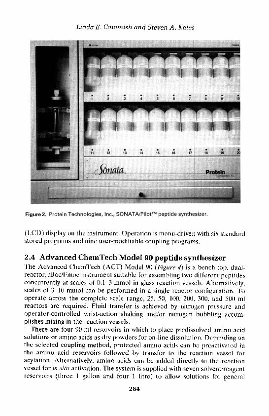
Linda E. Cammish and Steven A. Kates
Figure2, Protein Technologies, Inc., SONATA/Pilot™ peptide synthesizer.
(LCD) display on the instrument. Operation is menu-driven with six standardstored programs and nine user-modifiable coupling programs.
2.4 Advanced ChemTech Model 90 peptide synthesizerThe Advanced ChemTech (ACT) Model 90 (Figure 4) is a bench-top, dual-reactor, tBoc/Fmoc instrument suitable for assembling two different peptidesconcurrently at scales of 0.1-3 mmol in glass reaction vessels. Alternatively,scales of 3-10 mmol can be performed in a single reactor configuration. Tooperate across the complete scale range, 25. 50, 100, 200, 300, and 500 mlreactors arc required. Fluid transfer is achieved by nitrogen pressure andoperator-controlled wrist-action shaking and/or nitrogen bubbling accom-plishes mixing in the reaction vessels.
There are four 90 ml reservoirs in which to place predissolved amino acidsolutions or amino acids as dry powders for on-line dissolution. Depending onthe selected coupling method, protected amino acids can be preactivated inthe amino acid reservoirs followed by transfer to the reaction vessel foracylation. Alternatively, amino acids can be added directly to the reactionvessel for in situ activation. The system is supplied with seven solvent/reagentreservoirs (three 1 gallon and four 1 litre) to allow solutions for general
284

13: Instrumentation for automated solid-phase peptide synthesis
Figure3. Protein Technologies Inc., Model PS3™ peptide synthesizer.
Figure4. Advanced ChemTech Model 90 peptide synthesizer.
285

Linda E. Cammish and Steven A. Kates
washing, Na-tBoc/Fmoc group removal, activation, and capping. Activationchemistries that can be incorporated include in situ activation via DCC, DIC.TBTU, PyBOP, or HBTU. Preactivated symmetrical anhydrides, HOBtesters, and pentafluorophenyl esters can also be used.
The system operates via mouse-driven software with a DOS-based IBMPS/2™ compatible computer equipped with standard protocols that can befurther modified by the user.
2,5 Advanced ChemTech Model 400 production-scalesynthesizer
Large-scale solid phase synthesis of peptides can be performed on the floor-standing ACT Model 400 batch system (Figure 5) that operates in a singlereactor mode with 0.1-5 kg of starting solid support (36). All operations and
Figure 5. Advanced ChemTech Model 400 production-scale synthesizer.
286

13: Instrumentation for automated solid-phase peptide synthesis
liquid transfer occur under nitrogen pressure. Additional nitrogen bubblingand/or oscillating enables fluid/resin mixing in the reaction vessel. The systemis compatible with Boc/Bzl and Fmoc/tBu methods and can be linked up tofour 200-litre solvent drums. Amino acids can be added as solids to 5-litre/10-litre flasks and automatically dissolved just prior to addition to the solidsupport. Alternatively, the amino acid can be predissolved, stored in 5-litre/10-litre flasks and added automatically when required. The instrument isequipped with a filtration device that allows for the removal of solid pre-activation by-products prior to addition to the reaction vessel. This featureenables compatibility with the common activation chemistry used.
The system is usually customized to cover the specific needs of a laboratory(additional solvents, safety features, and integration into existing laboratoryinfrastructure) and is of stainless steel construction with only glass and Tefloncontact areas to allow operation in a clean room environment. Software andhardware are designed specifically for compliant operation.
2.6 ABIMED EPS 221 synthesizerThe EPS 221 (Figure 6) is an automated Fmoc peptide synthesizer based on arobotics system with a motor-driven syringe for reagent transfer. Movementof the robot in an X-Y-Z direction allows the system to perform allsolvent/reagent delivery steps. The system runs at synthesis scales up to 100umol on 1-3 columns with a total of 45 amino acid residue positionsdistributed over all column locations. Reagents are delivered to the columnby a needle that is connected to a motor-driven syringe. The standardprotocol for the instrument begins with a 2-3 ml DMF wash delivery to thesolid support to swell the resin for 10 min. A piperidine-DMF (1:4) solution isthen taken up from the derivatives rack and pushed slowly through the resin,followed by another DMF wash. Amino acids are activated using a 0.9 Msolution of PyBOP and a 4 M solution of N-methylmorpholine (NMM). Afivefold excess of the activated amino acid solution is pushed slowly throughthe resin bed. Gas is blown through the resin to achieve mixing. After thecoupling reaction the solid support is again washed with DMF. Thesynthesizer is operated from the keyboard controller of the samplepreparation robot equipped with an eight-line display and floppy disk drive.
3. Continuous-flow peptide synthesisIn traditional batch peptide synthesis, all reactions are carried out essentiallyin a discontinuous fashion with sequential treatment of acylating anddeprotecting agents in a shaken, stirred, vortexed, or bubbled reaction vessel.The excess of reagents is removed by filtration and repeated washing steps. Incontinuous-flow peptide synthesis (37), systems were developed originallywith the aim of improving the efficiency and speed by removing excess
287

Linda E. Cammish and Steven A. Kates
Figure 6. ABIMED EPS 221 synthesizer.
reagents with & continual solvent flow through a packed column containing asolid support. A glass column with filters at the top and bottom replaces thereaction vessel. The system incorporates a positive displacement pump toenable continuous fluid flow at a variety of flow rates. Valves provide forselection of solvent and reactant flow through the resin bed to waste as well asfor the introduction and recirculation of the acylating species during thecoupling reaction.
Under the pumped flow solvent conditions in continuous-flow instrument-ation, polystyrene resins can create high pressures that can halt a synthesis. Touse these polymeric supports in continuous-flow systems, a minimum of a 4:1ratio of glass to polystyrene bead is required. The incorporation of poly-ethylene glycol spacers into polystyrene provides a support, namely PEG-PS,which is compatible with and recommended for continuous-flow systems.
288

13: Instrumentation for automated solid-phase peptide synthesis
PEG-PS supports have a defined and reproducible structure that comprisesapproximately 60 wt% of polyethylene glycol providing a support that isphysically robust (38). PEG-PS resins swell appreciably in a range of solventsand are superior to polystyrene supports for the construction of morecomplex peptides, including those containing cyclic (lactam and disulphides),branched, and post-translational modified moieties.
Continuous-flow instrumentation was designed for Fmoc/tBu-basedmethods as Na-protecting group removal proceeds under milder conditions(piperidine) than Boc/Bzl chemistry (TFA) and would, therefore, be morecompatible with a reciprocating pump. Another advantage to Fmoc chemistryis the absorbance of the fluorene chromophore in an accessible region of theUV spectrum (39). Liquid flow effluent from the reaction column can bepassed through a UV detector system to enable continuous spectrophoto-metric monitoring of both acylation and deprotection reactions. Additionally,the detection method provides confirmation that the various operations areproceeding normally and that the instrument function is correct.
In a typical UV trace, the recirculation of the soluble, activated amino acidderivative is indicated by a characteristic oscillating pattern with an initialdamped harmonic motion-type pattern (40, 41). The maximum absorbancedecreases as the concentration of the Fmoc amino acid in solution is reduceddue to coupling to the solid support. Diffusion processes result in slightbroadening of successive UV peaks. After several recirculations through theresin bed, a uniform reactant concentration and a consistent UV absorbancethroughout the system are observed. The wash-out of excess reagents withDMF is very rapid as shown by a sharp fall in the optical density of thecolumn effluent during the washing step. The facile release and elimination ofthe Fmoc species, generally observed as a sharp rise and fall in UV absorb-ance, can easily monitor the deprotection step. The height and shape of thispeak provides valuable data with regard to the rate of Fmoc group removal,which can indicate the difficulty of assembling a particular sequence. Trouble-some Fmoc deprotections can produce UV data peaks which are character-istically broadened and reduced in height.
3.1 PE Biosystems Pioneer™ peptide synthesis systemThe Pioneer (Figure 7) peptide synthesis system (42) was introduced recentlyas a successor to the 9050Plus instrument, which was first commercialized in1988. The Pioneer combines dual simultaneous column capability with a highthroughput multiple peptide synthesis option (discussed in Section 4.1). It isan Fmoc/tBu continuous-flow system that in its standard mode of operationfunctions with two columns operating simultaneously with independent con-trol of solvent, reagent, and amino acid delivery. Fluid flow is via program-mable positive displacement pumps that deliver with a flow rate of up to50 ml/min and require no flow-rate calibration. Each column can function
289

Linda E. Cammish and Steven A. Kates
Figure 7. PE Biosystems Pioneer™ peptide synthesis system.
at 0.02-2.0 mmol scale range. The two separate pump systems enable simul-taneous operation of each column at a different scale. An integral UVspectrophotometer monitors in real time all reactions that occur on bothcolumns.
The solid support is placed in glass reaction columns that have filters at thetop and bottom. Adjustable end pieces allow for different scales of synthesisto be performed within the same reaction column. The standard column sizeis 15 X 100 mm, capable of containing up to 2.5 g of PEG-PS resin. A large-scale column (25 x 150 mm) is also provided to enable synthesis scales of upto 2.0 mmol.
Fmoe-amino acid derivatives are stored as dry powders in vials positionedin order of the sequence in the racks of a transport system. Similar to afraction collector, the transport system moves in the X,Y-direction, oneposition per cycle, and a probe is used for point-of-use dissolution of afourfold excess of the Fmoc-amino acid residues. The probe delivers activatorsolution to the vial and then bubbles the derivative with nitrogen to form ahomogeneous solution. The pump transfers the amino acid solution via theprobe through the column for a recirculation/coupling step. To ensure highconcentration, a low recirculation volume is used. The end of the probe isequipped with a filter to ensure that no particulates enter the system and toeliminate blockages. Both of the two transport systems possess a maximum of101 amino acid positions for scales of synthesis not greater than 0.2 mmol. Forscales up to a maximum of 0.5 and 2.0 mmol, alternative rack geometries are
290

13: Instrumentation for automated solid-phase peptide synthesis
provided with 68 and 24 amino acid positions, respectively. A range ofactivation chemistries can be used, including carbodiimide (DIC), penta-fluorophenyl ester, aminium salts (TBTU, HBTU, and HATU) andphosphonium salts (PyBOP and PyAOP).
The system incorporates eight solvent/reagent reservoirs including bottlesfor three activators to perform mixed acylation strategies, a main wash,piperidine-DMF (1:4), capping, solvent exchange, and additional wash. Pre-programmed protocols are provided to allow for automated removal of allyl-based protecting groups to enable a fully automatic Fmoc-based synthesis ofcyclic and branched chain peptides (43). Additionally, programmes are availablefor fast (22-min cycle time), standard (45-min cycle time), extended (75-mincycle time), capping, solvent exchange, and reduced diketopiperazine cycles.
The system can be operated from stand-alone control via an LCD and amembrane keypad screen on the instrument. Seven pre-programmed proto-cols are provided and a maximum of 36 sequences containing 101 amino acidsin length in each of the eight user profiles is allowed. Alternatively, for moreflexibility, full protocol editing, chemical editing, and database capability, thesystem can be controlled from a computer workstation with icon-driven easy-to-use Windows® 95-based software.
Real-time on-line UV monitoring is provided for both columns withsoftware enabling full feedback control. If the UV data determine a difficultdeprotection, the system can automatically extend the deprotection and thesubsequent coupling of the next incoming amino acid (44).
A unique characteristic of the Pioneer peptide synthesis system is theoption of a multiple peptide synthesis (MPS) accessory with 16-columncapacity. A maximum of two MPS units can be added to each Pioneer,extending the capability for simultaneous synthesis from 2 to 32 differentpeptides. The novel design allows independent operation of both columns,MPS accessories, or a combination of the two. The MPS unit possesses scalesat 0.025, 0.05, and 0.1 mmol. Twenty-four different predissolved amino acidderivatives can be incorporated to elongate a sequence. The appropriateamino acid solution is delivered in eightfold excess to the columns, togetherwith the activator solutions.
4. Multiple peptide synthesis systemsA range of MPS systems is available commercially which generally synthesizeat lower scales and with limited flexibility in terms of protocol and chemicalediting capability (45-48).
4.1 PE Biosystems Pioneer MPS optionThe Pioneer MPS option, described in Section 3.1, is a 16-column accessorythat can be added to the PE Biosystems Pioneer peptide synthesis system. A
291

Linda E. Cammiah and Steven A. Kates
total of two MPS options can be added on to each Pioneer, extending itscolumn capacity from 2 to 32, running at scales of 0.025.0.05, or 0.1 mmol.
4.2 Protein Technologies, Inc. SYMPHONY/Multiplex™peptide synthesizer
The SYMPHONY/Multiplex (Figure 8), introduced in 1993, is a batchtBoc/Fmoc peptide synthesizer with 12 independent reaction vessels (49). Thekey component is the patented matrix valve system that enables differentsolutions to be transferred to each of the 12 reactors simultaneously. Thisfeature allows the assembly of 12 different peptides under optimal conditionsfor each sequence with regard to scales and customized coupling reactions.Solution flow is controlled via chemically resistant membrane valves arrangedto provide a matrix system with common solution ports but separate deliverylines for each reaction vessel, A combination of gas pressure and an externalvacuum pump operates the valve system. Solution transfer is achieved bynitrogen pressure.
The reusable glass reaction vessels for the solid supports have an internalvolume of 16 ml and enable synthesis scales up to 0.1 mmol. Alternatively,disposable plastic vessels with 24 ml volume can be used. Resin agitation is
Figure 8. Protein Technologies, Inc. SYMPHONY/Multiplex peptide synthesizer.
292

13: Instrumentation for automated solid-phase peptide synthesis
accomplished by occasional nitrogen bubbling through a 40 u.m filter at thebottom of the reaction vessel.
The system is a floor-standing instrument that includes a vented cabinet forstorage of solvents and reagents. Six solvent/reagent reservoirs are availabletypically for DMF or NMP as a main wash solvent, piperidine-DMF (1:4) fordeprotection, DCM as a secondary reagent, HBTU solution for activation,acetic anhydride as a capping solution, and a TFA-based cleavage cocktail.
The SYMPHONY/Multiplex system includes 20 X 100 ml reservoirs forpredissolved amino acid solutions. The standard 100 mM amino acid con-centration provides a fivefold excess. The software is designed primarily for insitu HOBt ester activation using HBTU, TBTU, PyBOP, or BOP activators.
In the SYMPHONY/Multiplex system, the activator solution consists ofHBTU or TBTU at a concentration equimolar with the amino acid solutionsin conjunction with two- to fourfold equivalents of a base such as NMM. For aspecified coupling step, the system transfers a determined number of aliquotsof amino acid solution to the solid support, followed by the same volume ofactivator solution. Subsequent to completion of addition, the resin is agitatedby gas bubbling for the specified coupling time.
Control of the synthesis parameters is by a stand-alone IBM-compatiblepersonal computer. The software is based on 'point and click' pull-downmenus, allowing access to Peptide, Program, Reaction Vessel operations,Manual operations, Set-up, and Data sections.
After chain elongation, peptides are released from the solid support in thereaction vessels according to cleavage protocols selected in the software.Crude products are transferred to centrifuge tubes in a vented compartmentfor ether precipitation and centrifugation.
4.3 Advanced ChemTech Models 348, 396, and 357bimolecular synthesizers
Advanced ChemTech provides a range of robotic bench-top MPS systemsthat vary in the number of peptides that can be constructed and the scale rangethat can be performed (50). All systems are batch, tBoc/Fmoc compatible anduse syringe pumps for fluid delivery. Model 357 (Figure 9) has a maximum of36 X 8.0 ml reaction-well reactor capacities (5-250 mmol scale); Model 348(Figure 10) has a maximum of 96-well capacity (5-150 (umol scale); and Model396 (Figure 11) has a maximum of 96-well capacity (5 u.mol-1 mmol). InModels 348 and 396 it is also possible to use 8-, 16-, and 40-well Teflon reactorblocks. Additionally, Model 357 can serve as a large-scale single reactorsystem capable of handling up to 36 g of resin. The 600 ml chamber allows forpooling and dividing of the solid support for the 'mix and split' technique(51-53).
A variable speed orbital mixer and/or nitrogen bubbling (only on theModel 357) is incorporated to stir solvents/reagents with the solid supports. A
293
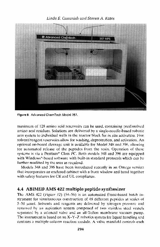
Linda E. Cammish and Steven A. Kates
Figure9. Advanced ChemTech Model 357.
maximum of 128 amino acid reservoirs can be used. containing predissolvedamino acid residues. Solutions are delivered by a single-needle-based roboticarm system to individual wells in the reactor block for in situ activation. Fivesolvent/reagent reservoirs allow for washing, deprotection, and activation. Anoptional on board cleavage unit is available for Model 348 and 396, allowingfor automated release of the peptides from the resin. OperaUon of thesesvsu-ms is via a Pentium* Class PC. Both models 348 and 396 are equippedwith Windows-based software with built-in standard protocols which can befurther modified by the user as required.
Models 348 and 396 have been introduced recently in an Omega versionthat incorporates an enclosed cabinet with a front window and hood togetherwith safety features for CE and UL compliance.
4.4 ABIMED AMS 422 multiple peptide synthesizerThe AMS 422 (Figure 12) (54-56) is an automated Fmoc-based batch in-strument for simultaneous construction of 48 different peptidcs at scales of5-50 umol. Solvents and reagents are delivered by nitrogen pressure andremoved by an aspiration system composed of two stainless steel vesselsseparated by a solenoid valve and an all-Teflon membrane vacuum pump.The instrument is based on an X-Y-Z robotics system tor liquid handling andcontains a multiple column reaction module. A valve manifold controls each
294
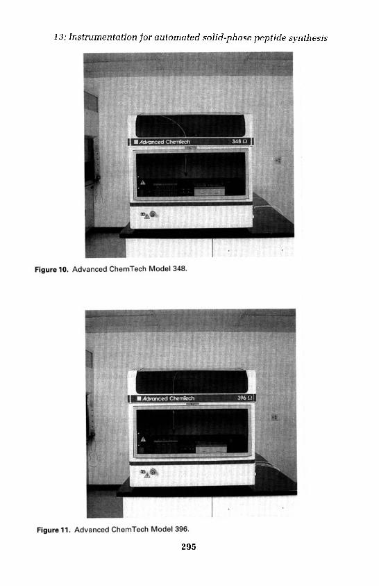
13: Instrumentation for automated solid-phase peptide synthesis
Figure 10, Advanced ChemTech Model 348.
Figure 11, Advanced ChemTech Model 396.
295
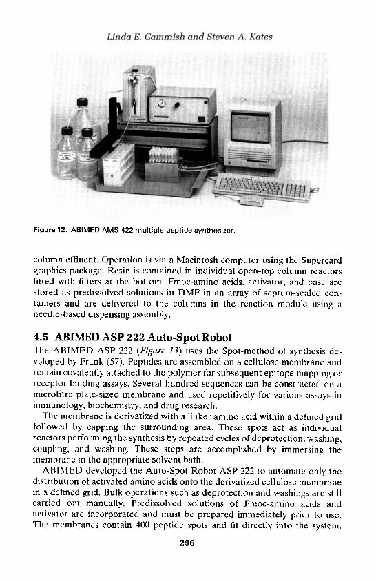
Linda E. Cammish and Steven A. Kates
Figure 12. ABIMED AMS 422 multiple peptide synthesizer.
column effluent. Operation is via a Macintosh computer using the Supercardgraphics package. Resin is contained in individual open-top column reactorsfitted with filters at the bottom. Fmoc-amino acids, activator, and base arcstored as predissolved solutions in DMF in an array of septum-sealed con-tainers and are delivered to the columns in the reaction module using aneedle-based dispensing assembly.
4.5 ABIMED ASP 222 Auto-Spot RobotThe ABIMED ASP 222 (Figure 13) uses the Spot-method of synthesis de-veloped by Frank (57). Peptides are assembled on a cellulose membrane andremain covalently attached to the polymer for subsequent epitope mapping orreceptor binding assays. Several hundred sequences can be constructed on amicrotitre plate-sized membrane and used repetitively for various assays inimmunology, biochemistry, and drug research.
The membrane is derivatized with a linker amino acid within a defined gridfollowed by capping the surrounding area. These spots act as individualreactors performing the synthesis by repeated cycles of deprotection, washing,coupling, and washing. These steps are accomplished by immersing themembrane in the appropriate solvent bath.
ABIMED developed the Auto-Spot Robot ASP 222 to automate only thedistribution of activated amino acids onto the derivatized cellulose membranein a defined grid. Bulk operations such as deprotection and washings are stillcarried out manually, Predissolved solutions of Fmoc-amino acids andactivator are incorporated and must be prepared immediately prior to use.The membranes contain 400 peptide spots and fit directly into the system.
296

13: Instrumentation for automated solid-phase peptide synthesis
Figure 13. ABIMED ASP 222 Auto-Spot Robot.
Typical membrane loadings are 0.5-0.6 umol/cm2. Distribution of the 44positions of the predissolved activated amino acids can be performed on fourmembranes simultaneously. Delivery of the amino acids to the membrane isvia a robotic arm system with a needle-based assembly connected to a motor-driven syringe. The ASP 222 is operated using Windows'*-based software onan IBM-compatible 383/484 computer to allow sequence entry manually orfrom external databases.
4.6 ZINSSER ANALYTIC SMPS 350 multiple peptidesynthesizer
Developed in 1989 in collaboration with Boehringer Ingelheim, the SMPS 350(Figure 14) can synthesize 3 X 48 peptides in a simultaneous manner usingFmoc chemistry. Two X-Y-Z robot arm systems with sample tips and twodispenser/dilutor systems perform the liquid handling and washing steps.Chain elongation occurs on polystyrene resin positioned in small disposableplastic reaction vessels with conical bottoms placed in supports similar to amicrotitre plate. A 3 X 48 microreactor or 3 X 30 minireactor capable ofcontaining a maximum of 15 and 50 mg, respectively, of solid support can beused. The system has a rack for 32 predissolved amino acid residues as well asthree solvent/reagent reservoirs for washing, deprotecting, and activatingreagents. Operation of the instrument is via DOS-based software on an IBM-compatible computer system.
297

Linda E. Cammish and Steven A, Kates
Figure 14. ZINSSER ANALYTIC SMPS 350 multiple peptide synthesizer.
4.7 ZINSSER ANALYTIC SOPHAS solid phasesynthesizer
SOPHAS (Figure 15) is an automated instrument capable of assembling 864sequences in a single run. Reactors can be selected from a 96-well plateformat to 25 ml vessels. Mixing is conducted by mechanic agitation. Thesystem uses four independent pipetting devices, each supported by twodilutors and a time/pressure delivery channel to transfer 4-5 solvents/reagentsor nitrogen. The system is operated by Windows® NT software.
4.8 SHIMADZU PSSM-8 peptide synthesizerThe PSSM-8 is a batch Fmoc instrument with eight disposable reaetion vesselscomprised of 2.5 ml syringes with filters at the bottom. The synthesizeroperates at a 5-50 umol/reactor scale. The system includes an amino acidstation equipped with 20 X 2.0 ml Eppendorf tubes/reactor positions forpredissolved derivatives. Fmoc-amino acids, solvents, and reagents aredelivered by syringes to the reactors via a needle-based assembly. Nitrogenbubbling agitates the solid support. Each reactor position is supplied withthree 30-ml reactor bottles to allow for activation and/or capping. The mainwash solvent and deprotecting reagent are located in reservoirs external tothe instrument. Syntheses are programmed via a separate computer systemfollowed by transfer of the data to a memory card,
298
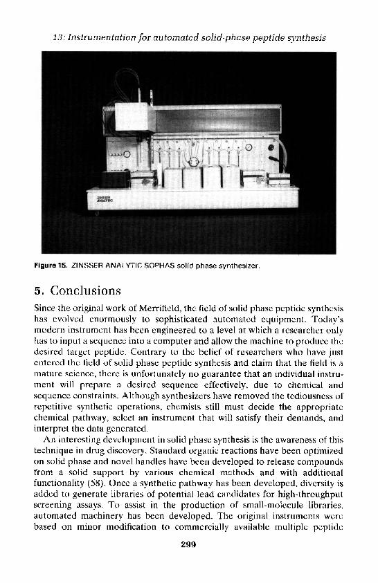
13: Instrumentation for automated solid-phase peptide synthesis
Figure 15. ZINSSER ANALYTIC SOPHAS solid phase synthesizer.
5. ConclusionsSince the original work of Merrifield, the field of solid phase peptide synthesishas evolved enormously to sophisticated automated equipment. Today'smodern instrument has been engineered to a level at which a researcher onlyhas to input a sequence into a computer and allow the machine to produce thedesired target peptide. Contrary to the belief of researchers who have justentered the field of solid phase peptide synthesis and claim that the field is amature science, there is unfortunately no guarantee that an individual instru-ment will prepare a desired sequence effectively, due to chemical andsequence constraints. Although synthesizers have removed the tediousness ofrepetitive synthetic operations, chemists still must decide the appropriatechemical pathway, select an instrument that will satisfy their demands, andinterpret the data generated.
An interesting development in solid phase synthesis is the awareness of thistechnique in drug discovery. Standard organic reactions have been optimizedon solid phase and novel handles have been developed to release compoundsfrom a solid support by various chemical methods and with additionalfunctionality (58). Once a synthetic pathway has been developed, diversity isadded to generate libraries of potential lead candidates for high-throughputscreening assays. To assist in the production of small-molecule libraries,automated machinery has been developed. The original instruments werebased on minor modification to commercially available multiple peptide
299

Linda E. Cammish and Steven A. Kates
synthesizers. Currently, there are a variety of solid phase organic synthesizersavailable commercially (59-63). Similar to peptide instrumentation, theseunits each possess unique features with regard to the level of automation, thenumber of reactors, and the scale. While this field is still at an early stage ofdevelopment, engineering designs that build versatility and ingenuity will beapplied to peptide instrumentation. Finally, as improvements continue in thechemistry for solid phase synthesis, machinery will need to adapt with regardto valving and plumbing to be able to implement these methods.
All registered trademarks are the properties of their respective owners.
References1. Merrifield, R. B. (1963). J. Am. Chem. Soc., 85,2149.2. Bodanszky, M. and Bednarek, M. A. (1989). J. Protein Chem., 8,461.3. Carpino, L. A., Beyermann, M., Wenschuh, H., and Biernert, M. (1996). Ace.
Chem. Res., 29,268.4. Wenschuh, H., Beyermann, M., Rothemund, S., Carpino, L., and Bienert, M.
(1995). Tetrahedron Lett., 36,1247.5. Sheehan, J. C. and Hess, G. P. (1955). J. Am. Chem. Soc., 77,1067.6. Sarantakis, D., Teichman, J., Lien, E. L., and Fenichel, R. (1976). Biochem.
Biophys. Res. Commun., 73,336.7. Konig, W. and Gieger, R. (1970). Chem. Ber., 103,788.8. Carpino, L. (1993). J. Am. Chem. Soc., 115, 4397.9. Dormoy, J. R. and Castro, B. (1979). Tetrahedron Lett., 20,3321.
10. Coste, J., Le-Nguyen, D., and Castro, B. (1990). Tetrahedron Lett., 31,205.11. Albericio, F., Cases, M., Alsina, J., Triolo, S. A., Carpino, L. A., and Kates, S. A.
(1997). Tetrahedron Lett., 38, 4853.12. Dourtoglou, V., Ziegler, J. C., and Gross, B. (1978). Tetrahedron Lett., 19,1269.13. Knorr, R., Trzeciak, A., Bannwarth, W., and Gillessen, D. (1989). Tetrahedron
Lett., 30,1927.14. Carpino, L. A., El-Faham. A., Truran, G. A., Minor, C. A., Kates, S. A., Griffin,
G. W., Shroff, H., Triolo, S. A., and Albericio, F. (1994). In Innovation andperspectives in solid phase synthesis - Peptides, proteins and nucleic acids (ed. R.Epton), p. 95. Mayflower Worldwide Ltd., Birmingham, UK.
15. Carpino, L. A., El-Faham, A., Minor, C. A., and Albericio, F. (1994). J. Chem.Soc., Chem. Commun., 201.
16. Kaiser, E., Colescott, R. L., Bossinger, C. D., and Cook, P. I. (1970). Anal.Biochem., 34,595.
17. Hodges, R. S. and Merrifield, R. B. (1975). Anal. Biochem., 65,241.18. Horn, M. and Novak, C. (1987). Am. Biotech. Lab., 5,12.19. Krchnak, V., Vagner, J., Safar, P., and Lebl, M. (1988). Coll. Czech. Chem.
Commun., 53, 2542.20. Young, S. C., White, P. D., Davies, J. W., Owen, D. E. I. A., Salisbury, S. A., and
Tremeer, E. J., (1990). Biochem. Soc. Trans., 18,1311.21. Chang, C. D. and Meienhofer, J. (1978). Int. J. Pept. Protein Res., 11, 246.
300

13: Instrumentation for automated solid-phase peptide synthesis
22. Meinenhofer, J., Waki, M., Heimer, E. P., Lambros, T. J., Makofske, R. C. andChang, C. D. (1979). Int. J. Pept. Protein Res., 13,35.
23. Chang, C. D., Felix, A. M., Jimenez, M. H., and Meienhofer, J. (1980). Int. J. Pept.Protein Res., 15,485.
24. Schafer-Nielson, C., Hansen, P. H., Lihme, A., and Heegard, P. M. H. (1989). J.Biochem. Biophys. Methods, 20,69.
25. Fox, J., Newton, R., Heegard, P., and Schafer-Nielson, C. (1990). In Innovationand perspectives in solid phase synthesis - Peptides, proteins and nucleic acids (ed.R. Epton), p. 141. SPCC Ltd., Birmingham, UK.
26. McFerran, N. V., Walker, B., McGurk, C. D., and Scott, F. C. (1991). Int. J. Pept.Protein Res., 37, 382.
27. Ozols, J. (1990). Methods Enzymol., 182,587.28. Edman, P. (1950). Acta. Chem. Scand., 4,277.29. Edman, P. (1950). Acta. Chem. Scand., 4,283.30. Laursen, R. A. (1971). Eur. J. Biochem., 20,89.31. Barany, G., Albericio, F., Kates, S. A., and Kempe, M. (1997). In Chemistry and
biological application of polyethylene glycol (ed. J. M. Harris and S. Zalipsky), p.239. American Chemical Society Books, Washington, DC.
32. Rapp, W. (1996). In Combinatorial peptide and nonpeptide libraries (ed. G. Jung),p. 425. VCH, Weinheim, Germany.
33. Geiser, T., Beilan, H., Bergot, B. J., and Otteson, K. M. (1988). InMacromolecular sequencing and synthesis: selected methods and applications (ed.D. H. Schlesinger), p. 199. Alan R. Liss, New York.
34. Fields, C. G., Lloyd, D. H., Macdonald, R. L., Otteson, K. M., and Noble, R. L.(1991). Peptide Res., 4,95.
35. PE Applied Biosystems Peptide Synthesis Technical Bulletin. Feedback monitor-ing kit for ABI431A peptide synthesizer.
36. Birr, C. (1990.) In Innovation and perspectives in solid phase synthesis (ed. R.Epton), p. 155. SPCC Ltd., Birmingham, UK.
37. Atherton, E. and Sheppard, R. C. (1989). In Solid phase peptide synthesis: apractical approach, p. 87. IRL Press, Oxford.
38. Zalipsky, S., Chang, J. L., Albericio, F. and Barany, G. (1994). Reactive Polymers,22,243.
39. Atherton, E. and Sheppard, R. C. (1987). In The peptides, analysis, synthesis,biology (ed. E. Gross and J. Meienhofer), Vol. 9, p. 1. Academic Press, New York.
40. Atherton, E., Brown, E., Sheppard, R. C., and Rosevear, A. (1981). J. Chem. Soc.,Chem. Commun., 1151.
41. Dryland, A. and Sheppard, R. C. (1986). J. Chem. Soc., Perkin Trans. 1,125.42. Daniels, S. B., Sole, N. A., Hantman, S. F., Gibney, B. R., Rabanal, F., and Kates,
S. A. (1998). In Peptides 1996: Proceedings of the Twenty-fourth European PeptideSymposium (ed. R. Ramage and R. Epton), p. 323. Mayflower Worldwide Ltd.,Birmingham, UK.
43. Kates, S. A., Daniels, S. B., and Albericio, F. (1993). Anal. Biochem., 212,303.
44. PerSeptive Biosystems Technical Bulletin (1998). Pioneer™ peptide synthesissystem: monitoring peptide synthesis.
45. Holm, A. and Meldal, M. (1989). In Peptides 1988 (ed. G. Jung and E. Bayer),p. 208. Walter de Gruyter, Amsterdam.
301

Linda E. Cammish and Steven A. Kates
46. Meldal, M., Holm, C. B., Bojesenm G., Jakobsen, M. H., and Holm, A. (1993). Int.J. Pept. Protein Res., 41,250.
47. Wang, Z. and Laursen R. A. (1992). Pept. Res., 5,275.48. Jung, G. and Beck-Singer, A. (1992). Angew. Chem., Int. Ed. Engl., 31,367.49. Crawford, J. K. (1997). In The 1997 American peptide symposium program and
abstracts, Nashville, Tennessee, pp. 2-103.50. Saneii, H. H., Shannon, J. D., Miceli, R. M., Fischer, H. D., and Smith, C. W.
(1994). In Proceedings of the Thirteenth American Peptide Symposium: Peptides,chemistry, structure and biology (ed. R. S. Hodges and J. A. Smith), p. 1018.Escom, The Netherlands.
51. Furka, A., Sebesty6n, F., Asgedom, M., and Dibo, G. (1991). Int. J. Pept. ProteinRes., 37,487.
52. Houghten, R. A., Pinilla, C., Blondelle, S. E., Appel, J. R., Dooley, C. T., andCuervo, J. H. (1991). Nature, 354, 84.
53. Lam, K. S., Salmon, S. E., Hersh, E. M., Hruby, V. J., Kazmierski, W. M., andKnapp, R. J. (1991). Nature, 354, 82.
54. Gausepohl, H., Boulin, C., Kraft, M., and Frank, R. W. (1992). Pept. Res., 5, 315.55. Gausepohl, H., Behn, C., Kreuzer, O., and Buettner, J. A. (1993). In Proceedings
of the 2nd Japanese Symposium on Peptide chemistry; Peptide chemistry 1992 (ed.N. Yanaihara), p. 107. Escom, The Netherlands.
56. Gausepohl, H., Behn, C., Schopfer, R. and Frank, R. W. (1997). In Innovation andperspectives in solid phase synthesis and combinatorial libraries (ed. R. Epton),p. 87. Mayflower Scientific, Birmingham, UK.
57. Frank, R. W. (1992). Tetrahedron, 48, 9217.58. Blackburn, C., Albericio, F., and Kates, S. A. (1997). Drugs of the Future, 22,1007.59. Gerhardt, J. and Rapp, W. (1998). In Proceedings of the Twenty-fourth European
Peptide Symposium (ed. R. Ramage and R. Epton), p. 150. Mayflower Scientific,Birmingham, UK.
60. Bouitn, J. A. and Fauchere, J.-L. (1996). In Proceedings of the 1995 InternationalSymposium on laboratory automation and robotics, p. 36.
61. Krchnak, V. and Lebl, M. (1997). In Innovation and perspectives in solid phasesynthesis and combinatorial libraries (ed. R. Epton), p. 93. Mayflower Scientific,Birmingham, UK.
62. Hoperich, P. D. (1996). Nature Biotechnology, 14,1311.63. Rivero, R. A., Greco, M. N., and Maryanoff, B. E. (1997). In A practical guide to
combinatorial chemistry (ed. A. W. Czarnik and S. H. DeWitt), p. 201. AmericanChemical Society, Washington.
302

14
Manual multiple synthesis methodsB. DORNER, J. M. OSTRESH, R. A. HOUGHTEN,
RONALD FRANK, ANDREA TIEPOLD, JOHN E. FOX,ANDREW M. BRAY, NICHOLAS, J. EDE, IAN W. JAMES, and
GEOFFREY WICKHAM
1. Simultaneous multiple peptide synthesis—theT-bag method
1.1 IntroductionSimultaneous multiple peptide synthesis enables the parallel synthesis of largenumbers of peptides. The T-bag (tea-bag) method (1) was developed alongwith other methods, e.g. pin synthesis, synthesis on paper plates, synthesis onparallel columns, and synthesis on cellulose (for reviews see ref. 2), astechnology to facilitate simultaneous multiple synthesis. Large numbers ofpeptides, peptidomimetics, and small organic molecules have been preparedusing the T-bag method to address different research fields, such asconformational analysis, structure activity analysis, synthesis methodologies,and antibody-antigen interaction studies (for reviews see refs 2 and 3). Usingthe T-bag method, more than 150 peptides can be prepared in parallel inflexible amounts, with easily enough material for biological tests andanalytical studies. The synthesis of peptides of length of up to 26 amino acidresidues has been reported (4). Moreover, the T-bag technology is easy toapply in practice and requires very little special equipment.
1.2 The T-bag method™T-bags (Figure 1) are prepared by containing solid phase resins withinpolypropylene mesh material. Polypropylene is rather chemically inert as wellas fairly thermally stable (to 150°C), allowing a wide range of chemicalreactions to be used for solid phase synthesis without affecting the bagmaterial. Polystyrene cross-linked with 1% divinylbenzene, 100-200 mesh, ismainly used as the solid support, but other types of base resin can be used aswell, e.g. TentaGel (5). The size of the resin beads must exceed the size of thepores of the polypropylene mesh material of the T-bags to avoid resin lossduring synthesis. Syntheses are carried out manually (1), using semi-

B. Dorner et al.
automation (6), or within a multiple peptide synthesizer (5). The preparationof T-bags for solid phase synthesis, starting with 100 mg resin per bag, isdescribed in Protocol 1.
Protocol 1. Preparation of polypropylene mesh bags
Equipment and reagents• Polypropylene mesh material (mesh with • Ruler
74 um openings, available from Polymer . Black ink pen (ink should have a highGroup, Inc., PO Box 308, Benson, NC 27504, carbon content; available through artUSA) supply stores
• Cardboard, about 30 cm x 30 cm, with a . Sealer (% inch seal, available fromtemplate outlined showing rectangles in McMaster Carr, 9630 Norwalk Blvd., Losthe size of the T-bags to be prepared Angeles, CA 90054, USA)
• Scissors • Solid phase synthesis resin
T-bag preparation
1. Cut two sheets of polypropylene mesh material of size large enough tofit all the T-bags needed for the multiple synthesis.
2. Mark a raster of 3 cm x 4 cm rectangles on one sheet of thepolypropylene mesh, using the cardboard template.
3. Mark a number with the ink pen on the bottom of each rectangle.
4. Put the unmarked sheet on top of the sheet with the ink-marked labelsand seal the lines between all the rectangles (the very top line remainsopen—three sides of the bags have to be sealed). The number ispermanently sealed into the polypropylene to ensure identification ofeach bag.
5. Cut the sheet into strips to form pockets and fill each with 100 mg ofresin.
6. Seal the open end of the strip and cut all the bags apart.
1.2.1 Simultaneous multiple peptide synthesisSynthesis using the T-bag method can be performed using either Boc (2, 6) orFmoc (7, 8) synthetic strategies. For all manipulations, enough solvent shouldbe used to cover the T-bags (about 3-4 ml per bag containing 100 mg of resin).To enable efficient washings and reactions, the reaction vessels (polyethylenebottles) should be shaken vigorously, preferably through the use of areciprocating shaker. Thus, during a T-bag synthesis of various sequences inparallel, the deprotection and washing steps can be performed with all bagscombined in a single polyethylene bottle as outlined in Figure 2. For theamino acid couplings, the bags are separated depending on the differentsequences to be prepared. Following the coupling reactions, two washingcycles are done separately before combining all the bags again for subsequent
304

14: Manual multiple synthesis methods
Figure 1. Labelled T-bag made out of polypropylene mesh material with a sealed edge,containing resin beads.
washing and deprotection steps. The final cleavage of the peptides from theresin can either be performed directly using the T-bags or the bags can beopened and the free resin portions treated with the necessary cleavagecocktails.
In addition to the parallel preparation of individual peptide sequences,simultaneous multiple peptide synthesis is used together with the divide,couple, recombine method (9) (also termed split-and-mix (10) or porlioning-and-mixing (11)) to prepare peptide combinatorial libraries containingmixtures of thousands to millions of peptides (12).
1.2.2 Simultaneous multiple peptidomimetic synthesisSimultaneous multiple peptidomimetic syntheses have been reported,including post-modification of resin-bound peptides (13, 14) and stepwisesynthesis of peptidomimetic compounds (15, 16). Furthermore, small organicmolecules, such as 2,4,5-trisubstituted thiomorpholin-3-ones (17), 4-ammo-3,4-dihydro-2(1H)-quinolinones (18), and primary amines (19) have beenprepared using the T-bag technology. In addition to amide formation,alkylation, reduction, reductive alkylation, thioether formation, nucleophilicadditions using Grignard reagents, and [2 + 2]-cycloaddition are examples ofreactions which can be performed on solid supports contained in T-bags.
2. Multiple peptide synthesis with the SPOT-technique
2.1 IntroductionSPOT-synthesis is a facile and very flexible technique for the simultaneousparallel solid phase chemical synthesis. Series of compounds or compound
305
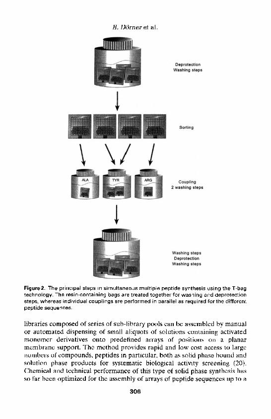
Figure2. The principal steps in simultaneous multiple peptide synthesis using the T-bagtechnology. The resin-containing bags are treated together for washing and deprotectionsteps, whereas individual couplings are performed in parallel as required for the differentpeptide sequences.
libraries composed of series of sub-library pools can be assembled by manualor automated dispensing of small aliquots of solutions containing activatedmonomer derivatives onto predefined arrays of positions on a planarmembrane support. The method provides rapid and low cost access to largenumbers of compounds, peptides in particular, both as solid phase bound andsolution phase products for systematic biological activity screening (20).Chemical and technical performance of this type of solid phase synthesis hasso far been optimized for the assembly of arrays of peptide sequences up to a
306

Figure3, A microtitre plate-adapted format of a manually prepared 96-spot membrane.Dark spots indicate stained (blue) free amino functionalities, light spots have turned toyellow after coupling with an Fmoc-amino acid HOBt ester.
length of 20 residues using conventional Fmoc/tBu chemistry. Other synthesesbased on amide bond formations, such as peptoids and polyamide nucleicacids (PNAs) (21), have been reported. In principle, chemistries that can becarried out at ambient temperature can be adapted to SPOT-synthesis.
SPOT-synthcsis is particularly flexible with respect to numbers and scalesthat can be accomplished. The arrays are freely selectable to fit the individualneeds of the experiment by the choice of membrane material, thickness, areaspecific anchor loading, and spot size (Table 1). The standard format used inmanual SPOT-synthesis was adapted to the 8 X 12 array of a microtitre platewith 96 spots (Figure 3).
An automated SPOT-synthesizer, the ASP222, has been developed atABIMED Analysen-Technik GmbH based on a Gilson pipetting workstation.This instrument can handle simultaneously up to six standard membranesheets. Moreover, automated spotting can be exploited to reduce the size ofspots and thus increase considerably the number per area (Table I), whichotherwise is extremely tedious to manufacture by hand. At present, up to 2200spots can be generated by pipetting only 30 nl reaction volumes onto amicrotitre plate-sized sheet (in total 13000 spots per working area of therobot). The instrument so far performs only the pipetting work; all washingsteps are carried out manually.
307

B. Dorner et al.
Table 1. Standard SPOT-array configurations (each one fitted to a sheet of the size of amicrotitre plate, 8x12 cm)
Format Membrane Spotted Spot size Positional Synthesis scaletypea volume (ul)b (mm) distance (mm) (nmol)
8 X 1 2 = 9 6 5 4 0 0.5/0.7 7 9 2 57X10 = 70 Chr1 1.0/1.5 8 10 5017X25 = 4 2 5 5 4 0 0.1/0.15 3 4 640X50 = 2000 50 0.03/0.05 1 2 1
aChromatography paper products from Whatman. Typical amino substitution is 0.2-0.4 umol/cm2 fortype 50 and 540 paper, 0.4-0.6 umol/cm2 for type Chr1 paper.bVolumes are given for array generation step/peptide assembly step.
2.2 Synthesis of peptide SPOT-arrays2.2.1 Preparation of the membrane supportsThe most widely used support material for SPOT-synthesis is made ofspecially selected, pure cellulose chromatography paper. Other materials suchas PVDF (Immobilon AV) (22) or polypropylene (21) have also been used.All membranes require a chemical derivatization to carry free aminofunctions. Paper is easily derivatized by the esterification with an N-Fmocprotected amino acid (e.g. Fmoc-pAla) to available hydroxyl functions on thecellulose fibres of the whole sheet followed by Fmoc-cleavage (20). The arraysof spot reactors on such amino-membranes are then generated by coupling asecond Fmoc-BAla or suitable linker/anchor acid, applying spotwise onlysmall aliquots of activated solution as given in Table 1 with either amicropipette or pipetting robot. After completion of this spot-reaction, allresidual amino functions between spots are blocked by acetylation. This arrayformation step requires very accurate pipetting. During peptide assembly,slightly larger volumes are dispensed and the spots then formed exceed thoseinitially generated in order to avoid incomplete couplings at the edges.
The choice of a linker/anchor compound determines the fate of the peptideproducts. Stable amide/ester bonds such as with the BAla-BAla spacer(Protocol 3) result in an array of immobilized peptides. Conventional linkerchemistry can be incorporated to yield cleavable peptides in different formats.Esterification reactions are problematic in this open reactor system; in suchcases, a preformed amino acid linker compound should be used. Such linkersshould allow a solid phase bound deprotection/pre-purification followed by amild final release reaction. Several options have been described (23-25).More sophisticated and superior amino membranes are now available fromseveral sources (ABIMED, AIMS, Jerini, PerSeptive).
Free amino functions on the spots can be stained with bromophenol blue(26) prior to the coupling reactions. This allows visual monitoring of theperformance of all the synthesis steps. Thus, a standard membrane for SPOT-
308

14: Manual multiple synthesis methods
synthesis displays an array of light blue spots on a white background. Eachspot can be marked by writing a number in pencil next to it (Figure 3). Thesenumbers refer to the corresponding peptide sequences that are assembled onthem and are a guide for rapid manual distribution of the solutions ofactivated amino acid derivatives at each elongation cycle using a suitablepipetting protocol. For automated pipetting, no pencil marking is necessary.Exact fixing of the membranes is ensured by the perforation for the holderpins.
Protocol 2. Generation of the SPOT-array
Equipment and reagents• A kit that includes all necessary items for • Software: special DOS-PC computer pro-
manual SPOT-synthesis is currently avail- grams for the generation of peptide listsable from GENOSYS and pipetting protocols are included in the
. Flat reaction troughs with good closing lids synthesis kit and the operation software ofmade of chemically inert material (poly- the ASP222ethylene, polypropylene) that have the . A stock solution of 10 mg/ml bromophenoldimensions slightly larger than the mem- blue indicator (Merck) in DMFbranes used . N,/V-diisopropylcarbodiimide (DIC)
. A micropipette adjustable from 0.5 to 10 ul . 2% v/v acetic anhydride solution in DMF(Eppendorf or Gilson) with corresponding 'plastic tips; alternatively, the ASP222 • Amme-free N,N-dimethylformamide (DMF)aspotting robot from ABIMED • Alcohol (methanol or ethanol) of technical
. Small (1.7 ml) plastic tubes (e.g. Eppendorf, gradesafe-twist) as reservoirs for amino acid • Amine-free W-methylpyrrolidinone (NMP)6
solutions . 20% v/v solution of piperidine in DMF;• A rocker table piperidine is toxic and should be handled. Two dispensers adjustable from 5 to 25 ml with gloves under a hood!
for DMF and alcohol stock containers • N-hydroxybenzotriazole (HOBt)c
• Amino-cellulose membranes from ABIMEDor AIMS
Methodd
1. Generate the list of peptides to be prepared and correspondinglydefine the array(s) required for your particular experiment accordingto number, spot size, and scale (see Table 1).
2. Mark the spot positions on the membranes with pencil dots formanual synthesis and place the membranes in the reaction trough orfix them on the platform of the ASP222-robot.e
3. Prepare a solution containing activated anchor compound in NMP(e.g. 0.3 M Fmoc-BAla-OH, 0.45 M HOBt, and 0.35 M DIC; or 0.3 MFmoc-BAla-OPfp, 0.075 M HOBt, and 0.35 M DIC).f
4. Leave this solution for 30 min and then spot aliquots of chosenvolume to all positions according to the array configuration prepared.
5. Leave for 15 min.
6. Repeat spotting once and let react for 30 min.
309

B. Dorner et al.
Protocol 2. Continued7. Wash the membrane with 20 ml of acetylation mix for 30 s, once
again for 2 min, and finally leave overnight in acetylation mix.
8. Wash the membrane three times with 20 ml DMF.
9. Incubate the membrane for 10 min with 20 ml piperidine/DMF.
10. Wash the membrane four times with 20 ml DMF.
11. Incubate the membrane with 20 ml of 1% bromophenol blue stock inDMF (must be a yellow solutionl). Repeat if traces of remainingpiperidine turn it into a dark blue solution. Spots should be stainedonly light blue!
12. Wash twice with 20 ml alcohol.
13. Dry with cold air from a hair-drier in between a folder of 3 mm paper.
14. Seal in a plastic bag and store at-20°C.
aAmine contamination is checked by addition of 6 ul of bromophenol blue stock solution to 1ml of DMF. If the resulting colour is yellow, then this batch can be used without furtherpurification."Amine contamination is checked as under DMF. If the resulting colour is yellow, then theNMP can be used without further purification. Most commercial products, however, are notacceptable. Then add alumina oxide (for chromatography, Fluka) and agitate gently overnight.Pass through a 20 um polyethylene sinter or cellulose filter. Divide the clear liquid into 100 mlportions and store tightly closed at -20°C.°HOBt is only available as the monohydrate. Dehydrate in a desiccator over phosphoruspentoxide at 50°C and 10-3 bar for at least 3 days. Store in a tightly closed container at roomtemperature.dAII volumes given below hold for a standard paper sheet of 8 x 12 cm (Table 7) and have tobe adjusted to more sheets, other paper qualities, or sizes. Solvents or solutions used inwashing/incubation steps are agitated gently on a rocker plate and aspirated or decanted afterthe time indicated.eMembranes can be cut easily into parts or pieces with scissors prior to or post synthesis asrequired for the particular experiment. Simply mark the cutting lines with a pencil. Spotpositions should be also marked with a pencil dot if treated individually after the synthesis.The pencil marking is sufficiently stable during the synthesis procedure.fAlthough more expensive, the pentafluorophenyl ester is preferred when working with theASP222 robot (see Chapter 13, Section 4.5).
2.2.2 Peptide assemblyAny series of individual peptide sequences can be freely arranged as a two-dimensional array on a SPOT membrane for systematic analysis with, forexample, overlapping fragments derived from a protein sequence(SPOTscan), stepwise N- or C-terminally truncated fragments (SPOTsize),substitution analogues (SPOTsalogue), etc. (20). Moreover, modern peptidelibrary screening approaches allowing the a priori delineation of peptideepitopes (27) have been incorporated into SPOT-synthesis (28, 29). Theseapproaches exploit the preparation of arrays of defined peptide mixtures(pools) by incorporation of randomized positions and the presentation of
310

14: Manual multiple synthesis methods
Binding of Streptavidin-AP to Ac-X-X-1-2-X-X-
A C* D E F G H I K L M N T Q R I
Position 1
Figure4. Example of a first screening round for the a priori delineation of peptidesbinding to streptavidin. Left: photograph of a membrane with 400 spots of a hexapeptidelibrary in the format Ac-XX12XX- (plus 25 reference spots in the upper fane) after probingwith a streptavidin-alkaline phosphatase conjugate. Bound protein was visualized with aphosphatase specific colour reaction (20). Right: spectral diagram display of Ac-XX12XX-showing the correlation of quantified signals to the defined amino acid residues atpositions 1 and 2 (X positions include all 20 proteinogenic amino acids); C* = Cys(Acm).Clearly, the XXHPXX- pool gives the strongest signal.
entire peptidc libraries (e.g. all 64 Mio-hexapeptides) as strategic sets of sub-libraries (Figure 4) . Randomized positions (X) within a peptidc sequenceassembled on a spot are introduced quite reliably by coupling with equimolaramino acid mixtures and applying these at a sub-molar ratio with respect toavailable amino functions on the spots. This is to allow all activatedderivatives to react quantitatively during a first round of spotting, thencompleting all peptide elongations by two to three successive repeats ofspotting. Using this coupling procedure, any position in a peptide sequencecan be randomized easily without special considerations or increase intechnical effort.
Protocol 3. Assembly of peptides on SPOTs
Equipment and reagents• For general information see Protocol 2• Monomer derivatives: * Fmoc-amino acids
are available from several suppliers insufficient quality. In situ prepared HOBtesters of these Fmoc-amino acids in NMPare used throughout for spotting reactions.Side-chain protection is Cys(Acm) orCys(Trt), Asp(OtBu), Glu(OtBu), His(Boc) or
His(Trt), Lys(Boc), Asn(Trt), Gln(Trt),Arg(Pbf), Ser(tBu), Thr(tBu), Trp(Boc), andTyr(tBu). Dissolve 1 mmol of each in each 5mt NMP containing 0,3 M HOBt to give 0.2M solutions. Divide into 100 ul aliquots incorrectly labelled Eppentorf tubes. Closetightly, freeze in liquid nitrogen, and storeat -70"C.
311
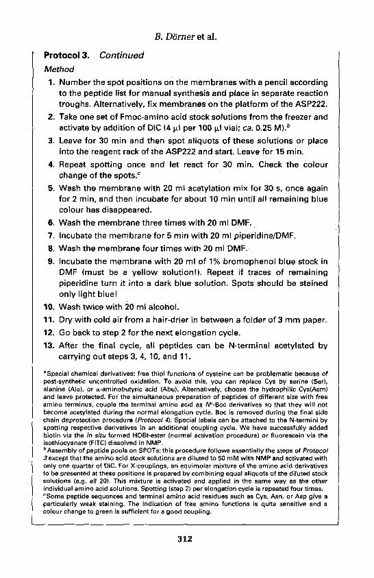
B. Dorner et al.
Protocol 3. Continued
Method
1. Number the spot positions on the membranes with a pencil accordingto the peptide list for manual synthesis and place in separate reactiontroughs. Alternatively, fix membranes on the platform of the ASP222.
2. Take one set of Fmoc-amino acid stock solutions from the freezer andactivate by addition of DIC (4 ul per 100 ul vial; ca. 0.25 M).b
3. Leave for 30 min and then spot aliquots of these solutions or placeinto the reagent rack of the ASP222 and start. Leave for 15 min.
4. Repeat spotting once and let react for 30 min. Check the colourchange of the spots.c
5. Wash the membrane with 20 ml acetylation mix for 30 s, once againfor 2 min, and then incubate for about 10 min until all remaining bluecolour has disappeared.
6. Wash the membrane three times with 20 ml DMF.
7. Incubate the membrane for 5 min with 20 ml piperidine/DMF.
8. Wash the membrane four times with 20 ml DMF.
9. Incubate the membrane with 20 ml of 1% bromophenol blue stock inDMF (must be a yellow solutionl). Repeat if traces of remainingpiperidine turn it into a dark blue solution. Spots should be stainedonly light blue!
10. Wash twice with 20 ml alcohol.
11. Dry with cold air from a hair-drier in between a folder of 3 mm paper.
12. Go back to step 2 for the next elongation cycle.
13. After the final cycle, all peptides can be N-terminal acetylated bycarrying out steps 3, 4, 10, and 11.
aSpecial chemical derivatives: free thiol functions of cysteine can be problematic because ofpost-synthetic uncontrolled oxidation. To avoid this, you can replace Cys by serine (Ser),alanine (Ala), or a-aminobutyric acid (Abu). Alternatively, choose the hydrophilic Cys(Acm)and leave protected. For the simultaneous preparation of peptides of different size with freeamino terminus, couple the terminal amino acid as /Va-Boc derivatives so that they will notbecome acetylated during the normal elongation cycle. Boc is removed during the final sidechain deprotection procedure (Protocol 4), Special labels can be attached to the N-termini byspotting respective derivatives in an additional coupling cycle. We have successfully addedbiotin via the in situ formed HOBt-ester (normal activation procedure) or fluorescein via theisothiocyanate (FITC) dissolved in NMP.bAssembly of peptide pools on SPOTs: this procedure follows essentially the steps of Protocol3 except that the amino acid stock solutions are diluted to 50 mM with NMP and activated withonly one quarter of DIC. For X-couplings, an equimolar mixture of the amino acid derivativesto be presented at these positions is prepared by combining equal aliquots of the diluted stocksolutions (e.g. all 20). This mixture is activated and applied in the same way as the otherindividual amino acid solutions. Spotting (step 2) per elongation cycle is repeated four times.cSome peptide sequences and terminal amino acid residues such as Cys, Asn, or Asp give aparticularly weak staining. The indication of free amino functions is quite sensitive and acolour change to green is sufficient for a good coupling.
312

14: Manual multiple synthesis methods
Protocol 4. Side-chain deprotection of SPOT assembled peptides
Equipment and reagents• For general information see Protocol 2 Trifluoroacetic acid is very corrosive and• Deprotection mix: 50% TFA, 3% triisobutyl- should be handled with gloves under a
silane, 2% water, and 45% DCM (v/v). hood!
Method
1. Prepare 40 ml of deprotection mix per standard membrane.
2. Place the dried membrane from step 10 of Protocol 3 in the reactiontrough (only one membrane per trough; do not stack severalmembranesl).
3. Add 20 ml deprotection mix, remove bubbles from under themembrane, close the trough tightly and agitate for 1 h.
4. Replace the deprotection mix with the remaining 20 ml and agitateagain for 1 h.
5. Wash the membrane four times for 2 min with 20 ml DCM.
6. Wash the membrane three times for 2 min with 20 ml DMF.
7. Wash the membrane three times for 2 min with 20 ml alcohol.
8. Wash the membrane three times for 10 min with 20 ml 1 M acetic acidin water.
9. Wash the membrane three times for 2 min with 20 ml alcohol.
10. Dry with cold air from a hair-drier in between a folder of 3 mm paper.
11. Seal in a plastic bag and store at -20°C.
2.3 Applications of peptide SPOT-arraysInitially, SPOT-synthesis was developed for rapid analysis of linear peptideepitopes recognized by antibodies. After blocking unspecific binding sites, thewhole membrane (or parts of it) is incubated with the anti-serum and de-tection of bound antibody is achieved by conventional immuno- or Western-blot procedures with colour dye development. If peptide spots have notbecome chemically, enzymatically, or otherwise irreversibly modified, peptidearrays on cellulose membranes are reusable many times (>20) when treatedcarefully. Signal patterns obtained from peptide arrays on SPOTs can bedocumented and quantitatively evaluated using modern image analysissystems as used with other two-dimensional analysis media such as electro-phoresis gels and blotting membranes.
The list of further applications, however, is rapidly expanding. These includepeptide interactions with proteins in general (cytokines, enzymes, receptors,
313

B. Dorner et al.
etc.), metal ions, and nucleic acids, as well as whole cells. Any labelling tech-nique such as radioisotopes (e.g. introduced by in vitro transciption/translation of cloned DNA) or fluorescent dyes is fully compatible. A fullreport on these is beyond the scope of this chapter. The user can followdescribed procedures (20) or simply adapt his/her own assay.
3. Manual multipeptide synthesis in block arraysDescribed here is a manual system to enable the synthesis of large numbers ofpeptides easily, cheaply, and in quantities of a few milligrams of each peptide.Approximately four to five coupling cycles can be completed in one workingday with a typical set of 48 different peptides completed in a week.
3.1 HardwareThe apparatus and procedures described here are performed in a device builtand supplied by Biotech Instruments Ltd. (30). The system is based on a set of48 wells drilled in a two-part polythene block, each well being a reactor for asingle peptide. The design of the system centres around trying to make afoolproof synthesis method, suited for either simple manual operation orsome form of mechanization. The device allows up to 48 different peptides tobe synthesized simultaneously within a few days, using virtually any type ofresin or activation chemistry at scales of 5-10 umol for each peptide. Theorganization of the synthesis is assisted by means of computer-generatedprintout, which guides the tracking of the synthesis and directs the operatorduring the coupling reactions.
The top block contains the 48 reaction wells with their centres spaced 19mm apart; the layout is shown in Figure 5. This spacing of the wells is exactlydouble that of the wells in a microtitre plate, and thus enables alternatechannels of a standard multichannel pipette to be used for reagent delivery if
Figure 5. Layout of the reaction wells in the top block.
314

Figure 6. Cross-section of the block system, showing the process of cleaving the peptidesoff the solid phase.
required. A short length of PTFE tubing fits into the outlet hole of each wellto drain the wells and to collect the cleaved peptide in a vial during the finalstage of synthesis. A PTFE frit, pore size 100 um, is placed in the bottom ofeach 10-mm-diameter well to retain the peptide-resin.
Figure 6 shows a cross-section of just four wells in the block, arranged as forthe cleavage reaction. Reagents and solvents are added from the top and,after reaction, allowed to flow through into the chamber below with theassistance of a small, solvent-resistant vacuum pump.
The complete fluid system is shown in Figure 7, where the solvents arecontained in pressurized bottles and are selected by a rotary valve. The flowto the four-way manifold is controlled by a two-way ON/OFF valve or pinchclamp in silicone tubing. The use of a manifold enables solvents to bedelivered to four wells simultaneously. Alternatively, the solvents can bedelivered by means of a multichannel pipette, operating with alternatechannels delivering into four wells at a time.
After all the synthesis cycles are completed, the peptides are simul-taneously cleaved off the resins and collected in glass vials placed under thereaction wells. The vials are held in a tray for stability and to maintain theirexact positioning under the drain tube for each of the wells. These peptidesolutions can then be dried and worked up in the usual way.
3.2 ChemistryThe method uses the standard Fmoc/tBu strategy with any convenientactivation procedure. For manual operation (Protocol 5), it can be somewhat
315

Figure 7. Entire fluidic assembly for the synthesis operation.
easier to use the OPfp active esters, as only two reagents have to be addedduring the coupling cycle, the Fmoc-amino acid OPfp ester and 1-hydroxy-benzotriazole. Free amino acids are more stable in solution but require themixing of three reagents. To ensure complete coupling reactions, a minimumof 5 X molar excess of the amino acids is used for all the synthesis steps, as thecost of the amino acids is not usually a major expense in the work. Anyconvenient solid phase can be used in this system, although the author hasmainly worked with Wang resins for this scale of synthesis. Both peptide C-terminal acids and amides can be synthesized simultaneously and unusualamino acids can easily be incorporated. Typically, the peptides are cleaved offthe resins using a cocktail of trifluoroacetic acid:l,2-ethanedithiol:water(95:2.5:2.5 v/v; Protocol 6), a mixture which is generally effective. As largenumbers of peptides are being made simultaneously, the same cleavagemixture must be used for every peptide regardless of amino acid content. Aphilosophy of maximum protection has been used by the author, againbecause it is not usually viable to treat peptides individually.
The times and steps for a typical synthesis cycle is shown in Table 2. Thetimes are not critical and if in doubt can be extended, as can the number ofwashes. After the deprotection step, it is advisable to include one wash stepwhich almost fills the wells in order to completely wash the sides and removeall the piperidine. The ether wash removes all solvent from the resin andcompletely dries the PTFE frit. This prevents any solvent normally heldwithin the resin from diluting the incoming activated Fmoc-amino acid andthe high surface tension of the dry frit prevents the amino acid mixture fromdraining through the frit. The subsequent addition of the amino acid/reagentmixture must be done gently to prevent it from draining through the frit.
316

14: Manual multiple synthesis methods
Table 2. The operations for one synthesis cycle
Step no.
123456789
101112
Solvent/reagent
Wash with DMFDeprotect with 20% piperidineDeprotect with 20% piperidineWash with DMF'Big'wash with DMFWash with DMFWash with etherVacuum dry resin b
Mix and add amino acidsAcylation reaction c
Wash with DMF
No. of washes/dwell timea
2X4min6min3x1 x1 x2x2min
60min3xEnd of cycle
aThe exact times are not critical.bEssential to prevent the reactants from draining through the frit.cFor short peptides, the acylation reaction time can be reduced to 30 min.
3.3 SoftwareA program written in Basic is used to edit and arrange all the 48 peptidesequences into an order. The problem of ensuring that the correct amino acidis added to any specific well is solved by the program printing a series ofoverlay sheets showing all the amino acids that are to be added during anyone cycle of the synthesis. The position of the printing on these overlayscorresponds exactly to the layout of the wells in the block and when pegged inplace over the top of the block shows exactly where the amino acids are to beadded into each well. When each amino acid is to be added to the deprotectedresin, a pipette is used to pierce the paper and deliver the reagents into thewell below. In this way, the overlay sheet not only serves to indicate whichamino acid should go into any one well, but also serves to show which well hasalready been filled with reagents.
Protocol 5. The synthesis cycle
Safety notes: All the steps involving manipulation of solvents must beperformed in an efficient fume hood.
Reagents
• Peptide synthesis grade DMF• N-deblock solution: 20% v/v piperidine in
DMF• Amino acid-derivatized resins (5 umol of
each resin per reaction well)• Solutions of the required Fmoc-amino acids.
For each amino acid addition, dissolve 25umol of the protected amino acid in 150 ulof DMF
• A solution of TBTU in DMF at a con-centration of 450 umol/ml (145 mg of TBTUin 1.00 ml of DMF); use 50 ul of this solutionper addition
• A solution of W-methyl morpholine (NMM)in DMF at a concentration of 500 umol/ml(55 ul of NMM diluted to 1.00 ml with DMF);use 50 ul of this solution per addition
• Diethyl ether
317

B. Dorner et al.
Protocol 5. Continued
Method (5 umol synthesis scale)1. At the start of the synthesis, place caps over each well to avoid any
spilt resin falling into the wrong reaction well.2. Following the positions shown on the first overlay sheet, add 5 umol
of the correct amino acid resin to each of the wells in turn and replacethe cap after each addition.
3. Remove the caps when all the resins have been added.4. Use the software to calculate the amounts and volumes of amino acid
solutions required. Weigh out the amino acids for the current day'swork in labelled tubes and dissolve them in the required volume ofDMF. Prepare the working volumes of TBTU and NMM solutions;these should be stable for the duration of the synthesis.
5. Following the procedure in Table 2, wash and N-deblock each of theamino acid resins.
6. After the ether wash and drying stage, the wells are ready for theaddition of the incoming amino acid.
7. Place the appropriate coupling overlay sheet over the block and peginto position.
8. Working with one amino acid at a time, mix 150 ul of the amino acidsolution with 50 ul of TBTU solution and 50 ul of NMM solution in anEppendorf tube.
9. Pipette this reaction mix through the paper overlay into the correctwell.
10. Repeat this procedure for all the additions for each amino acid in turn.When the process is complete, all the wells will have a puncture markin the paper indicating that the additions are complete.
11. After the coupling time is over, the excess reagents can be pumpedthrough to waste and the resins washed with solvent ready for thenext cycle.
Protocol 6. Peptide cleavage
Safety notes: This process must be performed in an efficient fume hood.The cleavage solution is corrosive, volatile, and very mobile, and istherefore difficult to control in a pipette.
Reagents• Prepare N x 2 ml of cleavage mixture, acetic acid + 2.5% 1,2-ethanedithiol + 2.5%
where N is the number of peptides to be v/v watercleaved, each containing 95% trifluoro-
318

14: Manual multiple synthesis methods
Method1. Follow the procedure in Table 2, steps 1 to 8, to remove the final
Fmoc-groups from the assembled peptides. Then wash and dry thepeptide-resins.
2. Prepare the correct volume of the cleavage cocktail.3. For each peptide to be cleaved, place a 4 ml glass collection vial
scribed with the peptide code number in the collection rack in thelower chamber and carefully fit the block assembly together ensuringthat the drain tubes enter all the vials. It is essential to scribe thetubes with a diamond pen as any writing with felt-tipped pen will beerased by the acid.
4. Add about 0.5 ml of the cleavage mix to each reaction well. A plasticPasteur pipette is best used for this operation as the vigorous actionof a stepper-type pipette can cause some of the resin to splash out ofthe wells. This solution will slowly drain through the frit into the vialbelow, simultaneously cleaving and extracting the peptide off theresin.
5. At approximately 20 min intervals, briefly apply the vacuum to drainany residual acid into the wells, then with the vacuum off.
6. Add a further 0.5 ml of the cleavage mixture, and repeat step 5.7. When all the cleavage mix has been added and drained through, turn
on the system vacuum pump. This will drain all the remaining acidinto the vials and start to evaporate the acid by drawing a rapidstream of air over the surface of the cleavage mix in the vial.
8. Keep the vacuum pump operating for about 2-4 h until all the acid hasbeen evaporated.
9. Add about 2 ml of cold diethyl ether to precipitate the peptides. If alight suspension is formed, the tubes can be centrifuged at about3000 r.p.m. to ensure complete precipitation of the peptide.
10. Carefully decant the ether supernatant and repeat step 9.11. Allow the peptides to air dry.12. Dissolve the peptides in 1 ml of 10% acetic acid and allow to stand for
about 1 h to complete the deprotection of any N-Boc-protectedtryptophan.
13. Lyophilize the peptide solutions.
4. Synthesis of peptides by the Multipin™ method4.1 IntroductionThe Multipin™ method is an effective, low cost, simultaneous multiplepeptide synthesis technology which gives researchers ready access to large
319
B. Dorneret al.
numbers of peptides (31-38). Chiron Technologies currently markets a rangeof Multipin™ products for both peptide and non-peptide solid phasesynthesis. This chapter gives an overview of a selection of the key proceduresused for peptide synthesis with commercially available Multipin™ kits.Synthesis is performed on plastic pins arranged in the 8 X 12 microtitre plateformat, thus allowing for the simultaneous preparation of sets of up to 96peptides (or multiples thereof).
The 'pin' consists of a radiation-grafted polypropylene 'crown' fitted to aninert polypropylene 'stem'. Graft polymers used with the Multipin™ systeminclude polystyrene, a methacrylamide copolymer (38), and poly(hydroxyethylmethacrylate) (34) (HEMA). We have found that the HEMA surface is bestsuited to peptide synthesis. Historically, peptides were prepared in a non-cleavable format on the crown surface for epitope mapping applications (39).Over the past decade, however, most peptides prepared by the Multipin™method have been synthesized on cleavable linkers. The linkers used forpeptide synthesis are outlined below.
Originally, Multipin™ peptide synthesis was performed using a Bocprotocol (39); however, due to more streamlined handling (required forefficient multiple synthesis), Fmoc/tBu synthetic protocols are now used. TheTFA-mediated cleavage method used with Fmoc/fBu synthesis is far moreeasily adapted for large numbers than the HF required with Boc synthesis.
Crowns with a loading of 20 umol yield around 20 mg of a typical 10-merpeptide. Larger quantities can be accessed by preparing multiple copies of atarget peptide, a simple process with simultaneous multiple synthesis. Whensmall numbers of peptides are being prepared, synthesis is often performedon unmounted crowns in glass vials or polypropylene tubes. The synthesiscycle used with the Multipin™ method is: coupling, washing, Fmoc-deprotection, and washing. Coupling reactions can be performed in the wellsof microtitre plates, in which case the washing and deprotection steps areperformed in polypropylene baths. As with other forms of solid phasesynthesis, the washing steps are critical. As noted in the protocols givenbelow, the pins must be fully immersed and soaked for the given time. Highquality solvents are always used for washing.
4.2 LinkersFigure 1 summarizes the linkers used for peptide synthesis by the Multipin™method. Most carboxylic acids are prepared on the 4-(hydroxymethyl)-phenoxyacetic acid handle 1 (40). To avoid diketopiperazine formationduring the synthesis of peptides containing a C-terminal Pro residue, thesterically hindered linker 2 is used (41). To minimize the risk of racemizationduring linker derivatization, Asp and Glu are linked via their side-chainfunctionality, as shown in 3. Similarly, Asn and Gln are prepared by couplingFmoc-Asp-OtBu and Fmoc-Glu-OtBu, respectively, to the Rink linker 4 (42).
320

Figure8. Linkers used on SynPhase™ crowns (n = 1, Asp; n = 2, Glu).
Primary peptide amides are prepared on the Rink linker 5. Although notcovered in this chapter, secondary amides have been prepared on the linkerdescribed by Barany and co-workers (43). Peptide aldehydes have beenprepared on the oxazolidine linker (44). The trityl linker (45) has also beenused for the preparation of protected peptide fragments and other specializedC-terminal endings such as hydroxamic acids (46).
4.3 Peptide synthesisAlthough a variety of solvents have been reported for peptide synthesis, weuse highly purified DMF for all coupling reactions (Protocol 7). Where Fmoc-protected amino acids require acid-labile side-chain protection, we recom-mend: Arg(Pbf), Asp(OtBu), Cys(Trt), Glu(OtBu), His(Boc), Lys(Boc),Ser(tBu), Thr(tBu), Trp(Boc), and Tyr(tBu). Although Fmoc-His(Boc)-OH isthermally labile and must be stored at -20°C, we have found that it couplesmore efficiently than either Fmoc-His(Trt)-OH or Fmoc-His(Bum)-OH.Fmoc-Asn(Trt)-OH and Fmoc-Gln(Trt)-OH are generally used with HBTU,HATU, or BOP activation; the non-side-chain protected derivatives of Asnand Gln can be used with DIC/HOBt activation in short peptides or at the N-terminal coupling (47).
For phosphopeptide synthesis, we routinely use Fmoc-Ser(PO(OBzl)OH)-
321

B. Dorner et al.
OH, Fmoc-Thr(PO(OBzl)OH)-OH, and Fmoc-Tyr(PO(OBzl)OH)-OH (seebelow) in preference to global post-synthesis phosphorylation (48). Peptidelactams are generally prepared using Fmoc-Asp(OAll)-OH, Fmoc-Glu(OAll)-OH, and Fmoc-Lys(Alloc)-OH, with the allyl-based protection being removedfrom the support-bound peptides with palladium tetrakistriphenylphosphine.
We use a range of coupling agents in our peptide synthesis. For simplicity ofuse, DIC is highly recommended: it is a liquid, it reacts very selectively withcarboxylic acids in the presence of amines, and it is economical. Typicalcoupling conditions are Fmoc-amino acid/DIC/HOBt (120 mM, 120 mM, 120mM). Recommended coupling times are 2-16 h. When preparing couplingsolutions, the activating agent is always added last, and the solution should beallowed to stand for 5 min prior to coupling to the solid support. For peptidesof moderate length (>20-mer), better results can be obtained using HBTU asfollows: Fmoc-amino acid/HBTU/HOBt/NMM (120 mM, 120 mM, 120 mM,240 mM) in DMF. Once again, an incubation time of 5 min is required prior toaddition of the coupling solution to the support-bound amine.
Other activating agents, such as BOP (49) and HATU (47, 50), have alsobeen applied to pins with good results. In the case of HBTU, BOP, andHATU, excess tertiary base such as NMM or DIPEA must be included foreffective coupling to occur. Furthermore, the uronium-based reagents HBTUand HATU should not be used in excess (with respect to amino acid) as theycan react with support-bound primary amines (51). Consequently, BOP/DIPEA (100 mM, 200 mM) is the preferred reagent when preparing cyclicpeptides via lactam formation on the pin surface. We have found HBTU/HOBt/DIPEA to be the activating agent of choice when coupling Fmoc-Ser(PO(OBzl)OH), Fmoc-Thr(PO(OBzl)OH)-OH, and Fmoc-Tyr(PO-(OBzl)OH)-OH (48). We recommend the use of TFFH/DIPEA (100 mM, 200mM) for sterically demanding couplings such as those to N-alkylated aminoacids and for couplings to unreactive amines such as anilines.
After each coupling reaction, the pins are subjected to a rigorous washingprotocol. This is conveniently performed in a bath, so that simultaneousmultiple washing is a straightforward process. Sufficient solvent is placed inthe bath so that the crowns are fully covered when the block of 96 pins isplaced in the bath. For consistency, each wash is timed. The standard washingprotocol used after either coupling or Fmoc deprotection steps is: DMF,MeOH, followed by air drying. Note that the HEMA polymer is well solvatedby MeOH. Fmoc deprotection is generally performed by soaking the pins in20% piperidine in DMF (v/v) for 30 min. Like washing, this is convenientlyperformed in a bath. Following deprotection, excess reagent is shaken fromthe pin surface and the pins are then subjected to the standard washingprotocol.
At the completion of peptide assembly, the peptides are side-chain de-protected and cleaved, usually concurrently. In the Multipin™ method,cleavage is performed using TFA solution containing scavengers. Cleavage
322

14: Manual multiple synthesis methods
can be performed either into either polypropylene tubes, which are rackedinto the 8 x 12 microtitre plate format, or into larger polypropylene tubes(Protocol 8). The latter method is convenient when multiple pins containingthe one sequence are cleaved together, and when larger volumes of ethersolution are required to extract thiol-based scavengers and by-products ofside-chain deprotection from the cleaved peptides. The cleavage solutions areevaporated using either a solvent resistant vacuum centrifuge or a stream ofdry nitrogen gas. The target peptides should be dissolved (or suspended) inaqueous acetonitrile and freeze dried to remove remaining volatile impuritiesprior to sampling for analysis and/or purification.
Protocol 7. Synthesis of peptides on pins
Equipment and reagents• Multipin™ multiple peptide synthesis • 1-Hydroxybenzotriazole hydrate (HOBt)
(MRS) kit (Chiron Technologies). The kit . BOP and HBTUincludes crowns and stems, plastic baths . Tetramethylfluoroformamidiniumand reaction trays, software (for IBM or hexafluorophosphate (TFFH) (PerSeptiveMacintosh), and a manual Biosystems)
. Solvent-resistant pipettors (e.g. Oxford, . Miscellaneous reagents: N,N-diisopropyl-Gilson) and tips ethylamine (DIPEA), N-methylrnorpholine
• Fluorenylmethyloxycarbonyl (Fmoc)- (NMM)protected amino acids with appropriate . Solvents: W,N-dimethylformamide (peptideside-chain protecting groups grade) (DMF), methanol (AR grade)
• N,N-Diisopropylcarbodiimide (DIC) (Sigma) • Piperidine
Method
If multiple sets of peptides are being synthesized, it is advisable to refer tothe manual provided with the Multipin™ Multiple Peptide Synthesis(MRS) kit. Use the software to generate the set of peptide sequences to besynthesized.
1. Mount crowns with attached stems into the block in the requiredformat, as indicated by software.
2. Fmoc-deprotection procedure:8 immerse crowns in 20% piperidine/DMF for 30 min. For multiple synthesis using the 8 x 1 2 format; theblock containing up to 96 crowns on stems can be immersed in a bathcontaining the deprotection solution.
3. Post-Fmoc-deprotection washing procedure: wash the crowns in turnwith DMF ( 2 x 5 min) and MeOH ( 2 x 5 min), and then air dry (15 min).
4. Coupling procedures
Procedure 1. DIC/HOBt (standard coupling): prepare the couplingsolution such that the final coupling concentrations are Fmoc-aminoacid/DIC/HOBt (120 mM:120 mM:120 mM) in DMF. Allow at least1.0-1.5 ml per l-series crown (loading = 20 u.mol). Add couplingsolution to crowns and couple for 2 h at room temperature.
323

B. Dorner et al.
Protocol 7. ContinuedProcedure 2. HBTU/HOBt/NMM (standard coupling): prepare thecoupling solution such that the final coupling concentrations areFmoc-amino acid/HBTU/HOBt/NMM (120 mM:120 mM:120 mM:240mM) in DMF. Add coupling solution to crowns and couple for 2 h atroom temperature.
Procedure 3. HBTU/HOBt/DIPEA (phospho-amino acids): same asHBTU/HOBt/NMM except DIPEA replaces NMM.
Procedure 4. TFFH/DIPEA (for sterically hindered unreactive aminoacids): prepare the coupling solution such that the final couplingconcentrations are Fmoc-amino acid/TFFH/DIPEA (120 mM:120mM:240 mM) in DMF. Add coupling solution to crowns and couple for2 h at room temperature.
Procedure 5. BOP/DIPEA (intramolecular lactam formation): preparethe coupling solution such that the final coupling concentrations areBOP/DIPEA (120 mM:240 mM) in DMF. Add coupling solution tocrowns and couple for 18 h at room temperature.
5. Post-coupling washing procedure: DMF ( 2 x 5 min), MeOH ( 2 x 5 min).
6. Air dry (15 min) the crowns.
"The procedures are illustrative of what is contained in the manual.
Protocol 8. Side-chain deprotection and cleavage of peptides
Equipment and reagents» Additionally, depending on the type of • Trifluoroacetic acid (TFA)
cleavage, solvent for the extraction of . Scavengers and reducing agents: 1,2-peptide from the crowns or for washing of ethanedithiol (EOT), anisole, and 2-the dried peptide might be required. mercaptoethanolAppropriate solvents include acetonitrile(HPLC grade), diethyl ether, and hexane
Method
1. Immerse of the crowns in TFA:EDT:anisole (38:1:1 v/v) for 2.5 h atroom temperature. Use 1.5-2.0 ml of the mixture per crown for the20 umol scale.
2. Remove the crowns by filtration.
3. Evaporate the TFA filtrate containing the peptide, using a gentlestream of dry nitrogen. Alternatively, the solutions can be evaporatedin a solvent-resistant vacuum centrifuge.
4. Triturate each peptide with 8 ml of a cold (4°C) mixture ofether:hexane:mercaptoethanol (1:2:0.003 v/v) for 30 min.
324

14: Manual multiple synthesis methods
5. Place the suspensions in centrifuge tubes.
6. Centrifuge the tubes for 6 min at approx. 2000 r.p.m. in a spark-proofcentrifuge. If there is a substantial pellet (the peptide), decant thesupernatant and repeat the extraction with 4 ml cold ether:hexane (1:2v/v) and centrifuge as above.
7. Decant the organic solvent wash and dry down the peptide pellet. If atany stage the peptide dissolves in the ether washes, do not discard thewashes but dry them down to recover the peptide.
8. Reconstitute or suspend the peptides in 1% AcOH(aq) in 40% v/vMeCN/water.
9. Lyophilize. The procedure yields peptides which are relatively free ofEDT, anisole, TFA, and deprotection by-products.
References1. Houghten, R. A. (1985). Proc. Natl. Acad. Sci. USA, 82,5131.2. Jung, G. and Beck-Sickinger, A. G. (1992). Angew. Chem., Int. Ed. Engl, 31,367.3. Pinilla, C., Appel, J., Blondelle, S. E., Dooley, C. T., Dorner, B., Eichler, J.,
Ostresh, J. M., and Houghten, R. A. (1995). Biopolymers, 37,221.4. Blondelle, S. E. and Houghten, R. A. (1991). Biochemistry, 30,4671.5. Eichler, J., Lucka, A. W., Pinilla, C., and Houghten, R. A. (1995). Mol Div., 1,
233.6. Pinilla, C., Appel, J. R., and Houghten, R. A. (1994). In Current protocols in
immunology (ed. J. E. Coligan, A. M. Kruisbeek, D. H. Margulies, E. M. Shevach,and W. Strober), p. 9.8.1. Wiley, New York.
7. Beck-Sickinger, A. G., Durr, H., and Jung, G. (1991). Pept. Res., 4,88.8. Eichler, J., Lucka, A. W., and Houghten, R. A. (1994). Pept. Res., 7,300.9. Houghten, R. A., Pinilla, C., Blondelle, S. E., Appel, J., Dooley, C. T., and
Cuervo, J. H. (1991). Nature, 354,84.10. Lam, K., Salmon, S. E., Hersh, E. M., Hruby, V. J., Kazmierski, W. M., and
Knapp, R. J. (1991). Nature, 354,82.11. Furka, A, Sebestyen, F., Asgedom, M., and Dib6, G. (1991). Int. J. Pept. Protein
Res., 37,487.12. Ostresh, J. M., Blondelle, S. E., Dorner, B., and Houghten, R. A. (1996). In
Methods in enzymology (ed. J. N. Abelson), Vol. 267, p. 220. Academic Press, SanDiego.
13. Ostresh, J. M., Husar, G., Blondelle, S. E., Dorner, B., Weber, P. A., andHoughten, R. A. (1994). Proc. Natl. Acad. Sci. USA, 91,11138.
14. Ostresh, J. M., Schoner, C. C., Hamashin, V. T., Meyer, J.-P., and Houghten R. A.(1998). J. Org. Chem., 63, 8622.
15. Dorner, B., Husar, G. M., Ostresh, J. M., and Houghten, R. A. (1996). Bioorg.Med. Chem., 4,709.
16. Nefzi, A., Dooley, C., Ostresh, J. M., and Houghten, R. A. (1998). Bioorg. Med.Chem. Lett., 8,2273.
325

B. Dorneret al.
17. Nefzi, A., Giulianotti, M., and Houghten, R. A. (1998). Tetrahedron Lett., 39,3671.
18. Pei, Y., Houghten, R. A., and Keily, J. S. (1997). Tetrahedron Lett., 38,3349.19. Katritzky, A. R., Xie, L., Zhang, G., Griffith, M., Watson, K., and Keily, J. S.
(1997). Tetrahedron Lett., 38,7011.20. Frank, R. (1992). Tetrahedron, 48,9217.21. Weiler, J., Gausepohl, H., Mauser, N., Jensen, O. N., and Hoheisel, J. D. (1997).
Nucleic Acids Res., 25,1792.22. Gao, B. and Esnouf, M. P. (1996). J. Immunol, 157,183.23. Adler, S., Frank, R., Lanzavecchia, A., and Weiss, S. (1994). FEBS Lett., 352,167.24. Hoffmann, S. and Frank, R. (1994). Tetrahedron Lett., 35,7763.25. Volkmer-Engert, R., Hoffmann, B., and Schneider-Mergener, J. (1997).
Tetrahedron Lett., 38,1029.26. Krchnak, V., Vagner, J., Safar, P., and Lebl, M. (1988). Collect. Czech. Chem.
Commun., 53,2542.27. Geysen, H. M., Rodda, S. J., and Mason, T. J. (1986). Mol. Immunol 23,709.28. Frank, R. (1994). In Innovations and perspectives in solid phase synthesis (ed. R.
Epton), p. 509. Mayflower Worldwide, Birmingham.29. Kramer, A., Volkmer-Engert, R., Malin, R., Reineke, U., and Schneider-
Mergener, J. (1993). Peptide Res., 6,314.30. Fox J. E. and Newton R. (1992). In Solid-phase synthesis: 2nd International
Symposium (ed. R. Epton R), p. 267. Mayflower Worldwide, Birmingham.31. Geysen, H. M., Meloen, R. H., and Barteling, S. J. (1984). Proc. Natl. Acad. Sci.
USA, 81,3998.32. Maeji, N. J., Bray, A. M., and Geysen, H. M. (1990). J. Immunol. Methods, 134,23.33. Bray, A. M., Maeji, N. J., Valerio, R. M., Campbell, R. A., and Geysen, H. M.
(1991). J. Org. Chem., 56,6659.34. Valerio, R. M., Bray, A. M., Campbell, R. A., DiPasquale, A., Margellis, C.,
Rodda, S. J., Geysen, H. M., and Maeji, N. J. (1993). Int. J. Pept. Protein Res., 42,1.
35. Bray, A. M., Jhingran, A. G., Valerio, R. M., and Maeji, N. J. (1994). J. Org.Chem., 59,2191.
36. Bray, A. M., Lagniton, L. M., Valerio, R. M., and Maeji, N. J. (1994). TetrahedronLett., 35,9079.
37. Valerio, R. M., Bray, A. M., and Maeji, N. J. (1994). Int. J. Peptide Protein Res.,44,158.
38. Maeji, N. J., Valerio, R. M., Bray, A. M., Campbell, R. A., and Geysen, H. M.(1994). Reactive Polymers, 22,203.
39. Rodda, S. J., Geysen, H. M, Mason, T. J., and Schoofs, P. G. (1986). Mol.Immunol., 23,603.
40. Atherton, E., Logan, C. J., and Sheppard, R. C. (1981). J. Chem. Soc., PerkinTrans. 1, 538.
41. Akaji, K., Kiso, Y., and Carpino, L. A. (1990). J. Chem. Soc., Chem Commun.,584.
42. Rink, H. (1987). Tetrahedron Lett., 28, 3787.43. Jense, K. J., Alsina, J., Songster, M. F., Vagner, J., Albericio, F., and Barany, G.
(1988). J. Am. Chem. Soc., 120,5441.44. Ede, N. J. and Bray, A. M. (1997). Tetrahedron Lett., 38,7119.
326

14: Manual multiple synthesis methods
45. Barlos, K. (1989). Tetrahedron Lett., 30,3943.46. Ede, N. J., James, I. W., Krywult, B,. M., Griffiths, R. M., Eagle, S. N, Gubbins, B.,
Leitch, J. A., Sampson, W. R., and Bray, A. M. (1999). Lett. Pept. Sci., 6,157.47. Bray, A. M., Valeric, R. M., DiPasquale, A., Greig, J., and Maeji, N. J. (1995). J.
Pept. Res., 1, 80.48. Perich, J. W., Ede, N. J., Eagle, S. N., and Bray, A. M. (1999). Lett. Pept. Sci., 6,91.49. Fournier, A., Wang, C,-T., and Felix, A. M. (1988). Int. J. Peptide Protein Res., 31,
86.50. Carpino, L. A. and El-Faham, A. (1994). J. Org. Chem., 59,695.51. Story, S. C. and Aldrich, J. V. (1994). Int. J. Peptide Protein Res., 43,292.
327

This page intentionally left blank

A1
Equipment and reagents for peptidesynthesis
1. Amino acid derivativesAdvanced ChemtechBachemNeosystem LaboratoireNovabiochemPE BiosystemsPeninsular LaboratoriesPeptides InternationalPierceSenn Chemicals
2. ResinsAdvanced Chemtech (polystyrene, Tentagel)Novabiochem (polystyrene, PEGA, Tentagel)PE Biosystems (PEG-PS)Peninsular Laboratories (polystyrene)Polymer Laboratories (polystyrene, polyamide, and PEGA)Peptides International (CLEAR)Rapp Polymere (Tentagel and polystyrene)Senn (polystyrene)
3. Special derivativesFmoc-Asp/Glu(ODmab)-OH, Fmoc-Asp/Glu-ODmabBachem, NovabiochemFmoc-Asn (Ac3AcNH-B-Glc)-OHBachem, NovabiochemFmoc-Dab/Dpr/Lys(Ddiv)-OH (Ddiv is known commercially as ivDde)Novabiochem

A1: Equipment and reagents for peptide synthesis
Fmoc-Ser/Thr/Tyr(PO(OBzl)OH)-OHNovabiochemFmoc-Ser (Ac3AcNH-a-Gal)-OHFmoc-Thr (Ac3AcNH-a-Gal)-OHBachemSAMA-OPfpNovabiochem4-Dodecylaminocarbonyl-fluoren-9-ylmethyl succinimidyl carbonate4-[5-(biotinylamino)pentylaminocarbonyl]fluoren-9-ylmethyl succinimidyl
carbonateItalformaco SpA, Via dei Lavoratori 54, Cinisello Balsamo, 20092 Milan,
Italy
4. SolventsAldrich/FlukaMerckRathburns
5. Equipment5.1 For multiple manual synthesisABIMED (membranes for SPOT synthesis)AIMS Scientific Products (membranes for SPOT synthesis)Biotech Instruments (manual synthesis blocks)Chiron Technologies (PINS and CROWNS)Genosys Biotechnologies (SPOT kits)Jerini Bio Tools GmbH, Rudower Chaussee 5, D-12489 Berlin, Germany(membranes for SPOT synthesis)PE Biosystems (membranes for SPOT synthesis)Polymer Group, Inc., PO Box 308, Benson, NC 27504, USA (mesh for T-bags)
5.2 Automated instrumentsABIMED (multiple, SPOT)Advanced Chemtech (batchwise, pilot scale, and multiple)CS Bio (batchwise synthesis)Multi Syn Tech (multiple)PE Applied Biosystems (batchwise synthesis)PE Biosystems (continuous-flow and multiple)Protein Technologies (batchwise synthesis, pilot scale, and multiple)Shimadzu (multiple)Zinsser Analytic (multiple)
330

List of suppliers
ABIMED, Analysen-Technik GmbH, Raiffeisenstrasse 3, 40764 Langenfeld,Germany.
Advanced Chemtech, 5609 Fern Valley Road, Louisville, Kentucky 40228,USA.
Advanced Chemtech Europe, Nieuwbrugstraat 73, B-1830 Machelen,Belgium.
AIMS Scientific Products GbR, Mascheroder Weg 1, D-38124 Braunschweig,Germany.
Aldrich Chemical Company Inc., 1001 West Saint Paul Avenue, Milwaukee,WI53233, USA.
AmershamAmersham International plc., Lincoln Place, Green End, Aylesbury,
Buckinghamshire HP20 2TP, UK.Amersham Corporation, 2636 South Clearbrook Drive, Arlington Heights, IL
60005, USA.AndermanAnderman and Co. Ltd., 145 London Road, Kingston-Upon-Thames, Surrey
KT17 7NH, UK.Bachem AG, Hauptstrasse 144, CH-4416, Bubendorf, Switzerland.Beckman InstrumentsBeckman Instruments UK Ltd., Oakley Court, Kingsmead Business Park,
London Road, High Wycombe, Bucks HP111J4, UK.Beckman Instruments Inc., PO Box 3100, 2500 Harbor Boulevard, Fullerton,
CA 92634, USA.Becton DickinsonBecton Dickinson and Co., Between Towns Road, Cowley, Oxford OX4 3LY,
UK.Becton Dickinson and Co., 2 Bridgewater Lane, Lincoln Park, NJ 07035, USA.Bio 101 Inc., c/o Statech Scientific Ltd, 61-63 Dudley Street, Luton,
Bedfordshire LU2 0HP, UK.Bio 101 Inc., PO Box 2284, La Jolla, CA 92038-2284, USA.Bio-Rad LaboratoriesBio-Rad Laboratories Ltd., Bio-Rad House, Maylands Avenue, Hemel
Hempstead HP2 7TD, UK.
A2
A2: List of suppliers
Bio-Rad Laboratories, Division Headquarters, 3300 Regatta Boulevard,Richmond, CA 94804, USA.
Biotech Instruments Ltd., Biotech House, 75A High Street, Kimpton, Herts.SG4 8PU, UK.
Boehringer MannheimBoehringer Mannheim UK (Diagnostics and Biochemicals) Ltd, Bell Lane,
Lewes, East Sussex BN17 1LG, UK.Boehringer Mannheim Corporation, Biochemical Products, 9115 Hague
Road, P.O. Box 504 Indianapolis, IN 46250-0414, USA.Boehringer Mannheim Biochemica, GmbH, Sandhofer Str. 116, Postfach
310120 D-6800 Ma 31, Germany.British Drug Houses (BDH) Ltd, Poole, Dorset, UK.Chiron Technologies Pty Ltd., 11 Duerdin Street, Clayton. 3168, Victoria.
Australia.C. S. Bio Co., 1300 Industrial Road, San Carlos, CA 94070, USA.Difco LaboratoriesDifco Laboratories Ltd., P.O. Box 14B, Central Avenue, West Molesey, Surrey
KT8 2SE, UK.Difco Laboratories, P.O. Box 331058, Detroit, MI 48232-7058, USA.Du PontDupont (UK) Ltd., Industrial Products Division, Wedgwood Way, Stevenage,
Herts, SG1 4Q, UK.Du Pont Co. (Biotechnology Systems Division), P.O. Box 80024, Wilmington,
DE 19880-002, USA.European Collection of Animal Cell Culture, Division of Biologies, PHLS
Centre for Applied Microbiology and Research, Porton Down, Salisbury,Wilts SP4 0JG, UK.
Falcon (Falcon is a registered trademark of Becton Dickinson and Co.).Fisher Scientific Co., 711 Forbest Avenue, Pittsburgh, PA 15219-4785, USA.Flow Laboratories, Woodcock Hill, Harefield Road, Rickmansworth, Herts.
WD3 1PQ, UK.FlukaFluka-Chemie AG, CH-9470, Buchs, Switzerland.Fluka Chemicals Ltd., The Old Brickyard, New Road, Gillingham, Dorset
SP8 4JL, UK.Gibco BRLGibco BRL (Life Technologies Ltd.), Trident House, Renfrew Road, Paisley
PA3 4EF, UK.Gibco BRL (Life Technologies Inc.), 3175 Staler Road, Grand Island, NY
14072-0068, USA.Arnold R. Horwell, 73 Maygrove Road, West Hampstead, London NW6 2BP,
UK.HybaidHybaid Ltd., 111-113 Waldegrave Road, Teddington, Middlesex TW11 8LL,
UK.
332

A2: List of suppliers
Hybaid, National Labnet Corporation, P.O. Box 841, Woodbridge, NJ. 07095,USA.
HyClone Laboratories 1725 South HyClone Road, Logan, UT 84321, USA.International Biotechnologies Inc., 25 Science Park, New Haven, Connecticut
06535, USA.Invitrogen CorporationInvitrogen Corporation 3985 B Sorrenton Valley Building, San Diego, CA.
92121, USA.Invitrogen Corporation do British Biotechnology Products Ltd., 4-10 The
Quadrant, Barton Lane, Abingdon, OX14 3YS, UK.Jerini Bio Tools GmbH, Rudower Chaussee 5, D-12489 Berlin, Germany.Sigma-Genosys Inc., 1422 Lake Front Circle, Suite 185, The Woodlands, TX
77380-3600, USA.Kodak: Eastman Fine Chemicals 343 State Street, Rochester, NY, USA.Life TechnologiesLife Technologies Inc., 8451 Helgerman Court, Gaithersburg, MN 20877,
USA.MerckMerck Industries Inc., 5 Skyline Drive, Nawthorne, NY 10532, USA.Merck, Frankfurter Strasse, 250, Postfach 4119, D-64293, Germany.MilliporeMillipore (UK) Ltd., The Boulevard, Blackmoor Lane, Watford, Herts WD1
8YW, UK.Millipore Corp./Biosearch, P.O. Box 255, 80 Ashby Road, Bedford, MA
01730, USA.MultiSynTech GmbH, Wullener Feld 4, Witten, Germany.Neosystem Laboratoire, 7 rue de Boulogne, 67100, MultiSynTech GmbH,
Wullener Feld 4, Witten, Germany. Strasbourg, France.New England Biolabs (NBL)New England Biolabs (NBL), 32 Tozer Road, Beverley, MA 01915-5510,
USA.New England Biolabs (NBL), c/o CP Labs Ltd., P.O. Box 22, Bishops Stortford,
Herts CM23 3DH, UK.Nikon Corporation, Fuji Building, 2-3 Marunouchi 3-chome, Chiyoda-ku,
Tokyo, Japan.Novabiochem, Calbiochem-Novabiochem AG, Weidenmattweg 4, CH-4448,
Laufelfingen, Switzerland.Nunc-Gibco, Life Technologies Ltd., 3 Fountain Drive, Inchinnan Business
Park, Paisley PA4 9RF, UK.Oncogene Research Products, 84 Rodgers Street, Cambridge, MA 02142,
USA.PE Applied Biosystems, 850 Lincoln Centre Drive, Foster City, CA 94404,
USA; Kelvin Close, Birchwood Science Park North, Warrington, Cheshire,WA3 7PB, UK.
333

A2: List of suppliers
PE Biosystems, 500 Old Connecticut Path, Framingham, MA 01701, USA;Kelvin Close, Birchwood Science Park North, Warrington, Cheshire, WA37PB, UK.
Peninsular Laboratories Inc., 601 Taylor Way, San Carlos, CA 94070, USA.Peptides International, Inc., 11621 Electron Drive, Louisville, Kentucky
40299, USA.Pierce, 3747 North Meridian Road, P.O. Box 117, Rockford, IL 61105, USA.Polymer Laboratories Ltd., Essex Road, Church Stretton, Shropshire SY6
6AX, UK.Perkin-ElmerPerkin-Elmer Ltd., Maxwell Road, Beaconsfield, Bucks. HP9 1QA, UK.Perkin-Elmer Ltd., Post Office Lane, Beaconsfield, Bucks, HP9 1QA, UK.Perkin-Elmer-Cetus (The Perkin-Elmer Corporation), 761 Main Avenue,
Norwalk, CT 0689, USA.Pharmacia Biotech Europe Procordia EuroCentre, Rue de la Fuse-e 62, B-
1130 Brussels, Belgium.Pharmacia BiosystemsPharmacia Biosystems Ltd. (Biotechnology Division), Davy Avenue,
Knowlhill, Milton Keynes MK5 8PH, UK.Pharmacia LKB Biotechnology AB, Bjorngatan 30, S-75182 Uppsala, Sweden.PromegaPromega Ltd., Delta House, Enterprise Road, Chilworth Research Centre,
Southampton, UK.Promega Corporation, 2800 Woods Hollow Road, Madison, WI 53711-5399,
USA.Protein Technologies, Inc., Mack Road, Box 4026, Woburn, MA 01888-4026,
USA.QiagenQiagen Inc., c/o Hybaid, 111-113 Waldegrave Road, Teddington, Middlesex,
TW11 8LL, UK.Qiagen Inc., 9259 Eton Avenue, Chatsworth, CA 91311, USA.Rapp Polymere GmbH, Ernst-Simon-Str. 9, D 72072 Tubingen, Germany.Schleicher and SchuellSchleicher and Schuell Inc., Keene, NH 03431A, USA.Schleicher and Schuell Inc., D-3354 Dassel, Germany.Schleicher and Schuell Inc., c/o Andermann and Company Ltd.Senn Chemicals AG, Industriestrasse 12, PO Box 267, CH-8157 Diesdorf,
Switzerland.Shandon Scientific Ltd., Chadwick Road, Astmoor, Runcorn, Cheshire WA7
1PR, UK.Shimadzu Corporation, Nishinokyo-Kuwabaracho 1, Nakagyo-ku, Kyoto 604,
Japan.Sigma Chemical CompanySigma Chemical Company (UK), Fancy Road, Poole, Dorset BH17 7NH, UK.
334

A2: List of suppliers
Sigma Chemical Company, 3050 Spruce Street, P.O. Box 14508, St. Louis,MO 63178-9916.
Sigma-Genosys Inc., 1442 Lake Front Circle, Suite 185, The Woodlands, TX77380-3600, USA.
Sorvall DuPont Company, Biotechnology Division, P.O. Box 80022,Wilmington, DE 19880-0022, USA.
StratageneStratagene Ltd., Unit 140, Cambridge Innovation Centre, Milton Road,
Cambridge CB4 4FG, UK.Strategene Inc., 11011 North Torrey Pines Road, La Jolla, CA 92037, USA.United States Biochemical, P.O. Box 22400, Cleveland, OH 44122, USA.Wellcome Reagents, Langley Court, Beckenham, Kent BR3 3BS, UK.Zinsser Analytic GmbH, Eschborner, Landstrasse 135, D-60489, Frankfurt,
Germany.
335

This page intentionally left blank

A3
Useful tables
Table 1. Amino acid derivatives
Amino acid
Fmoc-Ala-OHFmoc-BAIa-OHFmoc-Arg(Mtr)-OHFmoc-Arg(Pbf)-OHFmoc-Arg(Pmc)-OHFmoc-Asn-OHFmoc-Asn(Trt)-OHFmoc-Asp-OtBuFmoc-Asp(OtBu)-OHFmoc-Cha-OHFmoc-Cys(Acm)-OHFmoc-Cys(Tacm)-OHFmoc-Cys(tBu)-OHFmoc-Cys(tButhio)-OHFmoc-Cys(Trt)-OHFmoc-Gln-OHFmoc-Gln(Trt)-OHFmoc-Glu-OtBuFmoc-Glu(OtBu)-OHFmoc-Gly-OHFmoc-His(Bum)-OHFmoc-His(Trt)-OH
Molecular weight Amino acid
311.3311.3608.7648.8662.8354.4596.7411.5411.5393.5414.5456.5399.5431.6585.7368.4610.7425.5425.5297.3463.5619.7
Fmoc-His(Boc)-OHFmoc-Hyp-OHFmoc-Hyp(tBu)-OHFmoc-lle-OHFmoc-Leu-OHFmoc-Lys(Boc)-OHFmoc-Lys(Ddiv)-OHFmoc-Met-OHFmoc-Nle-OHFmoc-Nva-OHFmoc-Orn(Boc)-OHFmoc-Phe-OHFmoc-Pro-OHFmoc-Ser(tBu)-OHFmoc-Ser(Trt)-OHFmoc-Sta-OHFmoc-Thi-OHFmoc-Thr(tBu)-OHFmoc-Trp-OHFmoc-Trp(Boc)-OHFmoc-Tyr(tBu)-OHFmoc-Val-OH
Molecular weight
477.5353.4457.2353.4353.4468.5574.6371.5353.4339.4454.5387.4337.4383.4569.7397.5393.4397.5426.5526.6459.6339.4
Table 2. Protecting groups
Name
AcetamidomethylAcetylBenzoylBenzylBenzyloxyBenzyloxycarbonylt-Butoxyt-Butoxycarbonyl
Abbrev.
AcmAcBzBzlBzIOZtBuOBoc
Formula
C3H6NOC2H3OC7H50C7H7
C,H70C8H,02
CtH90C5H,02
Mol.wt.
72.08743.046105.11791.134107.125135.14473.116101.126

A3: Useful tables
Table 2. Continued
Name
f-Butoxymethylf-Butylf-Butylthio2,6-Dichlorobenzyl4,4'-Dimethoxybenzhydryl1-(4,4-Dimethyl-2,6-dioxocyclohexylidene)3-methylbutylFluorenylmethoxycarbonylFormyl4-Methoxybenzyl4-Methoxy-2,3,6-trimethylbenzenesulphonyl4-Methoxytrityl4-Methyltrityl2,2,5,7,8-Pentamethylchroman-6-sulphonylTosylTrifluoroacetylTrityl
Abbrev.
BumtButButhio2,6-Di-CI-BzlMbhDdivFmocForMeOBzlMtrMmtMttPmcTosTfaTrt
Formula Mol.wt.
C5H,,O 87.143C4H9 57.117C4H9S 89.181C7H5CI2 160.024C,5H15O2 227.286C10H13O2 207.295C15H11O2 223.254CHO 29.018C8H90 121.160C10H,3OS 213.278CMH17O 273.357C20H17 257.358C14H1903S 267.369C7H7O2S 155.197C2F30 97.017C19H15 243.331
Table 3. Amino acid residues
Amino acid
(3-AlanineAlanine2-Aminobutyric acid4-Aminobutyric acid6-Aminocaproic acida-Aminoisobutyric acida-Aminosuberic acidArginineAsparagineAspartic acid4-ChlorophenylalanineCitrullinep-CyclohexylalanineCysteineCystine3,4-Dehydroproline3,5-Diiodotyrosine2-Fluorophenylalanine3-Fluorophenylalanine4-FluorophenylalanineGlutamicacidGlutamineGlycineHistidineHomocitrullineHomoserine
3-Letter code
BAlaAlaAbuyAbuEAhxAibAsuArgAsnAspPhe(pCI)CitChaCysCysA-ProTyr(3,5-di-l)Phe(2-F)Phe(3-F)Phe(4-F)GluGlnGlyHisHcihSer
Formula Residue weight
C3H5NOC3H5NOC4H7NOC4H7NOC6H11NOC4H7NOC8H13N03
C6H12N4OC4H6N202
C4H5N03
C9H8CINOC6H11N302
C9H16NOC3H5NOSC6H8N202S2
C5H5NOC9H7I2N02
C9H8FNOC9H8FNOC9H8FNOC5H7N03
C5H8NZ02
C2H3NOC6H,N30C7H13N302
C4H7NO2
71.07971.07985.10685.106113.16185.106261.314156.189114.105115.089181.624157.173153.226103.145204.27195.103414.971165.169165.169165.169129.116128.13257.052137.142171.201101.107
338

A3: Useful tables
Table3. Continued
Amino acid
HydroxyprolineB-HydroxyvalineIsoleucineLeucineLysineMethionine4-NitrophenylalanineNorleucineNorvalineOrnithinePenicillaminePhenylalaninePhenylglycineProlinePyroglutamineSarcosineSerineStatineB-(2-Thienyl)alanineThreonineTryptophanTyrosineValine
3-Lettercode
HypVal(BOH)lieLeuLysMetPhe(4-NO2)NleNvaOrnPenPhePhgProPyr,GlpSarSerStaThiThrTrpTyrVal
Formula
CBH7N02
C5H9NO2
C8H,NOCeH^NOC6H12N20C5H9NOSC9H8N203
C6H,,NOC5H9NOCBH10N20C6H9NOSC9H9NOC8H7NOC5H7NOC6H6NO2
C3H6NOC3H5NO2
C8H15N02
C7H7NOSC,H,NO2
C,,H,0N20C9H9N02
C5H9NO
Residue weight
113.117115.133113.161113.161128.175131.198192.176113.16199.134114.148131.198147.178133.15297.118111.10171.07987.079157.214153.205101.106186.215163.17899.134
339

This page intentionally left blank
Index
acetal resin, use in preparation of peptidealdehyde 253
acetamidomethyl group 22, 82, 84loss of during TFA cleavage 90
2-acetamido-4-O-(2-acetamido-2-deoxy-3-D-glucopyranosyl)-2-deoxy-p-D-glucopyranosyl amine 204
acetic acid, use in cleavage of protectedpeptide fragments 218
acetic anhydride, use of in N-acetylation 166use as a capping reagent 268
acetyl group, removal from carbohydratemoieties 202,211
S-acetylmercaptoacetic acidpentafluorophenyl ester, use of inpeptide-protein conjugation 232-4,237-9
acid fluorides 59aggregation, effects of resins, solvents and
additives on 30, 118-19parameters 120predicting 119-21prevention through use of Hmb protection
30, 122prevention through use of oxazolidine
dipeptides 30symptoms 117-18
air oxidation, formation of disulphide bonds94-6
aldehydes, preparation of 153-66, 253-8use in peptide ligation 247-53
N-alkyl peptide amides, preparation of
alkyltrichlorosilane-sulphoxide oxidation103-104
allyloxycarbonyl, removal 173-4allyl ester, removal 173-4amine tests 61-2ammonium iodide, use in reduction of
methionine sulphoxide 79, 81arginine, 8-lactam formation 33
NG-Mtr protection 20, 66ornithine formation 33NG-Pbf protection 20,65NG-Pmc protection 20,65
aspargine, aspartimide formation(see aspartimide formation)
deamidation 33incomplete deprotection of Asn(Trt) 69preparation of peptides containing Asn as
C-terminal residue 46,51,320Mtt protection 22,69Trt protection 21,69
aspartic acid, aspartimide formation(see aspartimide formation)
OAll protection 21,173-4ODmab protection 21,174-6
aspartimide formation 32-4,36prevention through use of Hmb protection
36,127minimisation with DBU 51
attachment of first residue 26,44determination of substitution level 62-3enantiomerization during 44to aminomethyl 51to hydroxymethyl 45-9to hydroxymethylbenzoyl resin 144to trityl resins 50,216-17
batchwise synthesis 279bis(tert-butyl)azodicarboxylate, use in
disulphide bond formation 106,1084-[5-(biotinylamino)pentylaminocarbonyl]
fluoren-9-ylmethyl succinimidylcarbonate 274-5
biotinylation of peptides 167block synthesis 314
cleavage 318-19peptide assembly 315-18
bovine serum albumin, use as a carrier protein230,233
building block approach to phosphopeptidesynthesis 183-6
branched peptides, synthesis of 169-71bromoacetylation of proteins 232-5bromophenol blue monitoring, use in
monitoring of coupling reaction 62,312
use in testing of the purity of DMF 310t-butoxycarbonyl, side-chain protection of Lys
23side-chain protection of Trp 25,65-7,73,
102t-butoxymethyl (see histidine)
capping 268-9chaotropic salts 119chemical efficiency, effects on overall yield 10chloranil test 61-2N-(2-chlorobenzyloxycarbonyl)succinimide
(Z(2-Cl)-OSu), use as a cappingreagent 268-9

Index
2-chlorotrityl, preparation of esters ofprotected peptide fragments 223-4
selective removal of on solid phase 177-82-chlorotrityl resin, attachment of protected
fragments via amino acid side chains225-7
attachment of C-terminal amino acid 216-17esteriflcation with protected peptide
fragments 220use in preparation of peptide hydroxamates
149-52CLEAR resin 15cleavage of peptide from the resin, with
ammonia 19,145-7with TFE in dichloromethane 17,18,218-19with hydrazine 147-8withmethanol 148-9with 1% TFA 17,18,73-4,225with 95% TFA 17,18,64-6
continuous-flow 287-9coupling methods, acid fluoride 59
DIC/HOBt 53HATU 57PyBOP 57symmetrical anhydride 55TBTU 57TFFH 59
conjugation of peptides and proteins 106,226cyanoalanine formation 33cyclizations, disulphide bridge formation
(see disulphide bond formation)lactam-bridge formation 172,176
cysteine, S-Acm protection 22,82, 84S-tBu 22,82,84enantiomerization of during coupling 89-90S-Mmt protection 23,82,83S-Phacm protection 86preparation of peptides containing Cys as
C-terminal residue 87-9side reactions associated with 90-1S-StBu protection 22, 85,86S-Trt protection 22,70, 82,83
cystine (see disulphide bond formation)
deacetylation of carbohydrate moieties 202,211
l,8-diazabicyclo-[5.4.0]undec-7-ene (DBU), asalternative base to piperidine 51
dichlorobenzoyl chloride, use of in attachmentof first amino acid 47
2,3-dihydro-2,5-diphenyl-4-hydroxy-3-oxo-thiophen-l,l-dioxide esters, use inesteriflcation of cysteine 88
diisopropylcarbodiimide (DIG),use of in formation of HOBt/HOAt esters
28,53,143,165,167,210,222,259,277-9, 309
use of in formation of symmetricalanhydrides 28,55,126,144
diketopiperazine formation 26,35,50,142,320
dimethylformamide, checking purity of 310disulphide bond formation, from free thiol
precursors 92-101air oxidation 94dimethyl sulphoxide-mediated oxidation 96oxidized-reduced glutathione 95potassium ferricyanide oxidation 97solid phase Ellman's reagent 99
disulphide bond formation, from protectedthiol precursors 101-105
alkyltrichlorosilane-sulphoxide oxidation103
iodine oxidation 101thallium(III) trifluoroacetate oxidation
102silver trifluoromethanesulfonate-DMSO-
aqueous HC1 deprotection/oxidation104
disulphide bond formation, directed 105-108with 2,2'-dithiopyridine 107with Boc-N=N-Boc 108with dithiobis(5-nitropyridine) 107
disulphide bond formation, regioselective 108disulphide bond, reduction of 932,2'-dithiobispyridine, use in disulphide bond
formation 1072,2'-dithiobis(5-nitropyridine), use in
disulphide bond formation 107drying of peptide resins 43dipeptide formation, undesired reaction 35,
46,52dimethyl sulphoxide, use of as an oxidant in
disulphide bond formation 96use of as a solvent in fragment condensation
222divinylbenzene cross-linked polystyrene 14
enzymatic deprotection of S-Phacm group86
enantiomerization, during attachment of firstamino acid 26, 31,46,47,50
during coupling of cysteine 26, 34, 89-90during esterification of cysteine 87-9
ethanedithiol, use as scavenger 65ethylmethyl sulphide, use as scavenger 65
Fmoc deprotection reaction 27,51deprotection elution curve 29,117use of DBU 51,52use of piperidine 51
Fmoc loading determination 62formylation of peptides 166
342

Index
fragment condensation 221-3enantiomerization during coupling 31,221-2
glutamic acid, OA11 protection 21,173-4ODmab protection 21,174-6
glutamine, preparation of peptides containingGln as C-terminal residue 46,51,320
pyroglumate formation 34,35Trt protection 22,69
glutaraldehyde, use in peptide-proteinconjugation 230
glutathione oxidation of cysteine 95glycopeptide synthesis, strategic
considerations 197glycoproteins, N-linked 195
0-linked 195glycosylated amino acids 203-209
preparation of Na-(9-fluorenylmethoxycarbonyl)-Nr-[2-acetamido-4-O-(2-acetamido-3,4,6-tri-O-acetyl-2-deoxy-p-D-glucopyranosyl)-3,6-di-O-acetyl-2-deoxy-B-D-glucopyranosyl]-L-asparagine 204
preparation of Na-(9-fluorenylmethoxycarbonyl)-3-O-(2-acetamido-3,4,6-tri-0-acetyl-2-deoxy-p-D-glucopyranosyl)-L-serinepentafluorophenyl ester 206
preparation of Na-(9-fluorenylmethoxycarbonyl)-3-O-(2,3,4,6-tetra-O-acetyl-a-D-mannopyranosyl)-L-threonine 208
preparation of Na-(9-fluorenylmethoxycarbonyl)-3-0-(2,3,4-tri-O-acetyl-fJ-D-xylopyranosyl)-L-serine pentafluorophenyl ester 207
preparation of Na-(9-fluorenylmethoxycarbonyl)-3-O-(2,3,4-tri-O-acetyl-oi-L-fucopyranosyl)-L-serine 208
Na-(9-fluorenylmethoxycarbonyl)-Nr-(2-acetamido-3,4,6-tri-O-acetyl-2-deoxy-p-D-glucopyranosyl)-L-asparagine 203
Na-(9-fluorenylmethoxycarbonyl)-3-O-(2-acetarnido-3,4,6-tri-O-acetyl-2-deoxy-a-D-galactopyranosyl)-L-serine203
7V*-(9-fluorenyhnethoxycarbonyl)-3-O-(2-acetamido-3,4,6-tri-0-acetyl-2-deoxy-a-D-galactopyranosyl)-L-threonine 203
HATU 57,140,152,277-9,322HBTU 57,277-9,324
histidineNim-Boc protection 23A^-t-butoxymethyl (Bum) protection 23,
32Nim-Mmt protection 225preparation of peptides containing
C-terminal 45,46Nim-Trt protection 23
HOAt (see diisopropylcarbodiimide)HOBt (see diisopropylcarbodiimide)HPLC analysis of peptides 63
monitoring of solid-phase peptide synthesis218
purification of chemoselectively-labelledpeptides 269-74
purification of protected fragments 219hydrazones, use in peptide-peptide ligation
248-51hydroxamates, preparation of 149-52N-2-hydroxy-4-methoxybenzyl amino acids
122incorporation of 125preparation of 123-5site selection for insertion of 127
4-hydroxymethylbenzoic acid linker (HMBA)19,45,141-9
attachment of C-terminal amino acid to 48,144
4-hydroxymethyl-3-methoxyphenoxybutyricacid linker (HMPB) 17,45
hyperacid-labile linker (HAL) 18
kieselguhr 14
ligation,hydrazone 248-51imidazole 262oxime 247perthioester 261thiazolidine 251-3thioester 258-61
linkers 15,17-19lysine,
NEAP-Alloc protection 24,173-4,175NE-Boc protection 23NE-Dde protection 24,175NE-Ddiv protection 24,170,171,175NE-Mtt protection 24,177,225
Macrosorb 14methionine,
reduction of sulphoxide 78-81sulphonium salts 78sulphoxide formation 78
343

Index
methoxycarbonylsulfenyl, use in directeddisulphide bond formation 105
methoxytrimethylbenzenesulphonyl, use inprotection of arginine 20,65
accelerated cleavage with TMSBr 66sulphonation of Trp 68
4-methoxytrityl, selective removal fromcysteine 177
N-methylmercaptoacetamide, use in reductionof methionine sulphoxide 81
4-methylmorpholine, use in glycopeptidesynthesis 201
5-(N-methyl-N-phenylcarbamoyl)-sulfenyl105
4-methyltrityl, selective removal from lysine177
MSNT/Melm method, of attachment of firstresidue 48
multiple antigen peptides 236-9,248-51
ninhydrin test 61
ornithine formation 33oxazolidine dipeptides 30oxazolone formation 31oximes, use in peptide-peptide ligation 247
PAL linker 18,45,822,2,5,7,8-pentamethylchroman-6-sulphonyl,
modification of tryptophan 69use in protection of arginine 20,65-6
2,2,4,6,7-pentamethyldihydrobenzofuran-5-sulphonyl, modification of tryptophan69
use in protection of arginine 20,65-6PEGA resin 15,16,42PEG-PS resin 15,16,42penicillin G acylase 86pentafluorophenyl esters 54
use in block synthesis 316phenylacetamidomethyl group, use in
protection of cysteine 86peptide aldehydes,
analysis of 166epimerization of aldehydic function
157preparation of using oxazolidine linker
161-6preparation by ozonolysis 157-60preparation of using Weinreb linker
153-5use in peptide-peptide ligation 247-58
peptide amides,preparation of N-alkylated amides 137-41preparation of by ammonolysis 19,145preparation of by TFA cleavage 18
peptide cleavage (see cleavage of peptide fromresin)
peptide esters, preparation of 148peptide hydroxamates, preparation of 149-52peptide glyceric ester, use in peptide ligation
255-8peptide isolation 70peptide purification 265peptide synthesis vessel 42peptide synthesis, manual 41
block (see block synthesis)PIN (see PIN synthesis)SPOT (see SPOT synthesis)T-bag (see T-bag synthesis)
peptide synthesizers, batchwise 279ABIMED EPS 221 Synthesizer 287Advanced ChemTech Model 90 Peptide
Synthesizer 284Advanced ChemTech Model 400
Production Scale Synthesizer 286PE Biosystems Model 433A Peptide
Synthesis System 280Protein Technologies, Inc.,
SONATA/Pilot™ Peptide Synthesizer283
Protein Technologies Inc., Model PS3™Peptide Synthesizer 283
peptide synthesizers, continuous-flow 287PE Biosystems Pioneer™ Peptide Synthesis
System 289peptide synthesizers, multiple 291
ABIMED ASP 222 Auto-Spot Robot 296ABIMED AMS 422 Multiple Peptide
Synthesizer 294Advanced ChemTech Models 348,396 and
357 Biomolecular Synthesizers 293PE Biosystems Pioneer MPS Option 291Protein Technologies, Inc.
SYMPHONY/Multiplex™ PeptideSynthesizer 292
SHIMADZU PSSM-8 Peptide Synthesizer298
ZINSSER ANALYTIC SMPS 350 MultiplePeptide Synthesizer 297
ZINSSER ANALYTIC SOPHAS SolidPhase Synthesizer 298
phenol, use as a scavenger 65phosphoamino acids,
Fmoc-O-benzyl-phosphoserine 184-6Fmoc-0-benzyl-phosphothreonine 184-6Fmoc-O-benzyl-phosphotyrosine 183-6Fmoc-phosphotyrosine 183-6
phosphoramidites, use in phosphopeptidesynthesis 187-91
344

Index
phosphorylation of unprotected hydroxyls onsolid phase 187-91
phosphopeptides, analysis ofby HPLC 192by mass spectrometry 192
phosphopeptides, synthesis ofby building-blocking approach 183by post-synthetic phosphorylation 187
PIN synthesis 161,318-25Polyhipe, 14potassium ferricyanide, use in disulphide bond
formation 97proline,
diketopiperazine formation 26,35,50,142,320
preparation of peptides containingC-terminal 50
protected peptide fragments 205analysis of 218attachment to resin via amino acid side
chains 225cleavage from resin 218condensation of 211esterification to 2-chlorotrityl resin
220final protection of 227purification of 219synthesis of 2-chlorotrityl esters of 224
pseudoproline derivatives 30PyAOP 28,57
use in esterification of cysteine 88PyBOP 28,57pyroglutamate,
formation of 34,35preparation of 166
pyrophosphate formation during use ofFmoc-phosphotyrosine 184
reagent B 82reagent K 65,82reagent R 65,82resins,
CLEAR 15drying of 43Macrosorb 14PEGA 15,16,42PEG-PS 15,16,42PEO-PS 15,16polystyrene 14storage of 43Tentagel 15
Rink acid linker 17,45Rink amide linker 18,45reduction,
of disulphide bonds 93of methionine sulphoxide 79-81of Weinreb linker 153,155
serine,O-tBu protection 24O-Trt protection 24,177,225
side reactions 32-6side-chain protecting groups, summary 20-5Sieber amide linker 18,45
reductive alkylation of 137use in preparation of N-alkyl peptide
amides 137silver trifluoromethanesulfonate, use in
removal of cysteine protection 104solid phase principle 9solubility,
increasing solubility using Hmb protection131
SPOT synthesis 305-14N-succinimidyl bromoacetate 233symmetrical anhydrides,
formation of 55,126,144use in attachment of first amino acid 46,
144SynPhase™ crowns (see PIN synthesis)
T-bag synthesis 303-305TBTU 28,57Tentagel 15TFFH 59-60,322-4thallium(III) trifluoroacetate, use in
disulphide bond formation 102thiazolidines, use in peptide-peptide ligation
252thioesters, preparation of 259
use in peptide-peptide ligation 260thioanisole,
incompatibility with cysteine protection65
use as a scavenger 65,78thiophosphorylated peptides, synthesis of
191threonine,
O-tBu protection 24O-Trt protection 24,177,225
2,4,5-trichlorophenyl formate, use inpreparation of N-formyl peptides 166
triethylsilane, use as a scavenger 65trifluoroethanol,
use in the cleavage of protected peptidefrom resin 218
trifluoroacetic acid,use in cleavage of peptide from the resin
17,18,64-6,73-4, 225triisopropylsilane, use as a scavenger 65trimethylsilyl bromide,
cleavage of peptides with 66reduction of methionine sulphoxide 79-80
trinitrobenzenesulphonic acid test 61
345
Index
trityl group, tyrosineselective removal on solid phase 177 O-2-Clt protection 24,177,225
trityl resins, O-tBu protection 242-chlorotrityl chloride 17,45,50,2164-(chloro(diphenyl)methyl)benzoyl 18,45,
50 UV monitoring of,tryptophan, Fmoc deprotection reaction 29,117
Nin-Boc protection 25,65,67,102 acylation reaction 289modification by sulphenyl chlorides
106preparation of peptides containing Wang resin 17,45
C-terminal 50 Weinreb amide-based linker 153-5sulphonation 68-9,72
346