0957-4484_18_22_225103
10
This content has been downloaded from IOPscience. Please scroll down to see the full text. Download details: IP Address: 39.233.202.133 This content was downloaded on 03/11/2013 at 09:23 Please note that terms and conditions apply. Characterization of enhanced antibacterial effects of novel silver nanoparticles View the table of contents for this issue, or go to the journal homepage for more 2007 Nanotechnology 18 225103 (http://iopscience.iop.org/0957-4484/18/22/225103) Home Search Collections Journals About Contact us My IOPscience
-
Upload
fannykinasih -
Category
Documents
-
view
2 -
download
0
description
pdf
Transcript of 0957-4484_18_22_225103

This content has been downloaded from IOPscience. Please scroll down to see the full text.
Download details:
IP Address: 39.233.202.133
This content was downloaded on 03/11/2013 at 09:23
Please note that terms and conditions apply.
Characterization of enhanced antibacterial effects of novel silver nanoparticles
View the table of contents for this issue, or go to the journal homepage for more
2007 Nanotechnology 18 225103
(http://iopscience.iop.org/0957-4484/18/22/225103)
Home Search Collections Journals About Contact us My IOPscience

IOP PUBLISHING NANOTECHNOLOGY
Nanotechnology 18 (2007) 225103 (9pp) doi:10.1088/0957-4484/18/22/225103
Characterization of enhancedantibacterial effects of novel silvernanoparticlesSiddhartha Shrivastava1,5, Tanmay Bera2,5, Arnab Roy1,Gajendra Singh3, P Ramachandrarao4 and Debabrata Dash1,6
1 Department of Biochemistry, Institute of Medical Sciences, Banaras Hindu University,Varanasi-221005, India2 Department of Metallurgy, Institute of Technology, Banaras Hindu University,Varanasi-221005, India3 Department of Anatomy, Institute of Medical Sciences, Banaras Hindu University,Varanasi-221005, India4 Defence Institute of Advanced Technology, Pune-411025, India
E-mail: [email protected]
Received 19 January 2007, in final form 14 February 2007Published 4 May 2007Online at stacks.iop.org/Nano/18/225103
AbstractIn the present study, we report the preparation of silver nanoparticles in therange of 10–15 nm with increased stability and enhanced anti-bacterialpotency. The morphology of the nanoparticles was characterized bytransmission electron microscopy. The antibacterial effect of silvernanoparticles used in this study was found to be far more potent than thatdescribed in the earlier reports. This effect was dose dependent and was morepronounced against gram-negative bacteria than gram-positive organisms.Although bacterial cell lysis could be one of the reasons for the observedantibacterial property, nanoparticles also modulated the phosphotyrosineprofile of putative bacterial peptides, which could thus affect bacterial signaltransduction and inhibit the growth of the organisms.
1. Introduction
Nanotechnology involves the tailoring of materials at atomiclevel to attain unique properties, which can be suitablymanipulated for the desired applications [1]. Most of thenatural processes also take place in the nanometre scale regime.Therefore, a confluence of nanotechnology and biology canaddress several biomedical problems, and can revolutionize thefield of health and medicine [2]. Nanotechnology is currentlyemployed as a tool to explore the darkest avenues of medicalsciences in several ways like imaging [3], sensing [4], targeteddrug delivery [5] and gene delivery systems [6] and artificialimplants [7]. Hence, nanosized organic and inorganic particlesare finding increasing attention in medical applications [8]due to their amenability to biological functionalization.Based on enhanced effectiveness, the new age drugs are
5 Both of these authors have contributed equally.6 Author to whom any correspondence should be addressed.
nanoparticles of polymers, metals or ceramics, which cancombat conditions like cancer [9] and fight human pathogenslike bacteria [10–14].
For a long time silver has been known to have adisinfecting effect and has found applications in traditionalmedicines and culinary items. Several salts of silver andtheir derivatives are commercially employed as antimicrobialagents [15]. Thus, nanoparticles of silver have aptlybeen investigated for their antibacterial property [11–14].Commendable efforts have been made to explore thisproperty using electron microscopy, which has revealed size-dependent interaction of silver nanoparticles with bacteria [13].Nanoparticles of silver have thus been studied as a medium forantibiotic delivery [16], and to synthesize composites for use asdisinfecting filters [17] and coating materials [18]. However,the bactericidal property of these nanoparticles depends ontheir stability in the growth medium, since this imparts greaterretention time for bacterium–nanoparticle interaction. There
0957-4484/07/225103+09$30.00 1 © 2007 IOP Publishing Ltd Printed in the UK

Nanotechnology 18 (2007) 225103 S Shrivastava et al
Figure 1. UV–visible absorption spectra during various stages ofreduction of silver ions to silver nanoparticles and after differentageing times. The inset pictures show the colour changes before (o),during (b) and after the process of reduction (a). The spectramaintain the pattern even after 15 days (c) and a month (d) ofpreservation under ambient conditions.
lies a strong challenge in preparing nanoparticles of silverstable enough to significantly restrict bacterial growth.
In the present investigation we report the synthesis ofhighly stable nanoparticles of silver endowed with significantantibacterial properties. Studies were carried out on bothantibiotic resistant and non-resistant strains of gram-negativeand a non-resistant strain of gram-positive bacteria. Amulti-drug resistant strain of gram-negative bacteria was alsosubjected to analysis to examine the antibacterial effect ofthe nanoparticles. Efforts have been made to understandthe underlying molecular mechanism of such antimicrobialactions. The effect of the nanoparticles was found to besignificantly more pronounced on the gram-negative strains,irrespective of whether the strains are resistant or not, than onthe gram-positive organisms. We observed a similar extent ofantibacterial effect at silver concentrations that are five timesless than the previously reported cases [11]. We attributethis enhanced antibacterial effect of the nanoparticles to theirstability in the medium as a colloid, which modulates thephosphotyrosine profile of the bacterial proteins and arrestsbacterial growth.
2. Experimental details
2.1. Materials
Four bacterial strains, namely Escherichia coli (ATCC25922), Staphylococcus aureus (ATCC 25923), ampicillin-
(a)
(b)
(c)
(d)
0 5 10 15 20 25 30 35 40 45 50Particle size (nm)
Per
cent
age
of p
artic
les
(%)
0
10
20
30
40
50
Figure 2. TEM micrographs showing spherical silver nanoparticles, and the SAED pattern. (a) Both mono-dispersed and agglomeratednanoparticles of silver; (b) the diffraction pattern of the nanoparticles; (c) higher magnification of the cluster reveals spherical nanoparticles ofsilver; (d) particle size distribution showing most of the particles in the size range of 10–15 nm.
2
Nanotechnology 18 (2007) 225103 S Shrivastava et al
(a) (b)
(c) (d)
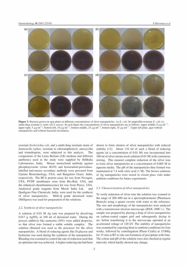
Figure 3. Bacteria grown on agar plates at different concentrations of silver nanoparticles. (a) E. coli; (b) ampicillin-resistant E. coli; (c)multi-drug resistant S. typhi; (d) S. aureus. In each figure the concentrations of silver nanoparticles are as follows: upper middle, 0 μg ml−1;upper right, 5 μg ml−1; bottom left, 10 μg ml−1; bottom middle, 25 μg ml−1; bottom right, 35 μg ml−1. Upper left plate, agar withoutnanoparticles and without bacterial inoculation.
resistant Escherichia coli, and a multi-drug resistant strain ofSalmonella typhus, resistant to chloramphenicol, amoxycilinand trimethoprim, were subjected to this analysis. Thecomponents of the Luria–Bertani (LB) medium and differentantibiotics used in the study were supplied by HiMediaLaboratories, India. Mouse monoclonal antibody againstphosphotyrosine (clone 4G10) and horseradish-peroxidase-labelled anti-mouse secondary antibody were procured fromUpstate Biotechnology, USA, and Bangalore Genei, India,respectively. The BCA protein assay kit was from Novagen,USA, PVDF membranes were from Bio-Rad, USA, andthe enhanced chemiluminescence kit was from Pierce, USA.Analytical grade reagents from Merck India Ltd. andQualigens Fine Chemicals, India, were used for the synthesisof silver nanoparticles. Milli-Q grade deionized water(Millipore) was used for preparation of the solutions.
2.2. Synthesis of silver nanoparticles
A solution of 0.01 M Ag ions was prepared by dissolving0.017 g AgNO3 in 100 ml of deionized water. During theprocess additives like ammonia (30%) were added dropwise,so that silver ions formed a stable soluble complex. Thesolution obtained was used as the precursor for the silvernanoparticles. A blend of reducing agents like D-glucose andhydrazine was used during the synthesis of the nanoparticles.Blending was essential to control the rate of reduction such thatan optimum rate was achieved. A higher reducing rate had been
shown to form clusters of silver nanoparticles with reducedstability [11]. About 110 ml of such a blend of reducingagents (at a concentration of 0.01 M) was incorporated into100 ml of silver nitrate stock solution (0.01 M) with continuousstirring. This ensured complete reduction of the silver ionsto form silver nanoparticles at a concentration of 0.005 M inaqueous media. The pH of the nanoparticles thus formed wasmaintained at 7.4 with citric acid (1 M). The brown solutionsof Ag nanoparticles were stored in closed glass vials underambient conditions for future experiments.
2.3. Characterization of silver nanoparticles
To verify reduction of silver ions the solution was scanned inthe range of 200–600 nm in a spectrophotometer (PharmaciaBiotech) using a quartz cuvette with water as the reference.The size and morphology of the nanoparticles were analysedwith a transmission electron microscope (JEOL 200C×). Thesample was prepared by placing a drop of silver nanoparticleson carbon-coated copper grid and subsequently drying inair, before transferring it to the microscope operated at anaccelerated voltage of 120 kV. The stability of nanoparticleswas examined by exposing them to ambient conditions for fourweeks, followed by centrifugation (Plasto Crafts) at 15 000gfor 15 min at RT to rule out formation of precipitate with time.The colour and pH of the solution were also checked at regularintervals, which hardly showed any change.
3
Nanotechnology 18 (2007) 225103 S Shrivastava et al
Time (h)
Time (h)
Times (h)
Times (h)
O.D
.O
.D.
O.D
.O
.D.
(a) (b)
(c) (d)
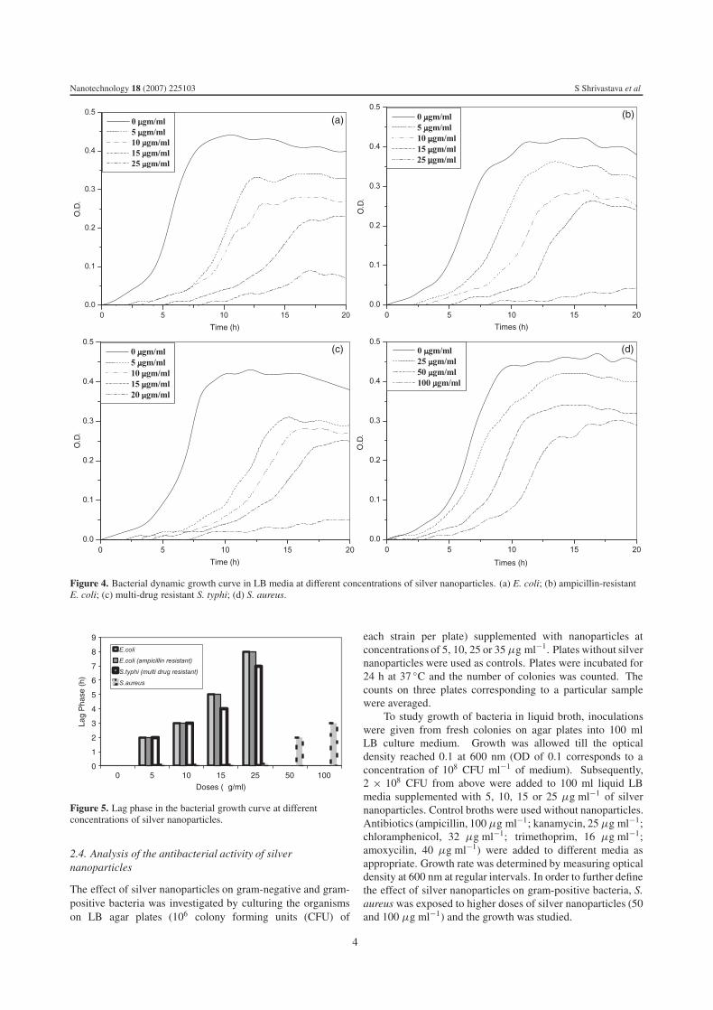
Figure 4. Bacterial dynamic growth curve in LB media at different concentrations of silver nanoparticles. (a) E. coli; (b) ampicillin-resistantE. coli; (c) multi-drug resistant S. typhi; (d) S. aureus.
00
1
2
3
4
5
6
7
8
9
5 10 15
Doses (µg/ml)
Lag
Pha
se (
h)
25 50 100
Figure 5. Lag phase in the bacterial growth curve at differentconcentrations of silver nanoparticles.
2.4. Analysis of the antibacterial activity of silvernanoparticles
The effect of silver nanoparticles on gram-negative and gram-positive bacteria was investigated by culturing the organismson LB agar plates (106 colony forming units (CFU) of
each strain per plate) supplemented with nanoparticles atconcentrations of 5, 10, 25 or 35 μg ml−1. Plates without silvernanoparticles were used as controls. Plates were incubated for24 h at 37 ◦C and the number of colonies was counted. Thecounts on three plates corresponding to a particular samplewere averaged.
To study growth of bacteria in liquid broth, inoculationswere given from fresh colonies on agar plates into 100 mlLB culture medium. Growth was allowed till the opticaldensity reached 0.1 at 600 nm (OD of 0.1 corresponds to aconcentration of 108 CFU ml−1 of medium). Subsequently,2 × 108 CFU from above were added to 100 ml liquid LBmedia supplemented with 5, 10, 15 or 25 μg ml−1 of silvernanoparticles. Control broths were used without nanoparticles.Antibiotics (ampicillin, 100 μg ml−1; kanamycin, 25 μg ml−1;chloramphenicol, 32 μg ml−1; trimethoprim, 16 μg ml−1;amoxycilin, 40 μg ml−1) were added to different media asappropriate. Growth rate was determined by measuring opticaldensity at 600 nm at regular intervals. In order to further definethe effect of silver nanoparticles on gram-positive bacteria, S.aureus was exposed to higher doses of silver nanoparticles (50and 100 μg ml−1) and the growth was studied.
4
Nanotechnology 18 (2007) 225103 S Shrivastava et al
(a) (b)
(c)
(d)
Time (h) Time (h)
Time (h) Time (h)
O.D
O.D
O.D O.D
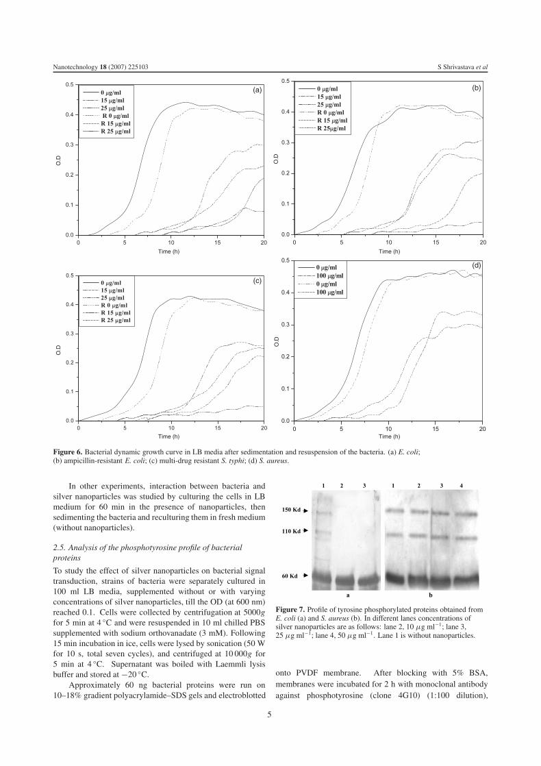
Figure 6. Bacterial dynamic growth curve in LB media after sedimentation and resuspension of the bacteria. (a) E. coli;(b) ampicillin-resistant E. coli; (c) multi-drug resistant S. typhi; (d) S. aureus.
In other experiments, interaction between bacteria andsilver nanoparticles was studied by culturing the cells in LBmedium for 60 min in the presence of nanoparticles, thensedimenting the bacteria and reculturing them in fresh medium(without nanoparticles).
2.5. Analysis of the phosphotyrosine profile of bacterialproteins
To study the effect of silver nanoparticles on bacterial signaltransduction, strains of bacteria were separately cultured in100 ml LB media, supplemented without or with varyingconcentrations of silver nanoparticles, till the OD (at 600 nm)reached 0.1. Cells were collected by centrifugation at 5000gfor 5 min at 4 ◦C and were resuspended in 10 ml chilled PBSsupplemented with sodium orthovanadate (3 mM). Following15 min incubation in ice, cells were lysed by sonication (50 Wfor 10 s, total seven cycles), and centrifuged at 10 000g for5 min at 4 ◦C. Supernatant was boiled with Laemmli lysisbuffer and stored at −20 ◦C.
Approximately 60 ng bacterial proteins were run on10–18% gradient polyacrylamide–SDS gels and electroblotted
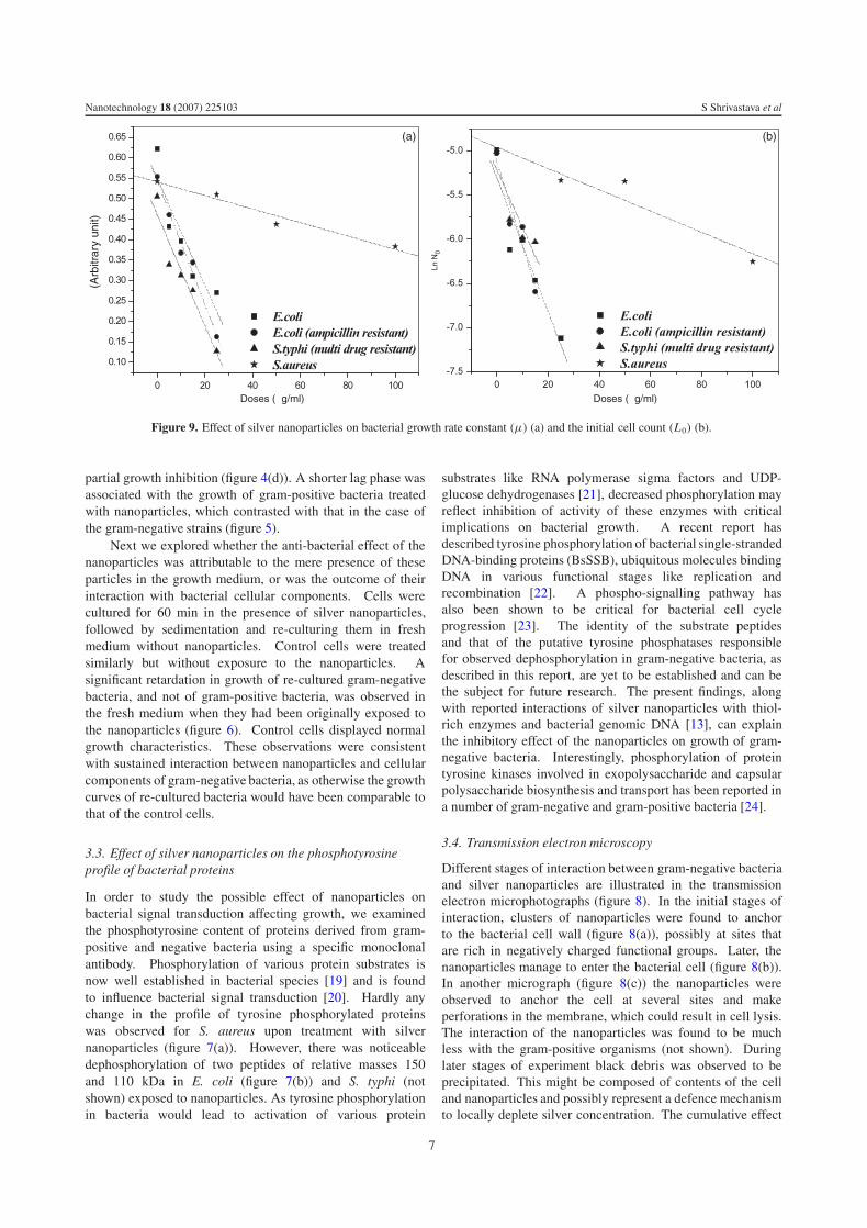
Figure 7. Profile of tyrosine phosphorylated proteins obtained fromE. coli (a) and S. aureus (b). In different lanes concentrations ofsilver nanoparticles are as follows: lane 2, 10 μg ml−1; lane 3,25 μg ml−1; lane 4, 50 μg ml−1. Lane 1 is without nanoparticles.
onto PVDF membrane. After blocking with 5% BSA,membranes were incubated for 2 h with monoclonal antibodyagainst phosphotyrosine (clone 4G10) (1:100 dilution),
5

Nanotechnology 18 (2007) 225103 S Shrivastava et al
(a)
(b)
(c)
Figure 8. TEM microphotographs showing different stages ofinteraction of silver nanoparticles with E.coli.
followed by 1 h incubation with peroxidase-conjugated anti-mouse IgG (dilution 1:5000). The signals were visualizedusing enhanced chemiluminescence.
3. Results and discussion
3.1. Reduction kinetics and morphology of silvernanoparticles
The reduction of silver ions was visibly evident from thecolour changes associated with it. Figure 1 shows UV–visibleabsorption spectra during various stages of reduction of theprecursors as they transformed into the silver nanoparticlesand after different ageing times during its preservation under
ambient conditions. The inset pictures show the colour changesbefore (o), during (b) and after the process of reduction (a). Thesilver nitrate solution showed a high at about 300 nm, whichgradually underwent red shift with appearance of a humpat 400 nm consistent with formation of silver nanoparticles.The sharp peak at 400 nm can be attributed to a narrowsize distribution of the particles formed in the solution. Asevident in figure 1 the particles showed hardly any changein the absorption spectra even after a month of ageing time,consistent with the highly stable nature of the nanoparticles.The rate of reaction was optimized to yield partially mono-dispersed silver nanoparticles of uniform size and shape, asshown in the electron micrograph in figure 2(a). The selectedarea electron diffraction (SAED) pattern obtained from thenanoparticles represented silver having face centred cubic (fcc)crystallographic structure. The different diffracting planeswere indexed as shown in figure 2(b). Figure 2(c) shows acluster of silver nanoparticles at higher magnification. Theparticles were found to be spherical or polyhedral in shape,having a mean size about 10–15 nm. This polyhedral shapecould be attributed to the controlled reaction rate, resulting ininitial nucleus formation and subsequent growth. Figure 2(d)represents the size distribution plot, suggesting that most of theparticles conformed to the size range of 10–15 nm.
3.2. Effect of silver nanoparticles on bacterial growth
LB agar plates incorporating increasing concentrations ofsilver nanoparticles were inoculated with 106 CFU fromdifferent bacterial strains. In case of non-resistant E. coli60% inhibition in bacterial growth was observed in platessupplemented with 5 μg ml−1 of nanoparticles. The extentof inhibition increased to 90% in plates with 10 μg ml−1
nanoparticles, whereas the concentration of 25 μg ml−1
ensured complete inhibition of bacterial growth (figure 3(a)).In the case of ampicillin-resistant E. coli and multi-drugresistant strains of S. typhi, 70–75% inhibition in growthwas observed in plates supplemented with 10 μg ml−1 ofnanoparticles, whereas 25 μg ml−1 or higher concentration ofnanoparticles elicited 100% inhibition in growth of bacteria(figures 3(b) and (c)). Interestingly, none of the doses of silvernanoparticles had hardly any effect on the growth of S. aureus(gram-positive) (figure 3(d)).
In further experiments, strains of gram-negative and gram-positive bacteria were inoculated in liquid LB media supple-mented with silver nanoparticles. Increasing concentrationof nanoparticles progressively inhibited the growth of non-resistant E. coli (figure 4(a)). The lag phase was found to bemore prolonged than that described in the earlier reports [11].This could be attributable to greater stability of the nanoparti-cles used in this study. The concentration of 25 μg ml−1 wasfound to be strongly inhibitory for bacteria, as it took about8 h to initiate any noticeable growth. Similar results wereobtained with either resistant or non-resistant strains of othergram-negative bacteria studied (figures 4(b) and (c)).
In contrast, silver nanoparticles were found to have aless significant effect on the growth of gram-positive bacteria(S. aureus). No reduction in bacterial growth was observedtill the concentration of nanoparticles rose to 25 μg ml−1,whereas the concentration of 100 μg ml−1 elicited only a
6
Nanotechnology 18 (2007) 225103 S Shrivastava et al
Doses (µg/ml)
µ (A
rbitr
ary
unit)
Ln N
0
Doses (µg/ml)
(a) (b)
Figure 9. Effect of silver nanoparticles on bacterial growth rate constant (μ) (a) and the initial cell count (L0) (b).
partial growth inhibition (figure 4(d)). A shorter lag phase wasassociated with the growth of gram-positive bacteria treatedwith nanoparticles, which contrasted with that in the case ofthe gram-negative strains (figure 5).
Next we explored whether the anti-bacterial effect of thenanoparticles was attributable to the mere presence of theseparticles in the growth medium, or was the outcome of theirinteraction with bacterial cellular components. Cells werecultured for 60 min in the presence of silver nanoparticles,followed by sedimentation and re-culturing them in freshmedium without nanoparticles. Control cells were treatedsimilarly but without exposure to the nanoparticles. Asignificant retardation in growth of re-cultured gram-negativebacteria, and not of gram-positive bacteria, was observed inthe fresh medium when they had been originally exposed tothe nanoparticles (figure 6). Control cells displayed normalgrowth characteristics. These observations were consistentwith sustained interaction between nanoparticles and cellularcomponents of gram-negative bacteria, as otherwise the growthcurves of re-cultured bacteria would have been comparable tothat of the control cells.
3.3. Effect of silver nanoparticles on the phosphotyrosineprofile of bacterial proteins
In order to study the possible effect of nanoparticles onbacterial signal transduction affecting growth, we examinedthe phosphotyrosine content of proteins derived from gram-positive and negative bacteria using a specific monoclonalantibody. Phosphorylation of various protein substrates isnow well established in bacterial species [19] and is foundto influence bacterial signal transduction [20]. Hardly anychange in the profile of tyrosine phosphorylated proteinswas observed for S. aureus upon treatment with silvernanoparticles (figure 7(a)). However, there was noticeabledephosphorylation of two peptides of relative masses 150and 110 kDa in E. coli (figure 7(b)) and S. typhi (notshown) exposed to nanoparticles. As tyrosine phosphorylationin bacteria would lead to activation of various protein
substrates like RNA polymerase sigma factors and UDP-glucose dehydrogenases [21], decreased phosphorylation mayreflect inhibition of activity of these enzymes with criticalimplications on bacterial growth. A recent report hasdescribed tyrosine phosphorylation of bacterial single-strandedDNA-binding proteins (BsSSB), ubiquitous molecules bindingDNA in various functional stages like replication andrecombination [22]. A phospho-signalling pathway hasalso been shown to be critical for bacterial cell cycleprogression [23]. The identity of the substrate peptidesand that of the putative tyrosine phosphatases responsiblefor observed dephosphorylation in gram-negative bacteria, asdescribed in this report, are yet to be established and can bethe subject for future research. The present findings, alongwith reported interactions of silver nanoparticles with thiol-rich enzymes and bacterial genomic DNA [13], can explainthe inhibitory effect of the nanoparticles on growth of gram-negative bacteria. Interestingly, phosphorylation of proteintyrosine kinases involved in exopolysaccharide and capsularpolysaccharide biosynthesis and transport has been reported ina number of gram-negative and gram-positive bacteria [24].
3.4. Transmission electron microscopy
Different stages of interaction between gram-negative bacteriaand silver nanoparticles are illustrated in the transmissionelectron microphotographs (figure 8). In the initial stages ofinteraction, clusters of nanoparticles were found to anchorto the bacterial cell wall (figure 8(a)), possibly at sites thatare rich in negatively charged functional groups. Later, thenanoparticles manage to enter the bacterial cell (figure 8(b)).In another micrograph (figure 8(c)) the nanoparticles wereobserved to anchor the cell at several sites and makeperforations in the membrane, which could result in cell lysis.The interaction of the nanoparticles was found to be muchless with the gram-positive organisms (not shown). Duringlater stages of experiment black debris was observed to beprecipitated. This might be composed of contents of the celland nanoparticles and possibly represent a defence mechanismto locally deplete silver concentration. The cumulative effect
7

Nanotechnology 18 (2007) 225103 S Shrivastava et al
of these factors would lead to retardation in bacterial growthbut not complete annihilation.
3.5. Prediction of bacterial growth in the presence of varyingconcentrations of silver nanoparticles
The dynamic growth rate of a bacterial species is representedby the equation
ln N = ln L0 + μt (1)
where N is the bacterial cell count at time t, L0 is the initialcell count and μ is the growth rate constant for the bacteria.The experimental data were plotted in the logarithmic scale toderive the values of μ and ln L0 corresponding to various dosesof the nanoparticles (x) studied. Linear relationships betweenx and μ (figure 9(a)) and x and L0 (figure 9(b)) were derivedfor each of the species of bacteria investigated as shown below:
μ = −ax + b (2)
ln L0 = −cx − d (3)
where a, b, c and d are the constants for the respective bacteriaas below.
Name of thebacterium a b c d
E. coli 0.0649 0.5494 0.3783 5.3061E. coli(ampicillinresistant)
0.0753 0.5435 0.4727 5.1190
S. typhi(multi-drugresistant)
0.0678 0.4618 0.3263 5.2115
S. aureus 0.0082 0.541 0.0601 4.9598
Similar relationships, though in complex forms, have beenproposed for several chemical species affecting the bacterialgrowth [25].
Thus, by replacing the values of μ and ln L0, equation (1)can be rewritten as
ln N = (bt − d) − (at + c)x . (4)
Equation (4) represents the dynamic growth rate of bacteria inthe presence of different concentrations of silver nanoparticlesin the medium. It can be noticed that slopes of the trend linesrepresenting gram-positive bacteria are an order of magnitudeless than those for the gram-negative strains (figures 9(a)and (b)), which is consistent with the observed impairment ofgrowth in the case of gram-negative bacteria in the presence ofsilver nanoparticles.
3.6. Possible mechanism for antibacterial action of silvernanoparticles
The differences between gram-positive and gram-negativebacteria essentially rest in the structure of their respectivecell walls. The gram-negative bacteria have a layer oflipopolysaccharide at the exterior, followed underneath by athin (about 7–8 nm) layer of peptidoglycan [26]. Although thelipopolysaccharides are composed of covalently linked lipidsand polysaccharides, they lack strength and rigidity. Negative
charges on the lipopolysaccharides [28] are attracted towardsweak positive charges available on silver nanoparticles [29].On the other hand, the cell wall in gram-positive bacteriais principally composed of a thick layer (about 20–80 nm)of peptidoglycan, consisting of linear polysaccharide chainscross-linked by short peptides to form a three dimensional rigidstructure [27]. The rigidity and extended cross-linking notonly endow the cell walls with fewer anchoring sites for thesilver nanoparticles but also make them difficult to penetrate.The extent of inhibition of bacterial growth reported in thisstudy was dependent on the concentration of nanoparticles inthe medium. Interaction between nanoparticles and the cellwall of bacteria would be facilitated by the relative abundanceof negative charges on the gram-negative bacteria, which wascongenial to the fact that growth of gram-negative bacteriawas more profoundly affected by the silver nanoparticles thanthat of the gram-positive organisms. As demonstrated byelectron microscopy, interaction with nanoparticles resulted inperforations in the cell wall, contributing to the antibacterialeffects of the nanoparticles. Inferences from the recultureexperiments were consistent with the entry of nanoparticlesinside bacterial cells and/or strong association with bacterialcellular components. Once inside the cell, nanoparticleswould interfere with the bacterial growth signalling pathwayby modulating tyrosine phosphorylation of putative peptidesubstrates critical for cell viability and division.
4. Conclusion
The silver nanoparticles synthesized and analysed in thisreport were found to have stronger antibacterial potency thanthose described in the earlier reports [11, 13]. The effectwas dose dependent and was more pronounced against gram-negative organisms than gram-positive ones. The antibacterialeffect of nanoparticles was independent of acquisition ofresistance by the bacteria against antibiotics. The majormechanism through which silver nanoparticles manifestedantibacterial properties was by anchoring to and penetratingthe bacterial cell wall, and modulating cellular signalling bydephosphorylating putative key peptide substrates on tyrosineresidues. However, further studies must be conducted to verifyif the bacteria develop resistance towards the nanoparticles andto examine cytotoxicity [30] of nanoparticles towards humancells before proposing their therapeutic use.
Acknowledgments
This research was funded by the Indian Council of MedicalResearch (ICMR) and the DST Unit on Nanoscience andTechnology (DST-UNANST), BHU. We sincerely thank DrG K Dey of the Bhaba Atomic Research Center, Mumbai,for carrying out the TEM analysis of the nanoparticlesand Professor O N Srivastava for encouragement. TBacknowledges Professor S Lele and Dr R K Mandal of BHUfor valuable discussions and encouragement.
References
[1] Gleiter H 2000 Nanostructured materials, basic concepts andmicrostructure Acta Mater. 48 1–12
[2] Curtis A and Wilkinson C 2001 Nanotechniques andapproaches in biotechnology Trends Biotechnol. 19 97–101
8

Nanotechnology 18 (2007) 225103 S Shrivastava et al
[3] Waren C W and Nie S 1998 Quantum dot bioconjugates forultra sensitive nonisotopic detection Science 281 2016–8
[4] Vaseashta A and Dimova-Malinovska D 2005 Nanostructuredand nanoscale devices, sensors and detectors Sci. Technol.Adv. Mater. 6 312–8
[5] Langer R 2001 Drug delivery: drugs on target Science293 58–9
[6] Roy K, Mao H Q, Huang S K and Leong K W 1999 Oral genedelivery with Chitosan-DNA nanoparticles generatesimmunologic protection in a murine model of peanut allergyNat. Med. 5 387–91
[7] Sachlos E, Gotora D and Czernuszka J T 2006 Collagenscaffolds reinforced with biomimetic composite nano-sizedcarbonate-substituted hydroxyapatite crystals and shaped byrapid prototyping to contain internal microchannels TissueEng. 12 2479–87
[8] Xu Z P, Zeng Q H, Lu G Q and Yu A B 2006 Inorganicnanoparticles as carriers for efficient cellular delivery Chem.Eng. Sci. 61 1027–40
[9] Farokhzad O C, Cheng J, Teply B A, Sherifi I, Jon S,Kantoff P W, Richie J P and Langer R 2006 Targetednanoparticle-aptamer bioconjugates for cancer chemotherapyin vivo Proc. Natl Acad. Sci. USA 103 6315–20
[10] Stoimenov P K, Klinger R L, Marchin G L andKlabunde K J 2002 Metal oxide nanoparticles as bactericidalagents Langmuir 18 6679–86
[11] Sondi I and Salopek-Sondi B 2004 Silver nanoparticles asantimicrobial agent: a case study of E. coli as a model forgram-negative bacteria J. Colloids Interface Sci. 275 177–82
[12] Panacek A, Kvitek L, Prucek R, Kolar M, Vecerova R,Pizurova N, Sharma V K, Nevecna T and Zboril R 2006Silver colloid nanoparticles: synthesis, characterization, andtheir antibacterial activity J. Phys. Chem. B 110 16248–53
[13] Morones J R, Elechiguerra J L, Camacho A, Holt K, Kouri J B,Ramirez J T and Yacaman M J 2005 The bactericidal effectof silver nanoparticles Nanotechnology 16 2346–53
[14] Baker C, Pradhan A, Pakstis L, Pochan D J and Shah S I 2005Synthesis and antibacterial properties of silver nanoparticlesJ. Nanosci. Technol. 5 244–9
[15] Holladay R, Moeller W, Mehta D, Brooks J, Roy R andMortenson M 2006 Silver/water, silver gels and silver-basedcompositions; and methods for making and using the sameApplication Number WO2005US47699 20051230 EuropeanPatent Office
[16] Li P, Li J, Wu C, Wu Q and Li J 2005 Synergistic antibacterialeffects of β-lactam antibiotic combined with silvernanoparticles Nanotechnology 16 1912–7
[17] Jain P and Pradeep T 2005 Potential of silvernanoparticle-coated polyurethane foam as an antibacterialwater filter Biotechnol. Bioeng. 90 59–63
[18] Li Y, Leung P, Yao L, Song Q W and Newton E 2006Antimicrobial effect of surgical masks coated withnanoparticles J. Hosp. Infec. 62 58–63
[19] Deutscher J and Saier M H 2005 Ser/Thr/Tyr proteinphosphorylation in bacteria—for long time neglected, nowwell established J. Mol. Microbiol. Biotechnol. 9 125–31
[20] Kirstein J and Turgay K 2005 A new tyrosine phosphorylationmechanism involved in signal transduction in Bacillussubtilis J. Mol. Microbiol. Biotechnol. 9 182–8
[21] Mijakovic I, Petranovic D, Bottini N, Deutscher J and RuhdalJensen P 2005 Protein tyrosine phosphorylation in Bacillussubtilis J. Mol. Microbiol. Biotechnol. 9 189–97
[22] Mijakovic I, Petranovic D, Macek B, Cepo T, Mann M,Davies J, Jensen P R and Vujaklija D 2006 Bacterialsingle-stranded DNA—binding proteins are phosphorylatedon tyrosine Nucleic Acids Res. 34 1588–96
[23] Iniesta A A, McGrath P T, Reisenauer A, McAdams H H andShapiro L 2006 A phosphor signaling pathway controls thelocalization and activity of a complex critical for bacterialcell cycle progression Proc. Natl Acad. Sci. USA103 10935–40
[24] Grangeasse C, Obadia B, Mijakovic I, Deutscher J,Cozzone A J and Doublet P 2003 Autophosphorylation ofthe Escherichia coli kinase Wzc regulates tyrosinephosphorylation of Ugd, a UDP-glucose dehydrogenaseJ. Biol. Chem. 278 39323–39
[25] Nostro P L, Ninham B W, Nostro A L, Pesavento G,Fratoni L and Baglioni P 2005 Specific ion effects on thegrowth rates of Staphylococcus aureus and Pseudomonasaeruginosa Phys. Biol. 2 1–7
[26] Madigan M and Martinko J 2005 Brock Biology ofMicroorganisms 11th edn (Englewood Cliffs, NJ: PrenticeHall)
[27] Baron S 1996 Medical Microbiology 4th edn (Galveston:University of Texas Medical Branch)
[28] Salton M R J and Kim K S 1996 Structure Baron’s MedicalMicrobiology 4th edn (Galveston: University of TexasMedical Branch)
[29] Sui Z M, Chen X, Wang L Y, Xu L M, Zhuang W C,Chai Y C and Yang C J 2006 Capping effect of CTAB onpositively charged Ag nanoparticles Physica E 33 308–14
[30] Braydich-Stolle L, Hussain S, Schlager J J andHofmann M C 2005 In vitro cytotoxicity of nanoparticles inmammalian germline stem cells Toxicol. Sci. 88 412–9
9

![Visible light photocatalytic H2-production activity of wide …cmsoep.physics.sjtu.edu.cn/doc/2015/0957-4484_26_34_345402.pdf · reaction [2]. ZnS is a well-known photocatalyst among](https://static.fdocuments.us/doc/165x107/5a8ac44e7f8b9a4a268c1398/visible-light-photocatalytic-h2-production-activity-of-wide-2-zns-is-a-well-known.jpg)