ANTEPARTUM FETAL MONITORING Reinaldo Figueroa, MD Winthrop-University Hospital.

Clinical Policy Bulletin: Antepartum Fetal Surveillance
Additional Information
Number: 0088
Policy
*Please see amendment for Pennsylvania Medicaid at the end ofthis CPB.
I. Aetna considers in-office and in-hospital antepartum fetal surveillance with non-stress tests (NST), contraction stress tests (CST), biophysical profile (BPP), modified BPP, and umbilical artery Doppler velocimetry medically necessary according to the American College of Obstetricians and Gynecologists (ACOG) Clinical Guideline on Antepartum Fetal Surveillance.
The following medical necessity guidelines apply:
A. Antepartum fetal surveillance using NST, CST, BPP, or modified BPP is considered medically necessary for women with risk factors for stillbirth due to utero-placental insufficiency. Accepted guidelines state that fetal testing should not begin until interventions can be undertaken. For most pregnancies at increased risk of stillbirth due to utero-placental insufficiency, testing is considered appropriate beginning at 32 to 34 weeks of gestation. Testing is considered medically necessary beginning at 26 weeks gestation for pregnancies with multiple or particularly worrisome high-risk conditions. Examples of such high-risk conditions include bleeding, chronic or pregnancy-induced hypertension, collagen vascular disease (including anti-phospholipid syndrome), fetal growth restriction, gestational diabetes, impaired renal function, maternal heart disease (New York Heart Association Class III or IV), oligohydramnios, significant isoimmunization, steroid-dependent or poorly controlled asthma (not an all-inclusive list).
B. If the clinical condition that has prompted testing persists, repeat testing (either weekly or twice-weekly, depending on the test used and the presence of certain high-risk conditions) is considered medically necessary until delivery. Repeat testing is also considered medically necessary for any significant deterioration in the maternal medical status or any acute diminution in fetal activity, regardless of the amount of time that has elapsed since the last test.
C. A CST or full BPP is considered medically necessary following an abnormal NST or modified BPP. (Subsequent management should then be predicated on the results of the CST or BPP, the gestational age, the degree of oligohydramnios (if assessed), and the maternal condition.)
D. Recent, normal antepartum fetal test results should not preclude the determination that intrapartum fetal monitoring is medically necessary.
E. Umbilical artery Doppler velocimetry is considered medically necessary only in pregnancies complicated by intra-uterine growth restriction, oligohydramnios, twin-twin transfusion syndrome and/or discordant fetuses. Discordant fetal growth is common in multiple gestation and usually is defined by a 15 to 25 % reduction in the estimated fetal weight of the smaller fetus when compared with the largest. (If used in this setting,

accepted guidelines indicate that decisions regarding timing of delivery should be made using a combination of information from the Doppler ultrasonography and other tests of fetal well-being, along with careful monitoring of maternal status.)
F. Middle cerebral artery Doppler velocimetry is considered medically necessary for pregnancy complicated by either twin-twin transfusion syndrome or suspected fetal anemia in conditions such as isoimmunization and parvovirus B-19 infection.
II. Aetna considers uterine artery Doppler studies experimental and investigational for risk assessment or screening during pregnancies because of insufficient evidence.
III. Aetna considers antepartum fetal surveillance with NST, CST, BPP, modified BPP, and umbilical artery Doppler velocimetry experimental and investigational for all other indications because their effectiveness for indications other than the ones listed above has not been established.
IV. Aetna considers Doppler studies of ductus venosus and vessels other than the middle cerebral artery and umbilical artery for fetal surveillance of impaired fetal growth experimental and investigational because their effectiveness for these indications has not been established.
V. Aetna considers measurement of serum YKL-40 for evaluation of pre-eclampsia or small-for-gestational age fetuses experimental and investigational because its effectiveness for these indications has not been established.
See also CPB 0106 - Fetal Echocardiograms and CPB 0127 - Home Uterine Activity Monitoring.
Background
Antepartum fetal surveillance is used to assess the risk of adverse perinatal outcome associated with utero-placental insufficiency, and is recommended for pregnancies that are at risk for hypoxia and stillbirth. Common tests include fetal movement assessment, non-stress tests (NST), contraction stress tests (CST), biophysical profile (BPP), modified BPP, and umbilical artery Doppler velocimetry. Some of the conditions under which antepartum fetal surveillance may be appropriate include the following:
Maternal conditions:
Anti-phospholipid syndrome Chronic renal disease Cyanotic heart disease Hemoglobinopathies (hemoglobin SS, SC, or S-thalassemia) Hypertensive disorders Hyperthyroidism (poorly controlled) Systemic lupus erythematosus Type 1 diabetes mellitus
Pregnancy-related conditions:
Decreased fetal movement Intrauterine growth restriction Isoimmunization (moderate to severe) Multiple gestation (with significant growth discrepancy) Oligohydramnios Polyhydramnios Post-term pregnancy (greater than 41 weeks gestation) Pregnancy-induced hypertension

Previous fetal demise (unexplained or recurrent risk)
Fetal Movement Assessment
A decrease in the maternal perception of fetal movement often but not invariably precedes fetal death, in some cases by several days. This observation provides the rationale for fetal movement assessment by the mother ("kick counts") as a means of antepartum fetal surveillance.
In a review on fetal movement assessment, Froen and colleagues (2008) noted that while almost all pregnant women adhere to it, organized screening by fetal movements has seen variable popularity among health professionals. Early results of screening were promising and fetal movement counting is the only antepartum testing method that has shown effect in reducing mortality in a randomized controlled trial comparing testing versus no testing. Although awareness of fetal movements is associated with improved perinatal outcomes, the quest to define a quantitative "alarm limit" to define decreased fetal movements has so far been unsuccessful, and the use of most such limits developed for fetal movement counting should be discouraged.
Non-Stress Test
The NST is based on the premise that the heart rate of a fetus that is not acidotic or neurologically depressed will temporarily accelerate with fetal movement. Heart rate reactivity is thought to be a good indicator of normal fetal autonomic function. Loss of reactivity is associated most commonly with the fetal sleep cycle but may result from any cause of central nervous system depression, including fetal acidosis and some medications.
To perform NST, the mother is asked to denote when the fetus moves. The fetal heart rate tracing is then evaluated for accelerations of the fetal heart rate corresponding with fetal movement. Alternatively, acoustic stimulation is applied to the maternal abdomen for 1 to 2 seconds and the fetal
heart rate is recorded. The acoustic stimulation may be repeated up to 3 times, each time for progressively longer durations (up to 3 seconds), to elicit fetal heart rate accelerations.
Contraction Stress Test
The CST measures the response of the fetal heart rate to uterine contractions. It relies on the premise that fetal oxygenation will be transiently worsened by uterine contractions. This test is rarely used in clinical practice at this time.
To perform CST, the fetal heart rate and uterine contractions are simultaneously recorded with an external fetal monitor. The test lasts until the mother has had 3 moderate strength contractions within a 10-min period. If contractions are not happening on their own, they may be induced using an intravenous dose of oxytocin
Biophysical Profile
BPP is comprised of 5 components:
Amniotic fluid index (determination of the amniotic fluid volume) Fetal breathing movements Fetal movement Fetal tone Non-stress test
Each component is assigned 2 points, resulting in a score ranging from 0 to 10, with scores from 8 to 10 considered normal, 6 considered borderline, and below 6 considered problematic.
A Cochrane review on BPP for fetal assessment in high-risk pregnancies (Lalor et al, 2008) concluded that there is currently insufficient evidence from randomized trials to support the use of BPP as a test of fetal wellbeing in high-risk pregnancies.

Modified Biophysical Profile
Modified BPP combines the NST (with the option of acoustic stimulation), as a short-term indicator of fetal acid-base status, with the amniotic fluid index as an indicator of long-term placental function.
Umbilical Artery Doppler Velocimetry
A variety of fetal and maternal blood vessels have been evaluated by Doppler wave form analysis to assess the risk of adverse perinatal outcome. The most commonly interrogated vessels are the umbilical arteries.
Umbilical artery Doppler flow velocimetry has been adapted for use as a technique of fetal surveillance, based on the observation that flow velocity waveforms in the umbilical artery of normally growing fetuses differ from those of growth-restricted fetuses. Abnormal flow velocity waveforms have been correlated histopathologically with small-artery obliteration in placental tertiary villi and functionally with fetal hypoxia and acidosis, as well as with perinatal morbidity and mortality.
At least 3 randomized clinical trials (RCTs) have evaluated the utility of umbilical artery Doppler velocimetry as a technique of antepartum fetal surveillance in pregnancies complicated by suspected intrauterine growth restriction. Women assigned to antepartum umbilical artery Doppler velocimetry have been shown to require less frequent antenatal monitoring and shorter durations of maternal hospitalization. The results of 1 RCT showed significantly lower rates of obstetric interventions in patients assigned to Doppler, such as antepartum admission and labor induction. Umbilical artery Doppler velocimetry has not been shown to impact other perinatal outcomes, such as gestational age at birth, birth-weight, Apgar scores, and cesarean birth rates.
Guidelines from the American College of Obstetricians and Gynecologists (ACOG, 1999) have concluded that, "[o]n balance, the available evidence suggests that primary antepartum surveillance of suspected intrauterine growth restriction with umbilical artery Doppler velocimetry can achieve at least equivalent (and possibly better) fetal and neonatal outcomes as primary antepartum surveillance based on results of the NST [non-stress test]. Furthermore, frequency of antepartum testing and certain aspects of obstetric intervention are reduced with use of Doppler." ACOG guidelines (1999) state that, "[i]f umbilical artery Doppler velocimetry is used, decisions regarding timing of delivery should be made using a combination of information from the Doppler ultrasonography and other tests of fetal well-being, such as amniotic fluid volume assessment, NST, CST [contraction stress test], and BPP [biophysical profile], along with careful monitoring of maternal status."
According to ACOG guidelines, "[n]o benefit has been demonstrated for umbilical artery velocimetry for conditions other than suspected intrauterine growth restriction, such as post term gestation, diabetes mellitus, systemic lupus erythematosus, or anti-phospholipid syndrome. Doppler ultrasonography has not been shown to be of value as a screening test for detecting fetal compromise in the general obstetric population, and its use for this purpose cannot be recommended."
American College of Obstetricians and Gynecologists (2000) guidelines on intra-uterine growth retardation (IUGR) reached the following conclusions about the clinical utility of Doppler ultrasound of the umbilical artery:
"Although Doppler velocimetry of the umbilical arteries is not useful as a screening technique for IUGR, it has been demonstrated to be useful once IUGR has been diagnosed. Not only are Doppler velocimetry findings normal in growth-restricted fetuses with chromosomal or other structural etiologies but Doppler velocimetry has been shown to both reduce interventions and improve fetal outcome in pregnancies at risk for IUGR. Thus, once IUGR is suspected or diagnosed, Doppler velocimetry may be useful as a part of fetal evaluation. Fetuses with normal flow patterns seem less likely to benefit from consideration of early delivery than do their counterparts with abnormal studies."
The American College of Radiology (2001) has concluded that Doppler studies are, in general, not
indicated for the initial assessment to determine if there is (probable) intrauterine growth retardation.
"Extensive research on Doppler analysis of uterine, umbilical, and various intrafetal vessels confirms a strong correlation between high resistance arterial wave form patterns (e.g., low, absent, or reversed diastolic flow in the umbilical artery) and subsequent IUGR, hypoxemic fetal morbidity, and mortality. The correlation is greatest in high-risk pregnancies, but insufficiently predictive in general, low-risk populations to be useful as a primary screening test. Some have argued that since Doppler appears to be applicable primarily in a population already defined as high risk, the clinical decisions as to when a fetus is distressed and requires emergent delivery will be made based on the BPP and heart rate monitoring, making the Doppler superfluous. A recently published meta-analysis of 20 controlled trials of Doppler ultrasonography found, however, that there is "compelling evidence" that knowledge of the Doppler findings improved perinatal outcome in high-risk pregnancies, reducing antenatal admissions, inductions of labor, and cesarean sections for fetal distress, and reducing the odds of perinatal death by 38 %."
Goffinet et al (1997) reviewed RCTs of umbilical artery Doppler velocimetry in average-risk pregnancies, and concluded that there is inadequate evidence to support its use in that clinical context:
"There is no evidence that routine umbilical Doppler in a general or low-risk population leads to any improvement in the health of women or their infants. Although other trials would be desirable before asserting a definite lack of benefit (due to the problem of statistical heterogeneity and lack of power), umbilical Doppler examination cannot be recommended as a routine test in low- risk pregnancies."
In a Cochrane review, Alfirevic and colleagues (2010) evaluated the effects on obstetric practice and pregnancy outcome of routine fetal and umbilical Doppler ultrasound in unselected and low-risk pregnancies. These investigators searched the Cochrane Pregnancy and Childbirth Group Trials Register (May 2010). Randomized and quasi-RCTs of Doppler ultrasound for the investigation of umbilical and fetal vessels waveforms in unselected pregnancies compared to no Doppler ultrasound were selected. Studies where uterine vessels have been assessed together with fetal and umbilical vessels have been included. Two authors independently assessed the studies for inclusion, assessed risk of bias, as well as carried out data extraction. These researchers included 5 trials involving 14,185 women. The methodological quality of the trials was generally unclear because of insufficient data included in the reports. Routine fetal and umbilical Doppler ultrasound examination in low-risk or unselected populations did not result in increased antenatal, obstetric and neonatal interventions, and no overall differences were detected for substantive short-term clinical outcomes such as perinatal mortality. There is no available evidence to assess the effect on substantive long-term outcomes such as childhood neurodevelopment and no data to assess maternal outcomes, particularly psychological effects. The authors concluded that existing evidence does not provide conclusive evidence that the use of routine umbilical artery Doppler ultrasound, or combination of umbilical and uterine artery Doppler ultrasound in low-risk or unselected populations benefits either mother or baby. They stated that future studies should be designed to address small changes in perinatal outcome, and should focus on potentially preventable deaths.
Middle Cerebral Artery Peak Systolic Velocity (MCA PSV) Doppler
Amniocentesis for amniotic fluid bilirubin levels is the most widely used test to predict the severity of fetal disease in red-cell alloimmunization. Many textbooks and guidelines recommend serial amniocentesis to monitor these pregnancies. However, the reliability of amniotic fluid bilirubin measurements has been questioned and these tests are of limited value in the second trimester. Furthermore, critical appraisal of the very few prospective studies is hampered by limitations in design or insufficient data given by the authors. Two strategies have been proposed by investigators as useful indicators of fetal anemia. Some advocate liberal or primary use of fetal blood sampling, while others promote the use of non-invasive ultrasonography and Doppler assessment of umbilical venous and middle cerebral artery peak systolic velocity (MCA PSV).
The most promising of these methods appears to be MCA PSV. Studies have shown a very good
correlation between MCA PSV and the degree of fetal anemia in red blood cell alloimmunized pregnancies known to cause immunological hydrops, that is, a low fetal hematocrit is associated with an increase in MCA PSV and the need to perform a transfusion. This screening method has been shown to have an overall sensitivity of 93 % to detect severe anemia, and a sensitivity of 88 % for moderate anemia. Specificity has been reported to be about 75 %. The false positive rate has been shown to increase following 33 weeks gestation. These high sensitivities and acceptable false-positive rates support the potential clinical applicability of the method to reduce the reliance on, and even replace, cordocentesis and amniocentesis with its attendant complications in Rh maternal alloimmunization pregnancies.
Uterine Artery Doppler
Abnormal uterine artery Doppler studies in the first and second trimester have been associated with subsequent adverse pregnancy outcomes including preeclampsia, fetal growth restriction, and perinatal mortality. However, there is insufficient evidence in the peer-reviewed published medical literature and from evidence-based clinical guidelines for the use of uterine artery Doppler in assessment of either average-risk or high-risk pregnancies. A Cochrane systematic evidence review (Neilson et al, 2003) of Doppler ultrasound for fetal assessment of high-risk pregnancies found that most randomized trials have examined ultrasound of the umbilical artery, not the uterine artery. Only 1 randomized study examined the clinical impact of uterine artery blood flow; in that study, both uterine artery and umbilical artery blood flow were measured. Based on the lack of evidence on the clinical utility of uterine artery blood flow measurements, the Cochrane reviewers concluded: "It is not clear if the study of utero-placental arteries makes any real contribution or not. Fetal vessels other than the umbilical artery can also be studied, especially using pulsed wave Doppler with or without color flow imaging; as yet, there is no evidence from controlled studies that these studies are of clinical value."
Guidelines from the ACOG have concluded that uterine artery Doppler is not an effective method for identifying women at risk for eclampsia. The ACOG guidelines on eclampsia and preeclampsia (2002) state that "Doppler velocimetry of the uterine arteries was reported not to be a useful test for screening pregnant women at low risk for preeclampsia." The ACOG guidelines on intrauterine growth restriction (2000) state that umbilical artery ultrasounds may be useful in the evaluation of the growth restricted fetus; however, these guidelines indicate no particular role for uterine artery Doppler ultrasound in the evaluation and management of intrauterine growth restriction pregnancies.
A review of the evidence for uterine artery Doppler studies prepared for the Society for Maternal Fetal Medicine (Scicione and Hayes, 2009) found that the predictive value of Doppler testing in a low-risk population of women appears to be low, and currently there are no available interventions to prevent adverse outcomes based on an abnormal result. The review found that effective interventions to prevent late pregnancy complications (e.g., preeclampsia, growth restrictions, and perinatal mortality) in women considered at low-risk with abnormal early pregnancy uterine artery Doppler studies are needed. The review concluded that, "[u]ntil such time as these are available, routine uterine artery Doppler screening of women considered at low risk is not recommended."
The review found that uterine artery Doppler screening of high-risk women (e.g., history of chronic hypertension or preeclampsia, prior fetal growth restriction, or stillbirth) with singleton gestations appears to identify those at substantially increased risk for adverse pregnancy outcomes (Scicione and Hayes, 2009). The review stated that abnormal testing in these women could potentially lead to increased surveillance (e.g., earlier and more frequent assessment of fetal growth and maternal clinical condition) and interventions that might improve clinical outcomes. Normal Doppler studies could potentially lead to a reduction in such testing and interventions. The review noted, however, that further study is needed to determine which high-risk conditions are amenable to such screening, what testing regimen is optimal for a normal or abnormal test in these women, and what interventions based on these findings will improve pregnancy outcomes.
The review concluded: "At this time, the evidence does not support routine screening with uterine
artery Doppler in any particular group of patients. Use of umbilical artery Doppler should be individualized, and a plan of management based on the results should be put in place. Because standards for the study technique, gestational age, and criteria for an abnormal test are lacking, uterine artery Doppler studies should not be considered to be a required medical practice in low or high risk populations."
Pedrosa and Matias A (2011) performed a systematic review of screening for pre-eclampsia (PE) with the combination of uterine artery Doppler (UAD), maternal history, mean arterial pressure and/or maternal serum markers. These researchers identified eligible studies through Medline searches, and, for each included study, they assessed the risk of bias and extracted relevant data. They reported the performance of screening tests according to the target population (low- or high-risk), the trimester of screening (first and/or second) and the subset of PE screened for (early and late). Several tests provided moderate or convincing prediction of early PE, but screening for late PE was poor. Although UAD is more accurate in the second trimester, these investigators found encouraging results for first-trimester screening when it was combined with other markers. Performance of screening was consistently lower in populations with risk factors for PE in the maternal history. The authors presented encouraging results for the prediction of early PE, even in the first trimester of pregnancy. The different performance of tests in screening for early versus late PE, and of low- versus high-risk populations, supports the concept that PE is a heterogeneous disease.
In a systematic review, Kuc et al (2011) examined the literature on the predictive potential of first-trimester serum markers and of UAD velocity waveform assessment (uterine artery [Ut-A] Doppler). Literature on the 7 most studied serum markers (A-disintegrin and metalloprotease 12 [ADAM 12], free β-subunit of human chorionic gonadotropin [fβ-hCG], Inhibin A, Activin A, PP13, placental growth factor [PlGF], and pregnancy-associated plasma protein A [PAPP-A]) and Ut-A Doppler was primarily selected. In the selected literature, a combination of these markers was analyzed, and where relevant, the value of maternal characteristics was added. Measurements of serum markers and Ut-A Doppler were performed between week 8 + 0 and 14 + 0 gestational age (GA). Low levels of PP13, PlGF, and PAPP-A and elevated level of Inhibin A have been found to be significantly associated with the development of PE later in pregnancy. The detection rates of single markers, fixed at 10 % false-positive rate, in the prediction of early-onset PE were relatively low, and ranged from 22 % to 83 %. Detection rates for combinations of multiple markers varied between 38 % and 100 %. Therefore, a combination of multiple markers yields high detection rates and is promising to identify patients at high-risk of developing PE. However, the authors stated that large scale prospective studies are needed to evaluate the power of this integrated approach in clinical practice.
In a prospective cohort study, Bezircioglu et al (2012) examined the diagnostic value of blood flow measurements in endometrial, myometrial and uterine vasculature by trans-vaginal Doppler ultrasonography in the differentiation of the neoplastic endometrial pathologies in women with post- menopausal bleeding. A total of 106 women who presented with post-menopausal bleeding were enrolled in this study. Endometrial thickness, pulsatility and resistance indices (PI and RI) of the uterine, myometrial and endometrial vasculature, endometrial histopathology were measured by trans- vaginal Doppler sonography. Dilatation and curettage were performed for all women. Sonographic and histopathological results were evaluated. Endometrial malignancy was diagnosed in 24 of the patients (22.7 %). Endometrial thickness was found to be higher in the patients with malign histopathology compared with the patients of benign histopathology. Statistically, uterine artery PI, RI, radial artery PI, spiral artery PI, and RI were also significantly lower in patients with malign histopathology. According to ROC curve analysis the endometrial thickness of 5 mm, uterine artery PI of 1.450, uterine artery RI of 0.715, radial artery PI of 1.060, and radial artery RI of 0.645 were defined as the cut-off points. In multi-variate regression model, only uterine artery PI was identified as independent determinant of malignant endometrium. The authors concluded that blood flow of uterine artery and also myometrial and endometrial vasculature displayed lower impedance in patients with malignant endometrium, but these lower indices are not already adequate for using as diagnostic tests.
The Society for Maternal-Fetal Medicine Publications Committee’s report on “Doppler assessment of
the fetus with intrauterine growth restriction” (Berkley et al, 2012) provided evidence-based guidelines for utilization of Doppler studies for fetuses with IUGR. Relevant documents were identified using PubMed (US National Library of Medicine, 1983 through 2011) publications, written in English, which describe the peri-partum outcomes of IUGR according to Doppler assessment of umbilical arterial, middle cerebral artery, and ductus venosus. Additionally, the Cochrane Library, organizational guidelines, and studies identified through review of the above were utilized to identify relevant articles. Consistent with US Preventive Task Force suggestions, references were evaluated for quality based on the highest level of evidence, and recommendations were graded. Summary of randomized and quasi-randomized studies indicated that, among high-risk pregnancies with suspected IUGR, the use of umbilical arterial Doppler assessment significantly decreases the likelihood of labor induction, cesarean delivery, and perinatal deaths (1.2 % versus 1.7 %; relative risk, 0.71; 95 % confidence interval: 0.52 to 0.98). Antepartum surveillance with Doppler of the umbilical artery should be started when the fetus is viable and IUGR is suspected. Although Doppler studies of the ductus venous, middle cerebral artery, and other vessels have some prognostic value for IUGR fetuses, currently there is a lack of randomized trials showing benefit. Thus, Doppler studies of vessels other than the umbilical artery, as part of assessment of fetal well-being in pregnancies complicated by IUGR, should be reserved for research protocols.
Goetzinger et al (2013) estimated the efficiency of 1st trimester Ut-A Doppler, ADAM12, PAPP-A, and maternal characteristics in the prediction of PE. These researchers conducted a prospective cohort
st study of patients presenting for 1 trimester aneuploidy screening between 11 and 14 weeks' gestation. Maternal serum ADAM12 and PAPP-A levels were measured by an immunoassay, and mean Ut-A Doppler PIs were calculated. Outcomes of interest included PE, early PE (defined as requiring delivery at less than 34 weeks' gestation), and gestational hypertension. Logistic regression analysis was used to model the prediction of PE using ADAM12 multiples of the median (MoM), PAPP A MoM, and Ut-A Doppler PI MoM, either individually or in combination. The sensitivity, specificity, and area under the receiver operating characteristic curves were used to compare the screening efficiency of the models using nonparametric U statistics. Among 578 patients with complete outcome data, there were 54 cases of PE (9.3 %) and 13 cases of early PE (2.2 %). Median ADAM12 levels were significantly lower in patients who developed PE compared to those who did not (0.81 versus 1.01 MoM; p = 0.04). For a fixed false-positive rate of 10 %, ADAM12, PAPP-A, and Ut-A Doppler parameters in combination with maternal characteristics identified 50 %, 48 %, and 52 % of patients
st who developed PE, respectively. Combining these 1 trimester parameters did not improve the predictive efficiency of the models. The authors concluded that 1st trimester ADAM12, PAPP-A, and Ut-A Doppler characteristics are not sufficiently predictive of PE. Combinations of these parameters do not further improve their screening efficiency.
-
An UpToDate review on “Prediction of preeclampsia” (Norwitz, 2014) states that “Studies of uterine artery Doppler velocimetry for prediction of preeclampsia are difficult to compare because investigators have used different Doppler sampling techniques, definitions of abnormal flow velocity waveform, populations, gestational age at examination, and criteria for the diagnosis of preeclampsia …. Although meta-analyses show that uterine artery Doppler analysis can predict women at increased risk of preeclampsia, we and most experts do not recommend these studies for screening purposes. Close clinical monitoring for preeclampsia is already a major component of prenatal care; improved identification of women at increased or decreased risk of a disease that cannot be prevented and has no treatment other than delivery is unlikely to improve maternal or fetal outcome. Furthermore, the false positive rate of this test is quite high, leading to excessive patient anxiety and health care costs. Further research is needed before screening with uterine artery Doppler can be recommended”.
Seravalli et al (2014) noted that 1st trimester screening for subsequent delivery of a SGA infant typically focuses on maternal risk factors and Ut-A Doppler. These investigators examined if incorporation of fetal umbilical artery (UA) and ductus venosus (DV) Doppler improves SGA prediction. They performed a prospective screening study of singletons at 11 to 14 weeks. Maternal
characteristics, serum concentrations of PAPP-A and free β-hCG were ascertained and Ut-A Doppler, UA, and DV Doppler studies were performed. These parameters were tested for their ability to predict subsequent delivery of a SGA infant. Among 2,267 enrolled women, 191 (8.4 %) delivered an SGA infant. At uni-variate analysis women with SGA neonates were younger, more frequently African-American (AA), nulliparous, more likely to smoke, have lower PAPP-A and free β-hCG levels. They had a higher incidence of Ut-A Doppler bilateral notching, higher mean Ut-A Doppler-PI z-scores (p < 0.001) and UA PI z-scores (p = 0.03), but no significant difference in DV-PI z-scores or in the incidence of abnormal qualitative UA and DV patterns. Multi-variate logistic regression analysis identifies nulliparity and AA ethnicity (p < 0.001), PAPP-A multiple of the median and bilateral notching (p < 0.05) as determinants of SGA infant. Predictive sensitivity was low; receiver operating characteristic curve analysis yields areas under the curve of 0.592 (95 % confidence interval [CI]: 0.548 to 0.635) for the combination of Ut-A Doppler and UA PI z-scores. The authors concluded that delivery of a SGA infant is most frequent in nulliparous women of AA ethnicity. Moreover, they stated that despite the statistical
association with Ut-A Doppler 1st trimester SGA prediction is poor and not improved by the incorporation of fetal Doppler.
In a Cochrane review, Alfirevic et al (2015) examined the effects of routine fetal and umbilical Doppler ultrasound on obstetric practice and pregnancy outcome in unselected and low-risk pregnancies. These investigators searched the Cochrane Pregnancy and Childbirth Group Trials Register (February 28, 2015) and reference lists of retrieved studies. Randomized and quasi-randomized controlled trials of Doppler ultrasound for the investigation of umbilical and fetal vessels waveforms in unselected pregnancies compared with no Doppler ultrasound were selected for analysis. Studies where uterine vessels have been assessed together with fetal and umbilical vessels have been included. Two review authors independently assessed the studies for inclusion, assessed risk of bias and carried out data extraction. In addition to standard meta-analysis, the 2 primary outcomes and 5 of the secondary outcomes were assessed using GRADE software and methodology. These researchers included 5 trials that recruited 14,624 women, with data analyzed for 14,185 women. All trials had adequate allocation concealment, but none had adequate blinding of participants, staff or outcome assessors. Overall and apart from lack of blinding, the risk of bias for the included trials was considered to be low. Overall, routine fetal and umbilical Doppler ultrasound examination in low-risk or unselected populations did not result in increased antenatal, obstetric and neonatal interventions. There were no group differences noted for the review's primary outcomes of perinatal death and neonatal morbidity. Results for perinatal death were as follows: (average risk ratio (RR) 0.80, 95 % CI: 0.35 to 1.83; 4 studies, 11,183 participants). Only 1 included trial assessed serious neonatal morbidity and found no evidence of group differences (RR 0.99, 95 % CI: 0.06 to 15.75; 1 study, 2,016 participants). For the comparison of a single Doppler assessment versus no Doppler, evidence for group differences in perinatal death was detected (RR 0.36, 95 % CI: 0.13 to 0.99; 1 study, 3,891 participants). However, these results were based on a single trial, and the authors would recommend caution when interpreting this finding. There was no evidence of group differences for the outcomes of caesarean section, neonatal intensive care admissions or pre-term birth less than 37 weeks. When the quality of the evidence for the main comparison of “All Doppler versus no Doppler” was assessed with GRADE software, the outcomes of perinatal death and serious neonatal morbidity data were graded as of low quality. Evidence for the outcome of stillbirth was graded according to regimen subgroups -- with a moderate quality rating for stillbirth (fetal/umbilical vessels only) and a low quality rating for stillbirth (fetal/umbilical vessels + uterine artery vessels). Evidence for admission to neonatal intensive care unit was assessed as of moderate quality, and evidence for the outcomes of caesarean section and pre-term birth less than 37 weeks was graded as of high quality. There was no available evidence to assess the effect on substantive long-term outcomes such as childhood neurodevelopment and no data to assess maternal outcomes, particularly maternal satisfaction. The authors concluded that existing evidence does not provide conclusive evidence that the use of routine UAD ultrasound, or combination of umbilical and UAD ultrasound in low-risk or unselected populations benefits either mother or baby. They stated that future studies should be designed to address small changes in peri- natal outcome, and should focus on potentially preventable deaths.
Allen et al (2016) evaluated the predictive accuracy for stillbirth of 2nd trimester UAD. These investigators searched MEDLINE, EMBASE and Cochrane databases from inception until March 2015 without language restrictions. Included studies were those that assessed the association of abnormal UAD parameters and stillbirth. Two independent reviewers selected studies, extracted data and assessed quality. Results for studies that were performed in the 2nd trimester were pooled and summary estimates of sensitivity, specificity, likelihood ratios and their 95 % CIs were obtained. Overall summary of test accuracy was provided by the diagnostic odds ratio (OR). Literature searches returned 338 relevant citations with 32 considered in full; 13 studies met search criteria, (85, 846 women, 508 stillbirths) and were included in the review. Bi-variate pooled estimate for sensitivity was 65 % (95 % CI: 38 to 85 %) and for specificity it was 82 % (95 % CI: 72 to 88 %). The positive likelihood ratio was 3.5 (95 % CI: 2.3 to 5.5) and negative likelihood ratio 0.43 (95 % CI: 0.22 to 0.85); the diagnostic OR was 8.3 (95 % CI: 3 to 22.4). The authors concluded that abnormal UAD indices are associated with a 3- to 4-fold increase in the risk of stillbirth. However, the heterogeneity was particularly high in the high-risk group rendering it impossible to draw firm conclusions.
Levine and colleagues (2016) stated that maternal prenatal stress is associated with pre-term birth, IUGR, and developmental delay. However, the impact of prenatal stress on hemodynamics during pregnancy remains unclear. These researchers carried out a systematic review to evaluate the quality of the evidence available to-date regarding the relationship between prenatal stress and maternal-fetal hemodynamics. The PubMed/Medline, Embase, PsycINFO, Maternity and Infant Care, Trip, Cochrane Library, and CINAHL databases were searched using the search terms pregnancy; stress; fetus; blood; Doppler; ultrasound. Studies were eligible for inclusion if prenatal stress was assessed with standardized measures, hemodynamics was measured with Doppler ultrasound, and methods were adequately described. A specifically designed data extraction form was used. The methodological quality of included studies was assessed using well-accepted quality appraisal guidelines. Of 2,532 studies reviewed, 12 met the criteria for inclusion; 6 reported that prenatal stress significantly affected maternal or fetal hemodynamics; 6 found no significant association between maternal stress and circulation. Significant relationships between prenatal stress and uterine artery RI and PI, umbilical artery RI, PI, and systolic/diastolic ratio, fetal MCA PI, cerebro-placental ratio (CPR), and umbilical vein volume blood flow were found. The authors concluded that there is limited evidence that prenatal stress is associated with changes in circulation. They stated that more carefully designed studies with larger sample sizes, repeated assessments across gestation, tighter control for confounding factors, and measures of pregnancy-specific stress are needed to clarify this relationship.
Also, an UpToDate review on “Overview of antepartum fetal surveillance” (Signore and Spong, 2016) states that “A number of investigators have explored the use of uterine artery Doppler for third trimester fetal assessment among women with complicated pregnancies, but its role in these settings has not been clearly defined”.
Serum YKL-40 (Chondrex/Chitinase 3-Like Protein 1):
YKL-40, a 38-kDA macrophage-derived glycoprotein, is a member of the "mammalian chitinase-like proteins". It is expressed and secreted by several types of solid tumors; however, the exact function of YKL-40 in cancer is unclear. YKL-40 exhibits growth factor activity for cells involved in tissue re-modeling processes; it may have a role in cancer cell proliferation, survival, and invasiveness, angiogenesis, and re-modeling of the extracellular matrix. Retrospective studies of patients with 8 different types of primary or advanced solid tumors suggested that serum concentration of YKL-40 may be a new biomarker in cancer patients. YKL-40 is neither organ- nor tumor-specific. Moreover, the pattern of its expression in certain tissues (e.g., human liver or cartilage) suggested that YKL-40 may also serve as an inflammatory marker involved in inflammatory states and vascular processes (Johansen et al, 2006).
In a prospective, case-control study, Madazli et al (2008) compared macrophage activation in normal and pre-eclamptic pregnancies by determining YKL-40 concentration and chitotriosidase activity in maternal and cord serum. Samples of maternal peripheral blood and umbilical venous blood were
collected from 28 pre-eclamptic and 24 normotensive pregnant women and their newborns. YKL-40 concentration and chitotriosidase activity were determined by enzyme-linked immunoassay and fluorometry, respectively. Chitotriosidase activity in maternal and cord serum and YKL-40 concentration in cord serum were significantly higher in pre-eclamptic pregnancies (p < 0.001), but there was no significant difference in maternal serum levels of YKL-40 between the case and control groups (p > 0.05). There was a significant positive correlation between diastolic blood pressure and (i) chitotriosidase activity in both maternal and cord serum and (ii) cord serum concentration of YKL-40 (r = 0.61, r = 0.84, and r = 0.58, respectively). The authors concluded that this study may be the first to demonstrate maternal and fetal macrophage activation in pre-eclampsia.
In a prospective, cohort study, Gybel-Brask et al (2014) examined if serum YKL-40 is increased in women developing pre-eclampsia or small-for-gestational age fetuses. These researchers also assessed the association between uterine artery pulsatility index, notching and serum YKL-40 levels. A total of 1,214 unselected pregnant women enrolled at nuchal translucency examination between 11(+3) and 13(+6) weeks of gestation were included in this study. All women had ultrasound and blood sample collection at the nuchal translucency scan, a 20-week malformation scan and 25-week and 32-week fetal growth examinations. Uterine artery Doppler was assessed and outcome was registered from medical records. Main outcome measures were pre-eclampsia, hypertension, and small-for-gestational age. Serum YKL-40 was associated with increasing maternal age (p < 0.0001), body mass index (BMI; p = 0.0002), primiparity (p = 0.0003), and hypertension (p = 0.015). Serum YKL-40 increased from 12 to 20 weeks and decreased from 20 to 25 and 25 to 32 weeks of gestation. No association was found between pre-eclampsia and serum YKL-40. Small-for-gestational-age at birth was significantly associated with a 5.4 % increase in serum YKL-40 at 32 weeks of gestation (95 % CI: 1.5 to 9.3, p = 0.005). An association was found between uterine artery pulsatility index at 32 weeks and small-for-gestational age (p = 0.0015); but not between YKL-40 and uterine artery notching (p = 0.83). The authors concluded that serum YKL-40 was not associated with pre-eclampsia. Increasing serum YKL-40 was related to maternal age, BMI and small-for-gestational age and may reflect an exaggerated inflammatory response.
Kucur et al (2014) examined if alterations in the serum levels of apelin and YKL-40 differ between early and late onset pre-eclampsia and whether there is a correlation between apelin and YKL-40 in women who subsequently develop early and late pre-eclampsia. A total number of 80 pregnant women, 40 with normal pregnancy and 40 with pre-eclampsia, were included in the present study. Both the normal pregnant and pre-eclamptic subjects were subdivided into 2 groups. Serum YKL-40 and apelin concentrations were measured. Mean maternal serum YKL-40 levels were lower in women who subsequently developed early (87.45 ± 3.07 versus 103.40 ± 4.29) or late (96.43 ± 4.06 versus 99.87 ± 3.63) pre-eclampsia than those who remained normotensive. The difference was significant in early-onset pre-eclamptic women (p < 0.05) rather than late-onset pre-eclamptic ones (p > 0.05). Mean maternal serum apelin levels were both higher in women who subsequently developed early (8.6 ± 3.6 versus 5.7 ± 1.2) or late (9.6 ± 2.5 versus 8.1 ± 1.8) pre-eclampsia than those who remained normotensive. The difference was significant in early-onset pre-eclamptic women (p < 0.05) rather than late-onset pre-eclamptic ones (p > 0.05). There was a significant negative correlation between serum apelin and YKL-40 levels (r = -0.48, p = 0.001). The authors concluded that circulating levels of apelin were significantly increased in early-onset pre-eclampsia, indicating the role of apelin in the discrimination of the early-onset of pre-eclampsia. On the other hand, maternal serum YKL-40 levels were not elevated significantly, indicating that adipose-derived apelin was primarily involved in the vascular pathogenesis of early-onset pre-eclampsia than macrophage-derived YKL-40.
Furthermore, UpToDate reviews on “Preeclampsia: Clinical features and diagnosis” (August and Sibai, 2015), “Prediction of preeclampsia” (Norwitz), and “Fetal growth restriction: Evaluation and management “ (Resnik, 2015) do not mention the use of YKL-40 as a biomarker.
CPT Codes / ICD-10 Codes / HCPCS Codes
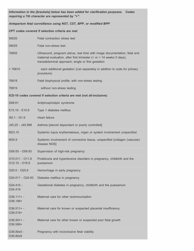
Information in the [brackets] below has been added for clarification purposes. Codes requiring a 7th character are represented by "+":
Antepartum fetal surveillance using NST, CST, BPP, or modified BPP:
CPT codes covered if selection criteria are met:
59020 Fetal contraction stress test
59025 Fetal non-stress test
76805 Ultrasound, pregnant uterus, real time with image documentation, fetal and maternal evaluation, after first trimester (> or = 14 weeks 0 days), transabdominal approach; single or first gestation
+ 76810 each additional gestation (List separately in addition to code for primary procedure)
76818 Fetal biophysical profile; with non-stress testing
76819 without non-stress testing
ICD-10 codes covered if selection criteria are met (not all-inclusive):
D68.61 Antiphospholipid syndrome
E10.10 - E10.9 Type 1 diabetes mellitus
I50.1 - I51.9 Heart failure
J45.20 - J45.998 Asthma [steroid dependant or poorly controlled]
M23.10 Systemic lupus erythematosus, organ or system involvement unspecified
M35.9 Systemic involvement of connective tissue, unspecified [collagen (vascular) disease NOS]
O09.00 - O09.93 Supervision of high-risk pregnancy
O10.011 - O11.9 Proteinuria and hypertensive disorders in pregnancy, childbirth and the O12.10 - O16.9 puerperium
O20.0 - O20.9 Hemorrhage in early pregnancy
O24.011 - O24.93 Diabetes mellitus in pregnancy
O24.410 -O24.419
Gestational diabetes in pregnancy, childbirth and the puerperium
O36.111+ -O36.199+
Maternal care for other isoimmunization
O36.511+ - O36.519+
Maternal care for known or suspected placental insufficiency
O36.591+ - O36.599+
Maternal care for other known or suspected poor fetal growth
O36.80x0 -O36.80x9
Pregnancy with inconclusive fetal viability

O36.812+ -O36.819+
Decreased fetal movements
O40.1xx+ - O40.9xx+
Polyhydramnios
O41.00x+ -O41.03x+
Oligohydramnios
O44.00 - O46.93 Placenta previa, premature separation of placenta [abruptio placentae], antepartum hemorrhage, not elsewhere classified
O48.0 - O48.1 Late pregnancy
O99.280 -O99.285
Other endocrine, nutritional and metabolic diseases complicating pregnancy, childbirth and the puerperium
O99.810 -O99.815
Abnormal glucose complicating pregnancy, childbirth and the puerperium
P55.0 - P55.9 Hemolytic disease of newborn
P56.0 Hydrops fetalis due to isoimmunization
P57.0 Kernicterus due to isoimmunization
ICD-10 codes not covered for indications listed in the CPB:
Z33.1 Pregnant state, incidental
Z34.00 - Z34.93 Encounter for supervision of normal pregnancy
CST or Full BPP:
CPT codes covered if selection criteria are met:
59020 Fetal contraction stress test
76818 Fetal biophysical profile; with non-stress testing
ICD-10 codes covered if selection criteria are met (not all-inclusive):
O28.0 - O28.9 Abnormal findings on antenatal screening of mother
O41.00x+ -O41.03x+
Oligohydramnios
Umbilical artery Doppler velocimetry:
CPT codes covered if selection criteria are met:
76820 Doppler velocimetry, fetal; umbilical artery [not covered for studies of ductus venosus and vessels for surveillance of impaired fetal growth]
ICD-10 codes covered if selection criteria are met:
O36.511+ -O36.599+
Maternal care for other known or suspected poor fetal growth
O36.821 - Maternal care for fetal anemia and thrombocytopenia
O36.829
O41.00x+ -O41.03x+
Oligohydramnios
O43.021 -O43.029
Fetus-to-fetus placental transfusion syndrome
Uterine artery Doppler studies:
CPT codes not covered for indications listed in the CPB:
93975 Duplex scan of arterial inflow and venous outflow of abdominal, pelvic, scrotal contents and/or retroperitoneal organs; complete study
93976 limited study
ICD-10 codes not covered for indications listed in the CPB:
O00.00 - O08.9 O09.00 - O09.93, O10.011 - O16.9, O20.0 - O26.93, O29.011 O29.93, O30.001 - O30.019, O30.031 O35.6xx9, O35.8xx+ O36.819+, O36.891+ O48.1, O60.00 O77.9 O80 - O82 O85 - O92.79 O98.011 - O9a.53
Complications of pregnancy, childbirth, and the puerperium
-
-
-
--
O28.0 - O28.9 Abnormal findings on antenatal screening of mother
Z30.011 - Z30.9 Z31.0 - Z31.9, Z33.1, Z34.00 - Z34.93, Z36, Z37.0 - Z37.9 Z39.0 - Z39.2 Z98.51 - Z98.52
Normal pregnancy, postpartum care and examination, encounter for contraceptive management, procreative management, outcome of delivery, and encounter for antenatal screening of mother
Middle cerebral artery Doppler velocimetry:
CPT codes covered if selection criteria are met:
76821 Doppler velocimetry, fetal; middle cerebral artery
ICD-10 codes covered if selection criteria are met (not all-inclusive):
O35.3xx+ Maternal care for (suspected) damage to fetus from viral disease in mother
O36.011+ - Maternal care for rhesus isoimmunization

O36.099+
O36.111+ -O36.199+
Maternal care for other isoimmunization
O36.511+ -O36.599+
Maternal care for other known or suspected poor fetal growth
O43.011 -O43.019
Fetomaternal placental transfusion syndrome
O43.021 -O43.029
Fetus-to-fetus placental transfusion syndrome
O98.511 - O98.53 Other viral diseases complicating pregnancy, childbirth and the puerperium [parvovirus B-19 infection]
Serum YKL-40:
CPT codes not covered for indications listed in the CPB:
83520 Immunoassay for analyte other than infectious agent antibody or infectious agent antigen; quantitative, not otherwise specified [not covered for serum YKL-40]
ICD-10 codes not covered for indications listed in the CPB:
O11.1 - O11.9 Pre-existing hypertension with pre-eclampsia
O14.00 - O14.93 Pre-eclampsia
O36.591+ - O36.599+
Maternal care for other known or suspected poor fetal growth [small-for gestational age fetuses]
-
The above policy is based on the following references:
1. American College of Obstetricians and Gynecologists (ACOG), Committee on Practice Bulletins -- Obstetrics. Antepartum Fetal Surveillance. ACOG Practice Bulletin No. 9. Washington, DC: ACOG; October 1999.
2. Huddleston JF. Intrapartum fetal assessment. A review. Clin Perinatol. 1999;26(3):549-568. 3. Penning S, Garite TJ. Management of fetal distress. Obstet Gynecol Clin N Am.
1999;26(2):259-274. 4. Dildy GA. The physiologic and medical rationale for intrapartum fetal monitoring. Biomed
Instrum Technol. 1999;33(2):143-151. 5. Low JA. Predictive value of electronic fetal monitoring for intrapartum fetal asphyxia with
metabolic acidosis. Obstet Gynecol. 1999;93(2):285-291. 6. Johnson TR, Paine LL, Strobino DM, et al. Population differences affect the interpretation of
fetal nonstress test results. Am J Obstet Gynecol. 1998;179(3 Pt 1):779-783. 7. Kunzel W. Intrauterine fetal death during pregnancy: Limitations of fetal surveillance. J Obstet
Gynaecol Res. 1998;24(6):453-460. 8. American College of Obstetricians and Gynecologists (ACOG). Special problems of multiple
gestation. ACOG Technical Bulletin No. 253. Washington, DC: ACOG; November 1998. 9. Manning F. Fetal assessment based on fetal biophysical profile scoring. Am J Obstet Gynecol.
1998;178(4):698-706. 10. Horio H, Murakami M, Chiba Y, et al. Fetal monitor for non-stress-test screening at home.
Biomed Instrum Technol. 1998;32(1):39-47.

11. Salamalekis E, Loghis C, Panayotopoulos N, et al. Non-stress test: A fifteen year clinical appraisal. Clin Exp Obstet Gynecol. 1997;24(2):79-81.
12. Smith-Leviton M, Petrikovsky B, Schneider EP. Practical guidelines for antepartum fetal surveillance. Am Fam Physician. 1997;56(8):1981-1988.
13. Ananth CV, Smulian JC, Vintzileos AM. Epidemiology of antepartum fetal testing. Curr Opinion Obstet Gynecol. 1997;9(2):101-106.
14. U.S. Preventative Services Task Force. Guide to clinical preventive services. 2nd ed. Baltimore, MD: Williams & Wilkins; 1996:433-442.
15. Thacker SB, Stroup DF, Peterson HB. Efficacy and safety of intrapartum electronic fetal monitoring: an update. Obstet and Gynecol. 1995;86(4 Pt 1):613 -620.
16. Crowe JA, Harrison A, Hayes-Gill BR. The feasibility of long-term fetal heart rate monitoring in the home environment using abdominal electrodes. Physiol Meas. 1995;16(3):195-202.
17. American College of Obstetricians and Gynecologists (ACOG). Fetal heart rate patterns: Monitoring, interpretation, and management. ACOG Technical Bulletin No. 207. Washington, DC: ACOG; July 1995.
18. American College of Obstetricians and Gynecologists (ACOG). Diabetes and pregnancy. ACOG Technical Bulletin No. 200. Washington, DC: ACOG; December 1994.
19. Naef RW 3rd, Morrison JC, Washburne JF, et al. Assessment of fetal well-being using nonstress test in the home setting. Obstet Gynecol. 1994;84(3):424-426.
20. Reece EA, Hagay Z, Garofalo J, Hobbins JC. A controlled trial of self-nonstress test versus assisted nonstress test in the evaluation of fetal well being. Am J Obstet Gynecol. 1992;166(2):489-492.
21. Gonen R, Braithwaite N, Milligan JE. Fetal heart rate monitoring at home and transmission by telephone. Obstet Gynecol. 1990;75(3 Pt 1):464-468.
22. Lacin S, Demir N, Koyuncu F, et al. Value of intraplacental villous artery Doppler measurements in severe preeclampsia. J Postgrad Med. 1996;42(4):101-104.
23. Ozcan T, Sbracia M, d'Ancona RL, et al. Arterial and venous Doppler velocimetry in the severely growth-restricted fetus and associations with adverse perinatal outcome. Ultrasound Obstet Gynecol. 1998;12(1):39-44.
24. van Asselt K, Gudmundsson S, Lindqvist P, et al. Uterine and umbilical artery velocimetry in pre-eclampsia. Acta Obstet Gynecol Scand. 1998;77(6):614-619.
25. Wang KG, Chen CP, Yang JM, et al. Impact of reverse end-diastolic flow velocity in umbilical artery on pregnancy outcome after the 28th gestational week. Acta Obstet Gynecol Scand. 1998;77(5):527-531.
26. American College of Obstetricians and Gynecologists (ACOG), Committee on Obstetric Practice. ACOG committee opinion. Utility of antepartum umbilical artery Doppler velocimetry in intrauterine growth restriction. Number 188, October 1997 (replaces no. 116, November 1992). Int J Gynaecol Obstet. 1997;59(3):269-270.
27. Harrington K, Carpenter RG, Goldfrad C, et al. Transvaginal Doppler ultrasound of the uteroplacental circulation in the early prediction of pre-eclampsia and intrauterine growth retardation. Br J Obstet Gynaecol. 1997;104(6):674-681.
28. Zimmermann P, Eirio V, Koskinen J, et al. Doppler assessment of the uterine and uteroplacental circulation in the second trimester in pregnancies at high risk for pre-eclampsia and/or intrauterine growth retardation: Comparison and correlation between different Doppler parameters. Ultrasound Obstet Gynecol. 1997;9(5):330-338.
29. Kingdom JC, Burrell SJ, Kaufmann P. Pathology and clinical implications of abnormal umbilical artery Doppler waveforms. Ultrasound Obstet Gynecol. 1997;9(4):271-286.
30. Mari GC, Deter RL, Carpenter R, et al. Noninvasive diagnosis by doppler ultrasonography of fetal anemia due to maternal alloimmunization. N Engl J Med. 2000;342:9-14.
31. Teixeira JM, Duncan K, Letsky E, et al. Middle cerebral artery peak systolic velocity in the prediction of fetal anemia. Ultrasound Obstet Gynecol. 2000;15:205-208.
32. Roberts AB, Mitchell JM, Lake Y, et al. Ultrasonographic surveillance in red blood cell alloimmunization. Am J Obstet Gynecol. 2001;184(6):1251-1255.
33. Bahado-Singh RO, Oz AU, Hsu C, et al. Middle cerebral artery Doppler velocimetric

deceleration angle as a predictor of fetal anemia in Rh-alloimmunized fetuses without hydrops. Am J Obstet Gynecol. 2000;183(3):746-751.
34. Oepkes D. Invasive versus non-invasive testing in red-cell alloimmunized pregnancies. Eur J Obstet Gynecol Reprod Biol. 2000;92(1):83-89.
35. Laks MP, Cohen T. Noninvasive diagnosis of fetal anemia by Doppler ultrasonography. N Engl J Med. 2000;343(1):66-67; discussion 67-68.
36. Saade GR. Noninvasive testing for fetal anemia. N Engl J Med. 2000;342(1):52-53. 37. Sherer DM. Prenatal ultrasonographic assessment of the middle cerebral artery: A review.
Obstet Gynecol Surv. 1997;52(7):444-455. 38. Sterne G, Shields LE, Dubinsky TJ. Abnormal fetal cerebral and umbilical Doppler
measurements in fetuses with intrauterine growth restriction predicts the severity of perinatal morbidity. J Clin Ultrasound. 2001;29(3):146-151.
39. Westergaard HB, Langhoff-Roos J, Lingman G, et al. Critical appraisal of the use of umbilical artery Doppler ultrasound in high-risk pregnancies: Use of meta-analyses in evidence-based obstetrics. Ultrasound Obstet Gynecol. 2001;17(6):466-476.
40. Deren O, Onderoglu L. The value of middle cerebral artery systolic velocity for initial and subsequent management in fetal anemia. Eur J Obstet Gynecol Reprod Biol. 2002;101(1):26-30.
41. Mari G, Detti L, Oz U, et al. Accurate prediction of fetal hemoglobin by Doppler ultrasonography. Obstet Gynecol. 2002;99(4):589-593.
42. Goffinet F, Paris-Llado J, Nisand I, Breart G. Umbilical artery Doppler velocimetry in unselected and low risk pregnancies: A review of randomised controlled trials. Br J Obstet Gynaecol. 1997;104(4):425-430.
43. Yla-Outinen A. EBM (evidence-based medicine) guidelines. Ultrasound scanning during pregnancy. Helsinki, Finland: Duodecim Medical Publications Ltd.; April 3, 2000.
44. Erskine RL, Ritchie JW. Umbilical artery blood flow characteristics in normal and growth-retarded fetuses. Br J Obstet Gynaecol. 1985;92:605-610.
45. Gudmundsson S, Marsal K. Umbilical and uteroplacental blood flow velocity waveforms in pregnancies with fetal growth retardation. Eur J Obstet Gynecol Reprod Biol. 1988;27:187-196.
46. Reuwer PJ, Bruinse HW, Stoutenbeek P, Haspels AA. Doppler assessment of the fetoplacental circulation in normal and growth-retarded fetuses. Eur J Obstet Gynecol Reprod Biol. 1984;18:199-205.
47. Karsdorp VH, van Vugt JM, van Geijn HP, et al. Clinical significance of absent or reversed end diastolic velocity waveforms in umbilical artery. Lancet. 1994;344:1664-1668.
48. Giles WB, Trudinger BJ, Baird PJ. Fetal umbilical artery flow velocity waveforms and placental resistance: Pathological correlation. Br J Obstet Gynaecol. 1985;92:31-38.
49. Nicolaides KH, Bilardo CM, Soothill PW, Campbell S. Absence of end diastolic frequencies in umbilical artery: A sign of fetal hypoxia and acidosis. BMJ. 1988;297:1026-1027.
50. Almstrom H, Axelsson O, Cnattingius S, et al. Comparison of umbilical-artery velocimetry and cardiotocography for surveillance of small-for-gestational-age fetuses. Lancet. 1992;340:936-940.
51. Johnstone FD, Prescott R, Hoskins P, et al. The effect of introduction of umbilical Doppler recordings to obstetric practice. Br J Obstet Gynaecol. 1993;100:733-741.
52. Newnham JP, O'Dea MR, Reid KP, Diepeveen DA. Doppler flow velocity waveform analysis in high risk pregnancies: A randomized controlled trial. Br J Obstet Gynaecol. 1991;98:956-963.
53. Omtzigt AM, Reuwer PJ, Bruinse HW. A randomized controlled trial on the clinical value of umbilical Doppler velocimetry in antenatal care. Am J Obstet Gynecol. 1994;170:625-634.
54. Pattinson RC, Norman K, Odendaal HJ. The role of Doppler velocimetry in the management of high risk pregnancies. Br J Obstet Gynaecol. 1994;101:114-120.
55. Trudinger BJ, Cook CM, Giles WB, et al. Umbilical artery flow velocity waveforms in high-risk pregnancy. Randomised controlled trial. Lancet. 1987;1(8526):188-190.
56. Tyrrell SN, Lilford RJ, Macdonald HN, et al. Randomized comparison of routine vs highly selective use of Doppler ultrasound and biophysical scoring to investigate high risk pregnancies. Br J Obstet Gynaecol. 1990;97:909-916.

57. Haley J, Tuffnell DJ, Johnson N. Randomised controlled trial of cardiotocography versus umbilical artery Doppler in the management of small for gestational age fetuses. Br J Obstet Gynaecol. 1997;104:431-435.
58. Nienhuis SJ, Vles JS, Gerver WJ, Hoogland HJ. Doppler ultrasonography in suspected intrauterine growth retardation: A randomized clinical trial. Ultrasound Obstet Gynecol. 1997;9:6-13.
59. Mason GC, Lilford RJ, Porter J, et al. Randomised comparison of routine versus highly selective use of Doppler ultrasound in low risk pregnancies. Br J Obstet Gynaecol. 1993;100:130-133.
60. Mari G, Deter RL. Middle cerebral artery flow velocity waveforms in normal and small-for-gestational-age fetuses. Am J Obstet Gynecol. 1992;166:1262-1270.
61. Ott WJ, Mora G, Arias F, et al. Comparison of the modified biophysical profile to a 'new' biophysical profile incorporating the middle cerebral artery to umbilical artery velocity flow systolic/diastolic ratio. Am J Obstet Gynecol. 1998;178:1346-1353.
62. Neilson JP, Alfirevic Z. Doppler ultrasound for fetal assessment in high risk pregnancies (Cochrane Review). In: The Cochrane Library, Issue 1, 2003. Oxford, UK: Update Software.
63. Irion O, Masse J, Forest JC, Moutquin JM. Prediction of pre-eclampsia, low birthweight for gestation and prematurity by uterine artery blood flow velocity waveform analysis in low risk nulliparous women. Br J Obstet Gynaecol. 1998;105:422-429.
64. Friedman SA, Lindheimer MD. Prediction and differential diagnosis. In: Chesley's hypertensive disorders in pregnancy. 2nd ed. MD Lindheimer, JM Roberts, FG Cunningham, eds. Stamford, CT: Appleton & Lange; 1999:201-227.
65. American College of Radiology (ACR), Expert Panel on Women's Imaging. Growth disturbances: Risk of intrauterine growth restriction. Reston, VA: ACR; 2001.
66. American College of Obstetricians and Gynecologists (ACOG), Committee on Practice Bulletins -- Obstetrics. Diagnosis and management of preeclampsia and eclampsia. ACOG Practice Bulletin No. 33. Washington, DC: ACOG; January 2002.
67. American College of Obstetricians and Gynecologists (ACOG), Committee on Practice Bulletins -- Obstetrics. Intrauterine growth restriction. ACOG Practice Bulletin No. 12. Washington, DC: ACOG; January 2000.
68. Banta DH, Thacker SB. Historical controversy in health technology assessment: The case of electronic fetal monitoring. Obstet Gynecol Surv. 2001;56(11):707-719.
69. Danish Centre for Evaluation and Health Technology Assessment (DACEHTA). Umbilical artery Doppler ultrasonography in high risk pregnancies - an health technology assessment. Copenhagen, Denmark: Danish Centre for Evaluation and Health Technology Assessment (DACEHTA); 2002.
70. Myers ER, Blumrick R, Christian AL, et al. Management of prolonged pregnancy. Evidence Report/Technology Assessment No. 53. Prepared by the Duke Evidence-based Practice Center under Contract No. 290-97-0014. AHRQ Publication No. 02-E018. Rockville, MD: Agency for Healthcare Research and Quality (AHRQ); May 2002.
71. Fretts RC, Elkin EB, Myers ER, Heffner LJ. Should older women have antepartum testing to prevent unexplained stillbirth? Obstet Gynecol. 2004;104(1):56-64.
72. Chauhan SP, Doherty DD, Magann EF, et al. Amniotic fluid index vs single deepest pocket technique during modified biophysical profile: A randomized clinical trial. Am J Obstet Gynecol. 2004;191(2):661-667; discussion 667-668.
73. Audibert F, Benchimol Y, Benattar C, et al. Prediction of preeclampsia or intrauterine growth restriction by second trimester serum screening and uterine Doppler velocimetry. Fetal Diagn Ther. 2005;20(1):48-53.
74. Madazli R, Kuseyrioglu B, Uzun H, et al. Prediction of preeclampsia with maternal mid-trimester placental growth factor, activin A, fibronectin and uterine artery Doppler velocimetry. Int J Gynaecol Obstet. 2005;89(3):251-257.
75. Barkehall-Thomas A, Wilson C, Baker L, et al. Uterine artery Doppler velocimetry for the detection of adverse obstetric outcomes in patients with elevated mid-trimester beta-human chorionic gonadotrophin. Acta Obstet Gynecol Scand. 2005;84(8):743-747.
76. American College of Obstetricians and Gynecologists. ACOG Practice Bulletin No. 75:

Management of alloimmunization. Obstet Gynecol. 2006;108(2):457-464. 77. Mahboob U, Mazhar SB. Role of Kleihauer test in Rhesus negative pregnancy. J Coll
Physicians Surg Pak. 2006;16(2):120-123. 78. Ozcan T, Thornburg L, Mingione M, Pressman E. Use of middle cerebral artery peak systolic
velocity and intrauterine transfusion for management of twin-twin transfusion and single fetal intrauterine demise. J Matern Fetal Neonatal Med. 2006;19(12):807-809.
79. Senat MV, Loizeau S, Couderc S, et al. The value of middle cerebral artery peak systolic velocity in the diagnosis of fetal anemia after intrauterine death of one monochorionic twin. Am J Obstet Gynecol. 2003;189(5):1320-1324.
80. Martinez JM, Bermudez C, Becerra C, et al. The role of Doppler studies in predicting individual intrauterine fetal demise after laser therapy for twin-twin transfusion syndrome. Ultrasound Obstet Gynecol. 2003;22(3):246-251.
81. Ohkuchi A, Minakami H, Shiraishi H, et al. Intrauterine death of one twin, with rescue of the other, in twin-twin transfusion syndrome. Ultrasound Obstet Gynecol. 2002;19(3):293-296.
82. Ropacka M, Markwitz W, Ginda W, Breborowicz GH. Ultrasound in the diagnosis of twin-to-twin transfusion syndrome--a preliminary report. Acta Genet Med Gemellol (Roma). 1998;47(3-4):227-237.
83. Suzuki S, Sawa R, Yoneyama Y, et al. Fetal middle cerebral artery Doppler waveforms in twin-twin transfusion syndrome. Gynecol Obstet Invest. 1999;48(4):237-240.
84. Hecher K, Ville Y, Nicolaides KH. Fetal arterial Doppler studies in twin-twin transfusion syndrome. J Ultrasound Med. 1995;14(2):101-108.
85. American College of Obstetricians and Gynecologists (ACOG). Multiple gestation: Complicated twin, triplet, and high-order multifetal pregnancy. ACOG Practice Bulletin No. 56. Washington, DC: ACOG; October 2004.
86. Kontopoulos EV, Quintero RA, Chmait RH, et al. Percent absent end-diastolic velocity in the umbilical artery waveform as a predictor of intrauterine fetal demise of the donor twin after selective laser photocoagulation of communicating vessels in twin-twin transfusion syndrome. Ultrasound Obstet Gynecol. 2007;30(1):35-39.
87. Chang YL, Chmait RH, Bornick PW, et al. The role of laser surgery in dissecting the etiology of absent or reverse end-diastolic velocity in the umbilical artery of the donor twin in twin-twin transfusion syndrome. Am J Obstet Gynecol. 2006;195(2):478-483.
88. American College of Obstetricians and Gynecologists (ACOG) Committee on Obstetric Practice, American Academy of Pediatrics (AAP) Committee on Fetus and Newborn. Guidelines for Perinatal Care. 6th Ed. Washington, DC: ACOG; 2007.
89. Meads CA, Cnossen JS, Meher S, et al. Methods of prediction and prevention of pre-eclampsia: Systematic reviews of accuracy and effectiveness literature with economic modelling. National Coordinating Centre for Health Technology Assessment (NCCHTA). Health Technol Assess. 2008;12(6):1-270.
90. Nabhan AF, Abdelmoula YA. Amniotic fluid index versus single deepest vertical pocket as a screening test for preventing adverse pregnancy outcome. Cochrane Database Syst Rev. 2008; (3):CD006593.
91. Frøen JF, Heazell AE, Tveit JV, et al. Fetal movement assessment. Semin Perinatol. 2008;32(4):243-246.
92. Lalor JG, Fawole B, Alfirevic Z, Devane D. Biophysical profile for fetal assessment in high risk pregnancies. Cochrane Database Syst Rev. 2008;(1):CD000038.
93. Grivell RM, Wong L, Bhatia V. Regimens of fetal surveillance for impaired fetal growth. Cochrane Database Syst Rev. 2009;(1):CD007113.
94. Sciscione AC, Hayes EJ. Uterine artery Doppler flow studies in obstetric practice. Am J Obstet Gynecol. 2009;201(2):121-126.
95. Phattanachindakun B, Boonyagulsrirung T, Chanprapaph P. The correlation in antepartum fetal test between full fetal biophysical profile (FBP) and rapid biophysical profile (rBPP). J Med Assoc Thai. 2010;93(7):759-764.
96. Maulik D, Mundy D, Heitmann E, Maulik D. Evidence-based approach to umbilical artery Doppler fetal surveillance in high-risk pregnancies: An update. Clin Obstet Gynecol.

2010;53(4):869-878. 97. Alfirevic Z, Stampalija T, Gyte GM. Fetal and umbilical Doppler ultrasound in normal pregnancy.
Cochrane Database Syst Rev. 2010;(8):CD001450. 98. Alfirevic Z, Stampalija T, Gyte GM. Fetal and umbilical Doppler ultrasound in high-risk
pregnancies. Cochrane Database Syst Rev. 2010;(1):CD007529. 99. Pedrosa AC, Matias A. Screening for pre-eclampsia: A systematic review of tests combining
uterine artery Doppler with other markers. J Perinat Med. 2011;39(6):619-635. 100. Kuc S, Wortelboer EJ, van Rijn BB, et al. Evaluation of 7 serum biomarkers and uterine artery
Doppler ultrasound for first-trimester prediction of preeclampsia: A systematic review. Obstet Gynecol Surv. 2011;66(4):225-239.
101. Bezircioglu I, Baloglu A, Cetinkaya B, et al. The diagnostic value of the Doppler ultrasonography in distinguishing the endometrial malignancies in women with postmenopausal bleeding. Arch Gynecol Obstet. 2012;285(5):1369-1374.
102. Society for Maternal-Fetal Medicine Publications Committee, Berkley E, Chauhan SP, Abuhamad A. Doppler assessment of the fetus with intrauterine growth restriction. Am J Obstet Gynecol. 2012;206(4):300-308.
103. American College of Obstetricians and Gynecologists. ACOG Practice bulletin no. 134: Fetal growth restriction. Obstet Gynecol. 2013;121(5):1122-1133.
104. Goetzinger KR, Zhong Y, Cahill AG, et al. Efficiency of first-trimester uterine artery Doppler, a-disintegrin and metalloprotease 12, pregnancy-associated plasma protein a, and maternal characteristics in the prediction of preeclampsia. J Ultrasound Med. 2013;32(9):1593-1600.
105. Norwitz ER. Prediction of preeclampsia. UpToDate [online serial]. Waltham, MA: UpToDate; reviewed September 2014.
106. Seravalli V, Block-Abraham DM, Turan OM, et al. First-trimester prediction of small-for-gestational age neonates incorporating fetal Doppler parameters and maternal characteristics. Am J Obstet Gynecol. 2014;211(3):261.e1-e8.
107. Johansen JS, Jensen BV, Roslind A, et al. Serum YKL-40, a new prognostic biomarker in cancer patients? Cancer Epidemiol Biomarkers Prev. 2006;15(2):194-202.
108. Madazli R, Kucur M, Gezer A, et al. Chitotriosidase and YKL-40 in normal and pre-eclamptic pregnancies. Int J Gynaecol Obstet. 2008;100(3):239-243.
109. Gybel-Brask D, Hogdall E, Johansen J, et al. Serum YKL-40 and uterine artery Doppler -- a prospective cohort study, with focus on preeclampsia and small-for-gestational-age. Acta Obstet Gynecol Scand. 2014;93(8):817-824.
110. Kucur M, Tuten A, Oncul M, et al. Maternal serum apelin and YKL-40 levels in early and late-onset pre-eclampsia. Hypertens Pregnancy. 2014;33(4):467-475.
111. Alfirevic Z, Stampalija T, Medley N. Fetal and umbilical Doppler ultrasound in normal pregnancy. Cochrane Database Syst Rev. 2015;4:CD001450.
112. August P, Sibai BM. Preeclampsia: Clinical features and diagnosis. UpToDate [online serial]. Waltham, MA: UpToDate; reviewed November 2015.
113. Norwitz ER. Prediction of preeclampsia. UpToDate [online serial]. Waltham, MA: UpToDate; reviewed November 2015.
114. Resnik R. Fetal growth restriction: Evaluation and management. UpToDate [online serial]. Waltham, MA: UpToDate; reviewed November 2015.
115. Allen RE, Morlando M, Thilaganathan B, et al. Predictive accuracy of second trimester uterine artery Doppler indices for stillbirth: A systematic review and meta-analysis. Ultrasound Obstet Gynecol. 2016;47(1):22-27.
116. Society for Maternal-Fetal Medicine (SMFM), Norton ME, Chauhan SP, Dashe JS. Society for maternal-fetal medicine (SMFM) clinical guideline #7: Nonimmune hydrops fetalis. Am J Obstet Gynecol. 2015;212(2):127-139.
117. Khalil A, Morales-Rosello J, Townsend R, et al. Value of third-trimester cerebroplacental ratio and uterine artery Doppler indices as predictors of stillbirth and perinatal loss. Ultrasound Obstet Gynecol. 2016;47(1):74-80.
118. Levine TA, Alderdice FA, Grunau RE, McAuliffe FM. Prenatal stress and hemodynamics in pregnancy: A systematic review. Arch Womens Ment Health. 2016;19(5):721-739.

119. Signore C, Spong C. Overview of antepartum fetal surveillance. UpToDate [online serial]. Waltham, MA: UpToDate; reviewed September 2016.

Copyright Aetna Inc. All rights reserved. Clinical Policy Bulletins are developed by Aetna to assist in administering plan benefits and
constitute neither offers of coverage nor medical advice. This Clinical Policy Bulletin contains only a partial, general description of plan
or program benefits and does not constitute a contract. Aetna does not provide health care services and, therefore, cannot guarantee
any results or outcomes. Participating providers are independent contractors in private practice and are neither employees nor agents
of Aetna or its affiliates. Treating providers are solely responsible for medical advice and treatment of members. This Clinical Policy
Bulletin may be updated and therefore is subject to change.
CPT only copyright 2015 American Medical Association. All Rights Reserved.
Copyright 2001-2017 Aetna Inc.Web Privacy Statement | Legal Statement | Privacy Notices | Member Disclosure

AETNA BETTER HEALTH® OF PENNSYLVANIA
Amendment toAetna Clinical Policy Bulletin Number: 0088
Antepartum Fetal Surveillance
There are no amendments for Medicaid.
www.aetnabetterhealth.com/pennsylvania updated 03/07/2017