Endocarditis is an inflammation of the endocardium, the membrane lining the chambers of the heart...
106
-
Upload
maurice-allison -
Category
Documents
-
view
214 -
download
0
Transcript of Endocarditis is an inflammation of the endocardium, the membrane lining the chambers of the heart...


Endocarditis is an inflammation of the
endocardium, the membrane lining the
chambers of the heart and covering the
cusps of the heart valves. Endocarditis refers to infection of the heart
valves by various microorganisms. Typically affects native valves, but also
nonvalvular areas or implanted mechanical
devices (e.g., mechanical heart valves).

Bacteria primarily cause endocarditis Fungi and other atypical microorganisms
can lead to the disease; hence, the more
encompassing term infective endocarditis is
preferred.

Severity Acute or subacute depending on the pace
and severity of the clinical presentation. Acute: Fulminating form associated with
◦ high fevers and systemic toxicity.
◦ Virulent bacteria, such as Staphylococcus aureus,
frequently cause this syndrome,
◦ if untreated, death may occur within days to
weeks.

Severity Subacute
◦ more indolent and caused by less-invasive
organisms, such as viridans streptococci,
◦ usually occurring in preexisting valvular heart
disease.

Etiology
Best classified based on The etiologic organism, The anatomic site of infection, Pathogenic risk factors. Infection also may follow surgical insertion
of a prosthetic heart valve, resulting in
prosthetic-valve endocarditis (PVE).3
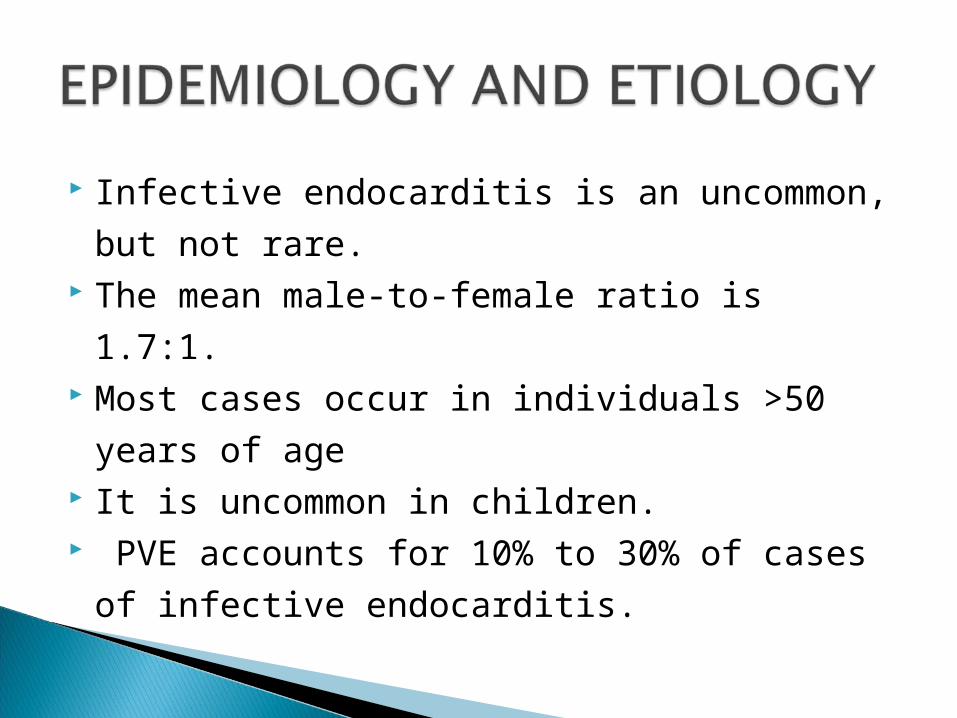
Infective endocarditis is an uncommon, but
not rare. The mean male-to-female ratio is 1.7:1. Most cases occur in individuals >50 years of
age It is uncommon in children. PVE accounts for 10% to 30% of cases of
infective endocarditis.

Most persons with IE have risk factors, such
as preexisting cardiac valvular abnormalities. Many types of structural heart disease result
in turbulent blood flow that increases the risk
for IE. A predisposing risk factor, however, may be
absent in up to 25% of cases Other conditions associated with a higher
incidence include diabetes, long-term
hemodialysis, and poor dental hygeine.
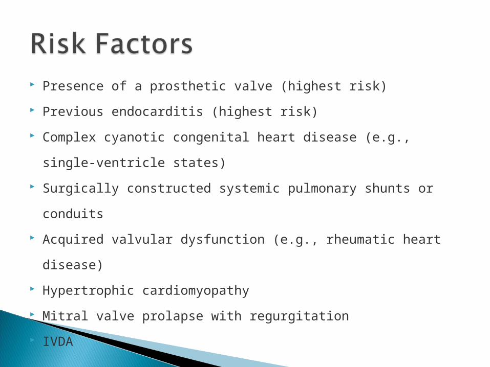
Presence of a prosthetic valve (highest risk)
Previous endocarditis (highest risk)
Complex cyanotic congenital heart disease (e.g., single-
ventricle states)
Surgically constructed systemic pulmonary shunts or conduits
Acquired valvular dysfunction (e.g., rheumatic heart disease)
Hypertrophic cardiomyopathy
Mitral valve prolapse with regurgitation
IVDA

Every organism causing human disease has been reported to cause IE.
Three groups of organisms result in a majority of cases: streptococci, staphylococci, and enterococci
The incidence of staphylococci, particularly S. aureus, continues to increase
Streptococci cause IE in patients with underlying cardiac abnormalities, such as mitral valve prolapse or rheumatic heart disease.
Staphylococci (S. aureus and coagulase-negative staphylococci) are the most common cause of PVE within the first year after valve surgery, and S. aureus is common in those with a history of IVDA.
Polymicrobial IE is uncommon, most often associated with IVDA.
Enterococcal IE follows GU manipulations (older men) or obstetric procedures (younger women).
isolation of the causative pathogen and determination of its antimicrobial susceptibilities offer the best chance for successful therapy
.

Table 115-1 Etiologic Organisms in I nfective Endocarditis
Agent Percentage of Cases (% )
Streptococci 60–80
Viridans streptococci 30–40
Other streptococci 15–25
Staphylococci 20–35
Coagulase positive 10–27
Coagulase negative 1–3
Enterococci 5–18
Gram-negative aerobic bacilli 1.5–13
Fungi 2–4
Miscellaneous bacteria <5
Mixed infections 1–2
"Culture negative" <5–24

The mitral and aortic valves are affected most commonly in cases involving a single valve.
Subacute endocarditis involves mitral valve. Acute disease involves aortic valve. 35% of cases involve concomitant infections
of aortic and mitral valves. Infection of tricuspid valve is less common,
and related to IVDA. pulmonary valve is rarley infected.
IE occurs via hematogenous spread It requires the sequential occurrence of several
factors. ◦The endothelial surface of the heart is damaged.◦Platelet and fibrin deposition occurs on the
abnormal epithelial surface (nonbacterial thrombotic endocarditis) .
◦Bacteremia gives organisms access to and results in colonization of the endocardial surface

◦Bacteremia is the result of trauma to a mucosal surface with a high concentration of resident bacteria, such as the oral cavity and gastrointestinal tract.
◦After colonization of the endothelial surface, a "vegetation" of fibrin, platelets, and bacteria forms. This allows unimpeded bacterial growth to concentrations as high as 109 to 1010 org/g tissue
Surgery may directly inoculate the valve with bacteria from the patient's skin or operating room personnel.
The recently placed nonendothelialized valve is more susceptible to bacterial colonization than are native valves.

Bacteria also may colonize the new valve from contaminated bypass pumps, cannulas, and pacemakers, or from a nosocomial bacteremia subsequent to an intravascular catheter.

Vegetations in IE may be single or multiple and vary in size from a few mls to cms.
Bacteria within the vegetation grow slowly and are protected from antibiotics and host defenses.

Adverse effects of IE and the resulting lesions include
(a) local perivalvular damage (b) embolization of septic fragments with
potential hematogenous seeding of remote sites.
(c) formation of antibody complexes.
Formation of vegetations may destroy valvular tissue
Continued destruction can lead to acute HF Valvular stenosis may occur. Abscesses can develop in the valve ring or in
myocardial tissue
Vegetations may be friable, and fragments may be released downstream.
Infected particles(septic emboli)can result in organ abscess or infarction.
Septic emboli from RT-side endocarditis lodge in the lungs, causing pulmonary abscesses. Emboli from LT-side affect organs with high blood flow, such as the kidneys, spleen, and brain
Circulating immune complexes consisting of Ag, Ab, and complement may deposit in organs, producing local inflammation and damage (e.g., glomerulonephritis in the kidneys).
Development of "mycotic" aneurysms, cerebral infarction, splenic infarction and abscess, and skin manifestations such as petechiae, Osler nodes, and Janeway lesions

Highly variable and nonspecific. Fever is most common (Table 115–2).
◦low grade, particularly in subacute cases. Heart murmurs inmajority of patients IE begins insidiously and worsens gradually. Patients present with nonspecific Sx
◦fever, chills, weakness, dyspnea, night sweats, weight loss, or malaise.

Patients with acute disease, such as those with a history of IVDA and S. aureus infective endocarditis, may appear with classic signs of sepsis.
Splenomegaly is a frequent finding in patients with prolonged endocarditis
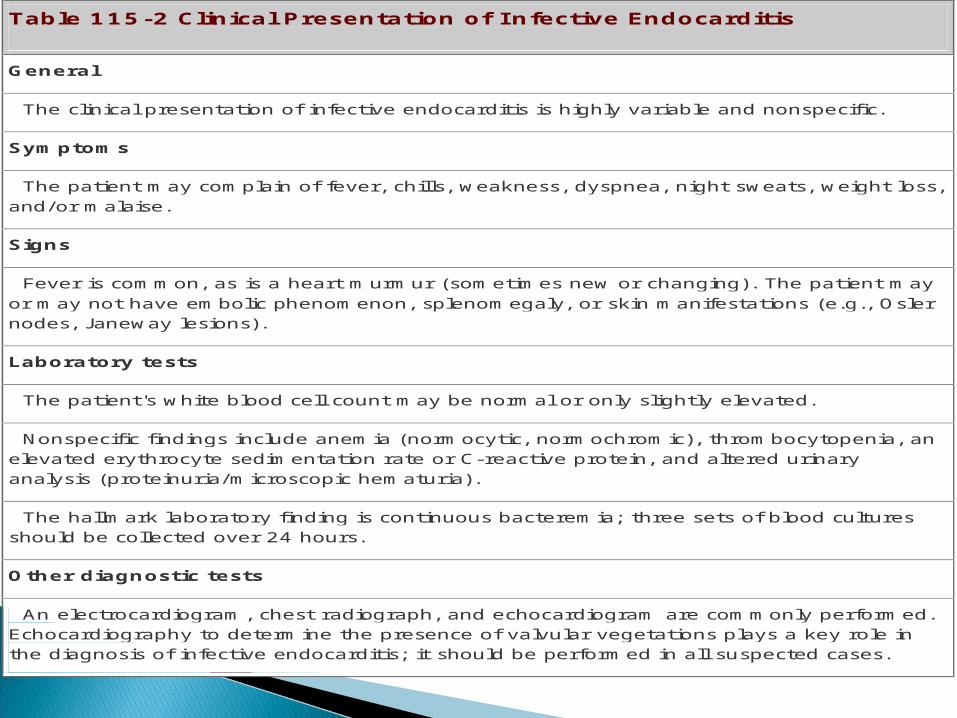
Table 115-2 Clinical Presentation of I nfective Endocarditis
General
The clinical presentation of infective endocarditis is highly variable and nonspecific.
Symptoms
The patient may complain of fever, chills, weakness, dyspnea, night sweats, weight loss, and/or malaise.
Signs
Fever is common, as is a heart murmur (sometimes new or changing). The patient may or may not have embolic phenomenon, splenomegaly, or skin manifestations (e.g., Osler nodes, J aneway lesions).
Laboratory tests
The patient's white blood cell count may be normal or only slightly elevated.
Nonspecific findings include anemia (normocytic, normochromic), thrombocytopenia, an elevated erythrocyte sedimentation rate or C-reactive protein, and altered urinary analysis (proteinuria/microscopic hematuria).
The hallmark laboratory finding is continuous bacteremia; three sets of blood cultures should be collected over 24 hours.
Other diagnostic tests
An electrocardiogram, chest radiograph, and echocardiogram are commonly performed. Echocardiography to determine the presence of valvular vegetations plays a key role in the diagnosis of infective endocarditis; it should be performed in all suspected cases.

Osler nodes—Purplish or erythematous subcutaneous nodules on pads of fingers and toes. Painful and tender. Not specific for infective endocarditis.
Janeway lesions—Hemorrhagic, painless plaques on palms of hands or soles of feet. Embolic in origin
Splinter hemorrhages—Thin, linear hemorrhages found under the nail beds of the fingers or toes. not specific for infective endocarditis.
Petechiae—Small erythematous, painless, hemorrhagic lesions anywhere on the skin but frequently on the anterior trunk, buccal mucosa and palate, and conjunctivae.
Clubbing of the fingers Roth spots—Retinal infarct with central pallor
and surrounding hemorrhage.

Emboli—in 1/3of cases and may result in significant complications.
Lt-side endocarditis can result in ◦renal artery emboli causing flank pain with
hematuria, ◦splenic artery emboli causing abdominal pain, ◦cerebral emboli, which may result in
hemiplegia or alteration in mental status. .
Emboli— Rt-side endocarditis may result in pulmonary emboli, causing pleuritic pain with hemoptysis

Anemia (normocytic, normochromic), and thrombocytopenia
WBC is NL or only slightly elevated, sometimes with a mild left shift.
Acute bacterial endocarditis, may present with an elevated WBC, consistent with a fulminant infection.
ESR is elevated in 90% to 100% of patients. C-reactive protein also may be elevated.

Urinary analysis: proteinuria and microscopic hematuria occurring in approximately 50% of individuals.
The hallmark is continuous bacteremia from shedding from vegetation into the blood.
95% of patients have +ve blood cultures. 3 sets of blood cultures, each from separate sites,
should be collected over 24 hours, and antibiotics should be withheld until adequate blood cultures are obtained.
Culture negative" endocarditis ◦clinical diagnosis of IE is likely but blood
culture is –ve.◦often the consequence of previous antibiotic therapy improperly collected blood cultures, unusual organisms. When blood cultures from these patients
are –ve after 48 to 72 hours, cultures should be held for a month to detect growth of fastidious organisms.

ECG rarely shows important diagnostic findings but may reveal heart block, suggesting extension of the infection.
CXR may provide more diagnostic information in patient with Rt-side IE.
Echocardiogram should be performed in all patients suspected of Septic pulmonary emboli.
Echocardiography ◦Transthoracic echocardiography (TTE) OR◦Transesophageal echocardiography (TEE),
depends on the clinical setting.
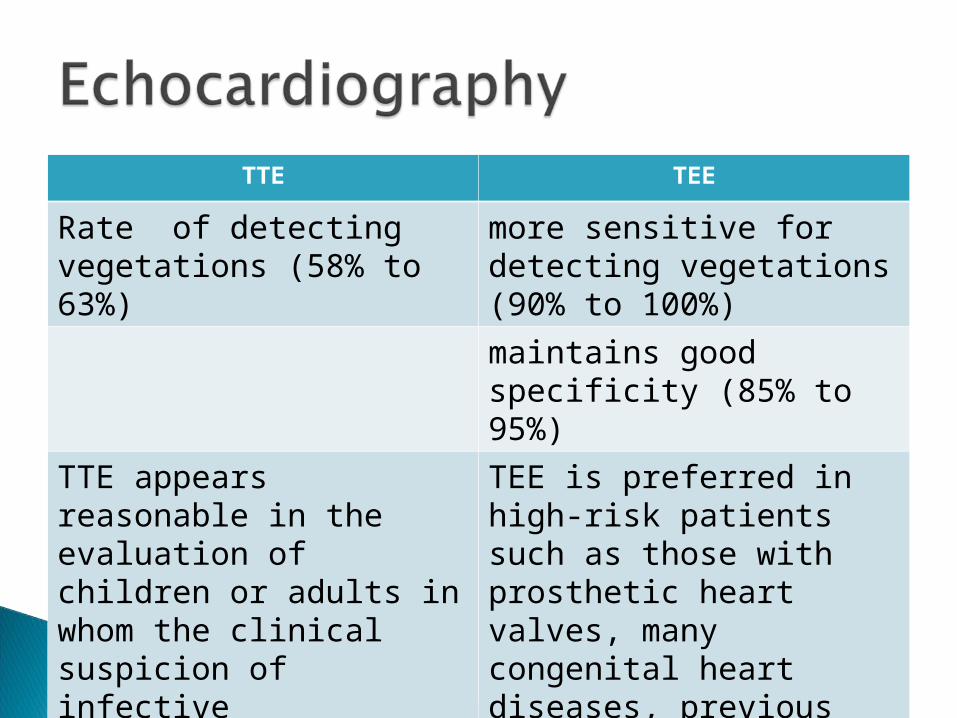
TEETTE
more sensitive for detecting vegetations (90% to 100%)
Rate of detecting vegetations (58% to 63%)
maintains good specificity (85% to 95%)TEE is preferred in high-risk patients such as those with prosthetic heart valves, many congenital heart diseases, previous endocarditis, new murmur, heart failure, or other stigmata of endocarditis
TTE appears reasonable in the evaluation of children or adults in whom the clinical suspicion of infective endocarditis is relatively low.
The identification of IE requires the integration of clinical, laboratory, and echocardiographic findings.
The Duke diagnostic criteria include major and minor variables (Table 115–3).
Based on the number of major and minor criteria, patients suspected of IE are categorized into: definite, possible or infective endocarditis rejected
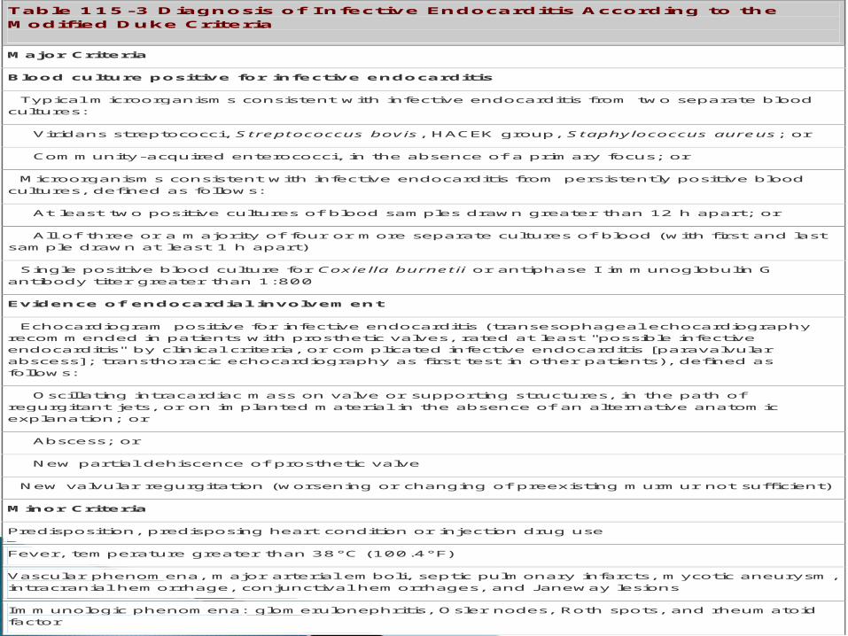
Table 115-3 Diagnosis of I nfective Endocarditis According to the Modified Duke Criteria
Major Criteria
Blood culture positive for infective endocarditis
Typical microorganisms consistent with infective endocarditis from two separate blood cultures:
Viridans streptococci, Streptococcus bovis, HACEK group, Staphylococcus aureus; or
Community-acquired enterococci, in the absence of a primary focus; or
Microorganisms consistent with infective endocarditis from persistently positive blood cultures, defined as follows:
At least two positive cultures of blood samples drawn greater than 12 h apart; or
All of three or a majority of four or more separate cultures of blood (with first and last sample drawn at least 1 h apart)
Single positive blood culture for Coxiella burnetii or antiphase I immunoglobulin G antibody titer greater than 1:800
Evidence of endocardial involvement
Echocardiogram positive for infective endocarditis (transesophageal echocardiography recommended in patients with prosthetic valves, rated at least "possible infective endocarditis" by clinical criteria, or complicated infective endocarditis [paravalvular abscess]; transthoracic echocardiography as first test in other patients), defined as follows:
Oscillating intracardiac mass on valve or supporting structures, in the path of regurgitant jets, or on implanted material in the absence of an alternative anatomic explanation; or
Abscess; or
New partial dehiscence of prosthetic valve
New valvular regurgitation (worsening or changing of preexisting murmur not sufficient)
Minor Criteria
Predisposition, predisposing heart condition or injection drug use
Fever, temperature greater than 38°C (100.4°F)
Vascular phenomena, major arterial emboli, septic pulmonary infarcts, mycotic aneurysm, intracranial hemorrhage, conjunctival hemorrhages, and J aneway lesions
Immunologic phenomena: glomerulonephritis, Osler nodes, Roth spots, and rheumatoid factor

The outcome for endocarditis is improved with ◦rapid diagnosis◦appropriate treatment (i.e., antimicrobial therapy,
surgery, or both)◦prompt recognition of complications should they
arise

Factors associated with increased mortality include
(a)Heart failure(b)Culture-negative endocarditis(c)Endocarditis caused by resistant organisms
such as fungi or gram-negative bacteria(d)left-sided endocarditis caused by S. aureus,(e)prosthetic-valve endocarditis

The presence of heart failure has the greatest negative impact on the short-term prognosis.
For native-valve IE, mortality range from 20% to 25%;
lower rates with viridans streptococci (4% to6%)
higher rates with lt-side IE caused by enterococci (15% to 25%) and staphylococci (25% to 47%).

higher rates of mortality with unusuall organisms (e.g., >50% for Pseudomonas aeruginosa).
mortality rate for Rt-side IE associated with IVDA is low (e.g., 10%).

Relapse after treatment will mostly occur within 1st 2 months after D/C of AB.
Relapse for viridans are low (2%), Relapse more likely with enterococcal
infection (8% to 20%) and PVE (10% to 15%).

After appropriate treatment and recovery, risk of morbidity and mortality following IE persist for years, although it gradually declines annually.
Morbidity remains elevated because of a greater likelihood of recurrent IE, heart failure, and embolism

The desired outcomes for treatment and prophylaxis of IE◦Relieve the signs and symptoms of the disease. ◦Decrease morbidity and mortality◦Eradicate the causative organism with minimal
drug exposure. ◦Provide cost-effective antimicrobial therapy◦Prevent infective endocarditis from occurring or
recurring in high-risk patients with appropriate prophylactic antimicrobials.

isolation of the infecting pathogen and determination of antimicrobial susceptibilities,
high-dose, parenteral, bactericidal antibiotics for extended
Treatment started in the hospital, but can be completed as outpatient if defervescence has occurred and follow up blood cultures show no growth.

Large doses of parenteral antimicrobials are necessary to achieve bactericidal concentrations within vegetations.
An extended duration of therapy is required, even for susceptible pathogens, because microorganisms are enclosed within valvular vegetations and fibrin deposits.
For most patients, 4 to 6 weeks of therapy is required
Surgery is an important adjunct in the management of endocarditis.
Valvectomy and valve replacement are performed to remove infected tissue and restore hemodynamic function. Echocardiographic features that suggest the need for surgery include ◦persistent vegetation ◦an increase in vegetation size after prolonged
antibiotic treatment◦Valve dysfunction, ◦perivalvular extension (e.g., abscess)
Surgery also may be considered in cases of PVE endocarditis ◦caused by resistant organisms (e.g., fungi or
gram-negative bacteria), ◦if there is persistent bacteremia or other
evidence of failure despite appropriate antimicrobial therapy.
◦
Treatment recommendations from the AHA provide guidance for the management of IE.
Guidelines use an evidence-based scoring system where recommendations are given a classification as well as level of evidence.
Class I recommendations are conditions for which there is evidence, general agreement, or both that a given procedure or treatment is useful and effective.
Class II recommendations are conditions for which there is conflicting evidence, a divergence of opinion, or both about the usefulness/efficacy of a procedure or treatment (IIa implies the weight of evidence/opinion is in favor of usefulness/efficacy whereas IIb implies usefulness/efficacy is less-well established by evidence/opinion).

Class III recommendations are conditions for which there is evidence, general agreement, or both that the procedure/treatment is not useful/effective and in some cases may be harmful.

Level of evidence is listed as A (data derived from multiple randomized clinical trials)
Level B (data derived from a single randomized trial or nonrandomized studies)
Level C (consensus opinion of experts).

B-Lactam antibiotics, such as penicillin G (or ceftriaxone), nafcillin, and ampicillin, remain the drugs of choice for streptococcal, staphylococcal, and enterococcal endocarditis, respectively

For some pathogens, as enterococci, synergistic antimicrobial combinations (including an aminoglycoside) is essential to obtain a bactericidal effect.
Combinations may decrease the emergence of resistant organisms during treatment (e.g., PVE caused by coagulase-negative staphylococci) and hasten clinical and microbiologic response (e.g., some streptococcal and staphylococcal infections).

Occasionally, combination treatment will result in a shorter treatment course
Streptococci are a common cause of infective endocarditis, with most isolates being viridans streptococci
These bacteria are common inhabitants of the human mouth and gingiva
During dental surgery, and even when brushing the teeth, these organisms can cause a transient bacteremia. In susceptible individuals, this may result in infective endocarditis.

Streptococcal endocarditis is usually subacute, and the response to medical treatment is good
in uncomplicated cases, response rates as high as 98% can be expected.
Viridans streptococci are penicillin-susceptible 10% to 20% are moderately susceptible.

This difference in in vitro susceptibility led to recommendations that the MIC be determined for all viridans streptococci and that the results be used to guide therapy.
Some streptococci are deemed tolerant to the killing effects of penicillin,
A tolerant organism is inhibited but not killed by an antibiotic normally considered bactericidal.

Tolerant strains do not respond as readily to B-lactam therapy as nontolerant ones
This phenomenon is primarily a laboratory finding with little clinical significance.
Treatment for tolerant strains is identical to that for nontolerant organisms.

An assortment of regimens can be used to treat uncomplicated, native-valve endocarditis caused by fully susceptible viridans streptococci (Table 115–4).
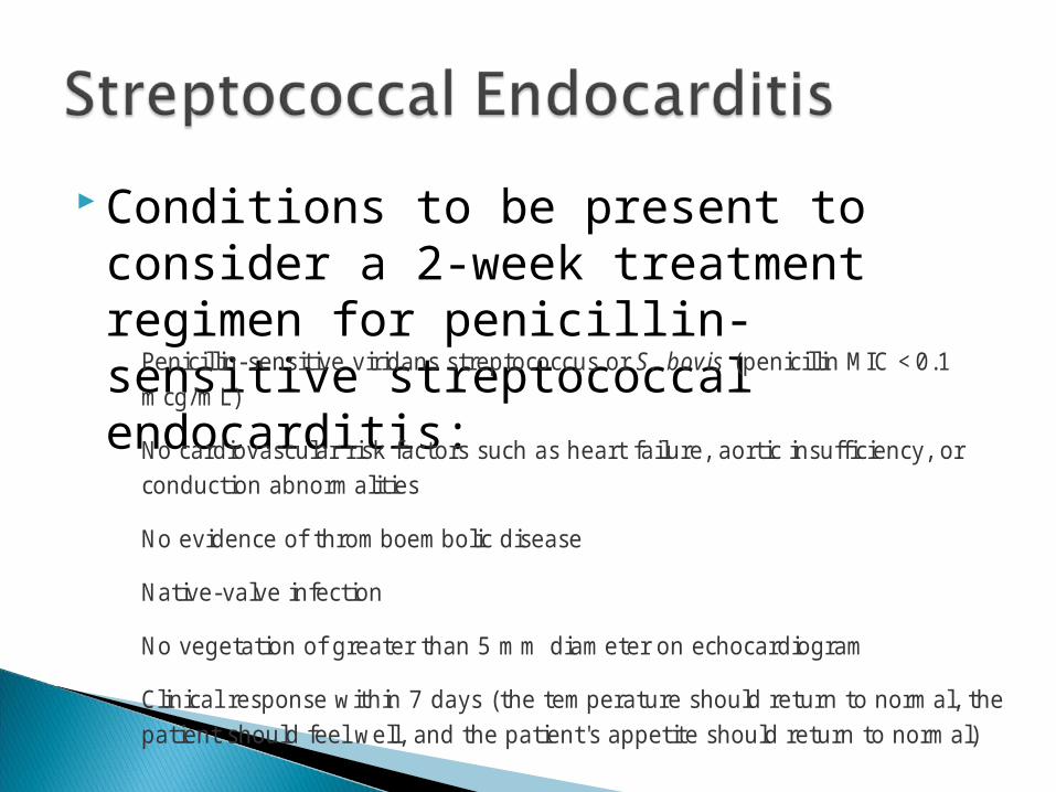
Conditions to be present to consider a 2-week treatment regimen for penicillin-sensitive streptococcal endocarditis: Penicillin-sensitive viridans streptococcus or S. bovis (penicillin MIC <0.1
mcg/mL)
No cardiovascular risk factors such as heart failure, aortic insufficiency, or conduction abnormalities
No evidence of thromboembolic disease
Native-valve infection
No vegetation of greater than 5 mm diameter on echocardiogram
Clinical response within 7 days (the temperature should return to normal, the patient should feel well, and the patient's appetite should return to normal)

If patient has immediate-type hypersensitivity to penicillin, vancomycin is chosen for IE caused by viridans streptococci.
addition of gentamicin is not recommended. A thorough allergy history must be obtained
before a second-line therapy is administered.
In patients with complicated infections or when the strep has an MIC of 0.12 to less than or equal to 0.5 mcg/mL, combination therapy with an AG and penicillin (higher dose) or ceftriaxone for the first 2 weeks is recommended, followed by penicillin or ceftriaxone alone for an additional 2 weeks (Table 115–5).

The rationale for combination therapy of penicillin-susceptible viridans streptococci is that enhanced activity
The combined treatment, however, is not superior to penicillin alone.
Few human data suggest that patients with endocarditis caused by these organisms respond less well to penicillin alone.

In patients with endocarditis of prosthetic valves caused by viridans streptococci, choices of treatment are similar.
treatment courses are extended to 6 weeks (Table 115–6).
If the organism is relatively resistant, gentamicin is recommended for 6 weeks.
Data support extended-interval dosing for the treatment of streptococcal infective endocarditis, and as compared with three times-daily dosing this approach may have greater efficacy
Endocarditis caused by staph. has become more prevalent because of ◦Increased IVDA◦More frequent use of peripheral & CVC◦Increased frequency of valve-replacement surgery.
S. aureus is the most common among IVDA and persons with venous catheters.
Coagulase-negative staphylococci (usually Staph. epidermidis) are prominent causes of PVE.

Any patient who develops staphylococcal bacteremia is at risk for endocarditis.
Parameters that predict higher risk of IE in patients with S. aureus bacteremia
(a) the absence of a primary site of infection(b) metastatic signs of infection(c) valvular vegetations detected by
echocardiography
Therapy for patients with left-sided, native-valve IE caused by MSSA is 6 weeks of nafcillin or oxacillin, often combined with a short course of gentamicin (Table 115–7).
For uncomplicated infections 4 weeks of monotherapy with nafcillin or oxacillin may be sufficient.
Addition of AG to nafcillin hastens the resolution of fever and bacteremia, but it does not affect survival or relapse rates and can increase renal toxicity
Twice or thee times-daily dosing of AG recommended.

In mild, delayed allergy to penicillin, first-generation cephalosporins (such as cefazolin) are effective alternatives, (see Table 115–7). The potential for a true immediate-type allergy should be assessed carefully
A penicillin skin test should be conducted before giving antibiotic treatment to any patient with IE caused by MSSA if there is a questionable penicillin allergy.

In a patient with a positive skin test or a history of immediate hypersensitivity to penicillin, vancomycin is chosen.
Patients who fail to respond to vancomycin should be considered for penicillin desensitization.

Duration and Prognosis. Antibiotic therapy should be continued for 6
weeks. Left-sided IE caused by S. aureus continues to
have a poor prognosis, with a mortality rate of 25% to 47%.
IE associated with IVDA have a more favorable response to therapy
Vancomycin is used in MRSA and coagulase-negative staphylococci (see Table 115–7)
Reports of S. aureus strains resistant to vancomycin are emerging.
There are currently no standard treatment regimens to treat S. aureus IE if it is resistant to methicillin and vancomycin.
There is emerging literature documenting success with daptomycin or linezolid in these patients.

The presence or lack of a prosthetic heart valve in patients with a methicillin-resistant organism guides therapy and determines whether vancomycin should be used alone or, combination therapy is necessary
(Table 115–8).

In IVDA the cause is frequently (60% to 70%) S. aureus,
the tricuspid valve is frequently infected, resulting in right-sided IE.
Most patients have no history of valve abnormalities, are usually otherwise healthy, and have a good response to treatment.

Treatment Right-sided MSSA is treated effectively (85%)
with a 2-week course of nafcillin or oxacillin plus an aminoglycoside.
Short-course vancomycin is ineffective. Concerns with resistance (e.g., ciprofloxacin)
and limited published data preclude routine use of oral antibacterial regimens for the treatment of IE in the IVDA.

PVE occurring within 2 months of surgery strongly suggests that the cause is staphylococci implanted during the procedure.
The risk of staphylococcal endocarditis remains elevated for up to 12 months after valve replacement.
Methicillin-resistant organisms are common, and vancomycin is the cornerstone of therapy.
Treatment Methicillin-resistant organisms are common,
and vancomycin is the cornerstone of therapy. Combination are recommended because of t
high morbidity and mortality. Rifampin may have unique activity against
infection that involves prosthetic material. Vancomycin is recommended with rifampin
for 6 weeks or more (see Table 115–8)

Treatment Vancomycin is recommended with rifampin
for 6 weeks or more (see Table 115–8) An aminoglycoside is added for the first 2
weeks if the organism is aminoglycoside-susceptible.
TreatmentMSSA penicillinase-resistant penicillin is
administered in place of vancomycin.

Treatment Infections after > 12 months parallels that of
native-valve endocarditis. antimicrobial therapy is based on the
identified organism and in vitro susceptibility. If other than staphylococci, treatment should
be at least 6 weeks A concomitant aminoglycoside is
recommended if streptococci or enterococci

E. faecalis and E. faecium Normal inhabitants of the GIT. Usually of low virulence but can become
pathogens in predisposed patients following genitourinary manipulations (older men) or obstetric procedures (younger women).
E. faecalis is the most common (90%) Enterococci cause 5% to 18% of cases. They are more resistant to therapy
Issues with therapy No single antibiotic is bactericidal MICs to penicillin are relatively high (1 to 25) Intrinsic resistance occurs to all
cephalosporins and relative resistance to AG. combinations of a cell wall active agent
(penicillin or vancomycin and an AG) are necessary for killing
Resistance to all available drugs is increasing
Issues with therapy relapse rates of monotherapy with penicillin
is 50% to 80%. relapse rate of penicillin-gentamicin or
susceptible strains is <15%.
Issues with therapy AG cannot penetrate the bacterial cell in the
absence of the penicillin, so enterococci usually appear rsistant to AG by susceptibility testing (low-level resistance).
In the presence of an agent that disrupts the cell wall AG can gain entry, attach to bacterial ribosomes, and cause rapid cell death.

Issues with therapy An aminoglycoside-vancomycin combination
is also synergistic against enterococci and is appropriate therapy for the penicillin-allergy.
Gentamicin is favored. Tobramycin and amikacin, cannot be
substituted routinely. Low serum concentrations of AG appear
adequate, (gentamicin peak of 3 to 4 mcg/mL)
Duration 4 to 6 weeks of ampicillin or high-dose
penicillin G plus an AG for cure (Table 115–9). Ampicillin has greater activity than penicillin
G. A 6-week course is recommended for
patients with symptoms lasting longer than 3 months and those with PVE.

Resistance the only way to distinguish high-level from
low-level resistance is by performing special susceptibility tests using 500 to 2,000 mcg/mL of the aminoglycoside.
High-level streptomycin-resistant = 60% high-level resistance to gentamicin =10% -
50%.
Resistance Use of vancomycin or ampicillin-sulbactam
with gentamicin should be considered B-lactamase–producing enterococci (esp. E. faecium)
VRE are reported increasingly (E. faecium) Because of the difficulty of treating MDR
enterococci, surgery and replacement of the infected cardiac valve may be the only cure.
Endocarditis caused by organisms such as Bartonella; Coxiella burnetii; Brucella, Candida, and Aspergillus spp.; Legionella; and gram-negative bacilli (e.g., Pseudomonas) is relatively uncommon.
Medical therapy is usually unsuccessful Cardiac surgery with extended course
antibacterial therapy is the recommended course
Fungi cause 2% - 4% of cases◦Undergone recent cardiovascular surgery◦Intravenous drug abusers◦Received prolonged treatment with intravenous
catheters or antibiotics,◦ Immunocompromised.
Combined medical–surgical approach is recommended using Amphotericin B

Sterile blood cultures are reported in 5% to 20% of patients
may occur as a result of ◦Unidentified subacute right-sided IE◦Previous antibiotic therapy◦Slow-growing fastidious organisms◦Nonbacterial etiologies (e.g., fungi)◦Improperly collected blood cultures.
The AHA guidelines provide general recommendations for culture-negative infective endocarditis (Table 115–11)
Signs and Symptoms Fever usually subsides within 1 week of
therapy. Persistence of fever may indicate
◦ Ineffective antimicrobial therapy◦ Emboli, infections of intravascular catheters◦ Drug reactions.
Low-grade fever may persist even with appropriate antimicrobial therapy.
Signs and Symptoms With defervescence, the patient should begin
to feel better, and other symptoms, such as lethargy or weakness, should subside.
Echocardiography should be performed when antibiotic therapy has been completed to determine new baseline cardiac function (i.e., ventricular size and function).
A TTE is usually sufficient

Blood Cultures Should be –ve within a few days, Response to vancomycin may be slower. If +ve beyond the first few days, may indicate
inactive antimicrobials OR doses are not producing adequate concentrations at the site of infection.
Blood Cultures Cultures should be rechecked until negative. During the remainder of therapy, frequent
blood culturing is not necessary. Additional blood cultures after successful
treatment (e.g., once or twice within the 8 weeks after treatment) to ensure cure.
Microbiologic Tests MICs should be determined The agent used should be tested, as well as
alternatives.

Serum Drug Concentrations Few data, however, support attaining any specific
serum concentrations in patients with infective endocarditis.
serum concentrations of the antimicrobial should exceed the MBC of the organisms.
Aminoglycoside concentrations rarely exceed the MBC for certain organisms, such as streptococci and enterococci, and concentrations have not been correlated with response, such as aminoglycosides and vancomycin for staphylococci

Serum Drug Concentrations AG peak serum concentrations are
recommended to be on the low side (3 to 4 mcg/mL for gentamicin).
Vancomycin: ensure adequate trough concentrations are achieved

Antimicrobial prophylaxis to prevent infective endocarditis in patients who are at the highest risk.
The objective is to diminish the likelihood of IE in high-risk individuals from procedures that result in bacteremia.
Key points Only a small number of cases of might be
prevented with prophylaxis for dental procedures
prophylaxis for dental procedures should be recommended only for patients with underlying cardiac conditions associated with the highest risk
To determine whether a patient needs prophylactic
Assess the patient's risk (Table 115–12) Assess if procedure results in bacteremia
(Table 115–13). A single 2-g dose of amoxicillin is for adults,
given 30 to 60 minutes before procedures (Table 115–14).
.
Key points in those with high-risk underlying cardiac
conditions, prophylaxis is recommended for all dental procedures involving manipulation of gingival tissue or the periapical region of teeth or perforation of the oral mucosa
administration of antibiotics is not recommended for patients who undergo a GU or GIT procedure