Prosthetic valve endocarditis: state of the heart · Prosthetic valve endocarditis: state of the...
15
803 ISSN 2041-6792 10.4155/CLI.12.70 © 2012 Future Science Ltd Review: Clinical Trial Outcomes Clin. Invest. (2012) 2(8), 803–817 Prosthetic valve endocarditis (PVE) is a rare but serious complication of cardiac valve replacement surgery and is associated with high rates of morbidity and mortality. Despite significant advances in our understanding of the pathogenesis and management of this disease process, the rates of undesirable outcomes, including mortality directly attributable to infection, remain unacceptably high. The purpose of this article is to provide an up-to-date review of the changing epidemiology, microbiology and pathogenesis of PVE as well as review state-of-the-art diagnostic approaches and treatment for PVE, including the controversies surrounding the role of surgery for optimal management. Keywords: antibiotic prophylaxis • antibiotic resistance • cardiovascular diseases • echocardiography • endocarditis • infection • microbiology • prosthetic valve • Staphylococcus • valve surgery Introduction & epidemiology Prosthetic valve endocarditis (PVE) is a dire complication of cardiac valve replacement surgery as it is associated with high rates of morbidity and mortality. Reported incidence of PVE varies based on the study population and year of publication. While overall risk of PVE was found to be 3.1% at 12 months and 5.7% at 60 months in an earlier study [1], a more recent investigation reported a lower risk of 1% at 12 months and 3% at 60 months among patients who underwent aortic valve replacement [2]. e overall incidence rate of PVE has been reported to be between 0.3 and 1.2% per patient per year [3]. Interestingly, the cumulative risk of PVE appears to be similar in mechanical and bioprosthetic valves [1,4]. As compared with allograſts, mechanical and prosthetic valves have a higher hazard for development of endocarditis in the first 6 months and 10–15 years following the initial surgery for infective endocarditis [5]. In general, the incidence of infective endocarditis increases with age and is higher in males [6]. With the increasing frequency of valve replacement and an aging population in the industrialized world, PVE now represents an increased proportion of overall endocarditis cases. In the most recent analysis from the International Collaboration on Endocarditis (ICE) Prospective Cohort Study (PCS), PVE represented 20.1% of all endocarditis cases [7]. Although one could argue that the ICE cohort could potentially represent a recruitment bias as most of the enrolled sites are tertiary referral centers, a recent French population-based cohort also had a similar percentage of patients with PVE [8]. In a separate analysis of the ICE-PCS registry, development of PVE was found to be an independent predictor of in-hospital mortality [9]. Mortality rates associated with PVE have also changed over time. e earliest reports describing the epidemiology of PVE emerged in the 1970s and the mortality in these publications ranged from 56 to 60% [10,11]. ree decades Prosthetic valve endocarditis: state of the heart Avish Nagpal*, Muhammad R Sohail & James M Steckelberg Division of Infectious Diseases, Department of Medicine, Mayo Clinic College of Medicine, Rochester, MN, USA *Author for correspondence: Tel.: +1 507 284 3309 Fax: +1 507 538 0001 E-mail: [email protected]
-
Upload
hoangkhanh -
Category
Documents
-
view
223 -
download
2
Transcript of Prosthetic valve endocarditis: state of the heart · Prosthetic valve endocarditis: state of the...

803ISSN 2041-679210.4155/CLI.12.70 © 2012 Future Science Ltd
Review: Clinical Trial Outcomes
Clin. Invest. (2012) 2(8), 803–817
Prosthetic valve endocarditis (PVE) is a rare but serious complication of cardiac valve replacement surgery and is associated with high rates of morbidity and mortality. Despite significant advances in our understanding of the pathogenesis and management of this disease process, the rates of undesirable outcomes, including mortality directly attributable to infection, remain unacceptably high. The purpose of this article is to provide an up-to-date review of the changing epidemiology, microbiology and pathogenesis of PVE as well as review state-of-the-art diagnostic approaches and treatment for PVE, including the controversies surrounding the role of surgery for optimal management.
Keywords: antibiotic prophylaxis • antibiotic resistance • cardiovascular diseases • echocardiography • endocarditis • infection • microbiology • prosthetic valve
• Staphylococcus • valve surgery
Introduction & epidemiologyProsthetic valve endocarditis (PVE) is a dire complication of cardiac valve replacement surgery as it is associated with high rates of morbidity and mortality. Reported incidence of PVE varies based on the study population and year of publication. While overall risk of PVE was found to be 3.1% at 12 months and 5.7% at 60 months in an earlier study [1], a more recent investigation reported a lower risk of 1% at 12 months and 3% at 60 months among patients who underwent aortic valve replacement [2]. The overall incidence rate of PVE has been reported to be between 0.3 and 1.2% per patient per year [3]. Interestingly, the cumulative risk of PVE appears to be similar in mechanical and bioprosthetic valves [1,4]. As compared with allografts, mechanical and prosthetic valves have a higher hazard for development of endocarditis in the first 6 months and 10–15 years following the initial surgery for infective endocarditis [5]. In general, the incidence of infective endocarditis increases with age and is higher in males [6].
With the increasing frequency of valve replacement and an aging population in the industrialized world, PVE now represents an increased proportion of overall endocarditis cases. In the most recent ana lysis from the International Collaboration on Endocarditis (ICE) Prospective Cohort Study (PCS), PVE represented 20.1% of all endocarditis cases [7]. Although one could argue that the ICE cohort could potentially represent a recruitment bias as most of the enrolled sites are tertiary referral centers, a recent French population-based cohort also had a similar percentage of patients with PVE [8]. In a separate ana lysis of the ICE-PCS registry, development of PVE was found to be an independent predictor of in-hospital mortality [9].
Mortality rates associated with PVE have also changed over time. The earliest reports describing the epidemiology of PVE emerged in the 1970s and the mortality in these publications ranged from 56 to 60% [10,11]. Three decades
Prosthetic valve endocarditis: state of the heart
Avish Nagpal*, Muhammad R Sohail & James M SteckelbergDivision of Infectious Diseases, Department of Medicine, Mayo Clinic College of Medicine, Rochester, MN, USA *Author for correspondence: Tel.: +1 507 284 3309 Fax: +1 507 538 0001 E-mail: [email protected]

www.future-science.com future science group804
Review: Clinical Trial Outcomes Nagpal, Sohail & Steckelberg
later, the ICE-PCS observational database reported an overall mortality rate of 22.8% [7]. Although the mortality rate has declined with time, it continues to be unacceptably high despite the significant advances in diagnostic techniques and improvements in medical and surgical management. The rate of complications from PVE also remains significantly high. According to the ICE-PCS database, patients with PVE were more likely to have an abscess compared with native valve endocarditis (NVE; 29.7 vs 11.7%) and had longer hospital stays (mean length of stay 33 days) compared with patients with NVE (mean length of stay 29 days) [7]. Almost half (48.9%) of the PVE patients underwent cardiac surgery during index hospitalization in this study. This number was comparable with the number of patients with NVE who underwent surgery (46.4%) during index hospitalization [7].
Traditionally, PVE has been classified as early, intermediate or late depending on the onset of symptoms of endocarditis following valve replacement surgery. This classification is based on the observational data that the microbiology, pathogenesis and many clinical features of the disease are directly linked to the timing of infection relative to valve replacement surgery [1,12,13].
Early-onset PVE is defined as that occurring within 60 days of surgery and it is usually a manifestation of healthcare-acquired infections. Staphylococcal aureus is the most common etiology during this time period [14]. Intermediate-onset PVE occurs from 60 to 365 days following valve replacement surgery. The microbiological profile of infections during this time represents a mix of healthcare- and community-acquired infections. The incidence of coagulase-negative Staphylococcus (CoNS) PVE is highest during this period [14]. Patients may present with low-grade clinical manifestations, a greater delay in diagnosis and a longer periannular extension of infection during this period and thus may carry a poorer prognosis [15]. Finally, late-onset PVE is classified as occurring more than 1 year following valve replacement surgery. Although S. aureus and CoNS remain important causes of PVE in this timeframe, primarily due to frequent exposures of the healthcare system in these patients, the microbiology of these late-onset infections resembles more closely that of NVE.
Based on published data, more than two-thirds of PVE cases occur within the first year following valve replacement surgery [7]. In the ICE-PCS study, median time to diagnosis of healthcare-associated PVE following a valve replacement procedure was 83.5 days, with 71% being diagnosed within the first
year of valve replacement [7]. It has been hypothesized that this is due to that fact that endothelialization of the mechanical valves takes about 1 year to complete and during this time period prosthetic valve material is exposed to pathogens in the bloodstream and prone to seeding or colonization by various bacterial species.
Interestingly, the long-term risk of endocarditis appears to be similar in bioprosthetic and mechanical valves [1,16]. This may seem contrary to the common wisdom that bioprosthesis have a lower risk of bacterial seeding and infection. However, it has been postulated that as bioprosthetic valves age, they are more prone to leaflet degeneration, which may translate into a higher risk of endocarditis, comparable with mechanical prosthesis.
The rate of PVE also varies based on geographic location and the healthcare delivery system. According to ICE-PCS data, the USA has a higher rate of healthcare-associated PVE infections as compared with Europe, Australia and South America [7]. While precise reasons for this difference are unclear, this may be partly attributable to greater use of intravenously (iv.) access devices and increased rates of complications in the US cohort.
In addition, the risk of PVE also varies based on the location of the valve prosthesis. The aortic valve is the most commonly infected (69.1%) prosthesis in PVE cases, followed by mitral valve or ring (50.4%). The tricuspid valve/ring and pulmonary valve are less frequently infected (9.4 and 5.6%, respectively) [7]. This may be secondary to a higher number of aortic valve replacements in general and the differences in flow dynamics and pressure gradients across these valvular prostheses.
MicrobiologyOverall, S. aureus is the most common pathogen responsible for PVE, accounting for 23.0% of total cases (Figure 1). Recent data suggest that an increasing number of these isolates are methicillin resistant. In the overall ICE-PCS database, 14.7% of all strains were methicillin-susceptible S. aureus (MSSA) and 6.5% were methicillin-resistant S. aureus (MRSA) [7]. However, rates of methicillin-resistance among S. aureus were disproportionately higher in the US population. CoNS are the next most common group of organisms responsible for PVE, accounting for 16.9% of all cases, followed by enterococci at 12.8%. Among CoNS, the most common species implicated in PVE is Staphylococcus epidermidis. Therefore, S. epidermidis bloodstream infection in patients with underlying prosthetic heart valves should be taken very seriously and consideration of underlying PVE is warranted. Staphylococcus lugdunensis is less frequently the

Prosthetic valve endocarditis: state of the heart Review: Clinical Trial Outcomes
future science group Clin. Invest. (2012) 2(8) 805
species responsible for CoNS in PVE cases, but has been associated with a more aggressive clinical course [17]. Unlike NVE, viridans group streptococci account for only 12.1% of all PVE cases.
The reported rate of culture-negative PVE in the ICE-PCS database was 11.2% [7]. The majority of these patients had received antibiotics within 7 days prior to the diagnosis of endocarditis, and thus prior antimicrobial use was the most common cause of culture-negative endocarditis cases.
Another important observation in this cohort was the high frequency of healthcare-associated infections. Overall, healthcare-associated PVE accounted for 36.5% (203 of 556 cases) of total infections. Of these, 141 were thought to be nosocomial and 62 were non-nosocomial healthcare-associated infections. Presence of an intravascular device was presumed to be associated with 42.9% of these infections, although the actual rate of causal association could be lower. As expected, staphylococci were responsible for the majority of healthcare-associated PVE cases; S. aureus accounted for 34.0% of all healthcare-associated infections (including MRSA in 13.3%). CoNS were responsible for 25.6% of the healthcare-associated PVE cases. In contrast with early PVE, healthcare-associated PVE was found to have higher rates of CoNS infections.
PathogenesisThe ability of a particular organism to cause infection in a foreign body is influenced by a number of different host, device and microbial factors. As discussed earlier, the incidence of mechanical valve infections are higher in the first year likely due to a lack of endothelialization. In contrast, bioprosthetic valves are at higher risk of causing PVE in the later years, which may be related to tissue degeneration over time. However, some of the key pathogenic mechanisms for PVE are related to the ability of a particular microorganism to adhere, persist and grow in a suitable environment in order to establish infection. In this regards, Gram-positive organisms such as S. aureus, CoNS and certain streptococcal species, possess a variety of unique mechanisms that
enhance their ability to perform these tasks. The initial adherence of the organism to endothelial
tissue is a critical event in establishing an infection. Since streptococci mostly adhere to damaged endothelium, while S. aureus is capable of infecting intact endothelial tissue, it is postulated that there are two separate pathways that are operating in this initial step of bacterial adherence to the heart valve [18]. The basic mechanism involved in adherence of bacteria to the endothelial tissues involves interaction between host extracellular matrix and microbial surface components recognizing adhesive matrix molecules (MSCRAMMs). Multiple different MSCRAMMs have been described in the literature (e.g., surface glucans, fimA, FBP-130, ssaB, scaA, PsaA for various species of streptococci; EfaA for Enterococcus fecalis; and clumping factor A and B, coagulase, fibrinogen-binding protein A and B for S. aureus) [18]. Multiple complex molecular interactions between these proteins enable the various bacterial strains to establish the initial colonization of the damaged or undamaged cardiac tissue.
The next step in the pathogenetic pathway is in situ bacterial persistence. This involves local production of tissue factor by monocytes, endothelial cells and fibroblasts. Tissue factor production triggers a coagulation cascade that ultimately leads to formation of fibrin from polymerization of fibrinogen. Platelets
Figure 1. Common pathogens responsible for prosthetic valve endocarditis. Adapted with permission from [7].
www.future-science.com future science group806
Review: Clinical Trial Outcomes Nagpal, Sohail & Steckelberg
are also activated during this time. The subsequent platelet aggregation and fibrin deposition lead to growth and maturation of endocarditis vegetation.
As the vegetation grows further, it may become fragmented by multiple factors including mechanical stress and dissolution by proteolytic enzymes. These fragments can then be carried hematogenously to other organs, such as the lungs, brain, spleen and kidneys, where the cycle of adherence, persistence and growth is repeated.
Besides knowledge of bacterial virulence factors, a basic understanding of microbial resistance mechanisms is critical for optimal management of PVE. Clinically, the resistance profile that is encountered most often is the one involving resistance of S. aureus to methicillin. The mechanism of this resistance involves alteration of PBP2a through a mecA gene encoded on the staphylococcal cassette chromosome mec (SCCmec) element. This leads to resistance of all the commonly used b-lactam class of antibiotics and cephalosporins. Traditionally, vancomycin has been the drug of choice for treating these organisms. However, more recently, reports of increasing minimal inhibitory concentrations (MICs) of MRSA to vancomycin have attracted attention [19,20]. Some studies have linked this elevated MIC amongst MRSA isolates with adverse clinical outcomes [21–24]. Moreover, subpopulations of S. aureus isolates with heteroresistance to vancomycin (hVISA) have been described and their clinical significance is being explored [25]. Given these concerns, recent guidelines have recommended that the goal trough levels of vancomycin should be increased to 15–20 µg/ml for certain infections including infective endocarditis [26]. Whether this approach of striving for higher vancomycin trough levels will be adequate in improving outcomes with hVISA infections remains to be seen. In a recent report, investigators described worse outcomes in patients infected with MSSA isolates who were treated with higher in vitro vancomycin MICs compared with active agents other than vancomycin [27]. This observation suggests that there are factors other than just reduced vancomycin susceptibility responsible for poor clinical outcomes in patients infected with hVISA isolates.
Clinical featuresClinical manifestations of PVE vary depending on the virulence of the causative microorganism and severity of the illness. The most common presenting symptom in patients with PVE is fever. However, other classic signs described with subacute bacterial endocarditis, such as Osler’s nodes (painful,
violaceous nodules in the pulp of fingers), Janeway lesions (macular, blanching and painless lesions on palms and soles) and Roth’s spots (exudative and edematous lesions on the retina), are infrequent in PVE cases due to acute presentation and the fact that S. aureus, which now accounts for the majority of these infections, has a more aggressive clinical course [7,28–30]. Also, clinicians should be aware that elderly and immunocompromised individuals commonly present with atypical features and this can lead to a delay in diagnosis.
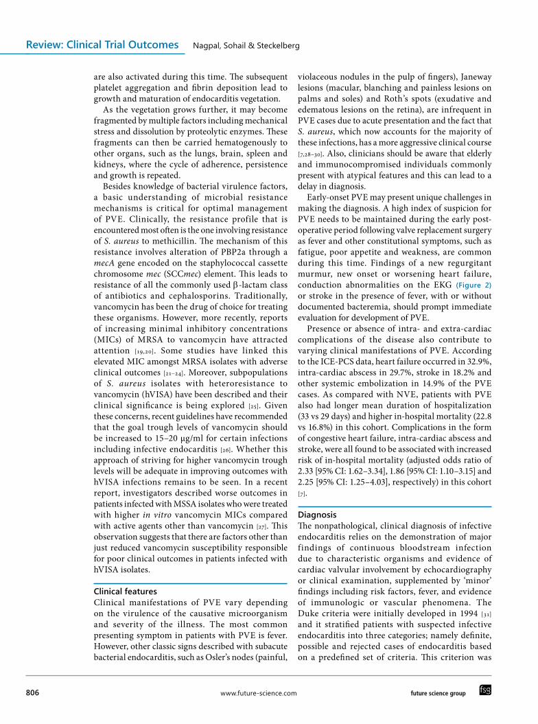
Early-onset PVE may present unique challenges in making the diagnosis. A high index of suspicion for PVE needs to be maintained during the early post-operative period following valve replacement surgery as fever and other constitutional symptoms, such as fatigue, poor appetite and weakness, are common during this time. Findings of a new regurgitant murmur, new onset or worsening heart failure, conduction abnormalities on the EKG (Figure 2) or stroke in the presence of fever, with or without documented bacteremia, should prompt immediate evaluation for development of PVE.
Presence or absence of intra- and extra-cardiac complications of the disease also contribute to varying clinical manifestations of PVE. According to the ICE-PCS data, heart failure occurred in 32.9%, intra-cardiac abscess in 29.7%, stroke in 18.2% and other systemic embolization in 14.9% of the PVE cases. As compared with NVE, patients with PVE also had longer mean duration of hospitalization (33 vs 29 days) and higher in-hospital mortality (22.8 vs 16.8%) in this cohort. Complications in the form of congestive heart failure, intra-cardiac abscess and stroke, were all found to be associated with increased risk of in-hospital mortality (adjusted odds ratio of 2.33 [95% CI: 1.62–3.34], 1.86 [95% CI: 1.10–3.15] and 2.25 [95% CI: 1.25–4.03], respectively) in this cohort [7].
DiagnosisThe nonpathological, clinical diagnosis of infective endocarditis relies on the demonstration of major findings of continuous bloodstream infection due to characteristic organisms and evidence of cardiac valvular involvement by echocardiography or clinical examination, supplemented by ‘minor’ findings including risk factors, fever, and evidence of immunologic or vascular phenomena. The Duke criteria were initially developed in 1994 [31] and it stratified patients with suspected infective endocarditis into three categories; namely definite, possible and rejected cases of endocarditis based on a predefined set of criteria. This criterion was

Prosthetic valve endocarditis: state of the heart Review: Clinical Trial Outcomes
future science group Clin. Invest. (2012) 2(8) 807
subsequently validated in a number of clinical studies [32–38].
With the availability of additional data, high proportion of cases that were categorized as ‘possible’ endocarditis, and development of a newer serological diagnostic techniques in the late 1990s, some modifications to the original Duke criteria were proposed in 2000 [39]. These revised criteria along with definitions are outlined in Boxes 1 & 2.
Despite the availability of this well-studied and published clinical criteria, diagnosis of infective endocarditis can be quite difficult in certain situations. Thus, it is important for clinicians to familiarize themselves with certain pearls and pitfalls associated with the diagnosis of PVE.
One of the most commonly encountered and frustrating clinical situations is administration of empiric antibiotics, without first obtaining blood cultures, when patients with prosthetic heart valves present to clinicians in outpatient or emergency room settings with fever. The importance of obtaining serial blood cultures, before initiating empiric antibiotic therapy, cannot be over-emphasized as identification of the causative organism is critical in choosing the appropriate antibiotic therapy for the patient. Even in situations where the patient is hemodynamically unstable and initiation of empirical antibiotics is of paramount importance, at least two sets of blood
cultures should still be drawn prior to the first dose of antibiotics.
Ideally, two different sets of blood cultures should be drawn 12 h apart. In more urgent situations, serial sets can be obtained 1 h apart. Multiple sets not only improve the yield of blood cultures, but also help to differentiate between contamination versus true bloodstream infection. Blood cultures are highly specific if separate sets of cultures are positive for the same organism. In cases where multiple blood culture were obtained prior to administration of any antibiotic therapy and are negative, serological tests for Coxiella burnetii, Bartonella, Brucella, Mycoplasma, Legionella and Chlamydia should be considered [40].
Whenever a patient has a high pretest probability of endocarditis (e.g., persistent bacteremia or fever of unknown origin in the presence of a prosthetic valve), an echocardiogram should be obtained. Transesophageal echocardiography (TEE) enables better visualization of the cardiac structures such as the atrial side of the mitral prosthesis, compared with trans-thoracic echocardiography (TTE) [41]. TEE is especially helpful in assessing the perivalvular extension of infection and detection of vegetations on prosthetic heart valves and cardiovascular implantable electronic devices leads. However, the advantages of TEE are not limited to patients with prosthetic heart valves and cardiac devices. Compared
Figure 2. EKG showing first-degree heart block due to paravalvular extension of infection.

www.future-science.com future science group808
Review: Clinical Trial Outcomes Nagpal, Sohail & Steckelberg
with TTE, TEE has better diagnostic yield for NVE (>95 vs 55%), prosthetic mitral valve endocarditis (82 vs 36%) [42], prosthetic valve abscesses (87 vs 28%) [43], and perivalvular regurgitation (95 vs 45%) [44]. Based on these data, TEE has become the imaging test of choice in the diagnosis of PVE.
In some instances, a TEE can be negative during the early stages of infection. Thus, if clinical suspicion of PVE persists, a repeat TEE should be performed in 7–10 days. This may allow visualization of previously undetected vegetations or abscesses that may have grown in size over time. TEE results may also be negative following embolization of previously present vegetation. Despite its relative superiority over TTE, TEE, in some cases, can still be negative and absence of vegetations should not be regarded as proof of absence of PVE.
In situations where pretest probability of endocarditis is low, and TEE is being performed to ‘rule out’ endocarditis, such as patients with short-lived nosocomial S. aureus bacteremia due to a vascular access device, it may be prudent to delay echocardiography until the bloodstream is rendered negative. This strategy may obviate unnecessary repeat echocardiography in situations where TEE is first obtained the very same day the positive blood culture is first reported and initial imaging is negative.
Other complimentary tests may help in the characterization of the disease by identifying associated complications and may help in deciding management interventions. For example, visualization of multiple focal lung nodules or infiltrates on a chest x-ray of a patient with tricuspid
or pulmonary PVE suggests the possibility of septic pulmonary emboli. Similarly, presence of a heart block on a EKG may indicate extension of the infective process to the adjacent myocardium and presence of ischemia or infarction on a EKG may indicate septic emboli to the coronary arteries [40]. Computed tomography (CT) or MRI of the head should be performed on a patient with underlying PVE and new neurological deficits as they may represent septic emboli to the brain. Similarly, in patients with underlying PVE, a new onset of left upper quadrant or flank pain or hematuria may indicate septic embolization to the spleen and kidneys, which can be investigated by performing a CT scan of the abdomen.
Newer imaging techniques, such as Gallium-67 citrate single photo emission computed tomography (SPECT) have been used to demonstrate the presence of peri-prosthetic abscesses in some case reports, but data regarding their routine use in diagnosis of local complications and cost–effectiveness are lackin [45–47].
Newer molecular diagnostic methods, developed in the last decade, appear to have some promising roles in the diagnosis of endocarditis. Most prominent among these is the broad range PCR assay utilizing the 16S ribosomal DNA gene for amplification followed by sequencing, which allows for identification of causative microorganisms without the aid of standard culturing techniques. This method has been found to be more sensitive than either the blood or the valve tissue culture for the diagnosis of endocarditis in cases where the infection is either caused by fastidious and slow growing or difficult to culture organisms, such as
Box 1. Classification of infective endocarditis according to the modified Duke criteria.
Definite infective endocarditis ■ Pathologic criteria- Microorganisms demonstrated by culture or histologic examination of a vegetation, a vegetation that has
embolized, or an intracardiac abscess specimen- Pathologic lesions, vegetation or intracardiac abscess confirmed by histologic examination showing active
endocarditis ■ Clinical criteria- Two major criteria- One major criterion and three minor criteria- Five minor criteria
Possible infective endocarditis ■ One major criterion and one minor criterion ■ Three minor criteria
Rejected ■ Firm alternate diagnosis explaining evidence of infective endocarditis ■ Resolution of infective endocarditis syndrome with antibiotic therapy for <4 days ■ No pathologic evidence of infective endocarditis at surgery or autopsy, with antibiotic therapy for <4 days ■ Does not meet criteria for possible infective endocarditis, as above
Adapted with permission from [39].

Prosthetic valve endocarditis: state of the heart Review: Clinical Trial Outcomes
future science group Clin. Invest. (2012) 2(8) 809
Tropheryma whipplei, Bartonella sp. and Coxiella sp., or where the growth of an organism on culture media is hampered by prior administration of antibiotics [48–
50]. However, there are certain limitations inherent to the PCR method and these include the possibility of contamination, inability to address viability of the detected organisms (bacterial DNA can persist in the valvular tissue even after completion of antimicrobial therapy), lack of standardization across different laboratories, unavailability of tissue in patients who do not undergo surgery and limited number of organisms available in the test library database. Most importantly, pure growth of the causative organism is still required for susceptibility ana lysis and the determination of MICs, unless resistance gene-specific PCR assays are used simultaneously to detect commonly occurring resistance patterns [50].
Fluorescence in situ hybridization is another molecular technique that can be used for the diagnosis of infective endocarditis. It involves the use of f luorescent-labeled probes to detect DNA and RNA. The most common target is the bacterial 16S ribosomal RNA. The probes can be applied to a fixed tissue or smear sample. The unique advantage of this technique is that it allows visualization and identification of microorganisms in the context of the surrounding environment and
thus can help distinguish between contaminants and true pathogens. Since it also has the potential to measure ribosomal content of bacteria in situ, it has the potential use in assessing treatment efficacy [50]. However, similarly to PCR assays, this technique also has certain limitations, including lack of standardization, limited library of probes and inability to study susceptibility and resistance profiles of etiological microorganisms.
Surgical treatmentThe optimal management of PVE is a subject of great debate as there have been no randomized-controlled trials comparing efficacy of medical treatment with the combined medical–surgical approach. Although there are multiple case series that describe outcomes with various approaches, the selection of patients for surgery is never standardized. Thus, the characteristics of patients managed with the medical–surgical approach are quite different from those managed with medical treatment alone. Therefore, practice guidelines have been published by a panel of experts convened by the American Heart Association and American College of Cardiology to aid clinicians in critical decision making regarding need for surgical intervention (Box 3) [51].
Box 2. Definition of terms used in the modified Duke criteria for the diagnosis of infective endocarditis.
Major criteria ■ Blood culture positive for IE- Typical microorganisms consistent with IE (Viridans streptococci, Streptococcus bovis, HACEK group, Staphylococcus aureus or
community-acquired enterococci; in the absence of a primary focus) from two separate blood cultures - Microorganisms consistent with IE from persistently positive blood cultures, defined as follows. At least two positive cultures of
blood samples drawn 12 h apart; all of three or a majority of >four separate cultures of blood (with first and last sample drawn at least 1 h apart); or single positive blood culture for Coxiella burnetii or anti-phase I IgG antibody titer >1: 800
■ Evidence of endocardial involvement- Echocardiogram positive for IE (TEE recommended in patients with prosthetic valves, rated at least ‘possible IE’ by
clinical criteria, or complicated IE [paravalvular abscess]; TTE as first test in other patients), defined as follows. Oscillating intracardiac mass on valve or supporting structures, in the path of regurgitant jets, or on implanted material in the absence of an alternative anatomic explanation; abscess; or new partial dehiscence of prosthetic valve
- New valvular regurgitation (worsening or changing of pre-existing murmur not sufficient)
Minor criteria ■ Predisposition, predisposing heart condition or injection drug use ■ Fever, temperature >38°C ■ Vascular phenomena: major arterial emboli, septic pulmonary infarcts, mycotic aneurysm, intracranial hemorrhage, conjunctival hemorrhages and Janeway’s lesions
■ Immunologic phenomena: glomerulonephritis, Osler’s nodes, Roth’s spots, and rheumatoid factor ■ Microbiological evidence: positive blood culture but does not meet a major criterion as noted above or serological evidence of active infection with organism consistent with IE
■ Echocardiographic minor criteria eliminatedBold text indicate the modifications. IE: Infective endocarditis; TEE: Transesophageal echocardiography; TTE: Trans-thoracic echocardiography. Adapted with permission from [39].

www.future-science.com future science group810
Review: Clinical Trial Outcomes Nagpal, Sohail & Steckelberg
In each individual case, the risks and benefits of surgery need to be carefully weighed. Surgical intervention is most beneficial when patients present with complications of PVE, such as worsening heart failure, prosthetic valve dehiscence, worsening regurgitation or perivalvular leak, valvular obstruction and cardiac abscess formation.
Surgery should also be considered in PVE cases with persistent bacteremia or relapse of infection after completion of an appropriate antibiotic course. The precise definition of persistent bacteremia is unclear as the duration of bacteremia varies depending on the causative organisms. While blood cultures usually turn negative within 48 h of initiating antibiotic therapy in cases of susceptible viridans group streptococci, they frequently remain positive for 7 days or more in cases of MRSA endocarditis despite appropriate vancomycin therapy [52].
Other relative indications of surgery include relapsing bacterial infection despite adequate therapy, fungal endocardit is , intracardiac abscess resulting in heart block, culture-negative endocarditis with recalcitrant fever despite 10 days of antibiotic therapy and recurrent emboli despite optimal antibiotic treatment. Similar to fungal endocarditis, S. aureus PVE is also considered to be a surgical disease by some surgeons [51,53].
The role of surgical intervention to prevent systemic embolization is not clearly defined. Vegetations greater than 10 mm at the aortic or mitral valve have a significantly higher association with embolization
as compared with the ones that measure less than 10 mm [54,55], especially during the first 2 weeks of therapy [56]. Thus, benefit of surgical intervention may be highest in this subset of patients in the earlier phase of their disease. Other echocardiographic features that may indicate the possible need for surgery are an increase in size of vegetation despite embolization and an increase in vegetation size despite appropriate antimicrobial therapy [57].
Surgery is not performed when the possibility of recovery is remote. This is usually seen in patients with high operative risks due to cardiopulmonary and neurological status, poor prognosis due to other severe comorbid conditions, a major cerebrovascular event with intracranial hemorrhage and history of multiple and technically difficult surgery with inoperability defined during a previous surgery [40,58].
Higher rates of operative intervention in patients with PVE require special attention to management of anticoagulation. Patients with PVE who are on warfarin should be switched to heparin, so that the anticoagulated state does not become a contraindication should the need for emergency surgery arise in these patients. Likewise, aspirin should also be discontinued in these patients [51]. Moreover, if neurological symptoms develop, anticoagulation should be discontinued until an intracranial hemorrhagic event has been excluded by appropriate imaging procedures, such as CT scanning.
Box 3. American Heart Association and American College of Cardiology practice guidelines to aid clinicians in critical decision making regarding need for surgical intervention.
Class I ■ Consultation with a cardiac surgeon is indicated for patients with infective endocarditis of a prosthetic valve (Level of Evidence C) ■ Surgery is indicated for patients with infective endocarditis of a prosthetic valve who present with heart failure (Level of Evidence B) ■ Surgery is indicated for patients with infective endocarditis of a prosthetic valve who present with dehiscence evidenced by cine fluoroscopy or echocardiography (Level of Evidence B)
■ Surgery is indicated for patients with infective endocarditis of a prosthetic valve who present with evidence of increasing obstruction or worsening regurgitation (Level of Evidence C)
■ Surgery is indicated for patients with infective endocarditis of a prosthetic valve who present with complications, such as, abscess formation (Level of Evidence C)
Class IIa ■ Surgery is reasonable for patients with infective endocarditis of a prosthetic valve who present with evidence of persistent bacteremia or recurrent emboli despite appropriate antibiotic treatment (Level of Evidence C)
■ Surgery is reasonable for patients with infective endocarditis of a prosthetic valve who present with relapsing infection (Level of Evidence C)
Class III ■ Routine surgery is not indicated for patients with uncomplicated infective endocarditis of a prosthetic valve caused by first infection with a sensitive organism (Level of Evidence C)
Level of Evidence A: Data derived from multiple randomized clinical trials or meta-analyses; Level of Evidence B: Data derived from a single randomized trial or nonrandomized studies; Level of Evidence C: Only consensus opinion of experts, case studies, or standard of care.Adapted with permission from [51].

Prosthetic valve endocarditis: state of the heart Review: Clinical Trial Outcomes
future science group Clin. Invest. (2012) 2(8) 811
Medical treatment ■ General principles
Antimicrobial therapy for PVE should be guided by the susceptibility profile of the causative organism. Blood cultures should be drawn prior to administration of any empiric antibiotics. Specific antimicrobial regimens depending on the causative microorganisms have been published by the American Heart Association and European Society of Cardiology and are readily available on their respective websites [3,57].
Initial antimicrobial therapy for PVE should be initiated in a hospital setting under close observation. If available, an infectious diseases expert should be consulted to guide antimicrobial management. The patient should be carefully monitored for any symptoms or signs suggestive of a worsening condition or development of intra- or extra-cardiac complications. Clinicians should also watch for any adverse effects from antimicrobial therapy that may necessitate use of supportive care or a change in treatment regimen.
In general, patients with PVE are treated with 6 weeks of antibiotic therapy, counting from the day of the first negative blood cultures. At least two sets of blood cultures should be obtained every 24–48 h until the bloodstream is cleared. The time to positivity may increase with ongoing antimicrobial therapy and thus premature conclusion of a negative blood culture should be avoided until the results are finalized, which is usually at 5 days for most microbiologic laboratories. When combination therapy is used, the drugs should be administered in close proximity to each other in order to maximize the synergistic effect.
If valve replacement surgery is performed during the course of antimicrobial treatment, valve and other infected tissues obtained during surgical procedure should be submitted for Gram stain and culture. If the tissue culture reveals bacterial growth despite negative blood cultures, antimicrobial therapy should be continued for 6 additional weeks, beginning from the day of surgery. However, positive-Gram stain alone (negative culture) may simply represent nonviable organisms and does not warrant restarting the entire treatment course.
Below we discuss some key issues regarding the antibiotic selection, including the need for combination therapy, and the duration of treatment for the most common pathogens responsible for PVE.
■ Staphylococcal PVEApproximately 90–95% of clinical S. aureus isolates are resistant to penicillin [44]. Additionally, an increasing number of staphylococcal strains are resistant to methicillin (31% of S. aureus and 68% of
CoNS in the ICE-PCS database) [59]. For all practical purposes, the antibiotic choices for both S. aureus and CoNS PVE are similar. The recommended treatment for methicillin-susceptible staphylococcal strains in patients with normal renal function is nafcillin (or oxacillin) 12 g/24 h iv. in six equally divided doses with rifampin 900 mg/24 h iv./orally in three equally divided doses for 6 weeks. In general, gentamicin 3 mg/kg/24 h iv./intramuscularly (im.), in equally divided doses is also recommended for the first 2 weeks of therapy. For methicillin-resistant strains, vancomycin in a dose of 30 mg/kg/24 h iv., in two equally divided doses, adjusted for renal function, should be used instead of nafcillin (or oxacillin).
The recommendation for the use of rifampin is based on experimental models of endocarditis in animals where combination antimicrobials with rifampin were shown to sterilize biofilm-associated foreign bodies infected by S. aureus [60]. This effect is partly attributable to better biofilm penetration by rifampin and its ability to interfere in replication of slowly growing bacteria where cell wall agents are not very effective. However, rifampin should never be used as a monotherapy because of a low barrier to emergence of resistance. Some experts believe that it may be prudent to wait and add rifampin only once the bloodstream has cleared.
If a patient has had a prior non-anaphylactic reaction to penicillin, a first generation cephalosporin, such as cefazolin can be substituted for nafcillin. For strains of staphylococci resistant to gentamicin, susceptibility testing should be performed for another aminoglycoside such as streptomycin. If the strain is resistant to all aminoglycosides, then either the treatment with aminoglycoside should be omitted or consideration may be given to using fluoroquinolones instead.
S. aureus strains that are fully resistant to vanco-mycin are very rare and a subject of case reports. However, there are increasing reports of creeping MICs in clinical S. aureus isolates that are associated with poor response to treatment as discussed above. In such cases, where clinical failure of vancomycin is apparent, and other scenarios where vancomycin cannot be used due to associated renal toxicity, few other options are available, but data are extremely limited. Recent reports have described successful treatment with daptomycin [61,62], which is approved by the US FDA for S. aureus bacteremia and right-sided endocarditis and is noninferior to vancomycin for left-sided endocarditis. Linezolid is another alternative agent for which limited data are available [63,64]. In general, bacteriostatic agents, such as linezolid, are best avoided in PVE if bactericidal

www.future-science.com future science group812
Review: Clinical Trial Outcomes Nagpal, Sohail & Steckelberg
agents are available. Moreover, PVE usually requires at least 6 weeks of antimicrobial therapy and such prolonged administration of linezolid carries the risk of bone marrow suppression, irreversible optic or peripheral neuropathy and lactic acidosis.
■ Streptococcal PVEIn general, uncomplicated PVE due to streptococci, most often caused by viridans group streptococci or Staphylococcus bovis, can be managed with medical therapy alone. Antibiotic choices for Streptococcal PVE vary based on penicillin MIC data. Depending on their susceptibility to penicillin, viridans group streptococci (Staphylococcus sanguis, Staphylococcus oralis, Staphylococcus salivarius, Staphylococcus mutans and Staphylococcus anginosus) and S. bovis are divided into three different categories; namely highly penicillin susceptible (MIC ≤0.12 µg/ml), relatively resistant to penicillin (MIC >0.12–≤0.5 µg/ml) and highly resistant to penicillin (MIC >0.5 µg/ml). Patients with PVE due to highly susceptible penicillin strains should be treated with 6 weeks of iv. penicillin 12–18 million units/24 h in four to six equally divided doses or as a continuous infusion. Alternatively, ceftriaxone 2 g iv./im. every 24 h can be used for the same 6 weeks duration with comparable outcomes.
Strains that are relatively or highly resistant to penicillin should be treated with 6 weeks of combination antimicrobial therapy consisting of iv. penicillin 24 million units/24 h, in four to six equally divided doses or ceftriaxone 2 g iv./im. every 24 h with gentamicin 3 mg/kg/24 h iv./im., in three equally divided doses.
PVE caused by nutritionally variant streptococci, such as Abiotrophia defective, Granulicatella sp. and Gemella sp., is rare and similarly to enterococcal PVE, is associated with a higher rate of complications and is difficult to cure. Moreover, susceptibility data can be difficult to interpret and even when available, may not correlate with clinical outcomes. Thus, infections with these organisms should be treated with combination antimicrobial therapy with penicillin or ceftriaxone along with gentamicin for 6 weeks, similar to PVE caused by enterococcal species.
Of note, bacteremia with S. bovis has been associated with gastrointestinal pathology, including malignancy [65–68]. Therefore patients with S. bovis PVE should undergo colonoscopy to look for the presence of an underlying occult gastrointestinal malignancy.
■ Enterococcal PVEE. faecalis followed by Enterococcus faecium are the
major enterococcal species of clinical importance in PVE. Enterococci are relatively resistant to the bactericidal action of b-lactam agents as they express penicillin-binding proteins with lower affinity to these drugs. Therefore, synergistic actions of aminoglycoside are required for treatment of enterococcal PVE. E. faecium in particular is frequently resistant to penicillin due to increased expression of low affinity PBP5 [69]. Penicillin resistance is somewhat less common in E. faecalis.
Besides penicillin resistance, high-level resistance to vancomycin is an emerging problem in enterococci. Six different phenotypes of vancomycin resistance are recognized [70]. Of these, VanA and VanB phenotypes can be harbored on plasmids and thus are the most clinically relevant. VanA phenotype confers high-grade resistance to vancomycin and teicoplanin. VanB phenotype confers variable resistance to vancomycin but retains susceptibility to teicoplanin. In addition, enterococci can acquire high-level aminoglycoside resistance through plasmid-mediated expression of aminoglycoside-modifying enzymes. This diminishes the affinity of aminoglycosides to their target sites. Another mechanism of resistance is through ArmA-mediated 16S ribosomal methylation, which blocks the target of aminoglycoside.
Some E. faecium strains can be resistant to both b-lactam agents and aminoglycosides. Others, such as Enterococcus gallinarum and Enterococcus casseliflavus, constitutively express VanC phenotype and thus are intrinsically resistant to vancomycin.
Antimicrobial therapy for enterococcal PVE is best guided by susceptibility testing. Penicillin susceptible strains should be treated with 6 weeks of combination therapy with iv. penicillin and gentamicin. If the strain is penicillin resistant, vancomycin can be used along with gentamicin. Rarely, penicillin resistance in Enterococcus spp. is caused by production of b-lactamase and in those cases ampicillin–sulbactam can be used along with gentamicin.
For gentamicin-resistant strains, susceptibilities should be obtained for streptomycin and if found to be susceptible, streptomycin should be used. A double b-lactam combination of ceftriaxone and ampicillin has been used for E. faecalis strains with high-level aminoglycoside resistance with reported microbiological and clinical cure [71]. This double b-lactam combination may also be useful in patients who have other contraindications to aminoglycoside use or have worsening renal function on therapy, but experience is limited.
Enterococci resistant to penicillin, aminoglycosides and vancomycin present a particularly difficult challenge. Case reports of successful treatment with

Prosthetic valve endocarditis: state of the heart Review: Clinical Trial Outcomes
future science group Clin. Invest. (2012) 2(8) 813
linezolid and quinupristin–dalfopristin have been published [63,72–74]. However, due to publication bias, reports of unsuccessful treatment are generally not available.
■ HACEK PVEHaemophilus parainf luenzae, Aggregatibacter ac t inomycetemcomitan s , Cardiobacter ium hominis, Eikenella corrodens and Kingella kingae constitute a group of slow-growing Gram-negative microorganisms that are part of normal oral flora. Collectively, these are known as HACEK organisms. Antimicrobial susceptibility can be difficult to perform in these slow-growing organisms and treatment is usually initiated while awaiting susceptibility data. Due to frequent occurrence of b-lactamase-producing strains, ampicillin resistance should be assumed and treatment with ceftriaxone, ampicillin–sulbactam or fluoroquinolones should be initiated. Recommended duration of treatment is 6 weeks.
■ Non-HACEK Gram-negative bacilli PVEInfections due to these organisms are rare, accounting for just over 2% of PVE cases in the ICE-PCS database [7]. Among these, Escherichia coli is the most frequently reported organism followed by Pseudomonas and Serratia. Infection usually occurs in the early phase following valve surgery and is frequently healthcare associated. Rate of complications and mortality is high with Gram-negative bacilli PVE. Susceptibility-guided antimicrobial therapy with third- or fourth-generation cephalosporins, extended spectrum penicillins, fluoroquinolones or carbapenems with or without aminoglycosides, and frequently surgical intervention, are the cornerstone of treatment.
■ Fungal PVEFungal PVE is extremely rare. Candida species and Histoplasma capsulatum are the most commonly reported fungal organisms responsible for PVE in the USA. Candida seeding of prosthetic heart valves is frequently a consequence of healthcare-associated bloodstream infection in patients with vascular access devices or surgery involving the gastrointestinal tract. Fungal PVE is usually characterized by large, dense and heterogeneous vegetations on the heart valves and is associated with a high rate of complications [75,76]. Outcomes are often poor even with the combined medical–surgical approach and relapse of infection is frequent.
For candida PVE, the recommended treatment approach consists of an initial parenteral induction with liposomal amphotericin B 5 mg/kg iv. once-daily
and 5-flucytosine 25–37.5 mg/kg every 6 h for 6–8 weeks followed by chronic long-term oral suppressive therapy with an azole (fluconazole 400 mg orally daily if susceptible). Echinocandins, such as caspofungin, micafungin or anidulafungin, can be used for Candida species if patients are unable to tolerate liposomal preparation of amphotericin B, which although better tolerated than amphotericin B, is still commonly associated with nephrotoxicity and electrolyte wasting, specifically potassium depletion. Echinocandins, however, are not active against dimorphic fungi such as histoplasma.
Fungal endocarditis should always be managed in close collaboration with an infectious diseases expert and an experienced cardiac surgeon.
■ Adverse effect monitoringTreatment of PVE involves long courses of parenterally administered antimicrobial therapy and thus close follow-up of patients is essential for successful outcomes. Patients should ideally be enrolled in an outpatient parenteral antibiotic treatment program to ensure a safe and effective treatment course. Infectious Diseases Society of America has published practice guidelines for outpatient parenteral antibiotic treatment [77], which are easily accessible at [101]. Clinicians should familiarize themselves with common side effects of antibiotics that are prescribed in the outpatient setting so as to monitor and detect any adverse effects and take corrective action when necessary.
OutcomesIn the ICE-PCS database, increasing age, healthcare-associated PVE, S. aureus PVE, complications in the form of heart failure, persistent bacteremia and stroke, were all associated with a higher in-hospital mortality rate of 30.5%. The mortality rate during the index hospitalization was 22.8% and approximately half the patients required surgical management [7]. In-hospital mortality rates following surgery for PVE have been reported to be 13–48% [30]. As compared with NVE, surgical intervention for PVE has been found to have higher rates of 30-day mortality (13 vs 5.6%) but long-term survival does not appear to be significantly different [78]. Also, redo aortic valve surgery for PVE has been shown to be associated with significantly higher in-hospital mortality as well as higher 1-, 3-, 5- and 10-year mortality as compared with redo aortic valve surgery for non-endocarditic causes [79]. In this group of patients, 5-year actuarial freedom from endocarditis was only 80% in patients who underwent redo surgery for PVE as compared with 95% in patients who underwent redo surgery for

www.future-science.com future science group814
Review: Clinical Trial Outcomes Nagpal, Sohail & Steckelberg
non-endocarditic causes. Furthermore, surgery for active PVE, defined as having been performed prior to completion of a standard course of antibiotics, has been shown to be associated with an increased risk of in-hospital mortality (overall response: 4.16; 95% CI: 1.14–12.2) in multivariate ana lysis of a single-center cohort of 141 patients operated upon by the same surgical team [80]. Mechanical respiratory and cardiac support, preoperation higher doses of catecholamines, emergent surgery, mitral valve replacement and age at operation, were found to be predictors of early mortality in another recent retrospective ana lysis [81].
Many of these studies are limited by a number of factors including being single-center retrospective observational studies. Additionally, lack of standardization in patient selection and procedure makes the results difficult to interpret. However, there appears to be a trend towards mortality benefit from surgery among patients who develop complications of PVE [30,82].
PreventionAmerican Heart Association guidelines for prevention of infective endocarditis have undergone numerous iterations over the last few years. The latest update to these guidelines was released in 2007
[83]. These revisions identify subgroups of patients who might be at a highest risk of complications if they were to develop infective endocarditis. People with prosthetic valves are recognized as one of these subgroups and thus perioperative prophylaxis with either amoxicillin 2 g orally or ampicillin 2 g iv./im. are considered reasonable for procedures that carry the highest risks of transient bacteremias, such as all dental procedures that involve manipulation of gingival tissue or periapical region of teeth or perforation of oral mucosa. Similarly, prophylaxis is also reasonable for these patients who are undergoing procedures involving incision or biopsy of respiratory mucosa, genitourinary and gastrointestinal procedures in the presence of an established infection and procedures on infected skin and skin structures.
Clindamycin, azithromycin, cephalexin, cefazolin or vancomycin, may be used in patients who are known to be allergic to penicillins. Of these, only vancomycin is active against Enterococcus sp., which is frequently a cause of gastrointestinal and genitourinary infections.
Challenges & future perspectiveAlthough significant progress has been made in
Executive summary
Epidemiology ■ Prosthetic valve endocarditis (PVE) accounts for approximately a fifth of all cases of infective endocarditis. ■ PVE is associated with high rates of morbidity and mortality (22.8%). ■ PVE can be early-onset (within 60 days of surgery), intermediate-onset (60–365 days) and late-onset (after 1 year of valve surgery).
Microbiology ■ Staphylococcus aureus is now the most common cause of PVE. ■ Healthcare-associated infections account for most cases of PVE.
Pathogenesis ■ Most cases of PVE occur within the first year after surgery, likely due to incomplete endothelialization of the mechanical valve during this timeframe.
Diagnosis ■ Diagnosis of PVE is guided by the modified Duke’s criteria. ■ Transesophageal echocardiography is the preferred imaging modality for PVE.
Treatment ■ Treatment often involves a combined medical and surgical approach. ■ Surgery is most beneficial for patients who present with acute heart failure symptoms, paravalvular extension of infection, valve dehiscence, worsening stenosis or regurgitation, recurrent emboli or persistent bloodstream infection despite appropriate therapy, and relapse of infection after an adequate treatment course.
■ More invasive organisms, such as Staphylococcus aureus, non-HACEK Gram-negative bacilli and fungi, are associated with a more severe presentation and frequently require surgical management.
■ Antibiotic therapy is usually given for 6 weeks or longer. ■ Organism-specific antibiotic treatment guidelines have been published by the American Heart Association and European Society of Cardiology and should be followed in most cases of PVE.
■ Infection due to resistant organisms, especially multidrug-resistant pathogens, poses an immediate and challenging problem in the management of these complicated infections.

Prosthetic valve endocarditis: state of the heart Review: Clinical Trial Outcomes
future science group Clin. Invest. (2012) 2(8) 815
our understanding and management of PVE, many unanswered questions remain. High-quality evidence in the form of randomized-controlled trials is lacking due to the rare occurrence of these infections. Nevertheless, the most important questions that remain unanswered involve decisions regarding the timing of surgical intervention and optimal duration of antimicrobial therapy. Molecular methods described above that are currently available need to be refined further and their role in the diagnosis of PVE needs to be clearly defined. Finally, we should continue to implement strict infection-control practices specifically pertaining to prevention of surgical-site infections and healthcare-associated bloodstream infections, in order to reduce the burden of early and intermediate cases of PVE.
Financial & competing interests disclosureThe authors have no relevant affiliations or finan-cial involvement with any organization or entity with a financial interest in or financial conflict with the subject matter or materials discussed in the manuscript. This includes employment, con-sultancies, honoraria, stock ownership or options, expert t estimony, grants or patents received or pending, or royalties. No writing assistance was utilized in the production of this manuscript.
ReferencesPapers of special note have been highlighted as:n of interestnn of considerable interest
1 Calderwood SB, Swinski LA, Waternaux CM, Karchmer AW, Buckley MJ. Risk factors for the development of prosthetic valve endocarditis. Circulation 72(1), 31–37 (1985).
2 Agnihotri AK, McGiffin DC, Galbraith AJ, O’Brien MF. The prevalence of infective endocarditis after aortic valve replacement. J. Thorac. Cardiovasc. Surg. 110(6), 1708–1720; discussion 1720–1704 (1995).
3 Habib G, Hoen B, Tornos P et al. Guidelines on the prevention, diagnosis, and treatment of infective endocarditis (new version 2009): the Task Force on the Prevention, Diagnosis, and Treatment of Infective Endocarditis of the European Society of Cardiology (ESC). Endorsed by the European Society of Clinical
Microbiology and Infectious Diseases (ESCMID) and the International Society of Chemotherapy (ISC) for Infection and Cancer. Eur. Heart J. 30(19), 2369–2413 (2009).
nn European Society of Cardiology’s guidelines for prevention, diagnosis and treatment of infective endocarditis.
4 Vongpatanasin W, Hillis LD, Lange RA. Prosthetic heart valves. N. Engl. J. Med. 335(6), 407–416 (1996).
5 Haydock D, Barratt-Boyes B, Macedo T, Kirklin JW, Blackstone E. Aortic valve replacement for active infectious endocarditis in 108 patients. A comparison of freehand allograft valves with mechanical prostheses and bioprostheses. J. Thorac. Cardiovasc. Surg. 103(1), 130–139 (1992).
6 Steckelberg JM, Melton LJ 3rd, Ilstrup DM, Rouse MS, Wilson WR. Influence of referral bias on the apparent clinical spectrum of infective endocarditis. Am. J. Med. 88(6), 582–588 (1990).
7 Wang A, Athan E, Pappas PA et al. Contemporary clinical profile and outcome of prosthetic valve endocarditis. JAMA 297(12), 1354–1361 (2007).
nn Large international multicenter prospective study describing epidemiology, clinical features and outcomes of prosthetic valve endocarditis.
8 Selton-Suty C, Celard M, Le Moing V et al. Preeminence of Staphylococcus aureus in infective endocarditis: a 1-year population-based survey. Clin. Infect. Dis. 54(9), 1230–1239 (2012).
9 Murdoch DR, Corey GR, Hoen B et al. Clinical presentation, etiology, and outcome of infective endocarditis in the 21st century: the International Collaboration on Endocarditis-Prospective Cohort Study. Arch. Intern. Med. 169(5), 463–473 (2009).
nn Most recent and comprehensive data from a large cohort of patients who were diagnosed and treated for infective endocarditis at a large number of participating institutions worldwide.
10 Slaughter L, Morris JE, Starr A. Prosthetic valvular endocarditis. A 12-year review. Circulation 47(6), 1319–1326 (1973).
11 Wilson WR, Jaumin PM, Danielson GK, Giuliani ER, Washington JA II, Geraci JE. Prosthetic valve endocarditis. Ann. Intern. Med. 82(6), 751–756 (1975).
12 Arvay A, Lengyel M. Incidence and risk factors of prosthetic valve endocarditis. Eur. J. Cardiothorac. Surg. 2(5), 340–346
(1988).
13 Grover FL, Cohen DJ, Oprian C, Henderson WG, Sethi G, Hammermeister KE. Determinants of the occurrence of and survival from prosthetic valve endocarditis. Experience of the veterans affairs cooperative study on valvular heart disease. J. Thoracic Cardiovasc. Surg. 108(2), 207–214 (1994).
14 Lee JH, Burner KD, Fealey ME et al. Prosthetic valve endocarditis: clinicopathological correlates in 122 surgical specimens from 116 patients (1985–2004). Cardiovasc. Pathol. 20(1), 26–35 (2011).
15 San Martin J, Sarria C, de las Cuevas C, Duarte J, Gamallo C. Relevance of clinical presentation and period of diagnosis in prosthetic valve endocarditis. J. Heart Valve Dis. 19(1), 131–138 (2010).
16 Sidhu P, O’Kane H, Ali N et al. Mechanical or bioprosthetic valves in the elderly: a 20-year comparison. Ann. Thorac. Surg. 71(Suppl. 5), S257–S260 (2001).
17 Fervenza FC, Contreras GE, Garratt KN, Steckelberg JM. Staphylococcus lugdunensis endocarditis: a complication of vasectomy? Mayo Clin. Proc. 74(12), 1227–1230 (1999).
18 Moreillon P, Que YA, Bayer AS. Pathogenesis of streptococcal and staphylococcal endocarditis. Infect. Dis. Clin. North Am. 16(2), 297–318 (2002).
19 Jones RN. Microbiological features of vancomycin in the 21st century: minimum inhibitory concentration creep, bactericidal/static activity, and applied breakpoints to predict clinical outcomes or detect resistant strains. Clin. Infect. Dis. 42(Suppl. 1), S13–S24 (2006).
20 Wang G, Hindler JF, Ward KW, Bruckner DA. Increased vancomycin MICs for Staphylococcus aureus clinical isolates from a university hospital during a 5-year period. J. Clin. Microbiol. 44(11), 3883–3886 (2006).
21 Hidayat LK, Hsu DI, Quist R, Shriner KA, Wong-Beringer A. High-dose vancomycin therapy for methicillin-resistant Staphylococcus aureus infections: efficacy and toxicity. Arch. Intern. Med. 166(19), 2138–2144 (2006).
22 Lodise TP, Graves J, Evans A et al. Relationship between vancomycin MIC and failure among patients with methicillin-resistant Staphylococcus aureus bacteremia treated with vancomycin. Antimicrob. Agents Chemother. 52(9), 3315–3320 (2008).
23 Sakoulas G, Moise-Broder PA, Schentag J, Forrest A, Moellering RC Jr, Eliopoulos GM.

www.future-science.com future science group816
Review: Clinical Trial Outcomes Nagpal, Sohail & Steckelberg
Relationship of MIC and bactericidal activity to efficacy of vancomycin for treatment of methicillin-resistant Staphylococcus aureus bacteremia. J. Clin. Microbiol. 42(6), 2398–2402 (2004).
24 Soriano A, Marco F, Martinez JA et al. Influence of vancomycin minimum inhibitory concentration on the treatment of methicillin-resistant Staphylococcus aureus bacteremia. Clin. Infect. Dis. 46(2), 193–200 (2008).
25 Liu C, Chambers HF. Staphylococcus aureus with heterogeneous resistance to vancomycin: epidemiology, clinical significance, and critical assessment of diagnostic methods. Antimicrob. Agents Chemother. 47(10), 3040–3045 (2003).
26 Rybak M, Lomaestro B, Rotschafer JC et al. Therapeutic monitoring of vancomycin in adult patients: a consensus review of the American Society of Health-System Pharmacists, the Infectious Diseases Society of America, and the Society of Infectious Diseases Pharmacists. Am. J. Health Syst. Pharm. 66(1), 82–98 (2009).
27 Holmes NE, Turnidge JD, Munckhof WJ et al. Antibiotic choice may not explain poorer outcomes in patients with Staphylococcus aureus bacteremia and high vancomycin minimum inhibitory concentrations. J. Infect. Dis. 204(3), 340–347 (2011).
28 Chu VH, Cabell CH, Benjamin DK Jr et al. Early predictors of in-hospital death in infective endocarditis. Circulation 109(14), 1745–1749 (2004).
29 Kuyvenhoven JP, van Rijk-Zwikker GL, Hermans J, Thompson J, Huysmans HA. Prosthetic valve endocarditis: analysis of risk factors for mortality. Eur. J. Cardiothorac. Surg. 8(8), 420–424 (1994).
30 Wang A, Pappas P, Anstrom KJ et al. The use and effect of surgical therapy for prosthetic valve infective endocarditis: a propensity analysis of a multicenter, international cohort. Am. Heart J. 150(5), 1086–1091 (2005).
31 Durack DT, Lukes AS, Bright DK. New criteria for diagnosis of infective endocarditis: utilization of specific echocardiographic findings. Duke Endocarditis Service. Am. J. Med. 96(3), 200–209 (1994).
n Describes the original Duke’s Criteria, which has since been validated in a number of studies. Publication of this article marked a significant change in the approach to diagnosis of infective
endocarditis.
32 Bayer AS. Revised diagnostic criteria for infective endocarditis. Cardiol. Clin. 14(3), 345–350 (1996).
33 Bayer AS, Ward JI, Ginzton LE, Shapiro SM. Evaluation of new clinical criteria for the diagnosis of infective endocarditis. Am. J. Med. 96(3), 211–219 (1994).
34 Cecchi E, Parrini I, Chinaglia A et al. New diagnostic criteria for infective endocarditis. A study of sensitivity and specificity. Eur. Heart J. 18(7), 1149–1156 (1997).
35 Dodds GA, Sexton DJ, Durack DT, Bashore TM, Corey GR, Kisslo J. Negative predictive value of the Duke criteria for infective endocarditis. Am. J. Cardiol. 77(5), 403–407 (1996).
36 Hoen B, Beguinot I, Rabaud C et al. The Duke criteria for diagnosing infective endocarditis are specific: analysis of 100 patients with acute fever or fever of unknown origin. Clin. Infect. Dis. 23(2), 298–302 (1996).
37 Hoen B, Selton-Suty C, Danchin N et al. Evaluation of the Duke criteria versus the Beth Israel criteria for the diagnosis of infective endocarditis. Clin. Infect. Dis. 21(4), 905–909 (1995).
38 Sekeres MA, Abrutyn E, Berlin JA et al. An assessment of the usefulness of the Duke criteria for diagnosing active infective endocarditis. Clin. Infect. Dis. 24(6), 1185–1190 (1997).
39 Li JS, Sexton DJ, Mick N et al. Proposed modifications to the Duke criteria for the diagnosis of infective endocarditis. Clin. Infect. Dis. 30(4), 633–638 (2000).
nn Modified Duke criteria for diagnosis of infective endocarditis. This is now the gold standard diagnostic tool used by clinicians worldwide to diagnose infective endocarditis.
40 Paterick TE, Paterick TJ, Nishimura RA, Steckelberg JM. Complexity and subtlety of infective endocarditis. Mayo Clin. Proc. 82(5), 615–621 (2007).
41 Jacob S, Tong AT. Role of echocardiography in the diagnosis and management of infective endocarditis. Curr. Opin. Cardiol. 17(5), 478–485 (2002).
42 Daniel WG, Mugge A, Grote J et al. Comparison of transthoracic and transesophageal echocardiography for detection of abnormalities of prosthetic and bioprosthetic valves in the mitral and aortic positions. Am. J. Cardiol. 71(2), 210–215 (1993).
43 Daniel WG, Mugge A, Martin RP et al. Improvement in the diagnosis of abscesses associated with endocarditis by transesophageal echocardiography. N. Engl. J. Med. 324(12), 795–800 (1991).
44 De Castro S, Cartoni D, d’Amati G et al. Diagnostic accuracy of transthoracic and multiplane transesophageal echocardiography for valvular perforation in acute infective endocarditis: correlation with anatomic findings. Clin. Infect. Dis. 30(5), 825–826 (2000).
45 McWilliams ET, Yavari A, Raman V. Aortic root abscess: multimodality imaging with computed tomography and gallium-67 citrate single-photon emission computed tomography/computed tomography hybrid imaging. J. Cardiovasc. Comput. Tomogr. 5(2), 122–124 (2011).
46 Thomson LE, Goodman MP, Naqvi TZ et al. Aortic root infection in a prosthetic valve demonstrated by gallium-67 citrate SPECT. Clin. Nucl. Med. 30(4), 265–268 (2005).
47 Yavari A, Ayoub T, Livieratos L, Raman V, McWilliams ET. Diagnosis of prosthetic aortic valve endocarditis with gallium-67 citrate single-photon emission computed tomography/computed tomography hybrid imaging using software registration. Circ. Cardiovasc. Imaging 2(6), e41–e43 (2009).
48 Breitkopf C, Hammel D, Scheld HH, Peters G, Becker K. Impact of a molecular approach to improve the microbiological diagnosis of infective heart valve endocarditis. Circulation 111(11), 1415–1421 (2005).
49 Casalta JP, Gouriet F, Roux V, Thuny F, Habib G, Raoult D. Evaluation of the LightCycler SeptiFast test in the rapid etiologic diagnostic of infectious endocarditis. Eur. J. Clin. Microbiol. Infect. Dis. 28(6), 569–573 (2009).
50 Moter A, Musci M, Schmiedel D. Molecular methods for diagnosis of infective endocarditis. Curr. Infect. Dis. Rep. 12(4), 244–252 (2010).
51 Bonow RO, Carabello BA, Chatterjee K et al. 2008 focused update incorporated into the ACC/AHA 2006 guidelines for the management of patients with valvular heart disease: a report of the American College of Cardiology/American Heart Association Task Force on Practice Guidelines (Writing Committee to revise the 1998 guidelines for the management of patients with valvular heart disease). Endorsed by the Society of Cardiovascular Anesthesiologists, Society for Cardiovascular Angiography and Interventions, and Society of Thoracic

Prosthetic valve endocarditis: state of the heart Review: Clinical Trial Outcomes
future science group Clin. Invest. (2012) 2(8) 817
Surgeons. J. Am. Coll. Cardiol. 52(13), e1–e142 (2008).
n These guidelines also include recommendations on surgical management of infective endocarditis.
52 Levine DP, Fromm BS, Reddy BR. Slow response to vancomycin or vancomycin plus rifampin in methicillin-resistant Staphylococcus aureus endocarditis. Ann. Int. Med. 115(9), 674–680 (1991).
53 John MD, Hibberd PL, Karchmer AW, Sleeper LA, Calderwood SB. Staphylococcus aureus prosthetic valve endocarditis: optimal management and risk factors for death. Clin. Infect. Dis. 26(6), 1302–1309 (1998).
54 Mugge A, Daniel WG, Frank G, Lichtlen PR. Echocardiography in infective endocarditis: reassessment of prognostic implications of vegetation size determined by the transthoracic and the transesophageal approach. J. Am. Coll. Cardiol. 14(3), 631–638 (1989).
55 Rohmann S, Erbel R, Gorge G et al. Clinical relevance of vegetation localization by transoesophageal echocardiography in infective endocarditis. Eur. Heart J. 13(4), 446–452 (1992).
56 Steckelberg JM, Murphy JG, Ballard D et al. Emboli in infective endocarditis: the prognostic value of echocardiography. Ann. Int. Med. 114(8), 635–640 (1991).
57 Baddour LM, Wilson WR, Bayer AS et al. Infective endocarditis: diagnosis, antimicrobial therapy, and management of complications: a statement for healthcare professionals from the Committee on Rheumatic Fever, Endocarditis, and Kawasaki Disease, Council on Cardiovascular Disease in the Young, and the Councils on Clinical Cardiology, Stroke, and Cardiovascular Surgery and Anesthesia, American Heart Association: endorsed by the Infectious Diseases Society of America. Circulation 111(23), E394–E434 (2005).
nn American Heart Association and Infectious Diseases Society of America Guidelines for diagnosis and management of infective endocarditis.
58 Knoll BM, Baddour LM, Wilson WR. Prosthetic valve endocarditis. In: Priniciples and Practice of Infectious Diseases. Mandell GL, Bennett JE, Dolin R (Eds). Churchill Livingstone Elsevier, PA, USA (2010).
59 Chu VH, Miro JM, Hoen B et al. Coagulase-negative staphylococcal prosthetic valve endocarditis – a contemporary update based on the International Collaboration on Endocarditis: prospective cohort study. Heart 95(7), 570–576 (2009).
60 Chuard C, Herrmann M, Vaudaux P, Waldvogel FA, Lew DP. Successful therapy
of experimental chronic foreign-body infection due to methicillin-resistant Staphylococcus aureus by antimicrobial combinations. Antimicrob. Agents Chemother. 35(12), 2611–2616 (1991).
61 Fowler VG Jr, Boucher HW, Corey GR et al. Daptomycin versus standard therapy for bacteremia and endocarditis caused by Staphylococcus aureus. N. Engl. J. Med. 355(7), 653–665 (2006).
62 Moore CL, Osaki-Kiyan P, Haque NZ, Perri MB, Donabedian S, Zervos MJ. Daptomycin versus vancomycin for bloodstream infections due to methicillin-resistant Staphylococcus aureus with a high vancomycin minimum inhibitory concentration: a case–control study. Clin. Infect. Dis. 54(1), 51–58 (2012).
63 Falagas ME, Manta KG, Ntziora F, Vardakas KZ. Linezolid for the treatment of patients with endocarditis: a systematic review of the published evidence. J. Antimicrob. Chemother. 58(2), 273–280 (2006).
64 Tascini C, Bongiorni MG, Doria R et al. Linezolid for endocarditis: a case series of 14 patients. J. Antimicrob. Chemother. 66(3), 679–682 (2011).
65 Murray HW, Roberts RB. Streptococcus bovis bacteremia and underlying gastrointestinal disease. Arch. Intern. Med. 138(7), 1097–1099 (1978).
66 Ballet M, Gevigney G, Gare JP, Delahaye F, Etienne J, Delahaye JP. Infective endocarditis due to Streptococcus bovis. A report of 53 cases. Eur. Heart J. 16(12), 1975–1980 (1995).
67 Leport C, Bure A, Leport J, Vilde JL. Incidence of colonic lesions in Streptococcus bovis and Enterococcal endocarditis. Lancet 1(8535), 748 (1987).
68 Klein RS, Catalano MT, Edberg SC, Casey JI, Steigbigel NH. Streptococcus bovis septicemia and carcinoma of the colon. Ann. Int. Med. 91(4), 560–562 (1979).
69 Rybkine T, Mainardi JL, Sougakoff W, Collatz E, Gutmann L. Penicillin-binding protein 5 sequence alterations in clinical isolates of Enterococcus faecium with different levels of beta-lactam resistance. J. Infect. Dis. 178(1), 159–163 (1998).
70 Courvalin P. Vancomycin resistance in Gram-positive cocci. Clin. Infect. Dis. 42(Suppl. 1), S25–S34 (2006).
71 Gavalda J, Len O, Miro JM et al. Brief communication: treatment of Enterococcus faecalis endocarditis with ampicillin plus ceftriaxone. Ann. Int. Med. 146(8), 574–579 (2007).
72 Furlong WB, Rakowski TA. Therapy with RP 59500 (quinupristin/dalfopristin) for prosthetic valve endocarditis due to enterococci with VanA/VanB resistance patterns. Clin. Infect. Dis. 25(1), 163–164
(1997).73 Rao N, White GJ. Successful treatment of
Enterococcus faecalis prosthetic valve endocarditis with linezolid. Clin. Infect. Dis. 35(7), 902–904 (2002).
74 Wareham DW, Abbas H, Karcher AM, Das SS. Treatment of prosthetic valve infective endocarditis due to multi-resistant Gram-positive bacteria with linezolid. J. Infect. 52(4), 300–304 (2006).
75 Boland JM, Chung HH, Robberts FJ et al. Fungal prosthetic valve endocarditis: Mayo Clinic experience with a clinicopathological analysis. Mycoses 54(4), 354–360 (2011).
76 Lusini M, Chello M, Pollari F, Covino E. Giant vegetation in prosthetic valve Candida albicans endocarditis. Eur. J. Cardiothorac. Surg. 34(2), 456 (2008).
77 Tice AD, Rehm SJ, Dalovisio JR et al. Practice guidelines for outpatient parenteral antimicrobial therapy. IDSA guidelines. Clin. Infect. Dis. 38(12), 1651–1672 (2004).
78 Manne MB, Shrestha NK, Lytle BW et al. Outcomes after surgical treatment of native and prosthetic valve infective endocarditis. Ann. Thoracic Surg. 93(2), 489–493 (2012).
79 Leontyev S, Borger MA, Modi P et al. Redo aortic valve surgery: Influence of prosthetic valve endocarditis on outcomes. J. Thoracic Cardiovasc. Surg. 142(1), 99–105 (2011).
80 Fayad G, Leroy G, Devos P et al. Characteristics and prognosis of patients requiring valve surgery during active infective endocarditis. J. Heart Valve. Dis. 20(2), 223–228 (2011).
81 Musci M, Hubler M, Amiri A et al. Surgical treatment for active infective prosthetic valve endocarditis: 22-year single-centre experience. Eur. J. Cardiothorac. Surg. 38(5), 528–538 (2010).
82 Chirouze C, Cabell CH, Fowler VG, Jr et al. Prognostic factors in 61 cases of Staphylococcus aureus prosthetic valve infective endocarditis from the International Collaboration on Endocarditis merged database. Clin. Infect. Dis. 38(9), 1323–1327 (2004).
83 Wilson W, Taubert KA, Gewitz M et al. Prevention of infective endocarditis: guidelines from the American Heart Association: a guideline from the American Heart Association Rheumatic Fever, Endocarditis, and Kawasaki Disease Committee, Council on Cardiovascular Disease in the Young, and the Council on Clinical Cardiology, Council on Cardiovascular Surgery and Anesthesia, and the Quality of Care and Outcomes Research Interdisciplinary Working Group. Circulation 116(15), 1736–1754 (2007).
■ Website101 www.idsociety.org