
˘ ˇ ˇ ˇ ˆ - Centers for Disease Control and Prevention · Patricia M. Quinlisk, Letters Editor...
173
Transcript of ˘ ˇ ˇ ˇ ˆ - Centers for Disease Control and Prevention · Patricia M. Quinlisk, Letters Editor...

��������������� ���������������������������������������� � ������ ������� !�"�#$$�
%���& ����������'�����������"���(���')
�������������� ������������������

EditorsJoseph E. McDade, Editor-in-ChiefAtlanta, Georgia, USAStephen S. Morse, Perspectives EditorNew York, New York, USA
Brian W.J. Mahy, Perspectives EditorAtlanta, Georgia, USAPhillip J. Baker, Synopses EditorBethesda, Maryland, USA
Stephen Ostroff, Dispatches EditorAtlanta, Georgia, USA
Patricia M. Quinlisk, Letters EditorDes Moines, Iowa, USA
Polyxeni Potter, Managing EditorAtlanta, Georgia, USA
International EditorsPatrice CourvalinParis, FranceKeith KlugmanJohannesburg, Republic of South AfricaTakeshi KurataTokyo, JapanS.K. LamKuala Lumpur, MalaysiaJohn S. MacKenzieBrisbane, AustraliaHooman MomenRio de Janeiro, BrazilSergey V. NetesovNovosibirsk Region, Russian FederationV. RamalingaswamiNew Delhi, IndiaDiana WalfordLondon, United Kingdom
Production EditorsMaria T. BritoKevin BurlisonTeresa M. HoodAnne D. MatherAva W. Navin
Retrieve the journal electronically on theWorld Wide Web (WWW) at http://www.cdc.gov/eid or from the CDC homepage (http://www.cdc.gov).
Announcements of new table of contentscan be automatically e-mailed to you. Tosubscribe, send an e-mail [email protected] with the following in thebody of your message: subscribe EID-TOC.
Electronic Access
Emerging Infectious DiseasesEmerging Infectious Diseases is published
six times a year by the National Center forInfectious Diseases, Centers for DiseaseControl and Prevention (CDC), 1600 CliftonRoad, Mailstop D 61, Atlanta, GA 30333,USA. Telephone 404-371-5329, fax 404-371-5449, e-mail [email protected].
All material published in EmergingInfectious Diseases is in the public domainand may be used and reprinted withoutspecial permission; proper citation, however,is appreciated.
Use of trade names is for identificationonly and does not imply endorsement by thePublic Health Service or by the U.S. Depart-ment of Health and Human Services.
Emerging Infectious Diseases is printed on acid-free paper that meets the requirements of ANSI/NISO 239.48-1992 (Permanence of Paper).
∞
The journal is distributed electronically and in hard copy and is available at no charge.
YES, I would like to receive Emerging Infectious Diseases.
Please print your name andbusiness address in the box andreturn by fax to 404-371-5449 ormail to EID Editor CDC/NCID/MS D 61 1600 Clifton Road, NE Atlanta, GA 30333
In Index Medicus/Medline, Current Contents, Excerpta Medica, and other databases
Moving? Please give us your new address (in the box) and print the number of your old
mailing label here__________
Editorial BoardDennis Alexander, Addlestone Surrey, United Kingdom (2003)Ban Allos, Nashville, Tennesee, USA (2003)Michael Apicella, Iowa City, Iowa, USA (2003)Abdu F. Azad, Baltimore, Maryland, USA (2002)Johan Bakken, Duluth, Minnesota, USA (2001)Ben Beard, Atlanta, Georgia, USA (2003)Barry J. Beaty, Ft. Collins, Colorado, USA (2002)Guthrie Birkhead, Albany, New York, USA (2001)Martin J. Blaser, New York, New York, USA (2002)S.P. Borriello, London, United Kingdom (2002)Donald S. Burke, Baltimore, Maryland, USA (2001)Charles Calisher, Ft. Collins, Colorado, USA (2001)Arturo Casadevall, Bronx, New York, USA (2002)Thomas Cleary, Houston, Texas, USA (2001)Anne DeGroot, Providence, Rhode Island, USA (2003)Vincent Deubel, Lyon, France (2003)J. Stephen Dumler, Baltimore, Maryland, USA (2002)Durland Fish, New Haven, Connecticut, USA (2002)Richard L. Guerrant, Charlottesville, Virginia, USA (2002)Scott Halstead, Arlington, Virginia, USA (2001)Seyed Hasnain, Hyderabad, India (2002)David L. Heymann, Geneva, Switzerland (2001)Dagmar Hulìnskà, Prague, Czech Republic (2001)Sakae Inouye, Tokyo, Japan (2003)Peter B. Jahrling, Frederick, Maryland, USA (2002)Mohamed A. Karmali, Guelph, Ontario, Canada (2002)Charles King, Cleveland, Ohio, USA (2003)Bruce R. Levin, Atlanta, Georgia, USA (2002)Myron Levine, Baltimore, Maryland, USA (2001)Stuart Levy, Boston, Massachusetts, USA (2002)Thomas J. Marrie, Edmonton, Alberta, Canada (2003)John E. McGowan, Jr., Atlanta, Georgia, USA (2002)Patrick S. Moore, New York, New York, USA (2002)Philip P. Mortimer, London, United Kingdom (2002)Fred A. Murphy, Davis, California, USA (2001)Barbara E. Murray, Houston, Texas, USA (2002)P. Keith Murray, Ames, Iowa, USA (2003)James M. Musser, Hamilton, Missouri, USA (2002)Rosanna W. Peeling, Geneva, Switzerland (2001)David H. Persing, Seattle, Washington, USA (2002)Richard Platt, Boston, Massachusetts, USA (2001)Didier Raoult, Marseille, France (2002)Leslie Real, Atlanta, Georgia, USA (2003)David Relman, Palo Alto, California, USA (2002)Rebecca Rico-Hesse, San Antonio, Texas, USA (2001)Pierre Rollin, Atlanta, Georgia, USA (2003)Nancy Rosenstein, Atlanta, Georgia, USA (2003)Connie Schmaljohn, Frederick, Maryland, USA (2001)Robert Shope, Galveston, Texas, USA (2002)Bonnie Smoak, Bethesda, Maryland, USA (2001)Rosemary Soave, New York, New York, USA (2001)P. Frederick Sparling, Chapel Hill, North Carolina, USA (2001)G. Thomas Strickland, Baltimore, Maryland, USA (2001)Jan Svoboda, Prague, Czech Republic (2001)Robert Swanepoel, Sandringham, South Africa (2002)Phillip Tarr, Seattle, Washington, USA (2001)Lucy Tompkins, Stanford, California, USA (2001)Timothy Tucker, Cape Town, South Africa (2003)Elaine Tuomanen, Memphis, Tennessee, USA (2002)David Walker, Galveston, Texas, USA (2002)Mary E. Wilson, Cambridge, Massachusetts, USA (2001)
The opinions expressed by authors contribut-ing to this journal do not necessarily reflect theopinions of the Centers for Disease Controland Prevention or the institutions with whichthe authors are affiliated.
Search EMERGING INFECTIOUS DISEASES at www.cdc.gov/eid

Cover: Detail of The Bull (1647) byPaulus Potter (1625-1654). Reproducedwith permission of the Mauritshuis,The Hague, the Netherlands. See p. 168.
International Update
LettersSynopses
International Editor’s Update—Russia ....... 1 S.V. Netesov &J.L. Conrad
Bovine Spongiform Encephalopathy P. Brown et al.and Variant Creutzfeldt-JakobDisease: Background, Evolutionand Current Concerns .................................. 6
Pesticides and Public Health: Integrated R.I. RoseMethods of Mosquito Management .............. 17
Quinolone and Macrolide Resistance in J. Engberg et al.Campylobacter jejuni and C. coli:Resistance Mechanisms and Trendsin Human Isolates......................................... 24
Geographic Subdivision of the Range of the J. Li et al.Malaria Parasite Plasmodium vivax ............ 35
Transferable Plasmid-Mediated Resistance A. Guiyoule et al.to Streptomycin in a Clinical Isolate ofYersinia pestis ............................................... 43
Nested Polymerase Chain Reaction for S. Pedraza-DíazAmplification of the Cryptosporidium et al.Oocyst Wall Protein Gene............................. 49
Preoperative Drug Dispensing as Predictor K.S. Kaye et al.of Surgical Site Infection .............................. 57
Lack of Evidence of Endogenous Avian A.I. Hussain et al.Leukosis Virus and Endogenous AvianRetrovirus Transmission to Measles,Mumps, and Rubella Vaccine Recipients ..... 66
A Flea-Associated Rickettsia D. Raoult et al.Pathogenic for Humans ................................ 73
Gastroenteritis in Sentinel General M.A.S. de Wit et al.Practices, the Netherlands ........................... 82
Active Bacterial Core Surveillance of the A. Schuchat et al.Emerging Infections Program Network ....... 92
High-Level CiprofloxacinResistance in Neisseriagonorrhoeae: First Reportfrom Israel ................................158M. Dan et al.
An Unusual BacteriumCausing a Brain Abscess ......... 159D.N. Atapattu et al.
First Glycopeptide-ResistantEnterococcus faeciumIsolate, from Blood Culture in Ankara, Turkey....................... 160A. Basustaoglu et al.
Antimicrobial-Drug Useand Methicillin-ResistantStaphylococcus aureus..................................................161
D.L. Monnet & N. Frimodt-Møller
Lack of Evidence forChloramphenicol Resistancein Neisseria meningitidis,Africa ........................................ 163
M.L.C. Tondella et al.
Perspectives
The opinions expressed by authorscontributing to this journal do notnecessarily reflect the opinions of theCenters for Disease Control andPrevention or the institutions withwhich the authors are affiliated.
Research

Research, cont’d. DispatchesLetters, cont’d.
Emerging Chagas Disease: Trophic Network A.R.L. Teixeira et al.and Cycle of Transmission of Trypanosomacruzi from Palm Trees in the Amazon ......... 100
Persistence and Variability of S. Valdezate et al.Stenotrophomonas maltophilia in CysticFibrosis Patients, Madrid, 1991-1998 .......... 113
Hospital Control and Multidrug-Resistant F.F. Willingham et al.Pulmonary Tuberculosis in FemalePatients, Lima, Peru ..................................... 123
Outbreak of West Nile Infection, A.E. Platonov et al.Volgograd Region, Russia, 1999 ................... 128
Rapid Identification of Corynebacterium S. Kombarovadiphtheriae Clonal Group Associated with et al.Diphtheria Epidemic, Russian Federation .. 133
Shigella spp. Surveillance in Indonesia: D. Subekti et al.The Emergence or Reemergenceof S. dysenteriae ............................................ 137
Tracking Cryptosporidium parvum L. Xiao et al.by Sequence Analysis ofSmall Double-Stranded RNA ....................... 141
Pathologic Studies of Fatal Cases in W.-J. Shieh et al.Outbreak of Hand, Foot, and MouthDisease, Taiwan ............................................ 146
Disseminated Neocosmospora vasinfecta O. Cornely et al.Infection in a Patient with AcuteNonlymphocytic Leukemia ........................... 149
Summary of the 5th International Conferenceon Legionella, Sept. 26-29, 2000 ................... 166
Announcement of the 4th Annual Conferenceon Vaccine Research, April 23-25, 2001 ...... 166
Erratum, Vol. 6 No. 4 .................................... 167
Electronic Access
Retrieve the journal electronicallyon the World Wide Web (WWW) athttp://www.cdc.gov/eid or from theCDC home page (http://www.cdc.gov).
Announcements of new table ofcontents can be automatically e-mailed to you. To subscribe, sendan e-mail to [email protected] withthe following in the body of yourmessage: subscribe EID-TOC.
DispatchesThe Cover
Dispatches
News and Notes
Iron Loading and Disease Surveillance ............................ 164M. Reyes & G. Imperatore
Reply to Dr. Reyes ................... 165E.D. Weinberg
Adventitious Viral Genomes inVaccines but Not in Vaccinees.................................................. 153
R.A. Weiss
Strengthening NationalPreparedness forSmallpox: An Update......................................................... 155J.W. LeDuc & P.B. Jahrling
Detail of The Bull byPaulus Potter.................................................. 168
DispatchesCommentary
DispatchesResearch Update

1Vol. 7, No. 1, January–February 2001 Emerging Infectious Diseases
International Update
International Editorsupdate
Emerging Infectious Diseasesin Russia, 1990-1999Sergey V. Netesov* and J. Lyle Conrad†*State Research Center of Virology andBiotechnology Vector, Koltsovo, NovosibirskRegion, Russian Federation; †MedicalEpidemiologist, Atlanta, Georgia, USA
Dr. Netesov, an interna-tional editor of this journal,is deputy director for re-search and head of theLaboratory of MolecularBiology of RNA viruses ofthe State Research Centerof Virology and Biotechnol-ogy Vector, Koltsovo, No-vosibirsk Region, Russia.His research interests in-clude the molecular evolu-tion and pathology of hu-
man RNA viruses, vaccine development, and themolecular epidemiology of viral hepatitis.
Russia, the world’s largest country, has apopulation of approximately 145 million and anarea of 17,075 km2, encompassing 7 geographic,10 time, and 3 climatic zones (1). This diversity,along with socioeconomic changes in the 1990s,substantially influences the country’s infectiousdisease rates. We discuss infectious disease datacollected since 1990 because data for earlier yearsare not available from officially published sources.
The system of health and epidemic surveil-lance in Russia, which was organized in the1920s, has been successful in eradicating someinfectious diseases and decreasing the rate ofothers. When epidemiologists graduate frommedical school, they are assigned to sanitaryepidemiologic surveillance stations throughoutRussia, in oblast (state), county, and city offices.Surveillance, disease reporting, sanitation in-spections, and outbreak investigations are theirmain functions. In 1993, the Central Moscow officeof regional sanitary epidemiologic surveillance
stations began publishing the monthly bulletinPopulation Health and Environment withcollated data that are distributed within andoutside Russia (www.fcgsen.ru). However, datacollection is limited by inaccurate informationfrom private clinics and diagnostic laboratories(especially those dealing with sexually transmit-ted diseases and HIV infection), which some-times do not report all the results of theiranalyses and diagnoses.
Availability of medical statistics in hospitalsand regional sanitary epidemiologic surveillancecenters is still limited by shortage of personalcomputers, incompatible software, and slowcommunications, which affect the speed, reliabil-ity, and validity of data. In addition, diagnosis inpolyclinics and hospitals, especially for gas-trointestinal and respiratory infections, isusually based on clinical signs and symptomsrather than laboratory identification of theinfectious agents or their markers. For example,data for rotaviral infections have been includedin disease statistics since at least 1990, althoughno laboratory reagent kits have been purchasedfor testing for markers of these infections in mostregions and no data were entered in regionalreports. Another example is influenza: immuno-fluorescent diagnostics are performed selectivelyand only during outbreaks. When the number ofpositive samples reaches a certain level, aninfluenza epidemic is declared. Influenza is thediagnosis recorded in the medical charts of allpatients with similar symptoms, and statisticsare coded accordingly.
Russia does not yet participate in theEuropean network for gastrointestinal diseases,the Enternet (2), although international coopera-tion in surveillance for such diseases aslegionellosis, meningococcal infections, andmalaria is improving. Increased surveillance andimproved diagnostic kits could increase thereported incidence of certain diseases.
Selected Bacterial Diseases
DiphtheriaIn the former Soviet Union, diphtheria was
controlled through vaccination. The large
Address for correspondence: J. Lyle Conrad, 1069 Burton Dr.NE, Atlanta, GA 30329; fax: 770-922-1971; e-mail:[email protected].
2Emerging Infectious Diseases Vol. 7, No. 1, January–February 2001
International Update
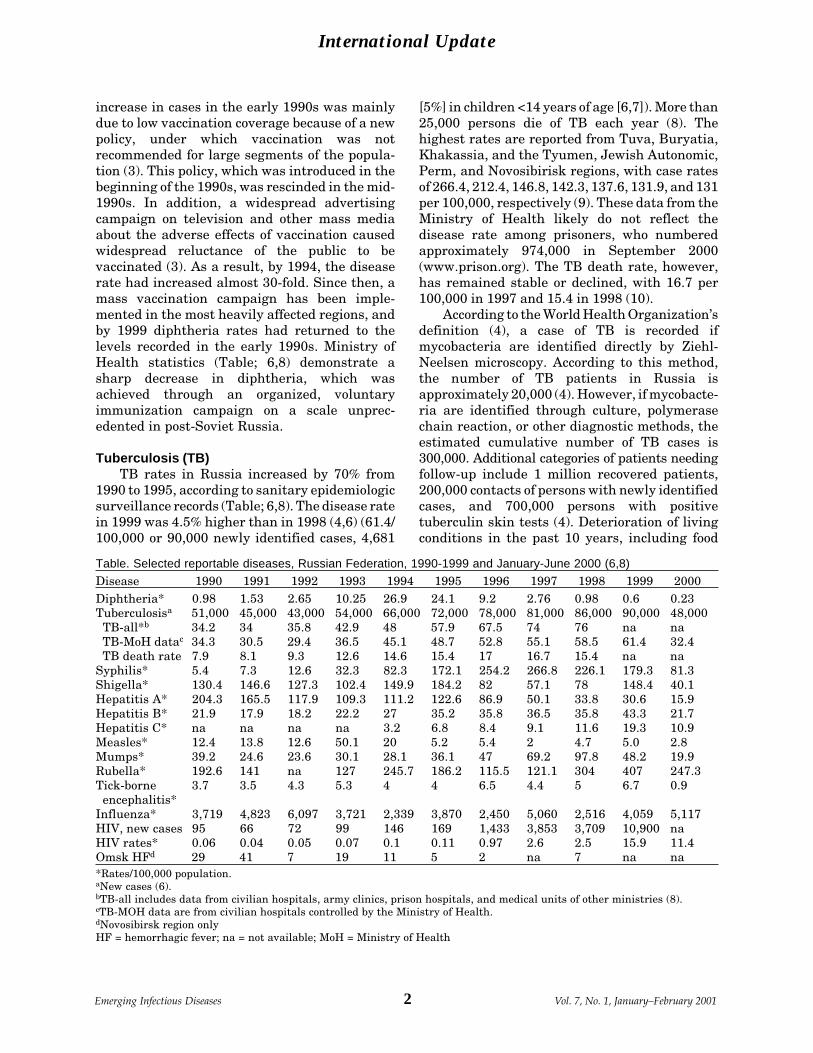
increase in cases in the early 1990s was mainlydue to low vaccination coverage because of a newpolicy, under which vaccination was notrecommended for large segments of the popula-tion (3). This policy, which was introduced in thebeginning of the 1990s, was rescinded in the mid-1990s. In addition, a widespread advertisingcampaign on television and other mass mediaabout the adverse effects of vaccination causedwidespread reluctance of the public to bevaccinated (3). As a result, by 1994, the diseaserate had increased almost 30-fold. Since then, amass vaccination campaign has been imple-mented in the most heavily affected regions, andby 1999 diphtheria rates had returned to thelevels recorded in the early 1990s. Ministry ofHealth statistics (Table; 6,8) demonstrate asharp decrease in diphtheria, which wasachieved through an organized, voluntaryimmunization campaign on a scale unprec-edented in post-Soviet Russia.
Tuberculosis (TB)TB rates in Russia increased by 70% from
1990 to 1995, according to sanitary epidemiologicsurveillance records (Table; 6,8). The disease ratein 1999 was 4.5% higher than in 1998 (4,6) (61.4/100,000 or 90,000 newly identified cases, 4,681
[5%] in children <14 years of age [6,7]). More than25,000 persons die of TB each year (8). Thehighest rates are reported from Tuva, Buryatia,Khakassia, and the Tyumen, Jewish Autonomic,Perm, and Novosibirisk regions, with case ratesof 266.4, 212.4, 146.8, 142.3, 137.6, 131.9, and 131per 100,000, respectively (9). These data from theMinistry of Health likely do not reflect thedisease rate among prisoners, who numberedapproximately 974,000 in September 2000(www.prison.org). The TB death rate, however,has remained stable or declined, with 16.7 per100,000 in 1997 and 15.4 in 1998 (10).
According to the World Health Organization’sdefinition (4), a case of TB is recorded ifmycobacteria are identified directly by Ziehl-Neelsen microscopy. According to this method,the number of TB patients in Russia isapproximately 20,000 (4). However, if mycobacte-ria are identified through culture, polymerasechain reaction, or other diagnostic methods, theestimated cumulative number of TB cases is300,000. Additional categories of patients needingfollow-up include 1 million recovered patients,200,000 contacts of persons with newly identifiedcases, and 700,000 persons with positivetuberculin skin tests (4). Deterioration of livingconditions in the past 10 years, including food
Table. Selected reportable diseases, Russian Federation, 1990-1999 and January-June 2000 (6,8)
Disease 1990 1991 1992 1993 1994 1995 1996 1997 1998 1999 2000Diphtheria* 0.98 1.53 2.65 10.25 26.9 24.1 9.2 2.76 0.98 0.6 0.23Tuberculosisa 51,000 45,000 43,000 54,000 66,000 72,000 78,000 81,000 86,000 90,000 48,000 TB-all*b 34.2 34 35.8 42.9 48 57.9 67.5 74 76 na na TB-MoH datac 34.3 30.5 29.4 36.5 45.1 48.7 52.8 55.1 58.5 61.4 32.4 TB death rate 7.9 8.1 9.3 12.6 14.6 15.4 17 16.7 15.4 na naSyphilis* 5.4 7.3 12.6 32.3 82.3 172.1 254.2 266.8 226.1 179.3 81.3Shigella* 130.4 146.6 127.3 102.4 149.9 184.2 82 57.1 78 148.4 40.1Hepatitis A* 204.3 165.5 117.9 109.3 111.2 122.6 86.9 50.1 33.8 30.6 15.9Hepatitis B* 21.9 17.9 18.2 22.2 27 35.2 35.8 36.5 35.8 43.3 21.7Hepatitis C* na na na na 3.2 6.8 8.4 9.1 11.6 19.3 10.9Measles* 12.4 13.8 12.6 50.1 20 5.2 5.4 2 4.7 5.0 2.8Mumps* 39.2 24.6 23.6 30.1 28.1 36.1 47 69.2 97.8 48.2 19.9Rubella* 192.6 141 na 127 245.7 186.2 115.5 121.1 304 407 247.3Tick-borne 3.7 3.5 4.3 5.3 4 4 6.5 4.4 5 6.7 0.9 encephalitis*Influenza* 3,719 4,823 6,097 3,721 2,339 3,870 2,450 5,060 2,516 4,059 5,117HIV, new cases 95 66 72 99 146 169 1,433 3,853 3,709 10,900 naHIV rates* 0.06 0.04 0.05 0.07 0.1 0.11 0.97 2.6 2.5 15.9 11.4Omsk HFd 29 41 7 19 11 5 2 na 7 na na*Rates/100,000 population.aNew cases (6).bTB-all includes data from civilian hospitals, army clinics, prison hospitals, and medical units of other ministries (8).cTB-MOH data are from civilian hospitals controlled by the Ministry of Health.dNovosibirsk region onlyHF = hemorrhagic fever; na = not available; MoH = Ministry of Health

3Vol. 7, No. 1, January–February 2001 Emerging Infectious Diseases
International Update
shortages, poverty, and severe overcrowding inprisons, is associated with increasing TB rates.Another important factor is the spread ofmycobacterial strains resistant to antibiotics,especially strains resistant to multiple drugs.Uncontrolled administration of antibiotics (e.g.,in prisons) promotes emergence of resistantstrains. Russia has a high rate of strainsresistant to a single drug (5,10), which may leadto an increase in the number of strains resistantto multiple drugs.
Sexually Transmitted DiseasesOne- to twofold annual increases in syphilis
incidence were recorded by the early 1990s, witha 50-fold increase in 1997 compared with 1990(Table); however, the rate of increase has slowedsince 1996 and even decreased from 1997 to 1999(7). These data may underestimate the incidence,as patients treated in private clinics are not fullyreported in official statistics. The decrease ingonorrhea incidence, which began in 1995 andcontinued until 1998, when the rate of gonorrheabecame half that of syphilis, may be alsoattributed to underreporting of these data byprivate clinics, which received official permissionfrom the Ministry of Public Health to treatgonorrhea. In 1997, the regions with the highestrates of syphilis were Tuva, Khakassia, andSakhalin, with 1,381, 1,314, and 1,217 cases per100,000, respectively (7).
BrucellosisIn the 1990s, 300 to 700 cases of brucellosis
occurred each year. No apparent long-termtrends were observed.
AnthraxAlthough many natural foci are located in
Russia, the number of anthrax cases per yearduring the past 10 years has never exceeded 100(e.g., 37 in 1998, 45 in 1999) (4).
Acute Bacterial Intestinal InfectionsIn 1998, dysentery rates were 37% higher
than in 1997; 114,800 cases were reported,including 66,000 in children. Shigellosis(Table) shows no long-term trends. In 1998,398,600 cases of acute intestinal infections ofunknown etiology were reported, including231,700 in children. The ratio of intestinalinfections with identified and unidentifiedcauses remains unchanged since 1990, indicating
lack of progress in developing and adaptingnew diagnostic tools.
Other Infectious DiseasesIn 1998, an increase was reported in cases of
zoonotic diseases such as typhus, whichincreased by 10%; borreliosis (Lyme disease),which increased by 25%; and tularemia, for whicha twofold increase was reported. No long-termtrends were noted. A cholera outbreak wasofficially recorded in Russia in 1998 in Dagestan(8 cases, 17 carriers), and three isolated caseswere reported elsewhere. Twenty cases ofepidemic typhus and 33 cases of Brill-Zinsserdisease were reported.
Resistance to AntibioticsAntibiotic resistance has increased in Russia
since antibiotics became available withoutprescription. In addition, a high concentration ofTB patients in prisons, combined with a massiveshortage of drugs in prison clinics, results infrequent self-treatment. This self-treatmentleads to inappropriate selection of drugs andresults in incomplete treatment, thus encourag-ing the emergence of drug-resistant strains. Adetailed study of this situation by the RussianAcademy of Medical Sciences and Academy ofSciences has just begun.
Selected Viral Diseases
HepatitisRates of hepatitis A decreased during the
1990s, but rates of hepatitis B and hepatitis Cincreased steadily (Table). Reasons may be asharp increase in intravenous drug use, lack ofhygiene, and high-risk sexual behavior. Half thepatients with acute hepatitis B and hepatitis Care 11 to 30 years of age (11). Mass vaccination ofchildren against hepatitis B and support for thedevelopment of a vaccine for hepatitis C areneeded to control these diseases.
PoliomyelitisPoliomyelitis increased during the war in
Chechnya (152 cases there in 1995) probablybecause of unavailability of vaccine in Chechnyaduring the conflict. Only six cases of acuteparalysis were recorded in 1998, none of whichwere caused by a wild-strain virus, as shown bylaboratory diagnostics (7). National immuniza-tion efforts against polio are continuing.

4Emerging Infectious Diseases Vol. 7, No. 1, January–February 2001
International Update
Measles, Mumps, and Rubella (MMR)Measles rates have decreased considerably
during the last 4 years through additionalvaccination of teenagers and children at sites ofmass outbreaks during 1992-94 (Table). Mumps,however, increased almost threefold from 1990 to1998. The vaccine may have degraded duringdelivery or storage under inadequate conditions.In addition, funds were insufficient for mumpsvaccination programs, and financial support waslacking in some regions for a second vaccinationat age 6 years. In 1997, a second vaccination wasrecommended in the national vaccinationschedule, with support from federal funds. Ratesof rubella remain high, sometimes increasing toepidemic levels. Russia was one of the fewEuropean countries that did not include rubellain the schedule of mandatory, state-fundedvaccines before 1997. The cost of support fordisabled children born to unvaccinated mothersis much higher than the cost of vaccination;therefore, rubella vaccine should be added to thenational vaccination schedule, ideally in the formof MMR.
Tick-borne encephalitisTick-borne encephalitis, a severe zoonotic
disease, is occasionally fatal (Table). The agent isa flavivirus transmitted through tick bites. Casesvary from 5,000 to 10,000 per year. Some regionaladministrations fund local vaccination programsfor children and adults at high risk. Adults canpay for the vaccination in most disease-endemicregions (Krasnoyarsk, Novosibirsk, Tomsk,Irkutsk, Omsk, and Kemerovo).
Omsk hemorrhagic feverOmsk hemorrhagic fever is a zoonosis caused
by a flavivirus; the infection is transmitted bymuskrats during trapping. During the past 10years, this disease has been reported only fromNovosibirsk Region (Province) (Table). Of theseven cases reported in 1998, one was fatal andthree were severe.
Hemorrhagic Fever with Renal SyndromeHemorrhagic fever with renal syndrome,
caused by a representative of the Bunyaviridaefamily, has many foci in Russia. The number ofcases ranged from 2,774 in 1990 to more than20,000 in 1997. A large outbreak in 1997 wasattributed to a surge in the population of rodents,the natural carriers of the agent. The disease rate
returned to an average annual level ofapproximately 5,000 cases by 1998.
RabiesIn Russia, the case rate of rabies in humans
remains constant (7 to 16 annual cases over thepast 10 years). All cases seem to be associatedwith ignorance of postexposure prophylaxis as aprotective measure. In 1998, animal rabies inNovosibirsk increased sharply in both domesticdogs and wild animals. The population wasinformed about the epidemic and the availabilityof vaccination if needed, and no human caseswere reported despite an increase in the numberof animal bites.
InfluenzaThe rate of influenza has been stable every
year except for peaks in 1992 and 1997 (Table). InRussia, this disease is diagnosed mainly byclinical symptoms, often without laboratoryconfirmation; the data represent a backgroundof 22 million to 23 million cases of acuterespiratory infection with unknown etiologyreported each year.
HIV InfectionThe number of HIV-infected persons in-
creased from 95 in 1990 to 3,709 in 1998, virtuallydoubling each year from 1993 to 1998 (Table). Thenumber of HIV-infected patients reached 15,569by September 1999 (12) and a report in theNovember 17, 2000, issue of Izvestia stated thatthe number of officially registered HIV-positivepersons had increased to 69,120. These officialstatistics on HIV may reflect only 10% to 20% ofthe actual number of carriers (12). A recent studyof the Irkutsk prison population identified morethan 1,400 HIV-infected prisoners (pers. commun.,office of public health, Irkutsk Region), althoughonly 30 cases had previously been reported fromthe entire region.
ConclusionsThree groups of diseases cause most concern
in Russia, as well as elsewhere: TB, viralparenteral hepatitis, and HIV infection. Publicmeasures for their control in Russia areinsufficient, mainly because of lack of funding fortreatment, vaccine prophylaxis, and healtheducation. Immunization of children againsthepatitis B is indicated. The development andintroduction of additional diagnostic tools for

5Vol. 7, No. 1, January–February 2001 Emerging Infectious Diseases
International Update
markers of intestinal and respiratory infectionand additional vaccination against mumps andrubella are needed. However, it is unlikely thatexisting public health funding will allowadditional improvements in the near future.
References 1. Goskomstat Rossii, Rossiiskii Statisticheskii Ezegodnik,
1998 Goskomstat, Moscow, p. 34-35, 101-102. 2. International Surveillance Network for the enteric
infections - Salmonella and VTEC 0157. Availablefrom: URL: http://www2.phls.co.uk/.
3. Tatochenko V. Vaccinoprophylactics. MeditsinskayaGazeta 1999;87:8-9.
4. Priimak A. The sorrowful date. Meditsinskaya Gazeta1999;22:1-4.
5. Centers for Disease Control and Prevention. PrimaryMultidrug-Resistant Tuberculosis—Ivanovo Oblast,Russia. MMWR Morbid Mortal Wkly Rep 1999;48:30.
6. Zdorov’e naseleniya I sreda obitaniya (PopulationHealth and Environment), Monthly InformationBulletin, Federal Center of Sanitary and EpidemicSurveillance, Ministry of Public Health of the RussianFederation, No. 1 (22), 1995; No 1 (34), 1996, No. 1(46),1997; No.1(58), 1998; No.1(70), 1999; No.1 (82), 2000;No. 7(88), 2000.
7. Press release of a conference on the sanitary andepidemic situation in the Russian Federation in 1998.Zdorov’e naseleniya I sreda obitaniya (PopulationHealth and Environment), Monthly InformationBulletin, Federal Center of Sanitary and EpidemicSurveillance, Ministry of Public Health of the RussianFederation. 1999;2:1-4.
8. Ministry of Public Health. The public health of thepopulation of Russia and activities of public healthservice in 1998. Moscow; 1999. p. 55.
9. Zdorov’e naseleniya i sreda obitaniya (PopulationHealth and Environment). Monthly InformationBulletin 1999;3:19-22.
10. Perepletchikov L. Tuberculosis made one step back butonly one. Meditsinskaya Gazeta 1999;86:3.
11. Shustov AV, Maksyutov AZ, Kiselev NN, Mishin VP,Tolokonskaya NP, Robertson BJ, et al. Prevalence ofhepatitis C virus infection markers and genotypesamong patients of the first municipal infectious diseaseclinical hospital of Novosibirsk. Voprosi virusologii2000;6:22-7.
12. Krasnoyarskii V. HIV is increasing and Moscow is notthe exception. Meditsinskaya Gazeta 1999;68:10.

6Emerging Infectious Diseases Vol. 7, No. 1, January–February 2001
Perspectives
Bovine Spongiform Encephalopathy
“The hungry Sheep look up, and are not fed,But swoln with wind, and the rank mist they drawRot inwardly, and foul contagion spread…”
John Milton, Lycidas (1637)
Bovine spongiform encephalopathy (BSE) or“mad cow disease” appears to have originatedfrom scrapie, an endemic spongiform encephal-opathy of sheep and goats that has beenrecognized in Europe since the mid-18th century(1). It has since spread to most sheep-breedingcountries and is widespread in the UnitedKingdom (UK), where until 1988 the renderedcarcasses of livestock (including sheep) were fed
to ruminants and other animals as a protein-richnutritional supplement.
During rendering, carcasses from which allconsumable parts had been removed were milledand then decomposed in large vats by boiling atatmospheric or higher pressures, producing anaqueous slurry of protein under a layer of fat(tallow). After the fat was removed, the slurrywas desiccated into a meat and bone mealproduct that was packaged by the animal foodindustry and distributed to owners of livestockand other captive animals (e.g., zoo andlaboratory animals, breeding species, pets).
Although elements of the ensuing story arestill disputed (including its origin from scrapie,rather than from unrecognized endemic BSE), itappears likely that changes in the renderingprocess that had taken place around 1980 allowedthe etiologic agent in infected carcasses to survive,contaminate the protein supplement, and infect
Bovine Spongiform Encephalopathy andVariant Creutzfeldt-Jakob Disease:
Background, Evolution, and Current Concerns
Paul Brown,* Robert G. Will,† Raymond Bradley,‡David M. Asher,§ and Linda Detwiler¶
*National Institute of Neurological Disorders and Stroke, National Institutesof Health, Bethesda, Maryland, USA; †National Creutzfeldt-Jakob Disease
Surveillance Unit, Western General Hospital, Edinburgh, Scotland; ‡CentralVeterinary Laboratory, New Haw, Addlestone, UK; §Center for Biologics
Evaluation and Research, Food and Drug Administration, Rockville,Maryland, USA; ¶Animal and Plant Health Inspection Service, U.S.
Department of Agriculture, Robbinsville, New Jersey, USA
Address for correspondence: Paul Brown, Building 36, Room4A-05, National Institutes of Health, 36 Convent Drive, MSC4122 Bethesda, MD 20892-4122; fax: 301-496-8275; e-mail:[email protected].
The epidemic of bovine spongiform encephalopathy (BSE) in the United Kingdom,which began in 1986 and has affected nearly 200,000 cattle, is waning to a conclusion,but leaves in its wake an outbreak of human Creutzfeldt-Jakob disease, most probablyresulting from the consumption of beef products contaminated by central nervoussystem tissue. Although averaging only 10-15 cases a year since its first appearance in1994, its future magnitude and geographic distribution (in countries that have importedinfected British cattle or cattle products, or have endogenous BSE) cannot yet bepredicted. The possibility that large numbers of apparently healthy persons might beincubating the disease raises concerns about iatrogenic transmissions throughinstrumentation (surgery and medical diagnostic procedures) and blood and organdonations. Government agencies in many countries continue to implement newmeasures to minimize this risk.
7Vol. 7, No. 1, January–February 2001 Emerging Infectious Diseases
Perspectives
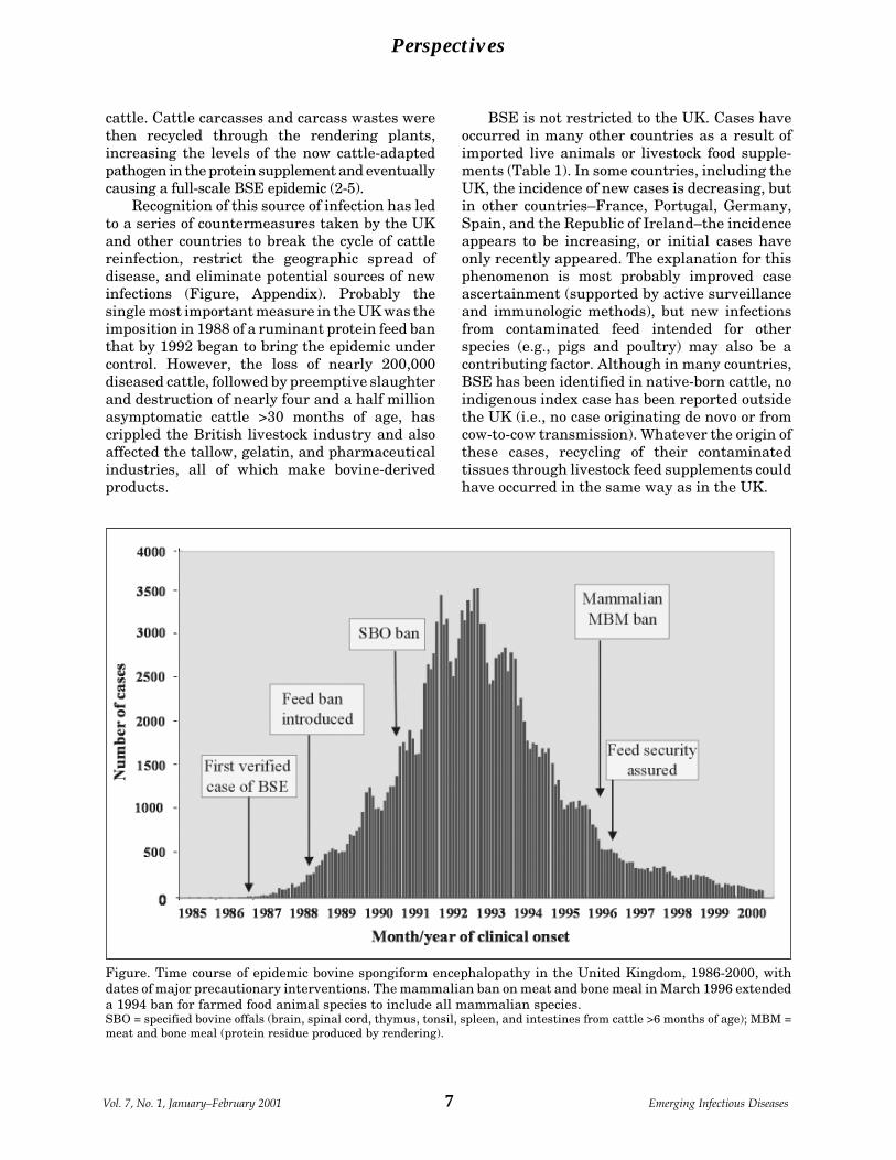
Figure. Time course of epidemic bovine spongiform encephalopathy in the United Kingdom, 1986-2000, withdates of major precautionary interventions. The mammalian ban on meat and bone meal in March 1996 extendeda 1994 ban for farmed food animal species to include all mammalian species.SBO = specified bovine offals (brain, spinal cord, thymus, tonsil, spleen, and intestines from cattle >6 months of age); MBM =meat and bone meal (protein residue produced by rendering).
cattle. Cattle carcasses and carcass wastes werethen recycled through the rendering plants,increasing the levels of the now cattle-adaptedpathogen in the protein supplement and eventuallycausing a full-scale BSE epidemic (2-5).
Recognition of this source of infection has ledto a series of countermeasures taken by the UKand other countries to break the cycle of cattlereinfection, restrict the geographic spread ofdisease, and eliminate potential sources of newinfections (Figure, Appendix). Probably thesingle most important measure in the UK was theimposition in 1988 of a ruminant protein feed banthat by 1992 began to bring the epidemic undercontrol. However, the loss of nearly 200,000diseased cattle, followed by preemptive slaughterand destruction of nearly four and a half millionasymptomatic cattle >30 months of age, hascrippled the British livestock industry and alsoaffected the tallow, gelatin, and pharmaceuticalindustries, all of which make bovine-derivedproducts.
BSE is not restricted to the UK. Cases haveoccurred in many other countries as a result ofimported live animals or livestock food supple-ments (Table 1). In some countries, including theUK, the incidence of new cases is decreasing, butin other countries–France, Portugal, Germany,Spain, and the Republic of Ireland–the incidenceappears to be increasing, or initial cases haveonly recently appeared. The explanation for thisphenomenon is most probably improved caseascertainment (supported by active surveillanceand immunologic methods), but new infectionsfrom contaminated feed intended for otherspecies (e.g., pigs and poultry) may also be acontributing factor. Although in many countries,BSE has been identified in native-born cattle, noindigenous index case has been reported outsidethe UK (i.e., no case originating de novo or fromcow-to-cow transmission). Whatever the origin ofthese cases, recycling of their contaminatedtissues through livestock feed supplements couldhave occurred in the same way as in the UK.

8Emerging Infectious Diseases Vol. 7, No. 1, January–February 2001
Perspectives
BSE has not occurred in the United States orother countries that have historically importedlittle or no live cattle, beef products, or livestocknutritional supplements from the UK. Eventhough rendering procedures in other countriesunderwent changes similar to those in the UKduring the late 1970s, BSE has apparentlyemerged solely within the UK. The mostplausible explanation is that the proportion ofsheep in the mix of rendered animal carcassesand the proportion of scrapie infections in suchsheep were probably higher in the UK thanelsewhere. These proportions were apparentlysufficient to bring very low levels of the etiologicagent in batches of rendered carcasses over thethreshold of transmission in the UK but not inother countries (5). An alternative explanationproposed in the recent Report of the BSE Inquiry(6) is that a pathogenic mutation occurred incattle in the 1970s.
Either of these two hypotheses satisfies theneed for an etiologic “seed” to survive the alteredrendering process and escalate through recyclingof an ever-larger number of infected carcasses.
However, the bovine origin hypothesis assumesthat a mutation occurred only in the UK and notin other countries where similar renderingprocesses would also have led to epidemic BSE ifmutations were occurring. In humans, mutationshave occurred all over the world, not just in theUK, and there is no reason to suppose thathumans differ in this respect from othermammalian species. It would therefore bepeculiar if the UK had the misfortune to host thecattle world’s only mutation.
Variant Creutzfeldt-Jakob Disease (vCJD)
How soon hath Time, the subtle thief of youth,Stol’n on his wing my three and twentieth year!
John Milton, Sonnet (1632)
Within weeks of identification of the first caseof BSE, concern was expressed about human risk(7-13), and as the epidemic unfolded, a series ofmeasures was taken to eradicate BSE andprevent potentially infected tissues from reach-ing the human food chain (Appendix). Asurveillance unit to monitor CJD was establishedin the UK in May 1990, and 3 years later,surveillance was extended to several otherEuropean countries, coordinated through theEuropean Union. By this means it was hoped thatany change in the epidemiology of CJD in the UKcould be detected quickly and that thesignificance of the change could be assessed bycomparison with the epidemiology of CJD incontinental Europe.
Concern was heightened by the discoverythat some exotic zoo ungulates, as well asdomestic and captive wild cats, were becominginfected (14-18). The ungulates and domestic catshad also been fed diets supplemented by meatand bone meal, and the wild cats had been feduncooked tissues, including cattle heads andspines. The possibility could therefore not beignored that the disease might also cross thespecies barrier to humans from the consumptionof beef or dairy products, or perhaps fromoccupational contact with cattle by ranchers,dairymen, or slaughterhouse workers.
What muted concerns about human infectionwas the presumption that BSE originated fromscrapie, and scrapie was not a human pathogen.Nevertheless, even those who considered humanrisk to be remote acknowledged that scrapiemight unpredictably show an altered host range
Table 1. Reported cases of bovine spongiformencephalopathy in the United Kingdom and othercountries (as of December 2000)
Native Imported TotalCountry cases cases casesUnited Kingdom 180,376a - 180,376Republic of Ireland 487 12 499Portugal 446 6 452Switzerlandb 363 - 363Franceb 150 1 151Belgium 18 - 18Netherlands 6 - 6Liechtenstein 2 - 2Denmark 1 1 2Luxembourg 1 - 1Germany 3 6 9Oman - 2 2Italy - 2 2Spainc - 2 2Canada - 1 1Falklands (UK) - 1 1Azores (Portugal)d - 1 1Data from Organization of International Epizootics (Paris)and Ministry of Agriculture, Fisheries, and Food (UK).aIncludes 1,287 cases in offshore British islandsbIncludes cases detected by active surveillance withimmunologic methodscOrigin and dates of imported cases are under investigation.dCase imported from Germany.

9Vol. 7, No. 1, January–February 2001 Emerging Infectious Diseases
Perspectives
after passage through cattle. Experimentalprecedents for such behavior were well known:passage of mouse-adapted strains of scrapiethrough hamsters altered their transmissibilityon back passage to rodents (19,20); humanstrains of kuru or CJD did not transmit to ferretsor goats until passaged through primates or cats(21); and a bovine strain of BSE did not transmitto hamsters until passaged through mice (22).Alternatively, if BSE originated from a spontane-ous mutation in cattle, experimental studies ofspecies susceptibility to this new strain oftransmissible spongiform encephalopathy (TSE)had not sufficiently advanced to predict thathumans would not be susceptible. Nevertheless,during the 10 years after the first case of BSE wasidentified, cases of CJD did not increase in groupsat high risk and continued to occur in the generalpopulation with the same spectrum of clinicaland neuropathologic features as before theappearance of BSE.
Then, from May to October 1995, the CJDSurveillance Unit was notified of three cases ofCJD in patients 16, 19, and 29 years of age(23,24). On neuropathologic examination, allthree patients had amyloid plaques, which wasunexpected in view of their occurrence in only5%-10% of sporadic cases of CJD. Thecomparative youth of the patients and thisunusual neuropathologic finding prompted asearch for similar features in patients whosedeaths might have been attributed to otherdiagnoses. In particular, cases of subacutesclerosing panencephalitis (SSPE) were scruti-nized in view of a report from Poland that cases ofCJD in three young patients had been identifiedby SSPE surveillance (25). No such cases werefound in a review of the UK SSPE register.
If CJD in young patients was not beingobscured by misdiagnosis, perhaps it reflectedincreased physician awareness through publicitysurrounding BSE and iatrogenic CJD in
recipients of contaminated growth hormone, orthe active CJD surveillance program institutedin the UK, or the availability of genetic andproteinase-resistant protein (PrP) immunocy-tochemistry. Although all these factors may havecontributed to ascertainment bias, most of theexcess cases were in older age groups, in whichCJD was now being diagnosed more often than inearlier decades.
By December 1995, the Surveillance Unit hadbeen informed of 10 suspected cases of CJD inpersons <50 years of age. Some were found tohave sporadic or familial CJD or some otherdisease; however, two of the patients, ages 29 and30 years, were later confirmed neuropathologi-cally to have CJD and, like the previous threeCJD patients, had extensive plaque deposition.As of January 1, 1996, the relationship betweenthese cases and BSE began to excite suspicion butremained tentative because critical informationjudged necessary to establish a probableconnection was still missing (Table 2).
During January, two additional cases of CJDin young patients were neuropathologicallyconfirmed, and a distinctive clinical syndromeassociated with plaque formation was beginningto emerge: young age at onset, early psychiatricsymptoms, prominent ataxia, absence of periodicelectroencephalographic activity, and a compara-tively prolonged illness. However, each of thesefeatures, alone or in combination, may also beseen in classic sporadic or familial CJD. Cautionwas further justified by a review of the records ofpre-1980 CJD patients in the UK, whichidentified three young patients who shared someof these features, and by the results of an inquiryabout young patients with CJD in otherEuropean countries, which showed an agedistribution similar to that in the UK. A majorconcern was that these seven apparently similarcases might represent a heterogeneous group ofpatients with sporadic and familial forms of CJD.
Table 2. Evolving assessment of criteria used to link bovine spongiform encephalopathy and variant Creutzfeldt-Jakob disease.
Assessment through early 1996Criteria Jan 1 Feb 1 Mar 1 Mar 8 Mar 20Novel clinical phenotype Uncertain Possible Probable Probable ProbableNovel neuropathologic phenotype Uncertain Possible Probable Probable ProbableDistinct from pre-1980 cases in UK Unknown Possible Probable Probable ProbableNo association with PRNP mutations Uncertain Uncertain Uncertain Probable ProbableDistinct from cases outside UK Unknown Unknown Unknown Possible Probable

10Emerging Infectious Diseases Vol. 7, No. 1, January–February 2001
Perspectives
Full comparative neuropathologic examinationof both pre- and post-1980 cases of CJD in youngpersons was needed, along with PRNP genesequence analysis of as many cases as possible.
During February 1996, an additional casewas referred to the Surveillance Unit with aclinical evolution similar to that of the previousseven patients, and neuropathologic examinationof recent and historical cases confirmed that therecent cases were indeed distinctive. In particu-lar, a morphologically unusual form of plaquewas present in all cases: the florid or “daisy”plaque in which an amyloid core was surroundedby “petals” of spongiform change. As of March 1,despite the likelihood that this group of patientshad a “new variant” of CJD, it was still unclearwhether mutations were involved and whethersuch a syndrome was also occurring outside theUK–both points essential to confirming theassociation of this variant disease with exposureto BSE.
On March 4, genetic analysis was completedfor six of the cases, and no pathogenic mutationwas identified. These results effectively ruled outa genetic cause for the syndrome (although theydid not rule out a genetic predisposition) and leftthe only remaining uncertainty–the geographicdistribution of the variant phenotype–to beresolved by the European CJD surveillancesystem. The answer came by March 20: none ofthe young CJD patients in other Europeancountries had the clinical and neuropathologicfeatures of the UK cases. In the preceding week,two more variant cases had beenneuropathologically confirmed, and a report onthe entire group of 10 cases concluded that anunrecognized variant of CJD occurring only inpersons <45 years of age was probably due toexposure to BSE (26).
This link has now been convincinglyestablished in laboratory studies showingidentical, distinctive biological and molecularbiological features of the pathologic agentisolated from BSE-infected cattle and humancases of vCJD (27-29). The source of contamina-tion appears to have been beef. However, musclehas never been reproducibly shown to contain theinfectious agent in any form of spongiformencephalopathy, whatever the affected species,and thus, infection most probably resulted frombeef products contaminated by nervous systemtissue. Contamination could have occurred in anyof the following ways: cerebral vascular emboli
from cranial stunning instruments used toimmobilize cattle before killing by exsan-guination; contact of muscle with brain or spinalcord tissue by saws or other tools used duringslaughter; inclusion of paraspinal ganglia in cutsof meat containing vertebral tissue (e.g., T-bonesteaks); and perhaps most importantly, thepresence of residual spinal cord and paraspinalganglia tissue in the paste of “mechanicallyrecovered meat” (a carcass compression extract)that could legally be added to cooked meatproducts such as meat pies, beef sausages, andvarious canned meat preparations. Measureshave since been taken to eliminate these sourcesof potential contamination and limit theconsequences of any contamination that mayalready have occurred (Appendix).
Although the amount of infectious tissueingested must be a critical determinant for thetransmission of BSE to humans in the form ofvCJD, the human genotype at polymorphic codon129 of the PRNP gene appears to play animportant role in susceptibility to infection. Theencoding alternatives, methionine (Met) andvaline (Val), are distributed in the generalCaucasian population in the approximateproportions of 50% Met/Val, 40% Met/Met, and10% Val/Val. All 76 vCJD patients tested havebeen homozygous for methionine, and theapparently single infecting strain of BSE may notbe able to replicate in any other human genotype.However, it is also possible that (as in theanalogous oral infection of kuru and in peripheraliatrogenic CJD infections) heterozygotes arecomparatively resistant to disease and become illafter longer incubation periods than those ofhomozygotes (30-33).
Predictions about the vCJD Outbreak
Think not but that I know these things; or thinkI know them not: not therefore am I shortOf knowing what I ought.
John Milton, Paradise Regained (1671)
The onset of illness in the first case of vCJDoccurred in early 1994, nearly a decade after thefirst case of BSE was recognized in cattle.Assuming that the earliest appearance of vCJDreflects the earliest exposure to BSE, thisincubation period is consistent with thosefollowing peripheral infections in experimentalanimals and in cases of iatrogenic CJD in

11Vol. 7, No. 1, January–February 2001 Emerging Infectious Diseases
Perspectives
Table 3. Chronology of variant Creutzfeld-Jakob disease(vCJD) in the United Kingdom and other Europeancountries, as of December 2000
Year of Unitedonset Kingdom France Ireland1994 8 11995 101996 111997 141998 171999a 20 (+4) 1 (+1) 12000a 1 (+2)aParentheses indicate still-living persons with probablevCJD or deceased persons whose diagnoses have not yet beenconfirmed by neuropathologic examination. In 2000,additional cases have been identified that do not yet meet theminimum clinical criteria for a premortem diagnosis of“probable” vCJD. Dates are for year of onset of illness, notyear of death.
humans. Through the end of November 2000, theoverall tally was 87 definite or probable cases ofvCJD in the UK, 2 confirmed and 1 probable casein France, and a single confirmed case in theRepublic of Ireland (Table 3). The Irish patienthad lived for some years in England; however,none of the French patients had lived in or visitedthe UK, so their infection must have come eitherfrom beef or beef products imported from the UK(approximately 5%-10% of the beef consumed inFrance) or from BSE-affected cattle in France.From a European standpoint, it would be muchmore troubling if imported beef were the source,as most European countries also imported beef orbeef products from the UK, although in smallerquantities.
Unlike the BSE epidemic, the vCJD outbreakhas shown only a modest increase during its first6 years, and the number of cases with onsets in2000 remains well below the previous year’stotal, although additional cases will certainly beidentified in coming months. The differencebetween BSE and vCJD may be due to the factthat, in humans, recycling of infected tissue hasnot occurred, and thus the epidemic will evolvemuch more slowly than in cattle, or the differencemay indicate a limited outbreak in humans due tovery small infectious doses that, except ingenetically susceptible persons, cannot sur-mount the combined effects of a species barrierand comparatively inefficient route of infection.
Much of the lingering uncertainty about theextent of the vCJD outbreak is attributable to the
fact that the incubation period of vCJD isunknown. If the average incubation period is 10to 15 years, the earliest patients with vCJDwould have been infected in the early 1980s,when BSE was still silently incubating in smallbut increasing numbers of cattle. In this case, thelarge increase in human exposure to contami-nated tissues during the late 1980s could lead toa parallel increase in cases of vCJD during thenext few years. If, however, the averageincubation period of vCJD is 5 to 10 years, theearliest human infections would have begun inthe mid- to late 1980s, when exposure to BSE wasmaximal. In this case, the outbreak of vCJDshould remain small because of measures toeliminate both animal and human exposure toBSE instituted from 1987 to 1997. Depending onassumptions about the incubation period andother variables, mathematical modeling predictsthat the total extent of the outbreak could rangefrom fewer than one hundred to hundreds ofthousands of cases (34-37).
If large numbers of infected persons aresilently incubating the disease, the potential forhuman-to-human iatrogenic spread of vCJD isvery real. Such apparently healthy personswould be subject to the same kinds of medical andsurgical procedures experienced by the generalpopulation, including endoscopies, vascularcatheterizations, operations for trauma orillness, and blood and organ donations. If, assuspected, the amount and distribution of theinfectious agent in tissues of persons with vCJDis greater than in other forms of CJD, theexposure of medical and surgical instruments topossibly infectious internal tissues and thetransfer of tissues as grafts and transplantsbecome a matter of much greater concern thanthe nearly negligible risk currently posed bycases of sporadic CJD.
Recent and Future Policy Decisions
A little onward lend thy guiding handTo these dark steps, a little further on…
John Milton, Samson Agonistes (1671)
Several governments have implementedpolicies to minimize the risk for human-to-human disease transmission through blooddonations from apparently healthy persons whomay be in the incubation phase of vCJD. In the

12Emerging Infectious Diseases Vol. 7, No. 1, January–February 2001
Perspectives
UK, where whole blood or blood products fromsome persons who later died of vCJD have beenadministered to others, all plasma is importedand all blood from UK donors is filtered toeliminate leukocytes, which are the most likelycarriers of infectivity in blood (38-40). In theUnited States, a blood donor policy excludesdonations from anyone who has lived in or visitedthe UK for a cumulative period of 6 months ormore during 1980 to 1996. The 6-month periodwas based on the fact that >80% of total USperson-years in the UK would be excluded andthat the 2%-3% deficit of blood donors resultingfrom the deferral could be absorbed by the bloodbanking industry without undue shortages.Several countries (Canada, Australia, NewZealand, Switzerland, Japan, and Germany)have since applied these criteria and formulatedsimilar policies.
Because of the possibility of widespreadinfection in the UK, concern extends beyondblood and organ donors to the safe use of medicaland surgical instruments, particularly thoseused in neurosurgery and ophthalmic surgery. Inthe absence of a screening test, a zero-risk policyis untenable because it would require termina-tion of the national organ donor program. Acompromise might be the temporary deferral oforgan donors–or perhaps only corneal donors–younger than 30 or 40 years of age. However, thismeasure might so diminish (and panic) the donorpopulation as to be inadvisable. Similarconsiderations apply to invasive medical andsurgical procedures: sound medical practicecannot be suspended on the basis of a theoreticalrisk for vCJD, and it would be unethical to denyneeded procedures to persons suspected ofhaving CJD. Under the circumstances, dispos-able instruments should be used wheneverpossible, and a standard sterilization protocol forreusable instruments should be implementedthat includes the most stringent possibledisinfectants (e.g., the combined use of 1 Nsodium hydroxide and autoclaving at 134°C, asrecommended in the recent World HealthOrganization guidelines on infection control forCJD [41]). No effective sterilization procedure yetexists for instruments or instrument parts toodelicate to withstand these harsh measures.Each such instrument must be disinfected to themaximum extent possible, for example bywashing repeatedly with detergent/proteinase
solutions and exposing the washed instrumentsto less harsh chemicals (e.g., 6 M urea or 4 Mguanidinium thiocyanate) that have shownmoderate to good disinfection of TSE tissueextracts (42-44).
An equally important issue is whether thebovine-adapted scrapie agent has recrossed thespecies barrier to sheep, carrying its newlyacquired ability to infect humans. The onlyreliable method to distinguish strains of TSE is atime-consuming comparison of incubation peri-ods and topographic features of brain lesionsafter injection into different strains of inbredmice (28). Glycotyping of PrP strains extractedfrom diseased brain tissue is much faster but hasnot been convincingly shown to discriminatereliably between BSE and scrapie. Moreover,neither method has been used to test a sheep-adapted strain of BSE (that is, after multiplepassages through sheep), which might have lostthe distinguishing characteristics found onprimary passage from cow to sheep.
If BSE did back-cross to sheep fed the samecontaminated meat and bone meal that infectedcattle, the consequences for humans will remainlimited to the same period of risk as BSE–roughly1980 through 1996–unless sheep BSE, like sheepscrapie, can be horizontally or maternallytransmitted. Without a test to discriminatebetween the two diseases, there would be nodefense against the development of endemic BSEin sheep and the consequent risk for humaninfection from sheep as well as cows. Therefore,global elimination of animal TSEs must seriouslybe considered.
Such a goal is more practical than it was evena few years ago. National programs to eliminatescrapie have historically relied on selectiveslaughter of blood lines or in some cases entireflocks in which scrapie was identified, and allsuch attempts have failed. Molecular genetictools are now available to guide scrapie-resistance breeding programs that until recentlydepended on field observation and classicalgenetics, and immunologic tools can detectpreclinical scrapie infection in tonsils, thirdeyelids, and possibly blood (45-48). The environ-mental durability of TSE pathogens will maketheir eradication difficult (49,50); however, theglobal elimination of TSE in sheep and otheranimals is a goal worth the expense, effort, andpatience that will be needed for its achievement.

13Vol. 7, No. 1, January–February 2001 Emerging Infectious Diseases
Perspectives
Dr. Brown is Senior Research Scientist in the Labo-ratory of Central Nervous System Studies at the NationalInstitutes of Health. His most recent research focuses onthe problem of iatrogenic Creutzfeldt-Jakob disease andon the potential for disease transmission through the ad-ministration of blood or blood products. He serves as con-sultant to the European CJD surveillance program andas Chair of TSEAC, the transmissible spongiform en-cephalopathy advisory committee of the United StatesFood and Drug Administration.
References 1. Brown P, Bradley R. 1755 and all that: a historical
primer of transmissible spongiform encephalopathy. BrMed J 1998; 317:1688-92.
2. Wells GAH, Scott AC, Johnson CT, Gunning RF,Hancock RD, Jeffrey M, et al. A novel progressivespongiform encephalopathy in cattle. Vet Rec1987;121:419-20.
3. Collee JG, Bradley R. BSE: a decade on-Part 1. Lancet1997; 349:636-42.
4. Collee JG, Bradley R. BSE: a decade on-Part 2. Lancet1997; 349:715-21.
5. Brown P. The risk of bovine spongiform encephalopa-thy (“mad cow disease”) to human health. JAMA1997;278:1008-11.
6. The BSE inquiry: report, evidence and supportingpapers of the inquiry into the emergence andidentification of Bovine Spongiform Encephalopathy(BSE) and variant Creutzfeldt-Jakob Disease (vCJD)and the action taken in response to it up to 20 March1996. Lord Phillips of Worth Matravers, Chairman.London: The Stationery Office. October 26, 2000.
7. Holt TA, Phillips J. Bovine spongiform encephalopa-thy. Br Med J 1988;296:1581-2.
8. BSE and scrapie: agents for change [editorial]. Lancet1988;ii:607-8.
9. Taylor DM. Bovine spongiform encephalopathy andhuman health. Vet Rec 1989;125:413-5.
10. Dealer SF, Lacey RW. Transmissible spongiformencephalopathies: the threat of BSE to man. FoodMicrobiol 1990;7:253-79.
11. Kimberlin RH. Bovine spongiform encephalopathy:taking stock of the issues. Nature 1990;345:763-4.
12. Will RG. Is there a potential risk of transmission of BSEto the human population and how may this be assessed?In: Bradley R, Savey M, Marchant B, editors. Sub-acutespongiform encephalopathies. Dordrecht: KluwerAcademic Publishers; 1991. p. 179-86.
13. Brown P. The clinical epidemiology of Creutzfeldt-Jakobdisease in the context of bovine spongiform encephalopa-thy. In: Bradley R, Savey M, Marchant B, editors. Sub-acute spongiform encephalopathies. Dordrecht: KluwerAcademic Publishers; 1991. p. 195-202.
14. Jeffrey M, Wells GAH. Spongiform encephalopathy in aNyala (Tragelaphus angasi). Vet Pathol 1988;25:398-9.
15. Fleetwood AJ, Furley CW. Spongiform enecphalopathyin an eland. Vet Rec 1990;126:408-9.
16. Wyatt JM, Pearson GR, Smerdon T, Gruffydd-JonesTJ, Wells GAH. Spongiform encephalopathy in a cat.Vet Rec 1990;126:513.
17. Kirkwood JK, Wells GAH, Wilesmith JW, CunninghamAA, Jackson SI. Spongiform encephalopathy in anArabian oryx (Oryx leucoryx) and a greater kudu(Tragelaphus strepsiceros). Vet Rec 1990;127:418-20.
18. Willoughby K, Kelly DF, Lyon DG, Wells GAH.Spongiform encephalopathy in a captive puma (Felisconcolor). Vet Rec 1992;131:431-4.
19. Kimberlin RH, Cole S, Walker CA. Temporary andpermanent modifications to a single strain of mousescrapie on transmission to rats and hamsters. J GenVirol 1987;68: 1875-81.
20. Kimberlin RH, Walker CA, Fraser H. The genomicidentity of different strains of mouse scrapie isexpressed in hamsters and preserved on reisolation inmice. J Gen Virol 1989;70: 2017-25.
21. Gibbs CJ Jr, Gajdusek DC, Amyx H. Strain variation inthe viruses of Creutzfeldt-Jakob disease and kuru. In:Prusiner SB, Hadlow WJ, editors. Slow transmissiblediseases of the nervous system. Volume 2. New York:Academic Press; 1979. p. 87-110.
22. Foster JD, Hope J, McConnell I, Bruce M, Fraser H.Transmission of bovine spongiform encephalopathy tosheep, goats, and mice. Ann NY Acad Sci 1994;724:300-3.
23. Britton TC, Al-Sarraj S, Shaw C, Campbell T, CollingeJ. Sporadic Creutzfeldt-Jakob disease in a 16-year-oldin the UK. Lancet 1995;346:1155.
24. Bateman D, Hilton D, Love S, Zeidler M, Beck J,Collinge J. Sporadic Creutzfeldt-Jakob disease in a 18-year-old in the UK. Lancet 1995;346:1155-6.
25. Kulczycki J, Jedrzejowska H, Gajkowski K, Tarnows-ka-Dziduszko E, Lojkowska W. Creutzfeldt-Jakobdisease in young people. Eur J Epidemiol 1991;5:501-4.
26. Will RG, Ironside, JW, Zeidler M, Cousens SN,Estibeiro K, Alperovitch A, et al. A new variant ofCreutzfeldt-Jakob disease in the UK. Lancet1996;347:921-5.
27. Collinge J, Sidle KC, Heads J, Ironside J, Hill AF.Molecular analysis of prion strain variation and theaetiology of ‘new variant’ CJD. Nature 1996;383:685-90.
28. Bruce ME, Will RG, Ironside JW, McConnell I,Drummond D, Suttie A, et al. Transmissions to miceindicate that ‘new variant’ CJD is caused by the BSEagent. Nature 1997;389:498-501.
29. Scott MR, Will R, Ironside J, Nguyen HB, Tremblay P,DeArmond SJ, et al. Compelling transgenetic evidencefor transmission of bovine spongiform encephalopathyprions to humans. Proc Natl Acad Sci U S A1999;96:15137-42.
30. Cervenáková L, Goldfarb LG, Garruto R, Lee H-E,Gajdusek DC, Brown P. Phenotype-genotype studies inkuru: implications for new variant Creutzfeldt-Jakobdisease. Proc Nat Acad Sci U S A 1998;95:13239-41.
31. Lee H-S, Brown P, Cervenáková L, Garruto RM, AlpersMP, Gajdusek DC, et al. Evidence for susceptibility ofthe 129MM PRNP genotype in epidemic kuru. J InfectDis 2000, in press.
32. d’Aignaux JH, Costagliola D, Maccario J, Billette deVillemeur T, Brandel J-P, Deslys JP, et al.Incubation period of Creutzfeldt-Jakob disease inhuman growth hormone recipients in France.Neurology 1999;53:1197-201.

14Emerging Infectious Diseases Vol. 7, No. 1, January–February 2001
Perspectives
33. Brown P, Preece M, Brandel J-P, Sato T, McShane L,Zerr I, et al. Iatrogenic Creutzfeldt-Jakob disease at themillennium. Neurology 2000;55:1075-81.
34. Cousens SN, Vynnycky E, Zeidler M, Will RG, SmithPG. Predicting the CJD epidemic in humans. Nature1997;385:197-8.
35. Ghani AC, Ferguson NM, Donnelly CA HagenaarsTJ, Anderson RM. Epidemiological determinants ofthe pattern and magnitude of the vCJD epidemic inGreat Britain. Proc R Soc London (Series B)1998;265:2443-52.
36. Donnelly CA, Ferguson NM. Predictions and scenarioanalysis for vCJD. In: Statistical aspects of BSE andvCJD: models for an epidemic. Boca Raton (FL): CRCPress LLC 1999:163-94.
37. Ghani AC, Ferguson NM, Donnelly CA, Anderson RM.Predicted vCJD mortality in Great Britain. Nature2000;406:583-4.
38. Brown P. Can Creutzfeldt-Jakob disease be transmit-ted by transfusion? Curr Opin Hematol 1995;76:472-7.
39. Brown P, Cervenáková L, McShane LM, Barber P,Rubenstein R, Drohan WN. Further studies of bloodinfectivity in an experimental model of tranmissiblespongiform encephalopathy, with an explanation ofwhy blood components do not transmit disease inhumans. Transfusion 1999;39: 1169-78.
40. Brown P, Cervenáková L. Reply to a letter to the editor.Transfusion 2000;40:754-5.
41. WHO infection control guidelines for transmissiblespongiform encephalopathies: report of a WHOConsultation. WHO/CDS/CSR/APH/2000.3. Geneva:March 23-26, 1999.
42. Kimberlin RH, Walker CA. Competition betweenstrains of scrapie depends on the blocking agent beinginfectious. Intervirology 1985;23:74-81.
43. Manuelidis L. Decontamination of Creutzfeldt-Jakobdisease and other transmissible agents. J Neurovirol1997;3:62-5.
44. Pocchiari M, Peano S, Conz A, Eshkol A, Maillard,Brown P, et al. Combination ultrafiltration and 6 Murea treatment of human growth hormone effectivelyminimizes risk from potential Creutzfeldt-Jakobdisease virus contamination. Horm Res 1991;35:161-6.
45. Roels S, Vanopdenbosch E, Langeveld JP, SchreuderBE. Immunohistochemical evaluation of tonsillartissue for preclinical screening of scrapie based onsurveillance in Belgium. Vet Rec 1999;145:524-5.
46. O’Rourke KI, Baszler TV, Besser TE, Miller JM, CutlipRC, Wells GA, et al. Preclinical diagnosis of scrapie byimmunohistochemistry of third eyelid lymphoid tissue.J Clin Microbiol 2000;38:3254-9.
47. Schmerr MJ, Jenny AL, Bulgin MS, Miller JM, HamirAN, Cutlip RC, et al. Use of capillary electrophoresisand fluorescent labeled peptides to detect the abnormalprion protein in the blood of animals that are infectedwith a transmissible spongiform encephalopathy. JChromatog A 1999;853:207-14.
48. Brown P, Cervenáková L, Diringer H. Blood infectivityand the prospects for a diagnostic screening test inCreutzfeldt-Jakob disease. J Lab Clin Med. In press2001.
49. Palsson PA. Rida (scrapie) in Iceland and itsepidemiology. In: Prusiner SB, Hadlow WJ, editors.Slow transmissible diseases of the nervous system.Volume 1. New York: Academic Press; 1979. p. 357-66.
50. Brown P, Gadjusek DC. Survival of scrapie virus after3 years’ interment. Lancet 1991;337:269-70.

15Vol. 7, No. 1, January–February 2001 Emerging Infectious Diseases
Perspectives
Appendix
Table A. Measures taken to prevent the spread of bovine spongiform encephalopathy to animals
Great European UnitedPrecautions Britaina Uniona StatesBSE made a notifiable disease June 1988 Apr 1990 Nov 1987BSE surveillance, with histologic examination of brains June 1988 May 1990 May 1990Ban on ruminant protein in ruminant feed July 1988Ban on export of UK cattle born before July 1988 feed ban July 1989Ban on import of live ruminants and most ruminant July/Nov 1989 products from all BSE countriesBan on export of UK cattle >6 months of age Mar 1990Ban on SBO for use in animal nutrition; ban on export of SBO and Sept 1990 feed containing SBO to EU countriesHigh-risk waste to be rendered at 133°C/3 bar/20 min (or other Nov 1990 approved procedure)Ban on export of SBO and feed containing SBO to non-EU countries July 1991Ban on MBM from SBO in fertilizer Nov 1991After Jan 1, 1995, rendering methods must sterilize BSE June 1994Ban on mammalian MBM in ruminant feed July 1994BSE surveillance includes immunohistologic features of brains Oct 1993Ban on mammalian protein in ruminant feedb Nov 1994 Aug 1997Ban on import of live ruminants and most ruminant products Dec 1997 (including meat products) from all countries of EuropeImmunologic testing for ruminant protein in animal feed July 1995Mammalian MBM prohibited from all animal feed/fertilizer Mar/Apr 1996Slaughtered cattle >30 months old (except certain beef cattle Mar 1996 >42 months old) ruled unfit for animal use (hides for leather excluded)Mammalian MBM and MBM-containing feed recalled June 1996All mammalian waste to be rendered at 133°C/3 bar/20 min July 1996 (or other approved procedure)Cattle tracing system improved Sept 1998Quarantine of 3 sheep flocks imported from Europe with possible Oct 1998 exposure to BSE (4 animals die with atypical TSE)BSE surveillance of fallen stock (downer cows) is intensified Oct 1998Proposal to eradicate scrapie is rejuvenated Nov 1999Allow export of deboned beef from cattle >30 months old born Aug 1999 after July 1996Prohibit use of animal protein, including MBM and blood meal Dec 2000 (but excluding milk, or fish meal for nonruminants) in feed for any farmed animal species (effective January 1, 2001)Prohibit importation of rendered protein and rendering wastes Dec 2000 originating or processed in EuropeaIn Northern Ireland and Scotland, dates of implementation sometimes differed from those shown for England and Wales; inaddition, individual European Union countries often adopted different measures on different dates.bSome exemptions, e.g., milk, blood, and gelatin.BSE: bovine spongiform encephalopathy; EU = European Union; MBM = meat and bone meal (protein residue produced byrendering); SBO = specified bovine offals (brain, spinal cord, thymus, tonsil, spleen, and intestines from cattle >6 months ofage); TSE = transmissible spongiform encephalopathy.

16Emerging Infectious Diseases Vol. 7, No. 1, January–February 2001
Perspectives
Table B. Measures taken to prevent the spread of bovine spongiform encephalopathy to humansGreat European United
Precautions Britaina Uniona StatesCompulsory slaughter of BSE-affected cattle Aug 1988Destroy milk from affected cattle (except for milk fed to cows’ own calves) Dec 1988Ban on import of UK cattle born after July 1988 feed ban July 1989Ban on SBO for domestic consumption Nov 1989Ban on export to EU of SBO and certain other tissues, including Apr 1990 Apr 1990 lymph nodes, pituitaries, and serumBan on export of live UK cattle (except calves <6 months old) June 1990 June 1990Ban on use of head meat after skull opened Mar 1992FDA recommends use of BSE/scrapie-free sources for materials used in Nov 1992 dietary supplements; request for safety plansCell lines used for biologicals should be BSE agent-free May 1993FDA requests that bovine source materials (except gelatin) used in Dec 1993 manufacture of regulated products be restricted to BSE-free countriesBone-in beef only from farms with no BSE for 6 years; if not BSE-free, July 1994 must be deboned with visible nervous and lymphatic tissue removedFDA requests that bovine-derived materials for animal use or for cosmetics Aug 1994 and dietary supplements not be sourced from BSE countriesThymus and intestines from calves <6 months old made SBO Nov 1994Import of beef only from UK cattle 1) >30 months, or 2) from herds July 1995 BSE-free for 6 years, or 3) if not BSE-free, deboned with visible nervous tissue and specified lymph nodes removedSBO ban broadened to include whole skull (SBM) Aug 1995MRM from bovine vertebral column banned and export prohibited Dec 1995Removal of lymph nodes and visible nervous tissue from bovine meat Jan 1996 >30 months exported to EUBan on export of all UK cattle and cattle products except milk Mar 1996SBM ban broadened to include entire head (excluding uncontaminated tongue) Mar 1996Slaughtered cattle >30 months (or certain beef cattle >42 months) ruled Mar 1996 unfit for animal or human use (hides excepted)FDA urges manufacturers of FDA-regulated human products to take May 1996 steps to assure freedom from BSE agentPartial lifting of export ban on tallow and gelatin June 1996SBM ban broadened to include certain sheep and goat heads, spleens, Sept 1996 and spinal cords (SRM)FDA recommends withdrawal of plasma and plasma products made from Dec 1996 pools to which persons who later died of CJD had contributedCNS tissues excluded from cosmetic products for use in EU Jan 1997BSE cohort cattle in UK ordered slaughtered and destroyed Jan 1997Proposed ban on SRM in cosmetics for use in EU (effective October 2000) July 1997SBM controls for cosmetics and medicinal products Mar 1997FDA request to manufacturers that no bovine gelatin from BSE countries be Sept/Dec 1997 used in injectable, implantable, or ophthalmic products; and that special precautions be applied to gelatin for oral and topical useBan on marketing cosmetic products containing SRM prepared before April 1, 1998 Mar 1998Allow export of beef and beef products from cattle >30 months in Mar 1998 certified BSE-free herds from Northern IrelandImportation of all plasma and plasma products for use in UK Aug 1998FDA limits plasma product withdrawals to pools at risk for Sept 1998 contamination by vCJD donorsSlaughter and destruction of offspring born to BSE-affected cattle after July 1996 Jan 1999FDA guidance to defer blood donors with >6 months cumulative Nov 1999 residence in UK during 1980-1996Leukodepletion of whole blood donations from UK residents Jul/Nov 1999Public FDA discussion about possible risk associated with vaccines July 2000 produced with bovine-derived materials from BSE countriesWithdrawal and destruction of a potentially tainted 1989 lot of polio Oct 2000 vaccine from one manufacturerSRM ban implemented (effective October 2000) July 2000Ban on slaughter techniques that could contaminate cattle carcasses with July 2000 brain emboli (e.g., pithing or pneumatic stun guns), effective Jan 2001All cattle >30 months old must have brain examinations for proteinase-resis- Dec 2000 tant protein (PrP) before entering the food chain (effective Jan-Jun 2001)aIn Northern Ireland and Scotland, dates of implementation sometimes differed from those shown for England and Wales; inaddition, individual EU countries often adopted different measures on different dates.CNS = central nervous system; EU = European Union; MRM = mechanically recovered meat; SBM = specified bovine materials(SBO plus entire head, including eyes but excluding tongue); SBO = Specified bovine offals (brain, spinal cord, thymus, tonsils,spleen, and intestines from cattle >6 months old); SRM = specified risk materials (SBM plus sheep and goat heads and spleensfrom animals of any age, and spinal cords from animals >1 year old).

17Vol. 7, No. 1, January–February 2001 Emerging Infectious Diseases
Perspectives
Vector-borne diseases (including a numberthat are mosquito-borne) are a major publichealth problem internationally. In the UnitedStates, dengue and malaria are frequentlybrought back from tropical and subtropicalcountries by travelers or migrant laborers, andautochthonous transmission of malaria anddengue occasionally occurs. In 1998, 90 con-firmed cases of dengue and 1,611 cases of malariawere reported in the USA (1) and denguetransmission has occurred in Texas (2). Othervector-borne diseases continue to pose a publichealth threat. Even though the reportedincidence of most of these diseases is low (in 1997,10 cases of eastern equine encephalitis, 115 ofLaCrosse, and 14 of St. Louis encephalitis [SLE]),occasional epidemics, e.g., of SLE (1,967 cases in1975 and 247 cases in 1990, mostly in Florida [3])have resulted in aerial applications of insecti-cides, primarily malathion. In addition, newvector-borne threats continue to emerge. In 1999,West Nile virus, an Old World flavivirus relatedto Saint Louis encephalitis virus, was firstrecorded in New York (4). The virus, which istransmitted by anthropophilic mosquitoes, causeda serious outbreak (62 cases, 7 deaths) andsignaled the potential for similar outbreaks inthe Western Hemisphere. Pesticides, whichtraditionally have been used in response to
epidemics, have a role in public health as part ofsustainable integrated mosquito managementfor the prevention of vector-borne diseases. Weassess the future use of pesticides in view ofexisting niche markets, incentives for newproduct development, Environment ProtectionAgency (EPA) registration, the Food QualityProtection Act (FQPA), and improved pestmanagement strategies for mosquito control.
Sustainable Integrated MosquitoManagement and Public Health
Mosquito control in the United States hasevolved from reliance on insecticide applicationfor control of adult mosquitoes (adulticide) tointegrated pest management programs thatinclude surveillance, source reduction, larvicide,and biological control, as well as public relationsand education. The major principles of integratedmosquito management are available at a newPublic Health Pest Control Manual internetwebsite (5). Adulticides still play a vital role whenflooding causes extreme numbers of nuisancemosquitoes or when outbreaks of diseases suchas SLE occur.
Surveillance programs track diseases har-bored by wild birds and sentinel chicken flocks;vector-borne pathogens in mosquitoes; adult andlarval mosquitoes and larval habitats (by aerialphotographs, topographic maps); mosquito traps;biting counts; and follow-up on complaints andreports by the public. When established mosquitolarval and adult threshold populations are
Pesticides and Public Health:Integrated Methods ofMosquito Management
Robert I. RoseU.S. Environmental Protection Agency, Washington, DC, USA
Address for correspondence: Robert I. Rose, USDA, APHIS,PPQ, Unit 147, 4700 River Road, Riverdale, MD 20737, USA;fax: 301-734-8669; e-mail: [email protected].
Pesticides have a role in public health as part of sustainable integrated mosquitomanagement. Other components of such management include surveillance, sourcereduction or prevention, biological control, repellents, traps, and pesticide-resistancemanagement. We assess the future use of mosquito control pesticides in view of nichemarkets, incentives for new product development, Environmental Protection Agencyregistration, the Food Quality Protection Act, and improved pest management strategiesfor mosquito control.
18Emerging Infectious Diseases Vol. 7, No. 1, January–February 2001
Perspectives
exceeded, control activities are initiated. Sea-sonal records are kept in concurrence withweather data to predict seasonal mosquito larvaloccurrence and adult flights.
Source reduction consists of elimination oflarval habitats or rendering of such habitatsunsuitable for larval development. Publiceducation is an important component of sourcereduction. Many county or state mosquito controlagencies have public school education programsthat teach children what they and their familiescan do to prevent mosquito proliferation. Otherforms of source reduction include open marshwater management, in which mosquito-produc-ing areas on the marsh are connected by shallowditches to deep water habitats to allow drainageor fish access; and rotational impoundmentmanagement, in which the marsh is minimallyflooded during summer but is flap-gated toreintegrate impoundments to the estuary for therest of the year.
Biological control includes use of manypredators (dragonfly nymphs and other indige-nous aquatic invertebrate predators such asToxorhynchites spp. predacious mosquitoes) thateat larvae and pupae; however, the most commonlyused biological control adjuncts are mosquito fish,Gambusia affinis and G. holbrooki. Naturallyoccurring Fundulus spp. and possibly Rivulus spp.,killifish, also play an important role in mosquitocontrol in open marsh water management androtational impoundment management. Likemany fish, mosquito fish are indiscriminatefeeders that may eat tadpoles, zooplankton,aquatic insects, and other fish eggs and fry (6).However, since they are easily reared, they havebecome the most common supplemental biologi-cal control agent used in mosquito control. Theentomopathogenic fungus, Laginidium gigan-teum, has been registered for mosquito control byEPA under the trade name Liginex, but productshave not become readily available. The pathogen-ic protozoon, Nosema algerae, has also notbecome available for technical reasons. Ento-moparasitic nematodes such as Romanomermisculicivorax and R. iyengari are effective and donot require EPA registration but are not easilyproduced and have storage viability limitations. Apredacious copepod, Mesocyclops longisetus, preyson mosquito larvae and is a candidate for localrearing with Paramecium spp. for food.
Mosquito traps (such as the New Jersey andthe Centers for Disease Control and Prevention
designs) have been used for monitoring mosquitopopulations for years. New designs usingmechanical control to capture adult mosquitoeshave now become available. These designs usecompressed carbon dioxide, burning propane,and octenol to attract mosquitoes and fans tocontrol air flow. The new technology is expensive:these traps may cost well over $1,000 each.Electric high-voltage insect traps (“bug zappers”)with “black” or ultraviolet light sources do notprovide satisfactory adult mosquito control andkill insects indiscriminately.
PesticidesPesticides used by state or local agencies to
control nuisance or public health pests havewarning labels and directions to minimize risksto human health and the environment. Thesepesticides are applied by public health employeeswho are specifically trained to follow propersafety precautions and directions for use. State orlocal mosquito control programs are funded bytaxes and subject to public scrutiny. Theenvironmental hazards precautionary state-ments on many mosquito insecticide labels statethat insecticides are toxic to birds, fish, wildlife,aquatic invertebrates, and honeybees. Because ofthe low rates of application used to controlmosquitoes and the special public health pestcontrol training of most applicators, hazard tonontargeted organisms is limited. However,honeybees may be killed if exposed whenforaging, so proper precautions are warranted.Human exposure in residential areas is alsouncommon because of the very low applicationrates, ultra low-volume methods (ULV), treat-ment at night when people are indoors, pesticideapplicator training, and public prenotificationbefore application. Pesticide applicators whomix, load, and apply the concentrated insecti-cides use personal protective equipment to avoidexposure and closed systems to pump insecti-cides from storage to spray equipment.
The Federal Food Drug and Cosmetic Act(FFDCA) 21 USC 9§406 is the regulation thatlimits the quantity of any poisonous ordeleterious substance added to food. A pesticideresidue is the pesticide or its metabolites in or onraw agricultural commodities or processed foodand feed. A tolerance is the maximum limit of apesticide residue considered safe. Tolerances arerelevant to adult mosquito control because winddrift may carry the pesticide over agricultural

19Vol. 7, No. 1, January–February 2001 Emerging Infectious Diseases
Perspectives
Table. Pesticides used for mosquito control in the United StatesName Trade name Formulation Application Advantage LimitationTemephos Abate G, EC Larvae Usually lowest Nontarget
cost effects, some resistance
Methoprene Altosid G, B, P, LC Larvae Residual Cannot be briquets, non- certain of per- target safety formance until
too late to retreat
Oils BVA, Oil Larvae, pupae Acts on pupae Oil film, subsur- Golden Bear face larvae
Monomolecular film Agnique Liquid Larvae, pupae Acts on pupae Subsurface larvaeBacillus thuringiensis Aquabac, WDG, AS, Larvae Nontarget Short window of israelensis (Bti) Bactimos, P,G,B safety, treatment
LarvX, Briquets con- opportunity.Teknar, trol 30+ days pupaeDunks
Bacillus sphaericus VectoLex G, WDG Larvae Nontarget Pupae, only (Bs) safety works in fresh
water
Malathion Fyfanon, ULV, Adults Tolerances OP, someAtrapa, thermal fog resistancePrentox
Naled Dibrom, ULV, EC, Adults Tolerances OP, corrosiveTrumpet thermal fog
Fenthion Batex ULV Adults None specified OP, Florida only, RUP, tolerances
Permethrin Permanone, ULV, Adults, Low vertebrate None specifiedAquaResilin, thermal fog, clothing treat- toxicityBiomist, clothing ment for ticksMosquito- treatment and mosquitoes Beater
Resmethrin Scourge ULV, Adults Low vertebrate RUP, nothermal fog toxicity tolerance for
residue on cropsSumithrin Anvil ULV, Adults Low vertebrate No tolerance
thermal fog toxicity
Pyrethrins Pyrenone, ULV, EC Adults, larvae Natural May be costlyPyronyl pyrethrum,
tolerancesAS = Aqueous Suspension; B = Briquets; EC = Emulsifiable Concentrate; G = Granules; LC = Liquid Concentrate; P = Pellets;ULV = Ultra Low Volume; WDG = Water-Dispersible Granule; OP = Organophospate insecticide; RUP = Restricted UseProduct
crops where residues subject to legal tolerancerequirements may occur. Crop tolerances arelisted in the Code of Federal Regulations (7).
LarvicidesDetection of large numbers of immature
mosquitoes in areas where source reduction orbiological control is not feasible may requirelarvicide treatment to prevent the emergence of
adult mosquitoes. Use of larvicides is lesscontroversial than use of adulticides, althoughuse of larvicides may lead to public concern abouttheir effects on untargeted beneficial aquaticarthropods and vertebrates (Table).
AdulticidesEffective sustainable integrated mosquito
management programs strive to prevent large

20Emerging Infectious Diseases Vol. 7, No. 1, January–February 2001
Perspectives
flights or swarms of mosquitoes through all themeasures described above, but heavy precipita-tion, flooding, high tides, environmental con-straints, inaccessible larval habitats, missedbreeding sites, human disease outbreaks, as wellas budget shortfalls, absent employees, orequipment failures, may necessitate use ofadulticides (Table). Some local mosquito controlprograms would use an integrated program ifthey had adequate resources, but may be solimited in funding and personnel that adulticidingtrucks are the only means of mosquitointervention.
Effective adult mosquito control with insecti-cides requires small droplets that drift throughareas where mosquitoes are flying. The dropletsthat impinge on mosquitoes provide the contactactivity necessary to kill them. Large dropletsthat settle on the ground or vegetation withoutcontacting mosquitoes waste material and maycause undesirable effects on nontargeted organ-isms. To achieve small droplets, special aerialand ground application ULV equipment is used.Insecticides are applied in a concentrated form ortechnical grade and at very low volumes such as 1oz (29.6 mL) per acre. Typically, aerialapplications produce spray droplets of 30 to50 microns measured as mass median diameter,with <2.5% of the droplets exceeding 100microns. Ground ULV applicators producedroplets of 8 to 30 microns, with none >50microns mass median diameter. Large dropletsof malathion, naled, and fenthion in excess of 50to 100 microns can damage automotive or similarpaint finishes.
Adulticide applications, particularly aerialapplications and thermal fogging, are quitevisible and contribute to public apprehension.Ground ULV application may be less alarmingthan aerial application but is not effective overlarge or inaccessible areas. Preferable aircurrents for ground applications are 3.2 kph to12.9 kph and not in excess of 16.1 kph. Excessivewind and updrafts reduce control, but light windis necessary for drifting spray droplets. Withinsecticide application by air using high-pressurepumps of 2,500 lbs psi, special nozzles, properaerial application altitude and wind drift,mosquito control is achievable for several milesdownwind with minimal spray deposit below theaircraft, as a result of improved atomization ofthe insecticide. This technology is beingdeveloped and needs validation under different
conditions with different mosquito species beforeit can be universally used. Thermal fogging,which was commonly used before ULV applica-tions became prevalent, continues to be used in afew areas in the United States and is still widelyused in other countries. The insecticide is dilutedwith petroleum oil and vaporized with heat into adense, highly visible fog of very small uniformdroplets, which allows tracking the plumedownwind to target areas. Although this fogreduces visibility, it may also penetratevegetation better than a ULV application. Smallelectric or propane thermal foggers are availablefor consumer use in retail stores at a cost ofapproximately $60.00.
Adult mosquitoes are easily controlled withinsecticides applied at extremely low rates. Forexample, malathion is applied at 3 fl oz per acre(219.8 mL/ha) for mosquitoes, while the rate foragriculture is as much as 16 fl oz per acre (1,172mL/ha).
Insecticide ResistanceVector resistance to certain larvicides and
adulticides has occurred periodically. Failure ofmosquito control indicating resistance must beverified by laboratory analysis or use of test kitsbecause other factors (improper equipmentcalibration, dilution, timing and other applica-tion errors, off-specification products, climaticfactors) can prevent insecticides from providingsatisfactory control in the field. Resistance mayoccur between insecticides within a class or couldbe passed from immature to adult stages subjectto the same insecticidal mode of action.Additionally, different species of mosquitoes mayinherently vary in susceptibility to differentlarvicides and adulticides. Insecticides withdifferent modes of action can be alternated toprevent resistance. Even though source reduc-tion and use of predators such as larvivorous fishare also used for sustainable integrated mosquitomanagement, only two chemical classes ofadulticides (organophosphates and pyrethroids)with different modes of action are available.Biological controls (including birds and bats) maybe present, but often not in sufficient numbers toprovide satisfactory alternative control, particu-larly in coastal areas where salt-marsh mosqui-toes are abundant or when human diseaseoutbreaks occur. Therefore, sustained integratedmosquito management requires alternative useof different classes of insecticides, in conjunction

21Vol. 7, No. 1, January–February 2001 Emerging Infectious Diseases
Perspectives
with resistance monitoring, source reduction,biological control, and public education.
RepellentsInsect repellents, primarily N,N-diethyl-
metatoluamide (DEET), are used to preventnuisance bites from mosquitoes (as well as ticks,biting flies, and mites) and may aid in loweringdisease transmission from these pests. However,they should not be relied upon to prevent diseasetransmission, particularly where Lyme diseaseor encephalitis are endemic or malaria, yellowfever, or other vector-borne diseases areprevalent. Repellents, mosquito coils, andpermethrin clothing treatment products aresubject to EPA pesticide registration perfor-mance requirements (8). Information on safe useof repellents is located at the EPA Office ofPesticide Programs website (9). Citronella and itsoil for mosquitoes and 30 other active ingredientsare exempted from EPA pesticide registration(10). However, some of these products may not beefficacious.
Future of Public Health PesticidesThe past decade has seen a sharp rise in
public apprehension concerning the use ofpesticides, although state and federal regula-tions are well established for the assessment andmitigation of their human and environmentalrisks. Response to public concern over safety ofpesticides prompted the FQPA, which includesprovisions to protect availability of public healthpesticides. However, public health pesticides arein jeopardy for the following reasons: In theUnited States, mosquito control programs areoften for nuisance rather than disease vectorcontrol and not many insecticides are registeredfor this use. None of the mosquito adulticidescommonly used were developed recently; theirregistrations are up to 44 years old. Mosquitocontrol is only a niche market compared withagricultural pest control, which includes pesti-cides for use on corn, soybeans, and cotton, aswell as the high-profit home, garden, andstructural pest control markets. As pesticidecompanies have merged to form multinationalconglomerates, the most profitable markets arethose that drive corporate decisions. At present,it may require $50 million or more to develop andregister a new pesticide with EPA. Furthermore,several years of the patent life elapse before costsare recouped and profits accrue.
Vector control uses of existing pesticides,particularly adulticides, often follow agriculturalregistration and commercialization as a means ofexpanding sales into new markets. Performancedata are not usually required for registration ofagricultural pesticides, but these data arerequired for registration of public healthpesticides. For mosquito control, these data areoften obtained under an experimental usepermit, which requires application to EPA,submission or reference to a portion of thepesticide registration requirements according toCFR 40 § 158 Data Requirements for Registra-tion and Reporting (7,8). Testing for mosquitoadulticides or larvicides is typically done byuniversities and mosquito control or abatementdistricts, although it may be done by companiesor state or federal research organizations, such asthe Department of Defense or the U.S.Department of Agriculture. In addition todefining dose rates, formulations, environmentalvariables, and effects that must be accommo-dated, testing under an experimental use permitprovides a means of market introduction throughuser and customer experience, presentations atprofessional society meetings, and journalpublications.
Pesticide marketing often involves distribu-tors or dealers who specialize in the market if themanufacturers do not deal directly. Profitmargins that add to price are required bydistribution chains. Public agencies solicitcompetitive bids for pesticides, which squeezemargins further, thus affecting marketingincentives. Mosquito adulticides are used at verylow rates of active ingredient per acre, whichlimits sales volumes and margins. Some seasonshave few mosquitoes, so sales are low. Productliability also plays an important role in reducingincentives because of possible personal and class-action lawsuits or court injunctions againstpesticides applied over populated areas.
The Federal Insecticide, Fungicide,and Rodenticide Act and FQPA
The Federal Insecticide, Fungicide, andRodenticide Act 7 USC 136 and FFDCA wereamended by the FQPA of 1996. Amendmentspertinent to mosquito nuisance and vectorcontrol include the following: review of apesticide’s registration every 15 years; expedit-ing minor use registrations; special provisions forpublic health pesticides; aggregate (all modes of

22Emerging Infectious Diseases Vol. 7, No. 1, January–February 2001
Perspectives
exposure from a single pesticide) and cumulative(all pesticides with the same mode of action) riskassessments; an additional safety factor of up to10 X for children; collection of pesticide useinformation; and integrated pest management.Special provisions for public health pesticidesinclude the following: risks and benefitsconsidered separately from those of otherpesticides; exemption from fees under certaincircumstances; development and implementa-tion of programs to control public health pests;Department of Health and Human Services(DHHS)-supported studies required forreregistration when needed; and appropriationsof $12 million for the first year after enactmentand similar funding as needed in succeedingyears to carry out public health pesticideprovisions of the Act. The Act describes aconsultation process between EPA and DHHSbefore any public health pesticide registration issuspended or canceled and allows additional timefor submission of data. The first group ofpesticides under review are the organophosphatecholinesterase inhibitors, including temephos,fenthion, naled, chlorpyrifos, and malathion.Should risk assessments result in detection ofrisk of concern to the Agency, cancellation ormitigations of use may follow, as exemplified byrecent chlorpyrifos and diazinon use cancella-tions. Risk assessments may be based on datafrom acute and chronic toxicology and exposurestudies, models that simulate exposure sce-narios, reports of adverse incidents to humansand wildlife, extrapolation, maximum label userate assumption, and worst-case exposurescenarios.
Even though the FQPA provisions wereintended by Congress to ensure that existingpublic health pesticide uses are not lost withouteconomically effective alternatives, the provi-sions may not be adequate. If FQPA results incancellation of major agricultural uses of apesticide that is also used in public health, it maybecome no longer profitable for the manufacturerto produce small quantities for mosquito control,thus ending production of the pesticide. Sinceadulticides used for mosquito control wereregistered decades ago, the data supporting theirregistrations may be insufficient to meet currentrequirements. The substantial cost involved inupdating the data required for reregistration willhave to be paid by pesticide registrants or theFederal government though the authorized and
appropriated funding in FQPA. Data to supportreregistration done at public expense are notproprietary. Registrants need proprietary data toprotect their market shares from genericpesticide competition from overseas manufactur-ers that can use public data to support their ownregistrations; therefore, they may not considerrequesting public funds to pay for new data tosupport existing registrations. However, ifgeneric safety studies applicable to several publichealth pesticides are required by EPA for allreregistrations, the data could be generated by atask force of registrants and county, state, andFederal public health agencies, which would thenrequest public funding under the provisions ofthe Act.
Although the development of new mosquitoinsecticides, particularly adulticides, is notexpected to accelerate in the near future,integrated pest management tools and tech-niques should improve as a result of FQPAfunding and the need to control continued vector-borne disease outbreaks. Integrated pest man-agement tools have strengths and weaknesses,and continued availability of adulticides iscritical. Therefore, implementation of the publichealth pesticide provisions of FQPA must includesubstantial comparative risk-benefit analyses ofthe significance of vector-borne disease impactsversus potential human and environmental toxiceffects of pesticides used to control public healthpests, both in the USA and other countriesaffected by EPA pesticide regulatory decisions.Public information and legislative campaignshave also become necessary to preserve theavailability and use of pesticides for diseasevector control as FQPA has been implementedand with the concurrent spread of West Nilevirus.
Dr. Rose is an arthropod biotechnologist with theAnimal and Plant Health Inspection Service of the U.S.Department of Agriculture.
References 1. Centers for Disease Control and Prevention. Summary
of notifiable diseases, United States, 1998. MMWRMorb Mortal Wkly Rep 1999:47:1-93. Available from:URL: http.cdc.gov/mmwr//preview/mmwrhtml/mm4753a1.htm
2. Rawlings JA, Hendricks KA, Burgess CR, CampmanRM, Clark GG, Tabony LJ, Patterson MA. Denguesurveillance in Texas, 1995. Am J Trop Med Hyg1998;59:95-9.

23Vol. 7, No. 1, January–February 2001 Emerging Infectious Diseases
Perspectives
3. Centers for Disease Control and Prevention. Confirmedarboviral encephalitis cases reported to CDC by statehealth departments, by type and state, United States1964-1997. Available from: URL: http://www.cdc.gov/ncidod/dvbid/arbor/arbocase.htm
4. Rappole JH, Derrickson SR, Hubálek Z. Migratorybirds and spread of West Nile virus in the WesternHemisphere. Emerg Infect Dis 2000;6:319-28.
5. University of Florida Entomology and NematologyDepartment. Public health pest control manual, thenational USA manual and related information.Available from: URL: http://www.ifas.ufl.edu/~pest/vector
6. Courtenay WR, Meffe, GK. Small fishes in strangeplaces; a review of introduced poeciliids. In: Meffe GK,Snelson EF, editors. Ecology and evolution of live-bearing fishes (Poeciliidae). New Jersey: Prentice Hall;1989. p. 319-31.
7. Protection of the Environment, 40 C.F.R. Parts 150 to180, July 2000 revision. Available from: URL: http://www.access.gpo.gov/nara/cfr/cfr-table-search.html
8. U.S. Environmental Protection Agency. ProductPerformance Test Guidelines, OPPTS 810.3700, InsectRepellents for Human Skin and Outdoor Premises.Washington: U.S. Government Printing Office; 1999.Available from: URL: http://www.epa.gov/OPPTS_Harmonized
9. U.S. Environmental Protection Agency. Using insectrepellents safely. EPA Office of Pesticide Programs.Available from: URL: http://www.epa.gov/pesticides/citizens/insectrp.htm
10. U.S. Environmental Protection Agency. Notice toManufacturers, Formulators, Producers and Regis-trants of Pesticide Products on Minimum RiskPesticides Exempted under FIFRA Section 25(b)Pesticide Registration (PR). Notice 2000-6, 2000.Labeling Unit, Registration Division (7505C). Wash-ington: Office of Pesticide Programs, EPA; 2000.Available from: URL: http://www.epa.gov/opppmsd1/PR_Notices/#2000

24Emerging Infectious Diseases Vol. 7, No. 1, January–February 2001
Synopses
Campylobacter jejuni subsp. jejuni (C. jejuni)and C. coli have been recognized since the late1970s as important agents of gastrointestinalinfections throughout the world; in the UnitedStates, these infections affect approximately 1%of the population each year (1). Contaminatedfood is the usual source of human infections;therefore, the presence of fluoroquinolone- andmacrolide-resistant strains in the food chain hasraised concerns that the treatment of humaninfections will be compromised. Most cases ofCampylobacter enteritis do not require antimi-crobial treatment, being brief, clinically mild, andself-limiting (2-4). However, a substantial propor-tion of these infections require treatment. Theseinclude severe and prolonged cases of enteritis,septicemia, and other extraintestinal infections.Erythromycin has been the most commonly usedagent for treating Campylobacter enteritis (2,5).
In the 1980s, the introduction of fluoroquino-lones, which are effective against most major
pathogens causing bacterial enteritis, offered anew approach to antibiotic intervention (6).Fluoroquinolones initially had good in vitroactivity for thermophilic Campylobacter species,as well as for members of the family ofEnterobacteriaceae.
Early clinical trials of both community-acquired acute diarrhea and traveler’s diarrheacaused by Campylobacter demonstrated thatpatients treated with a fluoroquinolone had goodclinical response (6,7). It soon became apparent,however, that resistance in Campylobacter spp.could arise in vivo, sometimes after only one ortwo administrations of fluoroquinolones (8).Moreover, Endtz and colleagues (9) reported asearly as 1991 that the emergence of quinolone-resistant C. jejuni and C. coli isolated fromhumans in the Netherlands coincided with theintroduction of fluoroquinolones in veterinarymedicine.
Fluoroquinolone resistance in Campylo-bacter from food animals is now recognized as anemerging public health problem. Smith et al.from Minnesota (10) found that patients infectedwith resistant C. jejuni had longer duration of
Quinolone and Macrolide Resistance inCampylobacter jejuni and C. coli:
Resistance Mechanisms and Trendsin Human Isolates
Jørgen Engberg,* Frank M. Aarestrup,† Diane E. Taylor,‡Peter Gerner-Smidt,* and Irving Nachamkin§*Statens Serum Institut, Copenhagen, Denmark;
†Danish Veterinary Laboratory, Copenhagen, Denmark; ‡University of Alberta, Edmonton, Alberta, Canada; §University of
Pennsylvania, Philadelphia, Pennsylvania, USA
Address for correspondence: Jørgen Engberg, Department ofGastrointestinal Infections, Division of Diagnostics, StatensSerum Institut, Artillerivej 5, DK-2300 Copenhagen S,Denmark; fax: 45-3268-8238; e-mail: [email protected].
The incidence of human Campylobacter jejuni and C. coli infections has increasedmarkedly in many parts of the world in the last decade as has the number of quinolone-resistant and, to a lesser extent, macrolide-resistant Campylobacter strains causinginfections. We review macrolide and quinolone resistance in Campylobacter and trackresistance trends in human clinical isolates in relation to use of these agents in foodanimals. Susceptibility data suggest that erythromycin and other macrolides shouldremain the drugs of choice in most regions, with systematic surveillance and controlmeasures maintained, but fluoroquinolones may now be of limited use in the empirictreatment of Campylobacter infections in many regions.

25Vol. 7, No. 1, January–February 2001 Emerging Infectious Diseases
Synopses
Resistance Mutation
Macrolide Domain V of 23S rRNAA2058 → → → → → GA2059 → → → → → G
Fluoroquinolone gyra → → → → → Thr - 86(higher MIC)Thr - 90Ala - 70(lower MIC)
gyra → → → → → Thr - 86 +parc→ → → → → Arg - 139
(highest MIC)
Figure 1. Macrolide and fluoroquinolone resistancemechanisms reported in Campylobacter species. Formacrolide resistance, mutations are at either positionshown (Escherichia coli coordinates) in up to all threecopies of ribosomal RNA (14,15, and CA Trieber & DETaylor, unpub. data). Fluoroquinolone resistancedepends on a mutation in the quinolone resistancedetermining region of DNA gyrase A (GyrA). Fortypical MICs see text and references 16-18. Thestrains with highest resistance levels had mutationsin both GyrA and topoisomerase IV ParC.
>
>
>
diarrhea than patients with fluoroquinolone-sensitive isolates. As Campylobacter infectionscan be serious in immunocompromised patients,the identified treatment failure raises theconcern that fluoroquinolone-resistant strainsmay increase Campylobacter-associated deathsin this group of patients.
Mechanism of Macrolide Resistancein Campylobacter
Erythromycin binds to the ribosome but,unlike larger macrolides, appears to causedissociation of the peptidyl-tRNA, rather thanblocking the peptidyltransferase activity (11).
In C. jejuni and C. coli, erythromycinresistance is chromosomally mediated and is dueto alteration of the ribosome (12); the resistancemechanism is not consistent with presence of anrRNA methylase, modification of the antibiotic,or efflux (13). Whole ribosomes or 50S subunitswere purified from erythromycin-resistant strainsand shown to bind much less erythromycin thanribosomes from sensitive strains. In a closelyrelated bacterium, Helicobacter pylori, resis-tance to clarithromycin is due to an alteration ofone of two adenine residues in the 23S rRNA atthe erythromycin-binding site (14). Sequencingof the 23S rRNA genes from erythromycin-resistant Campylobacter spp. identified muta-tions at these same sites, which are most probablyresponsible for resistance (Figure 1) (15).
Mechanism of FluoroquinoloneResistance in Campylobacter
Fluoroquinolone resistance in C. jejuniappears to be due most often to mutations in thegenes encoding subunits of DNA gyrase (gyrA)and only occasionally to topoisomerase IV (parC)(Figure 1). DNA gyrase purified from quinolone-resistant mutants of C. jejuni was 100-fold lesssensitive to inhibition by quinolones than thewildtype gyrase (19). Cloning and sequencing ofthe C. jejuni gyrA gene demonstrate thatmutations in gyrA at positions Thr-86, Asp-90,and Ala-70 were responsible for resistance(16,17). Mutations at Thr-86 are associated withhigher level resistance to nalidixic acid (MIC 64-128 µg/mL) and ciprofloxacin (MIC 16-64 µg/mL)than mutations at Asp-90 or Ala-70. C. jejuniisolates resistant to even higher levels ofquinolones (ciprofloxacin MIC of 125 µg/mL) carrytwo mutations, one in gyrA Thr-86 and the other inthe topoisomerase IV subunit parC at Arg-139 (18).
Evidence of efflux of fluoroquinolones inC. jejuni (20) also exists. Passage of the bacteriaon pefloxacin-containing agar has led to theisolation of a fluoroquinolone-resistant strain.This strain was also resistant to tetracycline,erythromycin, chloramphenicol, and several β-lactams. The pefloxacin-resistant strain carried amutation at Thr-86 of gyrA, likely responsible, inpart, for fluoroquinolone resistance. Broad-specificity efflux pumps in C. jejuni, which causefluoroquinolone resistance, have not yet beenshown to be clinically relevant.
Use of Macrolides andQuinolones in Food Animals
Antibiotics of the macrolide-lincosamidegroup have been used in treating food animalsworldwide for several decades. The mostcommonly used agents have been lincomycin andtylosin for controlling dysentery and Myco-plasma infections in swine and spiramycin fortreating mastitis in cattle. For the past 20 years,tylosin has also been the most commonly usedagent for growth promotion in swine productionworldwide, whereas spiramycin has been

26Emerging Infectious Diseases Vol. 7, No. 1, January–February 2001
Synopses
Table 1. Veterinary licensing of fluoroquinolones in selected countries
Country Substance Licensing year Animal speciesAustria (22) Enrofloxacin 1992 Cattle, pigs, poultry
Danofloxacin 1996 PoultryDifloxacin 1998 Poultry
Canadaa Enrofloxacin 1987 (withdrawn in 1997) Turkey (egg dip)Denmark (22) Enrofloxacin 1991 Cattle, pigs, poultry
Danofloxacin 1993 PoultryDifloxacin 1998 Poultry, turkeyMarbofloxacin 1998 Cattle, pigs, dogs, cats
Finland (22) Enrofloxacin 1992 (oral use withdrawn Pigs in 1999)
Difloxacin 1998 PoultryFrance (22) Enrofloxacin 1991 Cattle, poultry
Danofloxacin 1996 CattleMarbofloxacin 1993 CattleDifloxacin 1998 Poultry
Italy (22) Enrofloxacin 1989 Cattle, pigs, poultryDifloxacin 1998 Poultry
Japanb Enrofloxacin 1991 Cattle, poultry1992 Pigs
Danofloxacin 1992 Poultry1993 Cattle, pigs
Ofloxacin 1992 PoultryOrbifloxacin 1993 Cattle, pigsDifloxacin 1996 PigsNorfloxacin 1998 Poultry
Netherlands (22) Enrofloxacin 1987 Cattle, pigs, poultryDifloxacin 1998 Poultry
Spain (22) Enrofloxacin 1986 Cattle, pigs, poultryDifloxacin 1998 Poultry
United Kingdom (22) Enrofloxacin 1993 Cattle, pigs, poultryDanofloxacin 1993 PoultryMarbofloxacin 1995 CattleDifloxacin 1998 Poultry
USAc Enrofloxacin Approx. 1987-88 Dogs, cats1996 Poultry
Sarafloxacin 1999 Cattle1995 Poultry
aRJ Irwin, Health Canada, 1999. pers. comm.bY Tamura, National Veterinary Assay Laboratory, Japan, 1999. pers. comm.cJL Watts. Pharmacia/Upjohn, Kalamazoo, Michigan, 1999. pers. comm.
commonly used in poultry. The use of macrolidesfor growth promotion has been banned in allEuropean Union countries since July 1999.
Several fluoroquinolones are available fortreating food animals, such as poultry, cattle,pigs, and fish, in many countries. Whileinformation on global use is limited, worldwideuse in food animals was estimated at 120 tons in1997; use in humans has been estimated at morethan 800 tons (21). Data are available only for theyear of veterinary licensing of fluoroquinolonesby country (Table 1). Licensing for use does not
necessarily mean that the drug is actually used,so even these data have to be considered withcaution. However, quinolone treatment ofCampylobacter-colonized broiler chickens hasinduced quinolone resistance under experimen-tal conditions (23).
Macrolide and Quinolone Resistancein Isolates from Food Animals andFoods of Animal Origin
Campylobacter is carried in the intestinaltract of wild and domestic animals and, as result

27Vol. 7, No. 1, January–February 2001 Emerging Infectious Diseases
Synopses
of fecal contact during processing, frequentlycontaminates foods derived from animals.C. jejuni is predominant in broilers and cattle butis infrequent in pigs (where C. coli predominates)(24). In food animals, the prevalence of resistanceto erythromycin is generally higher in C. coli, inparticular in C. coli isolates from pigs, than inC. jejuni (24-26). In a recent study from Spain(27), rates of erythromycin and quinoloneresistance in C. coli from pigs were 81% and100%, respectively. High erythromycin resis-tance in pigs may be related to extensiveveterinary use of macrolides (5,28).
In food products of animal origin, theoccurrence of Campylobacter is much higher inpoultry than in other categories, e.g., pork or beef(29). Therefore, Campylobacter resistance dataare primarily based on poultry products,especially broiler meat. For a number ofcountries, fluoroquinolone-resistance rates aresimilar in isolates from poultry products andhumans (10,25,27,30-32). In the United King-dom, enrofloxacin (a derivative of ciprofloxacin)was first licensed in late 1993; before then,domestically bred chickens were less fre-quently infected with quinolone-resistantcampylobacters than imported chicken prod-ucts. Using a simple model, researchers wereable to correlate the previously observedresistance percentage in domestically acquiredcases with estimates of the amount of importedchicken consumed in the United Kingdom (32).In recent data from Spain and Taiwan, rates oferythromycin resistance were 17% and 17%,respectively, in C. jejuni isolated from foods,whereas for C. coli the figures were 50% and 83%,respectively (27,31).
Transmission of Resistant Campylobacterfrom Animals to Humans
Campylobacteriosis is primarily a zoonosis.Evidence to indicate that fresh raw meat,especially poultry, is a major source of infection isample, even though other sources such as rawmilk, water, and pets may contribute to humaninfection (1,5,33-38).
Studying the transmission of antimicrobialresistance from animals (especially poultry tohumans) has been difficult because the chain oftransmission is often complex. The number ofmacrolide- and fluoroquinolone-resistant iso-lates from humans is influenced by severalfactors including veterinary use of macrolides
(approved for use as antimicrobial growthpromoters or as therapeutic drugs) andfluoroquinolones (only approved as therapeuticdrugs) at a given location (24,39); associationwith recent or current antimicrobial treatment ofpatients; the origin of isolates (children vs.adults; inpatients vs. outpatients); travel (10,40-45); and sampling strategy and susceptibilitytesting procedures (no consensus as to method,media, culture conditions, or breakpoints [43,46]).These factors stress the need for cautiousinterpretation and comparison of data fromdifferent centers. However, several studies haveshown that food animals can be a substantialsource of infection in humans and that the sameserotypes and genotypes can be isolated fromhumans and food animals (29,36,37,47-49). DNAprofiling of Danish C. jejuni serotype O:2 strainsusing pulsed-field gel electrophoresis with fourrestriction enzymes identified common geno-types in humans, poultry, cattle and swine (SLWOn, EM Nielsen, and J Engberg, unpub. data).Typing data on resistant isolates is sparse, butSmith and colleagues (10) found DNA finger-prints of quinolone-resistant C. jejuni from U.S.-produced poultry identical to those of resistant C.jejuni from domestically acquired infections inhumans. Therefore, the susceptibility of humansstrains originating in animals to antibiotics canbe related to the exposure of animal strains toantibiotic agents used in farming.
Is There a Link Between Macrolide andFluoroquinolone Use in Humans andResistant Campylobacter Infections?
Some investigators suggest that resistance inC. jejuni and C. coli is driven by use of antibioticsfor treating human infections rather than byveterinary use. Induction of macrolide resistanceduring treatment has been reported infrequently(50). However, induction of macrolide resistancemay play a role in areas with a large reservoir ofasymptomatic Campylobacter carriers and fre-quent use of macrolides in humans.
Induction of fluoroquinolone resistanceduring treatment is well recognized and oftenreported (8,51-53). A predicted 10% of patientstreated with a fluoroquinolone for Campylobacterenteritis harbor quinolone-resistant Campylo-bacter strains (6). Recently, Ellis-Pegler (53)found that fluoroquinolone resistance developedin 18% to 28% of patients in their prospectivetrial. Development of resistance has been

28Emerging Infectious Diseases Vol. 7, No. 1, January–February 2001
Synopses
Figure 2. Trends for quinolone resistance rates (in percentages) among Campylobacter coli and C. jejuni combinedfrom human sources around the world. The bars represent both nalidixic acid and fluoroquinolone resistance andare based on mean values of resistance from numerous reports (9,17,24,27,39,43,56-58,61-64,72-75,78,88, pluspers. comm. from Feirel G and Rautelin H, and unpub. data from Nachamkin I).
reported within 24 hrs of treatment, butprolonged therapy, e.g., in immunosuppressedpatients, is also a risk factor (52,54).
Smith et al. (10) showed that use of aquinolone before culture accounted for amaximum of 15% of resistant isolates during1996 to 1998. In addition, an increasing numberof reports claim that fluoroquinolone-resistantstrains have been isolated from patients who hadnot received medical treatment, suggesting thatstrains were already fluoroquinolone resistantbefore causing the infection (7,31,32,55-57).Since human-to-human transmission of C. jejuniand C. coli is rare (9), patients infected with resis-tant Campylobacter are not an important source ofresistant Campylobacter for other humans.
Before fluoroquinolones were introduced inveterinary medicine, they were widely used inhuman medicine in a number of countries,
including the Netherlands and the United States(since 1985 and 1987, respectively), withoutemergence of quinolone resistance inCampylobacter in humans. In contrast, emergingquinolone resistance in humans often coincideswith or follows the approval of fluoroquinolonesin animal husbandry (Table 1, Figure 2). Thus,while human macrolide and fluoroquinolone usecontributes to the increase in resistance inhumans, their relative contribution to increase inresistance compared to the use of these agents inhusbandry appears to be small.
Frequency of MacrolideResistance in Human Isolates
Data on erythromycin and azithromycinresistance in C. jejuni, C. coli, and the twoorganisms combined, isolated from humansaround the world since 1989, differ by country

29Vol. 7, No. 1, January–February 2001 Emerging Infectious Diseases
Synopses
Table 2. Erythromycin and azithromycin resistance rates (%) in Campylobacter in humans, worldwide, since 1989
Country C. jejuni C. coli C. jejuni and C. coli ReferenceAustria 0.7 5.5 <1-1.4 (58 & pers. comm.a)Canada 0-3.3 - - (59,60)Denmark 0 14.0 0-4 (24,61,62)Finland - - <1-3 (43,63, & pers. comm.b)France 1.1 12.2 3.5 (64)Hungary - - 0 (65)Italy 1.2-6 16-68.4 7.8-11.6 (66-69)Japan 0.8 - - (66)New Zealand - - 1.5 (70)Singapore - - 51 (71)Spain 0-11.0 0-35.0 3.2-7.3 (17,27,56,57,72)Sweden 6.4c 11.1c 7.3c (44)Taiwan 10.0 50.0 18.3 (31)Thailand - - 0-31.0 (73,74)United Kingdom 1 13 1.8 (75)United States 0-7.8 - - (10,76-78, & unpub. datad)aG Feierl, 2000, pers. comm.bH Rautelin, 1999, pers. comm.c90% of isolates were acquired abroad.dI Nachamkin, 2000, unpub. data.
and species (Table 2). Almost all studies report ahigher frequency of erythromycin resistance inC. coli than in C. jejuni (0% to 11% in C. jejuni vs.0% to 68.4% in C. coli). Trends over time forerythromycin resistance show stable and lowrates in Japan, Canada, and Finland, but recentdevelopment of resistance in Thailand andSweden (45,73).
Trends over Time forQuinolone Resistance
Resistance to fluoroquinolones inCampylobacter has clearly increased over thepast decade in many parts of the world (Figure 2).Before 1989, resistance was rare. With theintroduction of enrofloxacin in veterinarymedicine (Table 1) and (probably less important)fluoroquinolones in human medicine in mainlandEurope (the Netherlands, Finland, France, andSpain), a rapid emergence of quinolone resis-tance in Campylobacter isolates from patientswas noted (8,9,43,55,64,89,90).
Surveillance data on resistance rates inhuman isolates from Asia soon indicated an equalincrease (84,91). Quinolones were approved forveterinary use in the United Kingdom and theUnited States in late 1993 and 1995, respectively;reports from these areas now show increasingquinolone-resistance profiles (10,39,88).
In the latest data from Taiwan, Thailand, andSpain, rates of fluoroquinolone resistance in
C. jejuni, or C. jejuni and C. coli combined were56.9%, 84%, and 75% to 88%, respectively(27,31,40,73). In these regions, where quinoloneresistance is highly endemic and Campylobacterspp. predominate, fluoroquinolones cannot berecommended for community-acquired bacterialdiarrhea. Although lower frequencies are re-ported from other regions, recent trends show aclear tendency of emerging quinolone resistancein many countries. Quinolone resistance inhuman isolates often coincides with or follows theapproval of fluoroquinolones for use in animalhusbandry (Table 1, Figure 2), although somedifferences in resistance rates between countriesmay be explained by differences in associationwith foreign travel, commerce, methods oftesting, and surveillance activity.
Multidrug ResistanceMultidrug resistance to macrolides and
fluoroquinolones must be considered highlyundesirable in Campylobacter as these twoclasses are generally advocated as first- andsecond-line drugs for antimicrobial treatment ofCampylobacter enteritis.
Additional resistance to other relevanttherapeutic agents poses a risk when there is noeffective antimicrobial regimen for Campylobacterinfections. Recently, Hoge et al. (73) found 100%co-resistance between Thai isolates resistant toazithromycin and ciprofloxacin in the last 2 years

30Emerging Infectious Diseases Vol. 7, No. 1, January–February 2001
Synopses
of surveillance. In addition, the level oftetracycline and ampicillin resistance in Thai-land is so high that these agents now have no rolein the treatment of Campylobacter or noncholeradiarrhea. Li et al. (31) reported that concomitantresistance rates among nalidixic acid-resistantC. jejuni isolates from their patients (exclusivelychildren) were as follows: gentamicin 2%,erythromycin 12%, clindamycin 12%, tetracyline97%, and ciprofloxacin 66%. All of these humanerythromycin-resistant C. jejuni isolates and90% of the C. coli isolates were concomitantlyresistant to clindamycin.
Consequences of Resistance for theClinical Decision-making Process
Distinguishing infections caused by differententeric pathogens is seldom possible, soantimicrobial-drug use in the clinical setting isnot confined to the treatment of Campylobacterspp. but rather to empiric treatment ofcommunity-acquired diarrhea in general. In-creased rates of resistance have also beenreported from nontyphoidal salmonellae (25,92),and documented failures in the treatment ofhuman Salmonella infections have been de-scribed (93). Therefore, having continuousinformation on the resistance patterns of themost common bacteria causing gastrointestinalinfections is critical.
Control MeasuresSurveillance of resistance in Campylobacter
is of paramount importance whenfluoroquinolones are used to treat humaninfections. Systematic surveillance and timelyreporting of antibiotic resistance patterns in C.jejuni and C. coli and other enteric pathogensfrom different regions should become a highpriority. The principal purpose of monitoringantimicrobial resistance trends in entericpathogens is to provide clinicians with data thatcan be used to select appropriate treatmentregimens. Surveillance should emphasize antibi-otics that are being used routinely to treatdiarrhea, as well as any alternatives, such asfluoroquinolones, macrolides, and gentamicin.Equally important is the accessibility of the datato those providing primary care. For quinolones,quantitative nalidixic acid susceptibility data aremore sensitive than fluoroquinolone susceptibil-ity data for detecting common first-stepmutations causing reduced susceptibility.
To circumvent the development of resistance,we have two options: infection control (zoonosescontrol) and prudent use of antibiotics. Improvedinfection control strategies along the chain“stable to the table” and guidelines for prudentuse of antimicrobial agents in food animalproduction should be developed (94,95). Toprevent further development of resistance inCampylobacter, limiting the use of macrolidesand fluoroquinolones for food animals as much aspossible is recommended. In Denmark,fluoroquinolones are not essential for treatmentof any type of infection in food animals, accordingto surveillance performed by the DanishVeterinary Laboratory, and their use is onlyrecommended on the rare occasion when no othertherapeutic option is available (22). Because ofthe selection for resistance, the use of macrolidesfor growth promotion has been banned in allEuropean Union countries since July 1999. Theeffect on the occurrence of resistance in bacteriain food animals is still not known. However,preliminary results suggest that macrolideresistance in C. coli from pigs in Denmark hasdecreased along with the decreased use of tylosin(FM Aarestrup, unpub. data).
ConclusionsReview of in vitro macrolide and quinolone
resistance prevalence and trends inCampylobacter isolated from humans showed atemporal relationship between use of quinolonesin food animals and resistant Campylobacterisolates in humans. The public health effects ofantibiotic use in agricultural practice, includingresistance of C. jejuni and C. coli to macrolidesand quinolones, should be estimated. Adequateactions for control are strongly needed in bothveterinary and human medicine. The publichealth issue of resistance in Campylobacter hasglobal dimensions because of ever-increasinginternational trade and travel.
This work was supported in part by the NaturalSciences and Engineering Research Council of Canada toD.E.T., a medical scientist with the Alberta HeritageFoundation for Medical Research.
Dr. Engberg is a physician at the Danish nationalreference laboratory for enteric pathogens at StatensSerum Institut. His research interests focus on the epi-demiologic, antimicrobial susceptibility, and moleculartyping aspects of Campylobacter.

31Vol. 7, No. 1, January–February 2001 Emerging Infectious Diseases
Synopses
References 1. Tauxe RV. In: Nachamkin I, Blaser MJ, Tompkins LS,
editors. Campylobacter jejuni: current status andfuture trends. Washington: American Society forMicrobiology; 1992. p. 9-19.
2. Blaser MJ, Mandell GL, Bennett JE, Dolin R, editors.Principles and practice of infectious diseases. 4th ed.New York: Churchill Livingstone Inc.;1995. p. 1948-56.
3. Allos BM, Blaser MJ. Campylobacter jejuni and theexpanding spectrum of related infections. Clin InfectDis 1995;20:1092-9.
4. Dryden MS, Gabb RJ, Wright SK. Empirical treatmentof severe acute community-acquired gastroenteritiswith ciprofloxacin. Clin Infect Dis 1996;22:1019-25.
5. Skirrow MB, Blaser MJ, Blaser MJ, Smith PD, RavdinJI, Greenberg HB, et al. editors. Infections ofgastrointestinal tract. New York: Raven Press; 1995. p.825-48.
6. Wistrom J, Norrby SR. Fluoroquinolones and bacterialenteritis, when and for whom? J Antimicrob Chemother1995;36:23-39.
7. Piddock LJ. Quinolone resistance and Campylobacterspp. J Antimicrob Chemother 1995;36:891-8.
8. Adler-Mosca H, Lüthy-Hottenstein J, MartinettiLucchini G, Burnens A, Altwegg M. Development ofresistance to quinolones in five patients withcampylobacteriosis treated with norfloxacin or cipro-floxacin. Eur J Clin Microbiol Infect Dis 1991;10:953-7.
9. Endtz HP, Ruijs GJ, van Klingeren B, Jansen WH, vander Reyden T, Mouton RP. Quinolone resistance inCampylobacter isolated from man and poultryfollowing the introduction of fluoroquinolones inveterinary medicine. J Antimicrob Chemother1991;27:199-208.
10. Smith KE, Besser JM, Hedberg CW, Leano FT, BenderJB, Wicklund JH, et al. Quinolone-resistant Campylo-bacter jejuni infections in Minnesota, 1992-1998. NEngl J Med 1999;340:1525-32.
11. Prescott JF, Baggot JD. Antimicrobial therapy inveterinary medicine. 2nd ed. Ames (IA): Iowa StateUniversity Press; 1993.
12. Taylor DE. In: Nachamkin I, Blaser MJ, Tompkins LS,editors. Campylobacter jejuni - current status andfuture trends. Washington: American Society forMicrobiology; 1992. p. 74-86.
13. Yan W, Taylor DE. Characterization of erythromycinresistance in Campylobacter jejuni and Campylobactercoli. Antimicrob Agents Chemother 1991;35:1989-96.
14. Taylor DE, Ge Z, Purych D, Lo T, Hiratsuka K. Cloningand sequence analysis of two copies of a 23S rRNA genefrom Helicobacter pylori and association of clarithromy-cin resistance with 23S rRNA mutations. AntimicrobAgents Chemother 1997;41:2621-8.
15. Trieber CA, Taylor DE. In: Mobley HLT, Nachamkin I,McGee D, editors. Abstracts and final program of the10th International Workshop on Campylobacter,Helicobacter and related organisms. Baltimore:University of Maryland School of Medicine; 1999;Abstract CA6. p. 3.
16. Wang Y, Huang WM, Taylor DE. Cloning and nucleotidesequence of the Campylobacter jejuni gyrA gene andcharacterization of quinolone resistance mutations.Antimicrob Agents Chemother 1993;37:457-63.
17. Ruiz J, Goni P, Marco F, Gallardo F, Mirelis B, JimenezDe Anta T, et al. Increased resistance to quinolones inCampylobacter jejuni: a genetic analysis of gyrA genemutations in quinolone-resistant clinical isolates.Microbiol Immunol 1998;42:223-6.
18. Gibreel A, Sjögren E, Kaijser B, Wretlind B, Sköld O.Rapid emergence of high-level resistance to quinolonesin Campylobacter jejuni associated with mutationalchanges in gyrA and parC. Antimicrob AgentsChemother 1998;42:3276-8.
19. Gootz TD, Martin BA. Characterization of high-levelquinolone resistance in Campylobacter jejuni. Antimi-crob Agents Chemother 1991;35:840-5.
20. Charvalos E, Tselentis Y, Hamzehpour MM, Köhler T,Pechere J-C. Evidence for an efflux pump immultidrug-resistant Campylobacter jejuni. AntimicrobAgents Chemother 1995;39:2019-22.
21. van Diest J, de Jong A. Overview of quinolone usage forfood-producing animals. In: Use of quinolones in foodanimals and potential impact on human health. Reportand proceedings of a WHO meeting. Geneva: WorldHealth Organization; 1999. p.97.
22. Antibiotic resistance in the European Union associatedwith therapeutic use of veterinary medicines. Reportand qualitative risk assessment by the committee forveterinary medical products. London: The EuropeanAgency for the Evaluation of Medical Products; 1999.
23. Jacobs Reitsma WF, Kan CA, Bolder NM. Theinduction of quinolone resistance in Campylobacter inbroilers by quinolone treatment. Lett Appl Microbiol1994;19228-31.
24. Aarestrup FM, Nielsen EM, Madsen M, Engberg J. Anti-microbial susceptibility patterns of thermophilic Campylo-bacter spp. from humans, pigs, cattle, and broilers in Den-mark. Antimicrob Agents Chemother 1997;41:2244-50.
25. Bager F, editor. Danmap 98 - Consumption ofantimicrobial agents and occurrence of antimicrobialresistance in bacteria from food animals, food andhumans in Denmark. Copenhagen, Denmark: DanishZoonosis Centre; 1999. p.3.
26. Cabrita J, Rodriguez J, Braganca F, Morgado C, Pires I,Penha Goncalves A. Prevalence, biotypes, plasmidprofile and antimicrobial resistance of Campylobacterisolates from wild and domestic animals fromNortheast Portugal. J Appl Microbiol 1992;73:279-85.
27. Saenz Y, Zarazaga M, Lantero M, Gastanares MJ,Baquero F, Torres C. Antibiotic resistance inCampylobacter strains isolated from animals, foods,and humans in Spain in 1997-1998. Antimicrob AgentsChemother 2000;44:267-71.
28. Moore JE, Madden RH, Kerr JR, Wilson TS, MurphyPG. Erythromycin-resistant thermophilic Campylo-bacter species isolated from pigs [see comments]. VetRec 1996;138:306-7.
29. Nielsen EM, Nielsen NL. Serotypes and typability ofCampylobacter jejuni and Campylobacter coli isolatedfrom poultry products. Int J Food Microbiol1999;46:199-205.
30. Endtz HP, Mouton RP, van der Reyden T, Ruijs GJ,Biever M, van Klingeren B. Fluoroquinolone resistancein Campylobacter spp. isolated from human stools andpoultry products [letter] [see comments]. Lancet1990;335:787.

32Emerging Infectious Diseases Vol. 7, No. 1, January–February 2001
Synopses
31. Li CC, Chiu CH, Wu JL, Huang YC, Lin TY.Antimicrobial susceptibilities of Campylobacter jejuniand coli by using E-test in Taiwan. Scand J Infect Dis1998;30:39-42.
32. Gaunt PN, Piddock LJ. Ciprofloxacin resistant Campylo-bacter spp. in humans: an epidemiological and laboratorystudy. J Antimicrob Chemother 1996;37:47-57.
33. Skirrow MB, Blaser MJ. In: Nachamkin I, Blaser MJ,Tompkins LS, editors. Campylobacter jejuni: currentstatus and future trends. Washington: AmericanSociety for Microbiology; 1992. p. 3-8.
34. Neimann J, Engberg J, Mølbak K, Wegener HC. 4thWorld Congress on Foodborne Infections andIntoxications (Proceedings). Berlin: Federal Institutefor Health Protection of Consumer and VeterinaryMedicine; 1998. p. 298-303.
35. Engberg J, Gerner-Smidt P, Scheutz F, Nielsen EM, OnSLW, Mølbak K. Water-borne Campylobacter jejuniinfection in a Danish town—a 6-week continuoussource outbreak. Clin Microbiol Infect 1998;4:648-56.
36. On SLW, Nielsen EM, Engberg J, Madsen M. Validityof SmaI-defined genotypes of Campylobacter jejuniexamined by SalI, KpnI, and BamHI polymorphisms:evidence of identical clones infecting humans, poultry,and cattle. Epidemiol Infect 1998;120:231-7.
37. Nielsen EM, Engberg J, Madsen M. Distribution ofserotypes of Campylobacter jejuni and C. coli fromDanish patients, poultry, cattle and swine. FEMSImmunol Med Microbiol 1997;19:47-56.
38. Doyle MP, Jones DM. In: Nachamkin I, Blaser MJ,Tompkins LS, editors. Campylobacter jejuni - currentstatus and future trends. Washington: AmericanSociety for Microbiology; 1992. p. 45-8.
39. Piddock LJV. Working Paper 20.09. Geneva: WorldHealth Organization; 1998. p. 1-9.
40. Gallardo F, Gascon J, Ruiz J, Corachan M, de AntaMTJ, Vila J. Campylobacter jejuni as a cause oftraveler’s diarrhea: clinical features and antimicrobialsusceptibility. J Travel Med 1998;5:23-6.
41. Mattila L, Peltola H, Siitonen A, Kyronseppa H, SimulaI, Kataja M. Short-term treatment of traveler’sdiarrhea with norfloxacin: a double-blind, placebo-controlled study during two seasons. Clin Infect Dis1993;17:779-82.
42. Friedman CR, Yang S, Rocourt J, Stamey K, Vugia D,Marcus R, et al. In: 36th annual meeting of theInfectious Diseases Society of America - Program andAbstracts. Denver, Colorado: The Infectious DiseasesSociety of America; 1998; Abstract 545 Fr, p. 179.
43. Rautelin H, Renkonen OV, Kosunen TU. Emergence offluoroquinolone resistance in Campylobacter jejuni andCampylobacter coli in subjects from Finland.Antimicrob Agents Chemother 1991;35:2065-9.
44. Sjögren E, Kaijser B, Werner M. Antimicrobialsusceptibilities of Campylobacter jejuni and Campylo-bacter coli isolated in Sweden: a 10-year follow-upreport. Antimicrob Agents Chemother 1992;36:2847-9.
45. Sjögren E, Lindblom GB, Kaijser B. Norfloxacinresistance in Campylobacter jejuni and Campylobactercoli isolates from Swedish patients. J AntimicrobChemother 1997;40:257-61.
46. Engberg J, Andersen S, Skov R, Aarestrup FM,Gerner-Smidt P. Comparison of two agar dilutionmethods and three agar diffusion methods includingthe E-test for antibiotic susceptibility testing ofthermophilic Campylobacter species. Clin MicrobiolInfect 1999;5:580-4.
47. Orr KE, Lightfoot NF, Sisson PR, Harkis BA, TveddleJL, Boyd P, et al. Direct milk excretion ofCampylobacter jejuni in a dairy cow causing cases ofhuman enteritis. Epidemiol Infect 1995;114:15-24.
48. Pearson AD, Greenwood MH, Donaldson J, HealingTD, Jones DM, Shahamat M, et al. Continuous sourceoutbreak of campylobacteriosis traced to chicken. JFood Prot 2000;63:309-14.
49. Owen RJ, Leeton S. Restriction fragment lengthpolymorphism analysis of the flaA gene of Campylo-bacter jejuni for subtyping human, animal and poultryisolates. FEMS Microbiol Lett 1999;176:345-50.
50. Funke G, Baumann R, Penner JL, Altwegg M.Development of resistance to macrolide antibiotics inan AIDS patient treated with clarithromycin forCampylobacter jejuni diarrhea. Eur J Clin MicrobiolInfect Dis 1994;13:612-5.
51. Segreti J, Gootz TD, Goodman LJ, Parkhurst GW,Quinn JP, Martin BA, et al. High-level quinoloneresistance in clinical isolates of Campylobacter jejuni. JInfect Dis 1992;165:667-70.
52. Tee W, Mijch A. Campylobacter jejuni bacteremia inhuman immunodeficiency virus (HIV)-infected andnon-HIV-infected patients: comparison of clinicalfeatures and review. Clin Infect Dis 1998;26:91-6.
53. Ellis-Pegler RB, Hyman LK, Ingram RJ, McCarthy M.A placebo controlled evaluation of lomefloxacin in thetreatment of bacterial diarrhoea in the community. JAntimicrob Chemother 1995;36:259-63.
54. Molina J, Casin I, Hausfater P, Giretti E, Welker Y,Decazes J, et al. Campylobacter infections in HIV-infected patients: clinical and bacteriological features.AIDS 1995;9:881-5.
55. Chatzipanagiotou S, Papavasiliou E, Malamou Lada E.Isolation of Campylobacter jejuni strains resistant tonalidixic acid and fluoroquinolones from children withdiarrhea in Athens, Greece [letter]. Eur J ClinMicrobiol Infect Dis 1993;12:566-8.
56. Reina J, Ros MJ, Serra A. Susceptibilities to 10antimicrobial agents of 1,220 Campylobacter strainsisolated from 1987 to 1993 from feces of pediatricpatients. Antimicrob Agents Chemother 1994;38:2917-20.
57. Sanchez R, Fernandez Baca V, Diaz MD, Munoz P,Rodriguez Creixems M, Bouza E. Evolution ofsusceptibilities of Campylobacter spp. to quinolonesand macrolides. Antimicrob Agents Chemother1994;38:1879-82.
58. Feierl G, Berghold C, Furpass T, Marth E. Furtherincrease in ciprofloxacin-resistant Campylobacterjejuni/coli in Styria, Austria. Clin Microbiol Infect1999;5:59-60.
59. Gaudreau C, Gilbert H. Antimicrobial resistance ofclinical strains of Campylobacter jejuni subsp. jejuniisolated from 1985 to 1997 in Quebec, Canada.Antimicrob Agents Chemother 1998;42:2106-8.

33Vol. 7, No. 1, January–February 2001 Emerging Infectious Diseases
Synopses
60. Gaudreau C, Gilbert H. In: Mobley HLT, Nachamkin I,McGee D, editors. Abstracts and final program of the10th International Workshop on Campylobacter,Helicobacter and Related Organisms. Baltimore:University of Maryland School of Medicine; 1999.Abstract CA3, p. 2.
61. Bager F, editor. DANMAP 97 - Consumption ofantimicrobial agents and occurrence of antimicrobialresistance in bacteria from food animals, food andhumans in Denmark. Copenhagen: Danish ZoonosisCentre; 1998. p.3.
62. Bager F, editor. DANMAP 99 - Consumption ofantimicrobial agents and occurrence of antimicrobialresistance in bacteria from food animals, food andhumans in Denmark. Copenhagen: Danish ZoonosisCentre; 2000.
63. Hänninen ML, Pajarre S, Klossner ML, Rautelin H.Typing of human Campylobacter jejuni isolates inFinland by pulsed-field gel electrophoresis. J ClinMicrobiol 1998;36:1787-9.
64. Megraud F. Les infections à Campylobacter en France(1986-1997). Bulletin Epidémiologique Annuel1998;2:83-4.
65. Varga J, Fodor L. Biochemical characteristics,serogroup distribution, antibiotic susceptibility andage-related significance of Campylobacter strainscausing diarrhoea in humans in Hungary. ZentralblBakteriol 1998;288:67-73.
66. Itoh T, Tadano K, Obata H, Shingaki K, Kai A, Saito K,et al. Emergence of quinolone-resistance in clinicalisolates of Campylobacter jejuni in Japan. In: NewellDG, Ketley J, Feldman RA, editors. 8th InternationalWorkshop on Campylobacters, Helicobacters andRelated Organisms. Abstracts from the meeting held atWinchester, United Kingdom, 10th-13th July 1995.New Haw, Addlestone, England: Central VeterinaryLaboratory; 1995. p. 83.
67. Piersimoni C, Crotti D, Nista D, Bornigia G, de Sio G.In: Newell DG, et al., editors. 8th InternationalWorkshop on Campylobacters, Helicobacters andRelated Organisms. Abstracts from the meeting held atWinchester, United Kingdom, 10th - 13th July 1995.New Haw, Addlestone, England: Central VeterinaryLaboratory; 1995; p. 88.
68. Crotti D, Medori MC, Fonzo G, Del Sante M, Silvestrini R.Clinical microbiology of Campylobacter enteritis in ourexperience. Clin Microbiol Infect 1999;7(Suppl. 3):267.
69. Crotti D, Fonzo G, D’Annibale ML, Medori, MC Luzzi I,Mobley HLT, et al., In: Mobley HLT, Nachamkin I,McGee D, editors. Abstracts and final program of the10th International Workshop on Campylobacter,Helicobacter and Related Organisms. Baltimore:University of Maryland School of Medicine; 1999.Abstract CA7. p. 4.
70. Dowling J, MacCulloch D, Morris AJ. Antimicrobialsusceptibility of Campylobacter and Yersinia entero-colitica isolates [letter]. N Z Med J 1998;111:281.
71. Lim YS, Tay L. A one-year study of entericCampylobacter infections in Singapore. J Trop MedHyg 1992;95:119-23.
72. Navarro F, Miro E, Mirelis B, Prats G. Campylobacterspp. antibiotic susceptibility [letter; comment]. JAntimicrob Chemother 1993;32:906-7.
73. Hoge CW, Gambel JM, Srijan A, Pitarangsi C,Echeverria P. Trends in antibiotic resistance amongdiarrheal pathogens isolated in Thailand over 15 years.Clin Infect Dis 1998;26:341-5.
74. Murphy GS, Jr, Echeverria P, Jackson LR, Arness MK,LeBron C, Pitarangsi C. Ciprofloxacin- and azithromy-cin-resistant Campylobacter causing traveler’s diar-rhea in U.S. troops deployed to Thailand in 1994. ClinInfect Dis 1996;22:868-9.
75. Frost JA, Thwaites RT. Drug resistance in C. jejuni, C.coli and C. lari isolated from humans in Wales andNorth West England during 1997. Working Paper20.10b. Geneva: World Health Organization; 1998.
76. Baker CN. The E-Test and Campylobacter jejuni.Diagn Microbiol Infect Dis 1992;15:469-72.
77. National antimicrobial resistance monitoring systemNARMS - 1997 annual report revised. Atlanta: Centersfor Disease Control and Prevention; 1998.
78. Nachamkin I. Antimicrobial susceptibility of Campylo-bacter jejuni and Campylobacter coli to ciprofloxacin,erythromycin and tetracycline from 1982 to 1992. MedMicrobiol Lett 1992;2:300-5.
79. Hirschl AM, Wolf D, Berger J, Rotter ML. In vitrosusceptibility of Campylobacter jejuni and Campylo-bacter coli isolated in Austria to erythromycin andciprofloxacin. Zentralbl Bakteriol 1990;272:443-7.
80. Feierl G, Pschaid A, Sixl B, Marth E. Increase ofciprofloxacin resistance in Campylobacter species inStyria, Austria. Int J Med Microbiol Virol ParasitolInfect Dis 1994;281:471-4.
81. Stobberingh E, van den Bogaard A, Mevius D, Endtz H.Examples of in-vitro quinolone resistance prevalencetrends in humans and animal isolates of food-borneSalmonella and Campylobacter. Working Paper 20.09.Geneva: World Health Organization; 1998.
82. Perez Trallero E, Urbieta M, Lopategui CL, Zigorraga C,Ayestaran I. Antibiotics in veterinary medicine and publichealth [letter; comment]. Lancet 1993;342:1371-2.
83. Reina J. Resistance to fluoroquinolones in Salmonellanon-typhi and Campylobacter spp. [letter; comment].Lancet 1992;340:1035-6.
84. Kuschner RA, Trofa AF, Thomas RJ, Hoge CW,Pitarangsi C, Amato S, et al. Use of azithromycin for thetreatment of Campylobacter enteritis in travelers toThailand, an area where ciprofloxacin resistance isprevalent. Clin Infect Dis 1995;21:536-41.
85. Bowler I, Day D. Emerging quinolone resistance incampylobacters [letter; comment]. Lancet 1992;340:245.
86. McIntyre M, Lyons M. Resistance to ciprofloxacin inCampylobacter spp. [letter; comment]. Lancet1993;341:188.
87. Bowler IC, Connor M, Lessing MP, Day D. Quinoloneresistance and Campylobacter species [letter]. JAntimicrob Chemother 1996;38:315.
88. Sam WIC, Lyons MM, Waghorn DJ. Increasing rates ofciprofloxacin resistant Campylobacter [letter]. J ClinPathol 1999;52:709.

34Emerging Infectious Diseases Vol. 7, No. 1, January–February 2001
Synopses
89. Shah PM, Schafer V, Knothe H. Medical and veterinaryuse of antimicrobial agents: implications for publichealth. A clinician’s view on antimicrobial resistance.Vet Microbiol 1993;35:269-74.
90. Reina J, Borrell N, Serra A. Emergence of resistance toerythromycin and fluoroquinolones in thermotolerantCampylobacter strains isolated from feces 1987-1991.Eur J Clin Microbiol Infect Dis 1992;11:1163-6.
91. Tee W, Mijch A, Wright E, Yung A. Emergence ofmultidrug resistance in Campylobacter jejuni isolatesfrom three patients infected with human immunodefi-ciency virus. Clin Infect Dis 1995;21:634-8.
92. Threlfall EJ, Ward LR, Rowe B. Resistance tociprofloxacin in non-typhoidal salmonellas fromhumans in England and Wales - the current situation.Clin Microbiol Infect 1999;5:130-4.
93. Mølbak K, Baggesen DL, Aarestrup FM, Ebbesen JM,Engberg J, Frydendahl K, et al. An outbreak ofmultidrug-resistant, quinolone-resistant Salmonellaenterica serotype Typhimurium DT 104. N Engl J Med1999;341:1420-5.
94. Pedersen KB, Aarestrup FM, Jensen NE, Bager F,Jensen LB, Jorsal SE, et al. The need for a veterinaryantibiotic policy. Vet Rec 1999;(July 10):50-3.
95. Use of quinolones in food animals and potential impacton human health. Report of a WHO meeting, Geneva,Switzerland, 2-5 June 1998. Geneva: World HealthOrganization; 1998.

35Vol. 7, No. 1, January–February 2001 Emerging Infectious Diseases
Synopses
Plasmodium vivax is the most common offour human malaria species, with a worldwidedistribution within approximately 16° to 20°north and south of the summer isotherms. Beforeits unexplained disappearance from Europe,P. vivax was probably present as far north asMoscow. Currently the organism is endemic inmany countries of Asia, the South Pacific, NorthAfrica, the Middle East, and South and CentralAmerica (1).
The biologic diversity included within thisspecies designation has justified the use of atrinomial system for naming, including thesubspecies, a taxonomic character given formalrecognition in the International Rules ofZoological Nomenclature. The concepts of“species” and “subspecies” are still hotly debated,camps often dividing between those referred to aslumpers and splitters. Although species hasvarious definitions, most depend on whetherpopulations share or do not share a common genepool. A subspecies is a population or group ofpopulations inhabiting a geographic subdivisionof the range of a species and differing from other
populations by diagnostic morphologic character-istics. It follows that subspecies cannot besympatric, as interbreeding would lead to the lossof identity (2). Some researchers think thatdistribution can be either geographically orecologically based (3).
In this study, we describe a large number ofNew and Old World P. vivax isolates. To assess ameasure of relationship between 10 differentparasite isolates, we measured the parasiteinfectivity of >25,000 mosquitoes in a 10-yearperiod, using a ratio of infectivity of Anophelesalbimanus to that of an internal control as ameasure of success. We also identified andmonitored a genomic and an organellar polymor-phic marker and applied these to 17 differentisolates from a wide geographic range. Ourresults were totally consistent with the geo-graphic separation based on developmentaldifferences in the mosquito. We determined thatindependently isolated strains of New WorldP. vivax are very similar to each other and, as agroup, are distinct from a broad distribution ofisolates collected in Asia and Oceania. This is thefirst report of genomic, organellar, and pheno-typic markers coalescing along geographicboundaries and justifies the naming of a newsubspecies of P. vivax.
Geographic Subdivision of the Range ofthe Malaria Parasite Plasmodium vivax
Jun Li,* William E. Collins,† Robert A. Wirtz,†Dharmendar Rathore,* Altaf Lal,† and Thomas F. McCutchan*
*National Institutes of Health, Bethesda, Maryland, USA; and†Centers for Disease Control and Prevention, Atlanta, Georgia, USA
Address for correspondence: Thomas F. McCutchan, NIAID,National Institutes of Health, 4 Center Drive, Room 4/126,Bethesda, Maryland 20892, USA; fax: 301-402-0079; e-mail:[email protected].
We examined geographically distinct isolates of Plasmodium vivax and categorizedthem according to developmental success in Anopheles albimanus. We found thatparasites from Central America and Colombia form a group distinct from those of Asia.New World isolates have a distinct chromosomal translocation and an episomalvariation in the open reading frame (ORF) 470 DNA sequence that distinguishes themfrom the other isolates tested. Old World types of P. vivax were introduced into theAmericas, and a remnant of this lineage remains in P. simium. It is indistinguishable fromOld World P. vivax to the extent determinable by using our encoded markers and theexamination of its developmental pattern in mosquitoes. The cohesive characteristicsthat separate types of P. vivax are predictors of range and potential for transmission andhence require taxonomic distinction.

36Emerging Infectious Diseases Vol. 7, No. 1, January–February 2001
Synopses
Geographic Differences inDevelopment of P. vivax Isolates
We examined the transmissibility of differentisolates of P. vivax and found that they vary in aselective way. The comparative feeding resultsfrom the study of 10 individual strains of P. vivaxare shown (Figure 1). For all parasite strainscollected from the New World infected mosqui-toes (as measured by oocyst count), the averageinfection rate was 30.6% for a single line ofCentral American An. albimanus and 51.9% for alaboratory line of An. freeborni, the positivecontrol. The Old World parasite strains, on theother hand, had an average infection rate of only0.25% for An. albimanus, while infecting thepositive control, An. freeborni, quite normally at63.4%. Methods and preliminary data on thecorrelation between different mosquito coloniesand parasite development in An. culicifacies havebeen reported (5).
Further experiments tested the effect of theorigin of An. albimanus on transmissibility ofP. vivax. Five New World An. albimanus colonies
were far more susceptible to each New Worldparasite than to any Old World parasite line(Table). The average infection rate for fivedifferent New World colonies was 21.2%. As thepositive control, An. freeborni was infected byparasites from the different areas with a meaninfection rate of 57.1%. Hence, the distinction ofdevelopmental success in An. albimanus did notrelate to characteristics of a single colony but wasa more general phenomenon. The singleexception to the Old World-New World separa-tion was P. simium, a New World monkeyparasite morphologically similar to P. vivax,which has been reported to successfully infectAn. freeborni but not to be very infectiveto An. albimanus (6).
Separation of New and Old World P. vivaxMalaria Indicated by Analysis of Nuclear-Encoded and Plastid DNA Markers
P. vivax isolates representing differentgeographic areas and one isolate of P. simium,a parasite of New World monkeys, were
Figure 1. Comparison of the relative developmental success of 11 different isolates of Plasmodium vivax inAnopheles albimanus and An. freeborni. The black bars and arrow represent An. albimanus, and the gray bars andarrow represent An. freeborni. The origin of each isolate is indicated on the map. American Type Culture collectionreference numbers are as follows: N. Korea T1206, Thai K1294, Vietnam C30151, New Guinea C30060,Nicaragua 30073, Panama C30138, and El Salvador-1 C30197. W. Pakistan, Salvador-2, and Colombia camedirectly from the Centers for Disease Control and Prevention (CDC). Analysis of feeding and infectivity are asdescribed (4).**Susceptibility of An. albimanus to different strains of P. vivax was tested by feeding the mosquito on infected Aotus monkeys(4). A total of six strains of An. albimanus collected from Panama, El Salvador, Colombia, and Haiti were used to test each of11 strains of P. vivax, 6 strains of which were from the New World, including 2 isolates from El Salvador and 1 each fromColombia, Haiti, Panama, and Nicaragua; 5 strains were from the Old World, including Chesson strain (New Guinea), WestPakistan strain (Pakistan), North Korea strain (Korea), Pakchong strain (Thailand), and Vietnam II strain (Vietnam). Theinfection rate was determined by dissecting and counting midgut oocysts or salivary glands during the second week after theinfectious blood meal. An. freeborni, which were originally from Marysville, California, and have been maintained in thelaboratory at CDC since 1944, were used as a positive control because this laboratory-selected colony has a high level ofsusceptibility to all strains of P. vivax.

37Vol. 7, No. 1, January–February 2001 Emerging Infectious Diseases
Synopses
characterized according to sequence poly-morphisms within the nuclear-encoded rRNAgenes and the 35-kb plastid genome. The NewWorld P. vivax isolates were identical underthese criteria, while the Old World P. vivax andP. simium also formed a distinct and relatedgroup. These parasites include most of theoriginal parasite lines tested for developmentalsuccess in An. albimanus.
Consistent Differences Between Oldand New World Isolates of P. vivax inS-type rRNA Genes: Result of aSingle-Type Gene Conversion
DNA sequence analysis of the cloned 18SrRNA genes from 17 isolates, including 5 of 7 OldWorld isolates and 5 of 7 New World isolates frominfectivity studies, indicated that all strainsshared similar sequence types, including 3 typesof genomic rRNA genes as we described for theSal-1 strain (7,8). In comparing the 18S rRNAsequences of New World and Old World isolates,however, we found a consistent polymorphismthat separated the two according to geographiclocation (Figure 2). While the genes expressed inblood stage (A type) and oocysts (O type)remained the same in all isolates, the S-typegenes transcribed in sporozoites were main-tained in either one or the other of two distinctforms (Figure 2). The New World type of S genehas been reported in a P. vivax isolate from ElSalvador (12), and the Old World type has beenreported to exist in the monkey malaria,P. simium (13; Li and McCutchan, unpub. data).The difference between the two types of S gene islocalized at the 3' end of the gene, including the lastvariable region, V8 (9). The A gene and two types ofthe S genes were differentiated on the basis offragment length (Figure 2). The assignment of typewas confirmed in all isolates by DNA sequence
analysis. The results indicate that one type of Sgene is shared by all strains or isolates fromCentral and South America. Parasites from Asia,the Pacific region, and Africa have a genotypesimilar to the S gene from the primate malaria P.cynomolgi and exactly like that of P. simium.
The structure of the variant New World Sgene appeared to be the result of a simpleconversion between the A and S genes (Figure 3).Further, the point of conversion between A and Sgenes appeared to be the same (within 9 bp) in allNew World isolates. The simplest conclusion isthat only one conversion occurred, and it existedin the progenitor of all New World strains thusfar examined. Expression of the two types of Sgene has been confirmed by sequencing of clonedreverse transcriptase/polymerase chain reactionfragments amplified from sporozoite rRNA (Liand McCutchan, data not shown).
Analysis of the Open Reading Frame(ORF) 470 in P. vivax Consistentwith Parasite AssociationsEstablished with Genomic Markers
This 35-kb circular DNA is maternallyinherited and highly conserved in sequence (14).We investigated the ORF 470, the small subunitrRNA, and the Clp gene for phylogeneticallyinformative sites. The results show that aconserved substitution of the ORF 470 has beenmaintained among the New World P. vivax,where an isoleucine is replaced by valine. Theparasites from Old World isolates maintain theisoleucine, as do their putative progenitors fromprimate malarias (Figure 4). The limited numberof maternal types is not unexpected because ofthe extensive plastid homogeneity found withinspecies of these parasites (14). We assume thatthis polymorphism is representative of twoseparate lineages.
DiscussionThe evolution of a new species or subspecies
from within the range of a single sexuallyreproducing population has been of centralinterest to biologists since before Darwin. Withregard to medicine and epidemiology, anunderstanding of the above would substantiallycontribute to designing procedures for pathogencontrol (15). We have shown that a partitionsubdivides the population range of P. vivax andhave presented one reason why such partitionsremain stably in place.
Table. Anopheles albimanus exhibits greater suscepti-bility to New World versus Old World Plasmodium vivax
Infection rateP. vivax Mosquito (No. positive/no. fed)New World An. albimanus 21.2%
(5,888/27,700)An. freeborni 57.1%
(13,227/25,555)Old World An. albimanus 0.4%
(10/2,508)An. freeborni 57%
(1,790/3,135)

38Emerging Infectious Diseases Vol. 7, No. 1, January–February 2001
Synopses
Figure 2. Sequences of Plasmodium vivax isolates are distinguished by variation in the 3' end of the S-type rRNAgene (9). The S-type gene is longer in Old World isolates and in P. simium. Oligonucleotide #902 (5'CAGCAAGCTGAATCGTAATTTTAA3') was used to detect type A rRNA, and #743 (5'ATCCAGATCCAATCCGACATA3')and #901 (5'GATAAGCACAAAATAGCGAAATGC3') were used to differentiate the two S-type rRNAs inmembrane blot hybridization. American Type Culture Collection reference numbers not designated in the Figure1 legend are as follows: Thai R112, Thai R115, Honduras-1 T09794, Honduras-2 T10595, Brazil-1 T40695, andThai K1090. Haiti, Brazil-2, and the West African isolates came directly from the Centers for Disease Control andPrevention (CDC).*†*The P. vivax SAL-1 strain, Africa strain, Pakistan strain, Brazil strain, and New World monkey vivax, P. simium, were fromCDC. P. vivax Vietnam strain, Chesson strain, and Panama strain were from the American Type Culture Collection. The fourThai strains were from Walter Reed Medical Center; they were collected from four geographically separate locations inThailand and their immunologic characteristics described with regard to the circumsporozoite protein gene (10). Theheparinized blood samples were stored in 5% glycerolyte at -70°C.†Purified DNA from frozen blood samples was processed with DNAzol reagents (GIBCO-Bethesda Research Laboratory,Gaithersburg, MD) according to the manufacturer’s instructions. The partial sequence of 18S rRNA genes that covers variableregions 7 and 8 was amplified with a pair of genus-conserved primers, #841 and #844 (9). The sequence of an open reading frame(ORF 470) on the 35-kb plastid-like DNA was amplified with a pair of oligonucleotide primers #1274 (5' GTAAAATTATATAAACCACC 3') and #1273 (5' GCACAATTTGAACGTAC 3') conserved in plastid-like organelle in Apicomplexa (11). Thesequence of circumsporozoite protein genes was amplified with oligonucleotide primers #1157 (5' AATGGAGTAAACTTCAATAATGTA 3') and #1160 (5' CTCCACAGGTTACACTGCATG 3'). The reaction was set up in a 100-µL reaction volumecontaining 20–50 ng DNA, 200 M of each deoxynucleoside triphosphate, 50 mM KCl, 10 mM Tris-HCl (pH8.3), 2 mM MgCl2,and 2.5 U Taq DNA polymerase (Perkin-Elmer Cetus, Norwalk, CT) at a three-step cycling with the following parameters:94°C/1 min for denaturation, 50°C/1 min for annealing, and 72°C/1–2 min for elongation for a total of 30 cycles. Conditions forseparation of polymerase chain reaction products on agarose electrophoresis and 32P-ending labeled probe hybridization weredescribed (8,9). Oligonucleotide #902 (5' CAGCAAGCTGAATCGTAATTTTAA 3') was used to detect type-A rRNA, and #743 (5'ATCCAGATCCAATCCGACATA 3') and #901 (5' GATAAGCACAAAATAGCGAAATGC 3') were used to differentiate the two S-type rRNAs in membrane blot hybridization. Oligonucleotide #1163 (5' AGAGCAGCTGGACAGCCAGCA 3') and #1165(5'GCWGGCAATCAACCAGGAGCA3') were used as probes to differentiate Sal-1 (classic) and PVK247 (variant) types ofcircumsporozoite protein genes, respectively. The gene coding for 18S ribosomal RNA was amplified from the DNA of Thaiisolates with oligonucleotides #566, 5' GGATAACTACGGAAAAGCTGTAGC 3', and #570, 5' CGACTTCTCCTTCCTTTAAAAGATAGG 3', as the 5' and 3' end primers, respectively. Both primers are conserved for the genus Plasmodium and cover mostof the transcribed sequences from approximately 140 downstream of the 5' end to 40 bp upstream of the 3' end.

39Vol. 7, No. 1, January–February 2001 Emerging Infectious Diseases
Synopses
Figure 3. The sequence of Plasmodium vivax from the Americas is distinguished from Old World isolates byanalysis of the 3' end of the S-type rRNA gene. The S-type rRNA sequences were determined from cloned amplifiedproducts of parasite DNA and RNA.
Figure 4. Polymorphism in the ORF 470 region of the35-kb plastid-like DNA was determined by DNAsequence analysis after amplification of DNA fromeach isolate with oligonucleotide primers #1274 (5'GTAAAATTATATAAACCACC3') and #1273 (5'GCACAATTTGAACGTAC3') (11).
The epidemiologic impact of barriers togenetic exchange is substantial. Such barrierscan come from both geologic and biologic sources.We have shown that differing vector-parasitecompatibility can create genetic isolation be-tween populations of a single parasite species. Inprinciple, the question of differing vector-parasite compatibility has been addressed by theobservation of historic events. When millions ofpotentially exposed soldiers returned home fromWorld War II, Korea, and Vietnam, reintroduc-tion of malaria was considered a seriouspossibility, given that P. vivax was onlyeliminated from the United States in the 1940sand the vectors were still in abundance. The factthat P. vivax was not reintroduced was, at thetime, surprising. How could the parasites fromAsia and America be different? Because P. vivaxcollected from the New World, Africa, and Asiahave indistinguishable morphologic features and
40Emerging Infectious Diseases Vol. 7, No. 1, January–February 2001
Synopses
life cycles, as well as many of the same surfaceantigens, it was assumed that they representedthe same parasite.
A dichotomy is revealed when one comparesthe relative developmental success of differentparasite isolates in An. albimanus andAn. freeborni. Both mosquitoes come from theAmericas, but An. albimanus is indigenous tomalarious areas and An. freeborni is not. Whendifferent isolates of P. vivax were fed to the NewWorld mosquitoes, an obvious division formedalong broad geographic lines. An. albimanuswere more easily infected by New Worldparasites than by Old World parasites, whereasno difference in infectivity was detected in thecontrol, An. freeborni. Hence, adaptation to themosquito, rather than general developmentalfitness, is the selective basis for separation.Examination of An. albimanus from five differentareas of the New World each revealed thedichotomy. It is of interest that the insect-parasiteassociation developed along broad geographic lines.
More than 25,000 mosquitoes were dissectedduring a 10-year period, and the resultant largenumbers have implications for the interpretationof results. Clearly, the feedings were repeatedmany times, and each can be considered aseparate experiment. The same protocol wasfollowed in each experiment, but slight variationsin factors such as the exact age of the mosquitoesand the source of infective blood must haveoccurred during the study period. The simpleconclusion that a large difference exists in thedevelopmental success of parasites from the Oldand New Worlds in An. albimanus was entirelyconsistent over the period, and all experimentsare in accord with that conclusion. Developmentof the same parasite strains was also tested in anOld World mosquito, An. culicifacies (Collins etal., unpub. data). As a group, Old World parasitesinfected An. culicifacies more efficiently and hada higher rate of oocyst production than parasitesfrom the New World, although some New Worldparasite lines had not lost the potential to infectAn. culicifacies. This indicates that while thelineage giving rise to New World parasites mayhave developed successfully in An. culicifaciesand other Old World mosquitoes, that potentialmay be lost during the process of adapting to anew environment.
The developmental mechanism involved indetermining success of a parasite isolate inmosquito colonies represents an unknown
number of loci. Data supporting the divisiondefined by infectivity studies were based on DNApolymorphisms in samples supplied from theAmerican Type Culture Collection (Rockville,MD) and the Centers for Disease Control andPrevention’s Division of Parasitic Diseases. Theepisomal marker is located within the ORF 470 ofthe 35-kb plastid (11), which is physicallyunlinked to genomic markers. Our genomicmarker is a polymorphism resulting from adefined ribosomal RNA gene transformation.Although we cannot definitively say that thedivision of groups is related to their geneticisolation (i.e., we cannot show that we havesaturated the genome with markers), that doesappear to be the case. The premise of ourinterpretation is that episomal and chromosomalribosomal markers are not linked to each otherand are unlikely to be linked to genes controllingparasite development in the mosquito.
P. simium carries genomic and maternalpolymorphisms identical to the Old World humanparasite P. vivax. Its ancestors are certainly morematernally related to the Old World P. vivax ofhumans than to the P. vivax that now dominatesin humans in South America. Thus, a lateraltransfer occurred during P. simium evolution, aspreviously indicated (13,16,17). P. simium andNew World P. vivax did not originate directlyfrom the same source, contrary to publishedspeculation on the parasite’s evolution. There-fore, P. vivax most likely entered the New Worldon two separate occasions and from differentgeographic locations.
Two types of repetitive epitope have beendescribed in the circumsporozoite protein gene ofP. vivax; their distribution appears to beworldwide (18). We confirm this finding in ourisolates (data not shown); hence, all our isolatesarose from a common ancestor. The separation ofthese two P. vivax subspecies may have startedbefore entry into the Americas, but it is likelythat isolation and adaptation to the newenvironment were strong forces in driving thefixation of the parasites that now exist there. It iswidely, although not universally, agreed thatP. vivax entered into the Americas within the last500 years. If true, this would suggest thatfixation and dispersal over a wide geographicarea happened within a few hundred years.Although this seems rapid, it is within the realmof what we know about malaria. The period ofisolation needed for subspecific differentiation
41Vol. 7, No. 1, January–February 2001 Emerging Infectious Diseases
Synopses
2The designation of separate species does not require that the two organisms cannot mate and produce viable progeny, only thatthis does not happen with frequency in natural situations.
1The biologic diversity inherent in P. vivax already justifies the use of a trinomial system for naming its members that includesthe designation of subspecies, a taxonomic character given formal recognition in the International Rules of ZoologicalNomenclature. A subspecies is a population or group of populations inhabiting a geographic subdivision of the range of a speciesand differing from other populations by diagnostic morphologic characteristics.
can occur in fewer than 300 generations. In anatural system involving Plasmodium, this couldhappen in as few as 60 years (19). Speculationwith regard to the evolution of malaria parasitesof neotropical monkeys suggests that alteredselective pressures on an isolated group ofmammalian malaria parasites may lead to theevolution of a new species in only a few hundredyears. The time of genetic separation needed toform new species is thus generously providedwithin the above range.
P. vivax of the New World clearly hasdistinctive features that relate to its potential forspread and thus require taxonomic distinction.Justifications for the designation of subspeciesare met;1 phenotypic differences exist amongparasites that occupy different sectors of theinclusive geographic range of P. vivax. There aresubspecies of Old World P. vivax separated on thebasis of biologic characteristics: relapse pattern(e.g., P. vivax hibernans and P. vivaxmultinucleatum) and morphologic characteris-tics such as multiple-cell invasion (20). It followsthat if these were sympatric, interbreeding wouldhave led to the loss of identity (2). The question iswhether species classification is justified for theNew World parasite. Historically, a majorbranching occurred in the P. vivax species, orspecies complex, which divided the New WorldP. vivax from known Old World parasites. Theproblem with either designation (species2 orsubspecies) is that it is unclear how many othertaxonomic distinctions are warranted within theNew World. For example, a study in Mexico linksa polymorphism within the circumsporozoiteprotein gene with transmission and geographicdistribution (21). Although this does not indicatethe existence of other restricted gene pools, thecircumsporozoite protein gene and transmissionbeing genetically associated, it does show thatfurther subdivisions are present within the NewWorld that are being affected by mosquitodistribution. We suggest that the New Worldparasite be given a subspecies designation,P. vivax collins, until it can be determinedwhether the New World group can be further
subdivided into phenotypically defined groupsoccupying portions of this parasite’s range. Thespecies designation P. collinsi should then beconsidered.
AcknowledgmentsWe thank Margery Sullivan for help in designing the
figures and critically reading the manuscript and NancyShulman for editorial advice and assistance.
Dr. Li was a senior postdoctoral fellow in the Labo-ratory of Parasitic Diseases at the National Institutes ofHealth during the course of this work. He is now in hissecond-year residency in pathology at New York Univer-sity. Upon completion of his residency, he plans to con-tinue his research efforts in infectious diseases.
References 1. Bruce-Chwatt LJ. Essential malariology. New York:
John Wiley and Sons; 1985. 2. Mayr E. Animal species and evolution. Cambridge:
Belknap Press of Harvard University Press; 1963. 3. Killick-Kendrick R. In: Peters W, editor. Rodent
malaria. London, New York: Academic Press; 1978. 4. Collins WE, Skinner JC, Pappaioanou M, Ma NS,
Broderson JR, Sutton BB, et al. Infection of Aotusvociferans (karyotype V) monkeys with different strains ofPlasmodium vivax. J Parasitol 1987;73:536-40.
5. Collins WE, Warren M, Huong AY, Skinner JC, SuttonBB, Stanfill PS. Studies of comparative infectivity offifteen strains of Plasmodium vivax to laboratory-reared anopheline mosquitoes, with special reference toAnopheles culicifacies. J Parasitol 1986;72:521-4.
6. Collins WE, McClure H, Strobert E, Skinner JC,Richardson BB, Roberts JM, et al. Experimentalinfection of Anopheles gambiae s.s., Anophelesfreeborni, and Anopheles stephensi with Plasmodiummalariae and Plasmodium brasilianum. J Am MosqControl Assoc 1993;9:68-71.
7. Li J, Wirtz RA, McCutchan TF. Analysis of malariaparasite RNA from decade-old Giemsa-stained bloodsmears and dried mosquitoes. Am J Trop Med Hyg1997;57:727-31.
8. Li J, Wirtz RA, McConkey GA, Sattabongkot J,McCutchan TF. Transition of Plasmodium vivaxribosome types corresponds to sporozoite differentia-tion in the mosquito. Mol Biochem Parasitol1994;65:283-9.
9. Li J, Wirtz RA, McConkey GA, Sattabongkot J, WatersAP, Rogers MJ, et al. Plasmodium: genus-conservedprimers for species identification and quantitation. ExpParasitol 1995;81:182-90.

42Emerging Infectious Diseases Vol. 7, No. 1, January–February 2001
Synopses
10. Wirtz RA, Burkot TR, Andre RG, Rosenberg R,Collins WE, Roberts DR. Identification of Plasmodi-um vivax sporozoites in mosquitoes using an enzyme-linked immunosorbent assay. Am J Trop Med Hyg1985;34:1048-54.
11. Wilson RJM, Denny PW, Preiser PR, Rangachari K,Roberts K, Roy A, et al. Complete gene map of theplastid-like DNA of the malaria parasite Plasmodiumfalciparum. J Mol Biol 1996;261:155-72.
12. Waters AP, McCutchan TF. Partial sequence of theasexually expressed SU rRNA gene of Plasmodiumvivax [published erratum appears in Nucleic Acids Res1989 May 11;17:3630-1]. Nucleic Acids Res17;1989;2135.
13. Escalante A, Barrio E, Ayala FJ. Evolutionary origin ofhuman and primate malarias: evidence from thecircumsporozoite protein gene. Mol Biol Evol1995;12:616-26.
14. Vaidya AB, Morrisey J, Plowe CV, Kaslow DC, WellemsTE. Unidirectional dominance of cytoplasmic inherit-ance in two genetic crosses of Plasmodium falciparum.Mol Cell Biol 1993;13:7349-57.
15. Gupta S, Ferguson N, Anderson R. Chaos, persistenceand evolution of strain structure in antigenicallydiverse infectious agents. Science 1998;280:912-5.
16. Lal AA, de la Cruz VF, Collins WE, Campbell GH,Procell PM, McCutchan TF. Circumsporozoite proteingene from Plasmodium brasilianum. Animal reser-voirs for human malaria parasites? J Biol Chem1988;263:5495-8.
17. Escalante AA, Freeland DE, Collins WE, Lal AA. Theevolution of primate malaria parasites based on thegene encoding cytochrome-b from the linear mitochon-drial genome. Proc Natl Acad Sci USA 1998;95:8124-9.
18. Kain KC, Brown AE, Webster HK, Wirtz A, KeystoneJS, Rodriguez MH, et al. Circumsporozoite genotypingof global isolates of Plasmodium vivax from dried bloodspecimens. J Clin Microbiol 1992;30:1863-6.
19. Simpson GC. Tempo and mode in evolution. New York:Columbia University Press; 1944.
20. Coatney GR, Collins WE, Warren M, Contacos PG. Theprimate malarias. Bethesda: Dept of Health, Educationand Welfare (US); 1971.
21. Gonzalez-Ceron L, Rodriquez MH, Nettel JC, VillarrealC, Kain KC, Hernandez JE. Differential susceptibili-ties of Anopheles albimanus and An. pseudopunctipen-nis to infections with coindigenous Plasmodium vivaxvariants VK210 and VK247 in southern Mexico. InfectImmun 1999;67:410-12.
43Vol. 7, No. 1, January–February 2001 Emerging Infectious Diseases
Research
Yersinia pestis is the causative agent ofplague, a disease transmitted from rodent torodent by the bites of fleas. Bubonic plague, themost common form of the disease, occurs throughrodent-to-human transmission by infected fleasof peridomestic animals (rats, cats) or wildrodents. Pneumonic plague, a less frequent buthighly severe form of the disease, is transmittedfrom human to human by infected dropletsspread by a patient with lung infection (1).
Public health measures and effective antibi-otic treatments have led to a drastic decrease inplague worldwide. However, the disease is noteradicated. Endemic plague foci persist in Africa,Asia, and North and South America. During thelast 15 years (1982 to 1996), 23,904 humanplague cases and 2,105 deaths were reported tothe World Health Organization by 24 countries(2). The most affected countries are Madagascarand Tanzania in Africa, Vietnam in Asia, andPeru in the Americas. Since the early 1990s, asteadily increasing trend in human plague caseshas been reported to the World HealthOrganization. This trend is partly attributable toepidemics in places where human plague had
disappeared for several decades and has led theWorld Health Organization to categorize plagueas a reemerging disease (3). The reasons forplague’s reemergence are not clear but may bepartly attributable to inadequate surveillance.
Streptomycin, chloramphenicol, and tetracy-cline, alone or in combination, are the referencedrugs to treat plague, whereas tetracycline orsulfonamides are recommended for prophylaxis(4). Classically, Y. pestis isolates are uniformlysusceptible to all antibiotics active against gram-negative bacteria (5-7). Recently, high-levelresistance to multiple antibiotics, including thoserecommended for plague prophylaxis andtherapy, was observed in a clinical isolate ofY. pestis in Madagascar (8).
We report high-level resistance to streptomy-cin (the reference antibiotic for plague treatment)in a second strain of Y. pestis isolated inMadagascar. The resistance genes were carriedby a plasmid that could conjugate at highfrequencies to other Y. pestis isolates.
Materials and Methods
Patient and StrainsIn our study of the second resistant Y. pestis
isolate, Y. pestis 16/95, we used the bacterial
Transferable Plasmid-MediatedResistance to Streptomycin in aClinical Isolate of Yersinia pestis
Annie Guiyoule,* Guy Gerbaud,* Carmen Buchrieser,*Marc Galimand,* Lila Rahalison,† Suzanne Chanteau,†
Patrice Courvalin,* and Elisabeth Carniel**Institut Pasteur, Paris, France; and †Institut Pasteur,
Antananarivo, Madagascar
Address for correspondence: Elisabeth Carniel, Laboratoire desYersinia, Institut Pasteur, 28 rue du Dr. Roux, 75724 Paris Cedex15, France; fax: 33-1-40-61-30-01; e-mail: [email protected].
Plasmid-mediated high-level resistance to multiple antibiotics was reported in aclinical isolate of Yersinia pestis in Madagascar in 1997. We describe a second Y. pestisstrain with high-level resistance to streptomycin, isolated from a human case of bubonicplague in Madagascar. The resistance determinants were carried by a self-transferableplasmid that could conjugate at high frequencies to other Y. pestis isolates. The plasmidand the host bacterium were different from those previously associated with multiple-drug resistance, indicating that acquisition of resistance plasmids is occurring in thisbacterial species. Emergence of resistance to streptomycin in Y. pestis represents acritical public health problem since this antibiotic is used as the first-line treatmentagainst plague in many countries.

44Emerging Infectious Diseases Vol. 7, No. 1, January–February 2001
Research
Table 1. Bacterial strain used in study of resistant Yersinia pestis isolate 16/95Strain Characteristics and plasmid contenta Source or referenceY. pestis16/95 pFra, pPla, pYV, pIP1203 Tra+b Smc Wild strain6/69 pFra, pPla, pYV Wild strain6/69cN Nald, pFra, pPla Spontaneous Nal mutant of pYV cured 6/696/69cNR Nal, Rife, pFra, pPla Spontaneous Rif mutant of 6/69cN6/69cN(pIP1203) Nal, pFra, pPla, pIP1203 Tra+ Sm Transconjugant 16/95 x 6/69cN
Y. pseudotuberculosisIP32790 pYV Wild strainIP32790cN Nal Spontaneous Nal mutant of pYV cured P32790IP32790cN(pIP1203) Nal, pIP1203 Tra+ Sm Transconjugant 16/95 x IP32790cN
Y. enterocoliticaIP864 pYV Wild strainIP864cN Nal Spontaneous Nal mutant of pYV cured IP864
Escherichia coliC600R thr leuB6 thi-1 lacY supE rpoB Spontaneous Rif mutant of C600, Bachmann (9)JM109 hsdR- supE gyrA Yanisch-Perron et al. (10)K802N hsdR- hsdM+ gal- met- supE gyrA Wood (11)K802N(pIP1203) pIP1203 Tra+ Sm Transconjugant 16/95 x K802NaPlasmid content = pFRa, pPla, and pYV are the endogenous plasmids of Y. pestis (12).bTra+ = self-transferable.cSm = streptomycin resistance.dNal = nalidixic acid resistance.eRif = rifampicin resistance.
strains listed in Table 1. The isolate, biotypeOrientalis and ribotype Q, was obtained in 1995in the Ampitana District of Madagascar from theaxillary bubo puncture of a 14-year-old boy beforeantibiotic treatment. No recent history of traveloutside the village was noted. Dead rats werefound inside his house before the onset of thedisease. The patient was treated with twice a dayintramuscular injections of streptomycin (2 g perday for 4 days) and oral trimethoprim-sulfamethox-azole (2 g per day for 10 days) and recovered.
Media and Resistance StudiesBrain-heart infusion broth and agar (Difco
Laboratories, Detroit, MI) were used. The MICsof antibiotics were determined on Mueller-Hinton agar (Sanofi Diagnostics Pasteur, Marnes-la-Coquette, France). The cultures were incu-bated for 18 hours at 37°C for Escherichia coliand for 48 hours at 28°C for Yersinia strains.Aminoglycoside-modifying enzymes were as-sayed by the phosphocellulose paper-bindingtechnique (13) in supernatants (centrifuged at100,000 x g) after ultrasonic bacterial disintegra-tion. Mating on filters was performed asdescribed previously (8). Transfer frequencieswere expressed as the number of transconjugants
per donor colony-forming unit after the matingperiod. Antibiotic concentrations for selectionwere 100 mg/L for ampicillin, 50 mg/L fornalidixic acid, 100 mg/L for rifampicin, and 25mg/L for streptomycin.
Nucleic Acid TechniquesIsolation of plasmid DNA, cleavage of
restriction fragments, and purification of DNAfragments from agarose type VII (SigmaChemical Co., St. Louis, MO) were performed asdescribed elsewhere (14). Pulsed-field gelelectrophoresis was performed for 18 hours witha CHEF-DRIII apparatus (Bio-Rad Laboratories,Richmond, CA), by using an electric field of 6 V/cm and an angle of 120°. Initial and final pulsetimes were 0.1 second and 6 seconds, respectively.Migration of the DNA fragments was performedin 0.5X Tris-Borate-EDTA buffer in a 0.9%agarose gel at 17°C. DNA sequencing reactionswere performed with a Taq BigDye Terminatorcycle sequencing kit (Applied Biosystems, FosterCity, CA) in a Perkin-Elmer 9700 thermocycler.The samples were loaded onto 4% polyacrylamidegels and electrophoresed on a Model ABI PRISM377 automatic DNA sequencer (Perkin-Elmer,Norwalk, CT). The nucleotide sequence of the
45Vol. 7, No. 1, January–February 2001 Emerging Infectious Diseases
Research
Table 2. Conjugative transfer of pIP1203Donor Recipient Frequency of transferYersinia pestis 16/95 Y. pestis 6/69cN 3 x 10⁻¹
Y. pseudotuberculosis IP32790cN 1 x 100
Y. enterocolitica IP864cN <10-7
E. coli K802N 2 x 10⁻⁵
Y. pestis 6/69cN (pIP1203) Y. pestis 6/69cNR 2 x 10⁻¹
Y. pseudotuberculosis IP32790cNR 5 x 10⁻¹
Y. pseudotuberculosis IP32790cN(pIP1203) Y. pestis 6/69cNR 3 x 10⁻²
Y. pseudotuberculosis IP32790cNR 2 x 10⁻²
E. coli C600R <10-7
Escherichia coli K802N (pIP1203) Y. pestis 6/69cNR 6 x 10⁻²
Y. pseudotuberculosis IP32790cN 1 x 10⁻³
E. coli C600R 1 x 10⁻⁵
Figure 1. Analysis of plasmid pIP1203. A) Agarose-gelelectrophoresis of EcoRV-digested plasmid DNA fromrepresentative Yersinia pestis strain 6/69 (1) and fromstreptomycin-resistant strain 16/95 (2). B) Pulsed-field gel electrophoresis of pIP1203 DNA extractedfrom Escherichia coli K802N transconjugant anddigested by EcoRV (1), EcoRI+EcoRV (2), and EcoRI(3). The arrow indicates the extra large-size DNAfragment in strain 16/95. The size of the molecularweight markers in kilobases is indicated at the left ofthe gels.
strA and strB genes and of flanking regions frompIP1203 has been deposited in the EMBL databank under accession number AJ249779.
Results
Streptomycin Resistance in Y. pestis 16/95Disk-agar diffusion tests showed that
Y. pestis 16/95 was resistant to streptomycin butremained susceptible to spectinomycin and otherantibiotics, including those recommended forplague therapy (chloramphenicol and tetracy-cline) and prophylaxis (sulfonamides and tetra-cycline) (4). The MICs of streptomycin andspectinomycin for this strain were 1,024 mg/Land 16 mg/L, respectively. High-level resistancewas due to the presence of a streptomycinphosphotransferase activity. No adenylyl trans-ferase activity was found (data not shown).
Transfer of Streptomycin Resistanceto Other Bacterial Species
Attempts were made to transfer streptomy-cin resistance from Y. pestis 16/95 by conjugationto recipient strains (MIC < 8 mg/L) (Table 2).Transfer occurred at high frequencies (3 x 10⁻¹
per donor CFU) to Y. pestis (MIC = 1,024 mg/L)and Y. pseudotuberculosis (MIC = 256 mg/L) andat lower frequencies to E. coli (MIC = 128 mg/L);transfer to Y. enterocolitica was not detected.Retransfer of streptomycin resistance fromY. pestis and Y. pseudotuberculosis transconju-gants to Y. pestis and Y. pseudotuberculosis alsooccurred at high frequencies (3 x 10⁻²) and wasless efficient when E. coli was used as therecipient strain.
Characterization of Plasmid pIP1203Plasmid DNA was extracted from Y. pestis 6/
69 and 16/95 and digested by EcoRV (Figure 1A).
46Emerging Infectious Diseases Vol. 7, No. 1, January–February 2001
Research
The restriction fragments in strain 6/69 correspondto those of the three Y. pestis resident plasmids,pPla, pYV, and pFra (12). Comparison of therestriction profiles of strains 6/69 and 16/95revealed that the latter contained at least oneextra large-size EcoRV fragment correspondingto an additional plasmid, designated pIP1203.Upon transfer to E. coli, the size of pIP1203 wasestimated (after single and double digestion withEcoRI and EcoRV) to be approximately 40 kb(Figure 1B).
Plasmid pIP1203 was stable after 100generations in Y. pestis 16/95 and E. coli K802N(frequency of loss <0.25%). In experimentsperformed by reciprocal conjugation to assess theincompatibility group, pIP1203 exhibited strongincompatibility with plasmid RP4 (data notshown), which belongs to the IncP group. Noincompatibility with prototype plasmids ofincompatibility groups Inc FI, FII, I1, I2, N, 6-C,7-M, 10-B-O, J, T, and W was observed (15).
Characterization of theStreptomycin-Resistance Genes
To clone the streptomycin-resistant determi-nant, DNA from plasmids pIP1203 and pUC18was partially digested with Sau3AI and BamHI,respectively, ligated, and introduced into E. coliJM109 (MIC of streptomycin = 2 mg/L). Thesmallest recombinant plasmid conferring
streptomycin resistance, pAT709, contained an11-kb insert. The resistance determinant wassubcloned by introducing a 2.7-kb HincIIfragment of the 11-kb insert into pUC18, whichgenerated pAT710. This recombinant plasmidconferred high levels of resistance to the new host(MIC of streptomycin = 512 mg/L) by synthesis ofa streptomycin phosphotransferase. Sequencedetermination of the insert in pAT710 revealedthat the base composition of this fragment was57.5% G+C, much higher than the average G+Ccontent of the Y. pestis genome (46%) (16). Twopotentially coding sequences of 801 bp and 834 bpidentified in the insert were identical to the strAand strB genes that encode an aminoglycoside 3"-O-phosphotransferase [APH(3")-Ib] and a 6-O-phosphotransferase [APH(6)-Id], respectively(17). The str genes were originally described inthe small, nonconjugative, broad-host-rangeIncQ plasmid RSF1010 (18). They have beensubsequently found as part of transposonTn5393 and related elements in phytopathogenicErwinia amylovora, Pseudomonas syringae pv.papulans, and Xanthomonas campestris pv.vesicatoria (19,20).
An 81-bp sequence identical to the invertedterminal repeat (IR) of Tn5393 was identifieddownstream from pIP1203 strA-strB genes(Figure 2). This IR is always present at the sameposition in the various genetic structures that
Figure 2. Genetic organization of the strA-strB genes. Schematic representation of the regions of Tn5393 andderivatives and of plasmids RSF1010, and pIP1203 carrying the strA and strB genes. IR, inverted repeat; tnpA,transposase; res, resolution site; tnpR, resolvase; IS1133 and IS6100, insertion sequences; korB and incC, geneshomologous to those involved in regulation and partition of plasmid R751, respectively; oriV, origin of vegetativereplication of R751. Direction of gene transcription is indicated by arrows.
47Vol. 7, No. 1, January–February 2001 Emerging Infectious Diseases
Research
carry the str genes. Upstream from strA, thesequence was identical to a portion of the tnpRresolvase-repressor gene of Tn5393, Tn5393a,and Tn5393b (20). The identity was interruptedafter 105 bp within the 3'-end of tnpR (Figure 2).Like Tn5393a, pIP1203 possessed the TAG motif,which represents a putative insertion target forIS1133 (20).
The tnpR-strA-strB-IR region of pIP1203,which displayed a Tn5393-like organization, wasflanked on both sides by sequences highly similarto portions of the broad-host-range IncP plasmidR751 (21). Upstream from the truncated tnpRgene, there was identity with the 3'-end of theincC2 and the 5'-end of the korB genes [positions3396 to 3820 of plasmid R751, numberingaccording to GenBank accession number U67194].Downstream from the IR, identity was foundwith a portion of plasmid R751 (positions 9796 to9947, numbering according to GenBank acces-sion number U67194), located in the vicinity ofthe oriV vegetative origin of replication.
DiscussionY. pestis strain 16/95, isolated in Madagascar
in 1995 from a human case of bubonic plague,carried the self-transferable plasmid pIP1203conferring resistance to streptomycin. The strainof Y. pestis 17/95 harboring the multidrug-resistance conjugative plasmid pIP1202 de-scribed in 1997 (8) was also isolated inMadagascar from a human case. However, thetwo strains differ in several aspects: they wereisolated in two districts of Madagascar(Ambalavao and Ampitana) that are 120 km (80miles) apart; strain 17/95 is of the typical ribotypeB, whereas strain 16/95 is of the newly described,Madagascar-specific, ribotype Q (22); plasmidpIP1202 carries multiple antibiotic resistancegenes, belongs to the Inc6-C group, and is 150 kbin size, whereas pIP1203 carries only thestreptomycin resistance determinants, belongsto the IncP group, and is 40 kb in size; andstreptomycin resistance is due to adenylylation ofthe drug in strain 17/95 and to phosphorylation instrain 16/95. Therefore, the two resistantY. pestis isolated in Madagascar correspond todistinct strains that have acquired differentconjugative plasmids.
The streptomycin resistance genes in pIP1203are part of the tnpR-strA-strB-IR clustercharacteristic of the Tn5393 group of transposons.This portion of the element is inserted in R751, a
broad-host-range plasmid belonging to the IncPgroup. The sequences flanking the tnpR-strA-strB-IR region in pIP1203 are separated byapproximately 6 kb in the original R751 backbone(21). This organization suggests that insertion ofa Tn5393-like element was associated withconcomitant (or subsequent) loss of a regioninvolved in the control of plasmid stability.Despite this deletion, pIP1203 appears to behighly stable in both Y. pestis and E. coli.
IncP plasmids are promiscuous; therefore,the original host of pIP1203 remains unknown.However, since this plasmid was extremelystable in Y. pestis 16/95, conferred high-levelresistance to streptomycin, and could transfer atremarkably high frequencies to other strains ofY. pestis, it is possible that pIP1203 was acquireda long time ago and is now well adapted to thisbacterial species.
It is not known where genetic transfer of theresistance plasmid took place. During its flea-host-flea cycle, Y. pestis may have been in contactwith the donor cell, either in its mammalian host(rodent or human) or the insect vector. Inmammals, Y. pestis circulates in a usually sterilemilieu (lymphatic vessels, spleen, liver, blood,and sometimes lungs). Contact with the bacterialdonor and transfer of pIP1203 may have occurredin the bloodstream at the premortem stage ofinfection, when gut bacteria invade the host.Alternatively, plasmid acquisition may havetaken place in the midgut of the flea, a nonsterileenvironment where Y. pestis is most likely to bein intimate contact with other microorganisms.
From a clinical and public health point ofview, this report is of great concern and indicatesthat surveillance of antibiotic resistance inY. pestis should become systematic worldwide.Streptomycin, an inexpensive, easy to use, andhighly effective drug against Y. pestis, representsthe therapy of choice for plague in Madagascar.Spread of plasmids pIP1202 and pIP1203 amongstrains of Y. pestis would render streptomycinineffective for plague treatment and could createeconomic and therapeutic problems in Madagas-car and other countries with endemic plague foci.
This work was supported in part by a Bristol-MyersSquibb Unrestricted Biomedical Research Grant inInfectious Diseases.
A. Guiyoule is a research technician. Her researchinterests are bacterial pathogenesis and antibiotic resis-tance.

48Emerging Infectious Diseases Vol. 7, No. 1, January–February 2001
Research
References 1. Pollitzer R. Plague. In: WHO Monograph Series 22
World Health; Geneva: World Health Organization;1954.
2. World Health Organization. Human plague in 1996.Wkly Epidemiol Rec 1998;73:366-9.
3. Schrag SJ, Wiener P. Emerging infectious diseases:what are the relative roles of ecology and evolution?Trends Evol Ecol 1995;10:319-24.
4. Barnes AM, Quan TJ. Plague. In: Gorbach SL, BartlettJG, Blacklow NR, editors. Infectious diseases.Philadelphia: W. B. Saunders Company; 1992:1285-91.
5. Rasoamanana B, Leroy F, Raharimanana C, ChanteauS. Surveillance de la sensibilité aux antibiotiques dessouches de Yersinia pestis à Madagascar de 1989 à1995. Arch Inst Pasteur Madagascar 1995;62:108-10.
6. Frean JA, Arntzen L, Capper T, Bryskier A, KlugmanKP. In vitro activities of 14 antibiotics against 100human isolates of Yersinia pestis from a southernAfrican plague focus. Antimicrob Agents Chemother1996;40:2646-7.
7. Smith MD, Vinh DX, Hoa NTT, Wain J, Thung D,White NJ. In vitro antimicrobial susceptibilities ofstrains of Yersinia pestis. Antimicrob AgentsChemother 1995;39:2153-4.
8. Galimand M, Guiyoule A, Gerbaud G, Rasoamanana B,Chanteau S, Carniel E, et al. Multidrug resistance inYersinia pestis mediated by a transferable plasmid. NEngl J Med 1997;337:677-80.
9. Bachman B. Derivations and genotypes of some mutantderivatives of Escherichia coli K-12. In: Neidhardt F,editor. Escherichia coli and Salmonella, cellular andmolecular biology, 2nd ed. Washington, DC: ASMPress; 1996:2460-88.
10. Yanisch-Perron C, Vieira J, Messing J. Improved M13phage cloning vectors and host strains: nucleotidesequences of the M13mp18 and pUC19 vectors. Gene1985;33:103-19.
11. Wood WB. Host specificity of DNA produced byEscherichia coli: bacterial mutations affecting therestriction and modification of DNA. J Mol Biol1966;16:118-33.
12. Ferber DM, Brubaker RR. Plasmids in Yersinia pestis.Infect Immun 1981;31:839-41.
13. Haas MJ, Dowding JE. Aminoglycoside-modifyingenzymes. Methods Enzymol 1975;43:611-28.
14. Sambrook J, Fritsch EF, Maniatis T. Molecular cloning:a laboratory manual. 2nd ed. Cold Spring Harbor (NY):Cold Spring Harbor Laboratory Press; 1989.
15. DNA insertion elements, plasmids, and episomes. In:Bukhari AI, Shapiro JA, Adhya SL, editors. ColdSpring Harbor (NY): Cold Spring Harbor LaboratoryPress; 1977. p. 601-38.
16. Bercovier H, Mollaret HH, Alonso JM, Brault J,Fanning GR, Steigerwalt AG, et al. Intra- andinterspecies relatedness of Yersinia pestis by DNAhybridization and its relationship to Yersiniapseudotuberculosis. Curr Microbiol 1980;4:225-9.
17. Shaw KJ, Rather PN, Hare RS, Miller GH. Moleculargenetics of aminoglycoside resistance genes andfamilial relationships of the aminoglycoside-modifyingenzymes. Microbiol Rev 1993;57:138-63.
18. Scholz P, Haring V, Wittmann-Liebold B, Ashman K,Bagdasarian M, Scherzinger E. Complete nucleotidesequence and gene organization of the broad-host-range plasmid RSF1010. Gene 1989;75:271-88.
19. Chiou CS, Jones AL. Nucleotide sequence analysis of atransposon (Tn5393) carrying streptomycin resistancegenes in Erwinia amylovora and other gram-negativebacteria. J Bacteriol 1993;175:732-40.
20. Sundin GW, Bender CL. Expression of strA-strBstreptomycin resistance genes in Pseudomonassyringae and Xanthomonas campestris and character-ization of IS6100 in X. campestris. Appl EnvironMicrobiol 1995;61:2891-7.
21. Thorsted PB, Macartney DP, Akhtar P, Haines AS, AliN, Davidson P, et al. Complete sequence of the IncPßplasmid R751: implications for evolution andorganisation of the IncP backbone. J Mol Biol1998;282:969-90.
22. Guiyoule A, Rasoamanana B, Buchrieser C, Michel P,Chanteau S, Carniel E. Recent emergence of newvariants of Yersinia pestis in Madagascar. J ClinMicrobiol 1997;35:2826-33.
49Vol. 7, No. 1, January–February 2001 Emerging Infectious Diseases
Research
The coccidian parasite Cryptosporidium isnow recognized as a major cause of waterbornediarrheal disease worldwide (1,2). The exactmodes of transmission, however, are oftenunclear, and the relative importance of foreigntravel; consumption of foods, beverages, or water;person-to-person transmission; and infectedanimals in disease transmission remain to beascertained (1,2).
Characterization of Cryptosporidium par-vum by phenotypic and genotypic methods (3-9)has identified two major types associated withhuman infection: one exclusively from humansand a single nonhuman primate (genotype 1 orhuman type) and a second in livestock as well ashumans (genotype 2 or calf type). Experimentalinfection of both calves and mice was successfulwith genotype 2 but not with genotype 1 (4).Polymorphic genes that identify these genotypesinclude the Cryptosporidium oocyst wall protein(COWP) gene (5), the thrombospondin-relatedadhesive proteins Cryptosporidium-1 (TRAP-C1[6]), and Cryptosporidium-2 (TRAP-C2 [4]).These observations concerning the two genotypesof C. parvum may reflect the epidemiology of twoparasites with distinct and exclusive transmissioncycles (4,9) and may represent two species (8).
We have described the identification bypolymerase chain reaction-restriction fragmentlength polymorphism (PCR-RFLP) of a singlehuman isolate with an unusual COWP genotype,designated genotype 3 (10). Several Cryptospo-ridium species are associated with humandisease, including C. felis, C. meleagridis, and anas-yet-unnamed species designated the dog type(11,12). DNA sequencing of multiple genes fromsix human isolates of COWP genotype 3 indicatesthat separate species status is justified; its 18SrDNA sequences are identical to those ofC. meleagridis (13). Since the host range of thevarious Cryptosporidium species and C. parvumgenotypes infectious to humans differs, theirepidemiology is also likely to differ.
We have described a simple DNA extractionmethod from whole feces, suitable for amplifica-tion of Cryptosporidium DNA, and have appliedit to 397 cryptosporidiosis cases, includingsporadic human and animal cases as well as casesfrom two large waterborne outbreaks (8,10). In218 sporadic human cases, DNA could not beamplified from 9% of samples for genotyping byPCR-RFLP analysis of the COWP gene (5,8),despite amplification of 18S rDNA fragments ordetection of oocysts by microscopy. The purposesof this study were to develop sensitive methodsfor identifying Cryptosporidium genotypes inDNA extracted from whole feces and to assess theapplication of these techniques to large numbersof samples.
Nested Polymerase Chain Reaction forAmplification of the Cryptosporidium
Oocyst Wall Protein Gene
Susana Pedraza-Díaz,* Corinne Amar,*Gordon L. Nichols,† Jim McLauchlin*
*PHLS Central Public Health Laboratory, London, UK;†PHLS Communicable Disease Surveillance Centre, London, UK
Address for correspondence: J. McLauchlin, Food SafetyMicrobiology Laboratory, PHLS Central Public HealthLaboratory, 61 Colindale Avenue, London, NW9 5HT, UK; fax:+44 20 8200 8264; e-mail: [email protected].
We developed a sensitive nested polymerase chain reaction procedure for theCryptosporidium oocyst wall protein (COWP) gene. Amplification and genotyping weresuccessful in 95.2% of 1,680 fecal samples, 77.6% by the unnested and 17.6% by thenested COWP procedure. The COWP gene was amplified from 2,128 fecal samples: 71from livestock animals and 2,057 from humans. This series included 706 cases fromseven drinking water-associated outbreaks and 51 cases from five swimming pool-associated outbreaks, as well as 1,300 sporadic cases.
50Emerging Infectious Diseases Vol. 7, No. 1, January–February 2001
Research
Materials and Methods
Fecal Samples, Oocyst Disruption,and DNA Extraction
Whole feces were collected from naturallyinfected humans and livestock animals; thesamples contained Cryptosporidium oocystsmorphologically indistinguishable from C. parvumby light microscopy (8,14). One sample of fecesfrom a sheep experimentally infected with astandard (Moredun) strain originally of cervineorigin (15) was included. Human feces were alsotested in which no Cryptosporidium was detectedbut Cyclospora oocysts or Giardia cysts weredetected by conventional techniques. All speci-mens were stored at 4°C without preservatives.Oocyst disruption and DNA purification wereperformed (8).
Staining and Light MicroscopySamples were reexamined by light micros-
copy after being stained by the modified Ziehl-Neelsen (MZN) acid-fast method (14) and theimmunofluorescence (IF) method (8) with ananti-Cryptosporidium-oocyst monoclonal anti-body designated MAB-C1 (16).
PCR-RFLP analysisPCR of two 18S rDNA fragments (reaction 1
[8,17] and reaction 2 [18]), COWP (5), TRAP-C1(6), and TRAP-C2 (4) gene fragments, as well asrestriction digestion with RsaI for the COWP andTRAP-C1 genes, was performed. The 18S rDNAreaction 2 (18) was modified as described byBornay-Llinares et al. (19) to include 4 mMMgCl2, with a program cycle of 95°C for 5 min, 45cycles of 94°C for 30 sec, 65°C for 30 sec, and 72°Cfor 1 min, followed by a final extension at 72°C for9 min.
For the nested-COWP procedure (N-COWP),a 769-bp fragment of the COWP gene wasamplified with primers BCOWPF (5'-ACCGCTTCTCAACAACCATCTTGTCCTC-3') andBCOWPR (5'-CGCACCTGTTCCCACTCAATGTAAACCC-3'), which encompasses the 553-bpfragment (5). Primers BCOWPNF and BCOWPNRwere designed by using the PRIME program inthe Genetics Computer Group Wisconsin pack-age (Madison, WI). PCR amplification wasperformed in 25-µl volumes with 2.5 µl of DNA in1x PCR buffer, 1.5 mM MgCl2, 250 µM of eachdNTP, 10 pmoles of each primer, and 1.25 units ofTaq DNA polymerase. Samples were subjected to
30 cycles of 94°C for 1 min, 65°C for 1 min, and72°C for 1 min, followed by a final extension at72°C for 10 min. This single reaction is referred toas the extended-COWP (E-COWP) reaction. Forthe N-COWP procedure, a 553-bp gene fragmentwas then amplified from 2.5 µl of the E-COWPmaterial as described (5), except that each primer(Cry9 and Cry15) was used at 10 pmoles. Positiveand negative controls for all PCR procedureswere included at all stages and for all batches.
For the N-COWP, E-COWP, COWP, 18SrDNA 1, 18S rDNA 2, TRAP-C1, and TRAP-C2gene fragments, 5-µl aliquots of the PCRproducts were analyzed by electrophoresis in 1%agarose-ethidium bromide gels. RsaI digestion ofN-COWP, COWP, and TRAP-C1 fragments wasresolved by electrophoresis in 3.2% typing-gradeagarose gels containing ethidium bromide. Allgels were recorded by using UV transillumina-tion and Polaroid Type 667 film.
DNA SequencingPCR products were purified in Microspin S-
400 HR (Pharmacia Biotech, St. Albans, UK) andcloned by using the TOPO-TA Cloning kit(Invitrogen, Groningen, the Netherlands). Plas-mid DNA was purified by using the PromegaWizard SV kit (Promega, Madison, WI), andclones were sequenced on an ABI377 automatedsequencer with BigDye terminator chemistrywith the M13(-21) primer at the Single ReactionDNA Sequencing Service (Cambridge BioscienceLtd., Cambridge, UK). Sequences were analyzedwith the Genetics Computer Group (GCG)program package (University of Wisconsin,Madison, WI).
Results
Nested COWP ProcedureAnalysis of the published genotype 2 COWP
gene sequence (GenBank accession numbersZ22537) led to design of two primers (BCOWPFand BCOWPR) to amplify a predicted E-COWP769-bp fragment, which includes the 553-bpfragment amplified by the previously describedCry15/Cry9 primers (5). Appropriately sizedfragments were generated by using the BCOWPFand BCOWPR primer pair from DNA extractedfrom human fecal samples containingCryptosporidium genotypes 1, 2, and 3. The threerespective amplicons were cloned and sequenced;the sequences are available from GenBank:

51Vol. 7, No. 1, January–February 2001 Emerging Infectious Diseases
Research
accession numbers af248741 (genotype 1),af248743 (genotype 2), and af248742 (genotype3). Identical sequences were obtained from thegenotype 2 sequence (accession number Z22537),with the exception of the insertion of threenucleotides.
The N-COWP amplification procedure for the553-bp COWP fragment from the 769-bp E-COWP amplicon was developed and initiallyassessed by using 76 DNA samples extractedfrom whole human feces, which had beengenotyped by using the COWP reaction. Identicalresults were obtained by the COWP andN-COWP procedures: 28 were genotype 1, 34genotype 2, and 5 genotype 3; both genotypes 1and 2 were recovered from nine samples. Noamplicons were detected by the N-COWPprocedure with DNA extracted from Toxoplasmagondii tachyzoites (two samples), Eimeria tenella
oocysts (two samples), and feces containingeither Cyclospora oocysts (10 samples) orGiardia cysts (11 samples).
N-COWP by Different PCR ProceduresTo assess the sensitivity of PCR procedures
for the N-COWP reaction, amplification of DNAwas compared with that from the E-COWP,COWP, TRAP-C1, and TRAP-C2, as well as thetwo 18S rDNA reactions. DNA samples preparedfrom whole feces were decimally diluted in waterto 10-4, and each dilution was tested by allprocedures. Samples were prepared from humanfeces containing genotypes 1, 2, or 3 and fromovine feces (Moredun strain) containing genotype2 (Table 1).
The N-COWP reaction strongly amplifiedDNA from all samples to a dilution of 10-3, withthe exception of the genotype 3 sample, in which
Table 1. Sensitivities of different polymerase chain reaction procedures for Cryptosporidium gene sequences
Dilutions of PCR procedurea
DNA sample N-COWP E-COWP COWP TRAP-C1 TRAP-C2 18S rDNA 1 18s rDNA 2Human, genotype 1Undiluted +++b ++ ++ ++ ++ ++ ++10-1 +++ ++ ++ ++ ++ ++ ++10-2 +++ + ++ — + + ++10-3 +++ — — — — + ++10-4 — — — — — — —Human, genotype 2Undiluted +++ ++ ++ ++ ++ ++ ++10-1 +++ + ++ + — ++ ++10-2 +++ — + + — + ++10-3 +++ — — — — ± ±10-4 — — — — — — —Human, genotype 3 (Cryptosporidium meleagridis)Undiluted +++ + + + — + ++10-1 +++ ± — — — + ±10-2 +++ — — — — — —10-3 — — — — — — —10-4 — — — — — — —Ovine, genotype 2 (Moredun strain)Undiluted +++ ++ ++ ++ ++ ++ ++10-1 +++ + ++ + + ++ ++10-2 +++ ± ± — — + ++10-3 +++ — — — — — ±10-4 — — — — — — —aPolymerase chain reaction (PCR) procedures used for gene fragments: N-COWP = nested Cryptosporidium oocyst wall proteingene (553 bp, this study); E-COWP = extended COWP (769 bp, this study); COWP = unnested procedure (553 bp, 5); TRAP-C1= thrombospondin-related adhesive protein Cryptosporidium 1 (506 bp, 6); TRAP-C2 = thrombospondin-related adhesiveprotein Cryptosporidium 2 (369 bp, 4); 18S rDNA 1 (422 bp, 8,17); 18S rDNA 2 (435 bp, 18).bSemiquantitative results on the basis of strength of ethidium bromide staining in electrophoresis gels: strong (+++), moderate(++), weak (+), very weak (±), and amplification not detected(—).

52Emerging Infectious Diseases Vol. 7, No. 1, January–February 2001
Research
amplification was achieved only up to 10-2 (Table1). The two 18S rDNA procedures moderatelyamplified DNA to 10-3, with the exception ofgenotype 3, which gave only a weak reaction inthe undiluted sample. The three PCRs used forgenotyping (COWP, TRAP-C1, and TRAP-C2) allgave weaker signals than the N-COWP proce-dure, and product was sufficient for restrictionenzyme digestion undiluted or at 10-1, except forgenotype 3, in which there was insufficientamplification with all reactions, and COWP, inwhich there was sufficient amplification from thesample containing genotype 1 to performgenotyping at the 10-2 dilution (Table 1).
Assessment of N-COWP ProcedureDNA was extracted from 1,680 fecal samples
in which hospital laboratories had reporteddetection of Cryptosporidium oocysts by conven-tional procedures; these samples were frompatients with diarrhea diagnosed in England,Northern Ireland, or Scotland during 1998-99.All samples were tested by the unnested COWPprocedure, and those in which no amplicons weredetected were retested by N-COWP. Sampleswere reexamined by microscopy if no amplifica-tion was detected by either COWP and N-COWP(except for two samples for which there wasinsufficient material) and a selection of othersamples: overall, 475 (28%) and 397 (24%) ofsamples were retested by IF and MZN,respectively. Amplification and genotyping weresuccessful in 95.2% of the samples, 77.6% byCOWP and 17.6% by N-COWP (Table 2). Of the43 samples in which no oocysts were detected, allwere negative by COWP, N-COWP, and 18SrDNA-1 PCR. DNA was amplified from two of the43 microscopy-negative samples by the 18S
rDNA-2 reaction. Five of these microscopy-negative samples did not amplify DNA whentested with TRAP-C1.
Of the 35 COWP- and N-COWP-negativesamples in which oocysts were detected afterreexamination (Table 2), DNA was amplifiedfrom 11 (31%) by either 18S rDNA amplifications:three and four samples by 18S rDNA reactions 1and 2, respectively, and four samples by both 18SrDNA amplifications.
Of the 1,600 samples in which DNA wasamplified by either COWP or N-COWP (Table 2),731, 209, and 210 were also tested by 18S rDNAreaction 1, 18S rDNA reaction 2, and TRAP-C2,respectively. DNA was amplified from 627 (86%),166 (79%), and 138 (66%) by 18S rDNA reaction 1,18S rDNA reaction 2, and TRAP-C2, respectively.Identical genotyping results were obtained byCOWP/N-COWP and Rsa1 digestion of theTRAP-C1 fragment in all 138 samples in whichamplification of the latter DNA fragment wasachieved: 55 were genotype 1 and 83 genotype 2.The proportions of genotype 1 and genotype 2amplifications were similar by COWP orN-COWP; however, there was a greater thantenfold increase in the proportions of both mixedgenotypes 1 and 2 and genotype 3 detection by N-COWP (Table 2).
COWP and N-COWP and Epidemiologic StudiesThe COWP gene was amplified from 2,128
cryptosporidiosis cases: 71 from livestock ani-mals and 2,057 from humans (Table 3). Amongthe samples from humans, a genotype wasestablished by N-COWP but not by COWP in 476(23.1%) of 2,057, 253 (35.8%) of 706, 13 (25.5%) of51, and 210 (16.2%) of 1,300 of all samples, andthose collected from drinking waterborne
Table 2. Cryptosporidium oocyst wall protein (COWP) gene analysis of DNA extracted from 1,680 human fecal samples
No. of COWP genotypesPCR amplication procedure samples (%) 1 2 1 & 2 3COWP gene fragment amplified Unnested 1,304 (77.6) 381 917 2 4 Nesteda 296 (17.6) 81 198 7 10COWP gene fragment not amplifiedb
Oocysts detected by microscopy 35 (2.1) Microscopy not reconfirmedc 2 (0.1) Oocysts not reconfirmed by microscopy 43 (2.6)aAll samples previously negative by unnested procedure.bBy both unnested and nested procedures.cInsufficient material available.PCR = Polymerase chain reaction.

53Vol. 7, No. 1, January–February 2001 Emerging Infectious Diseases
Research
outbreaks, swimming-pool outbreaks, and spo-radic cases, respectively. Of samples fromlivestock animals, 10 (14.1%) of 71 genotypeswere established by N-COWP but not COWP.Genotype 1 was found in 38.6% of the humansamples, genotype 2 in 59.6%, both genotype 1and 2 were detected in 1.0%, and genotype 3(C. meleagridis) in 0.7%.
Genotyping results were obtained from 706patients infected during seven drinking water-associated outbreaks: genotype 1 was predomi-nantly recovered from patients in outbreaks 1 to4, and genotype 2 from most of the patients inoutbreaks 5 to 7 (Table 3). Genotyping resultswere obtained from 51 patients during fiveswimming pool-associated outbreaks (Table 3).Two of these outbreaks (8 and 10) were due to asingle genotype, and the remaining three (9, 11,and 12) involved both genotypes 1 and 2 (Table 3).Two samples from swimming pool outbreak 6,which were from a single patient, yieldedgenotype 1 at first and both genotypes 1 and 2 sixdays later. Of 1,300 sporadic cases, 34.0% weregenotype 1, 64.1% genotype 2, 1% were bothgenotypes 1 and 2, and 9% were genotype 3(C. meleagridis).
ConclusionsHuman cryptosporidiosis has multiple poten-
tial host reservoirs of infection and multiple
routes of transmission (1,2). Molecular biologicmethods have allowed identification of two majorgenotypes within C. parvum (the principalinfectious agent for human cryptosporidiosis)with two transmission cycles. The application ofgenotyping techniques may therefore provide abetter understanding of the epidemiology ofcryptosporidiosis, including different routes oftransmission.
Epidemiologic studies of cryptosporidiosishave incorporated results from genotypingC. parvum (4,26-29), although these have beenapplied to relatively few samples. For example,among the estimated 400,000 cases associatedwith the 1993 waterborne outbreak in Milwaukee(30), genotyping data are available for fivepatients (all genotype 1 [4,26]). However,C. parvum genotype 1 was implicated inoutbreaks associated with drinking and food, aswell as person-to-person transmission in a day-care center and attendance at a water park (4,26-29). C. parvum genotype 2 was also associatedwith waterborne outbreaks, contaminated applejuice, and contact with cows (4,26,27). Toinvestigate the epidemiology of cryptosporidiosis,we have described simple methods for theextraction of cryptosporidium DNA from wholefeces and applied genotyping techniques toseveral hundred samples (8,10). We applied thesetechniques, together with the development and
Table 3. Cryptosporidium oocyst wall protein (COWP) gene analysis of DNA from 2,057 humans and 71 livestock animals
COWP genotypes1 2 1 & 2 3 Reference
Humans 2,057 cases 795 1,227 20 15 This studyDrinking water-associated outbreaks 1 140 2 3 0 20,21 2 158 14 1 1 22 3 4 0 0 0 20 4 15 0 0 0 23 5 0 6 0 0 24 6 0 25 0 0 24 7 4 331 0 2 24Swimming pool-associated outbreaks 8 3 0 0 0 24 9 6 3 0 0 25 10 0 10 0 0 25 11 9 1 3 0 25 12 14 2 0 0 25Sporadic human cases 442 833 13 12 8, this studyLivestock animals Calves, lambs 0 71 0 0 This study

54Emerging Infectious Diseases Vol. 7, No. 1, January–February 2001
Research
application of a sensitive PCR protocol (N-COWP),to >2,000 samples. Our techniques are less laborintensive than other methods (11,26,27) andallow analysis of large numbers of samples: weestimate that 1,000 samples can be extracted andgenotyped within 6 months by one scientistworking full time.
The N-COWP genotyping protocol is moresensitive than three unnested procedures(COWP, TRAP-C1, and TRAP-C2) also used forgenotyping. The higher copy number of the 18SrDNA genes means that PCR procedures forthese are likely to be more sensitive than thosefor the COWP, TRAP-C1, and TRAP-C2 genesequences, and our data are consistent with thisobservation: the 18S rDNA genes occur as fivecopies (31), but the COWP, TRAP-C1 and TRAP-C2 genes occur as single copies per genome (32).Nested procedures for a single copy gene (thedihydrofolate reductase gene) and 18S rDNAgenes were most sensitive when 11 PCRtechniques for genotyping of Cryptosporidiumwere compared, although these studies wereperformed on DNA extracted from foursemipurified oocyst suspensions (33).
One of the 18S rDNA amplifications reportedelsewhere for genotyping (33) also amplifiedDNA from different Cryptosporidium species.However, Sulaiman and colleagues (33) reportedthat a COWP gene can be amplified fromC. serpentis and C. muris (although the PCRproducts were faint) and that these are distinctfrom C. parvum. C. wrairi (5) and C. meleagridis(34) can be distinguished by PCR-RFLP analysis.We also reported that a single base mismatch (Tto C substitution) occurs in the Cry9 COWPprimer annealing region in genotype 3(C. meleagridis) (13), which may account for theincreased efficiency in amplification with the N-COWP procedure, as well as the faint amplifica-tions reported for C. serpentis and C. muris (33).We are investigating the use of our extractionand PCR protocols described for identification ofCryptosporidium species, especially in samplesthat did not amplify COWP sequences but didamplify cryptosporidium 18S rDNA and in whichoocysts were detected by microscopy.
A diagnosis of cryptosporidiosis can beestablished by examination of stained fecalsmears prepared either directly from feces orafter concentration (flotation) procedures (14).Although symptomatic cryptosporidiosis in hu-mans is generally associated with large numbers
of oocysts in the feces, infections occur in whichoocysts cannot be detected by microscopy (14,35).Our DNA extraction method is based on wholefeces; therefore, target DNA may be derived notonly from oocysts, but also from other stages inthe life cycle of this parasite. However, as foundin experiments seeding DNA into feces, oocystsare the most likely source of DNA and theestimated sensitivity of the N-COWP reaction isequivalent to <500 oocysts/g of feces (Pedraza-Díaz et al., unpub data). Future studies willexamine specimens from patients with diarrheadue to Cryptosporidium (and other intestinalpathogens) to establish the true sensitivity of thismethod for patient samples without detectableoocysts. Failure to detect oocysts may result fromdegradation of both oocysts and DNA, althoughDNA has been isolated and successfullyamplified from whole fecal samples stored at 4ºCfor >5 years (8).
The N-COWP procedure detected a higherproportion of samples containing both genotypes1 and 2. Further DNA sequence-based analysisindicates that these are true dual infections, notinfections due to recombination within C. parvum(Pedraza-Díaz et al., unpub data). The increasedrate of mixed infections identified by the N-COWP procedure is consistent with our datasuggesting that the two genotypes may occur infeces at differing concentrations (8). Previouslyundetectable mixtures of genotypes may alsoexplain apparent genetic changes due to selectivegrowth as a result of host specificity after passagethrough different animals (7,36).
In this large series of cryptosporidiosis cases,all samples from livestock yielded genotype 2,consistent with previous results (9). Of >2,000samples from humans, 38.6% were due togenotype 1, 59.6% to genotype 2, both genotypes 1and 2 were recovered from 1%, and 0.7% were dueto genotype 3 (C. meleagridis). There arerelatively few comparative data analyzing largerseries from humans, although Sulaiman et al.(26) reported that of 50 human cases, 82% weredue to genotype 1 and 18% to genotype 2. Theseresults with respect to the proportions of theC. parvum genotypes 1 and 2 differed markedlyfrom our data for the United Kingdom; however,further results showed marked seasonal andgeographic differences (34).
Data are presented here on 709 patientsinfected during seven drinking-waterborneoutbreaks (51% of the microbiologically confirmed

55Vol. 7, No. 1, January–February 2001 Emerging Infectious Diseases
Research
cases). Four of the outbreaks were almostexclusively due to genotype 1 and three togenotype 2. Data from field epidemiologicobservations (23,24) suggest that contaminationof water by sheep feces was involved in the threeoutbreaks due to genotype 2. All outbreaksoccurred in the spring when lambing (as well asoutbreaks of cryptosporidiosis in sheep) occursmost commonly in the United Kingdom (37). Incontrast, the likely source of C. parvum in thefour drinking-waterborne outbreaks predomi-nantly due to genotype 1 was by contaminationwith human sewage; these occurred throughoutthe year. Outbreaks 1 and 2 occurred after heavyrain (20-22), and untreated sewage overflowingfrom storm drains may be a contributing factor.
Among the five outbreaks associated withswimming pools, one was due to genotype 1, oneto genotype 2, and the remaining three to bothgenotypes 1 and 2. Outbreaks in swimming poolsmay be associated with fecal contamination froma single infected person (especially in toddlerpools), so that a single genotype is recovered fromthe patients. However, outbreaks may also be dueto more general problems such as contaminationwith sewage, poor disinfection, or inadequatemaintenance of filtration equipment (25).
Our data on 1,300 sporadic cases, as well asfurther epidemiologic analysis (34), indicate thatpatients reporting contact with animals or farmswere almost all infected by genotype 2; the springpeak in cases was almost exclusively due togenotype 2; genotype 1 was significantly morecommon in patients infected during the latesummer-autumn peak and in those with a historyof foreign travel; and there were distinctgeographic variations in the distribution of thegenotypes.
In summary, we described methods for theanalysis of Cryptosporidium genotypes anddemonstrated their application to a large seriesof samples. These approaches, together with thedevelopment of more discriminatory typingmethods (28), should increase understanding ofthe epidemiology of cryptosporidiosis. Methods ofimproved sensitivity, such as those describedhere, will also be invaluable in detection andgenotyping of Cryptosporidium in environmen-tal samples.
AcknowledgmentWe thank colleagues in clinical microbiology laborato-
ries for the donation of specimens.
The work of Dr. Pedraza-Díaz is funded by BiomedGrant PL 962557 from the European Commission and isjointly supervised by the Public Health Laboratory Serviceand the Imperial College of Science and Technology andMedicine, London, United Kingdom.
Dr. Pedraza-Díaz is a research student in the Cen-tral Public Health Laboratory of the PHLS. Her workfocuses on the development of molecular biological meth-ods for the study of intestinal infectious diseases.
References 1. Fayer R, Speer CA, Dubey JP. The general biology of
Cryptosporidium. In: Fayer R, editor. Cryptosporidi-um and cryptosporidiosis. Boca Raton: CRC Press;1997. p. 1-41.
2. Meinhardt PL, Casemore DP, Miller KB. Epidemiologicalaspects of human cryptosporidiosis and the role ofwaterborne transmission. Epidemiol Rev 1996;18:118-36.
3. Awad-el-Kariem FM, Robinson HA, Dyson DA, EvansD, Wright S, Fox MT, et al. Differentiation betweenhuman and animal strains of Cryptosporidium parvumusing isoenzyme typing. Parasitology 1995;110:129-32.
4. Peng MM, Xiao L, Freeman AR, Arrowood MJ,Escalante AA, Weltman AC, et al. Genetic polymor-phisms among Cryptosporidium parvum isolates:evidence of two distinct human transmission cycles.Emerg Infect Dis 1997;3:567-73.
5. Spano F, Putignani L, McLauchlin J, Casemore DP,Crisanti A. PCR-RFLP analysis of the Cryptosporidiumoocyst wall protein (COWP) gene discriminatesbetween C. wrairi and C. parvum isolates of human andanimal origin. FEMS Microbiol Lett 1997;150:209-17.
6. Spano F, Putignani L, Guida S, Crisanti A. Cryptosporid-ium parvum: PCR-RFLP analysis of the TRAP-C1(thrombospondin-related adhesive protein of Cryptospo-ridium-1) gene discriminates between two allelesdifferentially associated with parasite isolates of animaland human origin. Exp Parasitol 1998;90:195-8.
7. Widmer G. Genetic heterogeneity and PCR detection ofCryptosporidium parvum. Adv Parasitol 1998;40:223-39.
8. McLauchlin J, Pedraza-Díaz S, Amar-Hoetzeneder C,Nichols GL. Genetic characterization of Cryptosporidi-um strains from 218 patients with diarrhea diagnosedas having sporadic cryptosporidiosis. J Clin Microbiol1999;37:3153-8.
9. Morgan UM, Xiao L, Fayer R, Lal AA, Thompson RCA.Variation within Cryptosporidium: towards a taxonomicrevision of the genus. Int J Parasitol 1999;29:1733-51.
10. Patel S, Pedraza-Díaz S, McLauchlin J, Casemore DP,Outbreak Control Team South and West Devon 1996,Incident Management Team and Further Epidemiolog-ical and Microbiological Studies Subgroup, NorthThames, 1997. The molecular characterisation ofCryptosporidium parvum from two large suspectedwaterborne outbreaks. Commun Dis Public Health1998;1:231-3.
11. Pieniazek NJ, Bornay-Llinares FJ, Slemenda SB, daSilva AJ, Moura INS, Arrowood M J, et al. NewCryptosporidium genotypes in HIV-infected persons.Emerg Infect Dis 1999;5:444-9.
56Emerging Infectious Diseases Vol. 7, No. 1, January–February 2001
Research
12. Morgan UM, Weber R, Xiao L, Sulaiman I, ThompsonRCA, Ndiritu W, et al. Molecular characterisation ofCryptosporidium isolates obtained from humanimmunodeficiency virus-infected individuals living inSwitzerland, Kenya, and the United States. J ClinMicrobiol 2000;38:1180-3.
13. Pedraza-Díaz S, Amar C, McLauchlin J. Theidentification and characterization of an unusualgenotype of Cryptosporidium from human feces asCryptosporidium meleagridis. FEMS Microbiol Lett2000;189:189-94.
14. Arrowood MJ. Diagnosis. In: Fayer R, editor.Cryptosporidium and cryptosporidiosis. Boca Raton:CRC Press; 1997. p. 43-64.
15. Blewett DA. Quantitative techniques in Cryptosporidi-um research. In: Angus KW, Blewett DA, editors.Proceedings of the First International Workshop onCryptosporidiosis. Edinburgh: The Animal DiseaseResearch Association; 1989. p 85-95.
16. McLauchlin J, Casemore DP, Harrison TG, Gerson PJ,Samuel D, Taylor AG. The identification ofCryptosporidium oocysts by monoclonal antibody.Lancet 1987;i:51.
17. Patel S, Pedraza-Díaz S, McLauchlin J. Theidentification of Cryptosporidium species and Cryptospo-ridium parvum directly from whole feces by analysis ofa multiplex PCR of the 18S rRNA gene and by PCR/RFLP of the Cryptosporidium outer wall protein(COWP) gene. Int J Parasitol 1999;29:1241-7.
18. Johnson DW, Pieniazek NJ, Griffin DW, Misener L,Rose JB. Development of a PCR protocol for sensitivedetection of Cryptosporidium oocysts in water samples.Appl Environ Microbiol 1995;61:3849-55.
19. Bornay-Llinares FJ, da Silva AJ, Moura INS, Myjak P,Pietkiewicz H, Kruminis-Lozowska W, et al. Identifica-tion of Cryptosporidium felis in a cow by morphologicand molecular methods. Appl Environ Microbiol1999;65:1455-8.
20. Bouchier I. Cryptosporidium in water supplies: Thirdreport of the group of experts. London: Her Majesty’sStationery Office; 1998.
21. McLauchlin J, Casemore DP, Moran S, Patel S. Theepidemiology of cryptosporidiosis: application ofexperimental sub-typing and antibody detectionsystems to the investigation of water-borne outbreaks.Folia Parasitol 1998;45:83-92.
22. Willocks L, Crampin A, Milne L, Seng C, Susman M,Gair R, et al. A large outbreak of cryptosporidiosisassociated with a public water supply from a deep chalkborehole. Commun Dis Public Health 1998;1:239-43.
23. Surveillance of waterborne disease and water quality:January to June 1998. Commun Dis Public Health1998;8:305-6.
24. Surveillance of waterborne disease and water quality.January to June 1999, and summary 1999. CommunDis Public Health 1999;9:305-7.
25. Surveillance of waterborne disease and water quality:July to December 1999. Commun Dis Public Health2000;10:65-7.
26. Sulaiman IM, Xiao L, Yang C, Escalante L, Moore A,Beard CB, et al. Differentiating human and animalisolates of Cryptosporidium parvum. Emerg Infect Dis1998;4:681-5.
27. Ong CSL, Eisler DL, Goh SH, Tomblin J, Awad-el-KariemFM, Beard CB, et al. Molecular epidemiology ofcryptosporidiosis outbreaks and transmission in BritishColumbia, Canada. Am J Trop Med Hyg 1999;61:63-9.
28. Cacció S, Homan W, Camilli R, Traldo G, Kortbeek T, PozioE. A microsatellite marker reveals population heterogene-ity within human and animal genotypes of Cryptosporidi-um parvum. Parasitology 2000;120:237-44.
29. Quiroz ES, Bern C, MacArthur JR, Xiao L, Fletcher M,Arrowood MJ, et al. An outbreak of cryptosporidiosislinked to a food handler. J Infect Dis 2000;181:695-700.
30. MacKenzie WR, Hoxie NJ, Proctor ME, Gradus MS,Blair KA, Peterson DE, et al. A massive outbreak inMilwaukee of Cryptosporidium infection transmittedthrough the public water supply. N Engl J Med1994;331:161-7.
31. Le Blancq SM, Khramtsov NV, Zamani F, Upton SJ,Wun TW. Ribosomal RNA gene organization inCryptosporidium parvum. Mol Biochem Parasitol1997;90:463-78.
32. Piper M, Bankier AT, Dear PH. A HAPPY map ofCryptosporidium parvum. Genome Res 1999;8:1299-1307.
33. Sulaiman IM, Xiao L, Lal AA. Evaluation ofCryptosporidium genotyping techniques. Appl EnvironMicrobiol 1999;65:4431-35.
34. McLauchlin J, Amar C, Pedraza-Díaz S, Nichols GL.Molecular epidemiological analysis of Cryptosporidiumspp. in the United Kingdom: results of genotypingCryptosporidium spp. in 1,705 fecal samples fromhumans and 105 fecal from livestock animals. J ClinMicrobiol 2000;38:3984-90.
35. Okhuysen PC, Chappell CL, Crabb JH, Sterling CR,DuPont HL. Virulence of three distinct Cryptosporidi-um parvum isolates for healthy adults. J Infect Dis1999;180:1275-81.
36. Carraway M, Tzipori S, Widmer G. A new restrictionfragment length polymorphism from Cryptosporidiumparvum identifies genetic heterogeneous parasitepopulations and genetic changes following transmis-sion from bovine to human hosts. Infect Immun1997;65:3958-60.
37. Casemore DP. Epidemiological aspects of humancryptosporidiosis. Epidemiol Infect 1990;104:1-28.

57Vol. 7, No. 1, January–February 2001 Emerging Infectious Diseases
Research
Approximately 325,000 surgical site infec-tions occur each year in the United States,generating additional hospital costs in excess of$1 billion (1,2). Surgical site infection is also amajor cause of increased hospital stay and death(3-6). Surgical site infection rates are anestablished measure of quality of clinical care(2,7), and reliable surveillance data are thefoundation of effective infection control pro-grams. However, to interpret surgical siteinfection surveillance rates, an effective riskadjustment system is needed. The NationalNosocomial Infections Surveillance (NNIS)program uses a risk adjustment system forsurgical site infection that includes three equallyweighted variables: wound class, procedureduration, and the American Society of Anesthesi-ologists (ASA) score (8). The ASA score, apreoperative rating assigned to each patient, isa measure of the patient’s general health statusand coexisting conditions (9). Scores rangefrom 1, representing a healthy person, to 5,
representing a patient not expected to survivelonger than 24 hours. The NNIS risk indexassigns one point to patients with an ASA score>3. The ASA score is the only marker ofcoexisting conditions in the NNIS risk index.
Although the ASA score predicts surgical siteinfection, length of hospital stay, and risk fordeath (9-11), it is limited as a risk adjustmentmeasure because of its subjectivity and poorinter-rater reliability (12-14). In a study in which304 anesthesiologists assigned ASA scores to 10hypothetical patients, the mean number ofpatients rated identically by the expert panel was5.9 (13). The range of ASA scores is limited to fivepotential values; furthermore, there is oftenlimited variation among patients undergoingsimilar procedures. Finally, the ASA score is notalways available or easily accessible. It istypically not assigned for outpatient surgicalprocedures or procedures not attended by ananesthesiologist. An alternative rating systemfor coexisting diseases that does not have theselimitations is needed.
A supplemental or alternative measure of apatient’s underlying risk for surgical siteinfection is the chronic disease score, a measure
Preoperative Drug Dispensing asPredictor of Surgical Site Infection
Keith S. Kaye,* Kenneth Sands,* James G. Donahue,†K. Arnold Chan,† Paul Fishman,‡ and Richard Platt†
*Beth Israel Deaconess Medical Center and Harvard Medical School,Boston, Massachusetts, USA; †Brigham and Women’s Hospital and
Harvard Medical School, Boston, Massachusetts, USA; ‡Group HealthCooperative of Puget Sound, Seattle, Washington, USA; and *†the Eastern
Massachusetts CDC Prevention Epicenter, Boston, Massachusetts, USA
Address for correspondence: Keith S. Kaye, Duke UniversityMedical Center, Box 3152, Durham, NC 27710; Fax: 919-684-3137; e-mail: [email protected].
The system used by the National Nosocomial Infection Surveillance (NNIS)program to measure risk of surgical site infection uses a score of 3 on the AmericanSociety of Anesthesiologists (ASA)-physical status scale as a measure of underlyingillness. The chronic disease score measures health status as a function of age, sex, and29 chronic diseases, inferred from dispensing of prescription drugs. We studied therelationship between the chronic disease score and surgical site infection and whetherthe score can supplement the NNIS risk index. In a retrospective comparison of 191patients with surgical site infection and 378 uninfected controls, the chronic diseasescore and ASA score were highly correlated. The chronic disease score improvedprediction of infection by the NNIS risk index and augmented the ASA score for riskadjustment.

58Emerging Infectious Diseases Vol. 7, No. 1, January–February 2001
Research
that predicts death, hospitalization, and use ofhealth resources (15-17). For an individualpatient, the chronic disease score is derived fromthe patient’s age, sex, and presence or absence ofeach of 29 chronic diseases, which are inferredfrom ambulatory pharmacy dispensing recordsfor the preceding 6 or 12 months.
We studied the relationships betweensurgical site infection and the chronic diseasescore and ASA score and evaluated whether thechronic disease score might improve or augmentthe NNIS risk index for surgical site infection.
Methods
Study DesignThis nested case-control study involved cases
of surgical site infection confirmed within 30 daysafter surgery, as well as individually matchedcontrols. The study population was drawn fromtwo patient cohorts (18,19) for which postopera-tive infection status and risk factors had beenrigorously established. Data sources and meth-ods of identifying surgical site infections havebeen described (18,19). We identified all adultmembers of the staff model component ofHarvard Pilgrim Health Care (a mixed-modelhealth maintenance organization) who under-went nonobstetric surgeries at Brigham andWomen’s Hospital from February 14, 1992,through March 6, 1993, and from May 19, 1997,through October 29, 1998. More than one surgerycould be included for the same patient. Surgerieswere excluded from analysis if they wereperformed on the same anatomic site within 3months of prior surgery, on different sites but <1month apart, or with infection as the indicationfor surgery. Patients were continuous membersof the health-care plan for the 6 months beforesurgery and had prescription drug benefits. TheNNIS classification system was used to catego-rize procedures on the basis of the InternationalClassification of Diseases, 9th Edition (ICD-9)code (20). Surgical site infections occurring in thehospital or after discharge were identified byscreening automated claims and electronicmedical record data (18,19) and were confirmedby chart review according to NNIS criteria.
Controls were selected from patients who didnot have a surgical site infection. Two controlswere matched to each case from proceduresperformed within the following 6 weeks. Inselecting controls, the greatest weight was given
to matching procedure type (ICD-9 code, NNISprocedure, or NNIS procedure group), then tosex, age, and duration of procedure. The chronicdisease score was calculated on the basis ofprescriptions dispensed for the 6 months beforesurgery, available from an automated pharmacyrecord that captures essentially all pharmacydispensing for health-plan members (17).Chronic disease scores have been reported on thebasis of 6 or 12 months of pharmacy dispensingdata. For this study, we used data from the 6months before each patient’s surgery, tominimize the number of exclusions caused byincomplete data. We used weights for diseaseclasses derived for 12 months of data, becausethese provided a more current assessment of theimportance of disease classes and because ouremphasis was on the relative ranking of infectedand uninfected patients, rather than on absoluterisk prediction. We also computed the admissionchronic disease score (a variant of the chronicdisease score based solely on hospital pharmacydispensing activity on the day of admission) byusing an automated hospital database thatcaptures all medications dispensed to hospital-ized patients.
The ASA score, type of anesthesia adminis-tered, and emergency nature of the operativeprocedure were obtained by chart review. Aprocedure was considered an emergency if it wascoded as such by the surgeon on the postoperativesurgical summary sheets. Age, sex, proceduretype, procedure duration, and wound class wereobtained from automated databases.
Data AnalysisThe chronic disease score was studied as a
continuous, ordinal, and dichotomous predictor.As an ordinal predictor, the score was dividedinto quartiles. All quartiles were entered into aconditional logistic regression model. The effectswere plotted to determine if they had increased ina linear fashion.
Choosing a breakpoint to create a dichoto-mous chronic disease score variable wasproblematic because of the overfitting that wouldbe introduced if the breakpoint were based on theresult of a single conditional logistic regressionmodel. Therefore, we generated 500 bootstrapsamples, each the same size as the entire dataset,and selected by resampling from our entiredataset. Then, using conditional logistic regres-sion to select among 50 candidate breakpoints

59Vol. 7, No. 1, January–February 2001 Emerging Infectious Diseases
Research
after the data were controlled for age, sex,duration of surgery, and emergency surgery, wearrived at the best chronic disease score for eachof the samples. A forward selection process wasused to build the models; this process selected thechronic disease score, by producing 500 break-point values, of which the median value waschosen (21,22). In addition, the distribution of the500 values was used to assess the stability androbustness of the final breakpoint. To determinea breakpoint for the admission chronic diseasescore, we followed the same procedure, exceptlogistic regression was used rather thanconditional logistic regression.
The ASA score was evaluated as both a 5-level ordinal variable, with values of 1,2,3,4, and5, and as a dichotomous variable (ASA >3 andASA <3, corresponding to the NNIS scoringsystem). The unadjusted relationship betweenchronic disease score and surgical site infectionwas analyzed by paired t test. All other analysesof the relationships between the chronic diseasescore and surgical site infection and betweenASA and surgical site infection were performedby using conditional logistic regression. Theunadjusted relationships between the admissionchronic disease score and surgical site infectionwere analyzed with the Wilcoxon rank sum testand Fisher’s exact test, because the cases ofinfection and their controls were no longerpaired. Missing ASA scores (they were typicallynot available for ambulatory surgery procedures)were coded as dummy variables. Univariaterelationships between other dichotomous vari-ables and surgical site infection were analyzedwith Cochran-Mantel-Haenszel summary statis-tics (for matched data) or Fisher’s exact test (forunmatched data).
Relationships between continuous and ordi-nal variables were analyzed by conditionallogistic regression for matched data and byStudent’s t test, Wilcoxon rank sum test, orlogistic regression for unmatched data. Thelinearity assumption for continuous and ordinalpredictors was examined in the same way as forthe continuous chronic disease score. Therelationships between the chronic disease score,the admission chronic disease score, and the ASAscore were analyzed by Spearman correlation.For matched data, multivariate analyses assess-ing surgical site infection as the outcome wereperformed by conditional logistic regression. Thefollowing variables were included in the matched
multivariate models: the chronic disease score,ASA score, type of anesthesia, emergency pro-cedure vs. regular procedure, procedure dura-tion, and wound class. The ability of these modelsto discriminate between infected and uninfectedpatients was compared by chi-square test toanalyze differences in -2 log likelihood values.
Interaction terms between the chronicdisease score and all other variables in the finalmodel were evaluated and retained if they werestatistically significant (p<0.05). For the un-matched analysis involving the admissionchronic disease score, logistic regression wasused. The following variables were included inthe multivariate models: the admission chronicdisease score, ASA score, sex, age, type ofanesthesia, emergency procedure vs. regularprocedure, procedure duration, and wound class.These regression models were analyzed foroverfitting by the bootstrap method (1,000bootstrap samples chosen as described).
An NNIS risk index score was calculated foreach patient by assigning one point each for acontaminated wound, an ASA score >3, andsurgical procedures lasting longer than theNNIS-derived 75th percentile for the duration ofthe procedure (23). Patients without an assignedASA score were assumed to have a score <3,because more than two thirds of these surgerieswere performed as outpatient procedures.
Two variant scores were also computed; inone of these, a chronic disease score >5,000 wassubstituted for ASA score >3, and in the other, apoint was added to the NNIS score for a chronicdisease score >5,000. The ability of these threescores to discriminate between infected anduninfected patients was compared by usingunconditional logistic regression and the chi-square test to analyze differences in -2 loglikelihood values. Unconditional regression wasused to reduce the impact of our original selectionprocess for uninfected controls. That process wasinfluenced by procedure type and duration, bothof which are part of the NNIS risk score. Analyseswere performed with SAS software (SASInstitute, Cary, NC), system for Windows, v6.12).
ResultsThe source population for cases and controls
was 9,037 patients who underwent 10,457operative procedures. One hundred ninety-sixconfirmed surgical site infections were identified(infection rate 2.1%), and 392 matched controls

60Emerging Infectious Diseases Vol. 7, No. 1, January–February 2001
Research
Table 1. Characteristics of the study population
Variable Cases (n=191) Controls (n=372) p value Odds ratio (95% CI)Mean age (yrs) 51.2 51.5 NA NAMale sex (%) 91 (47.6) 182 (48.9) NA NANNIS wound class, con- 8 (4.2) 13 (3.5) NA NA taminated or infected (%)Surgical NA NA speciality (%) General 54 (28.3) 107 (28.8) Cardiac 41 (21.5) 78 (21.0) Orthopedic 31 (16.2) 63 (16.9) Plastic 17 (8.9) 30 (8.1) Urologic 14 (7.3) 27 (7.3) Vascular 10 (5.2) 19 (5.1) Gynecologic 5 (2.6) 10 (2.7) Neurosurgical 4 (2.1) 8 (2.2) Other 15 (7.9) 30 (7.8)General anesthesia (%) 132 (69.1) 222 (59.7) 0.004 2.19 (1.29,3.72)Duration of surgery, 105 (55-211) 83 (40-154) <0.001a 1.75 (1.34,2,29)a
minutes (IQR)Emergent 11 (5.8) 10 (2.7) 0.07 2.21 (0.94,5.21) procedure (%)ASA score (%) 0.002b 2.0 (1.4, 2.9)b
Missing 30 (15.7) 80 (21.5)1 32 (16.8) 83 (22.3)2 62 (32.5) 130 (34.9)3 59 (30.9) 74 (19.9)4 8 (4.2) 5 (1.4)5 0 0Chronic disease scoreMedian (IQR) 2,219 (1,101-5,673) 1,641 (809-3,588) 0.09 NAaValues are for duration as a 6-level ordinal variable. Risk is per-unit increase in duration category.bASA as a four-level ordinal variable, excluding missing values. Risk is per-unit increase in ASA category, missing valuesexcluded.OR = odds ratio.ASA = American Society of Anesthesiologists physical status scale; IQR = Interquartile range.
were selected. Drug dispensing data for the full 6months before surgery were not available for 15patients (5 cases and 10 controls), who weretherefore excluded. If a case was excluded, itsmatched controls were also excluded (an additional10 patients were excluded by this criterion).
The final study group included 191 cases and372 controls (Table 1). The groups werecomparable with regard to age (mean 51 years)and sex. General anesthesia (as opposed to othertypes of anesthesia) was associated with surgicalsite infection (Table 1). Although procedureduration was included in the multivariatematching process, longer duration was associat-ed with surgical site infection. Proceduresclassified as emergency had borderline associa-tion. Other recognized risk factors, such asprocedure type and NNIS wound class, were notsignificantly associated with infection because ofthe matching procedure that was used to select
uninfected controls. ASA score was associatedwith infection, but chronic disease score wasmarginally associated.
The relationships were calculated betweensurgical site infection rates and ASA scores forthe 80% of patients for whom scores had beenassigned and between surgical site infectionrates and the chronic disease scores (Figure 1).Patients with missing ASA scores were includedin the chronic disease score groupings. The riskfor surgical site infection increased with ASAgroup scores. In this group of patients (selected sothat one third had a surgical site infection) 27.8%of those with an ASA score of 1, representing 25%of the study population, had surgical siteinfections, while 61.5% of those with scores of 4,representing 3% of the study population, hadinfections. No patients had scores of 5.
For the chronic disease score groups, theproportion with surgical site infections also

61Vol. 7, No. 1, January–February 2001 Emerging Infectious Diseases
Research
Figure 1 (A and B). Risk for surgical site infection ingroups proportionate to ASA groupings for both theAmerican Society of Anesthesiologists (ASA)-physical status score (A) and chronic disease score(B). The width of each bar is proportional to thesample size in that particular group. The percentageabove each bar represents the proportion of personsin the group with infection.
Table 2.a. Relationships between chronic disease score, ASA score, and surgical site infection
Cases Controls Unadjusteda Adjustedb
Variable (n-191) (n=372) p value OR (95% CI) p value OR (95% CI)Chronic disease 55 (28.8) 60 (16.1) <0.001 2.6 (1.6-4.2) 0.001 2.6 (1.5-4.7) score >5,000 (%)ASA >3 (%) 67(35.1)c 79 (21.2)d <0.001 3.1 (1.7-5.5) 0.03 2.0 (1.1-3.7)aUnadjusted values are controlled for age, sex, and procedure duration because of the matching process used to selectuninfected subjects.bAdjusted for chronic disease score >5,000, ASA >3, type of anesthesia, emergent nature of the procedure, procedure duration(ordinal), and wound class.cThirty (15.7%) cases had missing ASA scores.dEighty-two (21.7%) controls had missing ASA scores.ASA = American Society of Anesthesiologists; OR = odds ratio; CI = confidence intervals
b. Relationships between admission chronic disease score, chronic disease score, ASA score and surgical site infection
Cases Controls Unadjusted Adjusteda
Variable (n=51) (n=67) p value OR (95% CI) p value OR (95% CI)Admission chronic 18 (35) 5 (7) <0.001 6.8 (2.3-19.9) 0.003 6.2 (1.9-20.4) disease score >4,500 (%)Chronic disease 22 (43) 17 (25) 0.05 2.2 (1.0-4.9) 0.32 1.6 (0.6-3.9) score >5,000 (%)ASA >3 (%) 39 (83)b 36 (58)c 0.007 3.5 (1.4-8.8) 0.03 3.4 (1.1-10.2)aAdjusted for sex, age, type of anesthesia, emergency nature of the procedure, procedure duration (ordinal) and wound class.bFour cases had missing ASA scores.cFive controls had missing ASA scores.
increased with the chronic disease group scores.The group with the lowest 25% of the chronicdisease scores had a surgical site infectionproportion of 29.7%, while those with the highest3% of scores had a surgical site infectionproportion of 40%. The ASA score and the chronicdisease score were strongly correlated (r=0.58,p<0.001). One major breakpoint in surgical siteinfection rates occurred between the lower 80%the upper 20% of the chronic disease score values,as determined by conditional logistic regression.Therefore, the chronic disease score wasanalyzed as a dichotomous variable, using abreakpoint of chronic disease scores >5,000 and<5000.
Relationships were calculated between surgi-cal site infection and chronic disease scores>5,000 and between surgical site infection andASA scores (Table 2a). In the unadjusted analysis(but with data controlled for the originalmatching variables of age, sex, and proceduretype), both the chronic disease score >5,000 andASA >3 were strong predictors of surgical siteinfection (for chronic disease score, odds ratio[OR] 2.6, 95% confidence interval [CI] 1.6-4.2,p<0.001; for ASA, OR 3.1, 95% CI 1.7-5.5,p<0.001). When added to a multivariate model

62Emerging Infectious Diseases Vol. 7, No. 1, January–February 2001
Research
Figure 2 (A, B, and C). Risk of surgical site infection indifferent risk index categories. The width of each bar isproportional to the sample size in that particulargroup; A) shows the traditional National NosocomialInfection Surveillance (NNIS) risk index categories;B) shows a modified NNIS risk index with chronicdisease score >5,000 substituted for ASA >3; C)shows a modified NNIS risk index, incorporatingboth chronic disease score >5,000 and the traditionalNNIS risk index categories. In each group, thepercentage of patients with infections is shown.
that already included the ASA score and otherfactors associated with surgical site infection, thechronic disease score improved the explanatoryvalue of the model (p<0.001). In this model,both ASA and the chronic disease score remainedsignificant predictors of surgical site infection(chronic disease score >5000, OR 2.6, 95% CI 1.5-4.7, p=0.001; ASA >3, OR 2.0, 95% CI 1.1-3.7,p=0.03).
A chronic disease score based solely onadmission medications (admission chronic dis-ease score) was also studied (Table 2b). From 191total cases of surgical site infection and 372uninfected controls, cases with admission andsurgery on the same day were excluded, leaving51 cases and 67 controls. The median admissionchronic disease score for cases was 2,218(interquartile range [IQR] 1,285-4,818) and forcontrols was 1,285 (IQR 1,209-2,729) (p=0.008,Wilcoxon rank sum test). The admission chronicdisease score was correlated with the 6-monthchronic disease score (p<0.001, r=0.45) and withthe ASA score (p=0.02, r=0.22). The admissionchronic disease score was analyzed as adichotomous variable, using a breakpoint ofchronic disease score >4,500 and chronic diseasescore <4,500. Admission chronic disease score>4,500, chronic disease score >5,000 and ASA >3were all associated with surgical site infection;the association was strongest for the admissionchronic disease score (for admission chronicdisease score, OR 6.8, p<0.001; for the 6-monthchronic disease score, OR 2.2, p=0.05; for ASA,OR 3.5, p=0.007).
After the data were controlled for anesthesiatype, emergency nature of surgery, sex, age,procedure duration, and wound class by logisticregression, the model that included the admis-sion chronic disease score had better predictivevalue for surgical site infection than the modelcontaining the chronic disease score based on 6months of preoperative medications (p<0.01) andthe model that included the ASA score (p<0.03).In these multivariate models, the admissionchronic disease score was a stronger predictor ofsurgical site infection than the chronic diseasescore based on 6 months of preoperativemedications and the ASA score (for the admissionchronic disease score, OR 6.2, p=0.003; for the 6-month chronic disease score, OR 1.6, p=0.32; forASA score, OR 3.4, p=0.03); the ASA score did notimprove the model already containing theadmission chronic disease score (p=0.1).
In this population, the NNIS risk index wascorrelated with infection status (Figure 2A). Asthe risk index score increased, the proportion ofinfected patients increased (27% for a risk scoreof 0, 100% for a risk score of 3). Both the risk indexthat substituted the chronic disease score for theASA score (Figure 2B) and the risk index thatadded the chronic disease score to theconventional NNIS score (Figure 2C) increasedprogressively in the proportion infected in eachrisk class. After the data were controlled for type
63Vol. 7, No. 1, January–February 2001 Emerging Infectious Diseases
Research
of anesthesia used and emergency nature of thesurgery, the risk index that included a chronicdisease score >5,000 in place of ASA >3 (Figure2B) was a stronger predictor of surgical siteinfection than the traditional NNIS score (Figure2A) (p<0.05). The risk index that included both achronic disease score >5,000 and an ASA >3(Figure 2C) was a better predictor of surgical siteinfection than both the traditional risk score(p<0.001) and the score that substituted thechronic disease score for an ASA >3 (p<0.01).
ConclusionsThe chronic disease score, like the ASA score
when other known risk factors for infection weretaken into consideration, was a strong predictorof postoperative surgical site infection. Thestrength of the association between the ASAscore and the chronic disease score wasimpressive since the two scores are derived bycompletely different methods. The chronicdisease score performed comparably with theASA score as a component of the NNIS risk index,and an NNIS-like index based on both the chronicdisease and ASA scores performed better thanthe conventional NNIS index. In our limitedsample of patients who were admitted to thehospital before the day of surgery, a chronicdisease score based solely on medicationsdispensed on the day of admission was a strongerpredictor of surgical site infection than either theASA or 6-month chronic disease scores. Thus,either the 6-month or admission chronic diseasescores might be considered as an alternative tothe ASA score and may provide better riskstratification.
Compared with the ASA score, the chronicdisease score has several potential advantages asa risk-adjuster for surgical site infection. It isobjective and, since it can be calculated from theadministrative databases of health insuranceorganizations, sometimes more readily availablethan the ASA score. Pharmacy databases areoften the most complete and accurate onesmaintained by health plans, and the informationin these systems is usually easy to interpret. Inaddition, pharmacy data can often be obtainedfrom third parties, such as pharmacy benefitmanagers, or directly from the pharmacies whereprescriptions were filled.
Since the chronic disease score can be derivedfrom electronic pharmacy data, it lends itself touse as a risk-adjuster in automated systems for
monitoring surgical site infection, particularly inpatients discharged from the hospital who do notreturn to the hospital for care. Such systemswould be applicable not only for the surveillanceand risk adjustment of surgical site infectionrates for individual hospitals, but also for largerpopulations such as members of a healthmaintenance organization or other healthinsurance group. The chronic disease score basedsolely on admission medications might beattractive for use in hospital-based surveillancesystems if the results in this study arereproducible in larger patient populations.
This study had some limitations. Since age isa component of the chronic disease score andcases were matched to controls by age, theobserved association between the chronic diseasescore and surgical site infection may beunderestimated. In addition, because compo-nents of the NNIS risk index were used to matchuninfected controls to cases, our results mayunderestimate the associations between surgicalsite infection and the traditional NNIS riskindex, the risk index using the chronic diseasescore in place of an ASA score, and the indexincorporating both the chronic disease and ASAscores. Additionally, all the procedures wereperformed at a single hospital, increasing thelikelihood that ASA scores were assigned moreconsistently than in the population at large. Suchan effect would have overestimated the utility ofthe ASA score. Restriction to a single institutionmay also affect the generalizability of theseresults. Although we do not have a readyexplanation for ways in which the chronic diseasescore could have a special relationship toinfection in this population, our findings shouldbe tested in other settings. Our relatively limitedsample size precludes investigation of whetherthe chronic disease score performed differentlyfor different types of procedures or specificgroups of patients. Validation of our model in alarger population with a more diverse set ofprocedure types would be worthwhile. Finally,the chronic disease score can only be assessedreadily for patients whose pharmacy dispensingdata are available. This information exists formost patients through pharmacy benefit sys-tems, but few hospitals have ready access to thisinformation.
Aspects of the association between chronicdisease score and surgical site infection thatwarrant additional exploration include assessing

64Emerging Infectious Diseases Vol. 7, No. 1, January–February 2001
Research
the value of the chronic disease score, eitheralone or in addition to the ASA score for specificprocedure types, and developing new weights forthe chronic disease categories to improvepredictive value. Evaluation of a chronic diseasescore based on admission medications using alarger sample size than the one used in this studyand including patients from multiple institutionswould be of interest.
In summary, the chronic disease scoreprovides a useful risk-adjuster for surgical siteinfection. It is objective and can often be obtainedfrom automated information available fromstandard health insurance claims data. It may beavailable when the ASA score is not, so it could beused either in place of the ASA score or inaddition to it. Further investigation of the chronicdisease score and its association with surgicalsite infection is warranted.
AcknowledgmentsWe thank E. John Orav for his expert statistical
guidance.
This study was presented in part at the Society forHealthcare Epidemiology of America annual meeting, SanFrancisco, California, April 1999, and at the 4th DecennialInternational Conference on Nosocomial and Health Care-Associated Infections, Atlanta, Georgia, March 2000.
This work was supported by the Harvard PilgrimHealth Care Foundation and Cooperative Agreement UR8/CCU115079 from the Centers for Disease Control andPrevention.
Dr. Kaye is the associate hospital epidemologist andan infectious disease attending physician at Duke Uni-versity Medical Center. His professional interests includehospital epidemiology and efficient animicrobial drug uti-lization. His academic interests focus on the study of out-comes and risk factors associated with multidrug-resis-tant bacterial pathogens and surgical site infections.
References 1. Mayhall CG. Surgical infections, including burns. In:
Wenzel RP, editor. Prevention and control ofnosocomial infections. 2nd ed. Baltimore: Williams &Wilkins; 1993:p. 614-64.
2. Wenzel RP. Nosocomial infections, diagnosis-relatedgroups, and study on the efficacy of nosocomialinfection control: economic implications for hospitalsunder the prospective payment system. Am J Med1985;78:3-7.
3. Cruse PJ, Foord R. The epidemiology of woundinfection: a 10-year prospective study of 62,939wounds. Surg Clin North Am 1980;60:27-40.
4. Cruse P. Wound infection surveillance. Rev Infect Dis1981;3:734-7.
5. Boyce JM, Potter-Bynoe G, Dziobek L. Hospitalreimbursement patterns among patients with surgicalwound infections following open heart surgery. InfectControl Hosp Epidemiol 1990;11:89-93.
6. Poulsen KB, Bremmelgaard A, Sorensen AI, RaahaveD, Petersen JV. Estimated costs of postoperativewound infections: a case-control study of marginalhospital and social security costs. Epidemiol Infect1994;113:283-95.
7. Haley RW, Culver DH, White JW, Morgan WM, EmoriTG, Munn VP, et al. The efficacy of infectionsurveillance and control programs in preventingnosocomial infections in US hospitals. Am J Epidemiol1985;121:182-205.
8. Centers for Disease Control and Prevention. Draftguidelines for the prevention of surgical site infection,1998. Federal Registister 1998;63:33168-92.
9. Cullen DJ, Apolone G, Greenfield S, Guadagnoli E,Cleary P. ASA Physical Status and age predictmorbidity after three surgical procedures [comments].Ann Surg 1994;220:3-9.
10. Vacanti CJ, VanHouten RJ, Hill RC. A statisticalanalysis of the relationship of physical status topostoperative mortality in 68,388 cases. Anesth Analg1970;49:564-6.
11. Garibaldi RA, Cushing D, Lerer T. Risk factors forpostoperative infection. Am J Med 1991;91:158S-163S.
12. Haynes SR, Lawler PG. An assessment of theconsistency of ASA physical status classificationallocation [see comments]. Anaesthesia 1995;50:195-9.
13. Owens WD, Felts JA, Spitznagel EL Jr. ASA physicalstatus classifications: a study of consistency of ratings.Anesthesiology 1978;49:239-43.
14. Salemi C, Anderson D, Flores D. American Society ofAnesthesiology scoring discrepancies affecting theNational Nosocomial Infection Surveillance System:surgical-site-infection risk index rates. Infect ControlHosp Epidemiol 1997;18:246-7.
15. Von Korff M, Wagner EH, Saunders K. A chronicdisease score from automated pharmacy data. J ClinEpidemiol 1992;45:197-203.
16. Fishman P, Goodman M, Hornbrook M, Meenan R,Bachman D, O’Keefe-Rosetti M. Risk adjustment usingautomated using pharmacy data: a global chronic diseasescore. 2nd International Health Economic Conference,June 8, 1999, Rotterdam, the Netherlands, 1999.
17. Clark DO, Von Korff M, Saunders K, Baluch WM,Simon GE. A chronic disease score with empiricallyderived weights. Med Care 1995;33:783-95.
18. Sands K, Vineyard G, Platt R. Surgical site infectionsoccurring after hospital discharge. J Infect Dis1996;173:963-70.
65Vol. 7, No. 1, January–February 2001 Emerging Infectious Diseases
Research
19. Sands K, Vineyard G, Livingston J, Christiansen C, PlattR. Efficient identification of postdischarge surgical siteinfections: use of automated pharmacy dispensinginformation, administrative data, and medical recordinformation. J Infect Dis 1999;179:434-41.
20. Emori TG, Culver DH, Horan TC, Jarvis WR, WhiteJW, Olson DR, et al. National nosocomial infectionssurveillance system (NNIS): description of surveillancemethods. Am J Infect Control 1991;19:19-35.
21. Hosmer DW, Lemeshow S. Applied logistic regression.New York: John Wiley & Sons; 1989.
22. Efron B, Tibshirani RJ. An introduction to thebootstrap. New York: Chapman & Hall; 1993.
23. Culver DH, Horan TC, Gaynes RP, Martone WJ, JarvisWR, Emori TG, et al. Surgical wound infection rates bywound class, operative procedure, and patient riskindex. National Nosocomial Infections SurveillanceSystem. Am J Med 1991;91:152S-7S.

66Emerging Infectious Diseases Vol. 7, No. 1, January–February 2001
Research
Vaccines effectively reduce and preventdeath and disease from many viral infections.However, vaccine production occasionally hasbeen complicated by inadvertent contaminationwith adventitious agents that may haveoriginated from cell substrates used to propagatevaccine strains. Examples of such contaminationinclude simian virus in early polio vaccinesgrown on monkey kidney cells and avian leukosisvirus (ALV) in yellow fever vaccines propagatedin chick embryos (1). Hepatitis B virus has alsobeen identified in yellow fever vaccines producedby using pooled human serum as a stabilizingagent (2). Exposure of vaccine recipients tocontaminated vaccines has been associated witheffects ranging from benign to demonstrable
transmission of infection, with or withoutsubsequent disease (2,3).
Reverse transcriptase (RT) activity, anindicator of retroviruses, has recently beendetected by sensitive polymerase chain reaction(PCR)-based RT assays in currently usedvaccines produced in chick embryo fibroblasts orembryonated eggs (4-7). The RT-positive vac-cines include measles, mumps, and yellow fevervaccines produced by several manufacturers inEurope and the United States (4,5). RT activitywas detected in the vaccines despite strictmanufacturing practices requiring that chickembryos and embryo fibroblasts be derived fromclosed, specific-pathogen-free chicken flocks.Such chickens are screened for known pathogens,including two exogenous avian retroviruses:reticuloendotheliosis virus and ALV (8).
The origin of RT activity in measles vaccineswas examined in two recent studies. RT activity
Lack of Evidence of EndogenousAvian Leukosis Virus and Endogenous
Avian Retrovirus Transmission to Measles,Mumps, and Rubella Vaccine Recipients
Althaf I. Hussain,* Vedapuri Shanmugam,* William M. Switzer,*Shirley X. Tsang,* Aly Fadly,† Donald Thea,‡ Rita Helfand,*William J. Bellini,* Thomas M. Folks,* and Walid Heneine*
*Centers for Disease Control and Prevention, Atlanta, Georgia, USA;†Avian Disease and Oncology Laboratory, U.S. Department of Agriculture,
East Lansing, Michigan, USA; ‡Harvard Institute for InternationalDevelopment, Cambridge, Massachusetts, USA
Address for correspondence: Walid Heneine, Centers forDisease Control and Prevention, 1600 Clifton Road, Mail StopG19, Atlanta, GA 30333; fax: 404-639-1174; e-mail:[email protected].
The identification of endogenous avian leukosis virus (ALV) and endogenousavian retrovirus (EAV) in chick cell-derived measles and mumps vaccines in currentuse has raised concern about transmission of these retroviruses to vaccine recipients.We used serologic and molecular methods to analyze specimens from 206 recipientsof measles, mumps, and rubella (MMR) vaccine for evidence of infection with ALV andEAV. A Western blot assay for detecting antibodies to endogenous ALV wasdeveloped and validated. All serum samples were negative for antibodies toendogenous ALV by Western blot analysis. Peripheral blood lymphocyte samples from100 vaccinees were further tested by polymerase chain reaction for both ALV and EAVproviral sequences; all were negative. Matching serum samples were tested byreverse transcriptase polymerase chain reaction for ALV and EAV RNA, and all 100samples were negative, providing no evidence of viremia. These findings do notindicate the presence of either ALV or EAV infection in MMR vaccine recipients andprovide support for current immunization policies.
67Vol. 7, No. 1, January–February 2001 Emerging Infectious Diseases
Research
in a vaccine manufactured in Europe wasassociated with particles containing endogenousavian virus (EAV) RNA (6). In the second study,we examined measles vaccines from a U.S.manufacturer and found evidence of both EAVand endogenous ALV (7): we detected particle-associated ALV and EAV-0 RNA sequences inboth vaccine and chick embryo fibroblastsupernatants and demonstrated neutralizationof RT activity in vaccines by anti-ALV RTantibodies. In addition, we observed ALV-likeparticles by electron microscopy in culturesupernatants from chick embryo fibroblasts thathad not been inoculated with vaccine viruses (7).
At least six subgroups of ALV (A-E and J)have been identified in chickens on the basis ofdifferences in envelope sequences (9). Onlysubgroup E viruses are expressed from endog-enous sequences that are part of the chickengerm line; all the other subgroups are exogenous.The endogenous ALV sequences are usuallyreferred to as endogenous viral (ev) loci. At least30 ev loci have been characterized in variouschicken strains (10). Although endogenous ALVsare not known to be pathogenic for chickens,related species of fowl are susceptible to infectionwith endogenous ALV (11). Disease associationsin these cross-species infections have not beenfully investigated (11). Exogenous-type ALVshave been shown to cause several neoplasticdiseases in infected chickens (9).
Less is known about EAV, which haselements distinct from but closely related tothose of the ALV family of endogenousretroviruses. EAV are also present in line-0chickens (ev-negative), which have been bred tohave no ev proviruses (12). EAV elements exist inat least 50 copies per chicken genome (13).
The observed association of the RT activity ofthese vaccines with endogenous retroviralparticles rather than exogenous retroviruses isconsistent with vaccine manufacturing regula-tions that require exogenous ALV andreticuloendotheliosis virus infections to beeliminated from source chickens. Endogenousretroviral particles are not addressed by currentmanufacturing guidelines because these par-ticles had not been associated with chick cell-derived vaccines.
The finding of RT activity in all measlesvaccine lots from different manufacturers testedsuggests that this occurrence is not sporadic andthat vaccine recipients may be universally
exposed to these retroviral particles (4,5,7,14).Surveillance for infection with ALV/EAV invaccine recipients is important for evaluating thesafety of these vaccines. This surveillance, whichwas recommended by the World HealthOrganization in a consultation meeting on RTactivity in chicken cell-derived vaccines, isneeded for policy decisions regarding the globaluse of these vaccines (15). We recently reportednegative PCR results for ALV and EAVsequences in peripheral blood lymphocytes from33 measles, mumps, and rubella (MMR) vaccinerecipients (7). However, these preliminaryresults do not fully reflect risks for transmissionof ALV and EAV because of the small number ofsamples analyzed and the lack of testing forantibodies and plasma viremia (7). We haveexpanded our surveillance for ALV and EAVinfection in recipients of MMR vaccines and herereport evidence that does not support infectionwith either ALV or EAV.
Materials and Methods
Study PopulationThe study population was 206 children
identified from two cohorts. Samples for 113 ofthe children were identified from repositoryserum specimens of the New York City PerinatalHIV Transmission Collaborative Study andPerinatal AIDS Collaborative TransmissionStudy (PACTS). All 113 children had documentedevidence of MMR vaccination; none were infectedwith HIV-1 (16). The remaining 93 childrenparticipated in a study of antibody responses toimmunization with the U.S.-manufactured MMRvaccine (17). Of 206 specimens analyzed, 32(15.5%) were collected 6 to 12 months and 158(76.7%) 12 to 24 months after the first MMRvaccination. Sixteen (7.8%) samples werecollected after the second MMR dose. Peripheralblood lymphocytes samples were available for100 of 113 children from the PACTS study. Alltesting was done anonymously with regard tochildren’s identity.
Endogenous ALV-based Western Blot AssayThe source of antigen for the Western blot
assay was the Rous-associated virus type 0 (RAV-0), a prototype endogenous ALV highly related tothe endogenous ALV particles found in MMRvaccine (9,10,18). RAV-0 was inoculated into15B1 chick embryo fibroblast cells. Infection with
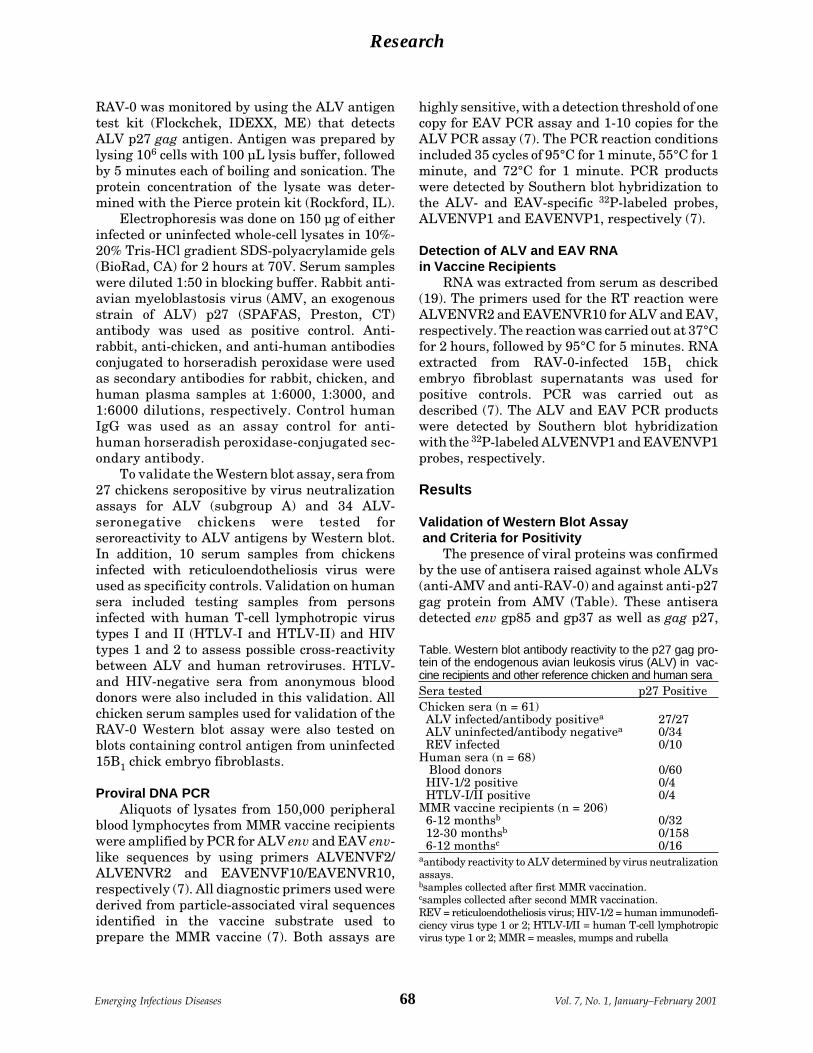
68Emerging Infectious Diseases Vol. 7, No. 1, January–February 2001
Research
RAV-0 was monitored by using the ALV antigentest kit (Flockchek, IDEXX, ME) that detectsALV p27 gag antigen. Antigen was prepared bylysing 106 cells with 100 µL lysis buffer, followedby 5 minutes each of boiling and sonication. Theprotein concentration of the lysate was deter-mined with the Pierce protein kit (Rockford, IL).
Electrophoresis was done on 150 µg of eitherinfected or uninfected whole-cell lysates in 10%-20% Tris-HCl gradient SDS-polyacrylamide gels(BioRad, CA) for 2 hours at 70V. Serum sampleswere diluted 1:50 in blocking buffer. Rabbit anti-avian myeloblastosis virus (AMV, an exogenousstrain of ALV) p27 (SPAFAS, Preston, CT)antibody was used as positive control. Anti-rabbit, anti-chicken, and anti-human antibodiesconjugated to horseradish peroxidase were usedas secondary antibodies for rabbit, chicken, andhuman plasma samples at 1:6000, 1:3000, and1:6000 dilutions, respectively. Control humanIgG was used as an assay control for anti-human horseradish peroxidase-conjugated sec-ondary antibody.
To validate the Western blot assay, sera from27 chickens seropositive by virus neutralizationassays for ALV (subgroup A) and 34 ALV-seronegative chickens were tested forseroreactivity to ALV antigens by Western blot.In addition, 10 serum samples from chickensinfected with reticuloendotheliosis virus wereused as specificity controls. Validation on humansera included testing samples from personsinfected with human T-cell lymphotropic virustypes I and II (HTLV-I and HTLV-II) and HIVtypes 1 and 2 to assess possible cross-reactivitybetween ALV and human retroviruses. HTLV-and HIV-negative sera from anonymous blooddonors were also included in this validation. Allchicken serum samples used for validation of theRAV-0 Western blot assay were also tested onblots containing control antigen from uninfected15B1 chick embryo fibroblasts.
Proviral DNA PCRAliquots of lysates from 150,000 peripheral
blood lymphocytes from MMR vaccine recipientswere amplified by PCR for ALV env and EAV env-like sequences by using primers ALVENVF2/ALVENVR2 and EAVENVF10/EAVENVR10,respectively (7). All diagnostic primers used werederived from particle-associated viral sequencesidentified in the vaccine substrate used toprepare the MMR vaccine (7). Both assays are
highly sensitive, with a detection threshold of onecopy for EAV PCR assay and 1-10 copies for theALV PCR assay (7). The PCR reaction conditionsincluded 35 cycles of 95°C for 1 minute, 55°C for 1minute, and 72°C for 1 minute. PCR productswere detected by Southern blot hybridization tothe ALV- and EAV-specific 32P-labeled probes,ALVENVP1 and EAVENVP1, respectively (7).
Detection of ALV and EAV RNAin Vaccine Recipients
RNA was extracted from serum as described(19). The primers used for the RT reaction wereALVENVR2 and EAVENVR10 for ALV and EAV,respectively. The reaction was carried out at 37°Cfor 2 hours, followed by 95°C for 5 minutes. RNAextracted from RAV-0-infected 15B1 chickembryo fibroblast supernatants was used forpositive controls. PCR was carried out asdescribed (7). The ALV and EAV PCR productswere detected by Southern blot hybridizationwith the 32P-labeled ALVENVP1 and EAVENVP1probes, respectively.
Results
Validation of Western Blot Assay and Criteria for Positivity
The presence of viral proteins was confirmedby the use of antisera raised against whole ALVs(anti-AMV and anti-RAV-0) and against anti-p27gag protein from AMV (Table). These antiseradetected env gp85 and gp37 as well as gag p27,
Table. Western blot antibody reactivity to the p27 gag pro-tein of the endogenous avian leukosis virus (ALV) in vac-cine recipients and other reference chicken and human seraSera tested p27 PositiveChicken sera (n = 61) ALV infected/antibody positivea 27/27 ALV uninfected/antibody negativea 0/34 REV infected 0/10Human sera (n = 68) Blood donors 0/60 HIV-1/2 positive 0/4 HTLV-I/II positive 0/4MMR vaccine recipients (n = 206) 6-12 monthsb 0/32 12-30 monthsb 0/158 6-12 monthsc 0/16aantibody reactivity to ALV determined by virus neutralizationassays.bsamples collected after first MMR vaccination.csamples collected after second MMR vaccination.REV = reticuloendotheliosis virus; HIV-1/2 = human immunodefi-ciency virus type 1 or 2; HTLV-I/II = human T-cell lymphotropicvirus type 1 or 2; MMR = measles, mumps and rubella
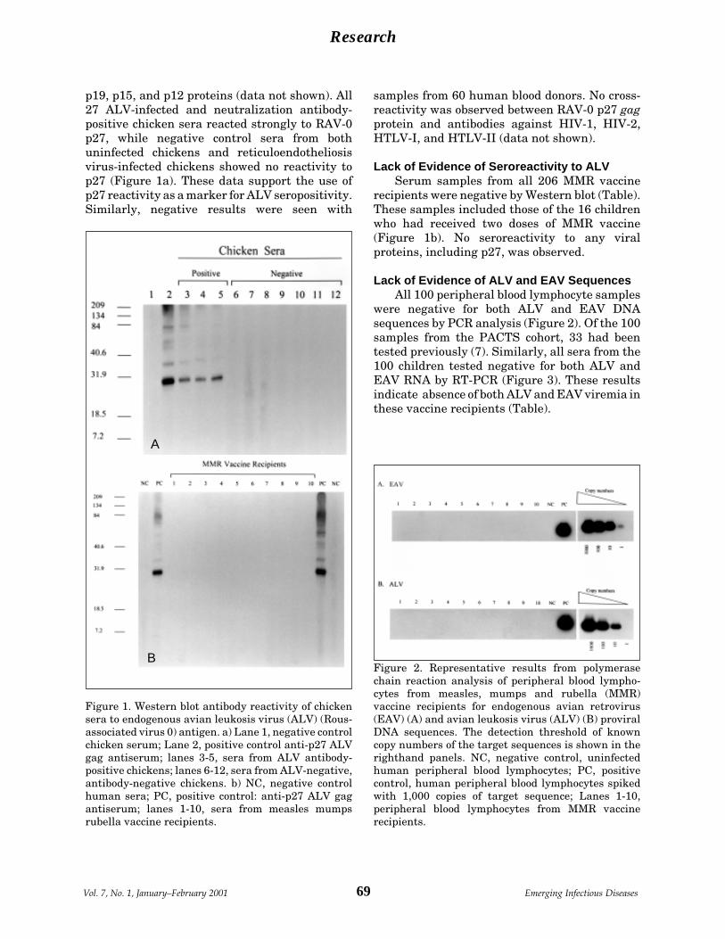
69Vol. 7, No. 1, January–February 2001 Emerging Infectious Diseases
Research
p19, p15, and p12 proteins (data not shown). All27 ALV-infected and neutralization antibody-positive chicken sera reacted strongly to RAV-0p27, while negative control sera from bothuninfected chickens and reticuloendotheliosisvirus-infected chickens showed no reactivity top27 (Figure 1a). These data support the use ofp27 reactivity as a marker for ALV seropositivity.Similarly, negative results were seen with
samples from 60 human blood donors. No cross-reactivity was observed between RAV-0 p27 gagprotein and antibodies against HIV-1, HIV-2,HTLV-I, and HTLV-II (data not shown).
Lack of Evidence of Seroreactivity to ALVSerum samples from all 206 MMR vaccine
recipients were negative by Western blot (Table).These samples included those of the 16 childrenwho had received two doses of MMR vaccine(Figure 1b). No seroreactivity to any viralproteins, including p27, was observed.
Lack of Evidence of ALV and EAV SequencesAll 100 peripheral blood lymphocyte samples
were negative for both ALV and EAV DNAsequences by PCR analysis (Figure 2). Of the 100samples from the PACTS cohort, 33 had beentested previously (7). Similarly, all sera from the100 children tested negative for both ALV andEAV RNA by RT-PCR (Figure 3). These resultsindicate absence of both ALV and EAV viremia inthese vaccine recipients (Table).
Figure 1. Western blot antibody reactivity of chickensera to endogenous avian leukosis virus (ALV) (Rous-associated virus 0) antigen. a) Lane 1, negative controlchicken serum; Lane 2, positive control anti-p27 ALVgag antiserum; lanes 3-5, sera from ALV antibody-positive chickens; lanes 6-12, sera from ALV-negative,antibody-negative chickens. b) NC, negative controlhuman sera; PC, positive control: anti-p27 ALV gagantiserum; lanes 1-10, sera from measles mumpsrubella vaccine recipients.
A
BFigure 2. Representative results from polymerasechain reaction analysis of peripheral blood lympho-cytes from measles, mumps and rubella (MMR)vaccine recipients for endogenous avian retrovirus(EAV) (A) and avian leukosis virus (ALV) (B) proviralDNA sequences. The detection threshold of knowncopy numbers of the target sequences is shown in therighthand panels. NC, negative control, uninfectedhuman peripheral blood lymphocytes; PC, positivecontrol, human peripheral blood lymphocytes spikedwith 1,000 copies of target sequence; Lanes 1-10,peripheral blood lymphocytes from MMR vaccinerecipients.

70Emerging Infectious Diseases Vol. 7, No. 1, January–February 2001
Research
ConclusionsAnalysis of MMR vaccines from different
manufacturers suggests that vaccine recipientsmay be universally exposed to endogenouschicken retroviral particles. We sought evidenceof persistent ALV and EAV infection in a largenumber of MMR vaccine recipients, and we wereunable to find any evidence of ALV or EAVsequences in peripheral blood lymphocytes,despite the use of highly sensitive PCR assays.Neither did we find evidence of ALV or EAVviremia, since all serum samples tested negativefor ALV and EAV RNA by RT-PCR analysis; thisfinding is of interest because ALV viremia iscommonly seen in chickens infected with ALV(18). All 206 serum samples tested by a validatedWestern blot assay were negative for ALVantibodies, indicating absence of antigenicexposure. These findings differ from those inpersons infected with human retroviruses, whousually seroconvert 2 weeks to 6 months afterexposure (20,21). The negative serologic data alsosuggest the low likelihood of nonviremic ALVinfection in cells other than peripheral bloodlymphocytes, which may not have been detectedby PCR testing. Our results overall show no
evidence of infection with either ALV or EAV inthese vaccine recipients. The lack of transmissionof ALV and EAV observed in 16 children who hadtwo MMR vaccinations provides additionalreassuring data.
Several factors, including a natural humanresistance to infection with endogenous ALV,may explain the lack of transmission of theseviruses to MMR vaccine recipients. However, fewor no data are available on the ability ofendogenous ALV to replicate in human cells.Resistance to endogenous ALV infection may, forinstance, be attributed to the absence of a humancell-surface receptor for the virus as well as toother intracellular blocks for ALV replication. Atumor necrosis factor receptor-related protein,referred to as SEAR, has been recently identifiedas a receptor for endogenous ALV in turkey cells(22). Plasmid-encoded expression of SEAR inhuman 293 cells can confer susceptibility toinfection by endogenous ALV, suggesting thathuman cells can support endogenous ALVreplication if virus entry is achieved (22). Humanserum can lyse ALV by complement activation(23); however, this protective mechanism has notbeen demonstrated for endogenous ALV and EAVparticles.
The presence of defective ALV and EAVparticles in vaccines may also explain the lack oftransmission of these viruses to vaccinerecipients. ev loci confer a variety of differentphenotypes, including infectious or defectiveparticles (9,10,18,24). However, it is not knownwhether the ALV particles in the vaccine are alldefective. The proportion of defective toinfectious ALV in different vaccine lots dependson the set of the ev loci in the chick embryofibroblast substrate preparation used for eachvaccine lot. Loci associated with noninfectiousviruses (ev-1, ev-3, and ev-6) have been identifiedin a chick embryo fibroblast substrate of a U.S.vaccine manufacturer (7). However, the presenceof many loci known to produce infectious ALV-Ecould not be determined (7).
EAV may represent the predominantretroviral particles in MMR vaccines (6,7).Therefore, our data, which indicate that exposureto EAV particles was not associated with EAVviremia or EAV-infected peripheral bloodlymphocytes in vaccine recipients, are important.Confirmation of our molecular results by EAV-specific serologic testing may, however, benecessary. The lack of evidence of transmission of
Figure 3. Reverse transcriptase-polymerase chainreaction analysis of sera from measles, mumps andrubella (MMR) vaccine recipients for the presence ofavian leukosis virus (ALV) (A) and endogenous avianretrovirus (EAV) (B) RNA. Lanes 1-10, samples fromMMR vaccine recipients. NC, negative control,uninfected human serum; PC, positive control, culturesupernatant from Rous-associated virus 0 (RAV-0)infected 15B1 chick embryo fibroblasts.

71Vol. 7, No. 1, January–February 2001 Emerging Infectious Diseases
Research
EAV to vaccinees is likely due to the presence ofdefective particles. No infectious EAVs have yetbeen isolated, nor has a full-length intact EAVprovirus been identified (25). However, ourunderstanding of the EAV family is limited.
The presence of ALV in chick-cell-derivedvaccines is not a new phenomenon; manyinstances of ALV contamination in yellow feverand measles vaccines have been documented(26,27). However, earlier vaccines had evidenceof exogenous rather than endogenous type ALV(27). Available data also suggest lack oftransmission of ALV to vaccine recipients(26,28,29). These studies examined 6 adults and41 children with measles vaccination and 227yellow fever vaccine recipients (26,27,29), and noevidence was found of neutralizing antibodies toan exogenous ALV. No increase in cancer rate hasbeen found in a study of recipients of exogenousALV-positive yellow fever vaccine (27).
Despite these reassuring data, the presenceof avian retroviral particles in chick embryofibroblast-derived vaccines raises questionsabout the suitability of primary chicken cellsubstrates for vaccine production and theadvisability of a change to RT-negative sub-strates. Chick embryo fibroblasts originatingfrom line 0 chickens could provide substratesthat do not express ALV-E; however, such cellsmay still produce EAV particles (7,12). Obtainingan RT-negative substrate may require asubstantial change from primary chicken cells toRT-negative cells from different species, such asimmortalized or diploid mammalian cells. Sincethe cell substrate is critical to the attenuation oflive vaccine viruses, any change in the cellsubstrate could have unpredictable effects on thesafety and efficacy of the vaccine and should beapproached cautiously.
In conclusion, we found no evidence of eitherendogenous ALV or EAV infection in recipients ofU.S.-made MMR vaccines. Our data indicate thatno change is warranted in current U.S. policiesfor the use of MMR vaccine.
AcknowledgmentsWe thank Alison Mawle, Harold Jaffe, and Rafael
Harpaz for critical reviews of the manuscript.
This work was supported in part by The NationalVaccine Program Office.
Dr. Hussain is a postdoctoral fellow at the Molecu-lar Epidemiology and Zoonoses Section, HIV andRetrovirology Branch, CDC, with research interests ininvestigating retroviral zoonotic infections.
References 1. Parkman PD. Safety of biopharmaceuticals: a current
perspective. Dev Biol Stand 1996;88:5-7. 2. Norman JE, Gilbert WB, Jay HH, Leonard BS.
Mortality follow-up of the 1942 epidemic of hepatitis Bin the U.S. Army. Hepatology 1993;18:790-7.
3. Fisher SG, Weber L, Carbone M. Cancer risk associatedwith simian virus 40 contaminated polio vaccine.Anticancer Res 1999;19:2173-80.
4. Boni J, Stadler J, Reigei F, Shupbach J. Detection ofreverse transcriptase activity in live attenuated virusvaccines. Clin Diagn Virol 1996;5:43-53.
5. Robertson JS, Nicolson C, Riley A-M, Bently M, DunnG, Corcoran T, et al. Assessing the significance ofreverse transcriptase activity in chick cell-derivedvaccines. Biologicals 1997;25:403-14.
6. Weissmahr RN, Schupbach J, Boni J. Reversetranscriptase activity in chicken embryo culturesupernatants is associated with particles containingendogenous avian retrovirus EAV-0 RNA. J Virol1997;71:3005-12.
7. Tsang SX, Switzer WM, Shanmugam V, Johnson JA,Goldsmith C, Wright A, et al. Evidence of avian leukosisvirus subgroup E and endogenous avian virus inmeasles and mumps vaccines derived from chickencells: investigation of transmission to vaccinerecipients. J Virol 1999;73:5843-51.
8. WHO Expert Committee on Biological Standardiza-tion. Requirements for measles, mumps and rubellavaccines and combined vaccines (live). Requirementsfor biological substances, no. 47. World Health OrganTech Rep Ser 1994;840:100-207.
9. Coffin JM, Tsichlis PN, Conklin KF, Senior A, RobinsonHL. Genomes of endogenous and exogenous avianretroviruses. Virology 1983;126:51-72.
10. Payne LN. Biology of avian retroviruses. In: Levy JA,editor. The retroviridae. New York: Plenum Press;1992. p. 299-403.
11. Weiss RA, Frisby DP. Are avian endogenous virusespathogenic? In: Yohn D, Blakeslee IR, editors.Proceedings of the tenth international symposium forcomparative research on leukemia and other diseases.Amsterdam: Elsevier;1982. p. 303-11.
12. Resnick RM, Boyce-Jacino MT, Fu Q, Faras AJ.Phylogenetic distribution of the novel avian endoge-nous provirus family EAV-0. J Virol 1990;64:4640-53.
13. Dunwiddie C, Faras AJ. Presence of reversetranscriptase-related gene sequences in avian cellslacking endogenous avian leukosis viruses. Proc NatlAcad Sci U S A 1985;82:5097-101.
14. Maudru T, Cooperating units, Peden KWC. Analysis ofcoded panel of licensed vaccines by polymerase chainreaction-based reverse transcriptase assays: a collabo-rative study. J Clin Virol 1998;11:19-28.

72Emerging Infectious Diseases Vol. 7, No. 1, January–February 2001
Research
15. WHO. Reverse transcriptase activity in chicken-cellderived vaccine. Wkly Epidemiol Rec 1998;28:209-12.
16. Thomas PA, Weedon J, Krasinski K Abrams E, ShafferN, Matheson P, et al. Maternal predictors of perinatalhuman immunodeficiency virus transmission. PediatrInfect Dis J 1994;13:489-95.
17. Helfand RF, Gary HE, Atkinson WL, Nordin JD,Keyserling HL, Bellini WJ. Decline of measles-specificimmunoglobulin M antibodies after primary measles,mumps and rubella vaccination. Clin Diagn LabImmunol 1998;5:135-8.
18. Crittenden LB. Retroviral elements in the genome ofthe chicken: implications for poultry genetics andbreeding. Crit Rev Poultry Biol 1991;3:73-109.
19. Mulder J, McKinney N, Christopherson C, Sninsky J,Greenfield L, Kwok S. Rapid and simple PCR assay forquantitation of human immunodeficiency virus type 1RNA in plasma: application to acute retroviralinfection. J Clin Microbiol 1994;32:292-300.
20. Lal RB, Heneine W. Testing of human T-lymphotropicvirus types I and II: serologic, virologic, and moleculardetection. Human T-cell lymphotropic virus type I NewYork: John Wily and Sons; 1996. p. 167-95.
21. Paul DA, Falk LA, Kessler HA, Chase RM, Blaauw B,Chudwin DS, et al. Correlation of serum HIV antigenand antibody with clinical status in HIV infectedpatients. J Med Virol 1987;22:357-63.
22. Adkins HB, Brojatsch J, Naughton J, Rolls MM, PesolaJM, Young JA. Identification of a cellular receptor forsubgroup E avian leukosis virus. Proc Natl Acad Sci US A 1997;94:11617-22.
23. Welsh RM, Cooper NR, Jensen FC, Oldstone MBA.Human serum lyses RNA tumor viruses. Nature1975;257:612-4.
24. Fadly AM. Avian retroviruses. Vet Clin North Am FoodAnim Pract 1997;1:71-85.
25. Jacino-Boyce MT, O’Donoghue K, Faras AJ. Multiplecomplex families of endogenous retroviruses are highlyconserved in the genus gallus. J Virol 1992;66:4919-29.
26. Dougherty RM, Harris RJC. Contaminant viruses intwo live virus vaccines produced in chicken cells. J HygCambridge 1966;64:1-7.
27. Waters TD, Anderson PS, Beebe GW, Miller RW.Yellow fever vaccination, avian leukosis virus, andcancer risk in man. Science 1972;177:76-77.
28. Levine S, Markham FS. The absence of serologicresponses by children and adults to avian leukosis virusin measles vaccine. Archiv fur die gesante Virusfors-chung 1965;15:305-10.
29. Richman AV, Auliso CG, Jahnes WG, Tauraso NM.Avian leukosis antibody response in individuals givenchicken embryo derived vaccines. Proc Soc Exp BiolMed 1972;139:235-7.
73Vol. 7, No. 1, January–February 2001 Emerging Infectious Diseases
Research
Rickettsia are intracellular Proteobacteriaassociated with arthropods, including body lice,fleas, ticks, and mites (1). R. typhi, the agent ofmurine typhus, is transmitted by rat fleas,Xenopsylla cheopsis. In 1990, when cat fleas(Ctenocephalides felis) were examined as pos-sible vectors of R. typhi, a novel Rickettsia-likeorganism was observed by electron microscopy inmidgut epithelial cells of the fleas. The agent,named the ELB agent for the EL Laboratory(Soquel, CA) (2), was detected in 1994 and 2000by polymerase chain reaction (PCR) in fourpatients from Texas and Mexico (3,4). Thetaxonomic position of this organism within thegenus Rickettsia was assessed by genomicsequence comparison, following the successfulamplification of a 17-kDa protein gene fragmentfrom infected flea tissue by PCR with genus-specific primers (5). In addition, the organismwas found to be transmitted transovarially infleas (5) and to be pathogenic in a human patient(6). In 1995, the name “R. felis” was proposed forthe ELB agent on the basis of its phenotypic
characteristics, as well as its clear genotypicdifferences from other known Rickettsia species(6). The organism was provisionally named “R.felis” but the name is not formally approved byInternational Society for Systematic and Evolu-tionary Biology as no strain was deposited in anyofficial collection. In 1997, the ELB agent wasdetected in two other flea species in the UnitedStates, C. felis and Pulex irritans (7). Althoughisolation in tissue culture was reported (3,8,9),contamination with R. typhi has hamperedsubsequent work (6), so no isolate of R. felis wasavailable when we began our study.
We describe methods used to cultivateseveral isolates of the ELB agent and itsmorphologic, antigenic, and genomic characteris-tics, as well as the results of a serosurvey withone of our type strains.
Materials and MethodsC. felis fleas (Flea Data Inc., Freeville, NY)
were divided into 20 groups of 5. After surfacesterilization by a 5-minute immersion in 70%methanol with 0.2% iodine, the fleas werewashed in sterile distilled water and frozen inliquid nitrogen. Frozen fleas were maceratedwith a sterile plastic spatula, suspended in0.8 mL of culture medium, and injected into shell
A Flea-Associated RickettsiaPathogenic for Humans
Didier Raoult,* Bernard La Scola,* Maryse Enea,*Pierre-Edouard Fournier,* Véronique Roux,*
Florence Fenollar,* Marcio A.M. Galvao,† Xavier de Lamballerie** Unité des Rickettsies, CNRS UPRESA 6020, France;
†Ouro Preto Federal University, Brazil
Address for correspondence: D. Raoult, Unité des Rickettsies,CNRS UPRESA 6020, Faculté de Médecine, Université de laMéditerranée, 27 Bd Jean Moulin, 13385 Marseille Cedex05, France; fax: 33-4-91-38-7772; e-mail: [email protected].
A rickettsia named the ELB agent, or “Rickettsia felis,” was identified by molecularbiology techniques in American fleas in 1990 and later in four patients from Texas andMexico. We attempted to isolate this rickettsia from infected fleas at varioustemperatures and conditions. A representative isolate of the ELB agent, the Marseillestrain, was characterized and used to develop a microimmunofluorescence test thatdetected reactive antibodies in human sera. The ELB agent was isolated from 19 of 20groups of polymerase chain reaction-proven infected fleas. The microimmunofluores-cence results provided serologic evidence of infection by the ELB agent in four patientswith fever and rash in France (2) and Brazil (2), supporting the pathogenic role of thisrickettsia. Our successful isolation of this rickettsia makes it available for use inserologic tests to determine its clinical spectrum, prevalence, and distribution.

74Emerging Infectious Diseases Vol. 7, No. 1, January–February 2001
Research
vials. Fifty microliters of the suspension wasretained for use as template in a Rickettsia-specific PCR targeting a fragment of the citratesynthase-encoding gene (gltA) (10).
Human embryonic lung fibroblasts (11) or theXTC-2 cell line derived from Xenopus laevis (12)were used for isolation by the shell vialcentrifugation technique (6). Subconfluent cellmonolayers were obtained by incubating the shellvials at 28°C for 48 hours after they were injectedwith 50,000 cells in 1 mL of Leibowitz-15 mediumwith L-glutamine and L-amino-acids (GIBCO,Rockville, MD), 5% (v/v) fetal calf serum and 2%(v/v) tryptose phosphate (GIBCO) for XTC-2cells, and Minimum Essential Medium (GIBCO)supplemented with 2 mM L-glutamine and 10%fetal bovine serum for human embryonic lungfibroblasts. Before injection with the flea extract,the medium was removed by aspiration.
After injection of both cell types withsuspensions of five fleas resuspended in 0.8 mL ofthe corresponding medium, the shell vials werecentrifuged at 700 X g for 1 hour at 20°C, and thesupernatant was discarded. After two washingsin sterile phosphate-buffered saline (PBS), 1 mLof fresh medium containing 4 µg/mLcotrimoxazole, an antibiotic used to preventcontamination, was added to the shell vials,which were incubated at 28°C. The cell culturemedium was replaced every 7 days for up to 30days. When rickettsiae could be detected in cellsin the discarded medium by Gimenez staining(13), the infected cell monolayer was harvestedand spread onto a subconfluent cell monolayer ofthe same cell line in a 25-cm2 tissue culture flask,which was also incubated at 28°C.
For further studies, we used our first isolateof the ELB agent, which we named the Marseillestrain. We attempted to infect mammalian celllines with the Marseille isolate on monolayers ofVero, MRC-5, and L-929 cells incubated with 5%CO2 at 28°C, 32°C, or 37°C, in the same mediumas human embryonic lung fibroblasts. Theultrastructure of the Marseille isolate wasstudied by electron microscopy (14), and thestrain was purified as for other rickettsia (15).
To quantify growth of the ELB agent, 1 µL of10-1, 10-2, 10-3, 10-4, 10-5, 10-6 dilutions of asuspension made of rickettsiae harvested from a25-cm2 tissue culture flask and purified from cellswere deposited onto 30-well microscope slides(Dynatech Laboratories Ltd., Billingshurt, UK),air dried, fixed with acetone for 10 minutes, and
then stained by the Gimenez technique. Thenumber of rickettsiae was estimated visually.
To raise polyclonal antisera against theMarseille isolate, 6- to 8-week-old Balb C micewere injected intraperitoneally with 1 mL of asolution containing v/v 106 purified Marseilleisolate in PBS and complete Freund’s adjuvant.Inoculation was repeated at 10, 20, and 30 days.At 40 days, blood was collected by intracardiacpuncture, and sera were stored at –20°C. Thesame procedure was performed with R. conorii(Moroccan strain, ATCC VR 141) and R. typhi(Wilmington strain, ATCC VR-144).
To determine the prevalence of antibodiesreactive with the organism in the generalpopulation in France, serum specimens from 100French blood donors were tested by microimmuno-fluorescence against the Marseille strain; 140serum samples from Brazilian blood donors,which had been sent to our laboratory to estimatethe seroprevalence of various rickettsioses, werealso tested. Microimmunofluorescence was alsoused to determine cross-reactivity between ourstrain of the ELB agent and other rickettsiae.Convalescent-phase serum specimens from 67patients with epidemiologic, clinical, and serolog-ic evidence of epidemic typhus, 16 patients withmurine typhus, and 97 French patients withfever and rash serologically and clinicallydiagnosed as Mediterranean spotted fever, weretested by microimmunofluorescence for antibod-ies against R. rickettsii, R. typhi, and theMarseille strain. Serum samples from 16Brazilian patients with unexplained febrile rashwere also included in these tests. For microim-munofluorescence, R. conorii strain Seven(Malish), R. rickettsii strain R (Bitterroot),R. prowazekii strain Brein L, and R. typhi strainWilmington were grown in Vero cells and purified(11). These antigens and the purified suspensionof the Marseille strain described above wereapplied at separate sites on each well of 30-wellmicroscope slides (Dynatech Laboratories, Ltd.),air dried, and fixed with acetone for 10 min.Microimmunofluorescence tests were performed(16), with immunoglobulin (Ig) G and IgM titersdetermined separately. To remove antibodiesagainst any host-cell components, antisera wereabsorbed with XTC-2 or Vero cells before beingused in the microimmunofluorescence. More-over, before detection of IgM, the antisera wereabsorbed with rheumatoid factor absorbent(Behring-Werke AG, Marburg, Germany). The

75Vol. 7, No. 1, January–February 2001 Emerging Infectious Diseases
Research
antisera were then applied to the fixed antigensat doubling dilutions from 1:4 to 1:2,048 (16).
Analysis of major proteins of the Marseilleisolate by sodium dodecyl-sulfate polyacrylamidegel electrophoresis (SDS-PAGE) were performedby using both heated and unheated antigens (17).Major immunogenic proteins were studied byWestern blot with purified unheated antigens(14,17).
For PCR amplification and sequencing ofgene fragments from macerated flea suspensionsand rickettsial isolates, genomic DNA wasextracted by using the QIAmp tissue kit (Qiagen,Hilden, Germany) according to the manufacturer’sinstructions. In addition to the 17-kDa antigen- andcitrate synthase-encoding genes determined byAzad et al. (5) and Higgins et al., respectively (3),we amplified and sequenced fragments of thegenes encoding the rickettsial outer membraneproteins A and B (ompA and ompB). Primerswere designed within conserved regions of thegenes, and amplifications were carried out as
described (Table 1,18). Sequencing reactions withthese primers were done with the dRhodamineTerminator sequencing kit (PE Applied Biosys-tems, Warrington, UK). The reaction productswere resolved on 5% polyacrylamide gels (Tebu,Le Perray en Yvelines, France) by an ABI PRISM377 DNA sequencer (PE Applied Biosystems).Amplifications of R. typhi by PCR fromsubcultures of the Marseille strain were performedby using the gltA-derived R. typhi-specific primersTY1f (5'-TGGGGAACTACCAAGTAGT-3') andTY1r (5'-ACCAGTGCTAATACATGCAA-3') asdescribed to determine the purity of the culture.DNA was extracted as described. DNA fromR. typhi cultured in Vero cells was used aspositive control.
Nucleotide sequences were aligned withsequences from other Rickettsia species inGenBank by using the multisequence alignmentsoftware CLUSTAL within the BISANCEenvironment (20). The phylogenetic relation-ships between our strain and other representative
Table 1. Primers used for PCR amplification and sequencing of the ELB agent strain Marseille-URRWFXCal2 Geneposition relative to open
Gene Primer Nucleotide sequence (5'-3') reading frame Reference17-kDa antigen 17kDFa GCTCTTGCAACTTCTATGTT 31-50 This manuscript
17KdRa CATTGTTCGTCAGGTTGGCG 464-445 This manuscriptCitrate synthase CSFEL1a TGATTCAGAATTTGCCGAAT 21-40 17
CS890ra GCTTTAGCTACATATTTAGG 890-871 17CS532ra GCCGCAATGTCTTATAAATATTCT 532-555 This manuscriptCS1273ra CATAACCAGTGTAAAGCTG 1273-1255 This manuscriptCS244rb CTTTAATATCATATCCTCGAT 244-224 17Rp877Pb GGGGACCTGCTCACGGCGG 797-815 18
rOmpB 120-M59a CCGCAGGGTTGGTAACTGC M59-M41 This manuscript120-807a CCTTTTAGATTACCGCCTAA 807-788 This manuscript120-607a AATATCGGTGACGGTCAAGG 607-626 This manuscript120-1497a CTATATCGCCGGTAATT 1497-1480 This manuscript120F-1351a TTTAGGAAACGCTGGTTCTC 1351-1370 This manuscript120F-2934a GCGTTAGTTGCGATAATACT 2934-2915 This manuscript120-2788a AAACAATAATCAAGGTACTGT 2788-2808 This manuscript120-3599a TACTTCCGGTTACAGCAAAGT 3599-3579 This manuscript120F-3440a GTTAATGCAACAACTACGGG 3440-3459 This manuscript120F-4341a GCATCGAAGAAGTAACGCTG 4341-4322 This manuscript120-4232a GGTTTCTCATTCTCTCTATATGG 4232-4254 This manuscript120-4879a TTAGAAGTTTACACGGACTTT 4879-4857 This manuscript120F-1749b CTATTAGTGGTAATATTGGTAC 1749-1770 This manuscript120F-2495b CATGGTCATTACCTGCATTACC 2495-2481 This manuscript120-4489b GTCTTCTGACGAAAACTACAA 4489-4509 This manuscript
ROmpA 190-70a ATGGCGAATATTTCTCCAAAA 70-90 18190-701a GTTCCGTTAATGGCAGCATCT 701-681 19
aPrimers used for both PCR amplification and sequencing of the ELB agent strain Marseille-URRWFXCal2.bPrimers used only for sequencing.

76Emerging Infectious Diseases Vol. 7, No. 1, January–February 2001
Research
Table 2. Time required to infect 90% of cells in a 174-cm2
cell culture flask after inoculation with 5 x10⁵ ELB agentstrain Marseille-URRWFXCal2
Incubation temperaturesCell type 28°C 32°C 37°CXTC-2 cells 6 days NDa NDa
Vero cells 14 days 14 days NoMRC-5 cells NGb NGb NGb
L-929 cells NGb NGb NGb
aND = not done. XTC-2 cells died at temperatures 32°C.bNG = no growth was obtained
Figure 1. Transmission electron micrograph of theELB agent in XTC-2 cells. The rickettsia are free in thecytoplasm and surrounded by an electron transparenthalo. Original magnification X 30,000.
strains based on the analysis of gltA and ompBsequences were determined by using the Phylipsoftware (21). The distance matrix generated byDNADIST was determined under the assump-tion of Jukes and Cantor (22) and used toconstruct a dendrogram by the neighbor-joiningmethod (23). Two other dendrograms wereconstructed by using data processing with themaximum-likelihood and parsimony programDNAPARS. Bootstrap replicates were performedby using SEQBOOT and CONSENSE in thePHYLIP software to estimate the node reliabilityof the trees obtained by the three methods (24).
To improve the sensitivity of PCR and detectrickettsial DNA from serum samples, wedesigned a nested PCR based on the amplifica-tion of the gltA gene. DNA was extracted from200 µL of serum by using the QIAmp blood kit(Qiagen) as recommended by the manufacturer.External primers designed for this purpose wereELB1f (5'-CTGCTTCTTGTCAGTCTAC-3') andELB1r (5'-GATTTTTTGTTCAGGGTCTTC-3'), andinternal primers were ELB2f (5'-GGAATCTTGCGACATCGA-3') and ELB2r (5'-CAGCCTACGGTTCTTGC-3'). The internal primers encom-passed a 952-bp gltA fragment allowing a reliableidentification of most rickettsial species aftersequencing (including R. felis, R. conorii, andR. rickettsii). Amplification, sequencing ofamplicons, and sequence analysis were done asdescribed. Forty-seven serum samples weretested with this technique, 27 from patients withrickettsial diseases (including those reactingserologically to the ELB agent) and 20 from blooddonors used as negative controls.
ResultsThe ELB agent was detected by PCR
amplification of a gltA fragment from all the fleasuspensions used to inoculate the cell culturemonolayers. Initial attempts to isolate therickettsia in human embryonic lung fibroblastsfailed, but growth was observed by Gimenezstaining and the rickettsiae were confirmed asthe ELB agent by PCR analysis: 100% homologywas observed with DNA amplified with the gltAsequence of the ELB agent in 19 of 20supernatants from the XTC-2 cell monolayersgrown at 28°C 7 and 14 days after inoculation.R. typhi DNA could not be detected by PCR in anyof the cell culture supernatants, whereas theprimer pair Ty1f and TY1r amplified positivecontrol DNA.
Initial isolation required 14 days, andsubsequent passages required 6 days to detectthe ELB agent. Cultures of all isolates wereeasily established in XTC-2 cells from the 19suspensions. The reference strain, Marseille-URRWFXCal2, has been deposited (accessionnumber I-2363) in the French National CultureCollection (Institut Pasteur, Paris, France) andwill be sent to the American Type CultureCollection. Before further studies of this strain,we reconfirmed by PCR that our strain of the ELBagent was not contaminated with R. typhi.
The Marseille strain grew most rapidly at28°C in XTC-2 cells, which died at temperatures>32°C (Table 2). Human embryonic lung fibroblastsdo not multiply at 28°C, as their optimal growthtemperature is 37°C, and therefore the ELBagent could not be cultivated in this cell line. Theorganism also grew in Vero cells incubated ateither 28°C or 32°C but at half the rate of growthobserved in XTC-2 cells. The MRC-5 and L-929cell lines were unable to support permanentgrowth of the Marseille strain. Electronmicroscopy showed the rickettsia to be presentand free in the cytoplasm but not in the nucleus ofthe cells (Figure 1).

77Vol. 7, No. 1, January–February 2001 Emerging Infectious Diseases
Research
Figure 2. Phylogenetic tree of members of the genus Rickettsia inferred from comparison of gltA sequences byusing the neighbor-joining method. Bootstrap values for the nodes are indicated.
Four gene fragments were successfullyamplified by PCR, and the base sequences of bothDNA strands of each segment were determinedtwice. Nonambiguous sequence data wereobtained between bases 51 and 444 of the 17-kDaantigen-encoding gene (394 base pairs), bases 41and 1236 of gltA (1196 bp), bases 91 to 665 ofompA (575 bp), and bases 1 to 1236 of ompB (1236bp). Sequences of the 17-kDa protein-encodinggene and gltA were 100% homologous with thosein GenBank (accession numbers M82878 andU33922, respectively). The GenBank accession
numbers for the Marseille strain of the ELBagent nucleotide sequence data reported in thispaper are as follows: citrate synthase-encodinggene, AF210692; 17-kDa protein-encoding gene,AF210693; outer membrane protein A-encodinggene, AF210694; and outer membrane protein B-encoding gene, AF2106695. Phylogenetic analy-sis inferred from the comparison of gltA andompB nucleotide sequences with the threeanalysis methods produced similar organiza-tions. The ELB agent clustered with R. akari andR. australis (Figure 2).
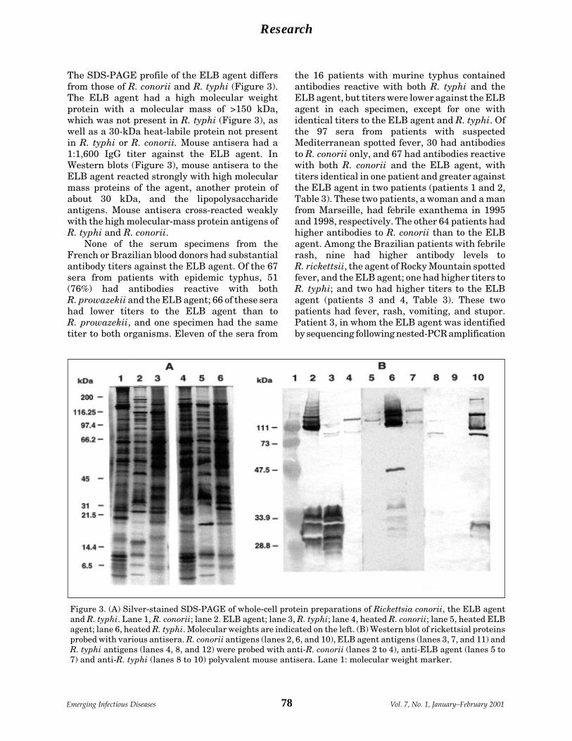
78Emerging Infectious Diseases Vol. 7, No. 1, January–February 2001
Research
The SDS-PAGE profile of the ELB agent differsfrom those of R. conorii and R. typhi (Figure 3).The ELB agent had a high molecular weightprotein with a molecular mass of >150 kDa,which was not present in R. typhi (Figure 3), aswell as a 30-kDa heat-labile protein not presentin R. typhi or R. conorii. Mouse antisera had a1:1,600 IgG titer against the ELB agent. InWestern blots (Figure 3), mouse antisera to theELB agent reacted strongly with high molecularmass proteins of the agent, another protein ofabout 30 kDa, and the lipopolysaccharideantigens. Mouse antisera cross-reacted weaklywith the high molecular-mass protein antigens ofR. typhi and R. conorii.
None of the serum specimens from theFrench or Brazilian blood donors had substantialantibody titers against the ELB agent. Of the 67sera from patients with epidemic typhus, 51(76%) had antibodies reactive with bothR. prowazekii and the ELB agent; 66 of these serahad lower titers to the ELB agent than toR. prowazekii, and one specimen had the sametiter to both organisms. Eleven of the sera from
the 16 patients with murine typhus containedantibodies reactive with both R. typhi and theELB agent, but titers were lower against the ELBagent in each specimen, except for one withidentical titers to the ELB agent and R. typhi. Ofthe 97 sera from patients with suspectedMediterranean spotted fever, 30 had antibodiesto R. conorii only, and 67 had antibodies reactivewith both R. conorii and the ELB agent, withtiters identical in one patient and greater againstthe ELB agent in two patients (patients 1 and 2,Table 3). These two patients, a woman and a manfrom Marseille, had febrile exanthema in 1995and 1998, respectively. The other 64 patients hadhigher antibodies to R. conorii than to the ELBagent. Among the Brazilian patients with febrilerash, nine had higher antibody levels toR. rickettsii, the agent of Rocky Mountain spottedfever, and the ELB agent; one had higher titers toR. typhi; and two had higher titers to the ELBagent (patients 3 and 4, Table 3). These twopatients had fever, rash, vomiting, and stupor.Patient 3, in whom the ELB agent was identifiedby sequencing following nested-PCR amplification
Figure 3. (A) Silver-stained SDS-PAGE of whole-cell protein preparations of Rickettsia conorii, the ELB agentand R. typhi. Lane 1, R. conorii; lane 2. ELB agent; lane 3, R. typhi; lane 4, heated R. conorii; lane 5, heated ELBagent; lane 6, heated R. typhi. Molecular weights are indicated on the left. (B) Western blot of rickettsial proteinsprobed with various antisera. R. conorii antigens (lanes 2, 6, and 10), ELB agent antigens (lanes 3, 7, and 11) andR. typhi antigens (lanes 4, 8, and 12) were probed with anti-R. conorii (lanes 2 to 4), anti-ELB agent (lanes 5 to7) and anti-R. typhi (lanes 8 to 10) polyvalent mouse antisera. Lane 1: molecular weight marker.
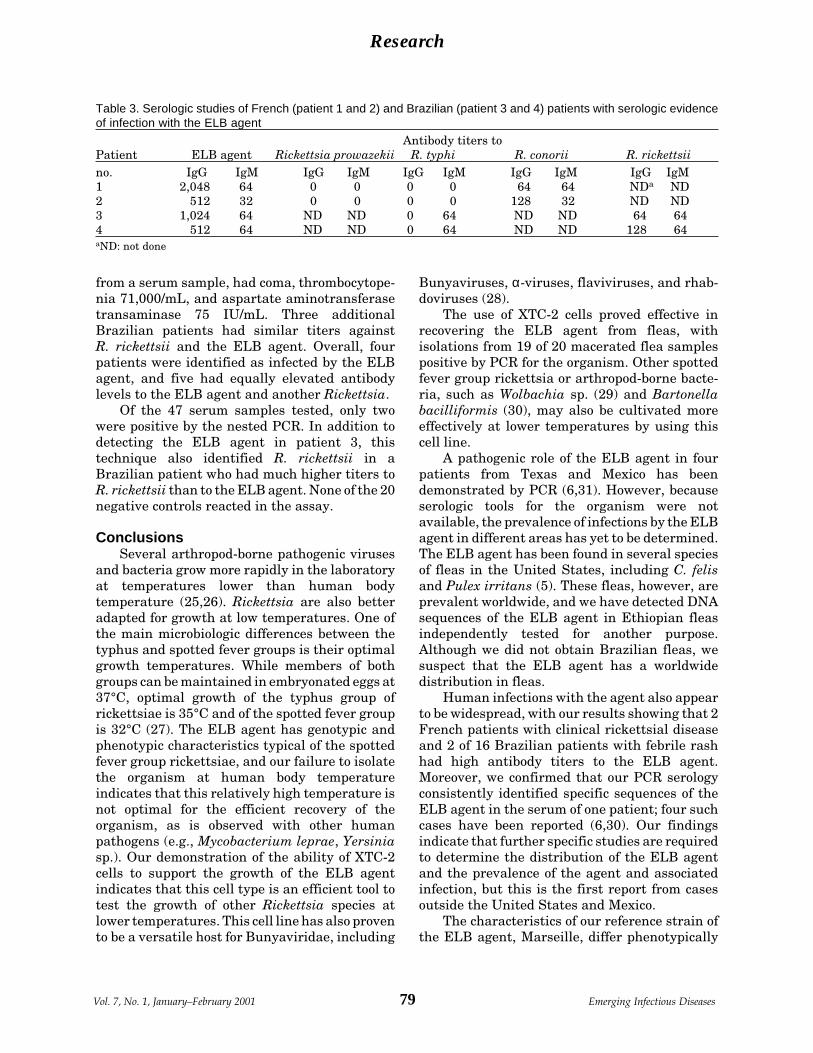
79Vol. 7, No. 1, January–February 2001 Emerging Infectious Diseases
Research
from a serum sample, had coma, thrombocytope-nia 71,000/mL, and aspartate aminotransferasetransaminase 75 IU/mL. Three additionalBrazilian patients had similar titers againstR. rickettsii and the ELB agent. Overall, fourpatients were identified as infected by the ELBagent, and five had equally elevated antibodylevels to the ELB agent and another Rickettsia.
Of the 47 serum samples tested, only twowere positive by the nested PCR. In addition todetecting the ELB agent in patient 3, thistechnique also identified R. rickettsii in aBrazilian patient who had much higher titers toR. rickettsii than to the ELB agent. None of the 20negative controls reacted in the assay.
ConclusionsSeveral arthropod-borne pathogenic viruses
and bacteria grow more rapidly in the laboratoryat temperatures lower than human bodytemperature (25,26). Rickettsia are also betteradapted for growth at low temperatures. One ofthe main microbiologic differences between thetyphus and spotted fever groups is their optimalgrowth temperatures. While members of bothgroups can be maintained in embryonated eggs at37°C, optimal growth of the typhus group ofrickettsiae is 35°C and of the spotted fever groupis 32°C (27). The ELB agent has genotypic andphenotypic characteristics typical of the spottedfever group rickettsiae, and our failure to isolatethe organism at human body temperatureindicates that this relatively high temperature isnot optimal for the efficient recovery of theorganism, as is observed with other humanpathogens (e.g., Mycobacterium leprae, Yersiniasp.). Our demonstration of the ability of XTC-2cells to support the growth of the ELB agentindicates that this cell type is an efficient tool totest the growth of other Rickettsia species atlower temperatures. This cell line has also provento be a versatile host for Bunyaviridae, including
Bunyaviruses, α-viruses, flaviviruses, and rhab-doviruses (28).
The use of XTC-2 cells proved effective inrecovering the ELB agent from fleas, withisolations from 19 of 20 macerated flea samplespositive by PCR for the organism. Other spottedfever group rickettsia or arthropod-borne bacte-ria, such as Wolbachia sp. (29) and Bartonellabacilliformis (30), may also be cultivated moreeffectively at lower temperatures by using thiscell line.
A pathogenic role of the ELB agent in fourpatients from Texas and Mexico has beendemonstrated by PCR (6,31). However, becauseserologic tools for the organism were notavailable, the prevalence of infections by the ELBagent in different areas has yet to be determined.The ELB agent has been found in several speciesof fleas in the United States, including C. felisand Pulex irritans (5). These fleas, however, areprevalent worldwide, and we have detected DNAsequences of the ELB agent in Ethiopian fleasindependently tested for another purpose.Although we did not obtain Brazilian fleas, wesuspect that the ELB agent has a worldwidedistribution in fleas.
Human infections with the agent also appearto be widespread, with our results showing that 2French patients with clinical rickettsial diseaseand 2 of 16 Brazilian patients with febrile rashhad high antibody titers to the ELB agent.Moreover, we confirmed that our PCR serologyconsistently identified specific sequences of theELB agent in the serum of one patient; four suchcases have been reported (6,30). Our findingsindicate that further specific studies are requiredto determine the distribution of the ELB agentand the prevalence of the agent and associatedinfection, but this is the first report from casesoutside the United States and Mexico.
The characteristics of our reference strain ofthe ELB agent, Marseille, differ phenotypically
Table 3. Serologic studies of French (patient 1 and 2) and Brazilian (patient 3 and 4) patients with serologic evidenceof infection with the ELB agent
Antibody titers toPatient ELB agent Rickettsia prowazekii R. typhi R. conorii R. rickettsiino. IgG IgM IgG IgM IgG IgM IgG IgM IgG IgM1 2,048 64 0 0 0 0 64 64 NDa ND2 512 32 0 0 0 0 128 32 ND ND3 1,024 64 ND ND 0 64 ND ND 64 644 512 64 ND ND 0 64 ND ND 128 64aND: not done
80Emerging Infectious Diseases Vol. 7, No. 1, January–February 2001
Research
from those reported for the lost R. felis strain (6).Our strain does not grow at 35°C or 37°C, and itsSDS–PAGE protein profiles and Western blotsdiffered from those reported for R. felis andR. typhi (3,7,8). Further studies will determine ifthe reactive 30-kDa heat-labile protein observedon Western blot could be a truncated rOmpAprotein, as predicted by genomic studies (31). ByPCR and sequencing, we identified our isolates asthe ELB agent on the basis of 100% gltA sequencehomology with the sequence available for R. felis(5,6). Therefore, discrepancies with previouslyreported phenotypic findings may result fromcontamination of R. felis cultures with R. typhi,which was reported after experiments by thegroup that described R. felis (9). In ourexperiments based on PCR techniques, however,we found no evidence of contamination of ourisolate with R. typhi. To avoid confusion betweenthe characteristics of our isolate and those of thepreviously characterized R. felis, we arepreparing a formal taxonomic characterization ofour isolate of the ELB agent.
In summary, our experiments have demon-strated the usefulness of XTC-2 cells in isolatingarthropod-associated microorganisms. This cellculture system allowed us to establish and makeavailable the Marseille strain of the ELB agent.In addition, we have identified likely cases ofinfection by the ELB agent. The techniques thatwe describe should facilitate further studies todetermine the prevalence and clinical spectrumof infection by this organism in humans.
AcknowledgmentsThe authors thank J. Georgi of the El Labs in Soquel,
California, for providing cat fleas, S.B. Calic and C.B.Chamone for providing Brazilian patients’ serum specimens,and P.J. Kelly for review of the translation.
Dr. Raoult is Director of the Unité des Rickettsies,the national reference center for rickettsiosis and WHOcollaborative center. The laboratory is mostly involved inthe study of emerging and reemerging bacteria and ar-thropod-borne diseases.
References 1. Raoult D, Roux V. Rickettsioses as paradigms of new or
emerging infectious diseases. Clin Microbiol Rev1997;10:694-719.
2. Adams JR, Schmidtmann ET, Azad AF. Infection ofcolonized cat fleas, Ctenocephalides felis (Bouché), witha rickettsia-like microorganism. Am J Trop Med Hyg1990;43:400-9.
3. Higgins JA, Radulovic S, Schriefer ME, Azad AF.Rickettsia felis: a new species of pathogenic rickettsiaisolated from cat fleas. J Clin Microbiol 1996;34:671-4.
4. Zavala-Velasquez JE, Sosa-Ruiz JA, Zavala-Castro J,Jimenez-Delgadillo B, Vado-Solis IE, Sanchez-Elias RA, etal. Rickettsia felis: the etiologic agent of three cases ofrickettsiosis in Yucatan. Lancet 2000;356:1079-80.
5. Azad AF, Sacci JB, Nelson WM, Dasch GA,Schmidtmann ET, Carl M. Genetic characterizationand transovarial transmission of a typhus-likerickettsia found in cat fleas. Proc Natl Acad Sci U S A1992;89:43-6.
6. Schriefer ME, Sacci JB Jr, Dumler JS, Bullen MG,Azad AF. Identification of a novel rickettsial infectionin a patient diagnosed with murine typhus. J ClinMicrobiol 1994;32:949-54.
7. Azad AF, Radulovic S, Higgins JA, Noden BH, TroyerJM. Flea-borne rickettsioses: ecologic considerations.Emerg Infect Dis 1997;3:319-27.
8. Radulovic S, Higgins JA, Jaworski DC, Dasch GA, AzadAF. Isolation, cultivation, and partial characterizationof the ELB agent associated with cat fleas. InfectImmun 1995;63:4826-9.
9. Radulovic S, Higgins JA, Jaworski DC, Azad AF. Invitro and in vivo antibiotic susceptibilities of ELBrickettsiae. Antimicrob Agents Chemother1995;39:2564-6.
10. Trilar T, Radulovic S, Walker DH. Identification of anatural cycle involving Rickettsia typhi infection ofMonopsyllus sciurorum sciurorum fleas from the nestsof the fat dormouse (Glis glis). Eur J Epidemiol1994;10:757-62.
11. La Scola B, Raoult D. Diagnosis of Mediterraneanspotted fever by cultivation of Rickettsia conorii fromblood and skin samples using the centrifugation-shellvial technique and by detection of R. conorii incirculating endothelial cells: a 6-year follow-up. J ClinMicrobiol 1996;34:2722-7.
12. Pudney M, Varma MG, Leake CJ. Establishment of acell line (XTC-2) from the South African clawed toad,Xenopus laevis. Experientia 1973;29:466-7.
13. Gimenez DF. Staining rickettsiae in yolk-sac cultures.Stain Technol 1964;39:135-40.
14. Teysseire N, Chiche-Portiche C, Raoult D. Intracellularmovements of Rickettsia conorii and R. typhi based onactin polymerization. Res Microbiol 1992;143:821-9.
15. Eremeeva ME, Balayeva NM, Ignatovich VF, Raoult D.Proteinic and genomic identification of spotted fevergroup rickettsiae isolated in the former USSR. J ClinMicrobiol 1993;31:2625-33.
16. Teysseire N, Raoult D. Comparison of Westernimmunoblotting and microimmunofluorescence fordiagnosis of Mediterranean spotted fever. J ClinMicrobiol 1992;30:455-60.
17. Laemmli UK. Cleavage of structural proteins duringthe assembly of the head of bacteriophage T4. Nature1970;227:680-5.
18. Roux V, Fournier PE, Raoult D. Differentiation ofspotted fever group rickettsiae by sequencing andanalysis of restriction fragment length polymorphismof PCR amplified DNA of the gene encoding the proteinrOmpA. J Clin Microbiol 1996;34:2058-65.
81Vol. 7, No. 1, January–February 2001 Emerging Infectious Diseases
Research
19. Roux V. Phylogenetic analysis and taxonomicrelationships among the genus Rickettsia. In: Raoult D,Brouqui P, editors. Rickettsiae and rickettsial diseasesat the turn of the third millennium. Marseille:Elsevier;1999. p. 52-66.
20. Dessen P, Fondrat C, Valencien C, Munier G.BISANCE: a French service for access to biomoleculardatabases. Cabios 1990;6:355-6.
21. Felsenstein J. PHYLIP-phylogeny inference package(version 3.2). Cladistics 1989;5:164-6.
22. Jukes TH, Cantor CR. Mammalian protein metabolism.In: Munro HN, editors. Evolution of protein molecules.New York: Academic Press; 1969. p. 121-32.
23. Saitou N, Nei M. The neighbor-joining method: a newmethod for sequences. J Mol Biol 1987;16:111-20.
24. Brown JKM. Bootstrap hypothesis tests for evolution-ary trees and other dendograms. Proc Natl Acad Sci US A 1994;91:12293-7.
25. Maurin M, Birtles RJ, Raoult D. Current knowledge ofBartonella species. Eur J Clin Microbiol Infect Dis1997;16:487-506.
26. Perry RD, Fetherston JD. Yersinia pestis: etiologicagent of plague. Clin Microbiol Rev 1997;10:35-66.
27. Weiss E, Moulder JW. The Rickettsias andChlamydias. In: Kreig NR, Holt JG, editors. Bergey’smanual of systematic bacteriology. Baltimore: Will-iams & Wilkins; 1984; p. 687-739.
28. Watret GE, Pringle CR, Elliott RM. Synthesis ofBunyavirus-specific proteins in a continuous cell line(XTC-2) derived from Xenopus laevis. J Gen Virol1985;66:473-82.
29. O’Neill SL, Pettigrew MM, Sinkins SP, Braig HR,Andreadis TG, Tesh RB. In vitro cultivation ofWolbachia pipientis in an Aedes albopictus cell line.Insect Mol Biol 1997;6:33-9.
30. Bouyer DH, Crocquet-Valdes PA, Walker DH.Expression and size determination of the rOmpaprotein of Rickettsia felis. Proceedings of the 15thmeeting of the American Society for Rickettsiology.Florida: ASR; 2000. p. 61.
31. Roux V, Rydkina E, Eremeeva M, Raoult D. Citratesynthase gene comparison: a new tool for phylogeneticanalysis, and its application for the rickettsiae. Int JSyst Bacteriol 1997;47:252-61.
82Emerging Infectious Diseases Vol. 7, No. 1, January–February 2001
Research
In industrialized countries, gastroenteritisincidence remains high, although improvedhygiene and treatment have decreased deathssubstantially (1,2). Up-to-date estimates of theincidence, disease burden (absence from work orschool, bed rest, and use of medication),microbiologic causes, transmission routes, andpathogen sources are necessary to controlgastroenteritis effectively and evaluate preven-tive measures. While numerous studies haveaddressed the incidence of specific pathogens inselected populations, little is known about theoverall incidence and relative contribution of thebroad range of pathogens that cause gastroen-teritis. In the Netherlands, data on the incidenceof gastroenteritis are available from studies inthe early 1990s in the general population and ingeneral practices and from continuous labora-tory-based surveillance (3,4). The incidence inthe general population was estimated to be 450per 1,000 person years in 1991 (5,6). Patientswere tested for Salmonella and Campylobacter
spp., which were detected in 1.5% and 4.6%,respectively. Of these patients, 10% reportedvisiting their general practitioner (GP), and anadditional 10% reported phoning their GP. Theincidence of gastroenteritis for which a GP wasconsulted was estimated to be 15 per 1,000person years in two regions from 1987 to 1991 (7)and 9 per 1,000 person years in 1992-1993 (8). Inthese two studies, stool samples were tested forbacterial pathogens; a small subset of these weretested for rotavirus and adenovirus. In mostpatients (67% in 1987 to 1991, 80% in 1992 to1993), no pathogen could be detected.
In the years following these studies, newpathogens have been identified, such asCyclospora, and diagnostic methods have beendesigned for others (Norwalk-like viruses,astrovirus). In addition, to meet the Europeanregulations on protection against zoonoticagents, preventive measures in the veterinarysector have been implemented to decreaseSalmonella infections in poultry (9-11). There-fore, in 1996, a new study was started amongpatients consulting a GP. The aims were toestimate the current incidence, study the role ofseveral pathogens, and identify risk factors for
Gastroenteritis in SentinelGeneral Practices, the Netherlands
Matty A.S. de Wit,* Marion P.G. Koopmans,*Laetitia M. Kortbeek,* Nan J. van Leeuwen,*
A.I.M. Bartelds,† and Yvonne T.H.P. van Duynhoven**National Institute of Public Health and the Environment,
Bilthoven, The Netherlands; †Netherlands Institute ofPrimary Health Care, Utrecht, the Netherlands
Address for correspondence: Matty de Wit, National Institute ofPublic Health and the Environment, Department of InfectiousDiseases Epidemiology, PO Box 1, 3720 BA Bilthoven, theNetherlands; fax: 31-30-274-4409; e-mail: [email protected].
From 1996 to 1999, the incidence of gastroenteritis in general practices and the roleof a broad range of pathogens in the Netherlands were studied. All patients withgastroenteritis who had visited a general practitioner were reported. All patients who hadvisited a general practitioner for gastroenteritis (cases) and an equal number of patientsvisiting for nongastrointestinal symptoms (controls) were invited to participate in a case-control study. The incidence of gastroenteritis was 79.7 per 10,000 person years.Campylobacter was detected most frequently (10% of cases), followed by Giardialamblia (5%), rotavirus (5%), Norwalk-like viruses (5%) and Salmonella (4%). Our studyfound that in the Netherlands (population 15.6 million), an estimated 128,000 personseach year consult their general practitioner for gastroenteritis, slightly less than in acomparable study in 1992 to 1993. A pathogen could be detected in almost 40% ofpatients (bacteria 16%, viruses 15%, parasites 8%).

83Vol. 7, No. 1, January–February 2001 Emerging Infectious Diseases
Research
microorganism-specific gastroenteritis. Uniqueto this study, controls were included and stoolsamples were tested for a broad panel of pathogens.The same network of GPs was used as in the 1992-1993 study, allowing a comparative analysis.
Materials and MethodsThe study was performed in cooperation with
the network of GPs from the ContinuousMorbidity Registration of the NetherlandsInstitute of Primary Health Care. The networkconsists of approximately 44 practices that cover1% of the Dutch population and are representa-tive of it regarding age, gender, regionaldistribution, and degree of urbanization. Allpractices reported the number of consultationsfor gastroenteritis by age group, gender, practice,and week of consultation (reporting study).
Approximately 34 of the practices (33 in 1996;35 in 1997; 36 in 1998; and 34 in 1999)participated in the case-control study. Patientswho consulted a participating GP were asked tocomplete a questionnaire and submit a stoolsample. For each case, a control was recruited onthe same day from patients who consulted theirGP for nongastrointestinal complaints. Controlswere matched by age group (<11 years, 12 yearsand older). Thirty-one GPs registered thenumber of the study package (containing aquestionnaire and a stool kit), which was handedout to a participant, on a registration form withage and gender. Participants collected the stoolsamples and completed the questionnaire athome, and then sent the samples and question-naires directly to the National Institute of PublicHealth and the Environment. The questionnaireaddressed health status, demographic character-istics, disease burden, clinical manifestations,and risk factors. Stool samples were tested forSalmonella, Campylobacter, Shigella, andYersinia by routine culture; for verocytotoxin-producing Escherichia coli by polymerase chainreaction for genes for verocytotoxin 1 andverocytotoxin 2 and eae gene; E. coli O157 byculture on Sorbitol McConkey Agar (12,13);rotavirus and adenovirus by enzyme-linkedimmunoassay (ELISA) (Rotaclone and Adenoclonefrom Meridian Diagnostics, Cincinnati, OH);astrovirus by ELISA (IDEIA from DAKODiagnostics, Cambridgeshire, UK); Norwalk-likeviruses by reverse transcriptase-polymerasechain reaction [14]; intestinal parasites bymicroscopy after fixation in sodium acetate,
acetic acid, and formalin; helminthic ova, cysts,and trophozoites by wet film (iodine stained orunstained); for cysts by Ridley concentration(iodine stained); for cysts of Cryptosporidium,Cyclospora, and Isospora belli, by Ridleyconcentration (Ziehl-Nielsen staining); for tro-phozoites of protozoa and, to a lesser extent, forcysts, by permanent stained smear (Haemaluinstaining) (15-20). Samples from May 1998 untilApril 1999 were also tested retrospectively forSapporo-like viruses (21). All stool samples werestored for future investigations. Standardizedprotocols were used.
The case definition of gastroenteritis used inthis study was three or more loose stools in 24hours; or diarrhea with two additional gas-trointestinal symptoms (vomiting, nausea, fever,abdominal cramps, abdominal pain, blood instool, mucus in stool); or vomiting with twoadditional gastrointestinal symptoms (diarrhea,nausea, fever, abdominal cramps, abdominalpain, blood in stool, mucus in stool) preceded by asymptom-free period of 2 weeks.
Statistical AnalysesCrude incidence was calculated from the
reporting study, corrected for incomplete years.Corrections for incompleteness of participation ofGPs and cases and for list inflation (persons nolonger belonging to the practice population) wereused to calculate an adjusted incidence. Listinflation was calculated on the basis of findings inan ongoing, population-based study, performedin the same general practices (22).
The response rate in cases and controls wasestimated by using the registration forms.Reported cases and cases in the case-controlstudy were compared by week, practice, agegroup, and gender to estimate the number ofcases in both and each study part. The total casesreported, recruited for the case-control study, orboth were used to estimate an incidence correctedfor nonresponse. A logistic regression model wasused to identify factors independently associatedwith total response. A variable was excluded ifexclusion did not significantly decrease the loglikelihood of the model. Because information onthe response of recruited cases was too limited tostudy effects of patients and practice characteris-tics, only the total of known cases to participatingcases was studied in this model.
The effects on incidence of age and gender ofthe patient, year of study, degree of urbanization,
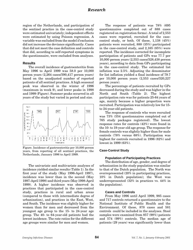
84Emerging Infectious Diseases Vol. 7, No. 1, January–February 2001
Research
region of the Netherlands, and participation ofthe sentinel practice in the case-control studywere estimated univariately; independent effectswere estimated by using Poisson regression. Avariable was excluded from the model if exclusiondid not increase the deviance significantly. Casesthat did not meet the case definition and controlsthat did, according to self-reported symptoms inthe questionnaire, were excluded from analyses.
ResultsThe overall incidence of gastroenteritis from
May 1996 to April 1999 was 58.0 per 10,000person years (2,264 cases/390,417 person years)based on the unadjusted number of reportedpatients of all sentinel practices. A high seasonalpeak was observed in the winter of 1996(maximum in week 9), and lower peaks in 1998and 1999 (Figure). Summer peaks occurred in allyears of the study but varied in period and size.
The univariate and multivariate analyses ofincidence yielded similar results (Table 1). In thefirst year of the study (May 1996-April 1997),incidence was lower than in the second (May1997-April 1998) and third years (May 1998-April1999). A higher incidence was observed inpractices that participated in the case-controlstudy, practices in rural and urban areas(compared to those with intermediate degree ofurbanization), and practices in the East, West,and South. The incidence was slightly higher forwomen than for men and decreased from theyoungest age group to the 15- to 24-year agegroup. The 40- to 64-year-old patients had thelowest incidence. The rate ratios for the differentage groups were similar for men and women.
The response of patients was 78% (695questionnaires completed out of 888 casesregistered on registration forms). A total of 2,553cases were reported, recruited for the case-control study, or both. Of these, 1,138 (45%)patients were recruited, 888 (35%) participatedin the case-control study, and 2,165 (85%) werereported. The incidence corrected for incompleteparticipation of patients and GPs was 77.7 per10,000 person years (2,553 cases/328,438 personyears), according to data from GPs participatingin the case-control study. The estimated listinflation was 2.5%. Adjusting the denominatorfor list inflation yielded a final incidence of 79.7per 10,000 person years (2,553 cases/320,227person years).
The percentage of patients recruited by GPsdecreased during the study and was higher in theNorth and South (Table 2). The highestparticipation rate was for patients 25-64 years ofage, mainly because a higher proportion wererecruited. Participation was relatively low for 15-to 24-year-old patients.
The response of controls who were recruitedwas 73% (554 questionnaires completed out of765 study packages registered). The lowestresponse rates for controls (42%) were found inthe 10- to 19-year-old age group. The response forfemale controls was slightly higher than for malecontrols (78% versus 68%). Participation washighest for controls recruited in 1996 (82%) andlowest in 1999 (63%).
Case-Control Study
Population of Participating PracticesThe distribution of age, gender, and degree of
urbanization in the study population was similarto that of the Dutch population. The North wasoverrepresented (20% in participating practices,10% in Dutch population); the West wasunderrepresented (32% in practices vs. 44% inthe population).
Cases and ControlsFrom May 1996 until April 1999, 985 cases
and 717 controls returned a questionnaire to theNational Institute of Public Health and theEnvironment. Of these, 878 cases and 581controls could be included in the analyses. Stoolsamples were examined from 857 (98%) patientsand 574 (99%) controls. The median age ofpatients (29 years) was significantly lower than
Figure. Incidence of gastroenteritis per 10,000 personyears, from reporting of all sentinel practices, theNetherlands, January 1996 to April 1999.

85Vol. 7, No. 1, January–February 2001 Emerging Infectious Diseases
Research
that of controls (37 years). Gastrointestinalcomplaints of longer than 1 month were morefrequent in patients (31.5%) than in controls(7.3%). The percentage of patients (18%) undertreatment of a specialist was similar to that ofcontrols, as was the number of GP consultationsin the last 3 months (1 consultation). Patientswere slightly more often born outside theNetherlands than controls, and the educationallevel of patients was higher than that of controls(both not statistically significantly).
Clinical Symptoms of PatientsAlmost all patients reported loose stools
(Table 3). Other commonly reported symptomswere frequent stools, abdominal pain, abdominalcramps, and nausea. Only 12 (1.4%) patientsreported vomiting and no diarrhea. In cases witha higher frequency of stools than normal, themedian of the maximum frequency was 6 times in24 hours (first quartile-third quartile: 4-8 timesin 24 hours). The median duration of symptomsbefore a GP was consulted was 6 days (first
quartile to third quartile: 3-20 days); 20% ofpatients had been symptomatic for more than 4weeks before consulting a GP.
Bed rest was required for 47% of patients fora median of 2 days (mean 3.1 days); 41% of thechildren who regularly visited a day-care centerhad to stay home for a median of 3 days (mean 3.2days), 58% of schoolchildren were absent fromschool for a median duration of 3 days (mean 3.8days), and 60% of working patients were absentfrom work for a median duration of 2 days (mean3.1 days). In 8% of cases, someone had to missschool or work for a median of 1.5 days (average2.0 days) to care for the patient; 56% of patientsused medication for gastroenteritis: 4% usedantibiotics, 18% analgesics, 27% antidiarrheicmedication, 10% oral rehydration solution, and21% additional medications.
Symptoms and Diagnoses of ControlsA small proportion of controls (6.7%) reported
consulting a GP for gastrointestinal symptomswhen they were recruited for the study but did
Table 1. Univariate and multivariate Poisson regression analyses of the incidence per 10,000 person years
Incidence per 10,000 Crude Univariate Poisson regression Multivariate Poisson regressionperson years incidence RRa 95% CIa RR 95% CIStudy year May 96-Apr 97 52.4 1.00 - 1.00 - May 97-Apr 98 62.4 1.19 1.08-1.32 1.19 1.08-1.32 May 98-Apr 99 65.6 1.25 1.13-1.38 1.23 1.11-1.36Participation in case- control study Yes 65.8 2.51 2.13-2.97 2.71 2.28-3.22 No 26.2 1.00 - 1.00 -Urbanization Low 57.0 1.04 0.92-1.17 1.18 1.04-1.34 Intermediate 55.5 1.00 - 1.00 - High 88.9 1.62 1.47-1.79 1.25 1.11-1.41Region North 28.7 0.39 0.34-0.45 0.35 0.29-0.40 East 71.3 0.97 0.88-1.07 0.93 0.83-1.05 West 76.0 1.00 - 1.00 - South 55.5 0.75 0.68-0.84 0.78 0.69-0.88Gender Male 56.8 1.00 - 1.00 - Female 63.4 1.12 1.03-1.21 1.12 1.04-1.22Age (in years) <1 360.4 5.53 4.49-6.82 5.54 4.50-6.83 1-4 221.5 3.40 2.94-3.94 3.36 2.90-3.89 5-14 65.1 1.00 - 1.00 - 15-24 51.7 0.79 0.67-0.94 0.79 0.67-0.93 25-39 55.9 0.86 0.75-0.98 0.84 0.73-0.97 40-64 36.5 0.56 0.48-0.65 0.56 0.48-0.65 >65 47.7 0.73 0.62-0.87 0.72 0.61-0.85aRR = relative risk; CI = confidence interval.
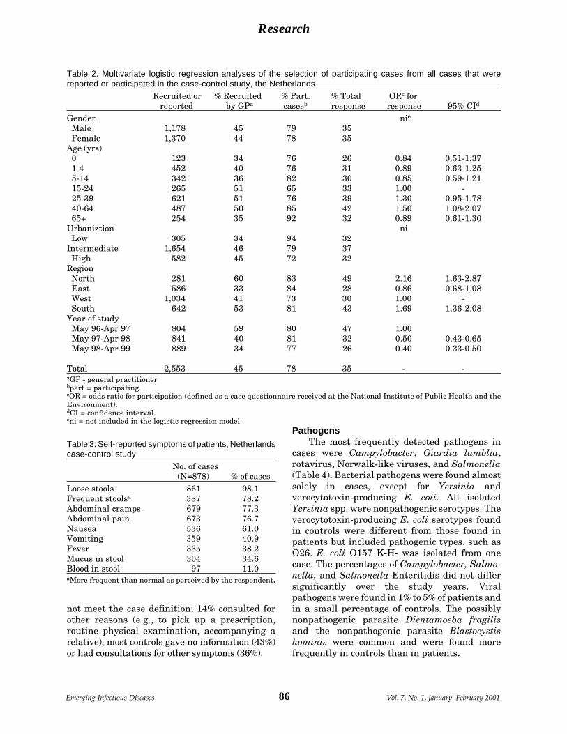
86Emerging Infectious Diseases Vol. 7, No. 1, January–February 2001
Research
PathogensThe most frequently detected pathogens in
cases were Campylobacter, Giardia lamblia,rotavirus, Norwalk-like viruses, and Salmonella(Table 4). Bacterial pathogens were found almostsolely in cases, except for Yersinia andverocytotoxin-producing E. coli. All isolatedYersinia spp. were nonpathogenic serotypes. Theverocytotoxin-producing E. coli serotypes foundin controls were different from those found inpatients but included pathogenic types, such asO26. E. coli O157 K-H- was isolated from onecase. The percentages of Campylobacter, Salmo-nella, and Salmonella Enteritidis did not differsignificantly over the study years. Viralpathogens were found in 1% to 5% of patients andin a small percentage of controls. The possiblynonpathogenic parasite Dientamoeba fragilisand the nonpathogenic parasite Blastocystishominis were common and were found morefrequently in controls than in patients.
Table 2. Multivariate logistic regression analyses of the selection of participating cases from all cases that werereported or participated in the case-control study, the Netherlands
Recruited or % Recruited % Part. % Total ORc for reported by GPa casesb response response 95% CId
Gender nie
Male 1,178 45 79 35 Female 1,370 44 78 35Age (yrs) 0 123 34 76 26 0.84 0.51-1.37 1-4 452 40 76 31 0.89 0.63-1.25 5-14 342 36 82 30 0.85 0.59-1.21 15-24 265 51 65 33 1.00 - 25-39 621 51 76 39 1.30 0.95-1.78 40-64 487 50 85 42 1.50 1.08-2.07 65+ 254 35 92 32 0.89 0.61-1.30Urbaniztion ni Low 305 34 94 32Intermediate 1,654 46 79 37 High 582 45 72 32Region North 281 60 83 49 2.16 1.63-2.87 East 586 33 84 28 0.86 0.68-1.08 West 1,034 41 73 30 1.00 - South 642 53 81 43 1.69 1.36-2.08Year of study May 96-Apr 97 804 59 80 47 1.00 May 97-Apr 98 841 40 81 32 0.50 0.43-0.65 May 98-Apr 99 889 34 77 26 0.40 0.33-0.50
Total 2,553 45 78 35 - -aGP - general practitionerbpart = participating.cOR = odds ratio for participation (defined as a case questionnaire received at the National Institute of Public Health and theEnvironment).dCI = confidence interval.eni = not included in the logistic regression model.
Table 3. Self-reported symptoms of patients, Netherlandscase-control study
No. of cases (N=878) % of cases
Loose stools 861 98.1Frequent stoolsa 387 78.2Abdominal cramps 679 77.3Abdominal pain 673 76.7Nausea 536 61.0Vomiting 359 40.9Fever 335 38.2Mucus in stool 304 34.6Blood in stool 97 11.0aMore frequent than normal as perceived by the respondent.
not meet the case definition; 14% consulted forother reasons (e.g., to pick up a prescription,routine physical examination, accompanying arelative); most controls gave no information (43%)or had consultations for other symptoms (36%).

87Vol. 7, No. 1, January–February 2001 Emerging Infectious Diseases
Research
In 37.5% of patients and 9.8% of controls, apathogen could be detected (excluding possiblynonpathogenic microorganisms, Table 4). FromMay 1998 until May 1999, these percentageswere increased by Sapporo-like viruses with 1.7%in patients and 0.6% in controls. When D. fragiliswas considered a pathogen, these percentageswere 44.1% and 22.9%, respectively. Thepercentage in which a pathogen was found wassimilar for the 50 controls who reportedgastrointestinal symptoms (but did not meet thecase definition) and controls without gastrointes-tinal symptoms.
Most stool samples of patients were receivedat the National Institute within 1 day ofcollection (57%), 21% after 2 days, 9% after 3days, 7% after 4 days, 3% after 5 days, and 2%after >6 days. No significant differences werenoted in the percentage of samples in which apathogen was detected for different postaldelays. For Campylobacter, however, a decreas-ing trend in the proportion of positive cases wasobserved with an increasing postal delay: 1 day:12% positive, 2 days: 10%, 3 days: 9%, 4 days: 6%,5 days 5%, 6 days or more: no Campylobacter.
Discussion
Incidence and Participation RateThis is the first GP-based national study in
the Netherlands covering the role of a wide rangeof microorganisms in a representative populationof gastroenteritis patients and controls. OnlyEngland has conducted a similar study.
The incidence of gastroenteritis in generalpractices was estimated at 79.7 per 10,000 personyears, suggesting that in the Netherlands, 1 inevery 125 persons, or 128,000 persons, will seekphysician care for gastroenteritis every year.This estimate was adjusted for list inflation andpartially for nonparticipation (15%) of GPs butnot for the number of underascertained casesabsent from both study components. In a similarstudy in England, underascertainment wasestimated to be 36% (23). The correction for listinflation (2.5%) in our study should be considereda minimum estimate because only patients whowere actively reported as no longer belonging tothe general practice population were counted; bycontrast, the more active approach in England(searching medical records of nonrespondents)
Table 4. Microorganisms detected in patients and controls
Patients Controls(N=857) (N=574)
N % N %Salmonella spp. 33 3.9 1 0.2 S. Enteritidis 12 1.4 0 0.0 S. Typhimurium 11 1.3 1 0.2 other Salmonellaea 9 1.1 0 0.0Campylobacter spp. 89 10.5 3 0.5 C. jejuni 77 9 3 0.5 C. coli 7 0.8 0 0.0 other species 5 0.6 0 0.0Yersinia spp. 6 0.7 6 1.1 Y. enterocoliticab 5 0.6 5 0.9 Y. frederikseniic 1 0.1 1 0.2Shigella spp. 1 0.1 0 0.0 S. flexneri 1 0.1 0 0.0VTECd 4 0.5 3 0.6Rotavirus 45 5.3 8 1.4Adenovirus 19 2.2 2 0.4Astrovirus 13 1.5 2 0.4Norwalk-like viruses 43 5.0 6 1.1Sapporo-like virusese 5 2.1 1 0.6Giardia lamblia 46 5.4 19 3.3Entamoeba histolytica/ 9 1.1 4 0.7 disparCryptosporidium 18 2.1 1 0.2Cyclospora 1 0.1 1 0.2(Possibly) nonpathogenic microorganisms Dientamoeba fragilis 88 10.3 84 14.6 Blastocystis hominis 185 21.7 172 30.0 Endolimax nana 14 1.6 14 2.4 Enteromonas hominis 2 0.2 1 0.2 Chilomastix mesnili 1 0.1 0 0.0 Entamoeba hartmanni 0 0.0 2 0.4 Enterobius vermicularis 0 0.0 1 0.2 Iodamoeba butschlii 0 0.0 1 0.2aS. Heidelberg, S. Thompson, S. Kottbus, S. Montevideo,S. Manhattan, S. Anatum, S. Arizona, Salmonella group B(not fully typeable).bSerotypes of Y. enterocolitica in patients: O6,31 (biotype1a), O6,30; O6,30 (biotype 1A), one not typeable, one nottyped. In controls: two O4 (biotype 1A); O65 (biotype 1A),O7,8 (biotype 1A); one not typeable.cSerotypes of Y. frederiksenii: O16a,58 (case); O16AB,29(control).dVTEC=verocytotoxin-producing Escherichia coli: positivefor eae gene and genes for either SLT1 or SLT2. VTECserotypes in cases: O157 K-H-, O98 K-, O145 K-, one nottypeable. In controls: O26, O145 K-, one not typeableeSapporo-like viruses have only been tested for the laststudy year (May 98–Apr 99; 240 cases and 160 controls).Genotypes in cases: 4 Sapporo, 1 London, genotype incontrol: Sapporo.

88Emerging Infectious Diseases Vol. 7, No. 1, January–February 2001
Research
yielded an estimate of 10% (23). Consequently,the incidence from our study must be considereda conservative, but the best available, estimate.
A previous estimate of the incidence ofgastroenteritis in a GP-based study in 1992 to1993 in the Netherlands was somewhat higher at90 cases per 10,000 person years (not correctedfor list inflation) (8). Since the percentages ofSalmonella and Campylobacter have alsodecreased since 1992-1993 (from 5% to 4% andfrom 14% to 10%, respectively), the decrease inincidence could partially be due to preventivemeasures in the poultry industry that focused onSalmonella Enteritidis, but as expected, alsocaused a slight decrease in Campylobacterinfections in poultry. Nevertheless, these mea-sures were not fully implemented until April1997, and therefore a decreasing trend in theincidence within the study period would beexpected, which was not observed for gastroen-teritis as a whole, nor for the percentages positivefor Salmonella and Campylobacter. Several otherfactors might have contributed to this decrease,such as a more widespread use of HazardAnalysis and Critical Control Points in the foodproduction industry, greater awareness andknowledge in the population about foodborneinfections, and a change in consultation behaviorbecause of GPs’ increasing deferral policy forgastroenteritis.
The low incidence in the first study year waspresumably due to the absence of a winter peak in1996 to 1997. Such peaks coincide with anincrease in infections with enteric viruses, suchas astrovirus, rotavirus, and calicivirus (24-26).Therefore, the variation in incidence in differentyears likely is due to the annual fluctuation of thepeaks in viral pathogens.
Our data indicate an incidence almostfourfold lower than the incidence in England. TheGP-based incidence of gastroenteritis estimatedfor England in a study from 1993 to 1996 was 330per 10,000 person years after correction for listinflation and underreporting (27). In Wales in1992, an incidence of 244 per 10,000 person yearswas found (28). Most likely, gastroenteritispatients’ higher consultation rate in Englandaccounts for the difference: approximately 1 in 6gastroenteritis case-patients in England consulta GP, whereas an estimated 1 patient in 10 to 1patient in 50 does in the Netherlands (5,6,8). Alower consultation rate could be explained by
Dutch GPs’ policy of deferring gastroenteritiscases. In the Dutch Guidelines for AcuteDiarrhea, a consultation by telephone isconsidered adequate for uncomplicated acutediarrhea and can be dealt with by the GP’sassistant (29). Of the 19 participating GPs whocompleted a questionnaire at the end of ourstudy, 12 (63%) reported fully or partiallydiscouraging patients with gastroenteritis fromvisiting their practice. In England, a deferralpolicy for consultations for gastroenteritis is notcommon.
The incidence of gastroenteritis was indepen-dently associated with degree of urbanizationand geographic region. The incidence was thelowest in the North, as was found in the study in1992 to 1993 (8). This cannot be explained by alower response rate (Table 2). A higher incidencewas found in areas with a high or low degree ofurbanization than in areas with an intermediatedegree of urbanization. Other studies also reporta higher incidence in urban areas (30). Possibly, ahigh population density causes an increased riskfor person-to-person transmission. In ruralareas, contact with animals, manure, and rawproducts could be more frequent than in moreurbanized regions, possibly leading to higherlevels of exposure to zoonotic pathogens. Ananalysis of risk factors for each pathogen will bepublished after an ongoing population-basedstudy is completed. The differences in incidencecould also reflect differences in consultationbehavior.
Patient-related factors that were indepen-dently associated with incidence were patient’sgender and age. The incidence was higher forwomen than for men and was clearly higher inthe youngest age groups, consistent with otherstudies (5,8,28,31-33). Although several studieshave reported a higher disease burden for theelderly, no increase in the incidence ofgastroenteritis was observed in our study (1).Possibly, the higher risk for gastroenteritis forthe elderly is limited to the relatively weakerpersons living in nursing homes (25). Becausethese homes usually have their own GP, theproportion of persons living in nursing homescould be underrepresented in this study.
PathogensBacteria were detected in 16.3% of patients,
viruses in 15.4%, and pathogenic parasites in

89Vol. 7, No. 1, January–February 2001 Emerging Infectious Diseases
Research
8.3%. The higher participation of the northernregion might have increased the percentage ofSalmonella Typhimurium because S. Typhimuri-um is predominant over S. Enteritidis in thisregion, in contrast to the rest of the Netherlands(34). The order of relative importance ofpathogens in general practices was similar forthe Netherlands and England, although thepercentages positive for viruses and bacteria inthis study were slightly lower than in Englandand the percentages positive for parasites wereslightly higher. Because bacteria and viruses aremore often detected in patients with acutediarrhea, whereas parasites tend to cause lessfulminant but intermittent and long-lastingsymptoms that might lead to delayed consulta-tion, these differences could be due to theexclusion of persons with symptoms lastinglonger than 2 weeks in the English study (35,36).In our study, 32% of patients had symptoms >2weeks’ duration (36). In addition, we usedformalin-fixed material to detect parasites, andfour different preparations were examined,which increased the sensitivity of microscopyexamination, compared to the use of nonfixedmaterial and the examination of three differentpreparations in the English study (37). The lowerparticipation of patients in the last 2 years of thestudy might have caused an underestimate ofviral pathogens because the seasonal viral peakwas relatively low in the first year (24,38). Inspite of a less sensitive method of testing forNorwalk-like viruses (electron microscopy inEngland versus reverse transcriptase-poly-merase chain reaction in the Netherlands), thepercentage of stool samples positive for Norwalk-like viruses is higher in England (36). The lowresponse for the younger age groups might alsohave reduced the percentage of rotavirus,Sapporo-like viruses, and to a lesser degreeNorwalk-like viruses, which are most common inyoung children (24). Differences in the proportionof specific pathogens in the English study and inour study may also be explained by differences inconsultation rates. A lower threshold forconsulting a GP might increase the proportion ofpathogens that cause relatively mild gastroen-teritis, such as Norwalk-like viruses.
Parasites that were (possibly) nonpathogenicwere more frequently found in controls than inpatients. A more detailed study of differencesbetween patients and controls with these parasites
might identify factors related to the developmentof disease after infection with these parasites.
Diagnostic DeficitIn spite of a diagnostic panel that included
most of the known pathogens that can causegastroenteritis, the percentage of patients inwhich no pathogen could be detected was 61%(including Sapporo-like viruses in 1998 andexcluding D. fragilis and nonpathogenic para-sites). Some cases could be noninfectious becausean exclusive distinction between infectious andnoninfectious intestinal disease cannot be madeclinically. The high percentage of patients withchronic gastrointestinal symptoms suggests thata substantial proportion might not be infectiousbut an expression of other illnesses, such asinflammatory bowel disease. The symptomsmight also be caused by intestinal microorgan-isms not included in this study or not yet known.Several pathogens, such as Campylobacter,verocytotoxin-producing E. coli, and torovirushave only recently been recognized as a cause ofgastroenteritis, and it is likely that newpathogens will be added to this list (39). Inaddition, we did not screen for some pathogens,such as some pathogenic E. coli (e.g., enterotoxi-genic E. coli, enteroinvasive E. coli) and bacterialtoxins (Bacillus spp, Clostridium difficile cytotoxin,C. perfringens enterotoxin), which were associat-ed with 15% and 6% of the cases, respectively, inthe English study (36). However, all stool samplesfrom our study were stored and are currently beingtested for Sapporo-like viruses and can be testedfor other pathogens. Also, intestinal symptomscan be caused by nonintestinal infections, such asinfluenza, not included in our study. Approxi-mately one quarter of gastroenteritis cases in anAmerican population-based study coincided withrespiratory disease (31). Detection of pathogensin the study is also influenced by logistics factorsand the sensitivity of the testing method. Thetiming of sampling was not ideal in many cases;because of the relatively long patient delay, somepathogens might no longer have been excreted inthe stool at the time of sampling. Somepathogens, such as Campylobacter, do not growin culture after a long delay in shipping. Atpresent, we are conducting a study ongastroenteritis in the community to elucidate therole of sampling time and referral behavior onpathogen-specific incidence.

90Emerging Infectious Diseases Vol. 7, No. 1, January–February 2001
Research
AcknowledgmentsWe thank the participating general practitioners and
the Netherlands Institute for Primary Health Care for theirindispensable cooperation in the data collection; Jan Vinjéfor his work on the diagnostic testing methods and support;Joke Admiraal, Denise Hoek, Nahid Nozari, Hanneke Deijl,Petra de Bree, Jeroen Meijer, and Sandy Altena for theirexcellent assistance in performing the diagnostic tests;Isabel Araya Segovia for administrative support; MartienBorgdorff for his work in the design and start of the study;and Nico Nagelkerke for his advice on data analyses.
Ms. de Wit works as an epidemiologist at the De-partment of Infectious Disease Epidemiology of the Na-tional Institute of Public Health and the Environment inthe Netherlands. Her work focuses on the epidemiologyof gastroenteritis in the Netherlands at the hospital, gen-eral practice, and community levels.
References 1. Guerrant RL, Hughes JM, Lima NL, Crane J. Diarrhea in
developed and developing countries: magnitude, specialsettings and etiologies. Rev Infect Dis 1990;12:S41-50.
2. Bern C, Martines J, de Zoysa I, Glass RI. The magnitude ofthe global problem of diarrhoeal disease: a ten-yearupdate. Bull World Health Organ 1992;70:705-14.
3. Esveld MI, van Pelt W, van Leeuwen WJ, Banffer JRJ.Laboratorium Surveillance Infectieziekten. RIVMreport no. 968902002. Bilthoven, the Netherlands:RIVM; June 1996.
4. Sprenger MJW, Schrijnemakers PM, Wijgergangs LM,Talsma E. Infectious Diseases Surveillance and Informa-tion System (ISIS). International Conference on EmergingInfectious Diseases; 1998 Mar 8-11; Atlanta. p. 123.
5. de Wit MAS, Hoogenboom-Verdegaal AMM, GoosenESM, Sprenger MJW, Borgdorff MW. A population-based longitudinal study on the incidence and diseaseburden of gastroenteritis and Campylobacter andSalmonella infection in four regions of the Netherlands.Eur J Epidemiol 2000;16:713-8.
6. Hoogenboom-Verdegaal AMM, Jong JC, During M,Hoogenveen R, Hoekstra JA. Community-based studyof the incidence of gastrointestinal disease in theNetherlands. Epidemiol Infect 1994;112:481-7.
7. Hoogenboom-Verdegaal AMM, Goosen ESM, DuringM, Engels GB, Klokman-Houweling JM, Laar MJWvan de. Epidemiologisch en microbiologisch onderzoekmet betrekking tot acute gastro-enteritis in dehuisartsenpeilstation in Amsterdam en Helmond,1987-1991. RIVM report no. 149101011. Bilthoven, theNetherlands: RIVM; 1994.
8. Goosen ESM, Hoogenboom-Verdegaal AMM, BarteldsAIM, Sprenger MJW, Borgdorff MW. Incidentie vangastro-enteritis in huisartsenpeilstations in Neder-land, 1992-1993. RIVM report no. 149101012.Bilthoven, the Netherlands: RIVM; 1995.
9. Council directive 92/117/EEC, concerning measures forprotection against specified zoonoses and specifiedzoonotic agents in animals and products of animalorigin in order to prevent outbreaks of food-borneinfections and intoxications. 1992 Dec. ProductschapVlee, Vleesen Eieren. Rijswijk: PVE; 1997a.
10. Productschap Vee, Vlees en Eieren (PVE). Plan vanaanpak preventie en bestrijding Salmonella in deeiersector. Rijswijk, the Netherlands: PVE; 1997a.
11. Productschap Vee, Vlees en Eieren (PVE). Plan vanaanpak Salmonella en Campylobacter in de pluimveev-leessector. Rijswijk, the Netherlands: PVE; 1997b.
12. Smith HR, Scotland SM. Isolation and identificationmethods for Escherichia coli O157 and other verotoxinproducing strains. J Clin Pathol 1993;46;10-17.
13. Edwards and Ewing. Identification of Enterobacteri-aceae. 4th ed. New York: Elsevier Science PublishingCo., Inc.; 1986.
14. Vinjé J, Koopmans MPG. Molecular detection andepidemiology of small round-structured viruses inoutbreaks of gastroenteritis in the Netherlands. JInfect Dis 1996;174:610-15.
15. Polderman AM, Rijpstra AC, editors. Medischeparasitologie, handleiding bij laboratorium diag-nostiek. Bohn Stafleu Van Loghum, the Netherlands;Houten/Zaventem; 1993; 218:146-7.
16. Belding DL, editor. Textbook of parasitology. 3rd ed,New York: Appleton-Century-Crofts; 1965. p. 1374.
17. Ash LR, Orihel TC, editors. Atlas of humanparasitology. 2nd ed. Chicago: American Society ofClinical Pathologists Press; 1984. p. 212.
18. Yang J, Scholten T. A fixative for intestinal parasitespermitting the use of concentration and permanentstaining procedures. Am J Clin Pathol 1977;67:300-4.
19. Garcia LS, section editor. Parasitology. Section 7.1-7.4. In: Isenberg HD, editor. Clinical microbiologyprocedures handbook. Washington: American Soci-ety for Microbiology.
20. van Gool T, Mank TG. Fixatives and permanent stainsin the laboratory diagnosis of intestinal protozoalinfections. Haarlem, the Netherlands: Van Gool andMank; 1999.
21. Vinjé J, Deijl H, van der Heide R, Lewes D, Hedlund K-O, Svensson L, et al. Molecular detection andepidemiology of Sapporo-like viruses. J Clin Microbiol2000;38:530-6.
22. Abbink F, Duynhoven YTHP, de Wit M, KoopmansMPG, van Leeuwen WJ, Kortbeek LM. A populationcohort study with a nested case-control study: a studydesign to estimate the incidence and aetiology ofgastroenteritis (GE) in the Netherlands. Proceeding4th World Congress of Foodborne Infections andIntoxications. 1998; Berlin, Germany. p. 766-9.
23. Sethi D, Wheeler J, Rodrigues LC, Fox S, Roderick P.Investigation of under-ascertainment in epidemiologi-cal studies based in general practice. Int J Epidemiol1999;28:106-11.
24. Koopmans MPG, van Asperen IA. Epidemiology ofrotavirus infections in The Netherlands. Acta PaediatrSuppl 1999;426:31-7.
25. Vinjé J, Altena SA, Koopmans MPG. The incidence andgenetic variability of small round-structured viruses(SRSV) in outbreaks of gastroenteritis. J Infect Dis1997;176:1374-8.
26. Borgdorff MW, Koopmans MPG, Goosen ESM,Sprenger MJW. Surveillance of gastroenteritis [letter].Lancet 1995;346:842-3.
91Vol. 7, No. 1, January–February 2001 Emerging Infectious Diseases
Research
27. Wheeler JG, Sethi D, Cowden JM, Wall PG, RodriguesLC, Tompkins DS, et al. Study of infectious intestinaldisease in England: rates in the community, presentingto general practice, and reported to nationalsurveillance. Br Med J 1999;318:1046-50.
28. Palmer S, Houston H, Lervy B, Ribeiro D, Thomas P.Problems in the diagnosis of foodborne infections ingeneral practice. Epidemiol Infect 1996;117:479-84.
29. Nederlands Huisartsen Genootschap [NetherlandsGeneral Practitioners Society]. Standaard acutediarree. Huisarts en Wetenschap 1993;36(9).
30. Roderick P, Wheeler J, Cowden J, Sockett P, Skinner R,Mortimer P, et al. A pilot study of infectious intestinaldisease in England. Epidemiol Infect 1995;114:277-88.
31. Monto AS, Koopman JS. The Tecumseh study:Occurrence of acute enteric illness in the community. JEpidemiol 1980;112:323-33.
32. Skirrow MB. A demographic survey of Campylobacter,Salmonella and Shigella infections in England.Epidemiol Infect 1987;99:647-57.
33. Simpson GE, Nagy GS, Fincks ES. A survey of acute gas-troenteritis in general practice. Med J Aust 1969;2:633-5.
34. Giessen AW, van Leeuwen WJ, van Pelt W. Chapter 8: Sal-monella enterica Serovar Enteritidis in the Netherlands:epidemiology, prevention and control. In: Saeed AM,editor. Salmonella enterica Serovar Enteritidis in humansand animals. Epidemiology, pathogenesis, and control.Ames (IA): Iowa State University Press;1999. p. 71-80.
35. Mank TG, Polderman AM, Zaat JOM, Roggeveen C,van Eijk JThM, Deelder AM. Persistent diarrhea in ageneral practice population in the Netherlands:prevalence of protozoal and other intestinal infections.In: Thesis intestinal protozoa and diarrhea in generalpractice. Haarlem, the Netherlands: Jos Mathôt BV;1997. p. 47-64.
36. Tompkins DS, Hudson MJ, Smith HR, Eglin RP,Wheeler JG, Brett MM, et al. A study of infectiousintestinal disease in England: microbiological findingsin cases and controls. Commun Dis Public Health1999;2:108-13.
37. Mank TG, Zaat JOM, Blotkamp J, Polderman AM.Comparison of fresh versus Sodium Acetate Acetic AcidFormalin preserved stool specimens for diagnosis ofintestinal protozoal infections. Eur J Clin MicrobiolInfect Dis 1995;14:1076-81.
38. de Wit MAS, Koopmans MPG, van der Blij JF,Duynhoven TYHP. Hospital admissions for rotavirusinfection in the Netherlands. Clin Infect Dis2000;31:698-704.
39. Mead PS, Slutsker L, Dietz V, McCaig LF, Bresee JS,Shapiro C, et al. Food-related illness and death in theUnited States. Emerg Infect Dis 1999;5:607-25.
92Emerging Infectious Diseases Vol. 7, No. 1, January–February 2001
Research
Bacterial infections are prototypical emerg-ing diseases (1), and their recent historychallenges the premature view that the battleagainst infectious diseases had been won. In thelast 25 years, disease caused by multidrug-resistant Streptococcus pneumoniae becameestablished on several continents, reaching theUnited States by the 1990s (2-4), and fatalinfections caused by S. pyogenes (group AStreptococcus), a problem of the 19th century (5),
have returned in toxic and necrotic forms (6). Bythe 1970s, group B Streptococcus replaced gram-negative bacteria and Staphylococcus aureus asthe leading cause of sepsis in newborns (7,8).Researchers tackled the public health challengeof developing vaccines to protect children againstthe major causes of bacterial meningitis:Haemophilus influenzae type b, S. pneumoniae,and Neisseria meningitidis (9,10). A critical stepfor response to microbial adaptation is establish-ing a reliable tracking system. We describeactive, population-based surveillance for seriousbacterial infections that was established by theCenters for Disease Control and Prevention
Active Bacterial Core Surveillance of theEmerging Infections Program Network
Anne Schuchat,* Tami Hilger,* Elizabeth Zell,* Monica M. Farley,†Arthur Reingold,‡ Lee Harrison,§ Lewis Lefkowitz,¶
Richard Danila,** Karen Stefonek,†† Nancy Barrett,‡‡ Dale Morse,§§and Robert Pinner,* for the Active Bacterial Core Surveillance Team
of the Emerging Infections Program Network*Centers for Disease Control and Prevention, Atlanta, Georgia, USA;
†Georgia Emerging Infection Program (Georgia Department of HumanResources, Division of Public Health, Emory University School of Medicine,and the Atlanta Veterans Administration Medical Center) Atlanta, Georgia,
USA; ‡California Department of Health Services and University of CaliforniaBerkeley School of Public Health, Berkeley, California, USA; §Maryland
Department of Health and Mental Hygiene and Johns Hopkins UniversitySchool of Public Health, Baltimore, Maryland, USA; ¶Tennessee Departmentof Health and Vanderbilt University Medical Center, Nashville, Tennessee,
USA; **Minnesota Department of Health, St. Paul, Minnesota, USA;††Oregon Department of Human Resources, Portland, Oregon, USA; ‡‡
Connecticut Department of Public Health, Hartford, Connecticut, USA; and§§New York State Department of Health, Albany, New York, USA
Address for correspondence: Anne Schuchat, Centers forDisease Control and Prevention, Mailstop C23, 1600 CliftonRd., Atlanta, GA 30333, USA; fax: 404-639-3970; e-mail:[email protected].
Active Bacterial Core surveillance (ABCs) is a collaboration between the Centersfor Disease Control and Prevention and several state health departments anduniversities participating in the Emerging Infections Program Network. ABCs conductspopulation-based active surveillance, collects isolates, and performs studies of invasivedisease caused by Streptococcus pneumoniae, group A and group B Streptococcus,Neisseria meningitidis, and Haemophilus influenzae for a population of 17 to 30 million.These pathogens caused an estimated 97,000 invasive cases, resulting in 10,000deaths in the United States in 1998. Incidence rates of these pathogens are described.During 1998, 25% of invasive pneumococcal infections in ABCs areas were notsusceptible to penicillin, and 13.3% were not susceptible to three classes of antibiotics.In 1998, early-onset group B streptococcal disease had declined by 65% over theprevious 6 years. More information on ABCs is available at www.cdc.gov/ncidod/dbmd/abcs. ABCs specimens will soon be available to researchers through an archive.
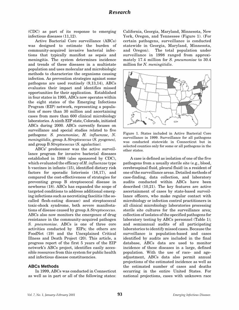
93Vol. 7, No. 1, January–February 2001 Emerging Infectious Diseases
Research
(CDC) as part of its response to emerginginfectious diseases (11,12).
Active Bacterial Core surveillance (ABCs)was designed to estimate the burden ofcommunity-acquired invasive bacterial infec-tions that typically manifest as sepsis andmeningitis. The system determines incidenceand trends of these diseases in a multistatepopulation and uses molecular and microbiologicmethods to characterize the organisms causinginfection. As prevention strategies against somepathogens are used routinely (9,13,14), ABCsevaluates their impact and identifies missedopportunities for their application. Establishedin four states in 1995, ABCs now operates withinthe eight states of the Emerging InfectionsProgram (EIP) network, representing a popula-tion of more than 30 million and ascertainingcases from more than 600 clinical microbiologylaboratories. A ninth EIP state, Colorado, initiatedABCs during 2000. ABCs currently focuses onsurveillance and special studies related to fivepathogens: S. pneumoniae, H. influenzae, N.meningitidis, group A Streptococcus (S. pyogenes),and group B Streptococcus (S. agalactiae).
ABCs’ predecessor was the active surveil-lance program for invasive bacterial diseasesestablished in 1988 (also sponsored by CDC),which evaluated the efficacy of H. influenzae typeb vaccines in infants (15), identified dietary riskfactors for sporadic listeriosis (16,17), andcompared the cost-effectiveness of strategies forpreventing group B streptococcal disease innewborns (18). ABCs has expanded the scope oftargeted conditions to address additional emerg-ing infections such as necrotizing fasciitis (the so-called flesh-eating disease) and streptococcaltoxic-shock syndrome, both severe manifesta-tions of disease caused by group A Streptococcus.ABCs also now monitors the emergence of drugresistance in the community-acquired pathogenS. pneumoniae. ABCs is one of three coreactivities conducted by EIPs; the others areFoodNet (19) and the Unexplained CriticalIllness and Death Project (20). This article, aprogress report of the first 5 years of the EIPnetwork’s ABCs project, identifies easily acces-sible resources from this system for public healthand infectious disease constituencies.
ABCs MethodsIn 1999, ABCs was conducted in Connecticut
as well as in part or all of the following states:
California, Georgia, Maryland, Minnesota, NewYork, Oregon, and Tennessee (Figure 1). (Forcertain pathogens, surveillance is conductedstatewide in Georgia, Maryland, Minnesota,and Oregon). The total population undersurveillance in 1998 ranged from approxi-mately 17.4 million for S. pneumoniae to 30.4million for N. meningitidis.
A case is defined as isolation of one of the fivepathogens from a usually sterile site (e.g., blood,cerebrospinal fluid, pleural fluid) in a resident ofone of the surveillance areas. Detailed methods ofcase-finding, data collection, and laboratoryaudits conducted within ABCs have beendescribed (10,21). The key features are activeascertainment of cases by state-based surveil-lance officers, who make regular contact withmicrobiology or infection control practitioners inall clinical microbiology laboratories processingsterile site cultures for the surveillance area;collection of isolates of the specified pathogens forlaboratory testing by ABCs personnel (Table 1);and semiannual audits of all participatinglaboratories to identify missed cases. Because thesurveillance is population-based and casesidentified by audits are included in the finaldatabase, ABCs data are used to monitorincidence of these diseases in a large, definedpopulation. With the use of race- and age-adjustment, ABCs data also permit annualprojections of the estimated incidence as well asthe estimated number of cases and deathsoccurring in the entire United States. Fornational projections, cases with unknown race
Figure 1. States included in Active Bacterial Coresurveillance in 1999. Surveillance for all pathogenswas conducted statewide in Connecticut but inselected counties only for some or all pathogens in theother states.
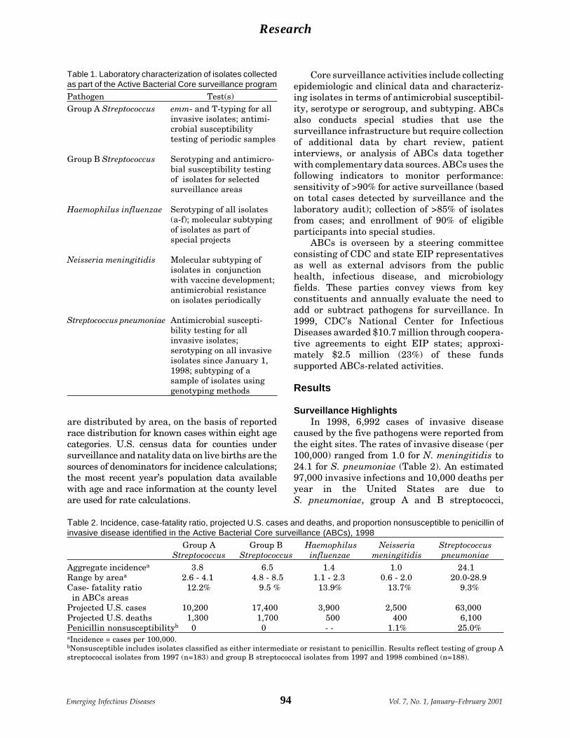
94Emerging Infectious Diseases Vol. 7, No. 1, January–February 2001
Research
are distributed by area, on the basis of reportedrace distribution for known cases within eight agecategories. U.S. census data for counties undersurveillance and natality data on live births are thesources of denominators for incidence calculations;the most recent year’s population data availablewith age and race information at the county levelare used for rate calculations.
Core surveillance activities include collectingepidemiologic and clinical data and characteriz-ing isolates in terms of antimicrobial susceptibil-ity, serotype or serogroup, and subtyping. ABCsalso conducts special studies that use thesurveillance infrastructure but require collectionof additional data by chart review, patientinterviews, or analysis of ABCs data togetherwith complementary data sources. ABCs uses thefollowing indicators to monitor performance:sensitivity of >90% for active surveillance (basedon total cases detected by surveillance and thelaboratory audit); collection of >85% of isolatesfrom cases; and enrollment of 90% of eligibleparticipants into special studies.
ABCs is overseen by a steering committeeconsisting of CDC and state EIP representativesas well as external advisors from the publichealth, infectious disease, and microbiologyfields. These parties convey views from keyconstituents and annually evaluate the need toadd or subtract pathogens for surveillance. In1999, CDC’s National Center for InfectiousDiseases awarded $10.7 million through coopera-tive agreements to eight EIP states; approxi-mately $2.5 million (23%) of these fundssupported ABCs-related activities.
Results
Surveillance HighlightsIn 1998, 6,992 cases of invasive disease
caused by the five pathogens were reported fromthe eight sites. The rates of invasive disease (per100,000) ranged from 1.0 for N. meningitidis to24.1 for S. pneumoniae (Table 2). An estimated97,000 invasive infections and 10,000 deaths peryear in the United States are due toS. pneumoniae, group A and B streptococci,
Table 1. Laboratory characterization of isolates collectedas part of the Active Bacterial Core surveillance program
Pathogen Test(s)Group A Streptococcus emm- and T-typing for all
invasive isolates; antimi-crobial susceptibilitytesting of periodic samples
Group B Streptococcus Serotyping and antimicro-bial susceptibility testingof isolates for selectedsurveillance areas
Haemophilus influenzae Serotyping of all isolates(a-f); molecular subtypingof isolates as part ofspecial projects
Neisseria meningitidis Molecular subtyping ofisolates in conjunctionwith vaccine development;antimicrobial resistanceon isolates periodically
Streptococcus pneumoniae Antimicrobial suscepti-bility testing for allinvasive isolates;serotyping on all invasiveisolates since January 1,1998; subtyping of asample of isolates usinggenotyping methods
Table 2. Incidence, case-fatality ratio, projected U.S. cases and deaths, and proportion nonsusceptible to penicillin ofinvasive disease identified in the Active Bacterial Core surveillance (ABCs), 1998
Group A Group B Haemophilus Neisseria Streptococcus Streptococcus Streptococcus influenzae meningitidis pneumoniae
Aggregate incidencea 3.8 6.5 1.4 1.0 24.1Range by areaa 2.6 - 4.1 4.8 - 8.5 1.1 - 2.3 0.6 - 2.0 20.0-28.9Case- fatality ratio 12.2% 9.5 % 13.9% 13.7% 9.3% in ABCs areasProjected U.S. cases 10,200 17,400 3,900 2,500 63,000Projected U.S. deaths 1,300 1,700 500 400 6,100Penicillin nonsusceptibilityb 0 0 - - 1.1% 25.0%aIncidence = cases per 100,000.bNonsusceptible includes isolates classified as either intermediate or resistant to penicillin. Results reflect testing of group Astreptococcal isolates from 1997 (n=183) and group B streptococcal isolates from 1997 and 1998 combined (n=188).

95Vol. 7, No. 1, January–February 2001 Emerging Infectious Diseases
Research
H. influenzae, and N. meningitidis. Despitecontinued availability of effective antimicrobialagents for each pathogen, approximately 1 in 10cases results in death (Table 2). Substantialgeographic variation exists in the incidence ofinvasive infections caused by each pathogen(Table 2). Among invasive S. pneumoniaeinfections, the proportion caused by drug-resistant organisms was three times higher insome areas than others (4); 8.4% of invasivepneumococci from New York were fully resistantto penicillin (MIC >2.0), while 25.4% of isolatesfrom Tennessee were penicillin resistant. Nopenicillin-nonsusceptible (intermediate or resis-tant) strains of group A or group B Streptococcushave been detected to date.
Recent temporal changes are most dramaticfor invasive group B streptococcal disease amonginfants less than 1 week old (i.e., early-onsetdisease), which declined 65% from 1993 to 1998(Figure 2), during a period when the incidence ofdisease in older infants and adults remainedstable (22). Data from ABCs provide a reliablestandard for evaluating alternative methods forsurveillance of drug resistance in S. pneumoniae,including sentinel surveillance methods (4) anduse of aggregate data from antibiograms frommultiple hospitals (23). The recent emergence ofserogroup Y meningococci, demonstrated by ABCs,suggests that vaccine companies should considerincorporating serogroup Y in new meningococcalvaccines. In addition, the diversity in the outermembrane proteins of serogroup B meningococ-cal strains suggests that vaccines against these
proteins may not be efficient means of preventingendemic serogroup B meningococcal disease.
Applied ResearchThe population-based collections of isolates
from ABCs are used to evaluate subtypingmethods (24), identify genetic mechanisms ofantimicrobial resistance, determine vaccineformulations (25,26), and assess capsularswitching among organisms (for vaccines basedon capsular types) (27,28). ABCs has identifiedpopulation-based risk factors for disease invarious age groups (Table 3). A case-control studyof invasive pneumococcal disease in youngchildren showed that attendance at day care wasassociated with a substantial attributable risk fordisease (29). A similar study of invasivepneumococcal disease in 18- to 64-year-old adultswho were not immunocompromised identifiedactive and passive smoking, in addition tohousehold contact with a child in day care, asindependent risk factors for disease (30). Modelsof age- and serogroup- or serotype-specific ratesof invasive meningococcal and pneumococcaldisease in the ABCs population have comparedthe potential impact of diverse immunizationstrategies for meningococcal and combinedmeningococcal-pneumococcal vaccines on diseaseprevention (32). The increased risk for pneumo-nia death occurring several days after illnessonset associated with antimicrobial-resistantstrains of S. pneumoniae was demonstrated byusing multistate clinical and epidemiologic datafrom ABCs (33).
Figure 2. Invasivegroup B streptococ-cal disease in in-fants less than 1week of age per1,000 live birthsand in adults >65years of age per100,000 population,Active BacterialCore surveillance,1993-1998 (adaptedfrom ref. 22).

96Emerging Infectious Diseases Vol. 7, No. 1, January–February 2001
Research
InfrastructureABCs provides participating state health
departments active contact with all acute-carehospitals and reference microbiology laborato-ries in the surveillance area. This networkprovides an infrastructure for public healthcommunication and education, as well as anetwork of key contacts available for response tonew or emerging concerns. Periodic surveys oflaboratories within ABCs determined theadequacy of methods used to detect group BStreptococcus from prenatal screening specimens(34), the computerization of clinical microbiologylaboratories and readiness for electronic labora-tory-based reporting, and the routine proceduresused by ABCs laboratories to detect antimicrobialresistance among S. pneumoniae, S. aureus, andseveral other organisms (35). ABCs and otherEIP personnel have provided assistance withmultistate response efforts to determine theburden of Creutzfeldt-Jakob disease (36) andcontributed to efforts to determine the rate ofrotavirus vaccine-related intussusception (37).Further, the presence of ABCs personnel in statehealth departments and academic institutionshas strengthened communication links requiredfor accurate reporting and feedback.
PreventionSince publication of consensus guidelines for
the prevention of group B streptococcal disease innewborns, ABCs assessed the implementation ofprevention practices and identified opportunitiesfor preventing more cases. ABCs showed thathospital obstetric programs’ adoption of policiesto prevent group B streptococcal infectionincreased significantly (38) and that hospitals
that had adopted or revised a policy in 1996 hadsignificantly fewer cases in 1997 (39). ABCs isalso tracking the characteristics of newborngroup B streptococcal cases that continue to occurdespite prevention guidelines to determinewhether these represent failures of intrapartumantibiotic prophylaxis or failure to offer suchprophylaxis to mothers at risk. In several EIPs,pilot prevention programs are in place to identifyefficient ways to reduce the incidence of diseasecaused by ABCs pathogens. These include amultifaceted program to reduce inappropriateantibiotic use in the Baltimore metropolitan areaand efforts to promote pneumococcal polysaccha-ride vaccine in populations at high risk inRochester, New York; Minneapolis-St. Paul,Minnesota; metropolitan Atlanta, Georgia; andPortland, Oregon. The Connecticut and Minne-sota health departments conducted demonstra-tion projects that integrated prevention ofgroup B streptococcal disease into routineperinatal care, building on successes withhepatitis B perinatal prevention programs andcontributing to reduction of perinatal HIVtransmission (40,41).
DiscussionNearly 100,000 invasive infections and
10,000 deaths caused by ABCs pathogens occurannually in the United States. Because few statesroutinely collect data and isolates for all of theseinfections, ABCs helps monitor disease andevaluate prevention programs at the nationallevel. ABCs has now developed robust estimatesof the magnitude of disease and deaths attributableto the five invasive pathogens (Table 2). Anumber of future priorities have been identified
Table 3. Population-based risk factors for invasive disease, identified by Active Bacterial Core surveillance
Age group, Factors associated with increasedPathogen other criteria risk by multivariate analysis ReferencesStreptococcus pneumoniae <5 years old Child-care attendance, underlying 29
conditions, lack of breast-feeding, household crowding
18-64 years old, Active or passive smoke exposure, 30 not immuno- black race, chronic diseases, household compromised contact with child in day care
Neisseria meningitidis All ages Active or passive smoke exposure, 31 underlying conditions, steroid use, attendance at new school
Group B Streptococcus <7 days old Black race, low birth weight, CDC, unpub. maternal age <20 years data

97Vol. 7, No. 1, January–February 2001 Emerging Infectious Diseases
Research
that take advantage of the careful characteriza-tion of isolates associated with invasive infection.Licensure and introduction of a seven-valentconjugate vaccine against S. pneumoniae neces-sitate evaluation of the impact of this newprevention tool on target populations (Table 4).Of particular interest will be evaluating whetherindirect effects similar to those seen with the Hibvaccine (42) occur. The large birth cohort undersurveillance through ABCs and the longitudinaldata on both early-onset cases and hospitalpolicies for disease prevention offer the opportu-nity to compare the two alternative strategies forgroup B streptococcal prevention (screening-based vs. risk-based) through two studies duringthe next few years. ABCs will continue tocontribute to tracking progress in Hib elimina-tion, monitor for emergence of other serotypes ofH. influenzae, and provide data on strain-specificdisease (e.g., serotype, serogroup, outer mem-brane type). Such information is valuable forevaluating new vaccines for group B Streptococ-cus and serogroup B meningococcus. ABCs datawill also be used to define clusters of invasivegroup A streptococcal disease and to model theimpact of possible strategies and new formula-tions of pneumococcal vaccines targeted againstpneumococcal pneumonia in adults (Figure 3) aswell as vaccines targeted against invasive groupA Streptococcus syndromes.
The molecular biology revolution and im-proved understanding of host-pathogen interac-tions offer great potential to advance knowledgeabout ABCs bacteria. Emerging antimicrobialresistance and other forms of pathogen adapta-tion (e.g., capsular switching) lend an urgency tosuch research. Specimens from invasive diseasesurveillance represent well-characterized, popu-lation-based collections with relevant clinical anddemographic information. These provide a
valuable resource for basic and applied researchfocused on issues as varied as new drug andvaccine development, evaluation of mechanismsof virulence and antimicrobial resistance, andgenetic research. ABCs is planning to make thesestrains available to outside researchers andindustry through a preserved collection. Such aspecimen bank could provide a lasting legacy ofthe work of hundreds of infection controlpractitioners, clinical microbiology laboratories,and ABCs surveillance collaborators.
To ensure that ABCs’ lessons learned withinthe EIP network reach other public healthconstituents, a number of efforts are under way.Additional details of the surveillance system andoutreach materials are available at http://www.cdc.gov/ncidod/dbmd/abcs. Other educa-tional materials are available at http://www.cdc.gov/ncidod/dbmd/gbs and at http://www.cdc.gov/ncidod/dbmd/antibioticresistance.For laboratories evaluating new strains of groupA Streptococcus, genetic sequencing data of allstrains described thus far are also available onthe web. A similar site for meningococcal isolatesis under development.
ConclusionsABCs is a model of collaboration between
public health and academia. The system providesreliable data that can be used to address criticalpublic health concerns, improve understandingof emerging infections, and help prevent theconsequences of these infections. While thepast 5 years have helped quantify themagnitude of disease caused by these patho-gens and document increasing antibioticresistance in some of them, the future providesseveral challenges. To remain a vital component
Table 4. Future priorities for Active Bacterial Coresurveillance (ABCs) project
1. Define invasive group A Streptococcus clusters.2. Determine effectiveness of screening vs. risk-
based prevention strategies for perinatal groupB streptococcal disease.
3. Determine feasibility of eliminating invasivedisease caused by Haemophilus influenzae type b.
4. Quantify culture-negative, polymerase chainreaction-positive meningitis.
5. Measure direct and indirect effects ofintroducing a seven-valent pneumococcalconjugate vaccine.
Figure 3. Age-specific incidence (per 100,000) andcase-fatality ratio (percent) of invasive pneumococcaldisease, Active Bacterial Core surveillance, 1998.
98Emerging Infectious Diseases Vol. 7, No. 1, January–February 2001
Research
in the nation’s efforts to prevent and controlemerging infectious diseases, ABCs will need toincorporate surveillance and research tools of the21st century, including electronic laboratory-based reporting, genotyping of pathogens, andimproved communication to promote behavioralchange and adoption of practice guidelines.
AcknowledgmentsWe thank the National Center for Infectious Diseases
Emerging Infections Program and infection controlpractitioners and clinical microbiologists who collaborate onABCs. We also thank the following additional members of theABCs team: California: Duc Vugia, Gretchen Rothrock, PamDaily, Lisa Gelling; Connecticut: James Hadler, MattCartter, Pat Mshar, Craig Morin, Aaron Roome, HeatherLinardos; Georgia: Paul Blake, David Stephens, KathrynArnold, Wendy Baughman, Katherine McCombs, SabrinaBurden, Patricia Martell-Cleary, Mathew Sattah; Maryland:Margaret Pass, Diane Dwyer; Minnesota: Ruth Lynfield,Catherine Lexau, Jean Rainbow, Karen White, Lori Triden,Brenda Sayler; New York: Perry Smith, Shelly Zansky,Barbara Damaske, Nancy Bennett, Glenda Smith, NellieDumas, Brian Sauders, Hwa Gan Chang; Oregon: PaulCieslak, Linda Duke; Tennessee: Allen Craig, WilliamSchaffner, Brenda Barnes, Carolyn Gilmore; CDC:Katherine Robinson, Chris Van Beneden, Cynthia Whitney,Nancy Rosenstein, Carolyn Wright, Kathleen Shutt, MelissaBerkowitz, Falgunee Parekh, Richard Facklam, BernardBeall, John Elliott, and Tanja Popovic.
Dr. Schuchat is chief of the Respiratory DiseasesBranch at the Centers for Disease Control and Preven-tion. Her research interests include group B streptococ-cal disease, evaluation of vaccines against meningitis andpneumonia, and prevention of perinatal infections.
References 1. Lederberg J, Shope RE, Oaks SCJ. Emerging
infections: microbial threats to health in the UnitedStates. Washington: National Academy Press; 1992.
2. Centers for Disease Control and Prevention. Definingthe public health impact of drug-resistant Streptococcuspneumoniae: report of a working group. MMWR MorMortal Wkly Rep 1996;45(No.RR-1):1-14.
3. Klugman KP. Pneumococcal resistance to antibiotics.Clin Microbiol Rev 1990;3:171-96.
4. Centers for Disease Control and Prevention. Geographicvariation in penicillin resistance in Streptococcuspneumoniae—selected sites, United States, 1997.MMWR Mor Mortal Wkly Rep 1999;48:656-61.
5. Charles D, Larsen B. Streptococcal puerperal sepsisand obstetric infections: a historical perspective. RevInfect Dis 1986;8:411-22.
6. Schwartz B, Facklam RR, Breiman RF. Changingepidemiology of group A streptococcal infection in theUSA. Lancet 1990;336:1167-71.
7. Freedman RA, Ingram DL, Gross I, Ehrenkranz PA,Warshaw JB, Baltimore RS. A half century of neonatalsepsis at Yale, 1928 to 1978. Am J Dis Child1981;135:140-4.
8. McCracken GH. Group B streptococci: the new challengein neonatal infections. J Pediatr 1973;82:703-6.
9. Centers for Disease Control and Prevention.Recommendations for use of Haemophilus influenzae bconjugate vaccines and a combined diphtheria, tetanus,pertussis and Haemophilus b vaccine. Recommendationsof the Advisory Committee on Immunization Practices(ACIP). MMWR Mor Mortal Wkly Rep 1993;42(RR-13):1-15.
10. Schuchat A, Robinson K, Wenger JD, Harrison LH, FarleyM, Reingold AL, et al. Bacterial meningitis in the UnitedStates in 1995. N Engl J Med 1997;337:970-6.
11. Centers for Disease Control and Prevention. Addressingemerging infectious disease threats: a preventionstrategy for the United States. Atlanta: Centers forDisease Control and Prevention; 1994.
12. Centers for Disease Control and Prevention. Preventingemerging infectious diseases: a strategy for the 21stcentury. Atlanta: Centers for Disease Control andPrevention; 1998.
13. Schuchat A, Whitney C, Zangwill K. Prevention ofperinatal group B streptococcal disease: a public healthperspective. MMWR Mor Mortal Wkly Rep 1996;45(No.RR-7):1-24.
14. Centers for Disease Control and Prevention. Prevention ofpneumococcal disease: recommendations of the AdvisoryCommittee on Immunization Practices (ACIP). MMWRMor Mortal Wkly Rep 1997;46(RR-8):1-24.
15. Wenger JD, Pierce R, Deaver K, Plikaytis BD, FacklamRR, Broome CV. Efficacy of Haemophilus influenzaetype b polysaccharide-diphtheria toxoid conjugatevaccine in US children aged 18-59 months. Lancet1991;338:395-8.
16. Schuchat A, Deaver K, Wenger JD, Plikaytis BD,Mascola L, Pinner RW, et al. Role of foods in sporadiclisteriosis. I: case-control study of dietary risk factors.JAMA 1992;267:2041-5.
17. Pinner RW, Schuchat A, Swaminathan B, Hayes PS,Deaver KA, Weaver RE, et al. Role of foods in sporadiclisteriosis. II: microbiologic and epidemiologicinvestigation. JAMA 1992;267:2046-50.
18. Mohle-Boetani J, Schuchat A, Plikaytis BD, Smith D,Broome CV. Comparison of prevention strategies forneonatal group B streptococcal infection: a population-based economic analysis. JAMA 1993;270:1442-8.
19. Centers for Disease Control and Prevention. TheFoodborne Diseases Active Surveillance Network,1996. MMWR Mor Mortal Wkly Rep 1997;46:258-61.
20. Perkins BA, Flood JM, Danila R, Holman RC, ReingoldAL, Klug LA, et al. Unexplained deaths due to possiblyinfectious causes in the United States: defining theproblem and designing surveillance and laboratoryapproaches. Emerg Infect Dis 1996;2:47-53.
21. Zangwill KM, Schuchat A, Wenger JD. Group Bstreptococcal disease in the United States, 1990: reportfrom a multistate active surveillance system. MMWRMor Mortal Wkly Rep 1992;41(SS-6):25-32.
22. Schrag S, Zywicki S, Farley MM, Reingold AL,Harrison LH, Lefkowitz LB, et al. Group Bstreptococcal disease in the era of intrapartumantibiotic prophylaxis, 1993-1998. N Engl J Med2000;342:15-20.

99Vol. 7, No. 1, January–February 2001 Emerging Infectious Diseases
Research
23. Chin AE, Hedberg K, Cieslak PR, Cassidy M, StefonekKR, Fleming DW. Tracking drug-resistant Streptococcuspneumoniae in Oregon: an alternative surveillancemethod. Emerg Infect Dis 1999;5:688-93.
24. Beall B, Facklam R, Thompson T. Sequencing the emm-specific PCR products for routine and accurate typing ofgroup A streptococci. J Clin Microbiol 1996;34:953-8.
25. Harrison LH, Dwyer DM, Johnson JA. Emergence ofserotype V group B streptococcal infection amonginfants and adults. J Infect Dis 1995;171:513.
26. Blumberg HM, Stephens DS, Modansky M, Erwin M,Elliott J, Facklam R, et al. Invasive group Bstreptococcal disease: the emergence of serotype V. JInfect Dis 1996;173:365-73.
27. Swartley JS, Marfin AA, Edupuganti S, Liu LJ, CieslakP, Perkins B, et al. Capsule switching of Neisseriameningitidis. Proc Natl Acad Sci USA 1997;94:271-6.
28. Gherardi G, Whitney C, Facklam R, Beall B. Majorrelated sets of antibiotic-resistant pneumococci in theUnited States as determined by pulsed-field gelelectrophoresis and pbp1a-pbp2b-pbp2x-dhf restrictionprofiles. J Infect Dis 2000;181:216-29.
29. Levine OS, Farley MM, Harrison LH, Lefkowitz L,McGeer A, Schwartz B. Risk factors for invasivepneumococcal disease in children: a population-basedcase-control study in North America. Pediatrics1999;103:e28.
30. Nuorti JP, Butler JC, Farley MM, Harrison LH,McGeer A, Kolczak MS, et al. Cigarette smoking andinvasive pneumococcal disease. N Engl J Med2000;342:681-9.
31. Fischer M, Harrison L, Farley M, Lefkowitz L, McGeerA, Schuchat A, et al. Risk factors for sporadicmeningococcal disease in North America. Philadelphia:Infectious Diseases Society of America; 2000.
32. Lingappa J, Zell E, Rosenstein N, Schuchat A, PerkinsBA, Active Bacterial Core Surveillance Team. Impact ofvaccination strategies using meningococcal conjugatevaccines in the United States. Philadelphia: InfectiousDisease Society of America; 1999.
33. Feikin DR, Schuchat A, Kolczak M, Barrett NL,Harrison LH, Lefkowitz L, et al. Mortality frominvasive pneumococcal pneumonia in the era ofantibiotic resistance, 1995-1997. Am J Public Health2000;90:223-9.
34. Centers for Disease Control and Prevention.Laboratory practices for prenatal group Bstreptococcal screening and reporting—Connecticut,Georgia, and Minnesota, 1997-1998. MMWR MorMortal Wkly Rep 1999;48:426-8.
35. Centers for Disease Control and Prevention. Laboratorycapacity to detect antimicrobial resistance. MMWRMor Mortal Wkly Rep 2000;48:1167-71.
36. Centers for Disease Control and Prevention. Surveillancefor Creutzfeldt-Jakob disease—United States. MMWRMor Mortal Wkly Rep 1996;45:665-8.
37. Centers for Disease Control and Prevention.Intussusception among recipients of rotavirus vaccine—United States, 1998-1999. MMWR Mor Mortal WklyRep 1999;48:577-81.
38. Centers for Disease Control and Prevention. Adoptionof hospital policies for prevention of perinatal group Bstreptococcal disease—United States, 1997. MMWRMor Mortal Weekly Rep 1998;47:665-70.
39. Factor SH, Whitney CG, Zywicki S, Schuchat A, theABC Surveillance Team. Effects of hospital policies onthe 1996 Group B streptococcal consensus guidelines.Obstet Gynecol 2000;95:377-82.
40. Roome A, Carley K, Melchreit R, Foye G, Hadler J.Testing pregnant women for HIV. A survey ofobstetricians and review of patient prenatal/obstetricmedical records—Connecticut 1996-1997. Conn Med1999;63:541.
41. Lynfield R, Rubin M, White K, Schuchat A, Moore K,Osterholm M, et al. Prenatal HIV screening practices inMinnesota. Philadelphia: Infectious Diseases Society ofAmerica; 1999.
42. Adams WG, Deaver KA, Cochi SL, Plikaytis BD, ZellER, Broome CV, et al. Decline of childhoodHaemophilus influenzae type b disease in the Hibvaccine era. JAMA 1993;269:221-6.

100Emerging Infectious Diseases Vol. 7, No. 1, January–February 2001
Research
The tropical moist broadleaf forests of LatinAmerica are an important region for conservationof biodiversity (1). In the Amazon Basin (area8,214,284 km2), blocks of original habitat are stillintact, while some ecoregions1 are almostcompletely converted or degraded, allowingmajor components of biodiversity to steadilyerode (1,2). Populations of several endangeredwildlife species have declined, and humanhabitat and land use are considered a threat tomost native species and communities.
Palm trees propagate in ecoregions of theAmazon Basin ecosystem amid other vegetationor in enormous palm forests. Of approximately2,800 palm species worldwide, 387 (13.8%) arenative to the basin (3,4). Palm trees have been
used to study the evolution of biological diversityand are excellent markers of ecologic fitness inthe Amazon Basin (2,4). In addition, these treesmay play an important role in the forestecosystem. Palm trees produce 15 tons of dryorganic material per hectare per year (threefoldmore than other species of trees) and recuperatemore rapidly after fire than other forest species.A single native palm tree may serve as shelterand food for diverse fauna (wild mammals,snakes, scorpions, spiders, amphibians, andmany species of insects). Palm trees are also animportant economic resource for residents of theAmazon region, who collect and sell palm roots,stipe, leaves, fruits, seeds, heart of palm, andinflorescences (4,5). The babassu palm (Attalaeaphalerata) reaches an average density of 200trees per hectare in the state of Maranhão, butlower densities were reported in the states ofPiaui, Goiás-Tocantins, and Mato Grosso (5), wherebabassu trees number an estimated 11 x 108.
Emerging Chagas Disease:Trophic Network and Cycle of
Transmission of Trypanosoma cruzifrom Palm Trees in the Amazon
Antonio R.L. Teixeira,* Pedro Sadi Monteiro,* José M. Rebelo,†Enrique R. Argañaraz,* Daniela Vieira,* Liana Lauria-Pires,*Rubens Nascimento,* Cássia A. Vexenat,* Antonio R. Silva,†
Steven K. Ault,‡ and Jackson M. Costa‡*University of Brasília, †Federal University of Maranhão, and ‡Pan-American
Health Organization/World Health Organization, Brasília, Brazil
Address for correspondence: Antonio R.L. Teixeira, Chagas’Disease Multidisciplinary Research Laboratory, Faculty ofMedicine, University of Brasília. P.O. Box 04536. Postcode70.919.970, Brasília, DF, Brazil; fax: 55+61 273-4645; e-mail:[email protected].
A trophic network involving molds, invertebrates, and vertebrates, ancestrallyadapted to the palm tree (Attalaea phalerata) microhabitat, maintains enzooticTrypanosoma cruzi infections in the Amazonian county Paço do Lumiar, state ofMaranhão, Brazil. We assessed seropositivity for T. cruzi infections in the humanpopulation of the county, searched in palm trees for the triatomines that harbor theseinfections, and gathered demographic, environmental, and socioeconomic data.Rhodnius pictipes and R. neglectus in palm-tree frond clefts or in houses were infectedwith T. cruzi (57% and 41%, respectively). Human blood was found in 6.8% of R. pictipesin houses, and 9 of 10 wild Didelphis marsupialis had virulent T. cruzi infections.Increasing human population density, rain forest deforestation, and human predation oflocal fauna are risk factors for human T. cruzi infections.
1In this study, a major ecosystem is defined as a set of ecoregions of comparable dynamics, response characteristics to disturbance,species diversity, and conservation needs. An ecoregion is a geographically distinct set of natural communities with similar species,ecologic dynamics, environmental conditions, and ecologic interactions critical for long-term persistence (1).

101Vol. 7, No. 1, January–February 2001 Emerging Infectious Diseases
Research
Not much attention has been given to humanhealth conditions in this ecosystem (6), probablybecause pathologic conditions in tropical broad-leaf forests are difficult to quantify in theseisolated, often impoverished communities. Nine-teen sylvatic species of triatomines were identifiedin the Amazon Basin (7-10), six in associationwith palm tree microhabitats (Table 1). Eleven of
these species were infected with Trypanosomacruzi or T. cruzi-like flagellates (17,18). None ofthese triatomine species, with the possibleexception of T. rubrofasciata, have adapted tohuman habitats in the Amazon Basin (19). Since1924, when T. cruzi infection in wild squirrelmonkeys (Crisotrix sciureus) was described (20),sporadic human T. cruzi infections have beenreported in the basin (21). However, this enzooticprotozoan infection received attention only after1969, when acute cases of human Chagas diseasewere described in Belém, State of Pará, Brazil(22,23). Further evidence shows that T. cruziinfections are endemic in the Amazon Basin (12-15,21-28) (Figures 1 and 2).
Spellerberg and Hardes (29) describe themajor threats to rain forest conservation asshifting agriculture, cattle ranching, logging, andindustrialization (mining, hydroelectric dams),to which we add land colonization. We
Table 1. Reservoir hosts, triatomines and palm treesparticipating in the life cycle of transmission ofTrypanosoma cruzi in the Amazon BasinMammal hosts of T. cruzia
Primata Marsupialia Edentata Rodentia Carnivora Artiodactyla ChiropteraTriatomines Belminus herreri Cavernicola lenti C. pilosab
Eratyrus mucronatusb
Microtriatoma trinidadensisb
Panstrongylus geniculatusb
P. rufotuberculatusb
P. lignariusb
P. arthruri Rhodnius brethesi R. nasutus R. neglectusb
R. paraensisb
R. pictipesb
R. prolixusb
R. robustusb
Triatoma maculata T. rubrofasciata T. rubrovariaPalm trees Acrocomia aculeata A. sclerocarpac
Astrocaryum aculeatum Attalaea phaleratac
A. vulgare Bactris gasipaes Euterpe oleraceac
E. precatoria Leopoldina piassaba Mauritia flexuosac
Maximiliana elegansc
M. regia Oenocarpus bacaba O. bataua O. mapora Phytelephas macrocarpa Scheelea martiana Sheelea sp.ca>100 mammal wildlife species are reservoirs of T. cruzi (11).bTriatomine species found with T. cruzi infection (12-14).cPalm species with triatomines infected with T. cruzi (13,15,16).
Figure 1. Human cases of acute human Trypanosomacruzi infections in the Amazon Basin (19-28). FrenchGuiana, 15; Colombia, 100; Ecuador, 14; Peru, 85; andBrazilian States: Amapá, 27; Acre, 7; Amazonas, 33;Pará, 57; and Maranhão, 50 cases. Insert shows Paçodo Lumiar county in the island São Luis, State ofMaranhão, an ecoregion vulnerable to humanpredation, where acute T. cruzi infections have beenidentified.

102Emerging Infectious Diseases Vol. 7, No. 1, January–February 2001
Research
county is part of the Tocantins moist forestecoregion (1). We worked in 15 villages,separated by partly deforested argilaceouspathways with scattered houses, where mud-walled, thatch-roofed houses are usually locatedbeneath or beside large palm trees. Thesurroundings consist of shady, partly deforestedareas, where dogs, cats, chickens, pigs, cows, andhorses live; no clear delineation separatesperidomestic areas from the dense rain foresthabitat of wild animals. The county’s economydepends on subsistence agriculture and fishing.Raising domestic animals, producing manioc rootflour and grains, and harvesting greens andfruits necessitate clearing areas of forest.
We conducted a serologic survey to assess theprevalence of T. cruzi infection in 25,451 countyresidents >1 and <75 years of age (72% were <30years of age) in these 15 villages with <1,200houses. Fingerprick blood samples from studyparticipants were collected onto Whatman (Clifton,NJ) 1-mm filter paper for seropositivity assess-ment. After air-drying at room temperature, eachset of 10 blood samples was sealed in clean plasticwrap and kept dry in an ice box during the day ofcollection. The blood samples were then storedfrozen until analysis. At the laboratory, filter paperblood samples were punched out and eluted in 100µL of phosphate-buffered saline (PBS), pH 7.4, asdescribed (30). The test was standardized forobtaining 5 µL of blood in 1 cm2 of the filter paper,and serum proteins were eluted in 100 µL of PBS,pH 7.4, yielded a 1:20 final dilution for screeningseropositivity. For quality control, 10% of thesamples were analyzed by a second examiner.
Trapping TriatominesThe strategy for trapping sylvatic triatomine
bugs derived from published work (12,17), as wellas observations by local residents that triatominesattracted by light fly from palm trees to houses atnight. A night visit to one house resulted incapturing two triatomines on the wall near a lightbulb; both these specimens had protozoanflagellates in the intestinal contents.
Researchers and field workers spoke toresidents at clubs or social organizations. Driedtriatomines were displayed, and communityleaders requested that triatomines in houses becaptured and stored (in a 5x3-cm translucentplastic container with holes in the cap). Thisstrategy proved efficient for collecting triatominesin the rainy season, when they invaded the houses.
hypothesized that certain effects of humaneconomic activity (population growth, land use,deforestation, lack of environmental educationprograms, and human predation on mammalsand birds) in a defined ecoregion of this majorecosystem may therefore pose a risk for outbreaksof acute Chagas disease. We evaluated these riskfactors in the Tocantins River moist forestecoregion (1), which is considered vulnerable tohuman intervention. We conducted a pilot studyto assess seropositivity for T. cruzi infections inthe human population of Paço do Lumiar County,state of Maranhão, Brazil, and to search fortriatomines that harbor these infections in palmtrees. We also gathered and analyzed demo-graphic, environmental, and socioeconomic data.
MethodsThe field study was carried out in Paço do
Lumiar county (population 55,000), 20 km fromSão Luiz, capital of the state of Maranhão. The
Figure 2. Human population growth and acute T. cruziinfections in the Brazilian Amazon Basin (19-28). InA, population density increased 2.5-fold in the lastthree decades; in B, acute T. cruzi infections increasedseveral-fold in the same timespan. = break in scale.
103Vol. 7, No. 1, January–February 2001 Emerging Infectious Diseases
Research
We systematically dissected 23 palm trees(A. phalerata) in backyards in five villages of thecounty. Each tree was cut into segments: stipe,crown shaft, fronds, petiole, and leaves. Eachsegment was carefully searched for insects,mammals, bird nests, and animal vestiges.
During microhabitat dissection, we captured67 nymphs and 95 adults of three species oftriatomines. The precipitin test was used to typeblood in the intestinal contents of 44 adult maleand female triatomines. The test consisted oftwo-dimensional immunodiffusion of blood in theinsects’ intestinal fluid against taxon-specificantisera (31,32).
Trapping and Identifying Wild AnimalsThe rich bird fauna in the research area
included Aratinga jandaia, Buzeo magnirostris,Colombina passerine, Coragyps atratus,Crotophaga ani, Guira guira, Otus choliba,Pitangus sulphuratus, Turdus fumigatus, andTyranus muscivora. We did not capture birdsbecause they are refractory to T. cruzi infections.However, we captured sylvatic mammals (Didel-phis marsupialis) near houses in the study area.These ancient marsupials eat palm-tree fruitsand rest and nest in the clefts between the stipeand their fronds. They leave these hiding placesat night to search for fruits, chicken eggs, babychicks, and food scraps. Using nylon net orwooden box-traps baited with mango andbanana, we captured 12 adult D. marsupialis butwere unable to trap Caluromys sp. seen in thepalm trees around the study area.
Biologic Characterization ofKinetoplastid Flagellates
Parasitic protozoa in the feces of insectvectors of Chagas disease infections or in bloodagar broth were demonstrated directly by lightmicroscopy. Flagellate protozoan infections inD. marsupialis were detected by xenodiagnosis orhemoculture (33). For xenodiagnosis, 20 first-instar uninfected nymphs of Dipetalogastermaximus took a blood meal from each adultD. marsupialis captured in the field. Thirty dayslater, the feces of the triatomines were examinedby microscopy for flagellates. Any parasiticflagellates in the feces of triatomines or inhemocultures were subjected to passage inweanling mice. This procedure consisted ofintraperitoneal injection of a saline dilution of themetacyclic flagellates into mice. Two weeks later,
trypomastigote forms of the parasite wereidentified in the blood of the mice, then 100 µL ofinfected blood was seeded in blood-agar slants,the supernatant of which yielded parasitic forms,which were used for mass production in nutrient-rich liver infusion tryptose medium. One isolatefrom R. pictipes (Rp1) and three isolates fromD. marsupialis (Dm1, Dm2, and Dm3) werecharacterized.
Phenotypic and Genotypic Characterizationof T. cruzi-like Isolates
Specific antibodies in sera from Chagasdisease patients with parasitologically confirmedT. cruzi infection were used as phenotypicmarkers for the counterpart herein calledT. cruzi-like parasitic infection. Binding ofantibodies to epimastigote forms grown in liverinfusion tryptose (LIT) medium and to amastigoteforms in sections of murine tissues was detectedby indirect immunofluorescence assay (30,34).Epimastigote and amastigote forms of thearchetype Berenice stock of T. cruzi were used aspositive controls. For negative controls, parasiticforms from both sources were treated with sera ofT. cruzi antibody-negative persons.
We extracted DNA of parasitic forms fromeach of three flagellate protozoa derived fromD. marsupialis and one isolate of R. pictipes. Theepimastigote forms grown in LIT were used forextraction of nuclear and kinetoplast DNA,essentially as described (35). DNA samples wereanalyzed by polymerase chain reaction (PCR)with specific primers for the constant regions ofminicircles of kDNA and for highly repetitivesequences of nuclear DNA of T. cruzi, asdescribed (35-37). In addition, we used the rDNAnested set of primers D71/72A, which can amplifysequences of 125 and 110 base pairs (bp),respectively, from type II or I parasites (38-41).The reactions were run in parallel with 100 pg ofprotozoan flagellates Rp1, Dm1, Dm2, and Dm3.As positive controls, we used 100 pg of DNAfrom T. cruzi Berenice (Type 1) and Dm28 (Type2). Negative controls were 100 pg of DNA fromLeishmania braziliensis (42) and T. rangeli(43,44).
Formalin-killed epimastigote forms ofT. cruzi-like flagellates from Rp1, Dm1, Dm2,Dm3, and Dm4 and the Berenice stock of T. cruziwere used. The probes consisted of a nuclearDNA sequence PCR amplified with primer setsTcZ1/2 (37). The probe was labeled with biotin
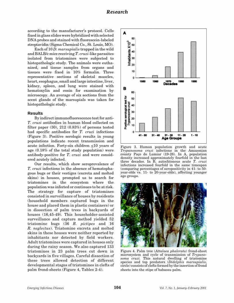
104Emerging Infectious Diseases Vol. 7, No. 1, January–February 2001
Research
according to the manufacturer’s protocol. Cellsfixed in glass slides were hybridized with selectedDNA probes and stained with fluorescein-labeledstreptavidin (Sigma Chemical Co., St. Louis, MO).
Each of 10 D. marsupialis trapped in the wildand BALB/c mice receiving T. cruzi-like parasitesisolated from triatomines were subjected tohistopathologic study. The animals were eutha-nized, and tissue samples from organs andtissues were fixed in 10% formalin. Threerepresentative sections of skeletal muscles,heart, esophagus, small and large intestine, liver,kidney, spleen, and lung were stained withhematoxylin and eosin for examination bymicroscopy. An average of six sections from thescent glands of the marsupials was taken forhistopathologic study.
ResultsBy indirect immunofluorescence test for anti-
T. cruzi antibodies in human blood collected onfilter paper (30), 212 (0.83%) of persons testedhad specific antibodies for T. cruzi infections(Figure 3). Positive serologic results in youngpopulations indicate recent transmission andacute infection. Forty-six children <10 years ofage (0.18% of the total study population) wereantibody-positive for T. cruzi and were consid-ered acutely infected.
Our results, which show seroprevalence ofT. cruzi infections in the absence of hematopha-gous bugs or their vestiges (excreta and moltedskins) in houses, prompted us to search fortriatomines in the ecosystem where thepopulation was infected or continues to be at risk.The strategy for capture of triatominesconsisted in surveillance of houses by residents(household members captured bugs in thehouse and placed them in plastic containers) orin dissection of palm trees in backyards ofhouses (16,45-49). This householder-assistedsurveillance and capture method yielded 52triatomine bugs (36 R. pictipes and 16R. neglectus). Triatomine excreta and moltedskins in these houses were neither reported byinhabitants nor detected by field workers.Adult triatomines were captured in houses onlyduring the rainy season. We also captured 133triatomines in 23 palm trees cut down inbackyards in five villages. Careful dissection ofthese trees allowed detection of differentdevelopmental stages of triatomines in clefts ofpalm frond-sheets (Figure 4, Tables 2-4).
Figure 3. Human population growth and acuteTrypanosoma cruzi infections in the Amazoniancounty Paço do Lumiar (19-28). In A, populationdensity increased approximately fourfold in the lastthree decades. In B, autochtonous acute T. cruziinfections increased fourfold in the same timespan(comparing percentages of seropositivity in 41- to 50-year-olds vs. 11- to 20-year-olds), affecting youngerage groups.
Figure 4. Palm tree (Attalaea phalerata) frond-sheetmicrosystem and cycle of transmission of Trypano-soma cruzi. This natural dwelling of triatominespecies and top predators (Didelphis marsupialis,circle) consists of clefts formed by the insertion of frondsheets into the stipe of babassu palm.
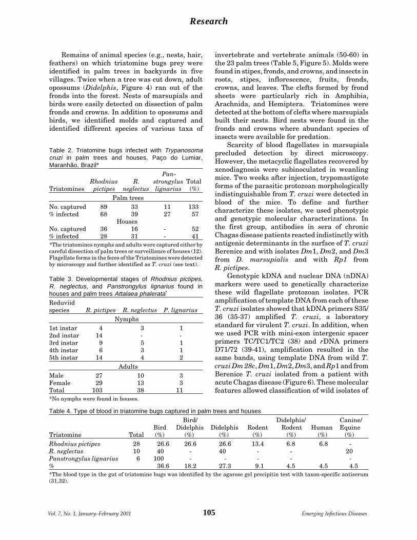
105Vol. 7, No. 1, January–February 2001 Emerging Infectious Diseases
Research
Remains of animal species (e.g., nests, hair,feathers) on which triatomine bugs prey wereidentified in palm trees in backyards in fivevillages. Twice when a tree was cut down, adultopossums (Didelphis, Figure 4) ran out of thefronds into the forest. Nests of marsupials andbirds were easily detected on dissection of palmfronds and crowns. In addition to opossums andbirds, we identified molds and captured andidentified different species of various taxa of
invertebrate and vertebrate animals (50-60) inthe 23 palm trees (Table 5, Figure 5). Molds werefound in stipes, fronds, and crowns, and insects inroots, stipes, inflorescence, fruits, fronds,crowns, and leaves. The clefts formed by frondsheets were particularly rich in Amphibia,Arachnida, and Hemiptera. Triatomines weredetected at the bottom of clefts where marsupialsbuilt their nests. Bird nests were found in thefronds and crowns where abundant species ofinsects were available for predation.
Scarcity of blood flagellates in marsupialsprecluded detection by direct microscopy.However, the metacyclic flagellates recovered byxenodiagnosis were subinoculated in weanlingmice. Two weeks after injection, trypomastigoteforms of the parasitic protozoan morphologicallyindistinguishable from T. cruzi were detected inblood of the mice. To define and furthercharacterize these isolates, we used phenotypicand genotypic molecular characterizations. Inthe first group, antibodies in sera of chronicChagas disease patients reacted indistinctly withantigenic determinants in the surface of T. cruziBerenice and with isolates Dm1, Dm2, and Dm3from D. marsupialis and with Rp1 fromR. pictipes.
Genotypic kDNA and nuclear DNA (nDNA)markers were used to genetically characterizethese wild flagellate protozoan isolates. PCRamplification of template DNA from each of theseT. cruzi isolates showed that kDNA primers S35/36 (35-37) amplified T. cruzi, a laboratorystandard for virulent T. cruzi. In addition, whenwe used PCR with mini-exon intergenic spacerprimers TC/TC1/TC2 (38) and rDNA primersD71/72 (39-41), amplification resulted in thesame bands, using template DNA from wild T.cruzi Dm 28c, Dm1, Dm2, Dm3, and Rp1 and fromBerenice T. cruzi isolated from a patient withacute Chagas disease (Figure 6). These molecularfeatures allowed classification of wild isolates of
Table 4. Type of blood in triatomine bugs captured in palm trees and houses
Bird/ Didelphis/ Canine/Bird Didelphis Didelphis Rodent Rodent Human Equine
Triatomine Total (%) (%) (%) (%) (%) (%) (%)Rhodnius pictipes 28 26.6 26.6 26.6 13.4 6.8 6.8 -R. neglectus 10 40 - 40 - - 20Panstrongylus lignarius 6 100 - - - - -% 36.6 18.2 27.3 9.1 4.5 4.5 4.5*The blood type in the gut of triatomine bugs was identified by the agarose gel precipitin test with taxon-specific antiserum(31,32).
Table 2. Triatomine bugs infected with Trypanosomacruzi in palm trees and houses, Paço do Lumiar,Maranhão, Brazil*
Pan-Rhodnius R. strongylus Total
Triatomines pictipes neglectus lignarius (%)Palm trees
No. captured 89 33 11 133% infected 68 39 27 57
HousesNo. captured 36 16 - 52% infected 28 31 - 41*The triatomines nymphs and adults were captured either bycareful dissection of palm trees or surveillance of houses (12).Flagellate forms in the feces of the Triatomines were detectedby microscopy and further identified as T. cruzi (see text).
Table 3. Developmental stages of Rhodnius pictipes,R. neglectus, and Panstrongylus lignarius found inhouses and palm trees Attalaea phalerata*
Reduviidspecies R. pictipes R. neglectus P. lignarius
Nymphs1st instar 4 3 12nd instar 14 - -3rd instar 9 5 14th instar 6 3 15th instar 14 4 2
AdultsMale 27 10 3Female 29 13 3Total 103 38 11*No nymphs were found in houses.

106Emerging Infectious Diseases Vol. 7, No. 1, January–February 2001
Research
Table 5. Trophic network in randomly selected palm trees of an Amazonian county*Network Trophic level† Palm tree localization Local given namesMetaphyta: Attalaea phalerata 1 Rainforest BabaçuMold: (50, 51) Meliolaceae 2 Stipe & crown shaft Bolor Meliola acristaeCatabotrydaceae Catobotrys decidiumMetazoa:Insecta (52, 53) ColeopteraCurculionidae Homalinotus coriaceus Rhynchophorus palmarum R. barbirosstris Amerrhynus inca 3 Stipe, fronds & crown BesouroChrysomelidae 3 Crown Barata do coqueiro Coraliomela brunea Mecistomela marginataScarabaeidae 3 Roots Rola bosta, broca raiz Strategus aloeusBruchidae 3 fronds Broca, bicho do côco Pachimerus nucleorum HomopteraAsphidae 3 Crown Pulgão Cerataphis lataniaeDiaspididae 3 Crown Cochonilha coqueiro Aspidiotus destructor LepidopteraBrassolidae 3 Crown Borboleta Brasssolis sophorae Brassopholis astyra astyra HymenopteraFormicidae Acromyrmex histrix A. landolti A. laticeps migrosetosus 2 & 3 Roots, stipe & fronds Formiga A. nobilis A. lundi carli HemipteraReduvidae (6, 7, 10, 17) 4 fronds & crown Barbeiro Rhodnius pictipes R. neglectus Panstrongylus lignarusAracnidea, Araneae (38) 4Theraphosidae fronds AranhaAmphibia 4 fronds Perereca Scinax spAves (56) 5 fronds & crown Aratinga jandaia Jandaia Buzeo magnirostris Gavião Colombina passerine Rolinha Coragyps atratus Urubú Crotophaga ani Anu Guira-guira Anu branco Otus choliba Caburé Pizangus sulphuratus Bem-te-ví Turdus fumigatus Sabiá Tyranus muscivora TesourinhaMarsupialia (54-59) 5 fronds Didelphis marsupialis Mucura Caluromys sp. Cuíca*Palm trees were randomly selected from five different localities (backyards) in the Paço do Lumiar county.†Trophic levels in microhabitat following the flux of energy: 1) Palm tree, predated by molds, insecta, birds and mammals; 2)molds, predated by Formicinae and Ponarinae; 3) Insects, predated by arachnids, amphibians and birds; (4) Arachnids andamphibians, predated by birds; 5) Birds and mammals, predated by hematophagus insects and man (50-60).

107Vol. 7, No. 1, January–February 2001 Emerging Infectious Diseases
Research
T. cruzi as phylogenetic type I, while Berenicewas T. cruzi II (11,38-41). These results werefurther confirmed by in situ hybridization of wildT. cruzi with a biotinilated 198-bp sequencederived from Berenice template DNA (Figure 7),which was amplified with specific nDNA primersTcz1/2 (37).
ConclusionsSeveral authors (7,8,17-19,12) described T. cruzi-like flagellate protozoans (instead of T. cruzi) inthe blood of various classes of mammals and inthe feces of triatomine species from the AmazonBasin. We detected blood flagellates in 9 of 10Didelphidae captured in backyards of houses inPaço do Lumiar county. In addition, we isolatedflagellates from feces of R. pictipes, which showedthe same morphologic features described forT. cruzi. In biological characterizations, these
flagellates induced low-level parasitemia inlaboratory mice. However, histopathologic le-sions in marsupials and laboratory mice weresimilar to those described in Chagas diseasepatients. Furthermore, nuclear DNA markersdisplayed all features of the T. cruzi standardsBerenice and Dm 28c T. cruzi, which differedfrom those shown by T. rangeli and L. braziliensis.These data confirm the parasitic flagellatespresent in triatomines and mammals in thisecoregion as T. cruzi.
Birds are refractory to T. cruzi infections (33),but several authors (54-60) describe marsupialsas important wildlife reservoirs of this parasiticprotozoon. We used traps with fruit baits tocapture 12 marsupials, which underwent karyo-typing and parasitologic and pathologic examina-tions. The karyotyping confirmed these mam-mals as D. marsupialis. Nine of 10 trappedmarsupials had protozoan blood flagellatesisolated by xenodiagnosis or hemoculture.Histopathologic study of heart sections from
Figure 5. The informal economy: man carrying ababassu palm frond to be sold as soft thatch. A singlepalm tree enclosing five trophic levels associated withthe Rhodnius pictipes (Hemiptera: Reduviidae)transmission of T. cruzi in the babassu microsystem.
Figure 6. Genotypic characterization of wild-typeflagellates by PCR amplification with rDNA and mini-exon specific primers derived from Trypanosoma cruzi.A, template DNAs amplified with mini-exon intergenicspacer primers (38): Blank, negative control; Tcb,archetypic type II T. cruzi Berenice; Rp1, Dm1, Dm2,and Dm3, flagellates isolated from Rhodnius pictipesand from Didelphis marsupialis; Dm28, standard typeI, sylvatic T. cruzi isolate. B, same template DNAsamplified with rDNA primers (39-41). Tcb yieldedtypical 300-bp band of type II lineage, whereas Rp1,Dm1, Dm2, and Dm3 and Dm28 yielded a 350-bp bandof type I T. cruzi lineage, with mini-exon spacerprimers. In addition, Tcb yielded a typical 125-bp bandof type II, whereas the sylvatic T. cruzi isolates yieldeda 110-bp band of type I, with rDNA primers. Thesefindings confirm sylvatic Rp1, Dm1, Dm2, and Dm3 asT. cruzi.

108Emerging Infectious Diseases Vol. 7, No. 1, January–February 2001
Research
these infected animals showed typical myocardi-tis, characterized by mononuclear cell infiltratesand target cell lysis. Inflammatory infiltrateswere seen in skeletal muscles, esophagus, andsmall and large intestines. Histopathologic studyof representative tissue sections taken from theonly opossum that did not have the parasitedetected showed no histopathologic alterations(data not shown).
American trypanosomiasis has been consid-ered an ancient zoonosis in which insect vectorsand mammal hosts sympatrically occupy vastareas of South America (33). Wild T. cruziinfections and the bug vectors are syntopicallyadapted to mammalian host habitats undernatural equilibrium. T. cruzi infections and humanChagas disease occur over a large geographicarea, limited by parallels 42° N to 42°S (33). TheTocantins moist forest, much of which is subjectto severe disturbance of the environmentalequilibrium, provided conditions for diseaseoutbreaks (61-64). The growing human popula-tions encroaching on that natural ecoregion arefed upon by the triatomine vector R. pictipes, andcases of acute T. cruzi infections in humans haveexponentially increased in the last three decades.
In this study, we describe a trophic networkof five levels comprising different speciesdwelling in palm tree microhabitats. A singleclass of top predator mammal (Didelphidae) wasfound in the study area. The absence of other taxaof top wild predators upon which bugs feed maycontribute to peridomiciliar and domiciliar
invasion during the wet season. This observationcontrasts with earlier descriptions of sevenfamilies of mammals, belonging to Primates,Edentates, Marsupials, Carnivores, Rodents,and Chiroptera classes (10,55,57), which werehosts for triatomines in relatively undisturbedecoregions. Elimination of a single class ofinvertebrate or vertebrate animals in a trophicnetwork may be a major risk factor leading tomore triatomine species entering houses andinitiating a new cycle of transmission of T. cruziinfection.
In our study, a child <10 years of age with apositive immunofluorescence test (see methods)was considered a host of acute T. cruzi infection.Considering the age-specific prevalence of T.cruzi infections in adults (30) and the fact that foreach acute case that is clinically identified anestimated 20 to 100 others are unrecognized (34),autochthonous human Chagas disease in theAmazon Basin may reach 7,860 to 39,300 cases.The latter figure is consistent with serologicevidence of T. cruzi infection in the BrazilianAmazon region presented in the national reporton Chagas disease (65). The characteristics oftransmission of infections described here do notindicate a need for insecticide spraying in theAmazon region, for the cycle of transmission ofT. cruzi is deeply embedded in a natural trophicnetwork comprising wild animals belonging toseveral classes and trophic levels.
Risk factors associated with the possibility ofemergence of endemic Chagas disease in the
Figure 7. In situ hybridization of Rhodnius pictipes-derived flagellates with an archetypic Berenice Trypanosomacruzi-specific probe. The byotinylated 198-bp sequence amplified with Tcz1/2 nuclear DNA probe (37) wasrevealed with fluorescein-conjugated streptavidin. This genotypic marker, representing 12.5% of total parasiticnuclear DNA, also confirms this sylvatic isolate as T. cruzi.

109Vol. 7, No. 1, January–February 2001 Emerging Infectious Diseases
Research
Amazon Basin have been described(10,12,17,19,25,54,55,62-64). First, the broadleafmoist rain forest ecosystem may be invaded bytriatomine species (T. infestans andT. rubrofasciata), which are considered complete-ly adapted to human domiciles, or by othertriatomines (P. megistus and T. brasiliensis, T.pseudomaculata, and T. sordida), which can befound in different ecosystems but frequentlyenter and colonize houses (64). Second, severalAmazon Basin triatomine species (R. pictipes,R. prolixus, R. neglectus, R. nausutus,T. vitticeps, T. rubrovaria and others) can adaptto human dwellings, where they could becomeimportant vectors of the T. cruzi infections (63-69). We found no vestiges of triatominecolonization in houses or their surroundings inour study area. We hypothesize that starvingadult R. pictipes and R. neglectus may leave theirnatural shelters at night to feed on human hosts,probably attracted by light in the houses.
Factors associated with triatomines flyingfrom palm tree to houses need to be clarified. Thescarcity of birds and mammals during the wetseason may be an important factor associatedwith anthropic predation and the presence ofT. cruzi-infected insects in houses in the rainyseason. Domiciliation of triatomines may not berequired for an increasing endemicity of Chagasdisease in the Amazon Basin (13,14,19). Forexample, sylvatic Rhodnius brethesi recently bitpeople harvesting palm fibers in Barcelos in thenorthwestern part of the State of Amazonas, 490km upriver from Manaus, leading to an acutecase of Chagas disease (13). Blood samples fromresidents of this locality tested by immunofluo-rescence assay had 12.5% positivity for anti-T. cruzi antibodies (70). These observationssuggest that to prevent transmission of T. cruziinfections to humans in the Amazon Basin, newstrategies are needed, which will not necessarilybe similar to those used in controlling endemicChagas disease in other ecosystems, such as theCerrado and Caatinga ecosystems in Brazil(1,63,66,67,71).
The risk for endemic Chagas disease in theAmazon Basin appears related to deforestationand new population settlements, shifting cultiva-tion, and rapid human colonization of the vectors’natural ecotope in the last three or more decades,as well as to lack of multiple blood sources (hosts)for the vectors in the rainy season. Amazon Basinperiurban and urban areas where acute cases of
Chagas disease have been reported did not showsigns of colonization of houses with triatominebugs. Instead, transmission of sylvatic T. cruzi tohumans has been associated with sylvatic species(R. pictipes, R. neglectus, P. lignarius), which flyfrom palm trees to houses. New prevention andcontrol strategies should take into considerationrisk factors leading to endemicity of the diseasein the Amazon. An entomologic and epidemio-logic system for surveillance of Chagas disease inAmazonia has been suggested (68,69).
Alternatively, the enormous task of control-ling emerging Chagas disease in the AmazonBasin should rely initially on an information,education, and communication program, whichencourages control measures by the householder(e.g., use of screens, bed nets, insecticide-treatedfabrics, and vegetation management) (69-73).Such a program for prevention of contact withtriatomines should be conducted directly incommunities, elementary schools, and churchesand social clubs, reinforced by social marketingand mass media communications. Furtherstudies are also needed for identifying new andintegrated (chemical and nonchemical) strate-gies required for controlling T. cruzi vectors inthe Amazon Basin, which may not necessarily besimilar to those already shown to be partiallyeffective in controlling the domestic vectors ofendemic Chagas disease in other ecosystems inthe Americas.
AcknowledgmentsWe thank the National Laboratory for Taxonomy of
Triatomines, Fundação Instituto Oswaldo Cruz, Rio deJaneiro, and the Chagas Disease Multidisciplinary ResearchLaboratory, University of Brasília, for performing theprecipitin test.
Dr. Teixeira is professor of pathology at the medicalschool of the University of Brasilia. His research inter-ests focus on endemic infections by kinetoplastic flagel-lates and the pathogeneses of Chagas disease and cuta-neous and visceral leishmaniasis.
References 1. Dinerstein E, Olson DM, Graham DJ, Webster AL,
Primm SA, Bookbinder MP, et al. A conservationassessment of the terrestrial ecoregions of LatinAmerica and the Caribbean. Ed. Washington: TheWorld Wildlife Fund/The World Bank; 1995.
2. La Foret en Jeu. L‘ Extractivisme en AmazonieCentrale. In: Emperaire L, editor. L’ Orston: InstitutFrançais de Recherche Scientifique pour leDéveloppement en Coopéracion. Paris: Unesco; 1996.

110Emerging Infectious Diseases Vol. 7, No. 1, January–February 2001
Research
3. Pindorama. São Paulo: Mercedes-Benz do Brazil; 1993. 4. Kahn F, Corradini L. Palmeiras da Amazonia. Ed.
Cendotec. Brasilia: Centro Franco Brasileiro deDocumentação Científica; 1994. Contato, specialvolume. p. 11-6.
5. Mapeamento e levantamento do potencial dasocorrências de babaçuais. Brasília: Núcleo deComunicação Social da Secretaria de TecnologiaIndustrial, Ministério da Indústria e do Comércio;1982. p. 11-62.
6. Moomen H. Emerging infectious diseases—Brazil.Emerg Infect Dis 1998;4:351-4.
7. Brasil RP, Silva AR, Arbarelli A, Vale JF. Distribuiçãoe infecção de triatomineos por Trypanosoma tipo cruzina Ilha de São Luis, Maranhão. Rev Soc Bras Med Trop1985;18:257-60.
8. Miles MA, Souza de AA, Póvoa M. Chagas diseases in theAmazon Basin. III. Ecotopes of ten triatomine bug species(Hemiptera: Reduvidae) from the vicinity of Belém, ParáState, Brazil. J Med Entomol 1981;18:266-78.
9. Miles MA, Arias JR, Souza AA. Chagas disease in theAmazon Basin. V. Peri-urban palms as habitats ofRhodnius robustus and Rhodnius pictipes triatominaevectors of Chagas disease. Mem Inst Oswaldo Cruz1983;78:391-8.
10. Lainson R, Shaw JJ, Fraiha H, Miles MA, Draper CC.Chagas disease in the Amazon Basin: Trypanosomacruzi infections in sylvatic mammals, triatomine bugsand man in the State of Pará, north Brazil. Trans R SocTrop Med Hyg 1979;73:193-204.
11. Recommendations from a satellite meeting. In:International Symposium to commemorate the 90thanniversary of the discovery of Chagas disease. Rio deJaneiro: Mem Inst Oswaldo Cruz 1999;94:429-32.
12. D’Alessandro A, Barreto P, Saravia N, Barreto M.Epidemiology of Trypanosoma cruzi in the oriental plainsof Colombia. Am J Trop Med Hyg 1984;33:1084-95.
13. Coura JR, Barret TV, Naranjo CM. Ataque depopulações humanas por triatomineos silvestres noAmazonas: uma nova forma de transmissão da infecçãoChagásica? Rev Soc Bras Med Trop 1994;27:251-3.
14. Valente VC, Valente SA, Noireau F, Carrasco HJ, MilesMA. Chagas disease in the Amazon Basin: associationof Panstrongylus geniculatus (Hemiptera: Reduviidae)with domestic pigs. J Med Entomol 1998;35:99-103.
15. Amunárriz MU, Chico ME, Guderian RH. Chagasdisease in Ecuador: a sylvatic focus in the AmazonRegion. J Trop Med Hyg 1991;94:145-9.
16. Whitlaw JT, Chaniotis BN. Palm trees in Panama. AmJ Trop Med Hyg 1978;27:873-81.
17. Almeida FB. Triatomíneos da Amazonia. Encontro detrês espécies naturalmente infectadas por trypanosomasemelhante ao cruzi no Estado do Amazonas(Hemiptera, Reduviidae). Acta Amáz (Manaus)1971;1:71-5.
18. Coura JR. Doença de Chagas como endemia naamazonia brasileira: risco ou hipótese? Rev Soc BrasMed Trop 1990; 23:67-70.
19. Fraiha Neto H, Valente SA, Valente VC, Pinto AYN.Doença de Chagas endemica na Amazonia. Rev AcadMed Pará, Belém 1995; 6:1-132.
20. Chagas C. Infection naturelle des singes du Pará(Crysotrix sciureus) par Trypanosoma cruzi. C R SocBiol (Paris) 1924;9:873-6.
21. Raccurt CP. Trypanosoma cruzi in French Guinea:review of accumulated data since 1940. Med Trop1996;56:79-87.
22. Shaw JJ, Lainson R, Fraiha H. Considerações sobre aepidemiologia dos primeiros casos autóctones de doençade Chagas registrados em Belém, Pará, Brasil. RevBras Saúde Pública, São Paulo 1969;3:153-7.
23. Ferraroni JJ, Melo JAN, Camargo ME. Molestia deChagas na Amazônia. Ocorrência de seis casossuspeitos, autóctones, sorologicamente positivos. ActaAmazônica 1977;3:438-9.
24. Silva AR, Mendes JR, Mendonca ML, Cutrin RN, BrazilRP. Primeiros casos agudos autoctones da doença deChagas no Maranhão e inquérito soro-epidemiologicoda população. Rev Soc Bras Med Trop 1985;18:269-70.
25. Rodrigues CI, Souza AA, Terceros R, Valente S. Doençade Chagas na Amazonia: I Registro de oito casosautóctones em Macapa. Rev Soc Bras Med Trop1988;21:193-7.
26. Acosta HMS, Ferreira CS, Carvalho ME. Humaninfection with Trypanosoma cruzi in Nasca, Peru: aseroepidemiologic survey. Rev Inst Med Trop São Paulo1997;39:107-12.
27. Chico MH, Sandoval C, Guevara AE, Calvopiña MH,Cooper PJ, Reed SG, et al. Chagas disease in Ecuador:evidence for disease transmission in an indigenouspopulation in the Amazon Region. Mem Inst OswaldoCruz 1997;92:317-20.
28. Corredor-Arjona A, Moreno CA, Agudelo CA, Bueno M,López MC, Cáceres E, et al. Prevalence of Trypanosomacruzi and Leishmania chagasi infection and risk factorsin a Colombian indigenous population. Rev Inst MedTrop São Paulo 1999;41: 229-4.
29. Spellerberg IF, Hardes S. Biological conservation.Cambridge: Cambridge University Press, 1992.
30. Williams-Blangero S, VandeBerg J, Blangero J,Teixeira ARL. Genetic epidemiology of seropositivityfor Trypanosoma cruzi infection in rural Goias, Brazil.Am J Trop Med Hyg 1997;57: 538-43.
31. Pedreira de Freitas JL, Siqueira AF, Ferreira OA.Investigações epidemiológicas sobre triatomíneos dehábitos domésticos e silvestres com auxílio da reação deprecipitina. Rev Inst Med Trop São Paulo 1960:2:90-9.
32. Siqueira AF. Estudos sobre a reação da precipitinaaplicada à identificação de sangue por triatomineos.Rev Inst Med Trop São Paulo 1960;2:41-53.
33. Teixeira ARL. The Stercorarian trypanosomes. In:Immune responses in parasitic infections: immunology,immunopathology and immunoprophylaxis. EJL Soulsby,editor. Boca Raton: CRC Press, 1987. pp. 125-45.
34. Teixeira ARL, Teixeira G, Macedo V, Prata A. Acquiredcell-mediated immunodepression in acute Chagasdisease. J Clin Invest 1978;62:1132-8.
35. Sturm NR, Degrave W, Morel CM, Simpson L. Sensitivedetection and schizodeme classification of Trypanosomacruzi by amplification of kinetoplast minicircle DNAsequences: use in the diagnosis of Chagas disease. MolBiochem Parasitol 1984;33:205-14.

111Vol. 7, No. 1, January–February 2001 Emerging Infectious Diseases
Research
36. Avila HA, Sigman DS, Coohen LM, Millikan RC,Simpson L. Polymerase chain reaction amplificationof Trypanosoma cruzi kinetoplast minicircle DNAisolated from whole blood lysates: diagnosis ofchronic Chagas disease. Mol Biochem Parasitol1991;48:211-22.
37. Moser DR, Kirchhoff LV, Donelson J. Detection ofTrypansoma cruzi DNA amplification using thepolymerase chain reaction. J Clin Microbiol1989;27:1477-82.
38. Fernandes O, Sturm NR, Campbell D. The mini-exongene: a genetic marker for zymodeme III of Trypanosomacruzi. Mol Biochem Parasitol 1998;95:129-33.
39. Briones MR, Souto RP, Stolf BS, Zingales B. The evolutionof two Trypanosoma cruzi subgroups inferred from rRNAgenes can be correlated with the interchange of Americanmammalian faunas in the Cenozoic and has implicationsto pathogenicity and host specificity. Mol BiochemParasitol 1999;104:219-32.
40. Souto RP, Zingales B. Sensitive detection and strainclassification of Trypanosoma cruzi by amplification ofa ribosomal RNA sequence. Mol Biochem Parasitol1993;63:45-52.
41. Souto RP, Vargas N, Zingales B. Trypanosoma rangeli:discrimination from Trypanosoma cruzi based on avariable domain from the large subunit ribosomal RNAgene. Exp Parasitol 1999;91:306-14.
42. Jorg ME. La modificación del biotopo peri-habitacionalen la profilaxis de la enfermedad de Chagas. Rev SocBras Med Trop 1989;22:91-5.
43. Vexenat AC, Santana JM, Teixeira ARL. Cross-reactivity of antibodies in human infections by thekinetoplastid protozoa Trypanosoma cruzi, Leishmaniachagasi and Leishmania (viannia) braziliensis. RevInst Med Trop São Paulo 1996;38:177-85.
44. Guhl F, Hudson L, Marinkelle CJ, Jaramillo CA,Bridge C. Clinical Trypanosoma rangeli infection as acomplication of Chagas disease. Parasitology1987;94:475-84.
45. Romaña CA, Pizarro JC, Rodas E, Guilbert E. Palmtrees as ecological indicators of risk areas for Chagasdisease. Trans R Soc Trop Med Hyg 1999;93:594-5.
46. Barreto MP, Siqueira AF, Correa FMA, Ferrioli F,Cavalheiro JR. Estudos sobre reservatórios e vetoressilvestres do Trypanosoma cruzi. VII: investigacões sobre ainfecção natural de gambás por Tripanossomos semelhantesao “T. cruzi.” Rev Bras Biol 1964;24:289-300.
47. Barreto MP, Siqueira AF, Filho, Cavalheiro JR.Estudos sobre reservatórios e vetores silvestres doTrypanosoma cruzi. XI: Observações sobre um foco datripanosomose Americana no Município de RibeirãoPreto, São Paulo. Rev Inst Med Trop São Paulo1966;8:103-12.
48. Barreto MP, Alburquerque RDR, Funayama GK.Estudos sobre reservatórios e vetores silvestres doTrypanosoma cruzi. XXXVI: investigações sobretriatomineos de palmeiras no Municipio De Uberaba,MG, Brasil. Rev Soc Bras Biol 1969; 29:577-88.
49. Barreto MP, Ribeiro RD, Rocha GM. Estudos Sobrereservatórios e vetores silvestres do Trypanosomacruzi. LXIX: Inquérito preliminar sobre triatomíneossilvestres na região do triângulo Mineiro MG, Brasil.Rev Bras Biol 1978; 38:633-7.
50. Hawksworth DL, Kirk PM, Sutton DN. Dictionary of thefungi. 8th edition. International Mycological Institute.Cambridge: University Press; 1995. p. 76, 272.
51. Fungos em plantas no Brasil. Empresa Brasileira dePesquisa Agropecuária, Centro Nacional de Pesquisade Recursos Genéticos e Biotecnologia, Ministério daAgricultura e do Abastecimento. Brasília: Serviço deProdução de Informação; 1988. p. 171.
52. Edwards PJ, Wratten SD. Ecologia das interações entreinsetos e plantas. São Paulo: Editora Pedagógica eUniversitária Ltda; 1981. Vol. 27.
53. Kempf WW. Levantamento das formigas da mataamazônica, nos arredores de Belém do Pará, Brasil.Studia Entomológica. Rio de Janeiro: Vozes Ltda; 1970.p. 321-43.
54. Naiff MF, Naiff RD, Barret TV. Vetores selváticos dedoença de Chagas na área urbana de Manaus (AM):atividade de vôo nas estações secas e chuvosas. Rev SocBras Med Trop 1998; 31:103-5.
55. Miles MA, Arias JR, Naiff RD, Souza AA, Povoa MM,Lima JAN, et al. Vertebrate hosts and vectors ofTrypanosoma rangeli in the Amazon Basin of Brazil.Am J Trop Med Hyg 1983; 32:1251-9.
56. Helmut S. Birds in Brazil. A natural history. NewJersey: Princeton University Press;1993.
57. Deane L. Animal reservoirs of Trypanosoma cruzi inBrazil. Rev Bras Malariol Doenças Tropicais1964;16:28-48.
58. Deane MP, Jansen AM. Another Trypanosoma, distinctfron T. cruzi, multiples in the lumen of the anal glandsof the opossum Didelphis marsupialis. Mem InstOswaldo Cruz 1986; 81:131-2.
59. Jansen AM, Madeira F, Carreira JC, Acosta-Medina,Deane MP. Trypanosoma cruzi in the opossumDidelphis marsupialis: a study of the correlations andkinetics of the systemic and scent gland infections innaturally and experimentally infected animals. ExpParasitol 1997;86:37-44.
60. Odun EP. Ecologia. Biblioteca Pioneira de BiologiaModerna. São Paulo: Livraria Pioneira; 1977. p. 19-148.
61. Morse SS. Factors in the emergence of infectiousdiseases. Emerg Infect Dis 1995;1:7-15.
62. Valente AS, Valente VC, Fraiha Neto A. Considerationson the epidemiology and transmission of Chagasdisease in the Brazilian Amazon. Mem Inst OswaldoCruz 1999;94:395-8.
63. Silveira AC, Rezende DF. Epidemiologia e controle datransmissão vetorial da doença de Chagas no Brasil.Rev Soc Bras Med Trop 1994;27(Supl III):11-22.
64. Silveira AC, Vinhaes MC. Elimination of vector-bornetransmission of Chagas disease. Mem Inst OswaldoCruz 1999; 94:405-11.
65. Camargo ME, Silva GR, Castilho EA, Silveira AC.Inquérito sorológico da prevalência de infecçãochagasica no Brasil, 1985-1980. Rev Inst Med Trop SãoPaulo 1984;26:192-204.
66. Dias JCP. A doença de Chagas e seu controle naAmérica Latina. Uma análise de possibilidades. Rio deJaneiro: Cad Saúde Pública 1993;9:201-9.
67. Moncayo A. Chagas disease: epidemiology andprospects for interruption of transmission in theAmericas. Wld Hlth Statist Quart 1992;45:276-9.

112Emerging Infectious Diseases Vol. 7, No. 1, January–February 2001
Research
68. Valente VC. Potential for domestication of Panstrongylusgeniculatus in the municipality of Muaná, MarajóIsland, State of Pará, Brazil. Mem Inst Oswaldo Cruz1999;94:399-400.
69. Feitosa VR. Implantação de um sistema de vigilânciaepidemológica (VE) de doença de Chagas na Amazônia.Ver Soc Brás Méd Trop 1995;28:84-7.
70. Coura JR, Junqueira ACV, Boia MN, Fernandes O.Chagas disease: from bush to huts and houses. Is it thecase of the Brazilian Amazon? Mem Inst Oswaldo Cruz1999;94:379-84.
71. Bos R. The importance of peridomestic environmentalmanagement for the control of the vectors of Chagasdisease. Rev Argent Microbiol 1988;20:58-62.
72. Kroeger A, Ordonez-Gonzalez J, Behrend M, Alvarez G.Bednet impregnation for Chagas disease control: a newperspective. Trop Med Int Health 1999;4:194-8.
73. Wood E, de Licastro SA, Casabe N, Picollo MI,Alzogaray R, Nicolas Zerba E. A new tactic for Triatomainfestans control: fabrics impregnated with beta-cypermethrin. Rev Panam Salud Publica 1999;6:1-7.

113Vol. 7, No. 1, January–February 2001 Emerging Infectious Diseases
Research
Pulmonary infection due to chronic microbialcolonization is the major cause of illness anddeath in cystic fibrosis (CF) patients. MucoidPseudomonas aeruginosa, which is involved inpulmonary damage, is the most frequentlyrecovered pathogen. In contrast, little informa-tion is available about the role of other nonfer-mentative gram-negative rods. An increasingincidence of Stenotrophomonas maltophilia iso-lates has been reported in some CF centers duringthe last decade (1-4). Although an associationbetween S. maltophilia colonization and lungdamage has been observed (2,3), the role of theorganism is still undetermined (5,6). In non-CFpatients (e.g., immunocompromised or intensive-care unit patients), exposure to wide-spectrumantimicrobial drugs, long-term antimicrobial ther-apy, previous pulmonary infections, and chronicrespiratory disease contribute to S. maltophiliaacquisition and increase the risk for respiratoryinfection with this organism (7,8). All these riskfactors are present in the CF population.
We analyzed S. maltophilia from respiratoryisolates of 25 CF patients of the same CF unitduring an 8-year period to determine a) theoverall and yearly incidence of S. maltophiliainfection or colonization and incidence asdetermined by molecular typing, ribotyping, andpulsed-field gel electrophoresis (PFGE); b) theage distribution of acquisition of S. maltophiliapulmonary infection or colonization in patientswith single or repeated episodes; c) thepersistence and variability of S. maltophiliaisolates in patients who had more than oneepisode and the degree of genomic similarityidentified among clones; and d) the epidemiologiclink between similar isolates from differentpatients. We also investigated pulmonaryfunction and other clinical aspects ofS. maltophilia-infected or colonized patients.
Materials and MethodsFrom 1991 to 1998, 25 CF patients (12 female
and 13 male) of 104 who were clinically andmicrobiologically followed at the Hospital Ramóny Cajal CF Unit had at least one positiverespiratory culture for S. maltophilia. CF wasdiagnosed by a positive sweat chloride test
Persistence and Variability ofStenotrophomonas maltophilia in
Cystic Fibrosis Patients,Madrid, 1991–1998
Sylvia Valdezate,*,† Ana Vindel,† Luis Maiz,*Fernando Baquero,* Hector Escobar,* and Rafael Cantón*
*Hospital Ramón y Cajal, Madrid, Spain; †Centro Nacionalde Microbiología, Instituto Carlos III, Madrid, Spain
Address for correspondence: Rafael Cantón, Servicio deMicrobiología, Hospital Ramón y Cajal, Carretera deColmenar, Km 9,100, 28034-Madrid, Spain; fax: 34-91-3368809; e-mail: [email protected].
During 1991 to 1998 at least one Stenotrophomonas maltophilia pulmonaryinfection was observed in 25 (24%) of 104 cystic fibrosis patients at the same unit of ourhospital in Spain. Ribotyping and pulse-field gel electrophoresis (PFGE)characterization of 76 S. maltophilia isolates from these patients indicated an overallclonal incidence of 47.1%, reflecting new strains in 44% of patients with repeatedpositive cultures for S. maltophilia. Six patients with repeated episodes werepersistently colonized (>6 months) with the same strain. S. maltophilia bacterial countswere higher (geometric mean, 2.9 x10⁸ cfu/mL) in patients with repeated episodes thanin those with a single episode (8.4 x10⁴ cfu/mL, p<0.01). Single episodes ofS. maltophilia occurred in patients <10 years of age (43% [6/14]), whereas chroniccolonization occurred more frequently in older patients (>16 years of age).

114Emerging Infectious Diseases Vol. 7, No. 1, January–February 2001
Research
(>60 mEq/L) in association with typical pulmo-nary and gastrointestinal findings or a positivefamily history. The age range of patients was <1to 32 years (median 14.5 years). Eleven patientswere homozygotes and eight heterozygotes for∆F508, the most common mutation in the cysticfibrosis transmembrane conductance regulator(CFTR) gene, and one patient was negative for∆F508. Mutation in this gene could not bedetermined in five patients. The mean number ofsputum samples examined was 6.7 specimens perpatient per year. All 25 patients were followed forat least 1 year during the study period (range 1 to8 years, mean 5.8 years). Culture results wereused to establish age at acquisition ofS. maltophilia. When available, retrospectivecultures obtained before 1991 were also takeninto account.
S. maltophilia colonization in CF patientswas considered persistent if positive cultureswere obtained for >6 months, regardless ofbacterial counts. Overall incidence was definedas the number of patients infected or colonizedwith S. maltophilia, independent of the numberof positive cultures during the study period. Thedenominator was the total number of patientsseen at the CF unit (104 patients). Yearlyincidence was defined as the number of patientswith new episodes of S. maltophilia infection orcolonization, with the denominator the number ofpatients seen per year in the CF unit. The overallincidence and yearly incidence were recalculatedwhen molecular typing data were available. Thesevalues were defined as overall clonal incidence andyearly clonal incidence, respectively, whichrepresent the incidence of S. maltophilia episodescaused by different clonal strains.
Bacteriologic Study and S. maltophilia IsolatesRespiratory secretions, mostly expectorated
sputum, were homogenized with N-acetyl-cysteine and processed by a modified quantita-tive technique (9). Columbia 5% blood,MacConkey, mannitol and salt, and a selectiveBurkholderia cepacia agar media were incubatedin air for 24 hours at 37°C, followed by 24 hours at25°C. In addition, bacitracin-chocolate agar wasplated and incubated in 5% CO2 for 48 hours andSabouraud-chloramphenicol and Sabouraud-chloramphenicol-cyclohexamide agar media for 4weeks at 30°C and 37°C. A culture forS. maltophilia was considered positive when anygrowth of this organism was observed, regardless
of bacterial count. Biochemical identification ofS. maltophilia isolates was performed both withthe API 20NE gallery (BioMerieux, Marcy-l´Étoile, France) and the semiautomatic PASCOsystem (Difco, Detroit, MI). Bacterial counts andco-colonization with other respiratory pathogenswere also considered in the analysis. The samemicrobiologic protocol was applied to all patients,regardless of clinical condition.
RibotypingDNA from all S. maltophilia isolates was
prepared by treatment with hexadecyl-trimethylammonium bromide (10). Ribotypingwas performed as described (11). BamH1,Bsu15I, EcoRI, and HindIII restriction endonu-cleases (Roche Diagnostic, Mannheim, Germany)were also tested in a representative number ofisolates. The best-defined restriction patternwith a higher number of bands was observed withBamH1 and HindIII. Digoxigenin-labeled phageλHindIII-digested DNA (Roche) was used as amolecular size marker. DNA fragments wereseparated by electrophoresis in 0.7% agarose gelsand were blotted onto nylon membranes.Membranes were hybridized with a digoxigenin-labeled rRNA probe with 16S+23S rRNAsequences of Escherichia coli (Roche) at 68ºC for18 hours (12). Differences in numbers and theposition of bands were considered.
Pulsed-Field Gel ElectrophoresisS. maltophilia DNA was prepared and
contained in agarose plugs for digestion with 30U of XbaI (Roche). Closely related isolates usingXbaI were reanalyzed with 20 U of SpeI (Roche)as described (13). Digested samples were meltedand loaded onto 1% agarose gels. PFGE wasperformed with the CHEF-DRII system (Bio-Rad, Hemel Hempstead, UK). Standard lambdaladders comprising 48.5-kbp concatemers wererun as molecular weight markers (Roche).Electrophoresis pulse times for XbaI-digestedDNA were 10 to 60 seconds for 24 hours, followedby a second ramp from 5 to 20 seconds for 5 hours.Both ramps were performed at 5.4 V/cm and 12ºC. For SpeI, pulse times were 25 to 45 seconds for20 hours at 6 V/cm and 12°C. Macrorestrictionfragments were visually compared and interpretedaccording to the criteria of Tenover et al. (14).
A genetic similarity dendogram was designedand calculated by the Dice correlation coefficient(15) and represented by UPGMA with Molecular

115Vol. 7, No. 1, January–February 2001 Emerging Infectious Diseases
Research
Analyst Software (BioRad) and a toleranceposition of 1%. Only well-resolved bandscorresponding to fragments exceeding 97.0 kbpwere included in the computer analysis.
Patient DataChart records from S. maltophilia-positive
CF patients were reviewed. Patients wereclassified according to age, sex, and severity oflung disease. Correlation between colonization orinfection with S. maltophilia and pulmonaryfunction was studied. Pulmonary function wastested in accordance with American ThoracicSociety Guidelines (16). Forced expiratoryvolume (FEV1) (% predicted) value was expressedas the percentage predicted according toKnudson norms for adjusting data for age,height, and sex (17). Trends in FEV1 wereestimated by comparing values at the time of thefirst recovery of S. maltophilia with thoseobtained within a year from the last isolation.P. aeruginosa and other pathogens commonlyencountered in CF were also recorded as outcomecriteria for evaluating the progression ofpulmonary disease.
Statistic AnalysisStatistical significance for comparison pro-
portions was calculated by Chi square or Fisher’sexact test with Epi-Info 6.04a. Quantitativevalues were compared by Student’s t test; p<0.05was considered statistically significant.
ResultsFrom 1991 to 1998, at least one respiratory
culture positive for S. maltophilia was observedin 25 of 104 patients. Thus, the overall incidenceof S. maltophilia-infected or -colonized patientswas 24%; yearly incidence was 2.9% to 14.0%(Figure 1). Fourteen (56%) of these 25 patientshad a single episode of S. maltophilia (SM-SEgroup), and 11 patients (44%) had repeatedepisodes (SM-RE group). No differences insampling frequency (number of sputum samplesstudied per year) or length of follow-up werefound between the two groups.
Eighty-eight S. maltophilia isolates wererecovered from these 25 patients. Seventy-sixisolates, 14 from the SM-SE group and 62 fromthe SM-RE group, were available for furtherstudy. PFGE results indicated an overall clonalincidence of 47.1%, reflecting new strains withdifferent PFGE profiles that had been acquiredby the SM-RE group (Figure 1). The highestyearly clonal incidences were detected in 1991and 1996.
In the SM-SE group, the median age atacquisition of S. maltophilia was 13.4 years(range <1 to 27 years). Nearly 43% of patients (6of 14) acquired S. maltophilia at 6 to 10 years ofage (Figure 2). In SM-RE patients, the medianage at first S. maltophilia isolation was 16.7years (range 3 to 32 years). In this group, 45% (5of 11) acquired S. maltophilia at 11 to 20 years ofage. PFGE analysis of all S. maltophilia strains
Figure 1. Yearlyincidence, yearlyclonal incidence,and yearly preva-lence of Stenotro-phomonas malto-philia acquisitionin 104 cystic fibro-sis patients, 1991–1998.

116Emerging Infectious Diseases Vol. 7, No. 1, January–February 2001
Research
indicated that nine new acquisitions occurred in11- to 15-year-old patients. Because of the smallsample size, differences in age of acquisitionbetween the groups could not be demonstratedwith statistical significance.
RibotypingTo select the suitable enzyme(s) for
S. maltophilia ribotyping, BamH1, Bsu15I,EcoRI, and HindIII endonucleases were used infive different strains isolated from the samepatient, resulting in 4, 4, 2, and 4 differentribotypes, respectively. The number of copies ofthe ribosomal rRNA operon in S. maltophilia was2 to 5 per isolate for BamHI, 2 to 4 Bsu15I forHindIII, and 4 to 5 for EcoRI, with hybridizationband sizes of 3 Kbp to 20 kbp. Greatheterogeneity in ribotypes, 21 with HindIII and20 with BamHI, was found among the 76 S.maltophilia isolates, with a Simpson index (15) of0.8992 and 0.9158, respectively. The geneticsimilarity was 29% to 100% for HindIII and 38%to 100% for BamHI.
PFGE AnalysisForty-seven well-defined profiles of genomic
DNA under XbaI digestion were obtained fromthe 76 S. maltophilia isolates. According to Tenovercriteria (14), 41 types and 6 subtypes wereconsidered. These 6 subtypes were associatedwith 3 of the 41 main subtypes. Fragment sizewas <48 kbp to >1,000 kbp. Discrimination basedon Simpson´s index peaked at 0.97. Geneticheterogeneity is illustrated by the dendogram ofthe 47 XbaI-PFGE profiles (Figure 3). Repeatedisolates displaying an identical PFGE profile from
Figure 2. Distribution of age of acquisition ofStenotrophomonas maltophilia in 25 cystic fibrosispatients with a single episode (SM-SE group) or withrepeated episodes (SM-RE group).
Figure 3. Percentages of genetic similarity between 47XbaI-PFGE clonal types from 76 Stenotrophomonasmaltophilia strains isolated in 25 cystic fibrosispatients of the same unit, 1991–1998.

117Vol. 7, No. 1, January–February 2001 Emerging Infectious Diseases
Research
the same patient or resulting from presumedpatient-to-patient transmission were excludedfrom the dendogram. Forty-one types displayedsimilarity coefficients from 25% to 75%; each wascoded with a number. Each subtype was codedwith a letter (similarity >80%). Strains sharing thesame XbaI digestion pattern could not be furtherdistinguished by SpeI. XbaI was more efficientthan SpeI in distinguishing between subtypes orclosely related strains; 14a and 14b subtypesshowed an indistinguishable PFGE pattern withSpeI. This was also the case with 16a and 16csubtypes.
Persistence and Variabilityof S. maltophilia Strains
The SM-SE group of 14 patients had 14different PFGE types. One of these PFGE
patterns (pattern 1a) was also seen in two of theSM-RE patients (patients 1 and 3). During thestudy period, each of the 11 patients in the SM-RE group had one to five strains with differentPFGE profiles. Strains from five patients (1, 3, 8,10, and 11) completely met the criteria forpersistence (Figure 4). The strains wererecovered from these patients during periods ofpersistence of 29, 86, 6, 9, and 8 months,respectively. A turnover of this predominantstrain with a different strain occurred in four ofthese patients. In patient 4, two strains with 4and 32 months of persistence were isolatedduring two different periods. All these patientswere considered persistently colonized withidentical S. maltophilia isolates (Figure 4).Variable colonization, defined as the isolation ofS. maltophilia strains with different PFGE
Figure 4. Persistence and variability of Stenotrophomonas maltophilia strains in 11 patients with repeatedepisodes of infection or colonization with this organism. S. maltophilia recovered is indicated by a ♦ (identicalisolates from the same patient), ◊ (different isolate from a single patient) or O (no isolate available). DistinctS. maltophilia isolates are identified by numbers, and a letter was added when a closely related strain wasobserved. + = time at which sputum specimens were obtained.

118Emerging Infectious Diseases Vol. 7, No. 1, January–February 2001
Research
Table. Demographic characteristics and co-colonization status of cystic fibrosis patients with Stenotrophomonasmaltophilia infection or colonization
Characteristics SM-SEa (14 patients) SM-REb (11 patients)Gender
Male 8 5Female 6 6Patient genotyped 11 9
Homozygous ∆F508 8 3Heterozygous ∆F508 3 5Other 0 1
Mean age at first S. maltophilia isolation (SD, years) 13.4 (7.3) 16.7 (7.4)FEV1
c measured 11 10FEV1 (% predicted) before S. maltophilia recovery (Mean [SD]) 68.7 (29.6) 74.2 (28.3)
>100 2 170–99 3 540–69 4 3<40 2 1FEV1 (% predicted) after S. maltophilia recovery (Mean [SD]) 63.8 (20.7) 62.9 (24.2)
ABPAc condition 2 1Death (%) 4 (28.5) 2 (18.2)S. maltophilia bacterial counts (geometric mean, cfu/mL) 8.4 x 10⁴ d 2.9 x 10⁸ d
Pseudomonas aeruginosa detected (%) 12 (85.7) 9 (81.8)Aspergillus detected (%) 7 (50.0) 7 (63.6)S. maltophilia co-colonization with:e
Only S. maltophilia detected (%) 1 (7.1) 1 (9.1)P. aeruginosa (%) 8 (57.1) 3 (27.2)Staphylococcus aureus (%) 6 (42.8) 3 (27.2)Burkholderia cepacia (%) 1 (7.1) 1 (9.1)Aspergillus spp. (%) 1 (7.1) 3 (27.2)Candida (%) 3 (21.4) 2 (18.2)
aSM-SE = CF patients with a single episode of S. maltophilia colonization.bSM-RE: CF patients with repeated episodes of S. maltophilia colonization.cFEV = Forced expiratory volume.dABPA: allergic bronchopulmonary aspergillosis.ep<0.05 comparing both groups.fPatients in the SM-RE group colonized with organisms in addition to S. maltophilia. When different co-colonizations occurredin the same patient, we recorded only the cocolonization that was at least twice as frequent.
profiles, was identified in five patients (patients2, 5, 6, 7, and 9).
Suspected Cross-TransmissionIn 1996, three patients, two in the SM-RE
group (patients 1 and 3) and another (patient 12)in the SM-SE group, shared S. maltophiliaisolates with indistinguishable ribotype andPFGE type under all restriction enzymes tested(profile 1a). Patient 1 was persistently colonizedwith this strain for 2 years, and patient 3 wastransiently colonized (Figure 4).
Bacterial Counts and Clinical FindingsSM-SE patients had higher S. maltophilia
bacterial counts when either the firstS. maltophilia isolate (geometric mean, 4.3 x10⁵
cfu/mL) or all isolates (2.9 x10⁸ cfu/mL) weretaken into account (p<0.05), compared with
patients with a single episode (8.4 x10⁴ cfu/mL). Asimilar rate of P. aeruginosa recovered from therespiratory tract during the study period wasnoted in both groups (Table). In contrast,Aspergillus spp. was detected more frequently inthe SM-RE group of patients. No statisticaldifferences were found when co-colonization wasevaluated. However, S. maltophilia co-coloniza-tion with Aspergillus spp. in the SM-RE grouphad a risk ratio of 3.8 compared with the SM-SEgroup.
Demographic and selected medical charac-teristics and results of respiratory tract cultureswere analyzed for S. maltophilia-infected or -colonized patients (Table). Before S. maltophiliacolonization, slightly lower pulmonary functionlevels (FEV1, % predicted) were observed inpatients with a single S. maltophilia episodethan in patients with repeated episodes (Table).

119Vol. 7, No. 1, January–February 2001 Emerging Infectious Diseases
Research
This value decreased in SM-RE patients from74.2 ± 28.3 (mean value ± SD) (first isolationof S. maltophilia) to 62.9 ± 24.2 (last isolation ofS. maltophilia), which could indicate a decreas-ing trend in FEV1 after the first episode.However, this difference was not statisticallysignificant. On the other hand, SM-SE patientshad a higher death rate (28.5%) than the SM-REgroup, but death rates in both groups were higherthan those observed in S. maltophilia-negativepatients (12.6%).
ConclusionsS. maltophilia, an essentially environmental
organism, is the fourth organism in prevalence inbronchial secretions of CF patients, afterP. aeruginosa, Staphylococcus aureus, andHaemophilus influenzae (5,18). Since it was firstreported in CF patients in 1979 (19), thisorganism has been investigated for its role in theprogression of CF pulmonary disease (5), andconsensus documents have emphasized theimportance of clinical microbiology laboratoriesin detecting its presence in CF respiratorysecretions (20). Despite some virulence factorsshared with P. aeruginosa, its potential forpathogenicity remains uncertain (21). We havereported a high incidence of S. maltophilia-colonized CF patients (30.7%) over a 5-yearperiod (3), but, as in other studies (2,6,22), we didnot address (through epidemiologic typingstudies) whether this high rate was a conse-quence of patient-to-patient transmission orwhether bacterial colonization was sporadicallyor chronically established.
The 1997 Cystic Fibrosis Foundation PatientRegistry from the United States (18), whichincluded 17,996 CF patients in a cross-sectionalstudy analyzing one respiratory sample perpatient per year, showed a percentage of positivecultures for S. maltophilia of 5.1%, a valueslightly higher than in 1996 (3.9%) and 1995(3.4%). In our study, the overall incidence, 24%, ishigher than that observed in other studies (10.6%to 16.6%) with a similar length of follow-up (2,22),but slightly lower than in studies with a longerfollow-up period (27.3%) (6). Consistent withother results, our data showed no clear trendtowards increasing or decreasing over the studyperiod (Figure 1).
The main purpose of our study was to applymolecular typing, both with ribotyping andPFGE, to S. maltophilia isolates recovered from
patients seen in our CF Unit. Among 76 isolates,47 PFGE profiles were identified, and theseresults were used to calculate the incidence ofepisodes of S. maltophilia colonization orinfection in our series. Without typing, theoverall incidence was 24% for the entire studyperiod; by PFGE the incidence was 47.1%. Thisresult clearly indicates that SM-RE patients hadnew episodes with different S. maltophiliastrains. Molecular typing also differentiatedpatients who were chronically infected orcolonized with the same strain (persistence) fromthose with repeated episodes with differentS. maltophilia strains (variability).
PFGE has been recommended for epidemio-logic studies of S. maltophilia isolates (13,23-25).The technique has been shown to be morediscriminatory than enterobacterial repetitiveintergenic consensus polymerase chain reactionand other molecular techniques for differentia-tion within this species (13). In our study,restriction endonuclease XbaI provides discrimi-natory patterns, with a high discrimination valueon Simpson´s index (0.97), enabling easyinterpretation of banding profiles. This enzymehas been used to study the stability ofS. maltophilia from a CF patient over a 15-monthperiod (26), the relationship between CF andenvironmental S. maltophilia isolates (13), andthe epidemiology of S. maltophilia isolates from ahematology department (27). Other studies havebeen based on DraI (25,28,29) and SpeI(23,27,30). In our study, XbaI was more efficientthan SpeI in distinguishing between subtypes orclosely related strains.
We observed only one positive culture ofS. maltophilia over the study period in 14patients, in accordance with the results of Demkoet al. (6), who showed that 50% of CF patients hadonly one positive culture of S. maltophilia over a13-year period. In contrast, 11 patients (44%)from our CF unit had repeated episodes ofS. maltophilia colonization or infection. Typingstudies, however, demonstrated different strainsin five patients and, with the exception of patient8, a persistent strain was characterized in theremaining six patients, but with a turnover withdistinct strains (Figure 4). Because of samplingbias, some of these patients may also have hadpersistent colonization. Of 11 patients withrepeated SM-RE isolates, 6 had evidence ofpersistent colonization (Figure 5). More frequentsampling could have increased this proportion.
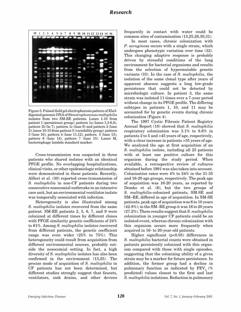
120Emerging Infectious Diseases Vol. 7, No. 1, January–February 2001
Research
Cross-transmission was suspected in threepatients who shared isolates with an identicalPFGE profile. No overlapping hospitalizations,clinical visits, or other epidemiologic relationshipwere demonstrated in these patients. Recently,Alfieri et al. (30) reported cross-transmission ofS. maltophilia in non-CF patients during twoconsecutive nosocomial outbreaks in an intensivecare unit, but an environmental ventilator isolatewas temporally associated with infection.
Heterogeneity is also illustrated amongS. maltophilia isolates recovered from the samepatient. SM-RE patients 2, 5, 6, 7, and 9 werecolonized at different times by different cloneswith PFGE similarity genetic coefficients of 24%to 61%. Among S. maltophilia isolates recoveredfrom different patients, the genetic coefficientrange was even wider (25% to 75%). Thisheterogeneity could result from acquisition fromdifferent environmental sources, probably out-side the nosocomial setting. In fact, a highdiversity of S. maltophilia isolates has also beenconfirmed in the environment (13,25). Theprecise mode of acquisition of S. maltophilia inCF patients has not been determined, butdifferent studies strongly suggest that faucets,ventilators, sink drains, and other devices
frequently in contact with water could becommon sites of contamination (13,25,28,30,31).
In most cases, chronic colonization withP. aeruginosa occurs with a single strain, whichundergoes phenotypic variation over time (32).This changing adaptive response is probablydriven by stressful conditions of the lungenvironment for bacterial organisms and resultsfrom the selection of hypermutable geneticvariants (33). In the case of S. maltophilia, theisolation of the same clonal type after years ofapparent absence suggests a long low-gradepersistence that could not be detected bymicrobiologic culture. In patient 3, the samestrain was isolated 11 times over a 7-year periodwithout change in its PFGE profile. The differingsubtypes in patients 1, 10, and 11 may beaccounted for by genetic events during chroniccolonization (Figure 4).
The 1997 Cystic Fibrosis Patient RegistryAnnual Report (18) showed that S. maltophiliarespiratory colonization was 3.1% to 8.6% inpatients 2 to 5 and >45 years of age, respectively,with a clear increase in patients >35 years of age.We analyzed the age at first acquisition of anS. maltophilia isolate, including all 25 patientswith at least one positive culture for thisorganism during the study period. Whenavailable, a retrospective review of culturesobtained before 1991 was also taken into account.Colonization rates were 4% to 24% in the 31-35and 16-20 age groups, respectively. The peak ageof acquisition was 16-20 years, as reported byDemko et al. (6), but the two groups ofS. maltophilia-colonized patients, SM-SE andSM–RE, differed in age of acquisition. In SM-SEpatients, peak age of acquisition was 6 to 10 years(42.8%); in the SM–RE group it was 16 to 20 years(27.2%). These results suggest that S. maltophiliacolonization in younger CF patients could be anisolated event, whereas chronic colonization withthis organism occurs more frequently whenacquired in 16- to 20-year-old patients.
Higher significant (p<0.05) differences inS. maltophilia bacterial counts were obtained inpatients persistently colonized with this organ-ism compared with those with single episodes,suggesting that the colonizing ability of a givenstrain may be a marker for future persistence. Inaddition, the former group had a decline inpulmonary function as indicated by FEV1 (%predicted) values closest to the first and lastS. maltophilia isolations. Reduction in pulmonary
Figure 5. Pulsed-field gel electrophoresis pattern of XbaI-digested genomic DNA of Stenotrophomonas maltophiliaisolates from two SM-RE patients. Lanes 1-10 frompatient 1 (persistence group): pattern 1a (lanes 1,3-6,8),pattern 1b (ln 7), pattern 1c (lane 9) and pattern 2 (lane2); lanes 10-15 from patient 5 (variability group): pattern3 (lane 10), pattern 4 (lane 11,12), pattern 5 (lane 13),pattern 6 (lane 14), pattern 7 (lane 15). Lanes M,bacteriophage lambda standard marker.
121Vol. 7, No. 1, January–February 2001 Emerging Infectious Diseases
Research
function could also reflect increased age or theeffect of other pathogens. In fact, a higher rate ofAspergillus spp. isolation was detected in CFpatients chronically colonized with S. maltophilia.However, a higher death rate was observed inpatients with a single episode of S. maltophilia(28.5%) than in patients with repeated episodes(18.2%), but both these values were higher thanthose obtained in S. maltophilia-negativepatients (12.6%). Demko et al. recently reported alower death rate in patients with long-termchronically S. maltophilia-positive cultures(7.7%) than in those with transient or acutepositive cultures (21.1%)(6). Moreover, thecombined death rate in S. maltophilia-positivepatients (19.0%) was slightly higher than inS. maltophilia-negative patients (16.5%). Incontrast, Goss et al. (34) demonstrated in a cohortstudy that S. maltophilia acquisition did notdecrease survival in patients with CF, butpatients with this organism had significantlylower FEV1 (% predicted) values. These datasuggest that isolation and persistence ofS. maltophilia could contribute to a progressionof clinical deterioration, particularly in patientswith lower pulmonary function. IncreasedS. maltophilia colonization may be observed inthe future as a result of improvements in lifeexpectancy.
This work was supported by research grant 2114/98from the Consejería de Educación y Cultura of theComunidad de Madrid, Spain.
Dr. Valdezate is a fellow in the Clinical MicrobiologyDepartment at the Ramón y Cajal Hospital and at theCentro Nacional de Microbiología (Instituto Carlos III)in Madrid, Spain. Her research interests focus on epide-miology and resistance of Stenotrophomonas maltophilia,mainly from cystic fibrosis patients.
References 1. Gladman G, Connor PJ, Williams RF, David TJ.
Controlled study of Pseudomonas maltophilia in cysticfibrosis. Arch Dis Child 1993;67:192-5.
2. Karpati F, Malmborg AS, Alfredsson H, Hjelte L,Strandvik B. Bacterial colonization with Xanthomonasmaltophilia: a retrospective study in a cystic fibrosispatient population. Infection 1994;22:258-63.
3. Ballestero S, Virseda I, Escobar H, Lucrecia L, Baquero F.Stenotrophomonas maltophilia in cystic fibrosis patients.Eur J Clin Microbiol Infect Dis 1995;14:728-79.
4. Burns JL, Emerson J, Stapp JR, Yim DL, Krzewinski J,Louden L, et al. Microbiology of sputum from patientsat cystic fibrosis centers in the United States. ClinInfect Dis 1998;27:158-63.
5. Denton M. Stenotrophomonas maltophila: an emergingproblem in cystic fibrosis patients. Rev Med Microbiol1997;8:15-9.
6. Demko CA, Stern RC, Doershuk CF. Stenotrophomo-nas maltophilia in cystic fibrosis: incidence andprevalence. Pediatr Pulmonol 1998;25:304-8.
7. Villarino ME, Stevens LE, Schable B, Mayers G, MillerJM, Burke JP, et al. Risk factors for epidemicXantomonas maltophilia infection/colonization inintensive care unit patients. Infect Control HospEpidemiol 1992;13:201-6.
8. Van Couwenberghe CJ, Farver TB, Cohen SH. Riskfactors associated with isolation of Stenotrophomonas(Xantomonas) maltophilia in clinical specimens. InfectControl Hosp Epidemiol 1997;18:316-21.
9. Wong K, Roberts MC, Owens L, Fife M, Smith AL.Selective media for the quantification of bacteria incystic fibrosis sputum. J Med Microbiol 1984;17:113-9.
10. Ausubel FM, Brent R, Kingston RE, Moore DD, SeidmanJG, Smith JA, et al. Current protocols in molecular biology.New York: Greene Publishing Associates and WileyInterscience; 1991( Suppl. 13):241-5.
11. Southern EM. Detection of specific sequences amongDNA fragments separated by gel electrophoresis. J MolBiol 1975; 98:503-17.
12. Stull TL, LiPuma JJ, Edlind TD. A broad-spectrumprobe for molecular epidemiology of bacteria: ribosomalRNA. J Infect Dis 1988;157:280-6.
13. Denton M, Todd NJ, Kerr KG, Hawkey PM, LittlewoodJM. Molecular epidemiology of Stenotrophomonasmaltophilia isolated from clinical specimens frompatients with cystic fibrosis and associated environ-mental samples. J Clin Microbiol 1998;36:1953-8.
14. Tenover FC, Arbeit RD, Goering RV, Mickelsen PA,Murray BE, Persing DH, et al. Interpretingchromosomal DNA restriction patterns produced bypulse-field gel electrophoresis: criteria for bacterialstrain typing. J Clin Microbiol 1995;33:2233-9.
15. Hunter PR, Gaston MA. Numerical index of the discrimi-natory ability of typing systems: an application of Sim-pson‘s index of diversity. J Clin Microbiol 1988;26:2465-6.
16. American Thoracic Society. Standardization of spirome-try: 1987 update. Am Rev Respir Dis 1987;136:1285-98.
17. Knudson RJ, Lebowitz MD, Holdberg CJ, Burrows B.Changes in the normal maximal expiratory flow-volume curve with growth and aging. Am Rev RespirDis 1983;127:725-34.
18. National CF Patient Registry 1997 Annual Report.Bethesda, MD: Cystic Fibrosis Foundation; 1998.
19. Blessing J, Walker J, Maybury B, Yeager AS, LewistonN. Pseudomonas cepacia and Pseudomonas maltophil-ia in cystic fibrosis patients [abstract]. Am Rev RespirDis 1979;119:262.
20. Gilligan P. Report on the consensus document formicrobiology and infectious diseases in cystic fibrosis.Clin Microbiol Newsletter 1996;18:83-7.
21. Denton M, Kerr KG. Microbiology and clinical aspectsof infection associated with Stenotrophomonas malto-philia. Clin Microbiol Rev 1998;11:57-80.
22. Gladman G, Connor PJ, Williams RF, David TJ.Controlled study of Pseudomonas cepacia andPseudomonas maltophilia in cystic fibrosis. Arch DisChild 1992;67:192-5.

122Emerging Infectious Diseases Vol. 7, No. 1, January–February 2001
Research
23. Laing FPY, Ramotar K, Read RR, Alfieri N, Kureishi A,Henderson EA, et al. Molecular epidemiology ofXanthomonas maltophilia colonization and infection inthe hospital environment. J Clin Microbiol 1995;33:513-8.
24. Marty N. Epidemiological typing of Stenotrophomonasmaltophilia. J Hosp Infect 1997;36:261-6.
25. Berg G, Roskot N, Smalla K. Genotypic and phenotypicrelationships between clinical and environmentalisolates of Stenotrophomonas maltophilia. J ClinMicrobiol 1999;37:3594-600.
26. Wüst J, Frei R, Gunthard H, Altwegg M. Analysis ofrestriction fragment length polymorphism and ribotyp-ing of multirresistant Stenotrophomonas maltophiliaisolated from persisting lung infection in a cysticfibrosis patient. Scand J Infect Dis 1995;27:499-502.
27. Fabe C, Rodríguez P, Cony-Makhoul P, Parneix P,Bebear C, Maugein J. Typage moleculaire parelectrophorese en champ pulse de souches deStenotrophomonas maltophilia isolees dans un serviced’hematologie. Pathol Biol Paris 1996; 44:435-41.
28. Talon D, Bailly P, Leprat R, Godard C, Deconnink E,Cahn J-Y, et al. Typing of hospital strains ofXanthomonas maltophilia by pulse-field gel electro-phoresis. J Hosp Infect 1994;27:209-17.
29. Yao JD, Conly JM, Krajden M. Molecular typing ofStenotrophomonas (Xanthomonas) maltophilia byDNA macrorestriction analysis and random amplifiedpolymorphic DNA analysis. J Clin Microbiol1995;33:2195-8.
30. Alfieri N, Ramotar K, Armstrong P, Spornitz ME, RossG, Winnick J, et al. Two consecutive outbreaks ofStenotrophomonas maltophilia (Xanthomonas malto-philia) in an intensive-care unit defined by restrictionfragment-length polymorphism typing. Infect ControlHosp Epidemiol 1999;20:553-6.
31. Weber DJ, Rutala WA, Blanchet CN, Jordan M, GergenMF. Faucet aerators: A source of patient colonizationwith Stenotrophomonas maltophilia. Am J InfectControl 1999;27: 59-63.
32. Govan JRW, Nelson JW. Microbiology of lung infectionin cystic fibrosis. Br Med Bull 1992;48:912-30.
33. Oliver A, Cantón R, Campo P. Baquero F, Blázquez J.High frequency of hypermutable Pseudomonasaeruginosa in cystic fibrosis. Science 2000;288:1251-3.
34. Goss CH, Aitken ML, Johnson WC, Campbell PW,Rubenfeld GD. Acquiring Stenotrophomonas malto-philia does not decrease survival in patients with cysticfibrosis. Pediatr Pulmunol 1999(Suppl 19):334-335(abstract)
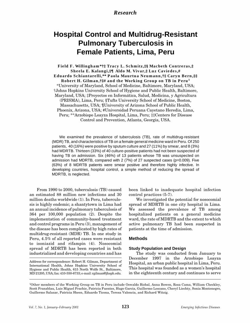
123Vol. 7, No. 1, January–February 2001 Emerging Infectious Diseases
Research
From 1990 to 2000, tuberculosis (TB) causedan estimated 88 million new infections and 30million deaths worldwide (1). In Peru, tuberculo-sis is highly endemic; a shantytown in Lima hadan annual incidence of pulmonary tuberculosis of364 per 100,000 population (2). Despite theimplementation of community-based treatmentand control programs in Peru (3), management ofthe disease has been complicated by high rates ofmultidrug-resistant (MDR) TB. In one study inPeru, 4.5% of all reported cases were resistantto isoniazid and rifampin (4). Nosocomialspread of MDRTB has been reported in bothindustrialized and developing countries and has
been linked to inadequate hospital infectioncontrol practices (5-7).
We investigated the potential for nosocomialspread of MDRTB in one city hospital in Lima.We assessed the prevalence of TB amonghospitalized patients on a general medicineward, the rate of MDRTB and the extent to whichactive pulmonary TB had been suspected inpatients at the time of admission.
Methods
Study Population and DesignThe study was conducted from January to
December 1997 in the Arzobispo LoayzaHospital, an urban public hospital in Lima, Peru.This hospital was founded as a women’s hospitalin the eighteenth century and continues to serve
Hospital Control and Multidrug-ResistantPulmonary Tuberculosis inFemale Patients, Lima, Peru
Field F. Willingham*†‡ Tracy L. Schmitz,‡§ Macbeth Contreras,‡Sheela E. Kalangi,‡¶ Aldo M. Vivar,‡ Luz Caviedes,#
Eduardo Schiantarelli,** Paola Maurtua Neumann,†‡ Caryn Bern,‡‡Robert H. Gilman,†‡# and the Working Group on TB in Peru1
*University of Maryland, School of Medicine, Baltimore, Maryland, USA;†Johns Hopkins University School of Hygiene and Public Health, Baltimore,Maryland, USA; ‡Proyectos en Informática, Salud, Medicina, y Agricultura
(PRISMA), Lima, Peru; §Tufts University School of Medicine, Boston,Massachusetts, USA; ¶University of Arizona School of Public Health,
Phoenix, Arizona, USA; #Universidad Peruana Cayetano Heredia, Lima,Peru; **Arzobispo Loayza Hospital, Lima, Peru; ‡‡Centers for Disease
Control and Prevention, Atlanta, Georgia, USA.
Address for correspondence: Robert H. Gilman, Department ofInternational Health, Johns Hopkins University School ofHygiene and Public Health, 615 North Wolfe St., Baltimore,MD 21205, USA; fax: 410-550-6733; e-mail: [email protected].
We examined the prevalence of tuberculosis (TB), rate of multidrug-resistant(MDR) TB, and characteristics of TB on a female general medicine ward in Peru. Of 250patients, 40 (16%) were positive by sputum culture and 27 (11%) by smear, and 8 (3%)had MDRTB. Thirteen (33%) of 40 culture-positive patients had not been suspected ofhaving TB on admission. Six (46%) of 13 patients whose TB was unsuspected onadmission had MDRTB, compared with 2 (7%) of 27 suspected cases (p=0.009). Five(63%) of 8 MDRTB patients were smear positive and therefore highly infective. Indeveloping countries, hospital control, a simple method of reducing the spread ofMDRTB, is neglected.
1Other members of the Working Group on TB in Peru include Oswaldo Bisbal, Anna Bowen, Rosa Cama, William Checkley,Scott Franzblau, Luis Miguel Frachie, Patricia Fuentes, Hugo Garcia, Guillermo Lescano, Cheryl Liechty, Sonia Montenegro,Guillermo Salazar, Patricia Sheen, Eduardo Ticona, Teresa Valencia, and Richard Witzig.

124Emerging Infectious Diseases Vol. 7, No. 1, January–February 2001
Research
a largely female patient population. We solicitedthe participation of all patients admitted to one ofthe hospital’s eight female internal medicinewards (an open room with 30 beds) during thestudy period. The most common admissiondiagnoses over the year of study werepneumonia, bronchiectasis, cardiac insufficiency,TB, cellulitis, diabetes mellitus and chronic renalfailure. The study protocol was approved by theinstitutional review boards of the Johns HopkinsUniversity and Loayza Hospital. All studyparticipants gave informed consent.
Patients who agreed to participate in thestudy answered a brief questionnaire andunderwent physical examination. The medicalrecords were reviewed. A tuberculin skin test(TST) (5 tuberculin units, Connaught, Swiftwater,PA) was administered and was read after 48 to 72hours. The TST was considered positive if thearea of induration measured >10 mm bothvertically and horizontally. At least one sputumspecimen >1 mL in volume was obtained;whenever possible, additional sputum specimenswere obtained on consecutive days.
Laboratory Testing for TB
Acid-fast Bacilli Smear MicroscopyAll samples were digested and concentrat-
ed by the standard N-acetyl-L-cysteine NaOH-Na citrate method for processing mycobacterialspecimens (8). Ziehl-Neelsen and Auraminestaining were performed by standard tech-niques (8).
CulturesMycobacterial growth indicator tubes (Becton
Dickinson, Sparks, MD) containing both 10%OADC (oleic acid, albumin, dextrose, andcatalase) (Becton Dickinson, Sparks, MD), and100 µL of PANTA Antimicrobic Supplement(Polymyxin B, Amphotericin B, Nalidixic acid,Trimethoprim, and Azlocillin) (Becton Dickinson)were injected with 500 µL of decontaminatedsputum sample according to the manufacturer’sspecifications. Löwenstein-Jensen slants (Difco,Detroit, MI) and Middlebrook 7H11 mediumplates (Difco, Detroit, MI) were injected with250 µL of decontaminated sample. Tubes wereincubated at 37°C and examined for mycobacte-rial growth at least weekly for up to 6 weeks witha 365-nm UV transilluminator. Löwenstein-Jensen slants and micro-agar 7H11 plates were
incubated at 37°C with and without 5% CO2 andexamined by light microscopy for mycobacterialgrowth at least weekly for 2 to 8 weeks afterinjection (8). Criteria for positive mycobacterialgrowth have been previously described by theCenters for Disease Control (9).
Sensitivity TestingThe microplate alamar blue assay was used
to determine mycobacterial drug resistance (10).Bacterial suspensions were prepared fromcolonies grown on Middlebrook 7H11 agar.Samples of the bacterial suspension (20 µL) weregrown in 96-well plates containing serialdilutions of anti-TB drugs (isoniazid, rifampin,ethambutol, streptomycin, capreomycin,ciprofloxacin) until control wells tested positivefor mycobacterial growth, usually in 5 to 6 days.Alamar blue reagent was then added to each well,and mycobacterial growth was identified by achange in media color from blue to pink. MIC wasdefined as the lowest drug concentration at whichno blue-to-pink color change was observed. MICsfor the panel of six anti-TB drugs weredetermined for each isolate.
Data AnalysisPatients were included in the study if they
completed the questionnaire, had a physicalexamination, and provided one adequate sputumspecimen. A patient was considered to haveMDRTB if the sputum exhibited growth in mediacontaining both isoniazid and rifampin. HIV testswere not performed as part of this study, but HIVtest results were available for some patients.
All data were entered twice, and the twodatabases were compared to eliminate data entryerrors. Data were analyzed with SPSS version7.5 (SPSS Inc., Chicago, IL) and Epi Info version6.0 (CDC, Atlanta, GA). The chi-square andFisher’s exact tests were used to measurestrengths of association for categorical variables.The Wilcoxon 2-sample test was used to comparecontinuous variables.
ResultsFrom January to December 1997, 250 (78%)
of 319 patients admitted to the ward had acompleted questionnaire and physical examina-tion and at least one adequate sputum specimen.Forty patients (16%) had sputum culturespositive for Mycobacterium tuberculosis, and 26of these had positive sputum smears. One patient

125Vol. 7, No. 1, January–February 2001 Emerging Infectious Diseases
Research
had a positive smear but a negative culture. Onlythree patients had a diagnosis of HIV infection;none of the three had a positive sputumspecimen. Of the 69 ward patients who declinedto participate or were unable to provide anadequate sputum specimen, 4 (6%) had beenadmitted with a diagnosis of suspected TB. If weassume all excluded patients to be negative forTB, the minimum estimated TB prevalence onthe ward was 13%.
Patients with a cough of any duration, acough that lasted >2 weeks, reported weight loss,hemoptysis, or a family history of TB were morelikely to have sputum cultures positive for TB(Table 1). Anorexia was associated with a lowerlikelihood of TB. Because of logistic constraints,we were able to place and read a TST at 48 to 72hours only on a subset of patients. Of the 67patients with TST results, a positive reading wasobserved in 11 (55%) of 20 culture-positivepatients compared with 10 (21%) of 47 patientswithout TB (p=0.007). Among culture-positivepatients, those with a positive TST response were
younger than those with a negative reading(median 23 years of age[range 19-66] vs. 47 years[range 25-88], p=0.02 by Wilcoxon 2-sample test).The socioeconomic status of patients with andwithout TB was similar.
Of the 181 patients who reported past BCGimmunization, 178 (98%) had a scar. No vaccinescars were observed among the 68 persons whoreported no history of BCG immunization.However, having a BCG scar was not associatedwith any apparent protective effect (Table 1). Thepresence of a BCG scar was not associated with apositive TST, even when TB culture positivepatients were excluded (p=0.7).
Of 40 patients with at least one positivesputum culture, 23 (58%) had strains resistant toat least isoniazid, 8 (20%) to rifampin, 4 (10%) toethambutol, and 1 (3%) to streptomycin. Nonewere resistant to ciprofloxacin or capreomycin.Eight patients (20%) had TB resistant to bothisoniazid and rifampin and were classified ashaving MDRTB. All 8 patients with resistance torifampin also had resistance to isoniazid, and 15patients had strains resistant to isoniazid but notto rifampin. Of the eight strains resistant to bothisoniazid and rifampin, one was also resistant toethambutol, one to streptomycin, and one to bothethambutol and streptomycin. Of 8 patients withMDRTB, 3 had a previous history of TBtreatment.
Culture-positive patients for whom TB wasthe admitting diagnosis differed from those inwhom TB was not suspected at the time ofadmission (Table 2). Patients whose TB had notbeen suspected were older and less likely to havethe classic findings of cough, hemoptysis, weightloss, and prior personal or family history of TB.Patients whose TB had not been suspected at thetime of admission were less likely to have apositive sputum smear, but this difference didnot reach statistical significance (p=0.16 byFisher’s exact test). However, patients whose TBhad not been suspected were significantly morelikely to have MDRTB. Six (75%) of 8 patientswith MDRTB were not suspected to have TB onadmission; 3 (50%) of these six were also smearpositive. Admitting diagnoses among culture-positive patients whose TB had not beensuspected on admission included two patientswith diabetes mellitus, one with systemic lupuserythematosus, and one with a lung lesionthought to be a hydatid cyst.
Table 1. Female patients admitted to a general medicineward of a hospital, Lima, Peru
Mycobacterium tuberculosisculture results
Positive Negativea
Characteristic N=40, n (%) N=209, n (%)Median age (range) 43 (18-96) 46 (14-92)Cough 35 (88)b 125 (60)b
Cough for > 2 weeks 25 (63)b 64 (31)b
Weight loss 33 (83)b 122 (58)b
Hemoptysis 12 (30)c 29 (14)c
Anorexia 22 (55)c 149 (71)c
Fever 24 (60) 108 (51)Dyspnea 22 (55) 107 (51)TST positived 11 (55)c 10 (21)c
BCG scar 28 (70) 150 (71)History of BCG 29 (73) 152 (72) vaccinationFamily history of TB 12 (30)c 32 (15) c
Prior history of TB 9 (23) 34 (16)Socioeconomic indicators Electricity in home 36 (90) 196 (93) Piped water 32 (80) 180 (86) Able to read and write 31 (78) 159 (76)aOne patient who was smear positive but culture negativewas excluded from the analysis.bP value < 0.01 by Mantel-Haenzsel chi-square test.cP value < 0.05 by Mantel-Haenzsel chi-square test.dA total of 67 patients, 20 M. tuberculosis culture-positiveand 47 M. tuberculosis culture-negative, had tuberculin skintests (TST).

126Emerging Infectious Diseases Vol. 7, No. 1, January–February 2001
Research
ConclusionsThe overall prevalence of TB among our
study patients was high: at least 13% of allpatients admitted to this general medicine wardhad active TB. Two-thirds of TB patients weresmear positive and therefore highly infectious,one-fifth had multidrug-resistant strains, and75% of the patients with MDRTB had not beensuspected of having TB when they entered thehospital. As in most Latin American hospitals, nomasks or other respiratory devices were used toprevent spread in this hospital, even when thepatient was known to be smear positive andhighly infectious.
Nosocomial outbreaks of MDRTB in theUnited States in the 1980s and early 1990sheightened enforcement of stringent hospitalcontrol measures (11), leading to measurabledecreases in TST conversion rates amonghospital staff (12). Although the rate of TB inPeru is approximately 20 times higher than thatof New York City (13), no concerted effort hasbeen made to improve TB control measures inPeruvian hospitals.
The spread of MDRTB threatens controlefforts (14). The fact that the majority of ourpatients with MDRTB had no history of pasttreatment of TB implies that person-to-person
transmission of multidrug resistant strainsoccurs in Peru. Our data suggest that hospitalwards may be one of the sites of transmission.
In developing countries where resources arelimited, TB control programs focus on identifica-tion and treatment of infectious cases (15).Although treatment is clearly an importantcomponent of control, person-to-person spread ofresistant strains makes isolation a high priorityfor preventing transmission. TST testing was notuseful in identifying the group in need ofscreening. Anergy, which was common amongculture-positive TB cases, was associated statisti-cally with older median age and was perhapsrelated to concurrent systemic illness and poornutritional status among hospitalized patients.
Although Peru has implemented an effectivecommunity-based TB control program, hospitalcontrol has not been a focus. Control measuressuch as isolation and respiratory precautions,stringently enforced in the past, were relaxedworldwide after the advent of inexpensive,effective anti-TB medications. After 50 years ofselective drug pressure, the outbreak of MDRTBin New York City (5) dramatically highlightedthe consequences of lapses in infection control.
Our data show that in countries or localeswith a known high prevalence of TB, hospitalsshould screen all patients with respiratorysymptoms by sputum smear within 12 hours ofadmission to hospital. Those found to be smear-positive should be placed in respiratory isolation,apart from TB-negative patients, until the smearbecomes negative. Hospital personnel shouldobserve respiratory precautions in caring forthese patients. A system of rapid culturediagnosis and susceptibility testing should beimplemented, allowing the presumptive diagno-sis of MDRTB within 2 weeks (16). Incombination, admission screening for TB, re-implementation of effective hospital respiratorycontrol, and rapid TB diagnosis can substantiallydecrease the transmission of TB, especiallyMDRTB, in countries like Peru.
AcknowledgmentsWe thank R. Black, D. Berg, and K. Laserson for helpful
comments and J.B. Phu and D. Sara for their assistance.
This study was supported in part by NIH grant numberU01-AI35894-03, World AIDS Federation grant number94.093, Fogarty, FIRCA TW00611 and ITREID and theanonymous RG-ER fund.
Table 2. Mycobacterium tuberculosis culture-positivepatients, by admission diagnosis, Lima, Peru
M. tuberculosis culture-positive patients
Suspected TB No suspected TBCharacteristic N=27, n (%) N=13, n (%)Median age (range) 27 (18-87)a 58 (22-96)a
Cough 27 (100)b 8 (62)b
Cough for > 2 weeks 20 (74)c 5 (39)c
Weight loss 25 (93)c 8 (62)c
Hemoptysis 10 (37) 2 (15)Fever 16 (59) 8 (62)Anorexia 13 (48) 9 (69)Dyspnea 17 (63) 5 (39)Prior history of TB 8 (30) 1 (8)Family history of TB 10 (37) 2 (15)Smear positive 20 (74) 6 (46)MDRTB 2 (7)c 6 (46)c
MDRTB and smear 2 (7) 3 (23) positiveap value < 0.05 by Wilcoxon 2-sample test.bp value < 0.01 by Fisher’s exact 2-tailed test.cp value < 0.05 by Fisher’s exact 2-tailed test.MDRTB = Multidrug-resistant tuberculosis.
127Vol. 7, No. 1, January–February 2001 Emerging Infectious Diseases
Research
Mr. Willingham, a fourth-year medical student atthe University of Maryland, performed this study afterhe completed his Masters in Public Health at the JohnsHopkins School of Public Health. His research interestsfocus on tuberculosis, infectious diseases, and publichealth.
References 1. Dolin PJ, Raviglione MC, Kochi A. Global tuberculosis
incidence and mortality during 1990-2000. Bull WorldHealth Organ 1994; 72:213-20.
2. Sanghavi DM, Gilman RH, Lescano-Guevara AG,Checkley W, Cabrera LZ, Cardenas V. Hyperendemicpulmonary tuberculosis in a Peruvian shantytown. AmJ Epidemiol 1998;148:384-9.
3. Raviglione MC, Dye C, Schmidt S, Kochi A. Assessment ofworldwide tuberculosis control. Lancet 1997;350:624-9.
4. Pablos-Mendez A, Raviglione MC, Laszlo A, Binkin N,Rieder HL, Bustreo F, et al. Global surveillance forantituberculosis-drug resistance, 1994-1997. N Engl JMed 1998;338:1641-9.
5. Edlin BR, Tokars JI, Grieco MH, Crawford JT,Williams J, Sordillo EM, et al. An outbreak ofmultidrug resistant tuberculosis among hospitalizedpatients with the acquired immunodeficiency syn-drome. N Engl J Med 1992; 326:1514-21.
6. Beck-Sague C, Dooley SW, Hutton MD, Otten J,Breeden A, Crawford JT, et al. Hospital outbreak ofmultidrug resistant Mycobacterium tuberculosis infec-tions. JAMA 1992;268:1280-6.
7. Kritski AL, Marques MJ, Rabahi MF, Vieira MA,Werneck-Barroso E, Carvalho CE, et al. Transmissionof tuberculosis to close contacts of patients withmultidrug-resistant tuberculosis. Am J Respir CritCare Med 1996;153:331-5.
8. Welch DF, Guruswamy AP, Sides SJ, Shaw CH,Gilchrist MJR. Timely culture for Mycobacteria whichutilizes a microcolony method. J Clin Microbiol1993;31:2178-84.
9. Kent BD, Kubica GP. Public health mycobacteriology:A guide for the level III laboratory. Atlanta:Department of Health and Human Services, Centersfor Disease Control, 1985;36-9, 47-69,185-7.
10. Franzblau SG, Witzig RS, McLaughlin JC, Torres P,Madico G, Hernandez A, et al. Rapid, low-technologyMIC determination with clinical Mycobacteriumtuberculosis isolates by using the microplate AlamarBlue assay. J Clin Microbiol 1998;36:362-6.
11. Stricof RL, DiFerdinando GT, Osten WM, Novick LF.Tuberculosis control in New York City hospitals. Am JInfect Control 1998;26:270-6.
12. Bangsberg DR, Crowley K, Moss A, Dobkin JF,McGregor C, Neu HC. Reduction in tuberculin skin-testconversions among medical house staff associated withimproved tuberculosis infection control practices. InfectControl Hosp Epidemiol 1997;18:566-70.
13. Hoyos C, Izquierdo G, Piscoya G, Romero M, Saldías J.[Incidence of infective diseases at an internal medicineservice]. Rev Gastroenterol Peru 1991;11:171-5.
14. Centers for Disease Control and Prevention. Multi-drug-resistant tuberculosis outbreak on an HIV ward –Madrid, Spain, 1991-1995. MMWR Morb Mortal WklyRep 1996;45:330-3.
15. Enarson DA, Grosset J, Mwinga A, Hershfield ES,O’Brien R, Cole S, et al. The challenge of tuberculosis:statement on global control and prevention. Lancet1995;346:809-19.
16. Caviedes L, Lee TS, Gilman RH, Sheen P, Speelman E,Lee EH, et al. Rapid, efficient detection and drugsusceptibility testing of Mycobacterium tuberculosis insputum by microscopic observation of broth cultures. JClin Microbiol 2000;38:1203-8.

128Emerging Infectious Diseases Vol. 7, No. 1, January–February 2001
Dispatches
West Nile (WN) virus is a member of theJapanese encephalitis (JE) antigenic complex ofthe genus Flavivirus, family Flaviviridae.Mosquito-borne WN virus fever is endemic inAfrica, the Middle East, and Southwest Asia. Theantigenically and genetically related Kunjinvirus is a WN virus counterpart in Australia andSoutheast Asia and has recently been taxonomi-cally classified as a subtype of WN virus. Untilrecently, WN virus infection in humans wasconsidered a relatively mild, influenzalikedisease with full recovery, although occasionally(<15% of cases) acute aseptic meningitis orencephalitis occurred (1). No large outbreak ofWN virus fever was reported in Europe untilAugust and September 1996, when more than500 clinical cases were observed in Romania(Bucharest region), with high rates of neurologicdisorders and death (up to 10%) (2). WN virus hadnever been detected in the Western Hemisphereuntil August 1999, when an outbreak of humanWN encephalitis in New York City (56 confirmedcases, 7 deaths) coincided with unusual deaths incrows and exotic birds (3-5).
The StudyIn August and September 1999, an outbreak
of acute viral infection consistent with arboviralinfection occurred in the Volgograd Region,Russia. Epidemiologic and clinical data werecollected and analyzed in the Center of Sanitaryand Epidemic Control for the Volgograd Regionin collaboration with the Commission of theRussian Ministry of Public Health. From July 25to October 1, 826 patients were admitted to areahospitals with the clinical diagnosis of acuteaseptic meningoencephalitis (code A86, ICD-10;84 patients), acute aseptic meningitis (codeA87.9; 308 patients), or acute viral infection withfever (code B34.9; 347 patients). Serum samplesfrom 318 patients were tested for WN virusantibody by immunoglobulin (Ig)M-captureenzyme-linked immunosorbent assay (ELISA)and indirect IgG ELISA (2,6); 183 (58%) samplesdemonstrated a level of anti-WN virus IgMindicative of acute infection. This proportion wasapproximately the same in patients with asepticmeningoencephalitis, aseptic meningitis, andacute fever. These 183 cases were consideredserologically confirmed WN virus cases; all 826cases were considered clinically compatible WNvirus cases. The total number of suspected overthuman WN virus cases was estimated to be 480.
Outbreak of West Nile Virus Infection,Volgograd Region, Russia, 1999
Alexander E. Platonov,* German A. Shipulin,* Olga Yu. Shipulina,*Elena N. Tyutyunnik,† Tatyana I. Frolochkina,‡
Robert S. Lanciotti,§ Svetlana Yazyshina,¶ Olga V. Platonova,* Igor L. Obukhov,¶ Alexander N. Zhukov,**
Yury Ya. Vengerov,† and Valenin I. Pokrovskii**Central Institute of Epidemiology, Moscow, Russia; †Moscow State
University for Medicine and Dentistry, Russia; ‡Ministry of Public Health,Moscow, Russia; §Centers for Disease Control and Prevention,
Fort Collins, Colorado, USA; ¶Russian State Institute for Control ofVeterinary Products, Moscow, Russia; **Center of Sanitary and
Epidemic Control for Volgograd Region, Volgograd, Russia
Address for correspondence: A.E. Platonov, Central Instituteof Epidemiology, Novogireevskaya Str. 3A, Moscow 111123,Russia; Fax: 7-095-305-5423; e-mail: [email protected].
From July 25 to October 1, 1999, 826 patients were admitted to Volgograd Region,Russia, hospitals with acute aseptic meningoencephalitis, meningitis, or feverconsistent with arboviral infection. Of 84 cases of meningoencephalitis, 40 were fatal.Fourteen brain specimens were positive in reverse transcriptase-polymerase chainreaction assays, confirming the presence of West Nile/Kunjin virus.
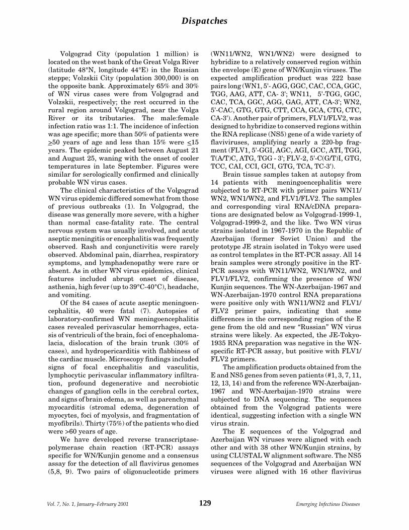
129Vol. 7, No. 1, January–February 2001 Emerging Infectious Diseases
Dispatches
Volgograd City (population 1 million) islocated on the west bank of the Great Volga River(latitude 48°N, longitude 44°E) in the Russiansteppe; Volzskii City (population 300,000) is onthe opposite bank. Approximately 65% and 30%of WN virus cases were from Volgograd andVolzskii, respectively; the rest occurred in therural region around Volgograd, near the VolgaRiver or its tributaries. The male:femaleinfection ratio was 1:1. The incidence of infectionwas age specific; more than 50% of patients were>50 years of age and less than 15% were <15years. The epidemic peaked between August 21and August 25, waning with the onset of coolertemperatures in late September. Figures weresimilar for serologically confirmed and clinicallyprobable WN virus cases.
The clinical characteristics of the VolgogradWN virus epidemic differed somewhat from thoseof previous outbreaks (1). In Volgograd, thedisease was generally more severe, with a higherthan normal case-fatality rate. The centralnervous system was usually involved, and acuteaseptic meningitis or encephalitis was frequentlyobserved. Rash and conjunctivitis were rarelyobserved. Abdominal pain, diarrhea, respiratorysymptoms, and lymphadenopathy were rare orabsent. As in other WN virus epidemics, clinicalfeatures included abrupt onset of disease,asthenia, high fever (up to 39°C-40°C), headache,and vomiting.
Of the 84 cases of acute aseptic meningoen-cephalitis, 40 were fatal (7). Autopsies oflaboratory-confirmed WN meningoencephalitiscases revealed perivascular hemorrhages, ecta-sis of ventriculi of the brain, foci of encephaloma-lacia, dislocation of the brain trunk (30% ofcases), and hydropericarditis with flabbiness ofthe cardiac muscle. Microscopy findings includedsigns of focal encephalitis and vasculitis,lymphocytic perivascular inflammatory infiltra-tion, profound degenerative and necrobioticchanges of ganglion cells in the cerebral cortex,and signs of brain edema, as well as parenchymalmyocarditis (stromal edema, degeneration ofmyocytes, foci of myolysis, and fragmentation ofmyofibrils). Thirty (75%) of the patients who diedwere >60 years of age.
We have developed reverse transcriptase-polymerase chain reaction (RT-PCR) assaysspecific for WN/Kunjin genome and a consensusassay for the detection of all flavivirus genomes(5,8, 9). Two pairs of oligonucleotide primers
(WN11/WN2, WN1/WN2) were designed tohybridize to a relatively conserved region withinthe envelope (E) gene of WN/Kunjin viruses. Theexpected amplification product was 222 basepairs long (WN1, 5'- AGG, GGC, CAC, CCA, GGC,TGG, AAG, ATT, CA- 3'; WN11, 5'-TGG, GGC,CAC, TCA, GGC, AGG, GAG, ATT, CA-3'; WN2,5'-CAC, GTG, GTG, CTT, CCA, GCA, CTG, CTC,CA-3'). Another pair of primers, FLV1/FLV2, wasdesigned to hybridize to conserved regions withinthe RNA replicase (NS5) gene of a wide variety offlaviviruses, amplifying nearly a 220-bp frag-ment (FLV1, 5'-GGI, AGC, AGI, GCC, ATI, TGG,T(A/T)C, ATG, TGG - 3'; FLV-2, 5'-C(G/T)I, GTG,TCC, CAI, CCI, GCI, GTG, TCA, TC-3').
Brain tissue samples taken at autopsy from14 patients with meningoencephalitis weresubjected to RT-PCR with primer pairs WN11/WN2, WN1/WN2, and FLV1/FLV2. The samplesand corresponding viral RNA/cDNA prepara-tions are designated below as Volgograd-1999-1,Volgograd-1999-2, and the like. Two WN virusstrains isolated in 1967-1970 in the Republic ofAzerbaijan (former Soviet Union) and theprototype JE strain isolated in Tokyo were usedas control templates in the RT-PCR assay. All 14brain samples were strongly positive in the RT-PCR assays with WN11/WN2, WN1/WN2, andFLV1/FLV2, confirming the presence of WN/Kunjin sequences. The WN-Azerbaijan-1967 andWN-Azerbaijan-1970 control RNA preparationswere positive only with WN11/WN2 and FLV1/FLV2 primer pairs, indicating that somedifferences in the corresponding region of the Egene from the old and new “Russian” WN virusstrains were likely. As expected, the JE-Tokyo-1935 RNA preparation was negative in the WN-specific RT-PCR assay, but positive with FLV1/FLV2 primers.
The amplification products obtained from theE and NS5 genes from seven patients (#1, 3, 7, 11,12, 13, 14) and from the reference WN-Azerbaijan-1967 and WN-Azerbaijan-1970 strains weresubjected to DNA sequencing. The sequencesobtained from the Volgograd patients wereidentical, suggesting infection with a single WNvirus strain.
The E sequences of the Volgograd andAzerbaijan WN viruses were aligned with eachother and with 38 other WN/Kunjin strains, byusing CLUSTAL W alignment software. The NS5sequences of the Volgograd and Azerbaijan WNviruses were aligned with 16 other flavivirus

130Emerging Infectious Diseases Vol. 7, No. 1, January–February 2001
Dispatches
strains by CLUSTAL W. The high level ofsequence similarity confirmed the WN virussource of the Volgograd cases (Table). Phyloge-netic trees of WN/Kunjin viruses and allflaviviruses derived from E and NS5 genesequences (5,8-10) have been previously de-scribed. We used smaller gene fragments, 165bases of E gene and 147 bases of NS5 gene;however, the deduced phylogenetic trees werepractically the same. Therefore, we limited theanalysis to the most similar strains, including theepidemiologically important WN-New-York-1999and WN-Romania-1996 strains, and some
representatives of other taxonomic subdivisions.For comparison, the designation of strainscoincides with the designation in publicationswhere the additional details of strain history aregiven (5,10).
The Volgograd and old Azerbaijan WN virusstrains clearly belonged to “lineage 1” of WNvirus isolates (5,9-10). Within lineage 1, theVolgograd patient strains were most closelyrelated to the current Kenya and Senegal strainsand the Romanian mosquito isolate (the identicalE gene fragment of 165 nucleotides [nt]). TheAzerbaijan isolates were more closely related to
Table. Percentage identity of nucleotide sequence for E and NS5 gene fragments among West Nile/Kunjin viruses
WN- WN- New York-
Kenya- 1999F1998 and and WN- WN-
WN- WN- WN- Azer- Azer- WN- WN- Kunjin- WN-Romania- Senegal- New York- baijan- baijan- Romania- Egypt- Aust- Nigeria
Virus strain 1996M 1993 1999H 1967 1970 1996H 1951 1960 (Wengler)WN-Volgograd- 100 100 98.2 93.9 93.9 93.9 92.3 86.7 74.5 1999-1 and WN- 96.6 94.6 95.2 92.5 85.0 78.2 Volgograd- 1999-11*
Romania-1996M 100 98.2 93.9 93.9 93.9 92.3 86.7 74.596.6 94.6 95.2 92.5 85.0 78.2
WN-Kenya-1998 98.2 93.9 93.9 93.9 92.3 86.7 74.5 and WN- Senegal-1993
WN-New York- 95.8 95.8 95.8 94.5 87.3 75.8 1999F and WN- 95.2 94.6 93.2 85.7 78.9 New York-1999H
WN-Azer- 100 100 97.6 88.5 75.8 baijan-1967 99.3 98.0 87.8 78.2
WN-Azerbaijan- 100 97.6 88.5 75.8 1970 97.3 88.4 78.2
WN-Romania- 97.6 88.5 75.8 1996H
WN-Egypt-1951 87.9 75.887.1 76.2
Kunjin-Aust-1960 75.172.8
WN-Nigeria (Wengler)*First lines in a row = percentage identity of nucleotide sequence for E gene fragment; second lines in a row = percentageidentity of nucleotide sequence for NS5 gene fragment (if available). The strains with identical sequences of E and NS5fragments are placed in the same row or column.
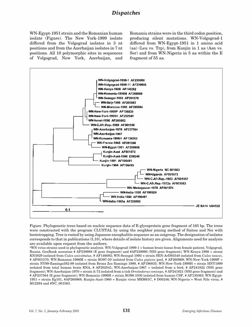
131Vol. 7, No. 1, January–February 2001 Emerging Infectious Diseases
Dispatches
WN-Egypt-1951 strain and the Romanian humanisolate (Figure). The New York-1999 isolatediffered from the Volgograd isolates in 3 ntpositions and from the Azerbaijan isolates in 7 ntpositions. All 10 polymorphic sites in sequencesof Volgograd, New York, Azerbaijan, and
Figure. Phylogenetic trees based on nucleic sequence data of E-glycoprotein gene fragment of 165 bp. The treeswere constructed with the program CLUSTAL by using the neighbor joining method of Saitou and Nei withbootstrapping. Tree is rooted by using Japanese encephalitis sequence as an outgroup. The designation of isolatescorresponds to that in publications (5,10), where details of isolate history are given. Alignments used for analysisare available upon request from the authors.aWN virus strains used in phylogenetic analysis. WN-Volgograd-1999-1 = human brain tissue from female patient, Volgograd,Russia, GenBank accession # AF239988 (E gene fragment) and #AF239990 (NS5 gene fragment); WN-Kenya-1998 = strainKN3829 isolated from Culex univittatus, # AF146082; WN-Senegal-1993 = strain SEN-ArD93548 isolated from Culex neavei,# AF001570; WN-Romania-1996M = strain RO97-50 isolated from Culex pipiens pool, # AF260969; WN-New-York-1999F =strain NY99-flamingo382-99 isolated from Bronx Zoo flamingo 1999, # AF196835; WN-New-York-1999H = strain HNY1999isolated from total human brain RNA, # AF202541; WN-Azerbaijan-1967 = isolated from a bird, # AF241822 (NS5 genefragment); WN-Azerbaijan-1970 = strain A-72 isolated from a tick Ornitodorus coniceps, # AF241821 (NS5 gene fragment) and# AF237564 (E gene fragment); WN-Romania-1996H = strain RO96-1030 isolated from human CSF, # AF130363; WN-Egypt-1951 = strain Eg101, #AF260968; Kunjin-Aust-1960 = Kunjin virus MRM61C, # D00246; WN-Nigeria = West Nile virus, #M12294 and #NC_001563.
Romania strains were in the third codon position,producing silent mutations. WN-Volgograd-1differed from WN-Egypt-1951 in 1 amino acid(aa) (Leu vs. Trp), from Kunjin in 1 aa (Asn vs.Ser) and from WN-Nigeria in 5 aa within the Efragment of 55 aa.

132Emerging Infectious Diseases Vol. 7, No. 1, January–February 2001
Dispatches
Although a few sequences of WN virus NS5gene were available, the data confirmed that theWN virus strains of lineage 1 were more close toeach other (the differences in 0-11 nt of 147) thanto Kunjin, and especially distant from WN-Nigeria (Wengler) strain (Table). Again, all 10polymorphic sites in sequences of lineage 1strains corresponded to the silent mutationsonly. WN-Volgograd-1 differed from Kunjin in 1aa (Lys vs. Arg) and from WN-Nigeria in 3 aa (Argvs. Lys, Arg vs. Lys, and Ile vs. Val) but differed,for example, from Saint Louis (AF013416) in 11aa within the NS5 fragment of 48 aa. Theisolation of WN virus from one of our clinicalbrain samples, Volgograd-1999-4, will makecomplete genome sequencing and further viro-logic investigations possible (11).
ConclusionsOur data, together with those of previous
publications, document several outbreaks ofemerging WN virus infection in regions wherethis disease was not found or was rarely found.(There were a few isolations of WN virus inRomania and the former Soviet Union before1996-99.) Some isolates demonstrate a highdegree of similarity (New York-1999 and Israel-1998; Volgograd-1999, Romania-1996-mosquitoisolate, Kenya-1998, and Senegal-1993;Azerbaijan-1967 and Romania-1996-human iso-late). Moreover, the last three large outbreakswere caused by genetically similar strains (WN-Romania-1996, WN-New York-1999, and WN-Volgograd-1999), indicating the wide circulationand emergence of potentially epidemic strains ofWN virus. All three cities, Bucharest, New York,and Volgograd, are located near large bodies ofwater and on bird migration pathways and allhad unusually dry summers the year of theoutbreak (12). Some clinical characteristics of therecent WN virus epidemics were unexpected,such as the high rate of neurologic disorders anddeath. These unusual characteristics may be dueto the expansion of new pathogenic WN virusstrain(s) or to the peculiarities of the human hostresponse.
AcknowledgmentsWe thank DK Lvov, VL Gromashevskii, AM Butenko, S.
Ya. Gaidamovitch, O Vyshemirskii, and VF Larichev forproviding the reference WN virus strains and helpfuldiscussions; DJ Gubler, J LeDuc, and P Henry for arrangingresearch collaboration between the Central Institute of
Epidemiology, Moscow, and the Centers for Disease Controland Prevention; and NV Rusakova, EM Krasnova, VAPetrov, and AM Alyushin for providing human specimens fortesting, and epidemiologic and clinical information foranalysis.
Dr. Platonov is head of a laboratory at the CentralInstitute of Epidemiology, Russia. His scientific interestsinclude the epidemiology, diagnosis, and pathogenesis ofbacterial and viral meningitis.
References 1. Hubálek Z, Halouzka J. West Nile fever—a reemerging
mosquito-borne viral disease in Europe. Emerg InfectDis 1999;5:643-50.
2. Tsai TF, Popovici F, Cernescu C, Campbell GL, NedelcuNI. West Nile encephalitis epidemic in southeasternRomania. Lancet 1998;352:767-71.
3. CDC. Update: West Nile virus encephalitis—NewYork, 1999. Morb Mortal Wkly Rep 1999;48:944-6.
4. Anderson JF, Andreadis TG, Vossbrinck CR, Tirrell S,Wakem EM, French RA, et al. Isolation of West Nilevirus from mosquitoes, crows, and a Cooper’s hawk inConnecticut. Science 1999;286:2331-3.
5. Lanciotti RS, Roehrig JT, Deubel V, Smith J, Parker M,Steele K, et al. Origin of the West Nile virus responsiblefor an outbreak of encephalitis in the northeastern U.S.Science 1999;286:2333-7.
6. Lvov DR, Butenko AM, Gaidamovitch SY, LarichevVPh, Leschinskaya EV, Zhukov AN, et al. Epidemicoutbreak of meningitis and meningoencephalitis inKrasnodar territory and Volgograd region provoked byof West Nile fever virus. Vopr Virusol 2000;45:37-8.(InRussian).
7. Vengerov YuYa, Frolochkina TI, Zhukov AN, ShipulinGA, Shipulina OYu, et al. West Nile virus infection asclinical and epidemiological problem. Epidemiologyand Infectious Diseases 2000;4:27-31.(In Russian).
8. Kuno G, Chang GJ, Tsuchiya KR, Karabatsos N, CroppCB. Phylogeny of the genus Flavivirus. J Virol1998;72:73-83.
9. Berthet FX, Zeller HG, Drouet MT, Rauzier J, DigoutteJP, Deubel V. Extensive nucleotide changes anddeletions within the envelope glycoprotein gene ofEuro-African West Nile viruses. J Gen Virol 1997;8(Pt9):2293-7.
10. Savage HM, Ceianu C, Nicolescu G, Karabatsos N,Lanciotti R, Vladimirescu A, et al. Entomologic andavian investigations of an epidemic of West Nile fever inRomania in 1996, with serologic and molecularcharacterization of a virus isolate from mosquitoes. AmJ Trop Med Hyg 1999;61:600-11.
11. Lvov DR, Butenko AM, Gromashevsky VI, LarichevVPh, Gaidamovitch SYa, Vyshemirsky OI, et al.Isolation of two strains of West Nile virus during anoutbreak in Southern Russia, 1999. Emerg Infect Dis2000;6:373-6.
12. Han LL, Popovici F, Alexander Jr JP, Laurentia V,Tengelsen LA, Cernescu C, et al. Risk factors for WestNile virus infection and meningoencephalitis, Romania,1996. J Infect Dis 1999;179:230-3.

133Vol. 7, No. 1, January–February 2001 Emerging Infectious Diseases
Dispatches
Molecular subtyping, primarily by multilocusenzyme electrophoresis (MEE) and ribotyping,has identified substantial genetic diversitywithin the Corynebacterium diphtheriae species,leading to the identification of a unique clonalgroup that emerged in Russia in 1990 at thebeginning of the current diphtheria epidemic (1).Strains of this clonal group belong to a distinctelectrophoretic type complex (ET8 complex) andare of ribotypes G1 and G4. Identification of thisclonal group has permitted precise monitoring ofthe epidemic’s growth and rapid detection ofimported cases in neighboring and otherEuropean countries.
Use of traditional subtyping methods inmonitoring the expansion of the epidemic clonehas helped differentiate epidemic, endemic, andimported cases and has allowed timely preven-tive measures. Even as the epidemic declines(from more than 50,000 cases in 1995 to 1,436cases in 1998), identifying organisms belongingto this epidemic clone in cases of suspectedimportation into locations where diphtheria israrely encountered continues to provide valuableinformation. Since both ribotyping and MEE are
time-consuming, taking 3-4 working days toproduce results, rapid methods that coulddistinguish the predominant clone would im-prove epidemic surveillance and preventionmeasures.
Random amplified polymorphic DNA (RAPD)is a simple and rapid molecular subtypingmethod. Recently, Nakao et al. (2) optimized andstandardized this assay for C. diphtheriae andshowed that the discrimination level obtained byRAPD correlated well with that of ribotyping;each of 20-plus ribotyping patterns wasassociated with one or more distinct RAPDpatterns. We compared these two techniques on alarge number of C. diphtheriae Russian isolatesfrom 1995 to 1997, focusing on the ability ofRAPD to identify the isolates of the epidemicG1/4 clonal group.
The StudyAll C. diphtheriae strains were collected by
the Russian Federal Diphtheria DiagnosisReference Laboratory. Of 199 isolates fromdifferent regions of Russia, 187 were isolatedfrom 1995 to 1997; 12 were isolated during 1993to 1994; 68 were recovered from clinicallydiagnosed diphtheria patients; and the remain-ing 131 isolates were obtained from carriers.
Identification, biotype, and toxigenicitydetermination were performed by standard
Rapid Identification ofCorynebacterium diphtheriaeClonal Group Associated with
Diphtheria Epidemic, Russian Federation
Svetlana Kombarova,* Chung Kim,† Viatcheslav Melnikov,*Michael Reeves,† Olja Borisova,*
Izabella Mazurova,* and Tanja Popovic*†*Gabrichevsky Institute of Epidemiology and Microbiology, Moscow, Russia;
†Centers for Disease Control and Prevention, Atlanta, Georgia, USA
Address for correspondence: Dr. Tanja Popovic, Centers forDisease Control and Prevention, Mail Stop G34, 1600 CliftonRd., Atlanta, GA 30333, USA; fax: 404-639-3172; e-mail:[email protected].
We used 199 Corynebacterium diphtheriae isolated from 1995 to 1997 inRussia to evaluate the ability of random amplified polymorphic DNA (RAPD) toidentify the unique clonal group that emerged there in 1990. Our data show thatRAPD can reliably, reproducibly, and rapidly screen a large number of strains toidentify the epidemic clonal group.

134Emerging Infectious Diseases Vol. 7, No. 1, January–February 2001
Dispatches
microbiologic methods (3,4). RAPD was per-formed by the Ready-To-Go RAPD Kit (PharmaciaBiotech, Piscataway, NJ)(2). RAPD type designa-tions were adopted from those previouslydocumented by Nakao et al. (2). Ribotyping wascarried out as previously described (1) with somemodifications (Table). MEE was performed aspreviously described (1). The genetic relatednessof the electrophoretic types (ETs) was illustratedas a dendrogram, generated by the average-linkage method of clustering the ETs (7) and byusing an SAS macro program described by Jacobs(8).
Of the 199 C. diphtheriae isolates, 185 werebiotype gravis, and 14 were biotype mitis. Allisolates were toxigenic by the Elek assay. Whenassayed by RAPD using primers 3 and 4, the 199isolates that were identical by primer 3 were alsoidentical by primer 4, with the single exception ofisolate B506. Of 185 isolates of the gravis biotype,183 were the G1/4 RAPD type. Isolate B325 had a
Table. Random amplified polymorphic DNA (RAPD)assay and ribotyping for 79 Russian Corynebacteriumdiphtheriae isolatesa,b
Number RAPDRibotype Biotype of strains Primer 3 Primer 4G1 G 38 G1/4 G1/4
M 2 G1/4 G1/4G4 G 25 G1/4 G1/4
M 1 G1/4 G1/4G 1 Newc G1/4
G4v G 1 G4v G4vM1 M 5 M1/1v M1/1vM1v M 5 M1/1v M1/1vNew M 1 New NewaG, biotype gravis; M, biotype mitis. Cultures were keptlyophilized at room temperature or were stored indefibrinated sheep blood and held at -70°C until needed.Before use, the strains were inoculated onto blood agar plates(trypticase soy agar with 5% sheep blood; Becton Dickinson,Cockeysville, MD) and were incubated at 37°C overnight.bDNA for ribotyping was isolated by the universal isolationprocedure (5). Hybridization of restricted DNA fragmentswas performed using a mixture of five digoxigenin-labeledoligonucleotide probes at 37°C for 4 hours as recentlydescribed by Regnault et al. (6). Posthybridization washeswere also performed at 37°C in 2X SSC (1 X SSC is 0.15 MNaCl plus 0.015 M sodium citrate), 0.1% sodium dodecylsulfate (SDS) for 2x5 minutes and in 0.1X SSC, 0.1% SDS for2x10 minutes. Detection was performed by using the DIGWash and Block Buffer Set (Boehringer MannheimBiochemicals, Indianapolis, IN), sheep anti-digoxigeninantibody conjugated with alkaline phosphatase, nitrobluetetrazolium chloride (NBT), and 5-bromo-4-chloro-3-indolylphosphate (BCIP).cNew = pattern had not been previously observed.
G4v pattern, and isolate B506 had the G1/4pattern only by primer 4. When primer 3 wasused on this isolate, a different pattern wasobserved.
The RAPD patterns of the 14 isolates of themitis biotype were distributed into three groups.The first group included three isolates of the G1/4RAPD type (B294, B399, B400). The second groupincluded 10 isolates that had RAPD patternstypes M1 and M1v. The third group included onlyone isolate (B306), which had a RAPD patterncompletely different from any previously assayedstrain.
Seventy-nine isolates were ribotyped. Of the186 isolates with RAPD G1/4 patterns (183 gravisand 3 mitis), 66 were selected to be ribotyped fortheir geographic and temporal diversity. Inaddition, all non-G1/4 isolates were ribotyped.The ribotyping results correlated extremely wellwith the RAPD data (Table, Figure). With oneexception, all isolates of the G1/4 RAPD type alsohad a G1 or G4 ribotype; 40 had the G1 ribotype,and 26 isolates possessed the G4 ribotype. Inaddition, the G4v ribotype was observed in theisolate with the G4v RAPD pattern. Isolate B306,which had an RAPD type not previouslyobserved, also had a ribotype that did notresemble any previously established ribotypes.Five M1 and five M1v ribotypes were identifiedamong the 10 M1/1v RAPD type isolates.
Twenty-nine isolates of RAPD types G1/4 andribotypes G1 or G4 were analyzed by MEE.Among all isolates of this group, seven individualETs, which clustered at a genetic distance of<0.12, were identified; all ETs were members ofthe previously defined ET8 complex. Only one tothree enzyme differences from the ET8 complexwere observed among the individual enzymetypes (data not shown). The ET8 complexcontains 27 ETs, which are related to each otherat a genetic distance of 0.20 and have a maximumof four enzyme differences within the complex.
ConclusionsThe C. diphtheriae epidemic clonal group
associated with the recent diphtheria epidemic inthe Russian Federation is characterized as beingof ribotypes G1 and G4 and belonging to the ET8complex. Detection of a unique epidemic clonalgroup has allowed continuous monitoring of thecirculation of existing clones and rapid detectionof new or unusual clones. The epidemicemphasized the need for continuous study of the
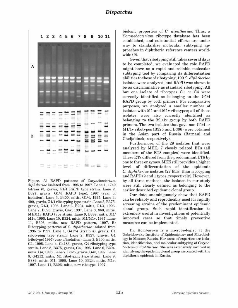
135Vol. 7, No. 1, January–February 2001 Emerging Infectious Diseases
Dispatches
biologic properties of C. diphtheriae. Thus, aCorynebacterium ribotype database has beenestablished, and substantial efforts are underway to standardize molecular subtyping ap-proaches in diphtheria reference centers world-wide (9).
Given that ribotyping still takes several daysto be completed, we evaluated the role RAPDmight have as a rapid and reliable molecularsubtyping tool by comparing its differentiationabilities to those of ribotyping; 199 C. diphtheriaeisolates were analyzed, and RAPD was shown tobe as discriminative as standard ribotyping. Allbut one isolate of ribotypes G1 or G4 werecorrectly identified as belonging to the G1/4RAPD group by both primers. For comparativepurposes, we analyzed a smaller number ofisolates with M1 and M1v ribotypes; all of theseisolates were also correctly identified asbelonging to the M1/1v group by both RAPDprimers. The two isolates that gave non-G1/4 orM1/1v ribotypes (B325 and B306) were obtainedin the Asian part of Russia (Barnaul andCheljabinsk, respectively).
Furthermore, of the 29 isolates that wereanalyzed by MEE, 7 closely related ETs (allmembers of the ET8 complex) were identified.These ETs differed from the predominant ET8 byone to three enzymes. MEE still provides a higherlevel of differentiation of the epidemicC. diphtheriae isolates (27 ETs) than ribotypingand RAPD (2 and 1 types, respectively). However,by all three methods, the isolates in our studywere still clearly defined as belonging to theearlier described epidemic clonal group.
Our data unambiguously show that RAPDcan be reliably and reproducibly used for rapidlyscreening strains of the predominant epidemicclonal group. Such rapid identification isextremely useful in investigations of potentiallyimported cases so that timely preventivemeasures can be implemented.
Dr. Kombarova is a microbiologist at theGabrichevsky Institute of Epidemiology and Microbiol-ogy in Moscow, Russia. Her areas of expertise are isola-tion, identification, and molecular subtyping of Coryne-bacterium diphtheriae. She was extensively involved inidentifying the epidemic clonal group associated with thediphtheria epidemic in Russia.
Figure. A) RAPD patterns of Corynebacteriumdiphtheriae isolated from 1995 to 1997. Lane 1, 1740(strain #), gravis, G1/4 RAPD type strain. Lane 2,B327, gravis, G1/4 (RAPD type), 1997 (year ofisolation). Lane 3, B400, mitis, G1/4, 1995. Lane 4,490, gravis, G1/4 ribotyping type strain. Lane 5, B375,gravis, G1/4, 1995. Lane 6, B294, mitis, G1/4, 1996.Lane 7, B325, gravis, G4v, 1997. Lane 8, 860, mitis,M1/M1v RAPD type strain. Lane 9, B389, mitis, M1/M1v, 1995. Lane 10, B324, mitis, M1/M1v, 1997. Lane11, B306, mitis, new RAPD pattern, 1997. B)Ribotyping patterns of C. diphtheriae isolated from1995 to 1997. Lane 1, G4174 (strain #), gravis, G1ribotyping type strain. Lane 2, B327, gravis, G1(ribotype) 1997 (year of isolation). Lane 3, B400, mitis,G1, 1995. Lane 4, G4183, gravis, G4 ribotyping typestrain. Lane 5, B375, gravis, G4, 1995. Lane 6, B294,mitis, G4, 1996. Lane 7, B325, gravis, G4v, 1997. Lane8, G4212, mitis, M1 ribotyping type strain. Lane 9,B389, mitis, M1, 1995. Lane 10, B324, mitis, M1v,1997. Lane 11, B306, mitis, new ribotype, 1997.
A
B
1 2 3 4 5 6 7 8 9 10 11

136Emerging Infectious Diseases Vol. 7, No. 1, January–February 2001
Dispatches
References 1. Popovic T, Kombarova S, Reeves M, Nakao H,
Mazurova IK, Wharton M, et al. Molecular epidemiolo-gy of diphtheria in Russia, 1985-1994. J Infect Dis1996;174:1064-72.
2. Nakao H, Popovic T. Use of random amplifiedpolymorphic DNA for rapid molecular subtyping ofCorynebacterium diphtheriae. Diagn Microbiol InfectDis 1998;30:167-72.
3. Efstratiou A, Maple PA. WHO manual for thelaboratory diagnosis of diphtheria. Geneva: WorldHealth Organization; 1994. Reference no. ICP-EPI038(C).
4. Mazurova IK, Melnikov V, Kombarova S. Manual forlaboratory diagnostis of diphtheria infection. Moscow:Russian Federation State Committee on SanitaryEpidemiologic Surveillance;1995.
5. Graves LM, Swaminathan B. Universal bacterial DNAisolation procedure. In: Persing DH, Smith TR, TenoverFC, White TJ, editors. Diagnostic molecular microbiol-ogy. Washington: American Society for Microbiology;1993. p. 617-21.
6. Regnault B, Grimont R, Grimont PAD. Universalribotyping method using a chemically labeledoligonucleotide probe mixture. Res Microbiol1997;146:649-59.
7. Selander RK, Daugant DA, Ochman H, Musser JM,Gilmore MN, Whittam TS. Methods of multilocus enzymeelectrophoresis for bacterial population genetic andsystematics. Appl Environ Microbiol 1986;51:873-4.
8. Jacobs D. SAS/GRAPH software and numericaltaxonomy. In: Proceedings of the 15th Annual UsersGroup International Conference. Cary (NC): SASInstitute; 1990. p. 1413-18.
9. Grimont PAD, Grimont F, Ruckly C. The Corynebacte-rium diphtheriae ribotype database. In: Program andabstracts of the 5th International Meeting of theEuropean Laboratory Working Group on Diphtheria.London: Public Health Laboratory Service; 1998. p. 31.

137Vol. 7, No. 1, January–February 2001 Emerging Infectious Diseases
Dispatches
Shigella spp. cause acute, debilitatingdiarrheal disease in humans (particularly youngchildren) worldwide (1). In developing countries,where affected populations are immunologicallycompromised by poor nutrition and backgroundinfections, deaths attributed to shigellosis arecommon. Four Shigella species are recognized aspathogenic to humans: S. sonnei, S. boydii,S. flexneri, and S. dysenteriae. Both S. sonnei andS. boydii are usually associated with mild illnessof short duration in which the stool may bewatery or bloody (2). S. flexneri is generally moresevere, lasts longer, and causes blood in stools.S. dysenteriae, particularly type 1, causes themost severe diarrheal illness, reflected in highdeath rates (3). S. flexneri is a principal cause ofendemic shigellosis in many developing coun-tries, while shigellosis in both endemic andepidemic form has been attributed toS. dysenteriae type 1 (2). Changes in theworldwide epidemiology of Shigella spp. have
been documented in the last decades of the 20thcentury. In industrialized regions, S. dysenteriaewas first replaced by S. flexneri, and then byS. sonnei (4,5); S. flexneri remains the leadingcause of shigellosis in most of the developingworld (2,6-8).
In Indonesia, the last cases of S. dysenteriaediarrhea were reported in 1985 from Jakarta(8,9). This report, based on a study using asystematic surveillance approach that included astandardized detailed bacteriologic examination,provides an Indonesia-wide geographic profile ofShigella spp.
The StudyFrom June 1998 through November 1999, a
total of 3,848 children and adults seekingtreatment for debilitating diarrheal disease wereidentified from eight hospital sites in Medan,North Sumatra; Padang, West Sumatra; Batam,Riau Island; Jakarta, Java Island; Denpasar,Bali (two hospitals); Pontianak, West Kalimantan;and Makassar, South Sulawesi. Rectal swabswere obtained from patients in the study beforeantibiotic therapy was administered. Specimenswere placed in Cary-Blair transport medium,
Shigella spp. Surveillance in Indonesia:the Emergence or Reemergence
of S. dysenteriae
Decy Subekti,* Buhari A. Oyofo,* Periska Tjaniadi,*Andrew L. Corwin,* Wita Larasati,* Maidy Putri,*
Cyrus H. Simanjuntak,† Narain H. Punjabi,* Jufri Taslim,‡Budhi Setiawan,§ Anak Agung G.S. Djelantik,¶ Luh Sriwati,**
Atiek Sumardiati,†† Eka Putra,‡‡James R. Campbell,* and Murad Lesmana*
*U.S. Naval Medical Research Unit No. 2, Jakarta, Indonesia; †NationalInstitute of Health Research and Development, Jakarta, Indonesia;
‡Municipal Health Service, North Jakarta, Indonesia; §Friendship Hospital,Jakarta, Indonesia; ¶Sanglah Hospital, Denpasar, Bali, Indonesia;
**Wangaya Hospital, Denpasar, Bali, Indonesia; ††Regional Office of HealthMinistry, Pontianak, Indonesia; and ‡‡Otorita Hospital, Batam, Indonesia
Address for correspondence: LCDR Buhari A. Oyofo,American Embassy Jakarta, Unit 8132 NAMRU TWO, FPO,AP 965208132, Indonesia; telephone: 62-21-421-4460; fax: 62-21-424-4507; e-mail: [email protected].
From June 1998 through November 1999, Shigella spp. were isolated in 5% ofsamples from 3,848 children and adults with severe diarrheal illness in hospitalsthroughout Indonesia. S. dysenteriae has reemerged in Bali, Kalimantan, and Batamand was detected in Jakarta after a hiatus of 15 years.

138Emerging Infectious Diseases Vol. 7, No. 1, January–February 2001
Dispatches
held at 4°C, and sent on wet ice within 2 to 4 daysafter collection to the U.S. Naval MedicalResearch Unit No. 2, Jakarta. Bacteriologicevaluation was performed by standard culturemethod (10). Species was confirmed by using API20E (Biomerieux, Marcy l’Etoile, France) andslide agglutination with specific Shigella antis-era (Difco Laboratories, Detroit, MI). Antibioticsusceptibility testing was accomplished by thedisk-diffusion method (11).
Overall, bacterial isolates of Shigella spp.were identified in 180 (5%) of 3,848 rectal swabs.The proportional contribution of S. flexneri,S. sonnei, and S. dysenteriae among shigellosiscases was 80%, 12%, and 8%, respectively. NoS. boydii was detected. The percentage ofrepresentation among the three species did notvary substantially by geographic location.Notable was the reemergence of S. dysenteriae inBali, West Kalimantan, and Batam, as well as inJakarta after a hiatus of >15 years (Table). Theproportional distribution of S. flexneri, S. sonnei,and S. dysenteriae for the 5- to 12-year-old and>12-year-old groups was similar. There appearedto be no consistency in the seasonal distributionpatterns of Shigella spp. (Figure). S. flexneri wasthe most frequently isolated organism, followedby S. sonnei and S. dysenteriae.
Other enteric pathogens isolated wereSalmonella spp. (95, 2.5%), Vibrio cholerae (80,2.1%), V. parahaemolyticus (38, 1%), V. choleraenon-O1 (9, 0.2%), and Campylobacter spp. (27,0.7%). Enterotoxigenic Escherichia coli weredetected in 225 (18.1%) of 1,240 specimens testedby the GM1 enzyme-linked immunoassay (12). Ofthe 541 specimens examined for rotavirus in theage groups < 5 years, 191 (35.3%) were positive.Tests to detect parasites showed Ascarislumbricoides in 8 (2%), Blastocystis hominis in 23(5.6%), Giardia lamblia in 3 (0.7%), and Endolimaxnana in 2 (0.5%) of 407 stool specimens examined.
Clinical presentations associated with non-S. dysenteriae included abdominal cramping(79%), vomiting (56%), and fever (48%); forS. dysenteriae, percentages of the same symp-toms were 100%, 64%, and 27%, respectively.Stool samples from patients with S. flexneri,S. sonnei, and S. dysenteriae were principallycharacterized by mucus in the absence of blood(45%) or mucus and visible blood (27%).
Overall, antibiotic susceptibility patternsshowed greater resistance to ampicillin,trimethoprim-sulfamethoxazole, chlorampheni-col, and tetracycline for S. flexneri (85%, 59%,82%, and 98%, respectively, from examination of144 isolates) and S. sonnei (32%, 79%, 37%, and100%, respectively, from examination of 22isolates), than for S. dysenteriae (36%, 43%, 7%,and 29%, respectively, from examination of 14isolates). There was no evidence of resistance toceftriaxone, norfloxacin, ciprofloxacin, or nalid-ixic acid, regardless of Shigella species.Antimicrobial resistance was only apparentamong isolates obtained from Jakarta, Bali, andPontianak.
ConclusionsOur study showed that a substantial
proportion (5%) of acute, debilitating diarrhealillness throughout Indonesia can be ascribed toshigellosis; moreover, S. dysenteriae was docu-mented from various geographic locations. Bothfindings suggest that greater attention should bepaid to highlighting the endemic and epidemiccommunity impact of this pathogen and thatlaboratory detection capabilities need to beenhanced. Recognition of emerging and orreemerging disease pathogens requires reliablebaseline and ongoing surveillance data.
Shifting patterns of antimicrobial-drug resis-tance, particularly in much of the developingworld, are generally a function of overuse and
Table. Distribution of Shigella spp. from patients with diarrhea in Indonesia, June 1998 through November 1999
No. specimens No. pos. for Proportional distribution of Shigella spp. (%)Sites tested Shigella (%) S. flexneri S. sonnei S. dysenteriaeJakarta 2495 122 (5) 97 (80) 17 (14) 8 (6)Makassar 146 4 (3) 3 (75) 1 (25) 0 (0)Denpasar (2) 607 35 (6) 29 (83) 2 (6) 4 (11)Pontianak 330 16 (5) 13 (81) 2 (13) 1 (6)Batam 99 1 (1) 0 (0) 0 (0) 1 (100)Padang 51 2 (4) 2 (100) 0 (0) 0 (0)Medan 120 0 (0) 0 (0) 0 (0) 0 (0)Total 3848 180 (5) 144 (80) 22 (12) 14 (8)

139Vol. 7, No. 1, January–February 2001 Emerging Infectious Diseases
Dispatches
misuse of antibiotic drug therapies. The spread ofdrug resistance is the result of poorly regulatedand enforced policies. Resistance to nalidixic acid(100%) among isolates of S. dysenteriae has beenreported in Bangladesh (13); however, we foundno such resistance. Nevertheless, other data fromIndonesia indicate increasing resistance. In aprevious report (8), 72% of Shigella spp. wereresistant to tetracycline, but less than 30% wereresistant to chloramphenicol, trimethroprim-sulfamethoxazole, or ampicillin. Previous studiesfrom Bangladesh and Tanzania (13,6) showedthat almost all tested isolates were resistant tothe antibiotics used for treatment. Similarantimicrobial resistance profiles for Shigella spp.were reported from Thailand (14), where highresistance to ampicillin, trimethroprim-sulfamethoxazole, chloramphenicol, and tetracy-cline was documented.
The reemergence of S. dysenteriae fromseveral locations in Indonesia should prove causefor concern to health officials, particularly inmonitoring acute, debilitating diarrheal out-breaks. The epidemic potential attributed toS. dysenteriae, as documented in Central America,Asia, and Africa, in conjunction with notably highdeath rates, warrants close attention to thisreemerging pathogen in Indonesia (13,15,16).
This research was supported by the U.S. Naval MedicalResearch and Center Command (work unit numbers 61101A-00101.EAX 2401 and 623002A810.0101.HIX.3411).
Dr. Subekti is a microbiologist and medical researchscientist who heads the Molecular Microbiology Sectionof the Enteric Diseases Program at U.S. Naval MedicalResearch Unit No. 2, Jakarta, Indonesia.
References 1. Dupont HL. Shigella. Infect Dis Clin North Am 1998;
2:599-605. 2. Keusch GT, Bennish ML. Shigellosis: recent progress,
persisting problems and research issues. Pediatr InfectDis J 1989;8:713-9.
3. World Health Organization. The management ofbloody diarrhea in young children 1994. Geneva: WorldHealth Organization; 1994.
4. Ashkenazi S, May-Zahav M, Dinari G, Gabbay U,Zilberberg R, Samra Z. Recent trends in theepidemiology of Shigella species in Israel. Clin InfectDis 1993;17:897-9.
5. Lee LA, Shapiro CN, Hargrett-Bean N, Tauxe RV. Hyper-endemic shigellosis in the United States: a review of sur-veillance data for 1967-1988. J Infect Dis 1991;64:894-900.
6. Navia MM, Capitano L, Ruiz J, Vargas M, Urassa H,Schellemberg D, et al. Typing and characterization ofmechanisms of resistance of Shigella spp.isolated fromfeces of children under 5 years of age from Ifakara,Tanzania. J Clin Microbiol 1999;37:3113-7.
7. Simanjuntak CH, Hasibuan MA, Siregar LO, KoimanI. Microbial etiology of acute diarrhea (Indonesian)Health Study. Indonesia 1983;10(2):1-9.
Figure. Seasonal isolates of Shigella spp. from patients with diarrhea in Indonesia (June 1998 - November 1999).
140Emerging Infectious Diseases Vol. 7, No. 1, January–February 2001
Dispatches
8. Subekti D, Lesmana M, Komalarini S, Tjaniadi P, BurrD, Pazzaglia G. Enterotoxigenic Escherichia coli andother causes of infectious pediatric diarrheas inJakarta, Indonesia. Southeast Asia J Trop Med PublicHealth 1993;24:420-4.
9. Simanjuntak CH, Hardjining S, Hasibuan MA,Pujarwoto Koiman I. Laboratory aspects of gastrointes-tinal infection in Indonesia, 1980-1985. Gastrointesti-nal Infection in Southeast SEAMIC Workshop, Japan;1998. p. 23-31.
10. Farmer III JJ, Kelly MT. Enterobacteriaceae. In:Balows A, Hausler WJ Jr, Herrmann KL, Isenberg HD,Shadomy HJ, editors. Manual of clinical microbiology.5th ed. Washington DC: American Society forMicrobiology; 1991. p. 360-83.
11. Barry AL, Thornsberry C. Susceptibility tests: Diffusiontest procedures. In: Balows A, Hausler WJ Jr, HerrmannKL, Isenberg HD, Shadomy HJ, editors. Manual of clinicalmicrobiology. 5th ed. Washington DC: American Societyfor Microbiology; 1991. p. 1117-25.
12. Svennerholm AM, Wikstrom M, Lindbland M,Holmgren J. Monoclonal antibodies against E. coli heatstable toxin (Sta) and their use in a diagnostic STganglioside GM1-enzyme-linked immunosorbent as-say. J Clin Microbiol 1986;24:585-90.
13. Sack RB, Rahman M, Yunus M, Khan EH.Antimicrobial resistance in organisms causing diarrhe-al disease. Clin Infect Dis 1997;24 (Suppl 1):S102-S105.
14. Moolasart P. Shigellosis in Thai people. JAMASoutheast Asia 1994;5:9-10.
15. Malakooti MA, Alaii J, Shanks GD, Phillips-HowardPA. Epidemic dysentery in western Kenya. Trans R SocTrop Med Hyg 1997;91:541-3.
16. Mendizabal-Morris CA, Malta L, Gangarosa EJ,Guzman G. Epidemic Shiga-bacillus dysentery inCentral America. Derivation of the epidemic and itsprogression in Guatemala, 1968-69. Am J Trop MedHyg 1971;20:927-33.

141Vol. 7, No. 1, January–February 2001 Emerging Infectious Diseases
Dispatches
Cryptosporidium parasites cause infection inhumans and other vertebrates. Two genotypes ofCryptosporidium parvum are responsible formost cases of human infection; the humangenotype (genotype 1 or anthroponotic genotype)is found almost exclusively in humans, whereasthe bovine genotype (genotype 2 or zoonoticgenotype) is found in both ruminants andhumans (1-4). In addition to zoonotic and person-to-person transmission, both genotypes ofC. parvum have caused waterborne andfoodborne outbreaks. Current genotyping toolspermit only differentiation of Cryptosporidiumparasites at the genotype level, which limitsability to track infection and contaminationsources in outbreaks.
Two double-stranded (ds) extrachromosomalviruslike RNAs have recently been identified inC. parvum (5). Both ds-RNAs have been found inall C. parvum oocysts examined. Sequenceanalysis of both the small and large ds-RNAsfrom seven C. parvum human genotype isolatesand five bovine genotype isolates showed distinctds-RNA sequences in isolates from the samegenotype (6), indicating that ds-RNA haspotential as a subgenotyping tool for Cryptospo-ridium. We report sequence diversity in the smallds-RNA of C. parvum human and bovinegenotype isolates and discuss the usefulness ofthis technique for laboratory investigations andfor tracking the source of cryptosporidiosisoutbreaks.
Tracking Cryptosporidium parvumby Sequence Analysis of
Small Double-Stranded RNALihua Xiao, Josef Limor, Caryn Bern, and Altaf A. Lal
Centers for Disease Control and Prevention, Atlanta, Georgia, USA
Address for correspondence: Lihua Xiao, Division of ParasiticDiseases, Centers for Disease Control and Prevention, MailStop F12, Atlanta, GA 30333, USA; fax: 770-488-4454; e-mail:[email protected].
We sequenced a 173-nucleotide fragment of the small double-stranded viruslikeRNA of Cryptosporidium parvum isolates from 23 calves and 38 humans. Sequencediversity was detected at 17 sites. Isolates from the same outbreak had identical double-stranded RNA sequences, suggesting that this technique may be useful for trackingCryptosporidium infection sources.
The StudyWe sequenced the small ds-RNA of 61
C. parvum isolates (23 isolates from cattle and 38from humans) (Table). Eighteen of the 38 humanisolates were from two foodborne outbreaks(Spokane, Washington, 1997; and Washington,D.C., 1998) and one waterborne outbreak(Minnesota, 1997) with well-defined infectionsources (7-9). These isolates had been genotypedby polymerase chain reaction (PCR)-restrictionfragment length polymorphism analysis of theSSU rRNA and TRAP-C2 genes (10,11). Allbovine isolates and the human isolates from theMinnesota outbreak were of the C. parvumbovine genotype, and the other human isolateswere of the C. parvum human genotype (Table).Total nucleic acid was extracted from purifiedoocysts or oocyst-containing fecal materials bythe phenol-chloroform method (11) and stored at-20°C before molecular analysis.
A 173-nucleotide fragment of small ds-RNAwas amplified by reverse-transcription (RT)-PCRwith the GeneAmp RNA PCR Core kit (PEApplied Biosystems, Foster City, CA), accordingto the manufacturer’s protocol. Random primerswere used, and the nucleic acid was preheated at65°C for 30 min. An aliquot (2 µL) of the RTmixture was used for PCR. The primers usedwere 5'-TGCAGTTTACTATCCAGTGG-3' and 5'-GCAGAAGGGTTCTATGATTC-3', and the PCRconditions were those described by Khramtsov etal. (5). PCR products were sequenced on an ABI377 Automated Sequencer (Perkin Elmer, FosterCity, CA). Sequence accuracy was confirmed bytwo-directional sequencing and sequencing of asecond RT-PCR product. Nucleotide sequences

142Emerging Infectious Diseases Vol. 7, No. 1, January–February 2001
Dispatches
Table. Cryptosporidium parvum isolates used in this study*Isolate Host Source Genotype ds-RNA sequence type6 Calf Ohio, 1996a Bovine B7 Calf Ohio, 1996 Bovine M11 Calf Ohio, 1996 Bovine G16 Calf Ohio, 1996 Bovine B28 Calf Ohio, 1996 Bovine G45 Calf Ohio, 1996 Bovine D46 Calf Ohio, 1996 Bovine G49 Calf Ohio, 1996 Bovine G50 Calf Ohio, 1996 Bovine D51 Calf Ohio, 1996 Bovine G53 Calf Ohio, 1996 Bovine B57 Calf Ohio, 1996 Bovine D3 Calf Oklahoma, 1996 Bovine D29 Calf Oklahoma, 1996 Bovine D89 Calf Pennsylvania, 1997 Bovine D21 Calf Idaho, 1996 Bovine F37 Calf Utah, 1996 Bovine M1346 Calf California, 1999 Bovine G1347 Calf California, 1999 Bovine G43 Human via calf Maryland, 1996b Bovine ABeltsville Calf Maryland, 1996 Bovine HAUCP Calf Alabama, 1996 Bovine AKSU-1 Calf Kansas, 1996 Bovine A1676 Human Peru, 1995c Human J1677 Human Peru, 1996 Human J1683 Human Peru, 1997 Human I1684 Human Peru, 1997 Human I1685 Human Peru, 1997 Human I1902 Human Kenya, 1999d Human Q1904 Human Kenya, 1999 Human Q1905 Human Kenya, 1999 Human R1911 Human Kenya, 1999 Human R1927 Human Kenya, 1999 Human R1935 Human Kenya, 1999 Human RHGA5 Human Georgia, 1995 Human NHNO3 Human New Orleans, 1997e Human NHNO6 Human New Orleans, 1997 Human EHNO23 Human New Orleans, 1998 Human PHNO27 Human New Orleans, 1998 Human NHNO30 Human New Orleans, 1998 Human NHNO32 Human New Orleans, 1998 Human NHNO35 Human New Orleans, 1998 Human OHNO52 Human New Orleans, 1999 Human NHMOB1 Human Minnesota outbreak, 1997 Bovine CHMOB3 Human Minnesota outbreak, 1997 Bovine CHMOB4 Human Minnesota outbreak, 1997 Bovine CHMOB5 Human Minnesota outbreak, 1997 Bovine CHWA1 Human Spokane outbreak, 1997 Human LHWA3 Human Spokane outbreak, 1997 Human LHWA4 Human Spokane outbreak, 1997 Human LHWA5 Human Spokane outbreak, 1997 Human LHWA6 Human Spokane outbreak, 1997 Human LHDC1 Human Washington, DC, outbreak, 1998 Human KHDC2 Human Washington, DC, outbreak, 1998 Human KHDC6 Human Washington, DC, outbreak, 1998 Human KHDC7 Human Washington, DC, outbreak, 1998 Human KHDC14 Human Washington, DC, outbreak, 1998 Human KHDC16 Human Washington, DC, outbreak, 1998 Human KHDC23 Human Washington, DC, outbreak, 1998 Human KHDC25 Human Washington, DC, outbreak, 1998 Human KaThe Ohio bovine samples were collected from four dairy farms in central Ohio over a 12-month period.bThe dates for laboratory isolates (Beltsville, KSU-1, and AUCP) were dates that oocyst passages were harvested for DNA extraction.cPeruvian samples 1683, 1684, and 1685 were taken from the same patient on different days.dKenyan samples were collected from patients visiting two hospitals in Nairobi.eThe New Orleans samples were from HIV-positive patients.ds = double-stranded

143Vol. 7, No. 1, January–February 2001 Emerging Infectious Diseases
Dispatches
from all isolates were aligned, and therelationship between isolates was assessed byunweighted pair group method with arithmeticmeans, by using the Wisconsin Package Version9.0 (Genetics Computer Group, Madison, WI).
Eighteen distinct nucleotide sequences wereobtained from the 61 isolates, dividing the 23isolates of C. parvum bovine genotype into 8subgenotypes (A,B,C,D,F,G,H, and M) and the 38isolates of the human genotype into 10subgenotypes (E,I,J,K,L,N,O,P,Q, and R).Subgenotype A sequence was identical to thatobtained from the laboratory isolate KSU-1,whereas others showed 1- to 13-nucleotidedifferences from KSU-1 at 17 positions over the173-nucleotide fragment of the small ds-RNA.Isolates of the C. parvum bovine genotypegenerally had more similarity in small ds-RNA
sequences to KSU-1 (subgenotype A) than thoseof the C. parvum human genotype. However, nonucleotide changes indicative of the genotypes(bovine or human) were present in the 173-nucleotide fragment (Figure 1).
Phylogenetic analysis was inconsistent inseparating isolates of the C. parvum bovinegenotype from those of the human genotype(Figure 2). However, isolates from the sameoutbreak clustered together: all isolates from theWashington, D.C., outbreak (subgenotype K); theSpokane outbreak (subgenotype L); and theMinnesota outbreak (subgenotype C) hadidentical ds-RNA sequences (Table, Figure 2).Similarly, a subgenotype (such as subgenotypesB, N, and R) was sometimes present in severalisolates from the same geographic location. Somesubgenotypes (for example, D and G) had broad
Figure 1. Sequence diversity in the 173-nucleotide fragment of double-stranded RNA of Cryptosporidium parvum.Dots denote nucleotides identical to the KSU-1 isolate of the C. parvum bovine genotype. Representativesequences for each subgenotype were deposited in GenBank under accession numbers AF266262 to AF266277.
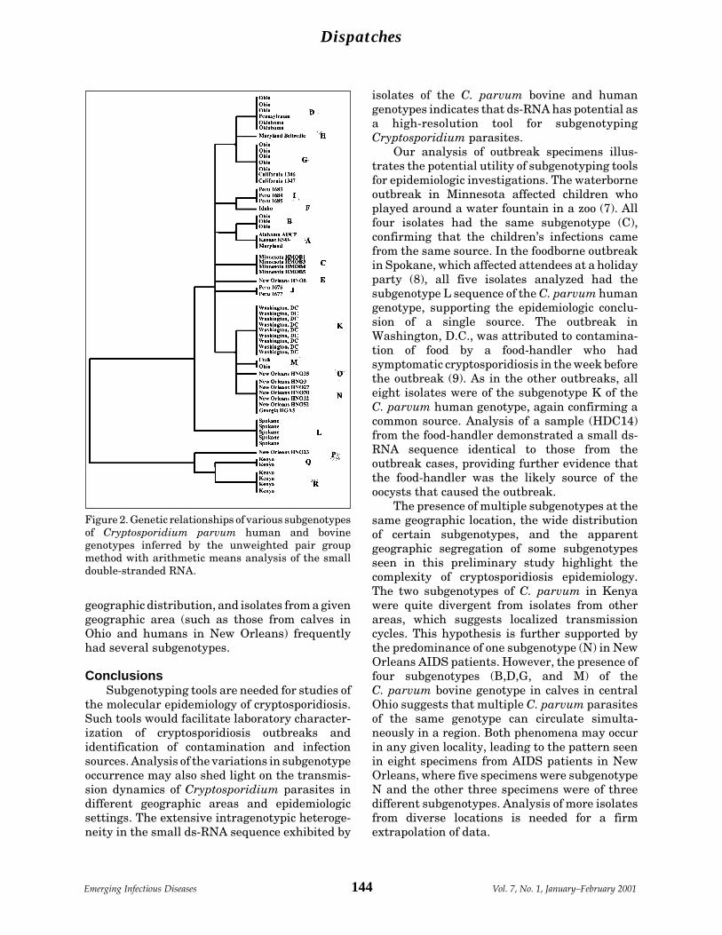
144Emerging Infectious Diseases Vol. 7, No. 1, January–February 2001
Dispatches
geographic distribution, and isolates from a givengeographic area (such as those from calves inOhio and humans in New Orleans) frequentlyhad several subgenotypes.
ConclusionsSubgenotyping tools are needed for studies of
the molecular epidemiology of cryptosporidiosis.Such tools would facilitate laboratory character-ization of cryptosporidiosis outbreaks andidentification of contamination and infectionsources. Analysis of the variations in subgenotypeoccurrence may also shed light on the transmis-sion dynamics of Cryptosporidium parasites indifferent geographic areas and epidemiologicsettings. The extensive intragenotypic heteroge-neity in the small ds-RNA sequence exhibited by
isolates of the C. parvum bovine and humangenotypes indicates that ds-RNA has potential asa high-resolution tool for subgenotypingCryptosporidium parasites.
Our analysis of outbreak specimens illus-trates the potential utility of subgenotyping toolsfor epidemiologic investigations. The waterborneoutbreak in Minnesota affected children whoplayed around a water fountain in a zoo (7). Allfour isolates had the same subgenotype (C),confirming that the children’s infections camefrom the same source. In the foodborne outbreakin Spokane, which affected attendees at a holidayparty (8), all five isolates analyzed had thesubgenotype L sequence of the C. parvum humangenotype, supporting the epidemiologic conclu-sion of a single source. The outbreak inWashington, D.C., was attributed to contamina-tion of food by a food-handler who hadsymptomatic cryptosporidiosis in the week beforethe outbreak (9). As in the other outbreaks, alleight isolates were of the subgenotype K of theC. parvum human genotype, again confirming acommon source. Analysis of a sample (HDC14)from the food-handler demonstrated a small ds-RNA sequence identical to those from theoutbreak cases, providing further evidence thatthe food-handler was the likely source of theoocysts that caused the outbreak.
The presence of multiple subgenotypes at thesame geographic location, the wide distributionof certain subgenotypes, and the apparentgeographic segregation of some subgenotypesseen in this preliminary study highlight thecomplexity of cryptosporidiosis epidemiology.The two subgenotypes of C. parvum in Kenyawere quite divergent from isolates from otherareas, which suggests localized transmissioncycles. This hypothesis is further supported bythe predominance of one subgenotype (N) in NewOrleans AIDS patients. However, the presence offour subgenotypes (B,D,G, and M) of theC. parvum bovine genotype in calves in centralOhio suggests that multiple C. parvum parasitesof the same genotype can circulate simulta-neously in a region. Both phenomena may occurin any given locality, leading to the pattern seenin eight specimens from AIDS patients in NewOrleans, where five specimens were subgenotypeN and the other three specimens were of threedifferent subgenotypes. Analysis of more isolatesfrom diverse locations is needed for a firmextrapolation of data.
Figure 2. Genetic relationships of various subgenotypesof Cryptosporidium parvum human and bovinegenotypes inferred by the unweighted pair groupmethod with arithmetic means analysis of the smalldouble-stranded RNA.

145Vol. 7, No. 1, January–February 2001 Emerging Infectious Diseases
Dispatches
A disadvantage of the ds-RNA subgenotypingtool is lack of specificity at the genotype level.Perhaps as a result of the use of a short fragmentas the target, this technique does not distinguishthe two genotypes of C. parvum and musttherefore be used in combination with routinegenotyping tools. Initial attempts targeted longerfragments of the large and small ds-RNAs.However, the RT-PCR that targeted longerfragments in amplifying samples of the C. parvumhuman genotype was much less efficient,probably because of sequence diversity at theprimer regions and lower efficiency of reversetranscription of longer fragments. A recentsequence analysis by Khramtsov et al. of fiveisolates of the C. parvum bovine genotype andseven isolates of the human genotype consis-tently separated the two genotypes in both thelarge and small ds-RNAs (6). It remains to bedetermined whether these primers and otherscan be developed for sensitive genotyping andsubgenotyping Cryptosporidium parasites.
AcknowledgmentsWe thank Anne Moore, Barbara Herwaldt, Michael
Arrowood, Bruce Anderson, William Shulaw, A. Morse, J.Inungu, Ronald Fayer, Robert H. Gilman, Lilia Cabrera,William Checkley, and Wangeci Ndiritu for providingCryptosporidium isolates.
This work was supported in part by funds from the FoodSafety Initiative, Centers for Disease Control andPrevention.
Dr. Xiao is a senior staff fellow in the Division ofParasitic Diseases, CDC. His research interests focus onthe molecular epidemiology of enteric protozoa andmalaria vaccine development.
References1. Morgan UM, Constantine CC, O’Donoghue P, Meloni
BP, O’Brien PA, Thompson RCA. Molecular character-ization of Cryptosporidium isolates from humans andother animals using random amplified ploymorphicDNA analysis. Am J Trop Med Hyg 1995;52:559-64.
2. Bonnin A, Fourmaux MN, Dubremetz JF, Nelson RG,Gobet P, Harly G, et al. Genotyping human and bovineisolates of Cryptosporidium parvum by polymerasechain reaction-restriction fragment length polymor-phism analysis of a repetitive DNA sequence. FEMSMicrobiol Lett 1996;137:207-11.
3. Peng MP, Xiao L, Freeman AR, Arrowood MJ,Escalante A, Weltman AC, et al. Genetic polymorphismamong Cryptosporidium parvum isolates supportingtwo distinct transmission cycle. Emerg Infect Dis1997;3:1-9.
4. McLauchlin J, Pedraza-Diaz S, Amar-Hoetzeneder C,Nichols GL. Genetic characterization of Cryptosporidi-um strains from 218 patients with diarrhea diagnosedas having sporadic cryptosporidiosis. J Clin Microbiol1999;37:3153-8.
5. Khramtsov NV, Woods KM, Nesterenko MV, DykstraCC, Upton SJ. Virus-like, double-stranded RNAs in theparasitic protozoan Cryptosporidium parvum. MolMicrobiol 1997;26:289-300.
6. Khramtsov NV, Chung PA, Dykstra CC, Griffiths JK,Morgan UM, Arrowood MJ, et al. Presence of double-stranded RNAs in human and calf isolates ofCryptosporidium parvum. J Parasitol 2000;86:275-82.
7. Centers for Disease Control and Prevention. Outbreakof cryptosporidiosis associated with a water sprinklerfountain–Minnesota, 1997. MMWR Morb Mortal WklyRep 1998;47:856-9.
8. Centers for Disease Control and Prevention. Foodborneoutbreak of cryptosporidiosis–Spokane, Washington,1997. MMWR Morb Mortal Wkly Rep 1998;47:565-6.
9. Quiroz ES, Bern C, MacArthur JR, Xiao L, Fletcher M,Arrowood MJ, et al. An outbreak of cryptosporidiosislinked to a foodhandler. J Infect Dis 2000;181:695-700.
10. Sulaiman IM, Xiao L, Yang C, Moore A, Beard CB,Arrowood MJ, et al. Differentiating human fromanimal isolates of Cryptosporidium parvum. EmergInfect Dis 1998;4:681-5.
11. Xiao L, Morgan U, Limor J, Escalante A, Arrowood M,Shulaw W, et al. Genetic diversity within Cryptosporid-ium parvum and related species of Cryptosporidium.Appl Environ Microbiol 1999;65:3386-91.
146Emerging Infectious Diseases Vol. 7, No. 1, January–February 2001
Dispatches
During April through July 1998, an outbreakof hand, foot, and mouth disease occurred inTaiwan; enterovirus 71 (EV71) was identified asthe main etiologic agent. The outbreak wasassociated with an unusually high death rate inyoung children. At least 55 fatal cases wereinitially reported (1,2) in healthy children, whohad refractory shock after an acute prodromalillness; many of them manifested neurologicdisorders during illness and died within 24 hoursof hospitalization (3). In many fatal cases, EV71was indicated as the etiologic agent by serologic,virologic, and polymerase chain reaction testsconducted on specimens from nonsterile sites,such as throat swabs or stool specimens. Whilethese results may indicate a precedent EV71infection in the fatal cases, they do not directlyimplicate EV71 as the causative agent ofneurologic disorders and eventual death. Incontrast, histopathologic examination in con-junction with special pathology techniques, suchas immunohistochemistry (IHC), in situ hybrid-ization, and electron microscopy, can provideunequivocal evidence linking a particular agent
to death. We report results of pathologicexamination of two fatal cases during thisoutbreak.
The StudyThe clinical and histopathologic features of
the first case were initially reported by Chang etal. (4); the second case was in a patient admittedto the same hospital with a similar clinicalcourse. Histopathologic features were similar inboth cases and showed severe and extensiveencephalomyelitis. An IHC technique using amonoclonal mouse anti-EV71 antibody and an insitu hybridization test using a digoxigenin-labeled enterovirus probe were performed onformalin–fixed, paraffin-embedded central ner-vous system (CNS) tissues and major organs ofboth patients. In the first case, positive stainingof enteroviral antigens and nucleic acids wasobserved in neurons, neuronal processes, andinflammatory foci at various CNS sites, includingthe cerebral cortex, brain stem, and all levels ofspinal cord (Figure 1A, B). No immunostaining orhybridization was present in lung, heart, liver,spleen, or kidney. Electron microscopy evalua-tion of spinal cord tissues showed a highlyvacuolated neuron containing scattered picor-navirus-like particles and viral inclusions(Figure 1C).
Pathologic Studies of Fatal Cases inOutbreak of Hand, Foot, and Mouth
Disease, Taiwan
Wun-Ju Shieh,* Shih-Ming Jung,† Chuen Hsueh,† Tseng-Tong Kuo,†Anthony Mounts,* Umesh Parashar,* Chen Fu Yang,*
Jeannette Guarner,* Thomas G. Ksiazek,* Jacqueline Dawson,*Cynthia Goldsmith,* Gwong-Jen J. Chang,* Steve M. Oberste,*
Mark A. Pallansch,* Larry J. Anderson,* Sherif R. Zaki,*and the Epidemic Working Group1
*Centers for Disease Control and Prevention, Atlanta, Georgia, USA; and†Chang Gung Memorial Hospital, Tauyuan, Taiwan, Republic of China
Address for correspondence: Wun-Ju Shieh, Centers forDisease Control and Prevention, 1600 Clifton Road, Mail StopG32, Atlanta, GA 30333, USA: fax: 404-639-3043; e-mail:[email protected].
In 1998, an outbreak of enterovirus 71-associated hand, foot, and mouth diseaseoccurred in Taiwan. Pathologic studies of two fatal cases with similar clinical featuresrevealed two different causative agents, emphasizing the need for postmortemexaminations and modern pathologic techniques in an outbreak investigation.
1Epidemic Working Group at Centers for Disease Control and Prevention: Jim Alexander, Betty Brown, David Shay, PatriciaGreer, Charles Humphrey, Tim Morken, and Tara Ferebee-Harris.

147Vol. 7, No. 1, January–February 2001 Emerging Infectious Diseases
Dispatches
The second case was negative for EV71 byIHC, in situ hybridization, polymerase chainreaction, and viral isolation. An IHC test using ananti-Japanese encephalitis antibody showedintense immunostaining of flaviviral antigens inneurons, neuronal processes, and inflammatoryfoci at various CNS sites (Figure 2A, B). Noimmunostaining of flavivirus was present inother major organs. Further study was conductedby injecting neurologic tissue of the patient intosuckling mice. CNS material from an inoculatedmouse was passed onto Vero-E6 cells andproduced cytopathic effect. Electron microscopyexamination of mouse brain and infected cellculture revealed flavivirus particles (Figure 2C),and polymerase chain reaction with sequencingconfirmed the isolate as Japanese encephalitisvirus.
ConclusionsIn addition to classic hand, foot, and mouth
disease, EV71 can cause severe CNS infectionswith a high death rate (5). Two previous EV71outbreaks were associated with neurologicdisorders and increased deaths; the first oneoccurred in Bulgaria in 1975 (5) and the second inMalaysia in 1997 (1,6). The neurologic disordersand clinical courses of acute viral CNS infectionscan be very similar regardless of causativeagents, and definitive diagnoses are sometimesvery difficult to establish without further
pathologic studies. A definitive laboratory andpathologic diagnosis is crucial for implementingeffective measures to control an outbreak (7); theNipah virus encephalitis outbreak in Malaysia(8,9) and the West Nile encephalitis outbreak inNew York City (10,11) lend additional support tothis statement.
Our report illustrates how two differentetiologic agents can cause similar diseasesduring an outbreak and emphasizes the need forpostmortem examination.
Dr. Shieh is a staff pathologist, Division of Viral andRickettsial Diseases, Centers for Disease Control andPrevention. His research interests include infectious dis-ease pathology, pathology and pathogenesis of viral en-cephalitides, molecular epidemiology, and infectious dis-ease outbreak investigations.
References 1. Centers for Disease Control and Prevention. Deaths
among children during an outbreak of hand, foot, andmouth disease –Taiwan, Republic of China, April-July1998. MMWR Morb Mortal Wkly Rep 1998;47:629-32.
2. Ho M, Chen ER, Hsu KH, Twu SJ, Chen KT, Tsai SF, etal. An epidemic of enterovirus 71 infection in Taiwan.Taiwan Enterovirus Epidemic Working Group. N EnglJ Med 1999;341:929-35.
3. Liu C, Tseng H, Wang S, Wang J, Su I. An outbreak ofenterovirus 71 infection in Taiwan, 1998: epidemiologicand clinical manifestations. J Clin Virol 2000;17:23-30.
4. Chang LY, Huang YC, Lin TL. Fulminant neurogenicpulmonary oedema with hand, foot, and mouth disease.Lancet 1998;352: 367-8.
Figure l. A). Positive immunostaining of EV71antigens in neuron and neuronal process. Originalmagnification, X158. B). Positive immunostaining ofEV71 antigens in necrotic area. Original magnifica-tion, X158. C). An array of picornavirus particles in aneuron (electron micrograph).
Figure 2. A). Positive immunostaining of Japaneseencephalitis antigens in neuron and neuronal process.Original magnification, X158. B). Positive immuno-staining of Japanese encephalitis antigens in necroticarea. Original magnification, X158. C). Flavivirusparticles (arrowheads) in isolates from mouse brain(electron micrograph).

148Emerging Infectious Diseases Vol. 7, No. 1, January–February 2001
Dispatches
5. Shindarov L, Chumakov M, Voroshilova M, Bojinov S,Vasilenko SM, Iordanov I, et al. Epidemiological, clinicaland pathomorphological characteristics of epidemicpoliomyelitis-like disease caused by enterovirus 71. J HygEpidemiol Microbiol Immunol 1979; 23: 284-95.
6. Lum LCS, Wong KT, Lam SK, Chua KB, Goh AY, LimWL, et al. Fatal enterovirus 71 encephalomyelitis. JPediatr 1998;133:795-8.
7. Zaki SR, Shieh W-J, and the Epidemic Working Groupat Ministry of Health in Nicaragua, et al. Leptospirosisassociated with outbreak of acute febrile illness andpulmonary haemorrhage, Nicaragua, 1995. Lancet1996;347:535-6.
8. Centers for Disease Control and Prevention. Outbreakof Hendra-like virus—Malaysia and Singapore, 1998–1999. MMWR Morb Mortal Wkly Rep 1999;48:265–9.
9. Chua KB, Bellini WJ, Rota PA, Harcourt BH, Tamin A,Lam SK, et al. Nipah virus: a recently emergent deadlyParamyxovirus. Science 2000;288:1432-5.
10. Centers for Disease Control and Prevention. Outbreakof West Nile-like viral encephalitis--New York, 1999.MMWR Morb Mortal Wkly Rep 1999;48:845-9.
11. Shieh WJ, Guarner J, Layton M, Fine A, Miller J, NashD, et al. The role of pathology in an investigation of anoutbreak of West Nile encephalitis in New York, 1999.Emerg Infect Dis 2000;6:370-2.

149Vol. 7, No. 1, January–February 2001 Emerging Infectious Diseases
Dispatches
Neocosmospora vasinfecta, a common plantpathogen in tropical and subtropical areas (1), islittle known as a pathogen in humans. We reporta case of fatal systemic N. vasinfecta infection inan African patient with leukemia. This is thethird reported case of a human infection with thisorganism and the first case in which the infectionwas disseminated.
The Case StudyA 38-year-old man was admitted to the
University Hospital of Cologne, Germany, with a4-week history of fatigue, vertigo, and bone pain.He had been living in Germany for 15 years andhad returned from a holiday in Nigeria 2 weeksbefore admission. Past medical history includedhepatitis C and malaria.
A bone marrow aspirate showed a predomi-nance of atypical promyelocytes, suggestingacute nonlymphocytic leukemia FAB M3. Thepatient was treated with high-dose all-trans-retinoic acid for 14 days, followed by a firstinduction chemotherapy regimen consisting ofcytarabine, daunorubicine, and thioguanine.Prophylactic treatment with fluconazole (400 mg/d)and trimethoprim-sulfamethoxazole (960 mg/d)was started because severe granulocytopenia,defined as an absolute neutrophil count below100/µL of up to 4 weeks’ duration, was anticipated.
Pain projecting to the lateral side of both feetdeveloped on day 12 of chemotherapy, when thepatient’s absolute neutrophil count had fallenbelow 100/µL. His feet were swollen andhyperthermic. Arthritis and deep vein thrombo-sis were ruled out. Pain resolved undersymptomatic treatment with acetaminophen. Onday 16 of induction therapy, bone marrowexamination yielded persistence of blast cells.Therefore, a second induction chemotherapy wasadministered, despite ongoing granulocytopenia.The regimen comprised high-dose cytarabine andmitoxantrone. On day 16 after the onset ofgranulocytopenia, fever developed. Results of achest X-ray and a high-resolution computerizedtomography scan were unremarkable. Broad-spectrum antibiotic treatment was started, butthe fever persisted. Several chest X-rays werenondiagnostic; however, after 24 days ofgranulocytopenia, another computerized tomog-raphy scan revealed interstitial and patchy lunginfiltrates. Because of persistent fever and newlydeveloping lung infiltrates under broad-spec-trum antibiotic therapy, invasive fungal infectionwas suspected, and conventional amphotericin Bwas administered (1 mg/kg/d). After 1 week ofantifungal therapy, the patient reported bilateralpleuritic chest pain. Because the patient’s conditionwas deteriorating, antifungal therapy wasaltered to 2 mg/kg/day liposomal amphotericin B,but pulmonary infiltrates persisted. Granulocy-topenia resolved after 34 days, and a bonemarrow aspirate showed complete remission of
Disseminated Neocosmospora vasinfectaInfection in a Patient with
Acute Nonlymphocytic Leukemia
Oliver A. Cornely,* Jens Chemnitz,* Hans-Georg Brochhagen,Karin Lemmer,† Heidi Schütt, Dietmar Söhngen,* Peter Staib,*Claudia Wickenhauser,* Volker Diehl,* and Kathrin Tintelnot†
*Universitätsklinik Köln, Köln, Germany, and†Robert Koch-Institute, Berlin, Germany
Address for correspondence: Oliver A. Cornely, Klinik I fürInnere Medizin, Universitätsklinik Köln, 50924 Köln,Germany; fax:+49-221-478-3611; e-mail: [email protected].
We report Neocosmospora vasinfecta infection following chemotherapy for acutenonlymphocytic leukemia. N. vasinfecta, a plant pathogen, was identified by culture andgenetic sequencing. Susceptibility testing revealed in vitro resistance for commonantifungals.
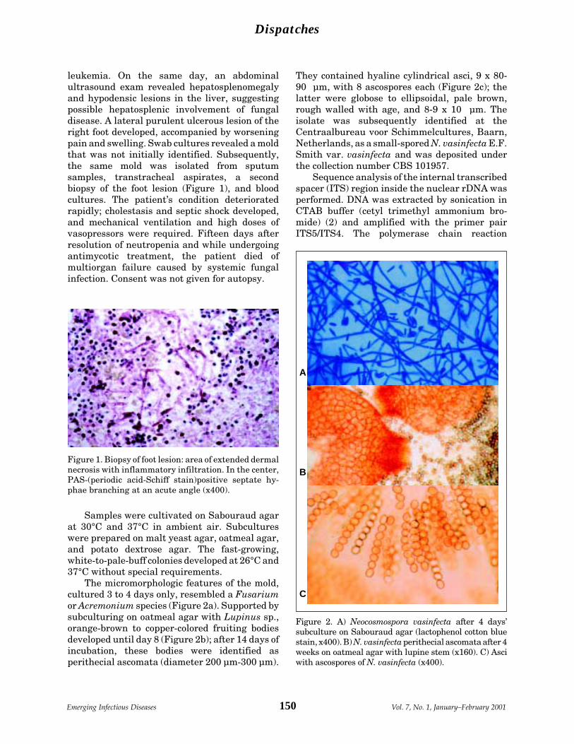
150Emerging Infectious Diseases Vol. 7, No. 1, January–February 2001
Dispatches
leukemia. On the same day, an abdominalultrasound exam revealed hepatosplenomegalyand hypodensic lesions in the liver, suggestingpossible hepatosplenic involvement of fungaldisease. A lateral purulent ulcerous lesion of theright foot developed, accompanied by worseningpain and swelling. Swab cultures revealed a moldthat was not initially identified. Subsequently,the same mold was isolated from sputumsamples, transtracheal aspirates, a secondbiopsy of the foot lesion (Figure 1), and bloodcultures. The patient’s condition deterioratedrapidly; cholestasis and septic shock developed,and mechanical ventilation and high doses ofvasopressors were required. Fifteen days afterresolution of neutropenia and while undergoingantimycotic treatment, the patient died ofmultiorgan failure caused by systemic fungalinfection. Consent was not given for autopsy.
Samples were cultivated on Sabouraud agarat 30°C and 37°C in ambient air. Subcultureswere prepared on malt yeast agar, oatmeal agar,and potato dextrose agar. The fast-growing,white-to-pale-buff colonies developed at 26°C and37°C without special requirements.
The micromorphologic features of the mold,cultured 3 to 4 days only, resembled a Fusariumor Acremonium species (Figure 2a). Supported bysubculturing on oatmeal agar with Lupinus sp.,orange-brown to copper-colored fruiting bodiesdeveloped until day 8 (Figure 2b); after 14 days ofincubation, these bodies were identified asperithecial ascomata (diameter 200 µm-300 µm).
They contained hyaline cylindrical asci, 9 x 80-90 µm, with 8 ascospores each (Figure 2c); thelatter were globose to ellipsoidal, pale brown,rough walled with age, and 8-9 x 10 µm. Theisolate was subsequently identified at theCentraalbureau voor Schimmelcultures, Baarn,Netherlands, as a small-spored N. vasinfecta E.F.Smith var. vasinfecta and was deposited underthe collection number CBS 101957.
Sequence analysis of the internal transcribedspacer (ITS) region inside the nuclear rDNA wasperformed. DNA was extracted by sonication inCTAB buffer (cetyl trimethyl ammonium bro-mide) (2) and amplified with the primer pairITS5/ITS4. The polymerase chain reaction
Figure 2. A) Neocosmospora vasinfecta after 4 days’subculture on Sabouraud agar (lactophenol cotton bluestain, x400). B) N. vasinfecta perithecial ascomata after 4weeks on oatmeal agar with lupine stem (x160). C) Asciwith ascospores of N. vasinfecta (x400).
Figure 1. Biopsy of foot lesion: area of extended dermalnecrosis with inflammatory infiltration. In the center,PAS-(periodic acid-Schiff stain)positive septate hy-phae branching at an acute angle (x400).
A
B
C
151Vol. 7, No. 1, January–February 2001 Emerging Infectious Diseases
Dispatches
product was purified from agarose gel by usingthe Gel Extraction Kit (Qiagen GmbH, Hilden,Germany) and directly sequenced with the primersITS2, ITS3, ITS4, and ITS5 (3). DNA sequencingwas performed by the dideoxynucleotide termi-nation method, using the dye deoxy terminatorchemistry and the ABI 377 automated sequencer(PE Applied Biosystems, Foster City, CA). Theresults were compared with the published ITSsequence, GenBank accession no. L36627.
When we compared our results to thepublished 544-bp ITS sequence, we founddifferences in three positions: T to C exchange atnucleotide position 72, C to T exchange atposition 102, and an insertion of a seventhC between positions 381 and 388.
The MICs of amphotericin B and 5-flucytosine were determined by the ATB Funguskit (Biomérieux SA, Marcy l’Étoile, France).Susceptibility testing to fluconazole, itraconazole,terbinafine, and voriconazole (Pfizer Inc., NewYork, NY) was performed by using a casitone-based microdilution system (4). The MIC foritraconazole was simultaneously determinedaccording to the National Committee for ClinicalLaboratory Standards (NCCLS) M27-A protocol(5). The inoculum had a final amount of 1x10⁴
conidia per microliter. Incubation time at 35°Cwas 44 hours for amphotericin B and 5-flucytosineand 30 hours for the other antifungals.
MICs were as follows: amphotericin B > 8 µg/mL-1, 5-flucytosine >128 µg/mL-1, fluconazole>128 µg/mL-1, itraconazole >2 µg/mL-1 versus >8µg/mL-1 (according to NCCLS), voriconazole 1 µg/mL-1 , and terbinafine 0.125 µg/mL-1.
ConclusionsIn the early days of incubation, the
micromorphologic features of N. vasinfectaisolates may be misleading, suggesting Fusariumor Acremonium species (1), which could lead to aninadequate therapeutic regimen because of thedifferent amphotericin B susceptibility of thesegenera. While high-dose amphotericin B intrave-nously is the first-line therapy in systemicfusarioses, polyene antimycotics are probablynot beneficial against infections caused byN. vasinfecta, as demonstrated in our patient andin a recently reported immunocompetent patientwith posttraumatic N. vasinfecta infection (6).
Our patient died of disseminated infection byN. vasinfecta despite high-dose antimycotic
therapy with amphotericin B and remission ofthe underlying hematologic malignancy. The invitro susceptibility test results confirmed theresistance of the isolate against amphotericin Band fluconazole and nearly all of the few otherantifungal agents available for systemic therapy.Not enough data on terbinafine are available toallow valid interpretation. In comparison to theMICs of voriconazole against other filamentousfungi such as Aspergillus fumigatus (7), thesusceptibility for voriconazole in our isolatemight not have been sufficient for adequateantifungal treatment.
Neither the mechanism of infection nor theincubation period could be determined. Ourpatient did not remember any injury, so infectionmost likely occurred through minimal trauma.Although he might have been infected during hisrecent stay in Nigeria, infection might haveoccurred in Africa considerably longer ago: Thelatency period for N. vasinfecta can be severalyears between infection and the symptomaticstage (8). The infection might never have beendetected had the patient remained immunocom-petent. Once the infection became systemic, itwas likely incurable.
The two previously reported patients (6,8,9),both from Senegal, had been cured by a radicalsurgical intervention that prevented dissemina-tion of disease, limiting it to the lowerextremities. Because of the poor susceptibility ofthe fungus, complete surgical excision promptedby early diagnosis is the only means to influencethe clinical outcome. Currently, no medicaltreatment option exists for a patient withsystemic disease caused by N. vasinfecta.
Imported mycoses should be taken intoconsideration in patients who have traveled orlived abroad, especially former residents oftropical and subtropical countries, even yearsafter their stay abroad. Recent epidemiologicdata on plants suggest that persons working incotton production might also be at risk (10).
This is the first case of disseminatedN. vasinfecta infection. Physicians may considerthis infection as a new differential diagnosis ofpurulent subcutaneous leg lesions in the severelyimmunocompromised host.
AcknowledgmentsThe authors thank Michael Seibold for performing the
antifungal susceptibility tests.

152Emerging Infectious Diseases Vol. 7, No. 1, January–February 2001
Dispatches
Dr. Cornely is a senior resident and clinical researchfellow at the Universitätsklinik Köln, Köln, Germany.His research interests include treatment of hematologicand oncologic malignancies, with a focus on supportivecare and infectious diseases in neutropenic hosts.
References 1. Cannon PF, Hawksworth DL. A revision of the genus
Neocosmospora (Hypocreales). Transactions of theBritish Mycological Society 1984;82:673-88.
2. Wedde M, Müller D, Tintelnot K, De Hoog GS, Stahl U.PCR-based identification of clinically relevantPseudallescheria/Scedosporium strains. Med Mycol1998; 36:61-7.
3. White TJ, Bruns T, Lee S, Taylor J. Amplification anddirect sequencing of fungal ribosomal RNA genes forphylogenetics. In: Innis N, Gelfand J, White T, editors.PCR protocols: a guide to methods and applications.New York: Academic Press; 1990. p. 315-22.
4. Seibold M, Werner E. Testing susceptibility of Candidaspecies to fluconazole and itraconazole using themicrodilution assay. Mycoses 1995;38:443-8.
5. National Committee for Clinical Laboratory Standards(NCCLS). Reference method for broth dilutionantifungal susceptibility testing of yeasts. M27-A.Villanova, PA: NCCLS; 1997.
6. Kac G, Piriou P, Gueho E, Roux P, Tremoulet J, DenisM, et al. Osteoarthritis caused by Neocosmosporavasinfecta. Med Mycol 1999;37:213-7.
7. Espinel-Ingroff A. In vitro activity of the new triazolevoriconazole (UK-109,496) against opportunisticfilamentous and dimorphic fungi and common andemerging yeast pathogens. J Clin Microbiol 1998;36:198-202.
8. Ben Hamida F, Achard JM, Westeel PF, Chandenier J,Bouzernidj M, Petit J, et al. Leg granuloma due toNeocosmospora vasinfecta in a renal graft recipient.Transplant Proc 1993;25:2292.
9. Chandenier J, Hayette MP, de Bièvre C, Westeel PF,Petit J, Achard JM, et al. Tuméfaction de la jambe àNeocosmospora vasinfecta chez un transplanté rénal.Journal de Mycologie Médicale 1993;3:165-8.
10. Baird R, Carling D. Survival of parasitic andsaprophytic fungi on intact senescent cotton roots.Journal of Cotton Science 1998;2:27-34.

153Vol. 7, No. 1, January–February 2001 Emerging Infectious Diseases
Commentary
Adventitious Viral Genomesin Vaccines but Not in Vaccinees
It is a pleasant change to write aboutviruses that might have emerged but haven’t.In this issue, Hussain and colleagues at theCenters for Disease Control and Prevention, theU.S. Department of Agriculture, and HarvardUniversity report that recipients of measles,mumps, and rubella (MMR) vaccine show noevidence of infection by endogenous avianretroviruses, even though viral genomes andreverse transcriptase activity have been de-tected in vaccine preparations. Influenza,yellow fever, and MMR vaccines are usuallyprepared in embryonated eggs or in cultures ofchick embryo fibroblasts (CEF). These fibro-blasts contain and express endogenousretroviral genomes (1). In any vaccine, adventi-tious agents in the cellular substrate maycontaminate the biological product. In live,attenuated vaccines, such contaminants are notinactivated, and endogenous retroviruses bytheir very nature as Mendelian transmittedgenomes are particularly difficult to eliminate.Endogenous retrovirus release also has ramifi-cations for pharmaceutical proteins made in cellsubstrates (e.g., monoclonal antibodies) and forxenotransplantation (2,3).
Some 45 years ago, it was found thatapparently healthy hens could transmit avianleukosis virus (ALV) vertically in eggs (4); laterit was demonstrated that live virus vaccinesmade in CEF were contaminated with infec-tious ALV (5). However, no increased risk forcancer was found in yellow fever vaccinees withthe longest presumed exposure to ALV (6).Nevertheless, vaccine manufacturers were soonrequired to use eggs or CEF from leukosis-freeflocks. To screen for ALV infection, a comple-ment fixation for ALV (COFAL) antigen testwas devised, and through pioneering work inthe 1960s, the existence of endogenousretroviruses came to light because many ALV-free birds were COFAL positive (7-9).
As a graduate student at the time, I ob-served that CEF of COFAL-positive embryoscomplemented envelope-defective Rous sarcomavirus, yielding pseudotype viruses with xenotro-pic properties. The endogenous virus wasgenetically transmitted in chickens but wasinfectious for other hosts such as quail andpheasant. Many copies of partial or complete
ALV genomes were located in chicken DNA (1).We showed that ALV had colonized the hostgerm line of red jungle fowl before domestica-tion to become chickens but after divergence ofthe genus Gallus into distinct species. Even so,it proved possible in the 1970s to breed whiteleghorns free of endogenous ALV genomes; suchchickens are now being introduced by Merck aspreferred substrates for vaccine production.
A second class of endogenous avianretroviral genome (EAV), discovered in 1985(10), is present in all breeds of chicken andcannot be eliminated. EAV can release nonin-fectious virus particles containing active re-verse transcriptase; and this is the genomemost commonly found in MMR and othervaccines (Hussain et al., this issue; 11). Themajor retroviral pathogen of meat-strainchickens is an infectious recombinant betweenALV gag and pol genes and an env gene relatedto EAV (12). This virus has not been observedto infect human cells.
May we assume, therefore, that chicken cellsubstrate vaccines are safe? With biologicalproducts, as with crossing the street, there is nosuch thing as absolute safety. The paper byHussain et al. is reassuring, and I agree withthe authors that no change in current U.S.policies (or WHO policies, for that matter) iswarranted, and the public should continue toenjoy the benefit of the vaccine. However, itmay be useful to probe the possibility of interac-tion between endogenous avian viruses and theinfectious components of MMR. We showed thatvesicular stomatitis virus (VSV) could assembleits glycoprotein G on avian retrovirus virionsand vice versa (13). Indeed, VSV G protein hasbecome an envelope of choice for retroviralvectors developed for gene therapy. By analogy,the assembly of the hemagglutinin and fusionglycoproteins of measles or mumps virusesmight confer a human host range onendogenous ALV or EAV particles. Thepossible generation of such pseudotypes orphenotypically mixed virions in vaccines maybe worthy of investigation.
In addition, with ultrasensitive techniques,such as polymerase chain reaction (PCR) geneamplification, we can detect viral genomes andreverse transcriptase activity more readily invaccine preparations. Virtually all vertebratesstudied, including humans, carry endogenousretroviral genomes as part of their natural

154Emerging Infectious Diseases Vol. 7, No. 1, January–February 2001
Commentary
genetic constitution (1,14). Therefore, almostany cell substrate for vaccine production (avian,rodent, or primate) is likely to contain andexpress (at low level) endogenous retroviralgenomes.
Vaccine contamination by adventitiousviruses in the cellular substrate has, of course,occurred before. In one instance, the discoveryof SV40 in rhesus macaque kidney cultures (15)soon led to the adoption of cynomolgus macaqueand later African green monkey (AGM) kidneysas the preferred substrate for polio vaccines.That was, perhaps, a near escape as AGMs arenow known to frequently harbor a strain ofsimian immunodeficiency virus (SIV) thatluckily does not appear to infect humans.Following the potential exposure of millions ofpolio vaccinees to SV40, no evidence was foundof increased cancer incidence (16). More re-cently, it has been reported that SV40 ispresent in some human cancers (17). Casesinclude pediatric tumors in patients born longafter SV40 was eliminated from polio vaccines.
Ironically, it was the misguided attention ofregulatory groups on hypothetical oncogenicDNA that led to vaccine contamination byadventitious oncogenic viruses in the first place.Fear of oncogenic DNA made tumor cell linestaboo as cellular substrates for vaccine produc-tion. Despite all we have learned aboutoncogenes and tumor suppressor genes inmultistep progression to cancer, the possibletrace of “oncogenic” DNA in vaccines preparedin established cell lines remained of greaterconcern to regulators than adventitious infec-tions in primary cells. It is high time to reevalu-ate the relative risks, so it is heartening thatthe Food and Drug Administration held aworkshop last year to begin that process.
Robin A. WeissUniversity College London, London, UK
References 1. Coffin J. Endogenous viruses. In: Weiss RA, Teich
NM, Varmus HE, Coffin J, editors. RNA tumorviruses. New York: Cold Spring Harbor LaboratoryPress;1982.p. 1109-203.
2. Weiss RA. Retroviruses produced by hybridomas. NEngl J Med 1982;307:1587.
3. Patience C, Takeuchi Y, Weiss RA. Infection ofhuman cells by an endogenous retrovirus of pigs. NatMed 1997;3:282-6.
4. Burmester BR, Gentry RF, Waters NF. The presenceof the virus of visceral lymphomatosis in embryonat-ed eggs of normal appearing hens. Poultry Sci1955;34:609-17.
5. Dougherty RM, Harris RJ, Biggs PM, Payne LN, GoffeAP, Churchill AE, et al. Contaminant viruses in two livevirus vaccines produced in chick cells. J Hyg 1966;64:1-7.
6. Waters TD, Anderson PS, Beebe GW, Miller RW.Yellow fever vaccination, avian leukosis virus, andcancer risk in man. Science 1972;177:76-7.
7. Dougherty RM, DiStefano HS. Lack of relationshipbetween infection with avian leukosis virus and thepresence of COFAL antigen in chick embryos.Virology 1966;29:586-95.
8. Dougherty RM, DiStefano HS, Roth FK. Virusparticles and viral antigens in chicken tissues free ofinfectious avian leukosis virus. Proc Natl Acad Sci US A 1967;58:808-17.
9. Payne LN, Chubb RC. Studies on the nature and geneticcontrol of an antigen in normal chick embryos whichreacts in the COFAL test. J Gen Virol 1968;3:379-91.
10. Dunwiddie C, Faras AJ. Presence of retrovirusreverse transcriptase-related gene sequences in aviancells lacking endogenous avian leukosis viruses. ProcNatl Acad Sci U S A 1985;82:5097-101.
11. World Health Organization. Reverse transcriptaseactivity in chicken-cell derived vaccine. WklyEpidemiol Rec 1998;73:209-12.
12. Bai J, Payne LN, Skinner MA. HPRS-103 (exogenousavian leukosis virus, subgroup J) has an env generelated to those of endogenous elements EAV-0 andE51 and an E element found previously only insarcoma viruses. J Virol 1995;69:779-84.
13. Weiss RA, Boettinger DE, Love D. Phenotypic mixingbetween vesicular stomatitis virus and avian RNAtumor viruses. Cold Spring Harbor Symp Quant Biol1975;39:913-8.
14. Patience C, Wilkinson DA, Weiss RA. Our retroviralheritage. Trends Genet 1997;13:116-20.
15. Sweet BH, Hilleman MR. The vacuolating virus,SV40. Proc Soc Exp Biol Med 1960;105:420-7.
16. Nathanson N, Shah K. Human exposure to SV40:review and comment. Am J Epidemiol 1976;103:1-12.
17. Butel JS, Lednicky JA. Cell and molecular biology ofsimian virus 40: implications for human infectionsand disease. J Natl Cancer Inst 1999;91:119-34.

155Vol. 7, No. 1, January–February 2001 Emerging Infectious Diseases
Research Update
Strengthening National Preparednessfor Smallpox: An Update
Concern that smallpox virus may be used asa biological weapon of mass destruction hasprompted calls for production of additionalvaccine and new research into variola virusdiagnostics and clinical interventions. Only 15.4million doses of smallpox vaccine, producedapproximately 20 years ago, exist in the UnitedStates (1). While virtually all lots remainpotent, additional vaccine would clearly beneeded in a national emergency involvingsmallpox virus. Global eradication of naturalsmallpox disease was declared in 1980; witheradication, most research activities involvingthe virus ended. Although the complete ge-nomic sequence of selected isolates of variolavirus is known (2), the diagnosis and treatmentof smallpox infection have not changed in thepast two decades. Recognizing the need foradvancement in these areas before variola virusstocks are destroyed, the World Health Organi-zation (WHO) passed a resolution (WHA 52.10)in 1999 extending the date of destruction of allremaining variola virus stocks until the end of2002. The midpoint of this period is an appro-priate time to review progress made in vaccineproduction and variola virus research and tooutline the next steps.
Vaccine ProductionOn September 20, 2000, the Centers for
Disease Control and Prevention (CDC) enteredinto an agreement with OraVax (Cambridge,MA) to produce a new smallpox vaccine. Likethe vaccine used to eradicate smallpox, the newvaccine will contain live vaccinia virus; how-ever, it will be produced in cell cultures bymodern vaccine production techniques. OraVaxwill coordinate full clinical testing of the vac-cine and submit a licensing application to theU.S. Food and Drug Administration (FDA) forthe prevention of smallpox in adults and chil-dren. Forty million doses of the new vaccinewill be produced initially, with anticipateddelivery of the first full-scale production lots in2004. The agreement calls for sustained annualproduction through 2020 to replace outdatedvaccine and allows for increased productionshould an emergency arise. The vaccine will beadministered with bifurcated needles (alsoproduced by OraVax), which create a localized
vaccine “pock” and confer protective immunity.The vaccine will be held in reserve as part ofthe national stockpile and be released only inthe event of a confirmed case of smallpox orwhen vaccination against vaccinia virus iswarranted. The agreement allows OraVax toproduce additional vaccine for other markets,including international buyers.
Variola Virus ResearchA research plan, implemented at CDC by
scientists from both the Department of Defenseand CDC and including extensive collaborationswith scientists from the National Institutes ofHealth and other organizations, is being under-taken with WHO concurrence. All work withlive variola virus is done under biosafety level 4containment conditions at CDC. Smallpox virusis officially retained at only two facilities in theworld: at CDC in the United States and theState Research Center of Virology and Biotech-nology in Novosibirsk, Russia. Research teamsfrom both institutions are coordinating activi-ties to avoid duplication and gain the maximumamount of information possible before finaldestruction of the virus.
Strain EvaluationOf 461 isolates in the smallpox virus collec-
tion at CDC, 49 were selected for furthercharacterization. These isolates, which includedboth variola major and variola minor, wereselected to represent the greatest diversity indate of collection and geographic region. Of the49 isolates tested for viability, 45 were success-fully recovered, and seed stocks were preparedfor subsequent studies. This group of 45 repre-sented isolates from as early as 1939 and aslate as the 1970s; all major geographic regionswere represented. Study of these isolates isbased on three research themes: application ofmodern serologic and genomic methods in thediagnosis of variola virus disease; determina-tion of candidate antiviral drug activity againstthis virus; and investigation of the pathogenesisof smallpox infection, especially through thedevelopment of a nonhuman primate model toreplicate human smallpox infection. The re-search team carefully outlined all experimentalwork to be undertaken with variola virus,incorporating suggestions from a peer group ofhighly qualified external experts from academiaand industry; the first set of experiments was

156Emerging Infectious Diseases Vol. 7, No. 1, January–February 2001
Research Update
conducted from January to July 2000 in theCDC maximum containment laboratory.
Serologic AssaysBecause enzyme immunoassay technology
was still in its infancy when smallpox waseradicated, during the first series of experi-ments, polyclonal and monoclonal antibodieshad to be produced for developing enzyme-linked immunosorbent assays to measurevariola virus-specific immunoglobulin (Ig) M,IgG, and antigen. These reagents are now beingevaluated by prototype assays with inactivatedviral antigens. This work will continue for theforeseeable future.
Nucleic Acid-Based DiagnosticsViral DNA was extracted from all 45 suc-
cessfully recovered isolates, was purified andinactivated, and is now being examined byrestriction fragment-length polymorphismdeveloped by an extended polymerase chainreaction assay that amplifies viral genome into20 overlapping products of approximately 10kilobases each. These products cover virtuallythe entire length of the viral genome andinclude sequences in essential genes and geneslikely needed for pathogenesis. Preliminaryresults indicate that the data thus generatedoffer a good low-resolution overview of geneticdiversity of variola viruses and are being usedto differentiate strains, infer phylogeny, andidentify as many as 10 additional variolaisolates for complete genome sequencing. Twoisolates, Somalia 77 and Congo 70, were specifi-cally suggested by WHO for sequencing, andthis work has begun. A dedicated sequence andbioinformatics facility being developed at CDCwill be used to undertake this effort and tobegin constructing a genomic signature data-base, not only for smallpox but also, over time,for other pathogens with bioterrorism potential.
Antiviral DrugsTwo hundred seventy-four antiviral drug
compounds were screened for activity andtherapeutic indices against variola, monkeypox,cowpox, camelpox, and vaccinia viruses by twocell culture assays. Many of these compoundswere provided for testing under collaborativearrangements facilitated by an orthopox antivi-ral research initiative of the National Instituteof Allergy and Infectious Diseases. Previous
studies identified a nucleoside phosphonateDNA polymerase inhibitor, cidofovir (Vistide),as being active against poxviruses, includingvariola. In the current trial, cidofovir and itsprodrug (cyclic HPMPC) were evaluated against31 strains of variola, which were selected tocover a wide geographic area and time span. Nosubstantial differences in inhibition amongstrains were observed, which suggests thatcidofovir-resistant strains are unlikely. The invitro inhibition was further characterized inmultiple cell lines to meet FDA requirements.However, another class of antiviral drugs, theS-adenosylhomocysteine hydrolase inhibitors,showed considerable variation in the 50%inhibitory dose between variola isolates; thiseffect should be investigated further.
Two approaches to the development of anoral prodrug of cidofovir yielded compoundswith improved antiviral activity. In addition,the current series of experiments identified 27other compounds, including completely newclasses of drugs, that appear to be activeagainst variola and other orthopoxviruses. Infact, 10 compounds had therapeutic indicesgreater than 200, while cidofovir had indicesgreater than 10; 3 compounds had therapeuticindices greater than 1,500. When work resumesin early 2001 with live variola virus, we willcontinue to evaluate these and additionalcompounds for activity, including analogsdesigned for oral administration. The mostpromising compounds emerging from this invitro testing will be evaluated in animal mod-els, e.g., cowpox and vaccinia in mice andeventually monkeypox virus challenge innonhuman primates. All promising compoundswill be tested against a battery of surrogateorthopox viruses to guide evaluation of newantiviral compounds after variola virus is nolonger available.
Animal ModelsA major goal of the current research is to
define an animal model that faithfully repli-cates human smallpox. Such a model would beextremely valuable in evaluating candidateantiviral drugs and novel diagnostic assays andin defining the pathogenesis of smallpox.Consequently, two groups of four cynomolgusmacaques were exposed to two variola virusstrains at a high dose (>108 PFU) by the aerosolroute. Clear evidence of infection was found; the
157Vol. 7, No. 1, January–February 2001 Emerging Infectious Diseases
Research Update
animals had transient fevers, perturbations incytokine titers in serum, and mild exanthemouslesions. A few of the monkeys showed signs ofbronchopneumonia, but none died or haddisease similar to the classic smallpox seen inhumans. Another series of experiments will beundertaken with different variola isolates toconfirm these preliminary observations andgenerate additional clinical material to validatethe diagnostic assays under development.
The results of the research now under way,coupled with the promise of renewed productionof smallpox vaccine, will better prepare theUnited States—and indeed the entire world—for the possibility that smallpox virus might beused as a terrorist weapon of mass destruction.
James W. LeDuc* and Peter B. Jahrling†*Centers for Disease Control and Prevention,
Atlanta, Georgia, USA; †United States Army MedicalResearch Institute of Infectious Diseases, Fort
Detrick, Frederick, Maryland, USA
References 1. LeDuc JW, Becher J. Current status of smallpox
vaccine. Emerg Infect Dis 1999;5:593-4. 2. Shchelkunov SN, Massung RF, Esposito JJ.
Comparison of the genome DNA sequences ofBangladesh-1975 and India-1967 variola viruses.Virus Res 1995;36:107-18.

158Emerging Infectious Diseases Vol. 7, No. 1, January–February 2001
Letters
High-Level Ciprofloxacin Resistancein Neisseria gonorrhoeae:First Report from Israel
To the Editor: We report a case of male gonococ-cal urethritis that persisted despiteciprofloxacin therapy. The isolate was found tobe highly resistant (MIC 32 µg/mL).
A 30-year-old man visited his family physi-cian with a 2-day history of urethral dischargeand dysuria. The symptoms began 7 days aftera single, unprotected orogenital contact with afemale hitchhiker. The patient denied exchangeof money for the act and reported no otherrecent sex partners or travel outside Israel.After a urethral swab was obtained for culture,the patient received a single dose ofciprofloxacin (500 mg orally). Growth of Neis-seria gonorrhoeae was subsequently reported.However, symptoms persisted, and a regimen ofdoxycycline (100 mg orally twice a day for 10days) was initiated. After temporary clinicalimprovement, the patient returned with wors-ening symptoms: bloody urethral discharge,severe dysuria, edema of the penis, and painfulerection. N. gonorrhoeae was reisolated from arepeat urethral swab. When a single dose ofceftriaxone (250 mg) was administered intra-muscularly, clinical cure was prompt.
Susceptibility testing was performed on thesecond isolate by using the E-test method (ABBiodisk, Solna, Sweden) on a medium contain-ing GC agar base and 1% defined growthsupplement. The MIC of ciprofloxacin was32 µg/mL, penicillin 1.5 µg/mL, tetracycline2 µg/mL, and ceftriaxone 0.016 µg/mL. Theisolate did not produce beta-lactamase. It wasclassified as a CMRNGPT phenotype(N. gonorrhoeae with chronomosomallymediated resistance to both penicillin andtetracycline).
Gonorrhea was considered a rare disease inIsrael in the 1990s: the average annual inci-dence was 0.89 reported cases per 100,000population (1). Most laboratories did not carryappropriate media, and susceptibility testing ofN. gonorrhoeae was not performed routinely.Quinolones and spectinomycin are the antibiot-ics most commonly used to treat the infection.Nevertheless, we are not aware of any instanceof clinical failure following fluoroquinolone
therapy. More recently, however, the incidenceof gonorrhea has been increasing (2). In re-sponse, a surveillance program for monitoringantimicrobial resistance in N. gonorrhoeae hasbeen launched.
Fluoroquinolones and cephalosporinsbecame the recommended drugs for treatmentof gonococcal infection after penicillin- andtetracycline-resistant N. gonorrhoeae appeared(3). Gonococcal strains with reduced in vitrosusceptibility to fluoroquinolones (MIC,0.125 µg/mL to 0.5 µg/mL) were first describedin the mid-1980s (4) and are now occurringworldwide (5).
Fluoroquinolone-resistant N. gonorrhoeae(ciprofloxacin MIC >1.0 µg/mL) emergedduring the 1990s and became well establishedin several Asian countries (6). In Japan, therate of ciprofloxacin resistance increased from6.6% in 1993-1994 to 24.4% in 1997-1998 (7).More recently, high-level resistance tociprofloxacin and reports of treatment failurehave appeared (8). Strains with ciprofloxacinMICs of >8.0 µg/mL were first isolated in 1994(6) and are detected mostly in the Far East.Two cases of gonococcal infection by strainswith an MIC of 16 µg/mL were recently re-ported in the United States (9). Gonococcalresistance to fluoroquinolones is associatedwith mutations in the genes encoding DNAgyrase (gyrA) and topoisomerase (parC) as wellas change in porin permeability and reducedintracellular drug accumulation (6). In view ofthe increasing resistance to fluoroquinolones,ceftriaxone, cefixime, or spectinomycin is nowrecommended if an infection was acquired inAsia or other areas with known fluoroquinoloneresistance (9).
Our patient reported no travel to the FarEast, and his sex partner, who could not belocated for follow-up, was not Asian; her travelhistory was unknown. The mode of transmis-sion of this infection was fellatio. Condoms areoften not used in this form of intercourse, evenby those who regularly use condoms for genito-genital sex, because of the mistaken belief thatinfection is not spread through this form ofintercourse. It is now well established that oralsex plays an important role in HIV transmis-sion (10), and condoms should be used with anyform of intimate sexual contact.

159Vol. 7, No. 1, January–February 2001 Emerging Infectious Diseases
Letters
Michael Dan,* Francesca Poch,* Daniel Shpitz,†and Bracha Sheinberg†
*Infectious Disease Unit, E. Wolfson Hospital,Holon, Israel, and †Maccabi Health Services,
Rishon-le-Zion, Israel
References 1. The Israel Center for Diseases Control. Notifiable
infectious disease in Israel, 1951-1995. Israel: State ofIsrael: Ministry of Health; 1996. pub. no. 201.
2. District Health Office, Tel-Aviv. Report on emergingissues, July-December 1999. State of Israel: Ministryof Health; Jan 10, 2000.
3. Centers for Disease Control and Prevention. Guide-lines for treatment of sexually transmitted diseases.Mor Mortal Wkly Rep 1998;47(RR-1):1-116.
4. Tapsall JW, Shultz TR, Phillips EA. Characteristicsof Neisseria gonorrhoeae isolated in Australiashowing decreased sensitivity to quinolone antibiot-ics. Pathology 1992;24:27-31.
5. Quinn TC. The impact of antimicrobial resistance onthe treatment of sexually transmitted diseases. InfectDis Clin North Am 1997;11:884-904.
6. Knapp JS, Fox KK, Trees DL, Whittington WL.Fluoroquinolone resistance in Neisseria gonorrhoeae.Emerg Infect Dis 1997;3:33-8.
7. Tanaka M, Nakayama H, Haraoka M, Saika T,Kobayashi I, Naito S. Antimicrobial resistance ofNeisseria gonorrhoeae and high prevalence ofciprofloxacin-resistant isolates in Japan, 1993 to1998. J Clin Microbiol 2000;38:521-5.
8. Tapsall JW, Limnios EA, Thacker C, Donovan B,Lynch SD, Kirky LJ, et al. High-level quinoloneresistance in Neisseria gonorrhoeae: a report of twocases. Sex Transm Dis 1995;22:310-11.
9. Centers for Disease Control and Prevention. Fluoroqui-nolone-resistant Neisseria gonorrhoeae–San Diego,California, 1997. Mor Mortal Wkly Rep 1998;47:405-8.
10. Dillon B, Hecht FM, Swanson M, Goupil-Sormany I,Grant RH, Chesney MA, et al. Primary HIV infec-tions associated with oral transmission. Presented atthe 7th Conference on Retroviruses and Opportunis-tic Infections, San Francisco, Jan 30–Feb 2, 2000(Abstract 473).
An Unusual BacteriumCausing a Brain Abscess
To the Editor: Intracranial abscesses are animportant cause of illness and death in aneurologic/neurosurgical unit. Early presump-tive clinical diagnosis supported by radiologicevidence (computerized axial tomography [CAT]scan and magnetic resonance imaging) is themainstay of diagnosis (1). Abscess contents areaspirated under stereotaxic guidance andcultured to isolate causative organisms and
determine their antibiotic sensitivities. Organ-isms isolated from brain abscesses are usuallystreptococci, anaerobic and facultative gram-negative bacilli, staphylococci, or pseudomonads(2).
A 24-year-old male farmer came to us withprogressive headache, dizziness, and a low-grade fever of 2 weeks’ duration. He had had apimple on his right cheek approximately 3weeks before, which had discharged “bluish”pus on forcible evacuation and subsequentlyhealed without treatment. No focal neurologicsigns were detected on physical examination.Because an intracranial space-occupying lesionwas suspected, a lumbar puncture was with-held. Later, a CAT scan of the patient’s headrevealed a right-sided temporoparietal space-occupying lesion approximately 3 cm in diam-eter, suggestive of a unilocular brain abscess.The abscess was needle aspirated under stereo-taxic guidance, and the pus was culturedaerobically and anaerobically. After 24 hours ofaerobic incubation on MacConkey agar at 37°C,a pure growth of violet-colored colonies appeared,identified as Chromobacterium violaceum bythe 20E API system (Biomerieux, France).
Other initial laboratory findings were asfollows: blood leukocyte count, 16,200 cells/µL(84% neutrophils, 15% lymphocytes, 1% eosino-phils); erythrocyte sedimentation rate(Westergren method), 22 mm/hour; C-reactiveprotein concentration, 96 mg/L; and fastingblood sugar concentration, 5.1 mmol/L. Bloodurea and C-reactive protein concentrationsafter 3 weeks of antibiotic treatment were 4.6mmol/L and <6 mg/L, respectively.
The organism was sensitive to imipenemand ciprofloxacin and resistant to cefotaximeand ceftriaxone, by the Stokes comparativedisk-diffusion antibiotic sensitivity testingmethod (3). Ciprofloxacin (as lactate) wasadministered intravenously, 400 mg twice aday, for 4 weeks. Repeated CAT scans, clinicalsymptoms, and serial C-reactive protein levelsindicated rapid regression of the abscessfollowed by complete cure.
C. violaceum is a gram-negative bacilluspresent in soil and aquatic environments oftropical and subtropical countries or regionssuch as Trinidad, Guyana, India, Malaysia,Florida, and South Carolina. It is a bacterium oflow virulence, occasionally causing skin
160Emerging Infectious Diseases Vol. 7, No. 1, January–February 2001
Letters
infections and disseminated disease involvingmultiple organs in immunocompromisedpatients. In such cases the disease can mimicsepticemic melioidosis (4,5).
In this previously healthy patient, infectionprobably originated from the facial abscess. Thepatient was negative for HIV antibody(Serodia), had no history of diabetes mellitus orother compromising illnesses, and had noevidence of immunodeficiency. In a previouscase of disseminated C. violaceum infection in ayoung patient, postmortem findings revealednumerous cortical infarcts and hemorrhages (6).Our isolate from a brain abscess is yet anothercase of a relatively avirulent saprophyticmicroorganism resulting in a deep-seatedinfection in a well-nourished, previouslyhealthy person.
Dhammika Nanda Atapattu,*Dhammika Priyal Jayawickrama,*† and
Vasanthi Thevanesam**University of Peradeniya, Peradeniya, Sri Lanka;
†General Hospital, Kandy, Sri Lanka
References 1. Mathisen GE, Johnson JP. Brain abscess. Clin Infect
Dis 1997;25:763-81. 2. Mandell GL, Bennett J, Dolin R. Principles and
practice of infectious diseases. 4th ed. New York:Churchill Livingstone; 1995: p. 887-99.
3. Stokes EJ, Ridgway GL, Wren MWD. Clinicalmicrobiology. 7th ed. London: Edward Arnold; 1993:p. 239-50.
4. Murray PR, Baron EJ, Pfaller MA, Tenover FC.Manual of clinical microbiology. 6th ed. Washington:ASM press; 1995: p. 503.
5. Mitchell RG. In: Parker MT, Duerden BI, editors.Miscellaneous bacteria. Topley and Wilson’sprinciples of bacteriology, virology and immunity, Vol.2. 8th ed. London: Edward Arnold; 1990: p. 589-91.
6. Ti TY, Tan WC, Chong APY. Non fatal and fatalinfections caused by Chromobacterium violaceum.Clin Infect Dis 1993;17:505-7.
First Glycopeptide-ResistantEnterococcus faecium Isolate fromBlood Culture in Ankara, Turkey
To the Editor: Glycopeptide-resistant entero-cocci infections are a major problem in hospi-tals. Infection or colonization by vancomycin-resistant enterococci was first reported inFrance (1) and the United Kingdom (2); sincethen, these organisms have been reported
throughout the world. In Turkey, vancomycinand teicoplanin have been used to treat seriousmethicillin-resistant Staphylococcus aureus andampicillin-resistant enterococci infections.
We describe the case of an acute myelocyticleukemia patient with vancomycin-resistantenterococci bloodstream infection. This is thefirst glycopeptide-resistant Enterococcusfaecium isolate from our hospital and fromAnkara, Turkey. The patient had not beencared for at another institution.
A 68-year-old man, hospitalized with acutemyelocytic leukemia, had fever episodes duringthe neutropenia following three courses ofremission-induction chemotherapy(daunorubicin+cytosine arabinoside). A combi-nation of antibiotics including vancomycin,ceftazidime (sometimes imipenem), andamikacin was administered with differentregimens during the 5 months of hospitaliza-tion. Blood, urine, and rectal swab culturesduring this period were positive for differentEnterobacteriaceae spp. but always negative forvancomycin-resistant enterococci. For long-termhospitalizations, our center routinely performssurveillance rectal swab cultures. At the end ofmonth 5, E. faecium was isolated from the bloodcultures, just 1 day before the patient’s death.
The strain was identified by conventionalmethods, commercial automatic systems (APIStrep-Biomerièux, France), and polymerasechain reaction. Susceptibility patterns showedthat the isolate was resistant to all antibioticsexcept ciprofloxacin and levofloxacin. When theE-test was used, MIC levels for vancomycin,teicoplanin, ciprofloxacin, and levofloxacin were256 µg/mL, 64 µg/mL, 0.75 µg/mL, and 1.5 µg/mL, respectively. VAN-A1 and Van-A2 typeresistance genes were detected by polymerasechain reaction. Hacettepe University microbiol-ogy laboratories confirmed these results (3,4).
After this strain was isolated, 1,266 stooland 176 rectal swab samples were taken fromhospital personnel in three sessions >1 weekapart, and patients were tested for vancomycin-resistant enterococci. Swab cultures from allenvironmental surfaces (bed rails, bedsidecommodes, carts, charts, doorknobs, faucethandles) were also examined. We injected allsamples with 5% sheep blood agar with vanco-mycin (6 mg/L); vancomycin-resistant E. faeciumwas not identified in any sample.

161Vol. 7, No. 1, January–February 2001 Emerging Infectious Diseases
Letters
This was the first case of high-level vanco-mycin-resistant enterococci with a class Aphenotype isolated from a person in our hospi-tal or in Ankara, Turkey. To prevent theorganism’s spread, we implemented the recom-mendations of the Hospital Infection ControlPractices Advisory Committee (5).
Ahmet Basustaoglu,* Hakan Aydogan,* CengizBeyan,* Atilla Yalcin,* Serhat Unal†
*Gülhane Military Medical Academy, Etlik Ankara,Turkey; †Hacettepe University, Ankara, Turkey
References 1. Leclercq R, Derlot E, Duval J, Courvalin P. Plasmid-
mediated resistance to vancomycin and teicoplanin inEnterococcus faecium. N Engl J Med 1988;319:157-61.
2. Uttley AH, George RC, Naidoo J, Woodford N,Johnson AP, Collins CH, et al. High levelvancomycin-resistant enterococci causing hospitalinfection. Epidemiol Infect 1989;103:173-81.
3. Dutka-Malen S, Evers S, Courvalin P. Detection ofglycopeptide resistance genotypes and identificationof the species level of clinically relevant enterococci byPCR. J Clin Microbiol 1995;33:24-7.
4. Handwerger S, Skoble J, Discotto LF, Pucci MJ.Heterogeneity of the VanA gene clusters in clinicalisolates of enterococci from the northeastern UnitedStates. Antimicrob Agents Chemother 1995;39:362-8.
5. Hospital Infection Control Practices AdvisoryCommittee (HICPAC). Recommendations forpreventing the spread of vancomycin resistance.Infect Control Hosp Epidemiol 1995;16:105.
Antimicrobial-Drug Useand Methicillin-ResistantStaphylococcus aureus
To the Editor: We read with great interest thedebate on the contribution of antimicrobialselection pressure to changes in resistance inSalmonella enterica serovar Typhimurium andthe comparison made with methicillin-resistantStaphylococcus aureus (MRSA) (1).
We strongly agree with Davis et al. thatinfection control practices must play a centralrole in successful MRSA control programs.However, we disagree that the antimicrobial-drug use practices that contribute to the controlof MRSA have not been scientifically defined. Ina recent review, we identified more than 20studies on consistent associations, dose-effectrelationships, and concomitant variations, allsupporting a causal relationship betweenantimicrobial-drug use and MRSA (2).
Since our review, seven other studieshave reported on the contribution ofantimicrobial-drug use to MRSA colonizationand infection in patients, or to high MRSA ratesin health-care settings (3-9). One study reportsa decrease in the rate of new MRSA cases aftermajor reduction in antimicrobial-drug use (5).Although a lower number of discharges and ashorter hospital stay recorded during the 2-yearpostintervention period have been proposed asother explanations (10), the sharp decrease innew MRSA cases after the new antibioticformulary was implemented (a delay of only afew months) supports the hypothesis thatreduced antimicrobial pressure contributed to thedecline. Additionally, at the recent 4th Decen-nial International Conference on Nosocomialand Healthcare-Associated Infections, at leastfive reports addressed either (a) antimicrobial-drug use and increased MRSA incidence or (b)antimicrobial-drug use as an independent riskfactor for MRSA acquisition or for persistentMRSA colonization after mupirocintreatment (11).
When antimicrobial classes are taken intoaccount separately, cephalosporins andfluoroquinolones are often identified as riskfactors for MRSA (2-5,8,11). The mechanismsthat would explain the participation of thesetwo classes are not fully understood. However,fluoroquinolones directly enhance the expres-sion of high-level oxacillin-resistant S. aureusin vitro (11, p.202). Another recent study showsthat sub-MIC levels of ciprofloxacin increaseadhesion of quinolone-resistant MRSA (12),which could explain persistent MRSA coloniza-tion and failure of mupirocin treatment inpatients who received a fluoroquinolone (11,p.197). MRSA outbreaks in surgical patientshave been controlled by isolating patients andabandoning third-generation cephalosporins forsurgical prophylaxis (3). As stated by Davis etal., dissemination of epidemic clones does notnecessarily require antimicrobial selectionpressure; however, the above studies suggestparticipation of antimicrobial drugs in MRSAcolonization and outbreaks.
Finally, when citing Dutch infection controlmeasures as an example of successful control ofMRSA, Davis et al. omit the fact that, amongEuropean countries, the Netherlands has thelowest antimicrobial-drug use in primary health

162Emerging Infectious Diseases Vol. 7, No. 1, January–February 2001
Letters
care (13) and one of the lowest in hospitals (14).Similarly, Nordic European countries reportboth very low MRSA prevalence and antimicro-bial-drug use (13,15). In Denmark, the preva-lence of MRSA peaked at approximately 18%among all S. aureus isolates (and approximately30% among blood isolates only) at the end of the1960s, then regularly decreased during the 10following years. This decrease has been attrib-uted to various interventions, including increas-ing awareness of hospital hygiene and anintensive campaign to teach physicians theprinciples of prudent antimicrobial-drug use.Indeed, the decade witnessed a decrease in theuse of streptomycin and tetracycline to whichthese MRSA strains were resistant. However,determining the relative contribution of theseinterventions to the disappearance of MRSAstrains from Denmark has not been possiblesince all were implemented at approximatelythe same time. Since the beginning of the1980s, the percentage of MRSA has remainedextremely low, and below 1% among bloodS. aureus isolates. Except for a very smallnumber of localized hospital outbreaks, DanishMRSA isolates now represent imported casesfrom countries with high prevalence. To pre-serve this low level, patients admitted fromforeign hospitals are isolated and screened forMRSA carriage. Health-care workers who havebeen working in foreign hospitals are alsoscreened before working in Danish hospitals.At the same time, both the overall level ofantimicrobial-drug use and the fraction repre-sented by broad-spectrum antimicrobial drugs,such as cephalosporins or fluoroquinolones,remain very low in Danish primary health careand hospitals, according to the 1999 report bythe Danish Integrated Antimicrobial ResistanceMonitoring and Research Programme (availablefrom: URL: http://www.svs.dk/dk/Organisation/z/forsider/Danmap%20forsider.htm).
Additional research is certainly needed tofully understand the relationship betweenantimicrobial use and MRSA. However, theevidence supports implementation of programsto control or improve prescriptions wheninfection control alone does not control MRSAor the organization and resources for a “search-and-destroy” MRSA control strategy are notavailable.
Dominique L. Monnet and Niels Frimodt-MøllerStatens Serum Institut, Copenhagen, Denmark
References 1. Davis MA, Hancock DA, Besser TE, Rice DH, Gay
JM. Reply to Drs. Angulo and Collignon. EmergInfect Dis 2000;6:437-8.
2. Monnet DL. Methicillin-resistant Staphylococcusaureus and its relationship to antimicrobial use:possible implications for control. Infect Control HospEpidemiol 1998;19:552-9.
3. Fukatsu K, Saito H, Matsuda T, Ikeda S, FurukawaS, Muto T. Influences of type and duration ofantimicrobial prophylaxis on an outbreak of methicil-lin-resistant Staphylococcus aureus and on theincidence of wound infection. Arch Surg1997;132:1320-5.
4. Hill DA, Herford T, Parratt D. Antibiotic usage andmethicillin-resistant Staphylococcus aureus: ananalysis of causality. J Antimicrob Chemother1998;42:676-7.
5. Landman D, Chockalingam M, Quale JM. Reductionin the incidence of methicillin-resistant Staphylococ-cus aureus and ceftazidime-resistant Klebsiellapneumoniae following changes in a hospital antibioticformulary. Clin Infect Dis 1999;28:1062-6.
6. Onorato M, Borucki MJ, Baillargeon G, Paar DP,Freeman DH, Cole CP, et al. Risk factors for coloniza-tion or infection due to methicillin-resistant Staphylo-coccus aureus in HIV-positive patients: a retrospec-tive case-control study. Infect Control Hosp Epidemiol1999;20:26-30.
7. Pujol M, Corbella X, Peña C, Pallares R, Dorca J,Verdaguer R, et al. Clinical and epidemiologicalfindings in mechanically-ventilated patients withmethicillin-resistant Staphylococcus aureus pneumo-nia. Eur J Clin Microbiol Infect Dis 1998;17:622-8.
8. Schentag JJ, Hyatt JM, Carr JR, Paladino JA,Birmingham MC, Zimmer GS, et al. Genesis ofmethicillin-resistant Staphylococcus aureus (MRSA),how treatment of MRSA infections has selected forvancomycin-resistant Enterococcus faecium, and theimportance of antibiotic management and infectioncontrol. Clin Infect Dis 1998;26:1204-14.
9. Soriano A, Martínez JA, Mensa J, Marco F, AlmelaM, Moreno-Martínez A, et al. Pathogenic significanceof methicillin resistance for patients with Staphylo-coccus aureus bacteremia. Clin Infect Dis2000;30:368-73.
10. Rice LB. Editorial response: a silver bullet forcolonization and infection with methicillin-resistantStaphylococcus aureus still eludes us. Clin Infect Dis1999;28:1067-70.
11. Abstracts of the 4th Decennial International Conferenceon Nosocomial and Healthcare-Associated Infections,Atlanta, Georgia, March 5-9, 2000. Infect Control HospEpidemiol 2000;21:120-122,124,135. (http://www.slackinc.com/general/iche/stor0200/abs1.pdf for pp.120, 121, 122, and 124; and http://www.slackinc.com/general/iche/stor0200/abs2.pdf for p. 135).

163Vol. 7, No. 1, January–February 2001 Emerging Infectious Diseases
Letters
12. Bisognano C, Vaudaux P, Rohner P, Lew DP, HooperDC. Induction of fibronectin-binding proteins andincreased adhesion of quinolone-resistant Staphylo-coccus aureus by subinhibitory levels of ciprofloxacin.Antimicrob Agents Chemother 2000;44:1428-37.
13. Cars O, Mölstad S, Melander A. Large variation inantibiotic usages between European countries[abstract MoP299]. Clin Microbiol Infect 2000;6(Sup-pl 1):216.
14. Janknegt R, Oude Lashof A, Gould IM, van der MeerJWM. Antibiotic use in Dutch hospitals 1991-1996. JAntimicrob Chemother 2000;45:251-6.
15. European Antimicrobial Resistance SurveillanceSystem. EARSS Newsletter. Apr 2000 (2). Availablefrom: URL: http://www.earss.rivm.nl/PAGINA/DOC/newslapril2000_arial.pdf
Lack of Evidence forChloramphenicol Resistancein Neisseria meningitidis, Africa
To the Editor: High-level chloramphenicolresistance has been reported in 11 epidemio-logically unrelated Neisseria meningitidisserogroup B strains in Vietnam and in a singlestrain in France, all isolated between 1987 and1996 (1). Resistance was mediated by achloramphenicol acetyltransferase (Cat) en-coded by a catP gene homologous toClostridium perfringens transposon Tn4451.While used infrequently in industrializedcountries, chloramphenicol is often used totreat patients with meningococcal disease inAfrica, especially during epidemics, when itfrequently becomes the drug of choice because itcan be administrated intramuscularly (2).
To evaluate the presence of meningococcalchloramphenicol-resistant isolates in Africa, weassessed the frequency of the catP gene in 33N. meningitidis strains of serogroup A from thecollection of the Centers for Disease Controland Prevention’s Epidemic InvestigationsLaboratory. The isolates, selected to give themaximum geographic and chronological repre-sentation, were collected during 1963 to 1998from Chad, Egypt, Gambia, Ghana, Niger,Nigeria, South Africa, Tanzania, and Uganda,mostly during outbreaks. Thirteen (39.3%) ofthe strains were isolated during the 1990s,when chloramphenicol resistance was firstdescribed in Vietnam. All isolates were charac-terized by multilocus enzyme electrophoresisand represented four major electrophoreticsubgroups (3,4). Chloramphenicol and penicillin
MICs were determined for all isolates, accord-ing to the recommendations of the NationalCommittee for Clinical Laboratory Standards,by the broth microdilution method usingMueller-Hinton broth with 5% lysed horse bloodincubated in 5% CO2 (5). All isolates weresusceptible to both chloramphenicol (MIC<2 µg/mL) and penicillin (MIC <0.06 µg/mL). Inaddition, we tested all isolates for the presenceof catP by polymerase chain reaction (PCR)using primers A, B, C, and D (1). Primers A andB, designed from the sequence of catP, amplifya 300-bp fragment only in chloramphenicol-resistant isolates. Primers C and D, designedon the basis of meningococcal sequences flank-ing the Tn4451-like insertion, amplify ~1200-bpfragment in resistant isolates and ~200-bpfragment in susceptible strains. StrainLNP13947 (kindly provided by Marc Galimand)was used as a positive control.
The catP gene was not detected in 32 of 33N. meningitidis serogroup A strains. Oneisolate that was negative with primers C and Dtested positive with primers A and B (M2786,Nigeria, 1963), which could suggest that catPwas present but in a different location in themeningococcal genome. However, the chloram-phenicol MIC of that strain was 2 µg/mL(susceptible). Repeated attempts to sequencethe A/B amplicon were not successful witheither primers A and B or another set of prim-ers internal to primers A and B, implying thatonly a portion of the catP gene was present or(even more likely, given the conserved natureof this gene) that the PCR result was a falsepositive.
Chloramphenicol resistance was firstdescribed in meningococcal serogroup B isolates(1), but only serogroup A strains were includedin this study since A is the most prevalentserogroup in Africa. (It accounts for mostepidemics in Sub-Saharan regions.) Althoughour small sample size limited the chances ofdetecting a rare event, the data suggest thatchloramphenicol resistance in Africa is rela-tively infrequent and that chloramphenicol isstill an appropriate agent to treat meningococ-cal disease.
The acquisition of plasmids encoding Cat,which enzymatically inactivate choramphenicol,is the most common mechanism of resistance ingram-positive and gram-negative organisms.

164Emerging Infectious Diseases Vol. 7, No. 1, January–February 2001
Letters
The catP gene has been found on variousbacterial chromosomes and conjugative plas-mids as part of the transposable elementTn4451. This transposon is derivative of theTn6338 that contains six genes, the largest ofwhich, TnpX, is required for the excision of thetransposon in both Escherichia coli andC. perfringens (6). The Tn4451 derivative thatlacked the functional TnpX gene was com-pletely stable in both organisms because it hadlost mobility as a result of these internaldeletions (6). The finding of catP inN. meningitidis within such a truncatedimmobile transposon (1) and the possibility oftransfer of this type of resistance among highlytransformable organisms such as Neisseria spp.are of great concern. Were the transposon tobecome a stable part of the meningococcalgenome, it could potentially be easily ex-changed. Interspecies recombination betweenantibiotic-resistant genes of N. meningitidisand commensal Neisseria spp. has occurred inpenicillin- and sulfonamide-resistant meningo-cocci (7-10). A similar occurrence may bepossible for N. meningitidis chloramphenicolresistance in Africa or other continents wherethis antibiotic is routinely used for treatment ofpatients with meningococcal diseases. Studieshave not yet demonstrated the clinical signifi-cance of chloramphenicol resistance caused bythe catP gene in meningococci. However, it ispossible that, in developing countries, patientswhose illness does not respond to antimicrobialagents may not be detected, or their isolatesmay not be obtained. Screening a selection ofisolates for catP may allow early detection ofchloramphenicol-resistant strains. Since detec-tion of increasing chloramphenicol resistancecould change recommendations for antimicro-bial-drug therapy, surveillance for antimicro-bial-drug resistance should be encouraged.
AcknowledgmentWe gratefully acknowledge Jasmine M. Mohammed
for performing chloramphenicol and penicillin MICs.
Maria-Lucia C. Tondella,Nancy E. Rosenstein, Leonard W. Mayer,
Fred C. Tenover, Sheila A. Stocker,Mike W. Reeves, and Tanja Popovic
Centers for Disease Control and Prevention,Atlanta, Georgia, USA
References 1. Galimand M, Gerbaud G, Guibourdenche M, Riou JY,
Courvalin P. High-level chloramphenicol resistance inNeisseria meningitidis. N Engl J Med 1998;339:868-74.
2. World Health Organization. Control of epidemicmeningococcal disease. WHO Practical Guidelines.2nd ed. Geneva: World Health Organization; 1998.p.1-84 (WHO/EMC/BAC/98.3).
3. Selander RK, Caugant DA, Ochman H, Musser JM,Gilmour MN, Whittam TS. Methods of multilocusenzyme electrophoresis for bacterial populationgenetics and systematics. Appl Environ Microbiol1986;51:873-84.
4. Caugant DA. Population genetics and molecularepidemiology of Neisseria meningitidis. APMIS1998;106:505-25.
5. National Committee for Clinical LaboratoryStandards. Methods for dilution antimicrobialsusceptibility tests for bacteria that grow aerobically.5th edition: approved standard, M7-A5. Wayne, PA:National Committee for Clinical LaboratoryStandards; 2000.
6. Bannam TL, Crellin PK, Rood JI. Molecular geneticsof the chloramphenicol-resistance transposon Tn4451from Clostridium perfringens: the TnpX site-specificrecombinase excises a circular transposon molecule.Mol Microbiol 1995; 16:536-51.
7. Spratt BG, Zhang QY, Jones DM, Hutchison A,Brannigan JA, Dowson CG. Recruitment of apenicillin-binding protein gene from Neisseriaflavescens during the emergence of penicillinresistance in Neisseria meningitidis. Proc Natl AcadSci U S A 1989;86:8988-92.
8. Sáez-Nieto JA, Lujan R, Martinez-Soares JV, BerronS, Vazquez JA, Vinas M, et al. Neisseria lactamicaand Neisseria polysaccharea as possible sources ofmeningococcal beta-lactam resistance by genetictransformation. Antimicrob Agents Chemother1990;34:2269-72.
9. Bowler LD, Zhang QY, Riou JY, Spratt BG.Interspecies recombination between the penA genes ofNeisseria meningitidis and commensal Neisseriaspecies during the emergence of penicillin resistancein N. meningitidis. J Bacteriol 1994;176:333-7.
10. Oppenheim BA. Antibiotic resistance in Neisseriameningitidis. Clin Infect Dis 1997;24(suppl 1):S98-S101.
Iron Loading and Disease Surveillance
To the Editor: We read with interest the articleby E. D. Weinberg entitled “Iron Loading andDisease Surveillance” (1). Dr. Weinberg pro-poses routine population screening of ironvalues by serum ferritin and transferrin satura-tion tests. Such screening could provide valu-able information for epidemiologic, diagnostic,

165Vol. 7, No. 1, January–February 2001 Emerging Infectious Diseases
Letters
prophylactic, and therapeutic studies of emerg-ing infectious diseases. However, populationscreening for hereditary hemochromatosis, theexample Dr. Weinberg uses to illustrate hisproposal, should await additional data (2-4). Atthis time, it is not known how many people withgenetic risk or biochemical evidence of ironoverload will actually become ill. Therefore, thebenefits of screening cannot be weighed againstthe risks of unnecessary treatment. Moreover,standardized, reliable methods for measuringand diagnosing iron overload are not available.
Without additional data, population screen-ing can actually be detrimental to those at riskfor disease. Persons with hereditary hemochro-matosis may face discrimination, includingdifficulties in acquiring health, life, or disabilityinsurance. Already, current blood safety policymakes it difficult for them to donate blood, eventhough blood donation is unlikely to havenegative consequences. In addition, the costs ofscreening for hemochromatosis are not rou-tinely covered by medical insurance nor has thecost-effectiveness of screening been determined.If routine screening is adopted, tracking ofpersons who test positive must be developed toensure that appropriate and continuing follow-up is provided and patient confidentiality ispreserved.
The Centers for Disease Control and Pre-vention recommends testing for persons whohave either a close relative with hemochromato-sis or who themselves experience the unex-plained symptoms compatible with the disease(severe weakness or fatigue; unexplained jointor abdominal pain) or its later complications(liver disease, diabetes, or heart problems;impotence; infertility; loss of menstrual periods)(2,5). Testing to exclude other causes of thesemedical problems should also be performed.Persons with elevated iron or liver functionmeasures should be monitored by their health-care provider.
Michele Reyes and Giuseppina ImperatoreCenters for Disease Control and Prevention,
Atlanta, Georgia, USA
References 1. Weinberg ED. Iron loading and disease surveillance.
Emerg Infect Dis 1999;5:346-52. 2. Cogswell ME, Mc Donnell SM, Khoury MJ, Franks
AL, Burke W, Brittenham G. Iron overload, publichealth, and genetics: evaluating the evidence forhemochromatosis screening. Ann Intern Med1998;129(Suppl 11);971-9.
3. Cogswell ME, Burke W, McDonnell SM, Franks AL.Screening for hemochromatosis. A public healthperspective. Am J Prev Med 1999;16:134-40.
4. EASL International Consensus Conference onHaemochromatosis. J Hepatol 2000;33:485-504.
5. Witte DL, Crosby WH, Edwards CQ, Fairbanks VF,Mitros FA. Practice guideline development task forceof the College of American Pathologists. Hereditaryhemochromatosis. Clin Chim Acta 1996;245:139-200.
Reply to Dr. Reyes
To the Editor: The article noted that nearly 50microbial genera contain strains that are morepathogenic in iron-loaded than in normal hosts.The article proposed “routine screening ofpopulations exposed to certain diseases” but notroutine screening of populations at large. A fewexamples of current interest include atheroscle-rosis (Coxiella and Chlamydia), septicemia(Capnocytophaga), Whipple’s disease(Tropheryma), tuberculosis (Mycobacterium),gastric ulcers (Helicobacter), hepatitis (hepatitisC), and AIDS (opportunistic pathogens).
Of course, the tissue or cell localization ofiron and the possible pathogen must be consid-ered. For instance, Legionella multiplies iniron-loaded alveolar macrophages but not inplasma. Thus, it would be expected that per-sons with untreated hemochromatosis withminimal macrophage iron but with high plasmairon would not be at risk for Legionnaires’pneumonia.
Eugene D. WeinbergIndiana University, Bloomington, Indiana

166Emerging Infectious Diseases Vol. 7, No. 1, January–February 2001
News and Notes
The 5th International Conference on LegionellaUlm, GermanySeptember 26-29, 2000
Twenty-five years after the first recognizedoutbreak of Legionnaires’ disease, thebacterium Legionella pneumophila is clearlyestablished as an enduring agent of pneumoniaand respiratory disease. This organism causesan estimated 15,000 cases of pneumonia in theUnited States each year. Since L. pneumophilawas first recognized, numerous advances haveoccurred in our understanding of how thisunique intracellular pathogen affects humansand how to control it and prevent disease.
These advances were highlighted at the 5thInternational Conference on Legionella, thefirst international symposium on this organismsince 1992. Sponsored by the University of Ulm,Deutsche Gesellschaft fur Hygiene andMikrobiologie, Vereinigung fur Allgemeine undAngewandte Mikrobiologie, American Societyfor Microbiology, and DeutscheForschungsgemeinschaft, the symposium hadmore than 200 attendees representing 35countries. The meeting was held in conjunctionwith the annual meeting of the EuropeanWorking Group on Legionella Infections.
Scientific sessions covered recent develop-ments in pathogenesis, immunology, ecology,clinical microbiology, epidemiology, surveil-lance, and prevention. The opening lecture wasby Joseph McDade, Editor-in-Chief of EmergingInfectious Diseases at the Centers for DiseaseControl and Prevention, who first identified thebacterium in 1977.
Substantial progress has been made in theareas of pathogenesis, development of policiesand guidelines to control legionellosis, andpotential alternative biocide treatments toprevent colonization of water systems by thebacterium. L. pneumophila survives as anintracellular parasite of protozoa and humanmacrophages. Researchers presented data thatfurther define the mechanisms by which thisbacterium enters the host cell, subvertsendosomal-lysosomal pathways, acquires
nutrients for multiplication, and escapes thehost cell. Investigators have identified manynovel genes that constitute an unprecedentedmodel for intracellular pathogens. In addition,sequencing of the L. pneumophila genome isnow >80% complete, with more than 2,000 openreading frames identified. Information on thegenome project is available at http://genome3.cpmc.columbia.edu/~legion/.
The meeting also highlighted two largeoutbreaks of Legionnaires’ disease that oc-curred in the past year: one associated with acooling tower at the Melbourne Aquarium andthe other with a whirlpool spa at a Dutch flowershow. Although substantial progress has beenmade in implementing guidelines to preventlegionellosis, it remains unclear if such preven-tive measures can suppress the occurrence oflarge focal outbreaks such as the ones in Aus-tralia and the Netherlands. The use of chlorina-tion, copper-silver ionization, monochloramine,and heat to control the bacterium continues tobe the subject of controversy. Several presenta-tions focused on the issue of travel-relatedlegionellosis, with much discussion on the bestmeans of detecting and preventing its occur-rence. The meeting hosted a formal debate onthe use of copper-silver ionization with a panelof five experts in this area.
The proceedings of the meeting will bepublished by ASM Press, Washington, DC, in2001. Chicago will host the 6th InternationalConference on Legionella in 2004.
Conference Announcement
Fourth Annual Conference on VaccineResearch: Basic Science-ProductDevelopment-Clinical and Field StudiesHyatt Regency Crystal City,Arlington, VirginiaApril 23-25, 2001
The conference is sponsored by the NationalFoundation for Infectious Diseases, in collabo-ration with the Centers for Disease Control andPrevention; National Institute of Allergy andInfectious Diseases, National Institutes of
Meeting Summary

167Vol. 7, No. 1, January–February 2001 Emerging Infectious Diseases
News and Notes
Erratum Vol. 6 No. 4In the article, “Communicating the Threat of
Emerging Infections to the Public,” by V. Freimuth etal., a reference was inadvertently omitted. On page342, column 1, first full paragraph, informationbeginning with the sentence “CDC collaborated withother federal agencies...” and continuing to the end ofthe paragraph was cited from the following reference.
Smith JY, Finkelstein BL, Govert B, Friedman CR, Gold BD,Swerdlow DC. Health communication campaign to increaseawareness of Helicobacter pylori and peptic ulcer disease. In:Abstracts of the American Public Health Association 126thAnnual Meeting and Exposition 1998; Abstract 3326.
We regret any confusion this omission may havecaused.
Health; International Society for Vaccines;Albert B. Sabin Vaccine Institute; World HealthOrganization; U.S. Department of Agriculture;and the Center for Biologics Evaluation andResearch, Food and Drug Administration.
For information, contact KathleenHanrahan, National Foundation for InfectiousDiseases, 4733 Bethesda Avenue, Suite 750,Bethesda, MD 20814-5228, USA; telephone:301-656-0003, x19; fax: 301-907-0878; e-mail:[email protected]; URL: http://www.nfid.org/confer-ences.

168Emerging Infectious Diseases Vol. 7, No. 1, January–February 2001
The Cover
The Bull (detail), 1647
Paulus Potter’s The Bull, a very famouspainting in its day, drew crowds to theMauritshuis Museum in The Hague during the18th and 19th centuries. The Bull is an exampleof Dutch realism, one of the most strikingfeatures of 17th century Dutch painting. Dutchrealism is not (as may be implied by the name)simply a highly accurate representation ofreality. For a number of artists, realism meantreproducing closely the way light fell on figuresand objects. At first glance, a painting mayappear an accurate representation of an object ora scene, but study of other related objects orscenes might have been used to create a bettercomposition. Paintings widely praised for theirrealism usually involved a great deal ofmanipulation on the artist’s part.
The Bull, with its down-to-earth subjectmatter, life-sized format, and astonishinglyrealistic details (flies on the bull’s coat, the cow’s‘damp’ nose), is the epitome of Dutch painting.Until a few years ago, the bull in the painting was
assumed to be the depiction of an existing bull;however, wide discrepancies between the differ-ent parts of the body suggest otherwise. Thedrooping dewlap and horns indicate a 2-year-oldanimal, but the teeth (six of which arepermanent) are those of a 3- to 4-year-old bull.The forequarters are very muscular, while thehindquarters are underdeveloped. The fore- andhindquarters are at an angle to the pictorialplane, while the animal’s body is parallel to it. Byall appearances, Potter composed the bull from anumber of preliminary studies of differentanimals.
The first design for The Bull was muchsmaller. Seams in the canvas indicate that thepainter initially intended to paint only the bull.At a later stage, he attached linen strips on eitherside and along the top of the original canvas, onwhich he added other animals, the farmer, andthe landscape.
Abstracted from The Mauritshuis Museum Catalog,Den Haag, the Netherlands
Instructions to Authors
Manuscript PreparationFollow “Uniform Requirements for Manuscripts Submitted to
Biomedical Journals” (Ann Intern Med 1997:126[1]36-47) (http://www.acponline.org/journals/annals/01jan97/unifreqr.htm).
Begin each of the following sections on a new page and in this order:title page, abstract, text, acknowledgments, references, tables, figurelegends, and figures.
Title page. Give complete information about each author (i.e., fullname, graduate degree(s), affiliation, and the name of the institution inwhich the work was done). Also provide address for correspondence(include fax number and e-mail address).
Abstract and key words. Avoid citing references in the abstract.Include up to 10 key words; use terms listed in the Medical SubjectHeadings from Index Medicus (http://www.nlm.nih.gov/mesh/meshhome.html).
Text. Double-space everything, including the title page, abstract,references, tables, and figure legends. Type only on one side of the paperand number all pages, beginning with the title page. Indent paragraphs5 spaces; leave no extra space between paragraphs. After a period, leaveonly one space before beginning the next sentence. Use Courier font size10 and ragged right margins. Italicize (rather than underline) scientificnames when needed.
Electronic formats. For word processing, use WordPerfect or MSWord. Send graphics in native format or convert to .TIF (Tagged ImageFile), or .EPS (Encapsulated Postscript) formats. The preferred font forgraphics files is Helvetica. Convert Macintosh files into one of thesuggested formats. Submit slides or photographs in glossy, camera-ready photographic prints.
References. Follow the Uniform Requirements style. Place referencenumbers in parentheses, not in superscripts. Number citations in order ofappearance (including in text, figures, and tables). Cite personalcommunications, unpublished data, and manuscripts in preparation orsubmitted for publication in parentheses in text. Consult List of JournalsIndexed in Index Medicus for accepted journal abbreviations; if a journalis not listed, spell out the journal title in full. List the first six authorsfollowed by “et al.”
Tables and figures. Create tables within the word processingprogram’s table feature (not columns and tabs within the wordprocessing program). For figures, use color as needed; send files, slides,photographs, or prints. Figures, symbols, lettering, and numberingshould be clear and large enough to remain legible when reduced. Placefigure keys within the figure.
Manuscript SubmissionInclude a cover letter verifying that the final manuscript has been
seen and approved by all authors.Submit three copies of the original manuscript with three sets of
original figures and an electronic copy (on diskette or by e-mail) to theEditor, Emerging Infectious Diseases, Centers for Disease Control andPrevention, 1600 Clifton Rd., MS D 61, Atlanta, GA 30333, USA; [email protected].
Types of Articles
Perspectives, Synopses, Research Studies, and Policy Reviews:Articles should be approximately 3,500 words and should include refer-ences, not to exceed 40. Use of subheadings in the main body of the textis recommended. Photographs and illustrations are encouraged. Pro-vide a short abstract (150 words) and a brief biographical sketch.Perspectives: Articles in this section should provide insightfulanalysis and commentary about new and reemerging infectious diseasesor related issues. Perspectives may also address factors known toinfluence the emergence of diseases, including microbial adaptation andchange; human demographics and behavior; technology and industry;economic development and land use; international travel andcommerce; and the breakdown of public health measures. If detailedmethods are included, a separate section on experimental proceduresshould immediately follow the body of the text.Synopses: This section comprises concise reviews of infectious diseasesor closely related topics. Preference is given to reviews of new andemerging diseases; however, timely updates of other diseases or topicsare also welcome. Use of subheadings in the main body of the text isrecommended. If detailed methods are included, a separate section onexperimental procedures should immediately follow the body of the text.Photographs and illustrations are encouraged.Research Studies: These articles report laboratory and epidemiologicresults within a public health perspective. Although these reports maybe written in the style of traditional research articles, they shouldexplain the value of the research in public health terms and place thefindings in a larger perspective (e.g., “Here is what we found, and hereis what the findings mean”).Policy Reviews: Articles in this section report public health policiesthat are based on research and analysis of emerging disease issues.Dispatches: These brief articles are updates on infectious diseasetrends and research. The articles include descriptions of new methodsfor detecting, characterizing, or subtyping new or reemergingpathogens. Developments in antimicrobial drugs, vaccines, or infectiousdisease prevention or elimination programs are appropriate. Casereports are also welcome. Dispatches (1,000 to 1,500 words) need not bedivided into sections. Provide a short abstract (50 words); references, notto exceed 10; figures or illustrations, not to exceed two; and a briefbiographical sketch.Book Reviews: Short reviews (250 to 500 words) of recently publishedbooks on emerging disease issues are welcome.Letters: This section includes letters that give preliminary data orcomment on published articles. Letters (500 to 1,000 words) should notbe divided into sections, nor should they contain figures or tables.References (not more than 10) may be included.News and Notes: We welcome brief announcements (50 to 150 words)of timely events of interest to our readers. (Announcements can be postedon the journal web page only, depending on the event date.) In thissection, we also include summaries (500 to 1,500 words) of conferencesfocusing on emerging infectious diseases. Summaries may provide ref-erences to a full report of conference activities and should focus on themeeting’s content.
Editorial Policy and Call for Articles
Emerging Infectious Diseases is a peer-reviewed journal established expressly to promote the recognition of new and reemerging infectiousdiseases around the world and improve the understanding of factors involved in disease emergence, prevention, and elimination.
The journal has an international scope and is intended for professionals in infectious diseases and related sciences. We welcome contributionsfrom infectious disease specialists in academia, industry, clinical practice, and public health, as well as from specialists in economics, demography,sociology, and other disciplines. Inquiries about the suitability of proposed articles may be directed to the Editor at 404-371-5329 (tel), 404-371-5449(fax), or [email protected] (e-mail).
Emerging Infectious Diseases is published in English and features the following types of articles: Perspectives, Synopses, Research Studies, PolicyReviews, and Dispatches. The purpose and requirements of each type of article are described in detail below. To expedite publication of information,we post journal articles on the Internet as soon as they are cleared and edited.
Chinese, French, and Spanish translations of some articles can be accessed through the journal’s homepage at www.cdc.gov/eid. Articles byauthors from non-English-speaking countries can be made simultaneously available in English and in the author’s native language (electronic versionof the journal only).


![Untitled-4 [] · ˘ ˘ ˇˆ ˙ ˘ ˘ ˝ ˛ ˘ ˇ ˇ˚˘ ˇ ˝ ˘ ˜˘ ! ˇ˘ ˇ˘ ˘ ˛ ˇ ˝˘ ˚ ˘ ˚ " ˘ ˇ # $ ˇ ˘ ˝ !!! ˇ !˘ ˇ ˝ " "ˇ ˇ ˛ ˝˜ ˆ % ˚˛ ˝! ˘ˆ](https://static.fdocuments.us/doc/165x107/5f2ad62a9e3f9d18cd6b754e/untitled-4-oe-.jpg)