© 2014 Sultan Qaboos University Recognition of Minerals ...
16
SQU Journal for Science, 2014, 19(2), 37-52 © 2014 Sultan Qaboos University 37 Recognition of Minerals Using Multispectral Remote Sensing Data: A Case Study in the Sultanate of Oman Sankaran Rajendran 1 * and Sobhi Nasir 2 1 Department of Earth Sciences, College of Science, Sultan Qaboos University, P.O. Box: 36, Al-Khod, PC 123, Muscat, Sultanate of Oman, 2 Center for Earth Sciences, Sultan Qaboos University, P.O. Box: 36, Al-Khod, PC 123, Muscat, Sultanate of Oman. *Email: [email protected]. ABSTRACT: The present study demonstrates the capability of a multispectral sensor for the detection of the minerals in the rocks surrounding the Rusayl and Al Jafnayn regions, Sultanate of Oman. The study of spectral absorptions of rocks and minerals in the visible and near infrared (VNIR) and short wavelength infrared (SWIR) spectral bands of the Advanced Spaceborne Thermal Emission and Reflection Radiometer (ASTER) using the Spectral Angle Mapper (SAM) supervised image classification technique has provided information on the occurrence of minerals in the rock types of the regions. The study shows the occurrence of carbonate minerals in the limestone formations and of poorly altered silicate minerals in the basic dyke rocks of the study regions. The analysis of minerals over the ancient terraces and recent alluvial deposits show that the deposit materials are derived from the dykes and foliated gabbro source rocks. The image interpretation is compared to the geological map, verified in the field and confirmed through laboratory analyses. The satellite data and the image processing techniques used have potential in the recognition of minerals in the rocks of the study region and could be used in similar arid regions elsewhere in the world. Keywords: Minerals mapping; Spectral absorptions; SAM; ASTER; Sultanate of Oman. ه بعذار عستشعت ام بياواستخذالمعادن بارف على ا التعلة دراسية مهطياف: حا المتعذد ا سلطىة عمان سان راجيىذران ىكر وبحي وصر ص ملخص: . حسهىات ع بسهطم وانجف انزساطك ي انصخىر ف يعادزف عه انخعاف عهطر يخعذد اسخشعا انذراست لذرة ا هذ حب يعهىياثى حمذ وانصخىر فعاد نهفص انطيخصا دراست احجشعت حت نزئت انفث انططالا ان وانصخىر ضعادطك حىاجذ انا حىل يف يعانمت حص نطزفخطظ انطت ان ساوخذاو، وباسخعكسع انشعا وانفضائ اد انحزاربعاىجت نزة انزاء لصحج انحزاء وح انح جتت ضاح انكزبىعادت حىاجذ اننحان انذراست ا حب انصىر.ت، بانخجىزة لهخغث، انكاه انسجذ يعاد، وحىازذاث صخىر انحجز انج وح انزواسب انغزت وفت انمذث انصخزذرجات نهكى انعادج دراست انا ب انذراست. كاطك يت، فنماعذت اار لىاطع انصخىر ان ضت انمىاطع وسب لادو ي انزوار هذ يصذ أثت عه انحذ.زو انطبمنجاب ا يت. حثبجخبزم اننخحانك ا طز وانخأكذ عذات وانخحمك انىنىجك انخزائظ انج طزخائج عزاث وانت انخفس حى دراساث وطزق يعانجتا بطمت انذراست، وانخ يت نهصخىر فكى انعاد انزف عه انخعخها فت إيكااعر انصال صىر اك يهاخذا اسخشابهتحهت اننماطمت ا ان فنعانى ا ي أخزي أياك ف. كلماتح ال مفتا: ،فص انطيخصا ا،عذسح ان ان)أسخز( خمذو انر انحزارسخشعاطمت ا، أ)ساو( انشاوفسح انطا ان، .ات ع سهط1. Introduction emote sensing is a cost-effective alternative technique to other expensive and time-consuming tools for mapping different lithological units and structures in geological studies. The technique is widely used in regions which have extremely rugged topography where it is difficult to do dense sampling and detailed conventional geological mapping. Multispectral satellite images acquired by remote sensing play a vital role in mapping of lithological R
Transcript of © 2014 Sultan Qaboos University Recognition of Minerals ...
SQU Journal for Science, 2014, 19(2), 37-52
© 2014 Sultan Qaboos University
37
Recognition of Minerals Using Multispectral Remote Sensing Data: A Case Study in the Sultanate of Oman
Sankaran Rajendran1* and Sobhi Nasir2
1Department of Earth Sciences, College of Science, Sultan Qaboos University, P.O. Box: 36, Al-Khod, PC 123, Muscat, Sultanate of Oman, 2Center for Earth Sciences, Sultan Qaboos
University, P.O. Box: 36, Al-Khod, PC 123, Muscat, Sultanate of Oman. *Email:
ABSTRACT: The present study demonstrates the capability of a multispectral sensor for the detection of the
minerals in the rocks surrounding the Rusayl and Al Jafnayn regions, Sultanate of Oman. The study of spectral
absorptions of rocks and minerals in the visible and near infrared (VNIR) and short wavelength infrared (SWIR)
spectral bands of the Advanced Spaceborne Thermal Emission and Reflection Radiometer (ASTER) using the
Spectral Angle Mapper (SAM) supervised image classification technique has provided information on the
occurrence of minerals in the rock types of the regions. The study shows the occurrence of carbonate minerals in
the limestone formations and of poorly altered silicate minerals in the basic dyke rocks of the study regions. The
analysis of minerals over the ancient terraces and recent alluvial deposits show that the deposit materials are
derived from the dykes and foliated gabbro source rocks. The image interpretation is compared to the geological
map, verified in the field and confirmed through laboratory analyses. The satellite data and the image processing
techniques used have potential in the recognition of minerals in the rocks of the study region and could be used in
similar arid regions elsewhere in the world.
Keywords: Minerals mapping; Spectral absorptions; SAM; ASTER; Sultanate of Oman.
المتعذد الأطياف: حالة دراسية مه التعرف على المعادن باستخذام بياوات الاستشعار عه بعذ
سلطىة عمان
صبحي وصر و ىكران راجيىذران س
حب هذ انذراست لذرة الاسخشعار يخعذد الأطاف عه انخعزف عه يعاد انصخىر ف ياطك انزسم وانجف بسهطت عا. حسهى :ملخص
حىل ياطك حىاجذ انعاد وانصخىر ض انطالاث انطفت انزئت نلأشعت ححج دراست الايخصاص انطف نهعاد وانصخىر ف حمذى يعهىياث
جت انحزاء وححج انحزاء لصزة انىجت نلابعاد انحزار انفضائ والإشعاع انعكس، وباسخخذاو ساوت انخطظ انطف نطزمت حصف يعان
وحذاث صخىر انحجز انجز، وحىاجذ يعاد انسهكاث، انخغزة لهلا بانخجىت، انصىر. حب انذراست انحانت حىاجذ انعاد انكزبىاحت ض
ت ض لىاطع انصخىر انارت انماعذت، ف ياطك انذراست. كا بج دراست انعاد انكىت نهذرجاث انصخزت انمذت وف انزواسب انغز
ي انجابزو انطبم.انحذثت عه أ يصذر هذ انزواسب لادو ي انمىاطع و
بااث وطزق يعانجت حى دراست انخفسزاث وانخائج ع طزك انخزائظ انجىنىجت وانخحمك انذا وانخأكذ ع طزك انخحانم انخبزت. حثبج
ف انطمت انماحهت انشابهت اسخخذايهاك صىر الألار انصاعت إيكاخها ف انخعزف عه انعاد انكىت نهصخىر ف يطمت انذراست، وانخ
.ف أياك أخزي ي انعانى
سهطت عا. ،اناسح انطف انشاو )ساو(، أطمت الاسخشعار انحزار انخمذو )أسخز( انسح انعذ، الايخصاص انطف، : مفتاح الكلمات
1. Introduction
emote sensing is a cost-effective alternative technique to other expensive and time-consuming tools for mapping
different lithological units and structures in geological studies. The technique is widely used in regions which
have extremely rugged topography where it is difficult to do dense sampling and detailed conventional geological
mapping. Multispectral satellite images acquired by remote sensing play a vital role in mapping of lithological
R

SANKARAN RAJENDRAN and SOBHI NASIR
38
formations and have the capability of providing information on the occurrence and distribution of minerals and rocks
[1-8]. Satellite data and image processing techniques are used to map ophiolite sequences, chromite potential zones,
hydrothermal mineralized zones, carbonates and carbonatite rocks in the arid region of the Sultanate of Oman [1, 9-12].
Studies using hyperspectral remote sensing data have also been carried out on the Oman ophiolite sequences [13-18].
However, study of the application of multispectral remote sensing technique to the occurrence of minerals within rocks
is limited. It is potentially important in the field of geological mapping and exploration of mineral resources and useful
for exploration and mining companies. Therefore, in the present study, an attempt is made to demonstrate the capability
of multispectral ASTER satellite data in the recognition of minerals and rock types of the Rusayl and Al Jafnayn
regions, near Muscat in the Sultanate of Oman (Figure 1) using VNIR and SWIR spectral bands by applying the
Spectral Angle Mapper (SAM) supervised image classification method. The spectral sensitivity of carbonate rocks of
the regions in ASTER VNIR-SWIR-TIR spectral bands has been studied by Rajendran and Nasir [19].
Figure 1. (a) Regional geology and structure map of the Oman Mountain area (after [20]) showing the location of the
Rusayl and Al Jafnayn region, (b) the ASTER RGB image (R:3; G:2; B:1) showing the sample locations in the study
region.
2. Geology of the Rusayl and Al Jafnayn regions
The geology in and around the Rusayl and Al Jafnayn region was studied by Rajendran and Nasir [19]. The
region mainly consists of sedimentary formations of Tertiary and Quaternary age which are underlain by an
allochthchonous unit, the Samail nappe (Figure 2). The major rock types of the region are lower nodular limestones
(Lm) and upper nodular limestones (Ulm) of Tertiary age distributed at the centre of the region. The alluvial fans and
wadi alluvium of Recent to Sub-Recent Quaternary age occur between the lower and upper nodular limestones. The
lower nodular limestone is associated with fossilized yellow marl formations (Ym- interlayered by brown sandstone,
and Yml- Yellow marl with large foraminifera), whereas the upper nodular limestone is mainly associated with the
Recent to Sub-Recent alluvial fan sediments (Rlm). The alluvial terrace consists of materials from dykes and gabbros
dominated by the iron-rich mafic silicate minerals. The occurrence and spatial distribution of other sedimentary
formations of the region are shown in Figure 2. Sheeted dyke (SD), consisting of doleritic and basaltic dykes occurs in
the SW of the region.
Rusayl and Jafnain
formations
Study area
a. b.
Al
Jafnayn
Rusayl
S1
S2
S3
S4
S5
S6
S7
S8

MINERSLS MAPPING USING REMOTE SENSING DATA
39
LEGEND
- Sheeted dyke (> 70%
doleritic and basaltic dykes SD
- High-level gabbro HG
Samail Nappe
Cg - Conglomerate
- Reef limestone Rl
Ulm
- Upper nodular limestone
Lm
- Lower nodular limestone
- Marl Ml
- Yellow marls with mollusks, gypsum,
ferruginous crust Ym
- Brown sandstone Sst
Yml
l
- Yellow marl with large foraminefera
Tertiary Formations
- Calcrete Cl
Late Tertiary-Quaternary Formations
- Ancient alluvial fans; terraces Af
- Active or sub-Recent slope deposits,
scree Asd
- Recent to Sub-Recent alluvial fans and
wadi alluvium Rlm
Rusayl
Al Jafnayn
Cg
HG SD
SD
SD
Sst
Ym
Yml
Yml
Lm
Lm
Lm
Ulm
Ulm
Ulm
Rl
Rl
Ml
Af
Af
Af
Cl
Asd
Asd
Rlm
Rlm
Rlm
Rlm
Rlm
Rlm
Rlm
0 2km
58 10’E 58 14’E 58 12’E
58 14’E 58 12’E 58 10’E
23 36’N
23 32’N
23 36’N
23 36’N
23 32’N
23 36’N
Figure 2. Geological map of the Rusayl and Al Jafnayn region [19, 21].
3. Spectral characteristics of minerals and rocks
Minerals in rocks respond uniquely to different electromagnetic wavelengths and create a diagnostic spectral
signature [22, 23] which conveys information on the composition of the rocks. Several studies on minerals and rocks
using absorption features of reflectance spectra in the spectral bands have been conducted by a number of authors [23-
32]. Hunt and Salisbury [33], Hunt et al. [34], Hunt and Ashley [35] and Blom et al. [36] have studied the spectra of
fresh and weathered surfaces of igneous rocks and stated that strong absorptions in the spectrum of visible and near
infrared region are due to the presence of iron contents. The ferrous iron (Fe+2
) in the octahedral coordination of
clinopyroxene (high Ca-pyroxene) causes an absorption of around 1.0-1.1 μm and in the six fold coordination of
olivine and pyroxenes, produces around 0.9-1.1 μm due to the electronic transition. Olivine produces broad absorption
around 1 μm. The ferric iron (Fe+3
) in weathering products is responsible for absorption at wavelengths shorter than
about 0.55 μm due to the charge transfer, giving rise to the visible red color which is characteristic of „iron staining‟.
The vibrations involving the OH stretch and metal-OH bend occur within the 2.0 to 2.5 μm wavelength region. The
Al-OH and Mg-OH minerals show absorptions close to 2.2 μm and 2.3 μm respectively [31, 37]. In addition to this, the
carbonate minerals such as calcite (CaCO3) and dolomite (CaMg(CO3)2) have diagnostic spectral absorption features
which can be used to distinguish them from each other [24, 26-29, 38]. Hunt and Salisbury [33] found that the calcite
absorption is centered at 2.35 μm and dolomite at 2.33 μm. In 1994, Van der Meer concluded that calcite absorption is
centered at 2.3465 μm and dolomite at 2.3039 μm. Recent studies carried out by Rajendran et al. [12], Rajendran and
Nasir [19, 39], Mars and Rowan [40], Combe et al. [15] and Clenet et al. [18] have described the absorption characters
of the carbonate and silicate minerals of certain rocks in Oman. Study of Rajendran et al. [12] shows that the major
rocks of the study region are the carbonate rocks (lower nodular limestones and upper nodular limestones) and the
sheeted basaltic dykes. There are also minor occurrences of the Recent to Sub-Recent alluvial fans and wadi alluvium.
The limestone predominantly consists of dolomite and calcite carbonate minerals, and the dykes are rich in pyroxenes,
amphiboles and plagioclase silicate minerals. The minerals of the rock types are recognisable from satellite images that
cover the wavelengths discussed above.
To understand in detail the absorption characters of such minerals and rocks in the VNIR and SWIR regions, the
spectral plots of the major minerals (Fig. 3a, spectra offset for clarity stacked from the USGS Spectral Library for
minerals, Envi 5) and rocks (Figure 3b, spectra stacked from the Johns Hopkins University Spectral Library, Envi 5)
are given in Figure 3. The carbonate minerals (calcite and dolomite) show absorptions of around 2.3 μm (line of
carbonate absorptions) due to C-O bonding in their contents, and the silicate minerals such as olivine, augite, enstatite,
hypersthene and hornblende show absorptions in the range of 0.9-1.1 μm (the region of iron absorptions) due to the
presence of ferrous iron in their coordination. Minerals like hornblende and mica show light and shallow absorptions in
1.4 μm and 2.3 μm due to the presence of the OH groups in their contents. The plagioclase and quartz show
absorptions around 0.6 μm, 1.4 μm and 2.1 μm. The absorptions of such mineral bearing rocks as limestone
(fossiliferrous limestone) and dykes (basalt and diabase) show strong major absorptions around 2.3 μm and 1.1 μm
respectively. The absorption of limestone around 2.3 μm is due to the presence of carbonate minerals in the rock and

SANKARAN RAJENDRAN and SOBHI NASIR
40
absorptions around 1.1 μm are due to the presence of the iron-rich major silicate minerals in the dyke rocks. Since the
ASTER spectral band 8 falls in SWIR region is characteristic to diagnostic CO32-absorption near 2.31–2.33 µm
wavelengths, the band can be used to detect the carbonate minerals and discriminate rocks bearing them. As well as
this, the ASTER band 3 can be used to detect the iron-rich minerals (which have absorptions around 0.9–1.1 µm
wavelengths) and discriminate mineral bearing rocks like dykes. Also, the hydroxyl and water molecule bearing rocks
have absorptions around 1.4 μm, 1.9 μm and 2.3 μm, and these can probably be detected in the ASTER bands 4 and
band 8. The ASTER band 6 facilitates the mapping of mica and clay minerals due to Al–OH absorption occurring close
to a 2.20 µm wavelength [40-42].
Figure 3. Spectral plots of (a) the major minerals (spectra offset for clarity) and (b) the rocks of the study region
showing the carbonate and iron absorptions in the VNIR-SWIR regions.
4. Satellite data
ASTER data have frequently been used to map silicate and carbonate rocks of arid regions, as well as for volcanic
studies [39, 40, 43-46]. There is an ASTER sensor on board the earth-observing system (EOS) TERRA platform, launched in
December 1999 [47]. This sensor records visible reflected radiation in three spectral bands (VNIR between 0.52 and 0.86 µm,
with 15-m spatial resolution), shortwave infrared reflected radiation in six spectral bands (SWIR between 1.6 and 2.43 µm,
with 30-m spatial resolution) and emitted thermal infrared radiation in five spectral bands (between 8.125 and 11.65 µm, with
90-m spatial resolution) (Table 1). It also records the data in band 3B (0.76-0.86 µm) with a backward looking angle that
enables the calculation of Digital Elevation Model (DEM). The inclusion of spectral bands in the SWIR region enhances the
surface mineralogical and lithological mapping.
In the present study, 14 ASTER Level 1B (L-1B) data (AST_L1B_00303292004065157_ 20101106004827_15688)
dated March 29, 2004 of the study region were obtained from NASA Land Processes Distributed Active Archive Center User
Services, USGS Earth Resources Observation and Science (EROS) Center (https://lpdaac.usgs.gov). The data were received
in Tag Image File (TIF) Format which provides files for each band containing the imagery and an ASCII text. meta file
containing the metadata. These were supplied in terms of scaled radiance at-sensor data with radiometric and geometric
corrections applied. The images were checked and found to be cloud cover of 0% and were rectified for sensor errors, such as
banding and other geometric distortions. The images were georeferenced in the UTM projection and WGS-84 ellipsoid
datum. We used nine VNIR and SWIR spectral bands of the study area resampled to 15 mts. spatial resolution. The data were
processed and interpreted to demonstrate the occurrence of minerals in the rocks using ENVI (5) and ArcGIS (10.1) software.
Basalt
Diabase Line of iron
absorptions
b.
Line of
carbonate
absorptions
Fossiliferrous
limestone
a. Quartz
Oligoclase
Biotit
e Hornblende
Olivine
Hypersthene
Enstatite
Augite
Calcite
Dolomite
Region of iron
absorptions
Line of carbonate
absorptions

MINERSLS MAPPING USING REMOTE SENSING DATA
41
Table 1. Sensor characteristics of ASTER instruments.
Sensors ASTER
Characteristics VNIR SWIR TIR
Spectral bands Band 01 0.52–0.60 Band 04 1.6–1.7 Band 10 8.125–8.475
with range (µm) Nadir looking
Band 02 0.63–0.69 Band 05 2.145–2.185 Band 11 8.475–8.825
Nadir looking
Band 03N 0.76–0.86 Band 06 2.185–2.225 Band 12 8.925–9.275
Nadir looking
Band 03B 0.76–0.86 Band 07 2.235–2.285 Band 13 10.25–10.95
Backward looking
Band 08 2.295–2.365 Band 14 10.95–11.65
Band 09 2.36–2.43
Spatial 15 30 90
Resolution (m)
Swath width 60 60 60
(km)
Radiometric 8 8 12
Resolution (bits)
Cross Track ± 318km (± 24 deg) ± 116km (± 8.55 deg) ± 116km (± 8.55 deg)
Pointing
5. Methodology
Initially, the major rock types of the Rusayl and Al Jafnayn region were discriminated by developing a simple
colour composite image [19, 48] using the ASTER spectral bands 8 (2.295–2.365 lm), 3 (0.78–0.86 lm), and 1(0.52–
0.60 lm). Here, the band 8 was chosen to show the carbonate minerals bearing limestone formations, and the ASTER
bands 1 and 3 are selected to characterize the iron bearing silicate minerals and dyke rocks of the region. Subsequently,
the minerals of the rock types of the region were studied using SAM method based on the “Spectral Hourglass” scheme
[12, 49-51]. SAM encompasses tools such as Minimum Noise Fraction (MNF) transformation, Pixel Purity Index (PPI)
and n-Dimensional visualizer, and classifies minerals based on library spectra [12, 49-52]. The SAM method is one of
the widely accepted and popular image processing techniques for hyperspectral image processing, which can also be
used in lower spectral resolution systems [12, 49-53]. The method applied on VNIR and SWIR spectral bands
determined the inherent dimensionality of MNF image data [54] which contains the increase of noise from MNF bands
1 to 9. These bands were further processed to determine the most spectrally pure pixels, and contain mineral
information of the image by PPI providing the PPI iteration value of 10,000 (the maximum), the default threshold value
of 2.5 and the SAM angle of 0.15 in radians. The default angle value of 0.10 set at the SAM procedure provided 8046
unclassified image pixels (which is about 2.5% of the total number of pixels of 3,21,114). By increasing the SAM
angle to 0.15, the pixels were fully classified mainly within the n-D class #1 and n-D class #7. The obtained pure pixels
typically correspond to the mixing endmembers computed by repeatedly projecting n-D scatter plots on a random unit
vector. Figure 5 shows the group of pure pixels in the n-Dimensional visualizer plot (Figure 5a) and the number of pure
pixels extracted as endmembers with chosen colors (Figure 5b). Figure 6 shows the spectra (n-D class Mean) derived,
based on endmembers with respect to the colors. Figure 7 is the SAM classified image showing the occurrence of
minerals of the region. Table 2 provides details of the number of pixels, classified by their relative percentages and the
total areas of their distribution. To study and understand the occurrence and spatial distribution of predominant
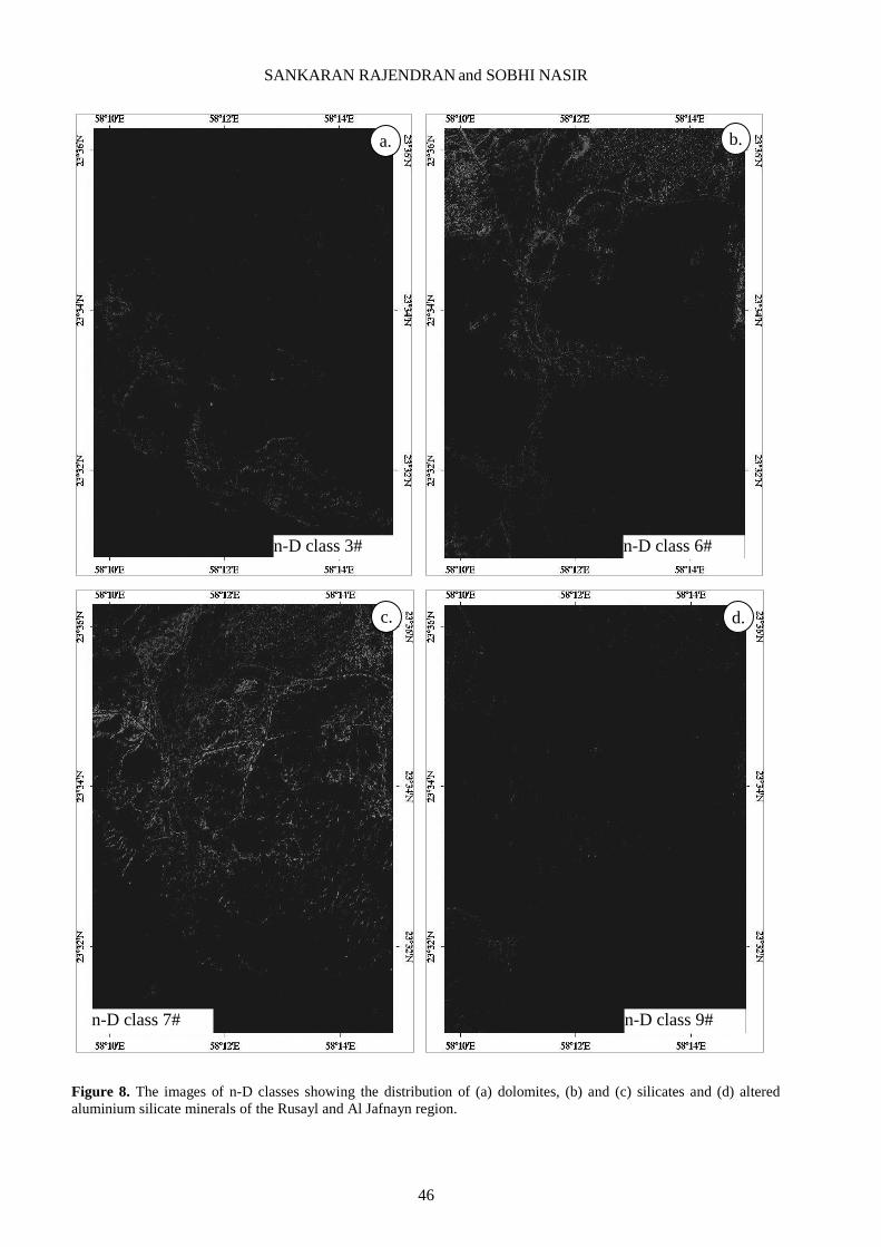
minerals of the rocks of the region, the images of n-D classes 3, 6, 7 and 9 are given in Figure 8. The regional
geological map [21] was used to verify the processed images and confirm the occurrence of minerals and rocks in the
study region.
Finally, the interpreted images were evaluated in field and laboratory studies. During the field work, traverse
based sample collection of minerals and rocks were carried out. The samples were used for spectral measurements,
megascopic, microscopic, and mineral studies at the laboratory of the Department of Earth Sciences and Central
Analytical and Applied Research Unit (CAARU), Sultan Qaboos University, to validate the occurrence of minerals in
the rocks and confirm the interpretation of images. The petrological characters of rocks and minerals of the study
region were studied under microscope by preparation of thin sections of samples. To confirm the capability of sensor
and characters of the spectral bands used in this study, the reflectance spectra of samples were measured using a PIMA
SP infrared spectrometer and studied. This instrument was fabricated for field spectroscopy by Integrated Spectronics
Pty Ltd., Australia to verify processed remote sensing data. The instrument identified and analyzed the spectral signal

SANKARAN RAJENDRAN and SOBHI NASIR
42
of minerals in the wavelength ranges from 1300 to 2500 nm with PIMA VIEW software (version 3.1). The spectral
resolution of the device was ~ 7 nm. It has a built-in wavelength calibration target plate and is capable of measuring
spectra from 10 seconds to around 5 minutes speed. Moreover, the samples used for spectral studies and other selected
samples were further analysed for identification of minerals to confirm the image interpretations, field and microscopic
studies by X-ray powder diffraction, using X'Pert PRO (P Analytical Company, The Netherlands) at CAARU, working
based on PW1710 (Cu: 1.54).
6. Mapping of rock types and recognition of minerals
The discrimination of similar rock types occurring in parts of Sultanate of Oman using ASTER spectral bands is
described by Rajendran et al. [10-12, 48] and Rajendran and Nasir [9, 19, 39, 51]. In this study, the colour composite
image developed [12, 39] and studied using the ASTER spectral bands 8 (2.295–2.365 lm), 3 (0.78–0.86 lm), and 1
(0.52–0.60 lm) of the study region is given in Figure 4. The image shows those formations rich in carbonate minerals
in shades of bright colors, discriminating them from the dark coloured sheeted dykes and ancient alluvial terrace
deposits. The alluvial fan materials which are derived from the dykes and gabbros appear in light brown, which
distinguishes them from wadi alluviums showing in shades of bright colours. However, the wadi alluviums are not
discriminated well from the adjacent source rocks rich in carbonate minerals. The alluvial fan deposits consisting of the
materials of dykes and gabbros derived from the mafic rock source region are clearly discriminated on the image. As
interpreted, the rocks and formations correlate with the geological map (Figure 2).
The SAM image (Figure 7) shows the occurrence and spatial distribution of different minerals that occur in
various proportions in different rock types of the region. The occurrence and spatial distribution of minerals within
different lithologies of the region can be identified from the selected colors of end member spectra (Figures 5a, b and
6) and interpreted with the geology of the region (Figure 2) [12, 51].
The SAM classified image (Figure 7) showed the area of 1.8117 km2 by red pixels based on n-D class #1. The
end member mean spectra of n-D class #1 (Figure 6) show the absorptions in bands 4 (1.6–1.7 μm) and band 7 (2.235–
2.285 μm). The clustered pixels along the wadi flow and built-up land regions (Figure 7) represent the presence of
vegetation. The strong absorptions in bands 4 and 7 (OH and CO3 stretched) are due to the chlorophyll content in the
vegetation occurring in these areas. The occurrence of vegetation was confirmed by the ASTER RGB (1-3) image of
the area (Figure 1b) and validated during the field check. The occurrence and spatial distribution of carbonate minerals
(calcites) over about 0.0447 km2 is shown in green, detected by n-D class #2, and about 30.3601 km
2 of dolomites are
represented in blue by the n-D class #3 [12, 51]. The spectra of both classes show carbonate absorptions (decrease in
trend) in band 8, whereas the absorption in band 5 may be due to the presence of Ca and Mg contents in the minerals.
The absorption in band 3 of the spectra may be due to the iron content present in the clay minerals (in marl) that are
associated with the dolomites, as discussed in section 7. The pixels representing the dolomite minerals are found in
areas where limestone formations occur (Figure 7).
Figure 8a shows the occurrence and spatial distribution of dolomite minerals in the lower nodular limestone and
upper nodular limestone regions (Figure 2). Such mineral-bearing carbonate formations were verified during the field
work [19] and studied in the laboratory as discussed in section 7. The minor occurrence and spatial distribution of
water and hydroxyl group bearing clay minerals in the marls which is associated in limestones were detected in the n-D
class #4 (about 2.0808 km2, in yellow) and n-D class #5 (about 0.3924 km
2 in cyan). The spectra belonging to these
minerals show absorptions in spectral bands 3, 5 and 7 due to the presence of iron, magnesium and calcium, and to the
hydroxyl group present in the minerals respectively. The minerals are found in the limestone formations that appear in
blue in the figure. The occurrence of such minerals was verified in the field where small streams originate, these
flowing both parallel and perpendicular to the limestone formations, and where the marls occur.
The n-D class #6 (12.2553 km2) and n-D class #7 (17.2129 km
2) are represented in magenta and maroon
respectively. These are more significantly distributed in the places where dyke and gabbro rocks and derived materials
are found in the wadi alluvium. The presence of such pixels are due to the occurrence of silicate minerals such as
clinopyroxene, orthopyroxene, amphibole, and plagioclase minerals, the basic constituents occurring in variable
proportions in the poorly altered rocks [12, 51]. Both the endmember spectra show poor absorptions in bands 3, 4 and 7
due to iron, magnesium and calcium, and aluminium contents in the minerals of the rock. The occurrence of pink pixels
separately from the maroon pixels, in the region of occurrence of dyke and gabbro rocks, represents the presence of
unaltered silicate minerals in the rocks. On the other hand, maroons mixed with pink colored pixels occur in areas of
wadi alluvium and clay minerals, and represent the presence of poorly altered silicates and aluminium silicate minerals.
Figures 8b and c show the occurrence and spatial distribution of silicate minerals. The n-D class #8 (0.229 km2, sea
green colored pixels) shows poor occurrences of silicate minerals and provides no significant information about their
presence in the region. The spectra show similar absorptions like n-D class #6 at bands 3, 4, 5 and 7, which may be due
to the presence of certain unaltered silicate minerals associated with other poorly altered minerals in the region. The
mean spectra of n-D class #9 (about 7.8338 km2 distinguished in the image purple pixels) occurs in the region of dyke
and gabbro rocks and the derived materials are found in the wadi alluvium. This may be due to the presence of certain
altered aluminosilicate minerals (Figure 8d) in the rocks. The n-D class #10 (about 0.0297 km2 in coral color) shows
poor occurrence of minerals and the spectra show absorption in band 4. This absorption may be due to the presence of
silica in the poorly or unaltered silicate minerals.
MINERSLS MAPPING USING REMOTE SENSING DATA
43
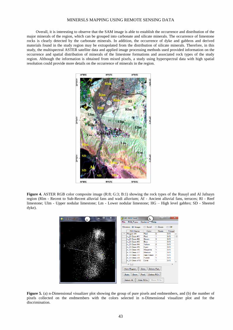
Overall, it is interesting to observe that the SAM image is able to establish the occurrence and distribution of the
major minerals of the region, which can be grouped into carbonate and silicate minerals. The occurrence of limestone
rocks is clearly detected by the carbonate minerals. In addition, the occurrence of dyke and gabbros and derived
materials found in the study region may be extrapolated from the distribution of silicate minerals. Therefore, in this
study, the multispectral ASTER satellite data and applied image processing methods used provided information on the
occurrence and spatial distribution of minerals of the limestone formations and associated rock types of the study
region. Although the information is obtained from mixed pixels, a study using hyperspectral data with high spatial
resolution could provide more details on the occurrence of minerals in the region.
Figure 4. ASTER RGB color composite image (R:8; G:3; B:1) showing the rock types of the Rusayl and Al Jafnayn
region (Rlm - Recent to Sub-Recent alluvial fans and wadi alluvium; Af - Ancient alluvial fans, terraces; Rl - Reef
limestone; Ulm - Upper nodular limestone; Lm - Lower nodular limestone; HG – High level gabbro; SD - Sheeted
dyke).
Figure 5. (a) n-Dimensional visualizer plot showing the group of pure pixels and endmembers, and (b) the number of
pixels collected on the endmembers with the colors selected in n-Dimensional visualizer plot and for the
discrimination.
a. b.
SD
SD
SD
Rlm
Rlm
Lm
Lm
Lm
Ulm
Ulm
Ulm
Rlm
Rlm
Rlm
Rlm
Af
Af
Rl
Rlm
Rlm
Rlm
Al
Jafnayn
Rusayl
HG

SANKARAN RAJENDRAN and SOBHI NASIR
44
Figure 6. Plot of endmember spectra (Class Means) of Rusayl and Al Jafnayn region.
Figure 7. SAM classified image showing the mineral distribution in the Rusayl and Al Jafnayn region (Rlm - Recent to
Sub-Recent alluvial fans and wadi alluvium; ; Rl - Reef limestone; Af - Ancient alluvial fans, terraces; Ulm - Upper
nodular limestone; Lm - Lower nodular limestone; HG – High level gabbro; SD - Sheeted dyke).
n-D Class Mean #1
n-D Class Mean #10
n-D Class Mean #9
n-D Class Mean #8
n-D Class Mean #7
n-D Class Mean #6
n-D Class Mean #5
n-D Class Mean #4
n-D Class Mean #3
n-D Class Mean #2
Lm
Ul
m
R
l
Rusayl
SD
SD
SD
Rlm
Rlm
Lm
Lm
Ul
m
Ulm
Rlm
Rlm
Rlm
Rlm
Af
Af
Rlm
Rlm
Rlm
Al
Jafnayn
Lm
HG
Rl

MINERSLS MAPPING USING REMOTE SENSING DATA
45
Table 2. Distribution of pixels in Rusayl and Al Jafnayn region in the SAM n-D classes.
7. Field and Laboratory studies
Field work was conducted to confirm the image interpretation discussed and to validate the occurrence of the
minerals and rocks of the study region. In the field, the lower and upper nodular limestone formations of Tertiary age
are exposed well on the surface and crop out at Wadi Rusayl, near to Rusayl village in the study region. Along the wadi
section, the formations exhibit nodular and massive limestones (Figure 9a and inset; S1 in Fig. 1b) with ophiolite
clasts. The formations are conformable and occur above the yellow marl (Figure 9b; S2 in Figure 1b) and marly
wackestone. The limestone formations are interbedded by multicoloured shale, sandstone and conglomerate (Figure 9c;
S3 in Figure 1b), which can be observed in the road section. The lower part of the formation consists of bivalves,
gastropods and small corals.
The upper boundary is conformable with the Rusayl Formation. The Rusayl Formation contains thick, resistant
and nodular microcrystalline limestone followed by soft multicolored shale and marl. Exposures of shales and marls
characterize the formation and are all highly weathered. The basal unit consists of poorly indurated, multicolored shale
and marl with occasional thin and microcrystalline limestones. The presence of ancient alluvial fan and terrace deposit
were verified in the field. The weathered surface materials occurring at ancient alluvial fans and terraces are a mixture
of gabbro and mafic fragmented materials. These appear dark in color and are easily distinguishable (Figure 9d; S4 in
Figure 1b). The surface materials on the terraces are loose, vary in size and are poor or unaltered (inset in Figure 9d).
The deposits are cemented by lime materials observed along the road section (Figure 9e; S5 in Figure 1b). The recent
wadi alluviums are friable, loose, vary in size and are poor or unaltered (Figure 9f; S6 in Figure 1b) on the surface
below which the materials are cemented. The occurrence of calcrete in the wadi deposits can be observed. The study of
samples under the microscope shows the frequent presence of major carbonate minerals and shells (Figure 9g, h) in the
limestone formations.
The barren exposures of doleritic and basaltic sheeted dykes and high level gabbro occurring near to Al Jafnayn
are verified in the road cuttings. The sheeted dykes vary in thickness, dark green to green in colour, fine grained and
exhibit chilled margins (Figure 10a, b and inset; S7 in Figure 1b). Epidotization is observed in the dyke rocks (Figure
10c). The vertical foliated gabbro (Figure 10d; S8 in Figure 1b) shows alternate layering of light and dark colored
felsic and mafic rich minerals (Figure 10e). Under the microscope, the dyke shows the presence of major minerals
such as plagioclase feldspar, pyroxene and magnetite (Figure 10f, g). Very few grains of olivine were observed. The
gabbro consists of mineral assemblages of olivine, hornblende, calcic-plagioclase and pyroxene (Figure 10h).
Plagioclase occurs as subhedral grains.
During the study, spectral measurements were taken over the limestone formations at different locations in the
field, and in the laboratory on the samples collected, using a portable PIMA SP infrared spectrometer to confirm the
occurrence and spectral absorption characters of minerals. The selected spectra collected over the limestones are given
in the spectral plot (Figure 11a, measurements around S2, S3 and S5 and lab samples). The collection of spectra in the
SWIR region, with a spectral resolution of 7 nm over the rocks, show narrow absorption sharp features at nearly 1400
nm, 1900 nm, and 2300 nm, and are well comparable with the studied spectra (Figure 3). The absorptions are mainly
due to the presence of major carbonate minerals, namely dolomite (Ca Mg (CO3)2), and hydroxyl group bearing clay
mineral, namely montmorrillonite in the samples. The spectra of gabbros (Figure 11b, measurements around S8 and
near S7 and lab samples) show absorptions around 2250 nm and 2350 nm. The absorption near 2250 nm may be due to
the influence of clinopyroxene minerals, and the absorptions near 2350 nm are due to the presence of OH bearing
epidote and chlorite minerals in the rock.
The study of spectra in the range of 300–3000 nm wavelength over the dyke rocks may provide more information
on the occurrence of silicate minerals. The presence of abundant minerals of dolomite in limestones and of epidote,
hornblende and chlorite minerals in gabbro rock samples were confirmed by XRD analyses (Figures 11c, d).
n-D Classesa No. of pixels % in total area Area in Km²
n-D Class Mean #1 8052 2.508 1.8117
n-D Class Mean #2 199 0.062 0.0447
n-D Class Mean #3 134934 42.021 30.3601
n-D Class Mean #4 9248 2.88 2.0808
n-D Class Mean #5 1744 0.543 0.3924
n-D Class Mean #6 54468 16.962 12.2553
n-D Class Mean #7 76502 23.824 17.2129
n-D Class Mean #8 1018 0.317 0.229
n-D Class Mean #9 34817 10.843 7.8338
n-D Class Mean #10 132 0.041 0.0297
Total 321,114 100.001 72.2504
a Unclassified: 0 points (0.000%) (0.0000 km
2).
SANKARAN RAJENDRAN and SOBHI NASIR
46
Figure 8. The images of n-D classes showing the distribution of (a) dolomites, (b) and (c) silicates and (d) altered
aluminium silicate minerals of the Rusayl and Al Jafnayn region.
n-D class 3#
a. b.
n-D class 6#
b.
d.
n-D class 9#
d. c.
n-D class 7#
c.

MINERSLS MAPPING USING REMOTE SENSING DATA
47
Figure 9. Field photographs showing (a) the massive Rusayl limestone formation (S1in Figure 1b), (b) the occurrence
of friable yellow marls and clays (S2 in Figure 1b), (c) the occurrences of interbedded multicoloured shale, sandstone
and conglomerate (S3 in Figure 1b), (d) the ancient alluvial terrace materials (S4 in Figure 1b), (e) the ophiolite clasts
in the limestone cements (S5 in Figure 1b), (f) the recent alluvial materials (S6 in Figure 1b), and the
microphotographs show (g) the dolomite minerals of massive limestones and (h) the fossil limestones (nicols crossed,
25x).
e.
Ancient alluvial deposits
d.
Ancient alluvial terrace Ancient alluvial
materials
b.
Yellow marls and clay deposits
a.
Massive thick limestone of
Rusayl formation
massive limestones
f.
Recent Wadi Alluvium
g.
Dolomite
Carbonate shell Dolomite
Carbonate shell
h.
Shale
Sandstone
Conglomerate
c.

SANKARAN RAJENDRAN and SOBHI NASIR
48
Figure 10. Field photographs show the occurrence of (a) and (b) the sheeted dykes (S7 in Figure 1b), (c) the chilled
margin of the dykes (S7 in Figure 1b), (d) the high level foliated gabbros (S8 in Figure 1b), (e) the foliated gabbro
shows the presence of mafic and felsic minerals (S8 in Figure 1b), and the microphotographs show the minerals of
sheeted dykes (f) nicols parallel (35x) and (g) nicols crossed (35x) and (h) gabbro (nicols crossed, 25x).
b.
Chilled margin in
sheeted dykes
e.
Foliated gabbro with mafic and
felsic minerals
a.
Sheeted dykes of Jafanyn
d.
High level foliated gabbro
c.
Sheeted dykes epidotized
f.
Plagioclase
Pyroxene
Dark spots are
magnetite
g.
Plagioclase
Pyroxene
Dark spots are
magnetite
h.
Plagioclase
Pyroxene
Hornblende

MINERSLS MAPPING USING REMOTE SENSING DATA
49
Figure 11. Laboratory spectral plots showing (a) carbonates absorptions in limestones, (b) epidote and chlorite OH
absorptions in gabbros. The results of XRD of the limestones show the presence of abundance of dolomite minerals
and the gabbros show the occurrence of epidote, hornblende and chlorite minerals in Rusayl and Al Jafnayn region.
8. Conclusion
In the present study, the occurrence of minerals in the rocks of the Rusayl and Al Jafnayn regions, Muscat, the
Sultanate of Oman is studied using the ASTER VNIR-SWR spectral bands, and by the SAM supervised classification
image processing method. The results of SAM clearly show the occurrence of carbonate minerals in limestone bearing
rocks and silicate minerals in the dyke rocks, and their spatial distributions. The image interpretations are verified in
the field by identification of minerals and rocks, and confirmed through laboratory analyses. The study has proved that
the technique is time and cost-effective in comparison to the time consuming classical field mapping of minerals and
rocks, and can be used in the difficult terrain of other similarly arid regions not reachable for conventional geological
mapping and where it is difficult to do exhaustive sampling.
9. Acknowledgements
The authors are thankful to NASA Land Processes Distributed Active Archive Center User Services, USGS Earth
Resources Observation and Science (EROS) Center (https://LPDAAC.usgs.gov) for providing the ASTER data. The
study is supported by Sultan Qaboos University Internal Grant IG/SCI/ETHS/14/02. XRD analytical help was extended
by Mr. Saif Amer Al-Maamari, CAARU. The authors are thankful to the anonymous reviewers and to the editor of the
journal for their valuable reviews and for providing comments and suggestions that have helped to improve the
manuscript.
Position [°2Theta] (Copper (Cu))
10 20 30 40 50 60
Counts
0
200
400
600
Ho
rn
ble
nd
e, m
ag
nesia
n
Ch
lorit
e I
Ib-4
Ep
ido
te
Ho
rn
ble
nd
e, m
ag
nesia
n
Alb
ite lo
w; H
orn
ble
nd
e, m
ag
nesia
n; Ep
ido
te; C
hlo
rit
e I
Ib-4
Alb
ite lo
w; M
icro
clin
e (
interm
ed
iate); C
hlo
rit
e I
Ib-4
Alb
ite lo
w; Ep
ido
te; M
icro
clin
e (
interm
ed
iate)
Alb
ite lo
w; C
hlo
rit
e I
Ib-4
Ep
ido
te; M
icro
clin
e (
interm
ed
iate)
Alb
ite lo
w; Ep
ido
te; C
hlo
rit
e I
Ib-4
Alb
ite lo
w; H
orn
ble
nd
e, m
ag
nesia
n; Ep
ido
te
Ho
rn
ble
nd
e, m
ag
nesia
n; Ep
ido
te; M
icro
clin
e (
interm
ed
iate); C
hlo
rit
e I
Ib-4
Alb
ite lo
w; Ep
ido
te; M
icro
clin
e (
interm
ed
iate)
Alb
ite lo
w; Ep
ido
te
Ho
rn
ble
nd
e, m
ag
nesia
n
Ho
rn
ble
nd
e, m
ag
nesia
nA
lbit
e lo
w; H
orn
ble
nd
e, m
ag
nesia
nA
lbit
e lo
w; Ep
ido
te; M
icro
clin
e (
interm
ed
iate); C
hlo
rit
e I
Ib-4
Ep
ido
te; M
icro
clin
e (
interm
ed
iate)
Ho
rn
ble
nd
e, m
ag
nesia
n; Ep
ido
te; M
icro
clin
e (
interm
ed
iate); C
hlo
rit
e I
Ib-4
Ho
rn
ble
nd
e, m
ag
nesia
n
Ho
rn
ble
nd
e, m
ag
nesia
n; Ep
ido
te; M
icro
clin
e (
interm
ed
iate)
Alb
ite lo
w; H
orn
ble
nd
e, m
ag
nesia
n; M
icro
clin
e (
interm
ed
iate); C
hlo
rit
e I
Ib-4
Alb
ite lo
w; H
orn
ble
nd
e, m
ag
nesia
n; Ep
ido
te
Alb
ite lo
w; H
orn
ble
nd
e, m
ag
nesia
n; Ep
ido
te; M
icro
clin
e (
interm
ed
iate); C
hlo
rit
e I
Ib-4
Ho
rn
ble
nd
e, m
ag
nesia
n; Ep
ido
te; M
icro
clin
e (
interm
ed
iate); C
hlo
rit
e I
Ib-4
Ho
rn
ble
nd
e, m
ag
nesia
n; Ep
ido
te; M
icro
clin
e (
interm
ed
iate); C
hlo
rit
e I
Ib-4
AUTO_CAARU-13-0098-I (GREEN SCH1)
d.
Ls 5
Ls 34
Ls 9
Ls 27
Ls 6
Ls 7
Line of carbonate
absorption
Ls 29
a. b.
Ga27
Line of epidote and
chlorite absorptions
Ga 21
Ga 25
Ga 3
Ga 6
Ga 9
Position [°2Theta] (Copper (Cu))
10 20 30 40 50 60
Counts
0
400
1600
Quart
z lo
wD
olo
mite
Dolo
mite
Quart
z lo
w
Dolo
mite
Dolo
mite
Dolo
mite
Dolo
mite
Quart
z lo
w
Dolo
mite
Dolo
mite
Dolo
mite
Dolo
mite
Dolo
mite;
Quart
z lo
wD
olo
mite
Dolo
mite
Dolo
mite;
Quart
z lo
w
Dolo
mite
Dolo
mite
Dolo
mite
Dolo
mite Dolo
mite
S5
c.

SANKARAN RAJENDRAN and SOBHI NASIR
50
References
1. Abrams, M.J., Rothery, D.A. and Pontual, A. Mapping in the Oman ophiolite using enhanced landsat thematic
mapper images. Tectonophys., 1988, 151, 387-401. 2. Bedell, R.L. Geological mapping with ASTER satellite: new global satellite data that is a significant leap in
remote sensing geologic and alteration mapping. Special Publication Geol. Soc. Nevada, 2001, 33, 329-334. 3. Ramadan, T. and Kontny, A. Mineralogical and structural characterization of alteration zones detected by orbital
remote sensing at Shalatein District, SE Desert, Egypt. J. Afr. Earth Sci., 2004, 40, 89-99. 4. Gad, S. and Kusky, T.M. Lithological mapping in the eastern desert of Egypt, the Barramiya area, using landsat
thematic mapper (TM). J. Afr. Earth Sci., 2006, 44, 196-202. 5. Zhang, X., Pazner, M., Duke, N. Lithologic and mineral information extraction for gold exploration using ASTER
data in the south Chocolate Mountains (California). Photogrammetry & Remote Sensing, 2007, 62, 271-282. 6. Amer, R., Kusky, T.M. and Ghulam, A. Lithological mapping in the central eastern desert of Egypt using ASTER
data. J. Afr. Earth Sci., 2010, 56(2–3), 75-82.
7. Rajendran, S., Thirunavukkarasu. A. Balamurugan. G. and Shankar. K. Discrimination of iron ore deposits of
granulite terrain of Southern Peninsular India using ASTER data. J. Asian Earth Sci., 2011a, 41, 99-106. 8. Tangestani, M.H., Jaffari, L. Vincent, R.K. and Maruthi, S.B.B. Spectral characterization and ASTER-based
lithological mapping of an ophiolite complex: A case study from Neyriz ophiolite, SW Iran. Remote Sensing
Environ., 2011, 115, 2243-2254. 9. Rajendran, S. and Nasir, S. Aster spectral analysis of ultramafic lamprophyres (carbonatites and aillikites) within
the Batain nappe, northeastern margin of Oman - a proposal developed for spectral absorption. Int. J. Remote
Sensing, 2013, 34(8), 2763-2795. 10. Rajendran, S., Hersi, Al-Harthy, O.S., Al-Wardi, A.R.., El-Ghali, A.M. and Al-Abri A.H. Capability of Advanced
Spaceborne Thermal Emission and Reflection Radiometer (ASTER) on discrimination of carbonates and
associated rocks and mineral identification of eastern mountain region (Saih Hatat Window) of Sultanate of Oman.
Carbonates and Evaporites, 2011b, 26, 351-364. 11. Rajendran, S., Al-Khirbash, S., Nasir, S., Al-Abri, A.H., Kusky, T.M. and Ghulam, A. ASTER detection of
chromite bearing mineralized zones in Semail Ophiolite Massifs of the northern Oman mountain: exploration
strategy. Ore Geol. Rev., 2012, 44,121-135. 12. Rajendran, S., Nasir, S., Kusky, T.M., Ghulam, A., Gabr, S. and El-Ghali, M. Detection of hydrothermal
mineralized zones associated with Listwaenites rocks in the Central Oman using ASTER data. Ore Geo. Rev.,
2013, 53, 470-488. 13. Pinet, P., Harris, E., Python, M., Ceuleneer, G., Launeau, P., Daydou, Y., Cord, A., Al Azri, H., Chabrillat, S.,
Lenot, X. and Chevrel, S. Hyperspectral Remote Sensing Approach for Rock Surface Mineralogy Mapping in Arid
Environment. IUGG XXXIII General Assembly, International Union of Geodesy and Geophys. Sapporo, Japan,
2003. 14. Pinet, P.C., Clenet, H., Rosemberg, C., Ceuleneer, G., Heuripeau, F., Harris, E., Daydou, Y., Baratoux, D.,
Chevrel, S., Launeau, P., Combes, J-P., Lemouelic, S. and Sotin, C. Mantle Rock Surface Mineralogy Mapping in
Arid Environment from Imaging Spectroscopy: The Case of the Maqsad Peridotitic Massif in Oman and
Implications for the Spectroscopic Study of Exposed Mafic Units on Mars, LPSC 37th, Houston, # 1346, 2006. 15. Combe, J.P., Launeau, P. Pinet, P. Despan, D. Harris, E. Ceuleneer, G. and Sotin, C. Mapping of an ophiolite
complex by high–resolution visible-infrared spectrometry. Geochem. Geophys. Geosystems, 2006, 7(8), 1-11. 16. Clenet, H., Pinet, Daydou, P.C. Heuripeau, Y. Rosemberg, F. and C. Ceuleneer, G. A sytematic testing approach
using the Modified Gaussian Model (MGM) for mafic mineralogy mapping in natural conditions (Earth, Mars).
Proc. Lunar Planetary Sci. Conf. XXXIX. 2008. 17. Roy, R., Launeau, P. Carrere, V. Pinet, P.C. Ceuleneer, G. Clenet, H. Daydou, Y. Girardeau, J. and Amri, I.
Geological mapping strategy using VNIR hyperspectral remote sensing: application to the Oman ophiolite (Sumail
Massif). Geochem. Geophys. Geosystems, 10: Q02004. doi:10.1029/2008GC002154, 2008. 18. Clenet, H., Ceuleneer, G., Pinet, P., Abily, B., Daydou, Y., Harris, E., Amri, I. and Dantas, C. Thick sections of
layered ultramafic cumulates in the Oman ophiolite revealed by an airborne hyperspectral survey: petrogenesis and
relationship to mantle diapirism. Lithos., 2010, 114, 265–281. 19. Rajendran, S. and Nasir, S. ASTER spectral sensitivity of carbonate rocks - study in sultanate of Oman, Adv.
Space Res., 2014b, 53, 656-673. 20. Robertson A.H.F. and Searle, M.P. The Northern Oman Tethyan Continental Margin: stratigraphy, structure,
concepts and controversies, the geology and tectonics of the Oman region. The Geol. Soc., London, 1990, 49, 3-25. 21. Ministry of Petroleum and Minerals, Geological Map, Oman (1:100,000). SEEB Sheet NF 40-3C, 1986. 22. Gupta, R.P. Remote Sensing Geology. 2nd Ed., Springer, Heidelberg, 2003. 23. Clark, R.N., King, T.V.V. Klejwa, M. Swayze, G.A. and Vergo, N. High spectral resolution reflectance
spectroscopy of minerals. J. Geophys. Res., 1990, 95, 12653-12680. 24. Van der MEER, F. Spectral reflectance of carbonate mineral mixtures and bidirectional reflectance theory:
quantitative analysis techniques for application in remote sensing. Remote Sensing Rev., 1995, 13, 67-94.

MINERSLS MAPPING USING REMOTE SENSING DATA
51
25. Clark, R.N. and Roush, T.L. Reflectance spectroscopy: quantitative analysis techniques for remote sensing
applications. J. Geophy. Res., 1984, 89, 6329-6340. 26. Gaffey, S.J. Reflectance spectroscopy in the visible and near infrared (0.35-2.55 microns): applications in
carbonate petrology. Geol., 1985, 13, 270-273. 27. Gaffey, S.J. Spectral reflectance of carbonate minerals in the visible and near infrared (0.35-2.55 microns): calcite,
aragonite, and dolomite. Am. Miner., 1986, 71, 151-162 28. Gaffey, S.J. Spectral reflectance of carbonate minerals in the visible and near infrared (0.35-2.55 microns):
anhydrous carbonate minerals. J. Geophys. Res., 1987, 92(B2), 1429-1440. 29. Crowley, J.K. Visible and near-infrared spectra of carbonate rocks: Reflectance variations related to petrographic
texture and impurities. J. Geophys. Res., 1986, 91(B5), 5001-5012. 30. Hunt, G.R. Spectroscopic Properties of Rocks and Minerals. In: Carmichael, R.S. (Ed.), Handbook of Physical
Properties of Rock. CRC Press, Boca Raton, 1982, 1, 295-385. 31. Hunt, G.R. Spectral signatures of particulate minerals in the visible and near infrared. Geophys. 1977, 42,501–513. 32. Hunt, G.R. and Salisbury, J.W. Visible and near infrared spectra of minerals and rocks: II. Carbonates. Mod.
Geol., 1971, 2, 23-30. 33. Hunt, G.R. and Salisbury, J.W. Visible and near infrared spectra of minerals and rocks: I. Silicate minerals. Mod.
Geol., 1970, 1, 283-300. 34. Hunt, G.R., Salisbury, J.W. and Lenhoff, C.J. Visible and near-infrared spectra of minerals and rocks. IX, Basic
and ultrabasic igneous rocks, Mod. Geol., 1974, 5, 15-22. 35. Hunt, G.R. and Ashley, P. Spectra of altered rocks in the visible and near infrared. Eco. Geol.1979, 74, 1613-1629. 36. Blom, R.G., Abrams, M.J. and Adams, H.G. Spectral reflectance and discrimination of plutonic rocks in the 0.45
to 2.45 μm region. J. Geophys. Res., 1980, 85, 2638-2648. 37. Hunt, G.R. Near-infrared (1.3-2.4) spectra of alteration minerals-potential for use in remote sensing. Geophys.,
1979, 44(12), 1974-1986. 38. Clark. R.N. Spectroscopy of Rock and Minerals and Principles of Spectroscopy. In: Rencz, A.N. (Ed.), Remote
Sensing for the Earth Sciences: Manual of Remote Sensing. 3th Ed., Vol.3, John Wiley & Sons, New York, 1999,
3-58. 39. Rajendran, S. and Nasir, S. ASTER mapping of limestone formations and study of caves, springs and depressions
in parts of Sultanate of Oman, Environ. Earth Sci., 2014a, 71, 133-146. 40. Mars J.C. and Rowan L.C. Spectral assessment of new ASTER SWIR surface reflectance data products for
spectroscopic mapping of rocks and minerals. Remote Sensing Environ., 2010, 114, 2011-2025. 41. Gad, S. Kusky, T.M. ASTER spectral ratioing for lithological mapping in the Arabian–Nubian shield, the
Neoproterozoic Wadi Kid area, Sinai, Egypt. Gondwana Res., 2007, 11(3), 326-335. 42. Rowan, L.C. and Mars, J.C. Lithologic mapping in the Mountain Pass Area, California using advanced spaceborne
thermal emission and reflection radiometer (ASTER) Data. Remote Sensing Environ., 2003, 84, 350-66. 43. Ninomiya, Y. Mapping quartz, carbonate minerals and mafic-ultramafic rocks using remotely sensed multispectral
thermal infrared ASTER data. SPIE, 2002, 4710, 191-202. 44. Abdeen, M.M., Thurmond, A.K. Abdelsalam, M.G. and Stern, R.J. Use of TERRA ASTER band-ratio images for
geological mapping in arid regions: the Neoproterozoic Allaqi suture, Egypt. J. Remote. Sens. & Space Sci., 2002,
5, 19-40. 45. Galvao, L.S., Filho, R.A. and Vitorello, C. Spectral discrimination of hydrothermal altered materials using ASTER
Short-wave infrared bands, evaluation in a tropical Savannah Environment. Inter. J. Appl. Earth Obs. Geoinfor.,
2005, 7, 107-114. 46. Mars, J.C. and Rowan, L.C. Regional mapping of phyllic- and argillic-altered rocks in the Zagros magmatic arc,
Iran, using Advanced Spaceborne Thermal Emission and Reflection Radiometer (ASTER) data and logical
operator algorithms. Geosphere, 2006, 2(3), 161-186. 47. Abrams, M. and Hook, S.J. Simulated ASTER data for geologic studies. IEEE Trans. Geosci. Remote Sens., 1995,
33(3). 48. Rajendran, S., Nasir, S. Kusky T.M. and Al-Khirbash, S. Remote sensing based approach for mapping of CO2
sequestered regions in Semail Ophiolite Massifs of the Sultanate of Oman. Earth Sci. Rev., 2014, 135, 122-140. 49. Kruse, F.A., Boardman, J.W. and Hunnington, J.F. Comparison of airborne hyperspectral data and EO-1 Hyperion
for mineral mapping. IEEE Trans. Geosci. Remote Sensing, 2003, 41(6), 1388-1400. 50. Gabr, S., Ghulam, A. and Kusky, T. Detecting areas of high-potential gold mineralization using ASTER data. Ore
Deposit Rev., 2010, 38, 59-69. 2010. 51. Rajendran, S and Nasir, S. 2014c. Hydrothermal altered serpentinized zone and a study of Ni-magnesioferrite-
magnetite-awaruite occurrences in Wadi Hibi, Northern Oman Mountain: discrimination through ASTER
mapping, Ore Geol. Rev., 62, 211-226. 52. Hecker, C.A., van der Meijde, M., van der Werff, H.M.A. and van der Meer, F.D. Assessing the influence of
reference spectra on synthetic SAM classification results. IEEE Trans. Geosci. and Remote Sensing, 2008, 46(12),
4162-4172.

SANKARAN RAJENDRAN and SOBHI NASIR
52
53. Kruse, F.A., Lefkoff, A.B. Boardman, J.B. Heidebreicht, K.B. Shapiro, A.T. and Barloon, P.J. The spectral image
processing system (SIPS)-interactive visualization and analysis of imaging spectrometer data. Remote Sensing
Environ., 1993, 44: 145-163.
54. Boardman, J.W. and Kruse, F.A. Automated spectral analysis: a geological example using AVIRIS data, north
Grapevine Mountains, Nevada. Proc., ERIM Tenth Thematic Conference on Geologic Remote Sensing.
Environmental Research Institute of Michigan, Ann Arbor, MI, 1994, I-407-I-418.
Received 2 March 2014
Accepted 15 June 2014