© 2013 Pearson Education, Inc. Connective Tissue Most abundant and widely distributed of primary...
31
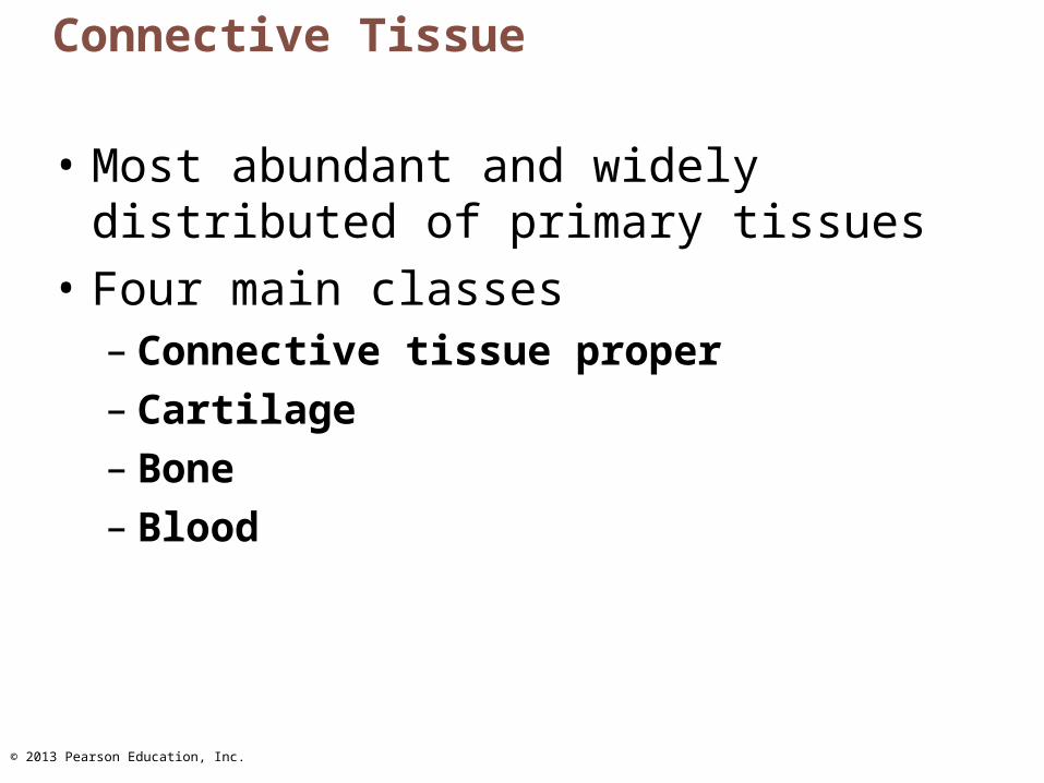
© 2013 Pearson Education, Inc. Connective Tissue • Most abundant and widely distributed of primary tissues • Four main classes – Connective tissue proper – Cartilage – Bone – Blood
-
Upload
brianna-sanders -
Category
Documents
-
view
216 -
download
0
description
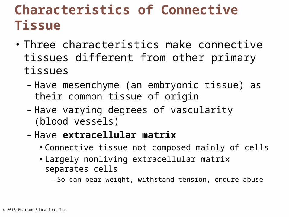
© 2013 Pearson Education, Inc. Characteristics of Connective Tissue Three characteristics make connective tissues different from other primary tissues –Have mesenchyme (an embryonic tissue) as their common tissue of origin –Have varying degrees of vascularity (blood vessels) –Have extracellular matrix Connective tissue not composed mainly of cells Largely nonliving extracellular matrix separates cells –So can bear weight, withstand tension, endure abuse
Transcript of © 2013 Pearson Education, Inc. Connective Tissue Most abundant and widely distributed of primary...
© 2013 Pearson Education, Inc.
Connective Tissue
• Most abundant and widely distributed of primary tissues
• Four main classes– Connective tissue proper– Cartilage– Bone – Blood

© 2013 Pearson Education, Inc.
Major Functions of Connective Tissue
• Binding and support• Protecting• Insulating• Storing reserve fuel• Transporting substances (blood)
© 2013 Pearson Education, Inc.
Characteristics of Connective Tissue
• Three characteristics make connective tissues different from other primary tissues– Have mesenchyme (an embryonic tissue) as
their common tissue of origin– Have varying degrees of vascularity (blood
vessels)– Have extracellular matrix
• Connective tissue not composed mainly of cells• Largely nonliving extracellular matrix separates
cells– So can bear weight, withstand tension, endure abuse

© 2013 Pearson Education, Inc.
Structural Elements of Connective Tissue
• Three elements– Ground substance– Fibers– Cells
• Composition and arrangement varies in different connective tissues

© 2013 Pearson Education, Inc.
Ground Substance
• Unstructured material that fills space between cells– Medium through which solutes diffuse between blood
capillaries and cells• Components
– Interstitial fluid– Cell adhesion proteins ("glue" for attachment) – Proteoglycans
• Protein core + large polysaccharides (chrondroitin sulfate and hyaluronic acid)
• Trap water in varying amounts, affecting viscosity of ground substance

© 2013 Pearson Education, Inc.
Connective Tissue Fibers
• Three types of fibers provide support– Collagen
• Strongest and most abundant type• Tough; provides high tensile strength
– Elastic fibers• Networks of long, thin, elastin fibers that allow for
stretch and recoil– Reticular
• Short, fine, highly branched collagenous fibers (different chemistry and form than collagen fibers)
• Branch, forming networks that offer more "give"
© 2013 Pearson Education, Inc.
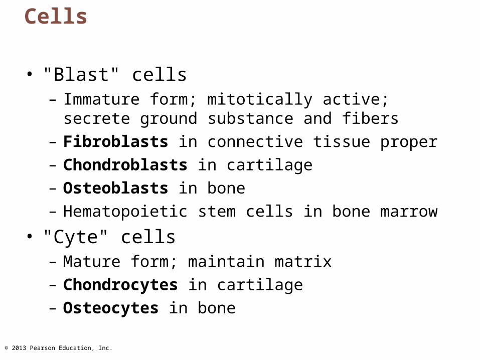
Cells
• "Blast" cells– Immature form; mitotically active; secrete ground
substance and fibers – Fibroblasts in connective tissue proper– Chondroblasts in cartilage– Osteoblasts in bone– Hematopoietic stem cells in bone marrow
• "Cyte" cells– Mature form; maintain matrix– Chondrocytes in cartilage – Osteocytes in bone

© 2013 Pearson Education, Inc.
Other Cell Types in Connective Tissues
• Fat cells– Store nutrients
• White blood cells– Neutrophils, eosinophils, lymphocytes– Tissue response to injury
• Mast cells– Initiate local inflammatory response against foreign
microorganisms they detect• Macrophages
– Phagocytic cells that "eat" dead cells, microorganisms; function in immune system
© 2013 Pearson Education, Inc.
Types of Connective Tissues: Connective Tissue Proper• All connective tissues except bone, cartilage and
blood• Two subclasses
– Loose connective tissues• Areolar• Adipose• Reticular
– Dense connective tissues (also called fibrous connective tissues)
• Dense regular• Dense irregular• Elastic
© 2013 Pearson Education, Inc.
Areolar Connective Tissue
• Support and bind other tissues– Universal packing material between other tissues
• Most widely distributed• Provide reservoir of water and salts• Defend against infection• Store nutrients as fat• Fibroblasts• Loose arrangement of fibers• Ground substance• When inflamed soaks up fluid edema

© 2013 Pearson Education, Inc.
Adipose Tissue
• White fat – Similar to areolar but greater nutrient storage– Cell is adipocyte
• Stores nutrients– Scanty matrix– Richly vascularized– Shock absorption, insulation, energy storage
• Brown fat– Use lipid fuels to heat bloodstream not to
produce ATP

© 2013 Pearson Education, Inc.
Reticular Connective Tissue
• Resembles areolar but fibers are reticular fibers
• Fibroblasts called reticular cells• Supports free blood cells in lymph nodes,
the spleen, and bone marrow

© 2013 Pearson Education, Inc.
Dense Regular Connective Tissue
• Closely packed bundles of collagen fibers running parallel to direction of pull– White structures with great resistance to
pulling– Fibers slightly wavy so stretch a little
• Fibroblasts manufacture fibers and ground substance
• Few cells• Poorly vascularized

© 2013 Pearson Education, Inc.
Dense Irregular Connective Tissue
• Same elements but bundles of collagen thicker and irregularly arranged
• Resists tension from many directions– Dermis– Fibrous joint capsules– Fibrous coverings of some organs

© 2013 Pearson Education, Inc.
Connective tissue proper: dense connective tissue, elastic
Description: Dense regularconnective tissue containing ahigh proportion of elastic fibers.
Function: Allows tissue to recoilafter stretching; maintains pulsatileflow of blood through arteries; aidspassive recoil of lungs followinginspiration.
Location: Walls of large arteries;within certain ligaments associatedwith the vertebral column; withinthe walls of the bronchial tubes.
Photomicrograph: Elastic connective tissuein the wall of the aorta (250x).
Aorta
Heart
Elasticfibers
Figure 4.8f Connective tissues.

© 2013 Pearson Education, Inc.
Cartilage
• Chondroblasts and chondrocytes• Tough yet flexible• Lacks nerve fibers• Up to 80% water - can rebound after compression• Avascular
– Receives nutrients from membrane surrounding it• Perichondrium
• Three types of cartilage:– Hyaline cartilage– Elastic cartilage– Fibrocartilage

© 2013 Pearson Education, Inc.
Bone
• Also called osseous tissue• Supports and protects body structures• Stores fat and synthesizes blood cells in cavities• More collagen than cartilage• Has inorganic calcium salts• Osteoblasts produce matrix• Osteocytes maintain the matrix• Osteons – structural units• Richly vascularized
© 2013 Pearson Education, Inc.
Blood
• Most atypical connective tissue – is a fluid• Red blood cells most common cell type• Also contains white blood cells and
platelets• Fibers are soluble proteins that precipitate
during blood clotting• Functions in transport

© 2013 Pearson Education, Inc.
Muscle Tissue
• Highly vascularized• Responsible for most types of movement• Three types
– Skeletal muscle tissue• Found in skeletal muscle• Voluntary
– Cardiac muscle tissue• Found in walls of heart• Involuntary
– Smooth muscle tissue• Mainly in walls of hollow organs other than heart• Involuntary

© 2013 Pearson Education, Inc.
Nervous Tissue
• Main component of nervous system– Brain, spinal cord, nerves– Regulates and controls body functions
• Neurons– Specialized nerve cells that generate and conduct
nerve impulses• Neuroglia
– Supporting cells that support, insulate, and protect neurons

© 2013 Pearson Education, Inc.
Covering and Lining Membranes
• Composed of at least two primary tissue types– An epithelium bound to underlying connective
tissue proper– Are simple organs
• Three types– Cutaneous membranes– Mucous membranes– Serous membranes
© 2013 Pearson Education, Inc.
Cutaneous Membranes
• Skin• Keratinized stratified squamous epithelium
(epidermis) attached to a thick layer of connective tissue (dermis)
• Dry membrane
© 2013 Pearson Education, Inc.
Mucous Membranes
• Mucosa indicates location not cell composition• All called mucosae
– Line body cavities open to the exterior (e.g., Digestive, respiratory, urogenital tracts)
• Moist membranes bathed by secretions (or urine)
• Epithelial sheet lies over layer of connective tissue called lamina propria
• May secrete mucus

© 2013 Pearson Education, Inc.
Serous Membranes
• Serosae—found in closed ventral body cavity• Simple squamous epithelium (mesothelium)
resting on thin areolar connective tissue • Parietal serosae line internal body cavity walls• Visceral serosae cover internal organs• Serous fluid between layers• Moist membranes• Pleurae, pericardium, peritoneum

© 2013 Pearson Education, Inc.
Tissue Repair
• Necessary when barriers are penetrated• Cells must divide and migrate• Occurs in two major ways
– Regeneration• Same kind of tissue replaces destroyed tissue• Original function restored
– Fibrosis• Connective tissue replaces destroyed tissue• Original function lost

© 2013 Pearson Education, Inc.
Steps in Tissue Repair: Step 1
• Inflammation sets stage– Release of inflammatory chemicals– Dilation of blood vessels– Increase in vessel permeability– Clotting occurs
© 2013 Pearson Education, Inc.
Steps in Tissue Repair: Step 2
• Organization restores blood supply– The blood clot is replaced with granulation
tissue– Epithelium begins to regenerate– Fibroblasts produce collagen fibers to bridge
the gap– Debris is phagocytized
© 2013 Pearson Education, Inc.
Steps in Tissue Repair: Step 3
• Regeneration and fibrosis– The scab detaches– Fibrous tissue matures; epithelium thickens
and begins to resemble adjacent tissue– Results in a fully regenerated epithelium with
underlying scar tissue

© 2013 Pearson Education, Inc.
Regenerative Capacity in Different Tissues
• Regenerate extremely well– Epithelial tissues, bone, areolar connective tissue,
dense irregular connective tissue, blood-forming tissue
• Moderate regenerating capacity– Smooth muscle and dense regular connective tissue
• Virtually no functional regenerative capacity– Cardiac muscle and nervous tissue of brain and
spinal cord– New research shows cell division does occur
• Efforts underway to coax them to regenerate better

© 2013 Pearson Education, Inc.
Developmental Aspects
• Primary germ layers– Superficial to deep: ectoderm, mesoderm,
and endoderm– Formed early in embryonic development– Specialize to form the four primary tissues
• Nerve tissue arises from ectoderm• Muscle and connective tissues arise from
mesoderm• Epithelial tissues arise from all three germ layers

© 2013 Pearson Education, Inc.
Aging Tissues
• Normally function well through youth and middle age if adequate diet, circulation, and infrequent wounds and infections
• Epithelia thin with increasing age so more easily breached
• Tissue repair less efficient• Bone, muscle and nervous tissues begin to
atrophy• DNA mutations possible increased cancer
risk