


Languages
Pages
Legal

VICKERS LIMITED • VICKERS INSTRUMENTSHAXBY ROAO • VORK
PURLEV W»Y CROYDON CRS 4HH
The M15c biological micro-scope is a versatile instrument,suitable in its simplest form forschools or, with the extensiverange and interchangeability ofaccessories, for research work.
The illumination is by either a6 volt 15 watt Kohler type illum-inator, a 25 watt mains voltageilluminator or a mirror for anexternal source. With the Kohlertype illuminator the transformeris built into the base of theinstrument with a variable rheo-stat and a colour temperaturecontrol meter.
A very high intensity source—100 watts at 12 volts, sufficientfor projection of high powerphase or polarizing images—isavailable from the tungsten halo-gen projection base, especiallydesigned to take the M15c micro-scope. With this light sourcehigh colour temperatures, up to3,300°K, can be obtained—animportant advantage for colourphotomicrography.
The precision ball bearingfocusing slides are controlledby concentric coarse and fine
f o c u s i n gknobs, the finecontrol beinggraduated to 2microns.All objectivesare par-focaland par-centraland the higher
powers of 20X magnification andabove incorporate a spring-loadedanti-crash device for specimenprotection.
All the viewing heads for theM15cmicroscopeareinterchange-able and the accessories availableinclude a photo-visual or projec-tion trip mirror unit, which allowsall the light to be deflected to theeye or alternatively to a cameraor a 6 inch diameter projectionscreen. A magnification changerwith a focusing Bertrand lens andgiving magnifications of 1X, 1.5Xand 2.5X can be fitted.
Let us send you further Information
Jnl. of Cell Sci., Vol. 2, No. 4
ELECTRONMICROSCOPISTS!
We can now offerover 70 chemicals for Electron Microscopy
from stock, including:
ACCELERATORSFIXATIVESPOLYMERSRESINS
CURING AGENTSCATALYSTSEPOXIDES'STAINS'
UNSATURATED MONOMERSAlso:MAXTAFORM GRIDS GRID BOXESGELATIN CAPSULES BEEM CAPSULESWATCHMAKERS BOW-SPRING
FORCEPS SCISSORS
Literature giving full detailsof this new service from CURRS is
available on request
GEORGE T. GURR LTD.136-14-4, New King's Road, London, S.W.6
Tel: RENown 5482/4Cables: MICROSTAIN. London, S.W.6
Details of Advertisement
rates and data for
Journal of Cell Science
may be obtained from:
Mr D. J. French
Cambridge University Press
Bentley House, P.O. Box 92
200 Euston Road
London, N.W. 1
or from
Miss Gretta Goldenman
Cambridge University Press
32 East 57th Street
New York, N.Y. 10022
The Journal ofGeneral Microbiology
Volume 49, Part 2, November 1967
J. Margaret Eadie Studies on the ecology ofcertain rumen ciliate protozoa
K. J. Bent Electrophoresis of proteins of 3Penicillium species on acrylamide gels
N. C. Khan & S. P. Sen Genetic transforma-tion in Pseudomonas
Margaret J. Thornley A taxonomic study ofAcinetobacter and related genera
R. Lahoz, F. Reyes, R. Beltra & C. Garcfa-Tapia The autolysis of Aspergillus terreus in aphysiologically acid medium
M. Polsinelli & S. Barlati Effect of periodateon competence in Bacillus subtilis
T. D. Hennessey Inducible /Mactamase inEnterobacter
Mary Barnes & M. S. Parker Use of theCoulter Counter to measure osmotic effectson the swelling of mould spores duringgermination
B. E. B. Moseley The isolation and some pro-perties of radiation-sensitive mutants ofMicrococcus radiodurans
J. Pearce & N. G. Carr The metabolism ofacetate by the blue-green algae, Anabaenavariabifis and Anacystis nidulans
O.Caryl Wallis & G. S. Coleman Incor-poration of 14C-labelled components ofEscherichia coli and of amino acids by Isotrichaintestinalis and Isotricha prostoma from thesheep rumen
O. W . Prozesky Arginine synthesis in Pro-teus mirabilis
Ursula B. Pearce & B. A. D. Stocker Phasevariation of flagellar antigens in Salmonella:abortive transduction studies
45s. net ($7.50)
Annual subscription £20 ($70.00)for twelve issues
Published for the Society forGeneral Microbiology
CAMBRIDGE
(ii)
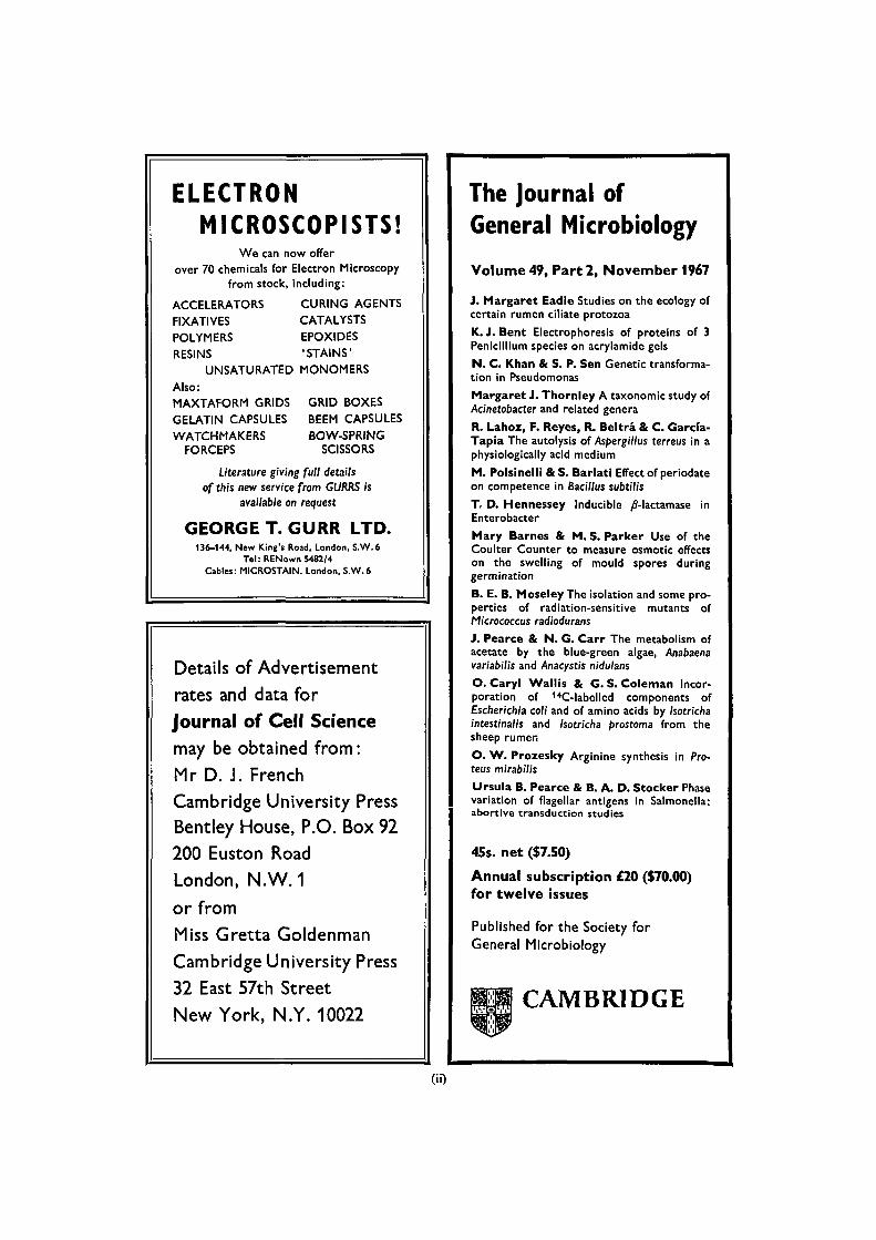
Focus on UltramicrotomyThis is the second of a series presenting the LKB UltrotomeIII by explaining Its ability to solve problems In Ultra-microtomy.
LKB ULTROTOME III SOLVESPROBLEM OF ORIENTATION.
The grain of the structural detail of many specimens, suchas fibers, films, membranes, muscle, skin and others, liesIn more than one direction. Therefore this structural detailwithin the specimen must be located and the cutting cor-rectly aligned to enable the best sections to be produced.
It is a great advantage to be able to produce sectionseither by cutting the specimen longitudinally or by makingtransverse cuts. The universal orientation head of theUltrotome III used together with the vise-type specimen
holder allows one and the same specimen to be adjustedin three directions perpendicular to each other without anyneed to loosen the specimen in the holder. Due to thegoniometer-type construction of the orientation head withits unique 45° arc displacement, the axis of the specimenblock can be positioned, and rigidly fixed at angles up to45° with respect to the axis of the specimen arm. Thisprovides the fastest and most precise structure orientationpossible without the need for any reembeddlng or otheradditional procedures.
Having all-round mobility, and a vernier scale on the arcwhich allows adjustments of 0.1°, the orientation-headneeds only one precision adjustment to enable cutting se-quences In two or three directions to be carried out.
This orientation head Is exclusive to the LKB Ultramicro-tome LKB 8800.
LKB INSTRUMENTS LTD.* 232 ADDINGTON RD. • S. CROYDON, SURREY CR2 8YD
OTHERHEADQUARTERSFOR SALESAND 8ERVICE
LKB-Produkter AB80s 76.Stockholm—Bromms I
USALKB Instruments Inc.12221 Perklewn Drive.Rockvllle Md. 20652
NETHERLANDSLKB-Produkten N.V.Zeekant 35.The Hague
DENMARKLKB Instrument A/SAmagerbrogade 34Copenhagen S
(nO
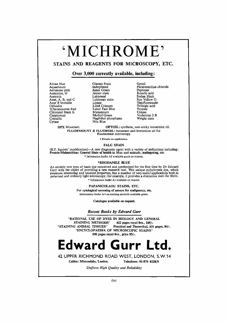
'MICHROME'STAINS AND REAGENTS FOR MICROSCOPY, ETC.
Over 3,000 currently available, including:
Alcian blue Giemsa Stain OptoilAquamount Indophenol Pararosaniline chlorideAstrazone pink Janus Green PeptonesAuramine, O Jenner stain Rosolic acidAurantia Laktoseal Sudan BlackAzur, A, B, and C Leishman stain Sun Yellow GAzur B bromide Lipase ThiofluoresceinCelloidin Lipid Crimson Trifalgic acidtChromosome Red Luxol Fast Blue TrypsinChlorazol black E Matamount UreaseClearmount Methyl Green Violamine 3 BCristalite Naphthyl phosphates Wright stainCytase Nile Blue
DPX Mountant. OPTOIL: synthetic, non-sticky immersion oil.FLUORMOUNT & FLUOROIL: mountant and immersion oil for
fluorescence microscopyt Details on application.
FALG STAIN(B.T. Squires' modification)—A new diagnostic agent with a variety of indications including:Protein-Malnutrition; General State of health in Man and animals; malingering, etc.
* Information leaflet A2 available gratis on request.
•RHODANILE BLUEAn entirely new type of basic dye conceived and synthesized for the first time by Dr EdwardGurr with the object of providing a new research tool. This unique polychrome dye, whichpossesses interesting and unusual properties, has a number of very useful applications both inpolarized and ordinary light microscopy; for example, it provides a distinctive stain for fibrin.
* Information leaflet Al available on request.
PAPANICOLAOU STAINS, ETC.For cytological screening of smears for malignancy, etc.
Information leaflet A 4 on staining methods available gratis.
Catalogue available on request.
Recent Books by Edward Gurr'RATIONAL USE OF DYES IN BIOLOGY AND GENERAL
STAINING METHODS' 422 pages royal 8vo., 105/-.'STAINING ANIMAL TISSUES' Practical and Theoretical, 631 pages, 84/-.
'ENCYCLOPAEDIA OF MICROSCOPIC STAINS'500 pages royal 8vo., price 95/-.
Edward Gurr Ltd.42 UPPER RICHMOND ROAD WEST, LONDON, S.W.14
Cables: Micromlabs, London. Telephone: 01-876 8228/9.
Uniform High Quality and Reliability
(iv)
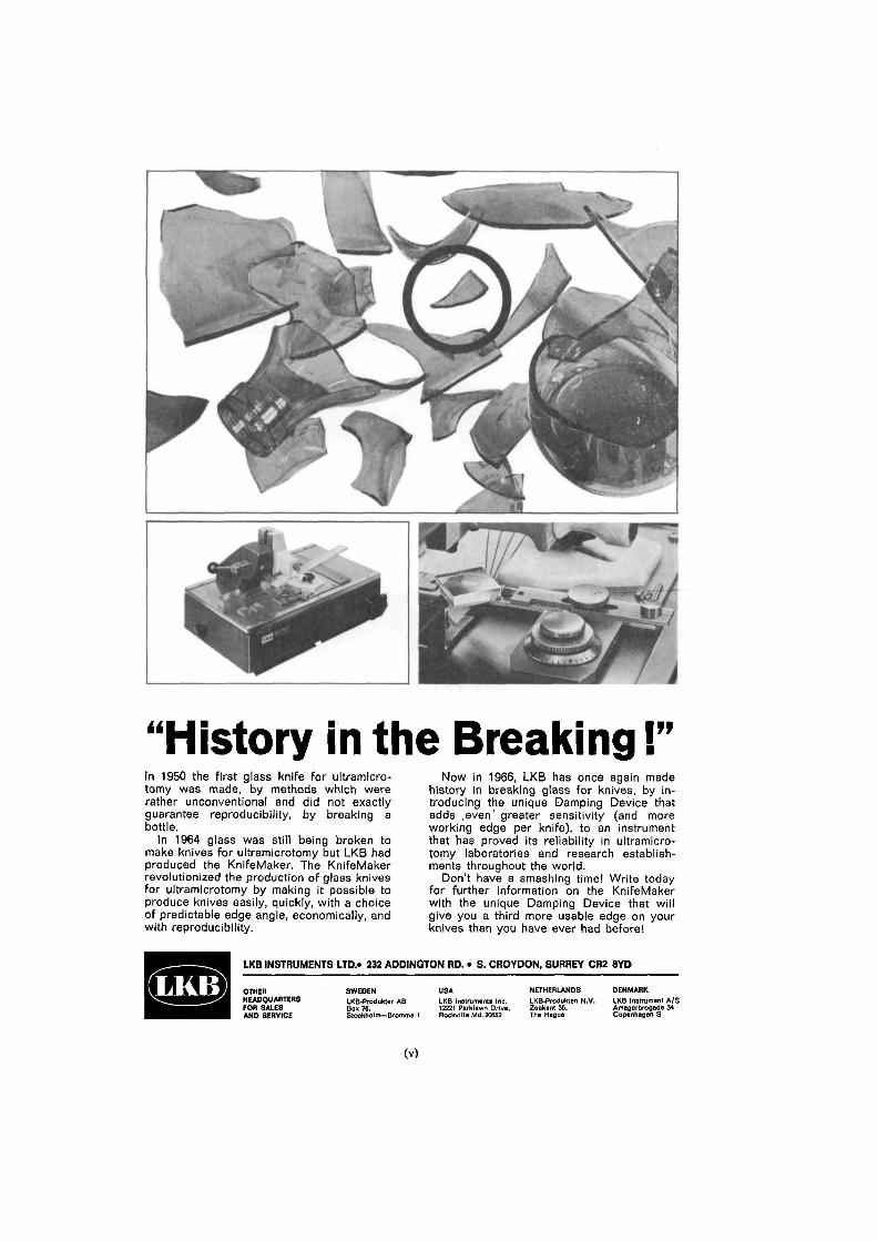
"History in the Breaking!"In 1950 the first glass knife for ultramicro-tomy was made, by methods which wererather unconventional and did not exactlyguarantee reproducibility, by breaking abottle.
In 1964 glass was still being broken tomake knives for ultramicrotomy but LKB hadproduced the KnifeMaker. The KnifeMakerrevolutionized the production of glass knivesfor ultramicrotomy by making it possible toproduce knives easily, quickly, with a choiceof predictable edge angle, economically, andwith reproducibility.
Now in 1966, LKB has once again madehistory in breaking glass for knives, by in-troducing the unique Damping Device thatadds .even greater sensitivity (and moreworking edge per knife), to an instrumentthat has proved its reliability in ultramicro-tomy laboratories and research establish-ments throughout the world.
Don't have a smashing time! Write todayfor further information on the KnifeMakerwith the unique Damping Device that willgive you a third more usable edge on yourknives than you have ever had before!
LKB INSTRUMENTS LTD.* 232 ADDINGTON RD. • S. CROYDON, SURREY CR2 8YD
OTHERHEADQUARTERSFOR SALESAND 8ERVICE
SWEDENLKB-Produkter ABBox 76,Stockholm—Bromma I
USALKB Instruments Inc.12221 Parklawn Drive.RockvllleMd. 20852
NETHERLANDSLKB-Produkten N.V.Zeekant 35.The Hague
DENMARKLKB Instrument A/SAmagerbrosade 34 -Copenhagen S
(V)
BRITISH SOCIETY FOR CELL BIOLOGY
ANNUAL GENERAL MEETING
The Annual General Meeting of the British Society for Cell Biology willbe held at the Middlesex Hospital Medical School, London, on the 4thand 5th April 1968.
There will be a Symposium entitled 'The Dynamics of Cell Membranes'followed by a session of general papers.
Further information may be obtained from the Secretary/Treasurer:
D R L. M. FRANKS
IMPERIAL CANCER RESEARCH FUND
LINCOLN'S INN FIELDS
LONDON, W.C.2
A descriptive list of the
JOURNALSPUBLISHED BY THE
CAMBRIDGE UNIVERSITY PRESS
may be had from
The Manager, Cambridge University Press, Bentley House
P.O. Box 92, 200 Euston Road, London, N.W. 1
Or in U.S.A. from
Cambridge University Press, American Branch
32 East 57th Street, New York, N.Y. 10022, U.S.A.
(vi)
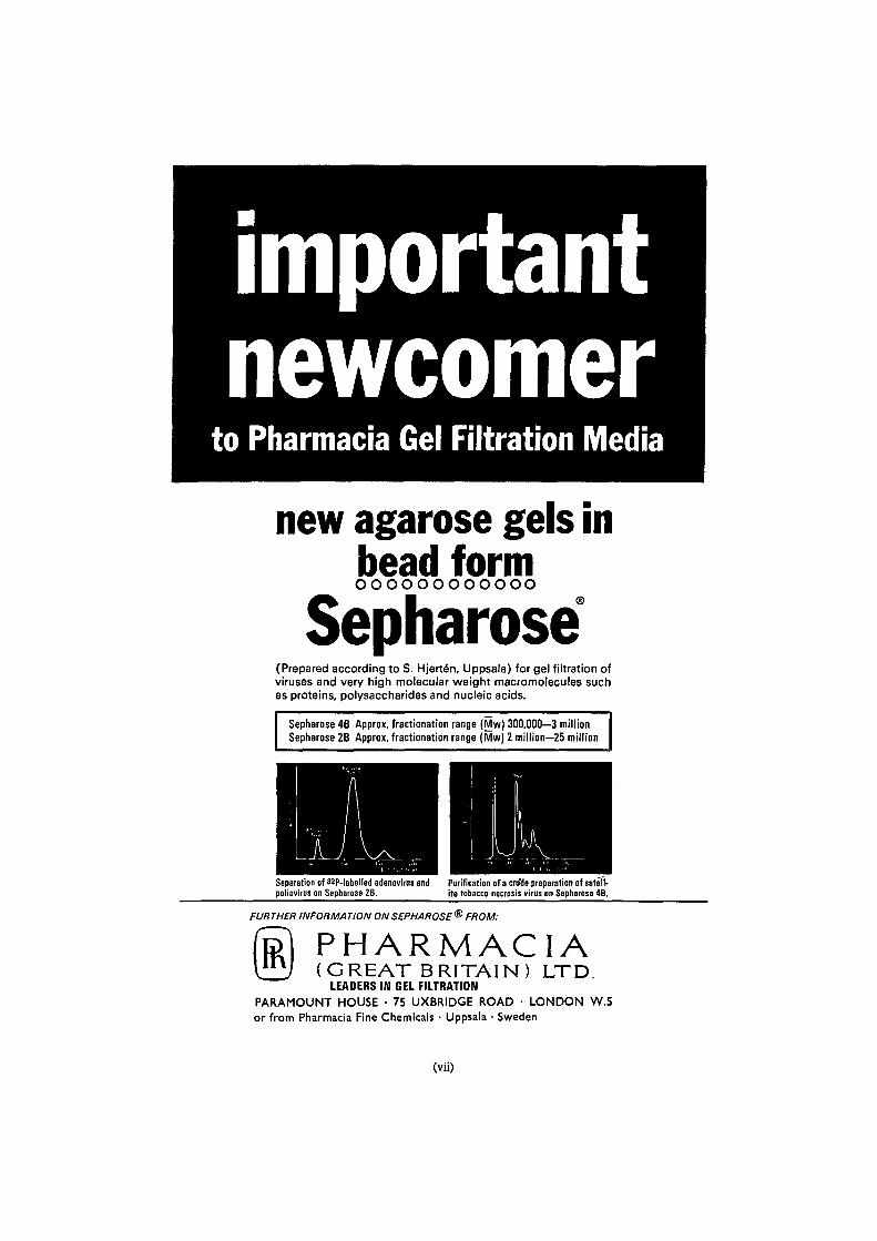
newcomerto Pharmacia Gel Filtration Media
new agarose gels inbead formoooooooooooo
Sepharose(Prepared according to S. Hjerte'n, Uppsala) for gel filtration ofviruses and very high molecular weight macromolecules suchas proteins, polysaccharides and nucleic acids.
Sepharose 4B Approx. fractionation range (Mw) 300,000—3 millionSepharose 2B Approx. fractionation range (Mw) 2 million—25 million
Separation of 32P-labelled adenovirus andpoliovirus on Sepharose 2B.
Purification of a cru'de preparation of satell-ite tobacco necrosis virus on Sepharose 4B.
FUR THER INFORMA TION ON SEPHAROSE ® FROM:
PHARMACIA(GREAT BRITAIN) LTD.
LEADERS IN GEL FILTRATIONPARAMOUNT HOUSE • 75 UXBRIDGE ROAD • LONDON W.5or from Pharmacia Fine Chemicals • Uppsala • Sweden
(vii)
CAMBRIDGEThe Journal of Experimental BiologyEdited by V. B. WIGGLESWORTH & A. J. RAMSEY
Volume 47, Number 2. October 1967
INGRID WALDRON. Mechanisms for the production of the motor output patternin flying locusts.INGRID WALDRON. Neural mechanism by which controlling inputs influence motoroutput in the flying locust.BARBARA G. WILLIAMS and E. NAYLOR. Spontaneously induced rhythm of tidalperiodicity in laboratory-reared Carcinus.
J. E. TREHERNE and S. H. P. MADDRELL. Axonal function and ionic regulation inthe central nervous system of a phytophagous insect {Carausius morosus).
M. E. J. HOLWILL and N. R. SILVESTER. Thermodynamic aspects of flagellaractivity.M. E. J. HOLWILL and M. A. SLEIGH. Propulsion by hispid flagella.
ANN E. KAMMER. Muscle activity during flight in some large lepidoptera.P. VOLPE, M. CARFAGNA and M. Di LORENZO. Extraretinal pigmentation andcolour discrimination. I. Choice of colour of substrate during oviposition inDrosophila melanogaster.
V. VIRABHADRACHARI, R. V. KRISHNAMOORTHY and V. PARVATHESWARARAO. Visualpigments in a tropical freshwater fish Etroplus maculatus (Teleostei).
JOHN BRADY. The relationship between blood ions and blood-cell density in insects.
DANIEL K. HARTLINE. Impulse identification and axon mapping of the nine neuronsin the cardiac ganglion of the lobster Homarus americanus.Y. PICHON and J. BOISTEL. Current-voltage relations in the isolated giant axon ofthe cockroach under voltage-clamp conditions.Y. PICHON and J. BOISTEL. Microelectrode study of the resting and action potentialsof the cockroach giant axon with special reference to the role played by the nervesheath.
30s. net. Annual Subscription (6 issues) £8
Published for the Company of Biologists, Limited
CAMBRIDGE UNIVERSITY PRESS
(viii)
Journal ofCELLSCIENCEFormerly the Quarterly Journal of Microscopical Science
VOLUME 2, 1967
Editors:
H. G. CALLAN A. V. GRIMSTONE
Editorial Board:
G. H. BEALE
H. G. DAVIES
J. B. FINEAN
E. G. GRAY
JEAN HANSON
H. HARRIS
J. HESLOP-HARRISON
S. J. HOLT
H. E. HUXLEY
B. JOHN
J. M. MITCHISON
D. H. NORTHCOTE
J. PAUL
SIR JOHN RANDALL
M. G. P. STOKER
Published for the Company of Biologists Limited
CAMBRIDGE UNIVERSITY PRESS
Printed in Great Britain at the University Printing House, Cambridge
CONTENTS
NUMBER 1 MARCH 1967
page i CALLAN, H. G.The organization of genetic units in chromosomes
9 WHITEHOUSE, H. L. K.
A cycloid model for the chromosome
23 HARRIS, H.
The reactivation of the red cell nucleus
33 BLENKINSOPP, W. K.
Mast cell proliferation in adult rats
39 OWEN, MAUREEN
Uptake of [3H]uridine into precursor pools and RNA in osteogenic cells
57 LING, N. R. and HOLT, P. J. L.
The activation and reactivation of peripheral lymphocytes in culture
71 MEISELMAN, N., KOHN, A. and DANON, D.
Electron microscopic study of penetration of Newcastle disease virus intocells leading to formation of polykaryocytes
77 JEFFS, R. A. and NORTHCOTE.'D. H.
The influence of indol-3yl acetic acid and sugar on the pattern of induceddifferentiation in plant tissue culture
89 BERRIDGE, M. J. and GUPTA, B. L.
Fine-structural changes in relation to ion and water transport in the rectalpapillae of the blowfly, Calliphora
113 GILLIS, J. M. and PAGE, SALLY G.
Localization of ATPase activity in striated muscle and probable sources ofartifact
119 MADDRELL, S. H. P. and TREHERNE, J. E.
The ultrastructure of the perineurium in two insect species, Carausiusmorosus and Periplaneta americana
129 SMITH, J. W., PETERS, T. J. and SERAFINI-FRACASSINI, A.
Observations on the distribution of the proteinpolysaccharide complexand collagen in bovine articular cartilage
137 MACGREGOR, H. C. and MACKIE, J. B.
Fine structure of the cytoplasm in salivary glands of Simulium
iv Contents
NUMBER 2 JUNE 1967
page 145 MACGREGOR, H. C.Pattern of incorporation of [3H]uridine into RNA of amphibian oocytenucleoli
151 HAY, ELIZABETH D. and GURDON, J. B.
Fine structure of the nucleolus in normal and mutant Xenopus embryos
163 GALL, J. G.
The light microscope as an optical diffractometer
169 BEHNKE, O. and FORER, A.
Evidence for four classes of microtubules in individual cells
193 WATKINS, J. F. and GRACE, D. M.
Studies on the surface antigens of interspecific mammalian cellheterokaryons
205 MALHOTRA, S. K. and EAKIN, R. T.
A study of mitochondrial membranes in relation to elementary particles
213 BAKER, T. G. and FRANCHI, L. L.
The fine structure of oogonia and oocytes in human ovaries
225 SMITH-SONNEBORN, JOAN and PLAUT, W.
Evidence for the presence of DNA in the pellicle of Paramecium
235 ODHIAMBO, T. R.
The fine structure and histochemistry of the fat body in the locust,Schistocerca gregaria
243 WlGGLESWORTH, V. B.Cytological changes in the fat body of Rhodnius during starvation,feeding and oxygen want
257 PERRY, MARGARET M.
Identification of glycogen in thin sections of amphibian embryos
265 MANTON, IRENE
Further observations on the fine structure of Chrysochromulina chitonwith special reference to the haptonema, 'peculiar' Golgi structure andscale production
273 NEVILLE, A. C.
Factors affecting the tertiary structure of resilin in locusts
281 GRIMSTONE, A. V., ROTHERAM, SUSAN and SALT, G.An electron-microscope study of capsule formation by insect blood cells
Contents v
NUMBER 3 SEPTEMBER 1967
page 293 STOKER, M. G. P.Transfer of growth inhibition between normal and virus-transformedcells: autoradiographic studies using-marked cells
305 BLENKINSOPP, W. K.
Effect of tritiated thymidine on cell proliferation
309 MACINTYRE, ELIZABETH and PONTEN, J.
Interaction between normal and transformed bovine fibroblasts inculture I. Cells transformed by Rous sarcoma virus
323 KEMP, R. B., JONES, B. M., CUNNINGHAM, I. and JAMES, M. C. M.
Quantitative investigation on the effect of puromycin on the aggregationof trypsin- and versene-dissociated chick fibroblast cells
341 CLARK, A. W.
The fine structure of the eye of the leech, Helobdella stagnalis
349 EAKIN, R. M., WESTFALL, JANE A. and DENNIS, M. J.
Fine structure of the eye of a nudibranch mollusc, Hermissenda crassicornis
359 CHAPMAN, J. A., ELVES, M. W. and GOUGH, J.
An electron-microscope study of the in vitro transformation of humanleucocytes I. Transformation of lymphocytes to blastoid cells in thepresence of phytohaemagglutinin
371 CHAPMAN, J. A., GOUGH, J. and ELVES, M. W.
An electron-microscope study of the in vitro transformation of humanleucocytes II. Transformation to macrophages
377 JOHNSTON, PATRICIA V. and ROOTS, BETTY I.
Fixation of the central nervous system by perfusion with aldehydes andits effect on the extracellular space as seen by electron microscopy
387 HESLOP-HARRISON, J. and MACKENZIE, A.
Autoradiography of soluble [2-14C]thymidine derivatives during meiosisand microsporogenesis in Lilium anthers
401 NUNEZ, E. A., GOULD, R. P., HAMILTON, D. W., HAYWARD, J. S. and
HOLT, S. J.
Seasonal changes in the fine structure of the basal granular cells of thebat thyroid
411 MANTON, IRENE
Further observations on scale formation in Chrysochromulina chiton
419 HAYES, R. L. and ALLEN, E. R.
Electron-microscopic studies on a double-stranded beaded filament ofembryonic collagen
vi Contents
page 435 ASHHURST, DOREEN E.The fibrillar flight muscles of giant water-bugs: an electron-microscopestudy
445 GAY, F. W. and ATTRIDGE, J. T.
The fine structure of cytoplasmic inclusions in a mycoplasma-likeinfection in mice
451 SPANSWICK, R. M. and COSTERTON, J. W. F.
Plasmodesmata in Nitella translucens: structure and electrical resistance
NUMBER 4 DECEMBER 1967
465 SABNIS, D. D. and JACOBS, W. P.
Cytoplasmic streaming and microtubules in the coenocytic marine alga,Caulerpa prolifera
473 CHOUINARD, L. A. and LEBLOND, C. P.
Sites of protein synthesis in nucleoli of root meristematic cells of Alliumcepa as shown by radioautography with [3H]arginine
481 TUCKER, J. B.
Changes in nuclear structure during binary fission in the ciliate Nassula
499 BENEDETTI, E. L. and EMMELOT, P.
Studies on plasma membranesIV. The ultrastructural localization and content of sialic acid in plasmamembranes isolated from rat liver and hepatoma
513 BRYAN, G. W., ZADYLAK, ARLENE H. and EHRET, C. F.
Photoinduction of plastids and of chlorophyll in a Chlorella mutant
529 Lu, B. C.Meiosis in Coprinus lagopus: a comparative study with light and electronmicroscopy
537 LEE, D. L. and ANYA, A. O.
The structure and development of the spermatozoon of Aspiculuristetraptera (Nematoda)
545 GRIFFIN, M. J. and Cox, R. P.
Studies on the mechanism of substrate induction and L-cyst(e)inerepression of alkaline phosphatase in mammalian cell cultures
557 O'BRIEN, T. P.
Cytoplasmic microtubules in the leaf glands of Phaseolus vulgaris
563 SLAUTTERBACK, D. B.
Coated vesicles in absorptive cells of Hydra
Contents vii
Pa8e 573 J°NES. D- G-An electron-microscope study of subcellular fractions of Octopus brain
587 FINCH, J. T., KLUG, A. and NERMUT, M. V.
The structure of the macromolecular units on the cell walls of Bacilluspolymyxa
591 THORNHILL, R. A.
The ultrastructure of the olfactory epithelium of the lamprey Lampeirufluviatilis
603 WlGGLESWORTH, V. B.
Polyploidy and nuclear fusion in the fat body of Rhodnius (Hemiptera)
617 TOOZE, J. and DAVIES, H. G.
Light- and electron-microscope studies on the spleen of the newt Trituruscristatus: the fine structure of erythropoietic cells

INFORMATION FOR CONTRIBUTORS
1 Manuscripts should be sent to The Editors,Journal of Cell Science, Department of Zoology,Cambridge, England.
2 Manuscripts must be typewritten, in doublespacing throughout (including tables, references andlegends). Each table should be typed on a separatesheet. Legends to figures should be typed in asingle series and placed at the end of the manuscript.Papers must be fully corrected by the author, and acharge will be made for excessive alteration in proof.
3 A short title of not more than 40 characters, foruse as page headings, should be supplied if the fulltitle is longer than this.
4 Manuscripts must contain a Summary of notmore than 500 words, placed immediately afterthe title page. Contributors should also send threecopies of an Abstract for distribution to abstractingjournals. The abstract must be not more than 100words long and should be headed by the author'sname and address and the title of the paper. Bothsummary and abstract must be intelligible withoutreference to the main text.
5 The list of References must be given inalphabetical order of authors' names. The titles ofjournals should be abbreviated in accordance withthe World List of Scientific Periodicals, 4th ed. (1963).The following style is used:
BARNICOT, N. A. & HUXLEY, H. E. (1965).
Electron microscope observations on mitoticchromosomes. Q. jfl microsc. Sci. 106, 197-214.
MAZIA, D. (1961). Mitosis and the physiology ofcell division. In The Cell, vol. 3 (ed. J. Brachet& A. E. Mirsky), pp. 77-412. New York andLondon: Academic Press.
Citations in the text are given in the following form:Jones & Smith (i960) or (Jones & Smith, i960).Where there are more than two authors the firstcitation should include all the names and subsequent
citations should be in the form (Jones et al. i960,).Where more than one paper by the same author(s)have been published in the same year they are citedas Jones (1960a), Jones (19606) etc.
6 Text figures should preferably be drawn abouttwice final size; very large drawings should beavoided. Photographic reproductions of drawingscannot always be satisfactorily reproduced. Themaximum printed size of a drawing is 5 in. by8 in. Lettering will be inserted by the printers andshould be indicated on drawings in faint bluepencil or on a tracing-paper overlay. It should be inlower case, and abbreviations should not be used ifthere is space for complete words.
7 Photographs should preferably be submitted thesame size as they are to appear. The maximum areafor a plate is 5J in. by 8i in. Where several photo-graphs make up a plate they should be accuratelymounted on one sheet of cardboard. Irregularlyshaped photographs or plates should be avoidedwherever possible. Lettering on plates will be in-serted by the printers and should be indicated eitheron a duplicate, marked set of prints or on a tracing-paper overlay bearing accurately marked outlinesof the objects indicated. Authors may be asked tocontribute to the cost of plates in excess of four.
8 Text figures and photographs should benumbered in a single series, all text figures pre-ceding the photographs. Each individual drawingor photograph should be numbered separately(Fig. 1, Fig. 2 and so on).
9 Where appropriate the magnifications ofillustrations should be indicated by scales drawn onthem. Magnifications may also be stated in thelegends.
10 Authors will receive 50 offprints free of chargeand may order additional copies when proofs arereturned.
Top Related