


Languages
Pages
Legal

1
ANNUAL NEPHROLOGY ESSAY COMPETITION
FOR MD/DNB MEDICINE/ PAEDIATRICS 2018
URINALYSIS
Submitted by:
Dr. Deepti Agarwal
Second year Post Graduate
Department of General Medicine
KMC Mangaluru
Outline
1. Introduction
2. Gross appearance and physical characteristics
a. Odor
b. Clarity
c. Color
d. Specific gravity
3. Chemical analysis
a. pH
b. Glucose
c. Protein
d. Blood
e. Bilirubin and urobilinogen
f. Ketone
g. Nitrite
h. Leucocyte esterase
4. Microscopy
a. Casts
b. Crystals
c. Cells
d. Microrganisms
5. Bibiliography
Word count: 3008

2
Abstract:
Urinalysis is one of the key tools to evaluate kidney and urinary tract disease. It can
yield ample amount of information when done with the right clinical context.
Urinalysis is most widely done using the dipstick, but there are limitations to the
same. Urine sediment analysis also plays a crucial role in diagnosis of renal diseases.
Urine microscopy and sediment analysis should ideally be done by the clinical
personnel (eg: treating clinician) in order to not to miss out on vital details.
Examination of urine is also called as uroscopy and is one of the oldest practices in
medicine dating back to Babylonian era. Richard Bright, an English physician in the
18th century was an early proponent of routine practice of urine analysis and its
application to renal disease.
Urinalysis is advantageous as it is non invasive, economical and readily available. In
addition to aiding in diagnosis of renal diseases, an abnormal urinalysis findings on a
routine evaluation may be the first evidence of an underlying renal disease. It can
also aid in monitoring the course of an established renal disease.
In this essay a brief review about urinalysis is presented under the headings of gross,
chemical and microscopic analysis.
3
1. Introduction:
Urinalysis is an essential aspect of evaluation of renal disease. Available almost
immediately and most informative when done with the right clinical context.
Someone has rightly said, “What an electrocardiogram is to a cardiologist is what a
urinalysis is to a nephrologist”
A complete urinalysis consists of gross analysis, chemical analysis (usually done via
dipstick) and microscopic analysis
A few instructions to be given to the patient while collecting the sample for routine
urinalysis are:
Collect specimen in a clean, dry wide mouthed, container
Clean the genitalia with water and provide a mid stream urine for analysis
o Midstream specimen is preferred as it avoids contamination with
periurethral and periprepucial organisms
In males, retract the prepuce and clean the glans with water. In females, labia
should be separated and washed with water
In patients with in dwelling catheter collect the sample directly from the
catheter tubing and not from the urobag
Routine examination should be performed within two hours of sample
collection
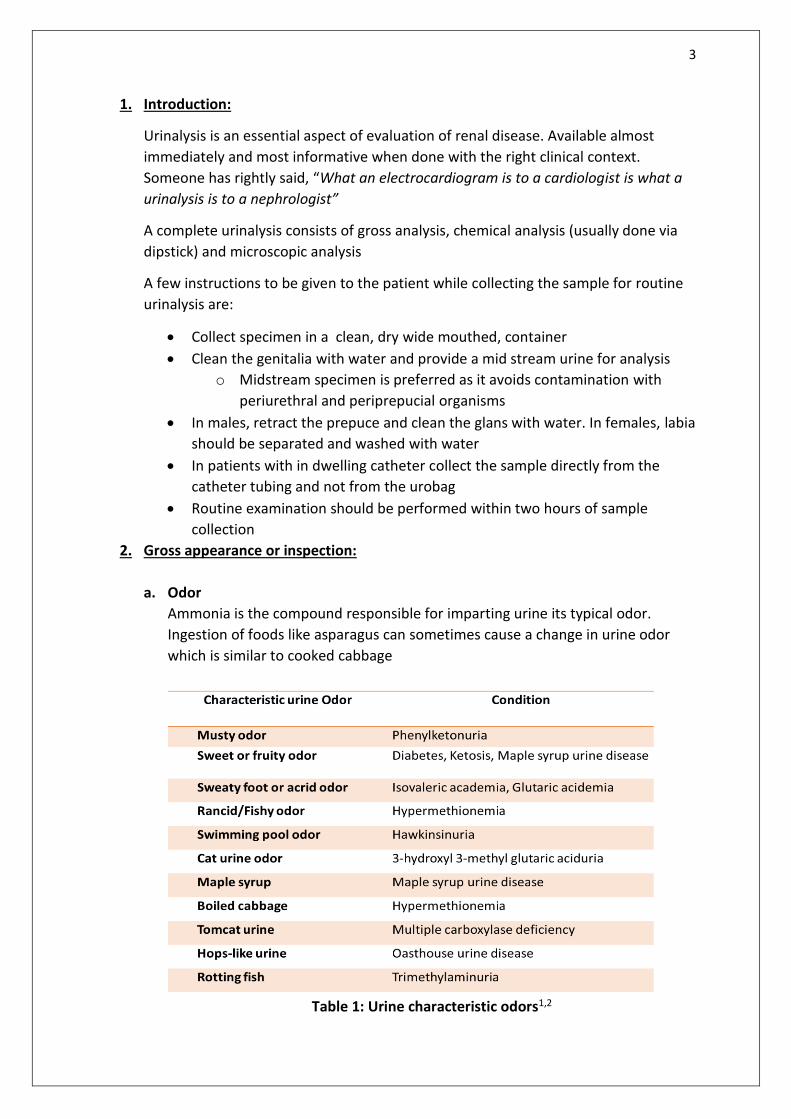
2. Gross appearance or inspection:
a. Odor
Ammonia is the compound responsible for imparting urine its typical odor.
Ingestion of foods like asparagus can sometimes cause a change in urine odor
which is similar to cooked cabbage
Table 1: Urine characteristic odors1,2

4
Conventionally it is taught that Urinary tract infection imparts a foul or feculent
odor but a few studies have shown that urine odor is often a misleading
predictor of UTI in more than one third of cases3
b. Clarity
Cloudy or turbid urine is most commonly associated with urinary tract infections.
It can also be caused due to other causes as below. Turbid white urine is
sometimes referred to as albinuria 4
Figure 2: Causes of turbid urine4
c. Color
Normal urine color varies from clear to dark yellow. Normal urine color is due to
urochrome pigment.
Table 2: Characteristic urine color 1,2
Causes of turbid urine or albinuria
•Chyluria
•Filariasis , Schistosomiasis , Postsurgery , Malignancy
•Hyperuricosuria
•Phosphaturia
•Hyperoxaluria
•Proteinuria
•Pyuria
•Lipiduria
•Caseous material from renal tuberculosis
•Congenital malformations of the lymphatic vessels
5
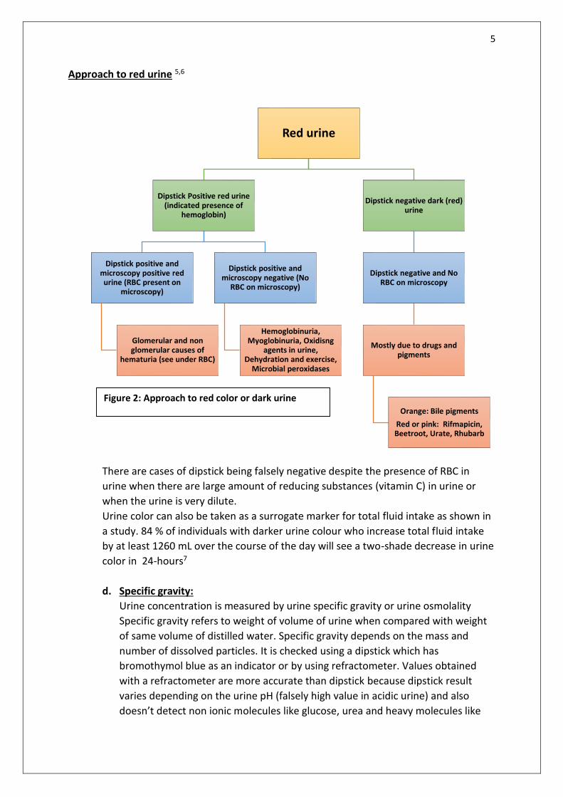
Red urine
Dipstick Positive red urine (indicated presence of
hemoglobin)
Dipstick positive and microscopy positive red urine (RBC present on
microscopy)
Glomerular and non glomerular causes of
hematuria (see under RBC)
Dipstick positive and microscopy negative (No
RBC on microscopy)
Hemoglobinuria, Myoglobinuria, Oxidisng
agents in urine, Dehydration and exercise,
Microbial peroxidases
Dipstick negative dark (red) urine
Dipstick negative and No RBC on microscopy
Mostly due to drugs and pigments
Orange: Bile pigments
Red or pink: Rifmapicin, Beetroot, Urate, Rhubarb
Approach to red urine 5,6
There are cases of dipstick being falsely negative despite the presence of RBC in
urine when there are large amount of reducing substances (vitamin C) in urine or
when the urine is very dilute.
Urine color can also be taken as a surrogate marker for total fluid intake as shown in
a study. 84 % of individuals with darker urine colour who increase total fluid intake
by at least 1260 mL over the course of the day will see a two-shade decrease in urine
color in 24-hours7
d. Specific gravity:
Urine concentration is measured by urine specific gravity or urine osmolality
Specific gravity refers to weight of volume of urine when compared with weight
of same volume of distilled water. Specific gravity depends on the mass and
number of dissolved particles. It is checked using a dipstick which has
bromothymol blue as an indicator or by using refractometer. Values obtained
with a refractometer are more accurate than dipstick because dipstick result
varies depending on the urine pH (falsely high value in acidic urine) and also
doesn’t detect non ionic molecules like glucose, urea and heavy molecules like
Figure 2: Approach to red color or dark urine
6
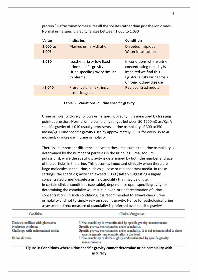
protein.8 Refractometry measures all the solutes rather than just the ionic ones.
Normal urine specifc gravity ranges between 1.005 to 1.030
Table 3 : Variations in urine specific gravity
Urine osmolality closely follows urine specific gravity. It is measured by freezing
point depression. Normal urine osmolality ranges between 50-1200mOsm/Kg. A
specific gravity of 1.010 usually represents a urine osmolality of 300 to350
mosm/kg. Urine specific gravity rises by approximately 0.001 for every 35 to 40
mosmol/kg increase in urine osmolality
There is an important difference between these measures: the urine osmolality is
determined by the number of particles in the urine (eg, urea, sodium,
potassium), while the specific gravity is determined by both the number and size
of the particles in the urine. This becomes important clinically when there are
large molecules in the urine, such as glucose or radiocontrast media. In these
settings, the specific gravity can exceed 1.030 ( falsely suggesting a highly
concentrated urine) despite a urine osmolality that may be dilute.
In certain clinical conditions (see table), dependence upon specific gravity for
determining the osmolality will result in over- or underestimation of urine
concentration. In such conditions, it is recommended to always check urine
osmolality and not to simply rely on specific gravity. Hence for pathological urine
assessment direct measure of osmolality is preferred over specific gravity9
Figure 3: Conditions where urine specific gravity cannot determine urine osmolality with
accuracy
7
3. Chemical analysis
a. pH
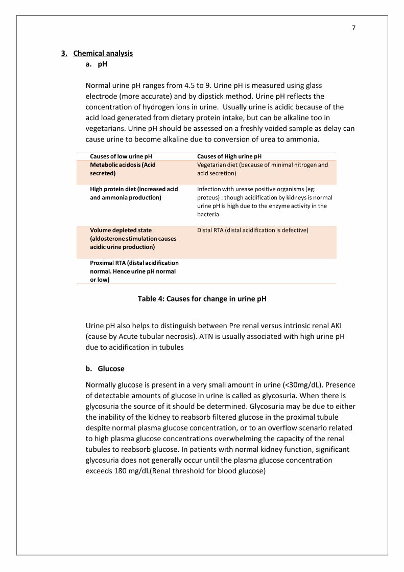
Normal urine pH ranges from 4.5 to 9. Urine pH is measured using glass
electrode (more accurate) and by dipstick method. Urine pH reflects the
concentration of hydrogen ions in urine. Usually urine is acidic because of the
acid load generated from dietary protein intake, but can be alkaline too in
vegetarians. Urine pH should be assessed on a freshly voided sample as delay can
cause urine to become alkaline due to conversion of urea to ammonia.
Table 4: Causes for change in urine pH
Urine pH also helps to distinguish between Pre renal versus intrinsic renal AKI
(cause by Acute tubular necrosis). ATN is usually associated with high urine pH
due to acidification in tubules
b. Glucose
Normally glucose is present in a very small amount in urine (<30mg/dL). Presence
of detectable amounts of glucose in urine is called as glycosuria. When there is
glycosuria the source of it should be determined. Glycosuria may be due to either
the inability of the kidney to reabsorb filtered glucose in the proximal tubule
despite normal plasma glucose concentration, or to an overflow scenario related
to high plasma glucose concentrations overwhelming the capacity of the renal
tubules to reabsorb glucose. In patients with normal kidney function, significant
glycosuria does not generally occur until the plasma glucose concentration
exceeds 180 mg/dL(Renal threshold for blood glucose)
8
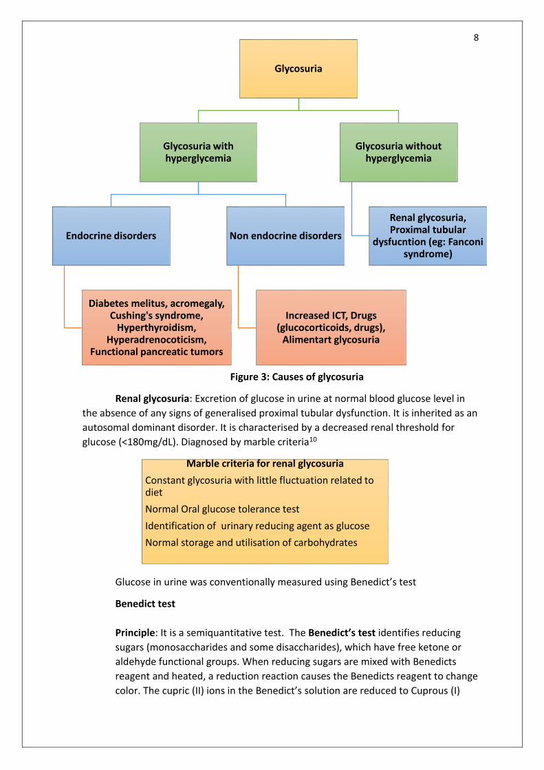
Glycosuria
Glycosuria with hyperglycemia
Endocrine disorders
Diabetes melitus, acromegaly, Cushing's syndrome,
Hyperthyroidism, Hyperadrenocoticism,
Functional pancreatic tumors
Non endocrine disorders
Increased ICT, Drugs (glucocorticoids, drugs),
Alimentart glycosuria
Glycosuria without hyperglycemia
Renal glycosuria, Proximal tubular
dysfucntion (eg: Fanconi syndrome)
Figure 3: Causes of glycosuria
Renal glycosuria: Excretion of glucose in urine at normal blood glucose level in
the absence of any signs of generalised proximal tubular dysfunction. It is inherited as an
autosomal dominant disorder. It is characterised by a decreased renal threshold for
glucose (<180mg/dL). Diagnosed by marble criteria10
Glucose in urine was conventionally measured using Benedict’s test
Benedict test
Principle: It is a semiquantitative test. The Benedict’s test identifies reducing
sugars (monosaccharides and some disaccharides), which have free ketone or
aldehyde functional groups. When reducing sugars are mixed with Benedicts
reagent and heated, a reduction reaction causes the Benedicts reagent to change
color. The cupric (II) ions in the Benedict’s solution are reduced to Cuprous (I)
Marble criteria for renal glycosuria
Constant glycosuria with little fluctuation related to diet
Normal Oral glucose tolerance test
Identification of urinary reducing agent as glucose
Normal storage and utilisation of carbohydrates

9
ions, which causes the color change. The color varies from green to dark red
(brick) or rusty-brown, depending on the amount of and type of sugar.
Results and interpretation:
Figure 4: Interpretation of Benedict’s test
Benedict’s test is not used commonly as urine dipsticks have replaced them. Urine
dipstick for glucose work on the following principle. Glucose is oxidised to gluconic
acid and hydrogen peroxide with glucose oxidase. Hydrogen peroxide than reacts
with reduced colorless chromogen to form a colored product. This color is charted
against a concentration gradient and an approximate value of urine glucose is
obtained. This test detects a concentration of 0.5 to 20g/L of glucose.
For precise quantitative assessment of urine glucose enzymatic methods like
Hexokinase methods should be used
Figure 5: Principle and results of urine dipstick for glucose
Yellow: 0.1-0.5 % sugar, Green: 0.5-1, Orange : 1-1.5%, Red: 1.5-2%, Brick red: >2%

10
Table 5: Causes of false positivity and negativity for glucose on dipstick
c. Protein
No consistent definition exists for proteinuria
Currently the following definitions are in use11
Type 24 hour urine excretion Protein creatinine ratio (urine protein/Urine creatinine)
Physiologic <150mg/ 24 hours Or <4mg/m2/hour
<0.5mg/mg
Pathologic
Microalbuminuria (now called moderately increased albuminuria
30-300mg albumin/day
Macroalbuminuria (now called severely increased albuminuria)
>300mg albumin/day
Non nephrotic range 4-40 mg/m2/hour <3gm/24hours
<3mg/mg <3000mg/gm
Nephrotic Range >40 mg/m2/hour >3gm/24hours
>3mg/mg or >3000mg/gm
Physiological proteinuria mainly is composed of Tomm Horsfall protein and albumin
excretion is <30mg/day
Figure 6:Types of proteinuria

11
Figure 7: Pathogenesis of proteinuria
Figure 8: Methods of testing for proteinuria
Semi quantitative methods
i. Routine urine dipstick
Simplest and least expensive methods
Principle: Uses tetrabromphenol blue buffered with citrate as an
indicator
Urine albumin binds to the reagent and changes the pH which results
in a spectrum of color change based on degree of change of pH
Lower threshold for detection : 15-20mg/dL
Semi quantitative
• Routine urine dipstick
• Sulfosalicylic acid
• Albumin sensitive tests
Quantitative
• Spot urine Protein to creatinine ratio
• Albumin to creatinine ratio
• 24 hour urine protein collection

12
Figure 8: Results and interpretation of urine dipstick
Figure 9: False positive and false negative for protein on dipstick
ii. Sulfosalycilic acid (SSA) test:
Detects all proteins in urine
Positive SSA and negative dipstick: Suspect non albumin proteins in
urine
Principle: SSA causes precipitation of proteins
Procedure: Mix one part urine supernatant with 3 parts SSA
False positive
•Concentrated urine: Abnormal result even when protein is normal
•Receive contrast 24hours prior
•Highly alkaline urine overhwhelming the dye’s buffer
•When antiseptics are used (Chlorhexidineand benzalkalonium)
•Delay in reading
•Stick left to soak in urine for long
False negative
•Highly dilute urine
•Presence of non albumin protein (Low molecular weight tubular, light chain protein, Globulin)
13
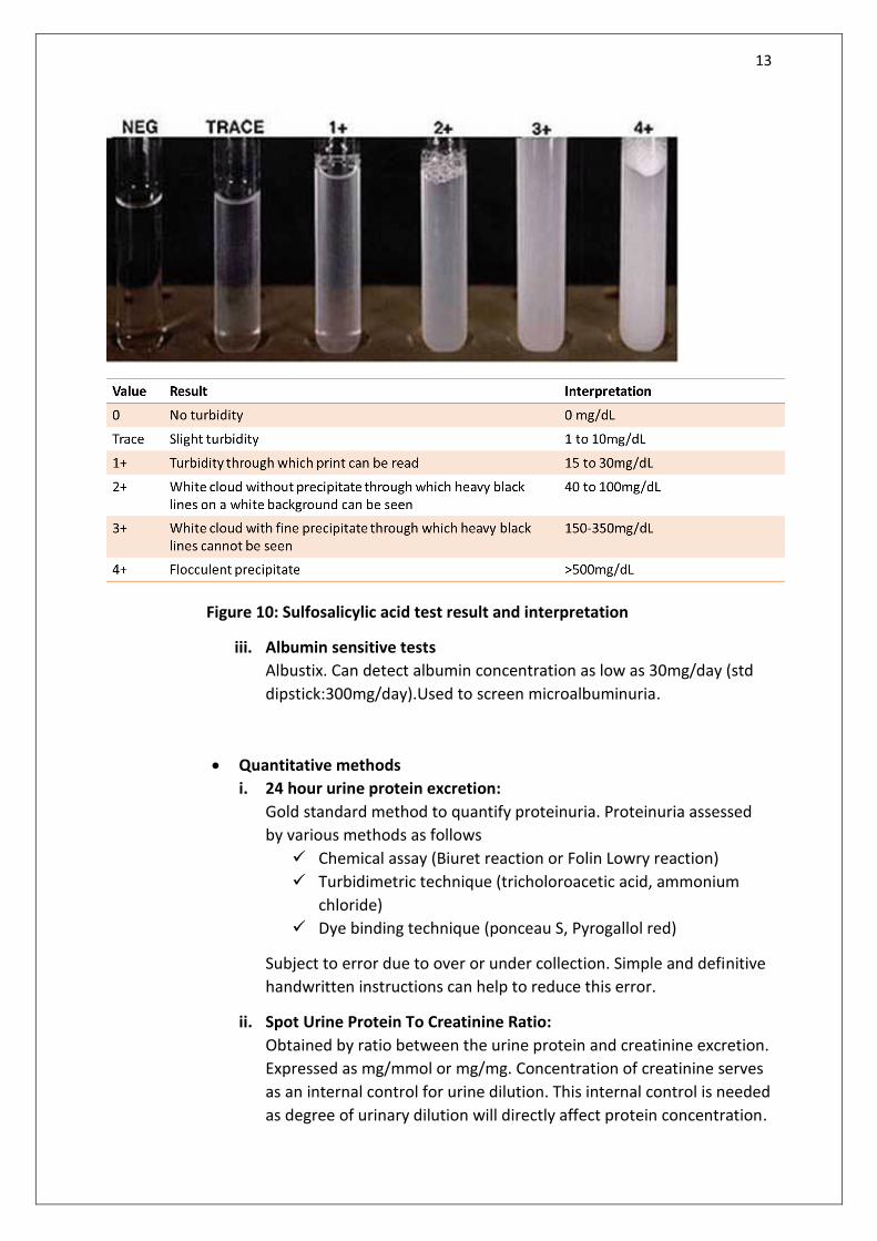
Figure 10: Sulfosalicylic acid test result and interpretation
iii. Albumin sensitive tests
Albustix. Can detect albumin concentration as low as 30mg/day (std
dipstick:300mg/day).Used to screen microalbuminuria.
Quantitative methods
i. 24 hour urine protein excretion:
Gold standard method to quantify proteinuria. Proteinuria assessed
by various methods as follows
Chemical assay (Biuret reaction or Folin Lowry reaction)
Turbidimetric technique (tricholoroacetic acid, ammonium
chloride)
Dye binding technique (ponceau S, Pyrogallol red)
Subject to error due to over or under collection. Simple and definitive
handwritten instructions can help to reduce this error.
ii. Spot Urine Protein To Creatinine Ratio:
Obtained by ratio between the urine protein and creatinine excretion.
Expressed as mg/mmol or mg/mg. Concentration of creatinine serves
as an internal control for urine dilution. This internal control is needed
as degree of urinary dilution will directly affect protein concentration.
14
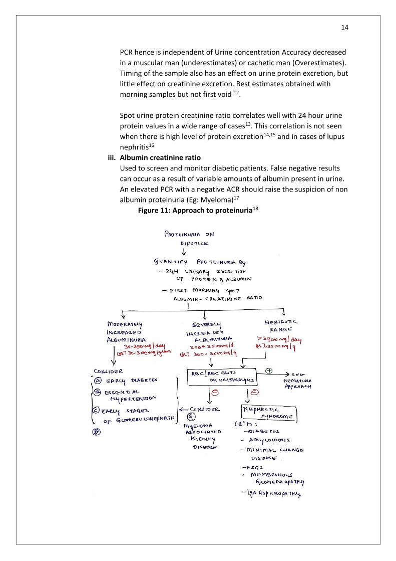
PCR hence is independent of Urine concentration Accuracy decreased
in a muscular man (underestimates) or cachetic man (Overestimates).
Timing of the sample also has an effect on urine protein excretion, but
little effect on creatinine excretion. Best estimates obtained with
morning samples but not first void 12.
Spot urine protein creatinine ratio correlates well with 24 hour urine
protein values in a wide range of cases13. This correlation is not seen
when there is high level of protein excretion14,15 and in cases of lupus
nephritis16
iii. Albumin creatinine ratio
Used to screen and monitor diabetic patients. False negative results
can occur as a result of variable amounts of albumin present in urine.
An elevated PCR with a negative ACR should raise the suspicion of non
albumin proteinuria (Eg: Myeloma)17
Figure 11: Approach to proteinuria18
15
b. Ketones
Ketone dipstick tests for aceto acetic acid using the nitroprusside reaction. It
does not test for beta hydorxybutyrate and acetone.
Causes of ketonuria:
Fasting
Vomiting
Strenuous exercise
Diabetic Ketoacidosis
Negative urine for ketones does not rule out ketosis
A study was done to check the efficacy of point of care betahydorxybutyrate
capillary assay versus urinary dipstick for detection of ketoacidosis and the
former was found to be more sensitive in picking up the cases of DKA19
d. Bile salts and bile pigments
Not a routinely done test after the advent of liver function test
Hay’s sulphur powder test was used for bile salts
Fouchet test for bile pigments.
Ehrlich aldehyde test for urobilinogen
Unconjugated hyperbilirubinemia has increased amounts of urobilinogen and
no bile salts and bile pigments in urine (Acholuric jaundice). Conjugated
hyperbilirubinemia has reduced urobilinogen and increased bile salts and bile
pigments in urine.
e. Blood
Urine dipstick detects haemoglobin by measuring the peroxidase activity.
Hemoglobin catalyses the oxidation of chromogen to produce a colored
product. Myoglobin also shows urine dipstick positivity. The approach to dipstick
positive and negative dark urine is discussed in the section under red urine.
f. Nitrite
Detects the bacteria that can reduce nitrates to nitrites by nitrate reductase
activity. Has low sensitivity but high specificity. Positive with most of the
gram negative uropathogenic bacteria. Negative with Staphylococcus albus,
Enterococcus and pseudomonas. It can be falsely positive if the diet is rich in
nitrates. Urine must incubate in the urinary bladder for atleast four hours to
allow adequate nitrate reduction, failing which results can be falsely
negative. A study done by Weix and colleagues suggested that a negative
urine nitrite test is a possible indicator that a microorganism is resistant to
the first and third-generation of cephalosporins. However, Grant et al
16
concluded that the detection of urine nitrites should not influence the use of
first-generation cephalosporins for urinary tract infections.20
g. Leucocyte esterase:
For detection of leucocyturia(25-50cells/ml of urine). Positive with
neutrophils, monocytes. Eosinophils and basophils. Negative with
lymphocytes. Positive test seen with UTI and interstitial nephritis.
False positive results are seen when formaldehyde used as urine preservative
False negative can be due to high glucose or high protein concentration. In
the presence of antibiotics too leucocyte esterase will be falsely negative.
17
4. Microscopy
a. Casts
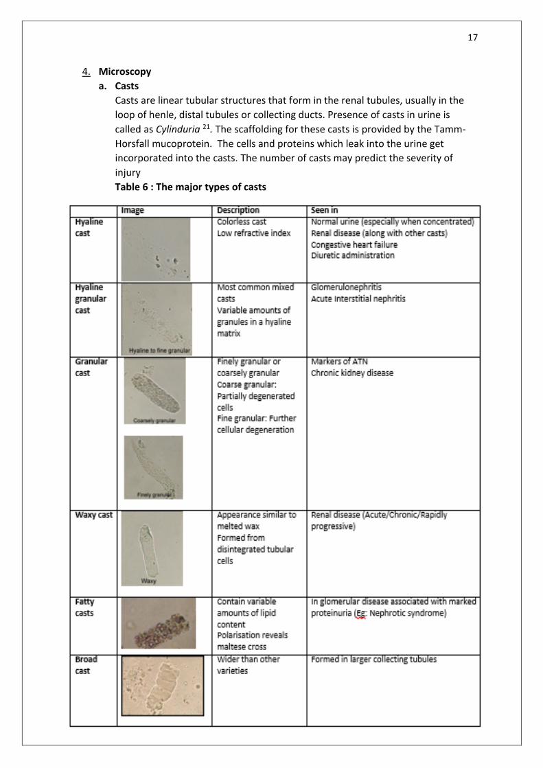
Casts are linear tubular structures that form in the renal tubules, usually in the
loop of henle, distal tubules or collecting ducts. Presence of casts in urine is
called as Cylinduria 21. The scaffolding for these casts is provided by the Tamm-
Horsfall mucoprotein. The cells and proteins which leak into the urine get
incorporated into the casts. The number of casts may predict the severity of
injury
Table 6 : The major types of casts

18
Other casts:
Myoglobin casts: Pigmented cylinders with myoglobin providing color.
Similar to haemoglobin casts. Seen in urine in patients with
Rhabdomyolysis secondary with AKI.
Bilirubin casts: Cylinders pigmented with bilirubin, which can stain any
particle contained in the cast. Observed in patients with jaundice
associated with increased direct conjugated bilirubin.
Mixed casts: Contain components of different nature, such as granules,
cells, and lipids.

19
Urine crystals
Common crystals
Uric acid and amorphous urate
Calcium oxalate crystals
Phosphate crystals (triple phosphate and calcium
phosphate)
Pathologic crystals
Cholesterol crystals
Cystine crystals
Dihydroxyadenine crystals
Tyrosine crystals
Leucine crystals
Crystals caused by drugs
Contain the drug itself
Other crystals
Hippuric acid crystals
Calcium carbonate crystals
Ammonium bicarbonate crystals
b. Crystals
Crystal formation in urine depends on many factors like
Presence of inhibitors of crystallisation
Urine pH: High and low urine pH promote crystallisation
Concentration of the constituent molecules
Temperature: Low temperature promotes crystallisation
Crystals are clinically significant when associated with renal failure, like22
Calcium oxalate crystals in ethylene glycol poisoning
Uric acid crystals in tumor lysis syndrome
Drug crystals like acyclovir in drug associated renal failure
Examination of urine for crystalluria should be done on urine as close as 37C
Crystals found in urine can be classified as follows
Crystals formed in
Acidic urine: Calcium oxalate, uric acid, amorphous urate
Alkaline urine: Calcium phosphate, triple phosphate and amorphous
phosphate

20
Common crystals
Uric acid, calcium oxalate and calcium phosphate crystals may have no
clinical significance and can reflect transient urinary supersaturation. Persistence presence
of these crystals requires evaluation.
Table 7: Common urinary crystals
21
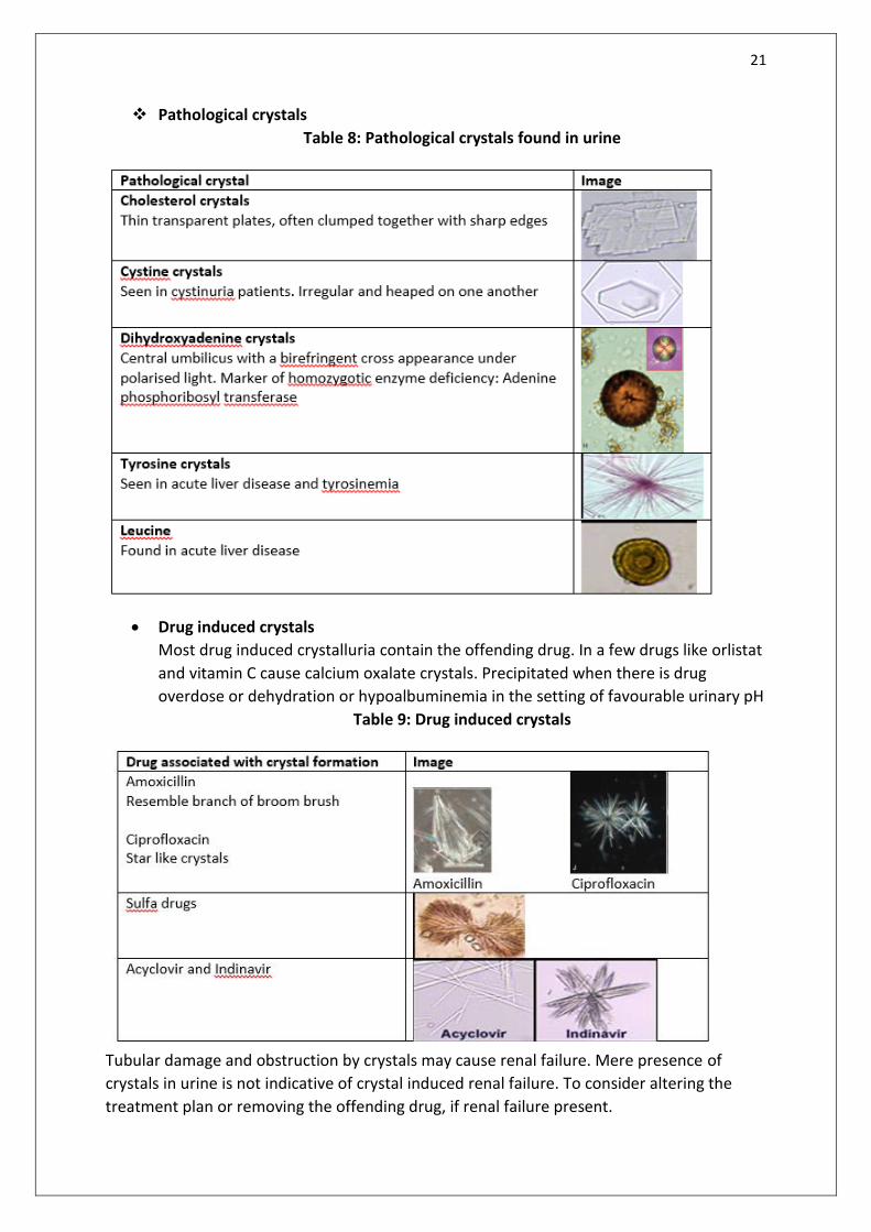
Pathological crystals
Table 8: Pathological crystals found in urine
Drug induced crystals
Most drug induced crystalluria contain the offending drug. In a few drugs like orlistat
and vitamin C cause calcium oxalate crystals. Precipitated when there is drug
overdose or dehydration or hypoalbuminemia in the setting of favourable urinary pH
Table 9: Drug induced crystals
Tubular damage and obstruction by crystals may cause renal failure. Mere presence of
crystals in urine is not indicative of crystal induced renal failure. To consider altering the
treatment plan or removing the offending drug, if renal failure present.

22
c. Cells
Red blood cells
Microscopic hematuria is defined 2-5 RBC per high power field in a centrifuged
urine sample18. Persistent or significant hematuria is defined as <3RBC/HPF on
three urinalysis, single urinalysis with >100 RBCs or gross hematuria18.
RBCs appear as translucent biconcave discs between 4 and 7 µm. RBC in urine
can be isomorphic or dysmorphic
Isomorphic (similar to circulating RBC): Associated with a non glomerular
hematuria from a genitourinary or external source. Urinary catheterization is also
a common cause in admitted patients23
Dysmorphic (showing varying degrees of anisopoikilocytosis): RBC appear
dysmorphic due to changes in urine pH, osmolality or protein concentration.
Dysmorphism arises due to passage of RBC through glomerular barrier or
tubules. Forms of dysmorphia associated with renal disease are anulocytes, ghost
cells, Schizocytes, codocytes and knizocytes. Acanthocytes are an unique form of
dysmorphic RBC characterized by round shape with one or two smaller round
protrusions, vesicles or blebs attached. Acanthocyturia is particularly useful to
identify non diabetic renal disease in diabtetics where hematuria is common24
Dysmorphic RBCs are seen in hematuria due to glomerular causes25. Clinical
application is hampered due to a lack of an uniform definition to define
dysmorphism.
A reasonable definition of glomerular hematuria is ≥40% dysmorphic RBCs or
≥5% acanthocytes26
Figure 12: A: non glomerular hematuria showing isomorphic RBC and arrow pointing to
crenated RBC (numerous spiculations or spikes and don’t signify any pathology) . B:
Glomerular hematuria with dysmorphic RBC and inset showing acanthocytes
23
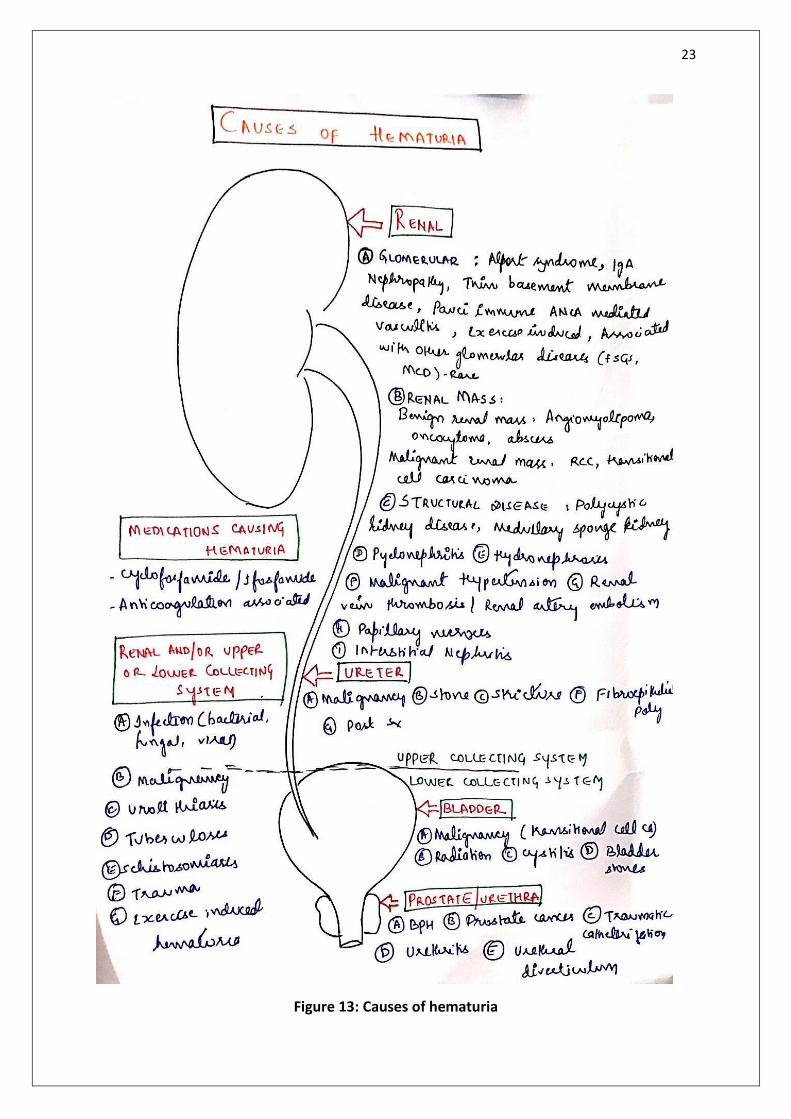
Figure 13: Causes of hematuria
24
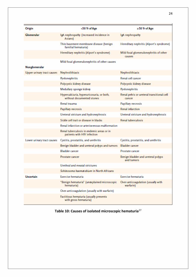
Table 10: Causes of isolated microscopic hematuria27

25
Figure 14a: Approach to a patient with hematuria
26
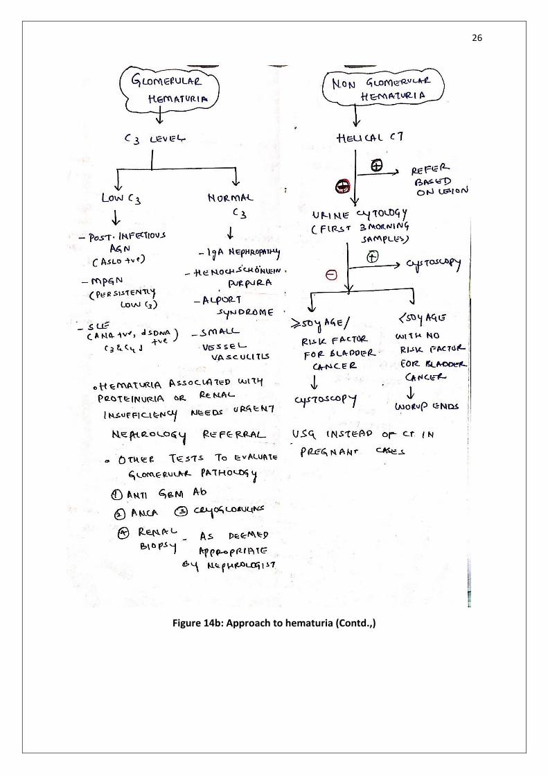
Figure 14b: Approach to hematuria (Contd.,)

27
White blood cells
Pyuria is defined as the presence of 10 or more white cells percubic millimeter in an
uncentrifuged urine specimen, 3 or more white cells per high-power field of unspun
urine, a positive result on Gram’s staining of an unspun urine specimen, or a urinary
dipstick test that is positive for leukocyte esterase28. Entire spectrum of WBCs can be
seen in urine, but most common ones are neutrophils and eosinophils
Neutrophils:
Urinary neutrophils are commonly associated with Bacteruria. In the absence
of urine culture positivity causes of sterile pyuria should be ruled out. Sterile pyuria
is the persistent finding of white cells in the urine in the absence of bacteria, as
determined by means of aerobic laboratory techniques (on a 5% sheepblood agar
plate and MacConkey agar plate)29.
Figure 16: Causes of sterile pyuria
Infectious causes
• Renal tuberculosis
• Urethritis
• Chlamydia balanitis
• Ureaplasma infection
• Viral infections
Non infectious causes
• Acute glomerulonephritis
• Acute tubulointerstitial nephritis
• Bladder tumor
• Foreign body
• Exercise
• Steroid and cyclophosphamide therapy
Figure 15: Urinary neutrophil:
Intermediate size when compared to
RBC and renal tubular epithelial cell.
Identified by their characteristic
granular cytoplasm and multilobed
nuclei.

28
Figure 17: Approach to sterile pyuria29

29
Eosinophils:
Detected by Wright’s or Hansel stain to urine sediment. Presence of
eosinophils in urine is called eosinophiluria, the causes of which are enlisted below30
Figure 18: Causes of eosinophiluria
Other WBCs seen in urine
Lymphocytes are also seen in urine in a few cases of transplant rejection
Causes of eosinophiluria
• Acute interstitial nephritis
• Transplant rejection
• Pyelonephritis
• Prostatitis and cystitis
• Atheroembolic disease
• Rapidly progressive glomerulonephritis
Urinary macrophage:
•Large cells ; 15 to >100µm diameter, seen in glomerular diseases or as oval fat bodies containing phagocytosed lipids
30
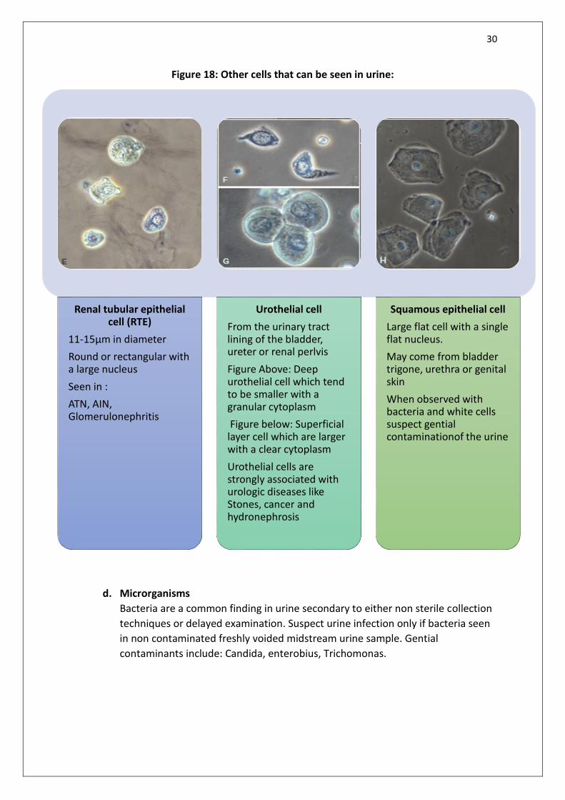
Figure 18: Other cells that can be seen in urine:
d. Microrganisms
Bacteria are a common finding in urine secondary to either non sterile collection
techniques or delayed examination. Suspect urine infection only if bacteria seen
in non contaminated freshly voided midstream urine sample. Gential
contaminants include: Candida, enterobius, Trichomonas.
Renal tubular epithelial cell (RTE)
11-15µm in diameter
Round or rectangular with a large nucleus
Seen in :
ATN, AIN, Glomerulonephritis
Urothelial cell
From the urinary tract lining of the bladder, ureter or renal perlvis
Figure Above: Deep urothelial cell which tend to be smaller with a granular cytoplasm
Figure below: Superficial layer cell which are larger with a clear cytoplasm
Urothelial cells are strongly associated with urologic diseases like Stones, cancer and hydronephrosis
Squamous epithelial cell
Large flat cell with a single flat nucleus.
May come from bladder trigone, urethra or genital skin
When observed with bacteria and white cells suspect gential contaminationof the urine

31
5. Bibiliography
1. Jhonson Richrd J, Feehally John FJ. Comprehensive Clinical Nephrology. 5th ed. Elsevier; 2015. 39-51 p.
2. Ross DG. Urinalysis. Imaging Technol Urol Princ Clin Appl. 2012;9781447124:127–30.
3. Struthers S, Scanlon J, Parker K, Goddard J, Hallett R. Parental reporting of smelly urine and urinary tract infection. Arch Dis Child. 2003;88(3):250–2.
4. Vera M, Molano A, Rodríguez P. Turbid white urine. NDT Plus. 2010;3(1):45–7.
5. LB B. When the urine is red. JAMA [Internet]. 1977 Jun 20;237(25):2753–4. Available from: http://dx.doi.org/10.1001/jama.1977.03270520063032
6. Turner neil, Lameire Robert, Goldsmith David J, Winearls Christopher G. Oxford textbook of clinical nephrology. 4th ed. Oxford university press; 2016. 36-43 p.
7. Perrier ET, Johnson EC, McKenzie AL, Ellis LA, Armstrong LE. Urine colour change as an indicator of change in daily water intake: a quantitative analysis. Eur J Nutr. 2016;55(5):1943–9.
8. De Buys Roessingh AS, Drukker A, Guignard JP. Dipstick measurements of urine specific gravity are unreliable. Arch Dis Child. 2001;85(2):155–7.
9. Voinescu GC, Shoemaker M, Moore H, Khanna R, Nolph KD. The relationship between urine osmolality and specific gravity. Am J Med Sci [Internet]. 2002;323(1):39–42. Available from: http://dx.doi.org/10.1097/00000441-200201000-00007
10. WD D, RF F, GASTON J. Diabetes mellitus and preexisting renal glucosuria. Arch Intern Med [Internet]. 1962 Aug 1;110(2):199–204. Available from: http://dx.doi.org/10.1001/archinte.1962.03620200059011
11. Turner neil, Lameire Robert, Goldsmith David J WCG. Oxford textbook of clinical nephrology. Oxford university press; 2016. 478-483 p.
12. Is W, Evidence THE. Testing for proteinuria. Diabetes Care [Internet]. 2004;9(January 2003):3–7. Available from: http://www3.interscience.wiley.com/cgi-bin/abstract/118802806/ABSTRACT
13. McIntyre NJ, Taal MW. How to measure proteinuria? Curr Opin Nephrol Hypertens. 2008;17(6):600–3.
14. LANE C, BROWN M, DUNSMUIR W, KELLY J, MANGOS G. Can spot urine protein/creatinine ratio replace 24 h urine protein in usual clinical nephrology? Nephrology [Internet]. 2006;11(3):245–9. Available from: https://doi.org/10.1111/j.1440-1797.2006.00564.x
15. Wahbeh A, Ewais M, Elsharif M. Comparison of 24-hour urinary protein and protein-to-creatinine ratio in the assessment of proteinuria. Saudi J Kidney Dis Transplant [Internet]. 2009;20(3):443–7. Available from: http://www.sjkdt.org/article.asp?issn=1319-2442;year=2009;volume=20;issue=3;spage=443;epage=447;aulast=Wahbeh

32
16. Birmingham DJ, Rovin BH, Shidham G, Nagaraja HN, Zou X, Bissell M, et al. Spot urine protein/creatinine ratios are unreliable estimates of 24 h proteinuria in most systemic lupus erythematosus nephritis flares. Kidney Int. 2007;72(7):865–70.
17. Atkins RC, Briganti EM, Zimmet PZ, Chadban SJ. Association between albuminuria and proteinuria in the general population: The AsuDiab study. Nephrol Dial Transplant. 2003;18(10):2170–4.
18. Jameson JL, Kasper DL, Fauci AS HS. Harrison’s Principle of Internal medicine. 20th ed. 2018. 294 p.
19. Arora S, Henderson SO, Long T, Menchine M. Diagnostic accuracy of point-of-care testing for diabetic ketoacidosis at emergency-department triage: β-hydroxybutyrate versus the urine dipstick. Diabetes Care. 2011;34(4):852–4.
20. Medows M, Nijres BM, Elbakoush F, Alali A, Patel R, Mohammad S. Can urinary nitrites or other urinalysis findings be a predictor of bacterial resistance of uncomplicated urinary tract infections? Int J Pediatr Adolesc Med [Internet]. 2016;3(1):12–7. Available from: http://www.sciencedirect.com/science/article/pii/S2352646716000065%5Cnhttp://linkinghub.elsevier.com/retrieve/pii/S2352646716000065
21. Caleffi A, Lippi G. Cylindruria. Clin Chem Lab Med. 2015;53:S1471–7.
22. Frochot V, Daudon M. Clinical value of crystalluria and quantitative morphoconstitutional analysis of urinary calculi. Int J Surg. 2016;36(PD):624–32.
23. Hockberger RS, Schwartz B, Connor J. Hematuria induced by urethral catheterization. Ann Emerg Med [Internet]. 1987 May 1;16(5):550–2.
24. Kohler H, Wandel E, Brunck B. Acanthocyturia - A characteristic marker for glomerular bleeding. Kidney Int. 1991;40(1):115–20.
25. Birch DF, Fairley KF, Whitworth JA, Forbes I, Fairley JK, Cheshire GR, et al. Urinary erythrocyte morphology in the diagnosis of glomerular hematuria. Clin Nephrol [Internet]. 1983 Aug;20(2):78—84.
26. Fogazzi GB, Edefonti A, Garigali G, Giani M, Zolin A, Raimondi S, et al. Urine erythrocyte morphology in patients with microscopic haematuria caused by a glomerulopathy. Pediatr Nephrol. 2008;23(7):1093–100.
27. Parmar M. Microscopic Hematuria - Correspondence. N Engl J Med. 2003;349(13):1292–3.
28. Horan TC, Andrus M, Dudeck MA. CDC/NHSN surveillance definition of health care–associated infection and criteria for specific types of infections in the acute care setting. Am J Infect Control [Internet]. 2008 Jun 1;36(5):309–32.
29. Moore T. Sterile Pyuria. J R Soc Med. 1940;33(9):593–9.
30. Corwin HL, Haber MH. Special Report: The Clinical Significance of Eosinophiluria [Internet]. Vol. 88, American Journal of Clinical Pathology. 1987. 520-522 p.
Top Related