


Languages
Pages
Legal


UPPER EXTREMITY LYMPHEDEMA & EDEMA MANAGEMENT
Christa Newgent MS,PT,CLT

Presentation Objectives
• Learn the pathophysiology of lymphedema & edema• Recognize the signs & symptoms of lymphedema vs. edema• Understand the available treatment for lymphedema &
edema• Understand the components of complete decongestive
therapy• Learn basic manual lymph drainage techniques• Know how to apply bandaging or taping to reduce swelling• Know when & how to recommend a compression garment

Anatomy & Physiology• Function• Anatomy• Overview of drainage
• Lymph Flow

Function of the Lymphatic System
• Transport fluid from the interstitium and transport it into the venous circulation
• Transport fat, proteins, and large molecules• Produce lymphocytes• Recognize and respond to foreign cells

Anatomy of the Lymphatic System
Lymphatic system is comprised of:• Lymphoid organs• Lymph vessels• Lymph nodes

Lymphoid Organs
• Lymph nodes: filter• Spleen: assists with removal of old cells• Thymus gland: makes T-‐cells; shrinks after puberty
• Tonsils: assists with immunity• Lymphocytes: circulates WBC• Intestines: assists with immunity

Lymph Vessels
Lymph vessels are subdivided into:• Capillaries• Pre-‐collectors• Collectors• Trunks

Lymph Nodes• 600-‐700 lymph nodes in the body• Majority are in the intestines• Functions include:1. Filtering station for noxious matter2. Produce lymphocytes3. Regulate concentration of protein in the lymph• Lymph nodes are generally located in adipose and not
palpable• They have more afferent vessels than efferent• Each lymph node receives fluid from a specific region• With regards to the superficial system, these are delineated
by Watersheds

Lymphatic Watersheds

Lymph Drainage
• Rate of flow determined by interstitial fluid pressure
• Two factors: intrinsic and extrinsic• Intrinsic: lymphangion pumping• Extrinsic: anything external that intermittently compresses a vessel-‐mm contraction, movement of body part, arterial pulsations, respiration, tissue mobilization (MLD)

Okay, let’s get started on this.

• Under normal conditions, all but capillary pressure tends to remain constant.
• Blood capillary pressure changes from 30-‐40 mmHg (arterial side) to 10-‐15mmHg (venous side)
• Capillary pressure high on arterial side leads to filtration
• Capillary pressure low on venous side leads to resorption

Three factors that can increase lymphatic load:
• Active hyperemia= dilation of the precapillary arteriole-‐blood flow increases-‐blood capillary pressure increases-‐filtration increases-‐lymphatic load increases
• Passive Hyperemia= obstruction of the venous return-‐blood backs up into the capillaries-‐blood capillary pressure increases-‐filtration increases-‐lymphatic load increases
• Hyperproteinemia=reduced resorption in the venous capillaries-‐increase lymphatic load

Sufficiency and Insufficiency of the Lymphatic System
• The lymph time volume is only about 10% of the maximum transport capacity = we have a significant built in functional reserve
• The lymph vessels respond to an increase in water load or protein load by increasing LTV = we dip into our functional reserve


Lymphatic System Failure
• High output failure is where the lymphatic load exceeds the transport capacity of a healthy lymphatic system = edema
• Low output failure is where the transport capacity drops below the physiologic lymph load due to organic or functional causes = lymphedema
• In low output failure the lymph system is unable to remove the necessary load from the interstitium, as a result, the colloid osmotic pressure will increase and effective resorption back into the blood capillary will be diminished.

Lymphedema
• The abnormal accumulation of protein rich fluid in the interstitium which usually causes chronic inflammation and reactive fibrosis of the affected tissues

Primary vs. Secondary• Primary: imperfect development of lymph vascular system:
aplasia, hypoplasia, hyperplasia• Secondary: lymph vascular system is damaged
-‐most common cause in the US is breast cancer
-‐other types of cancer-‐in 3rd world countries-‐filariasis is most common cause-‐surgery-‐trauma-‐infection-‐CVI-‐obesity

Stages of Lymphedema
• Stage 0 – latency -‐ feeling of heaviness• Stage 1 – reversible -‐ better when limb raised against gravity
• Stage 2 – spontaneously irreversible – gravity isn’t doing anything
• Stage 3 -‐ Elephantiasis
Usual signs and symptoms• Onset may be slow or rapid• Progressive• Pitting • Often starts distally• Cellulitis is common• Discomfort (ache/heavy)• Skin changes• ulcerations

LYMPHEDEMA vs. EDEMA • Edema & lymphedema are both conditions manifesting as swelling, however their etiologies differ.
• Lymphedema results from inability of the lymph system to remove sufficient fluid.
• Edema is excess fluid as symptom of a known disease (trauma, infection, cardiac origin).
• Means of treatment are common to both lymphedema and edema.

Traumatic EdemaCaused by increased capillary permeability.
CHARACTERISTICS
• Localized edema at site of trauma
• There is a specific known time of onset (surgery date)
• It is temporary• Edema is soft, pitting, with
localized discomfort• Joint movement is limited
KEYS TO MANAGEMENT
• Responds well to regional manual lymphatic drainage
• Limit inflammation with ice.• Control with compression• Joint and muscle pumping
assists with reducing edema

Complete Decongestive Therapy

CDT Contraindications (general)
• Absolute:-‐acute infection -‐decompensated CHF-‐acute DVT
• Relative:-‐kidney disease-‐caution on neck MLD in patients over 60 y.o.-‐malignant disease-‐osteoporosis

4 Components to CDT
• Skin Care• Exercise• Manual Lymphatic Drainage• Compression

Meticulous Skin Care
• Decreases risk of infection• Keep skin supple and clean• Avoid injury• Clean injuries immediately• First sign of infection-‐call doc!!!!

Exercise
• Performed with compression bandage/garment on
• PROM/AROM, strength, stretching• Include diaphragmatic breathing• Sequential (proximal to distal to proximal)

Manual Lymphatic Drainage -‐MLD
• Increases lymphangio activity• Increases resorption of protein rich fluid• Promotes relaxation• Analgesic effect• 10-‐15 min. sessions for regional management of edema

MLD TechniquesCommon characteristics:• Gentle & predominantly circular stretching of the skin
• There is a pressure phase (promotes drainage in desired direction) & relaxation phase (lead to refilling of lymph vessels)
• Application of technique is 1/sec & 5-‐7 reps per “area”
• Stationary Circles• Pump• Scoop• Rotary

Stationary Circle

Pump Technique

Scoop Technique
Regional MLD Pattern
1. Axillary lymph nodes (stationary circle)2. Upper arm (stationary circles and/or pump)3. Elbow and cubital fossa (stationary circle)4. Forearm (pump and scoop)5. Back of hand (stationary circle)6. Fingers (pump)

Compression
• Reduces filtration rate• Improve efficiency of muscle and joint pumps• Prevents reaccumulation of evacuated edema fluid
• Breaks down deposits of indurated tissue

Compression Contraindications/Precautions
• Arterial disease• ABI 0.8 or below• Acute infection• Acute DVT• Neurologic conditions• Diabetes• Hypertension/cardiac edema• Must be cleared by MD

Law of Laplace
• Pressure applies is inversely proportional to the radius.
• Pressure = tension/radius• Tension = apply with even tension in each bandage
• Radius = the smaller the radius, the greater the pressure

Types of BandagesShort Stretch• Gentle band, comprilan, rosidal• Provide low resting pressure and high working pressure
• Minimally elastic which prevent circulatory compromise and tourniquet effect
Long Stretch• Ace• Provide high resting pressure and low working pressure

Principles & Goals of Bandaging
• Apply multiple “snug” layers• Use varying widths• Overlap about 50%• Even tension• Increase tissue pressure
• Prevent re-‐accumulation of fluid
• Maintain functional mobility• Break up fibrosis (foam
inserts)• Pressure needs to be
effective and comfortable

How to apply Compression Bandaging
• Goal is to create a pressure gradient beginning distally
• Rebandage daily• Factors affecting pressure: type of bandage, application, and number applied

Bandaging vs. Compression Garments
• Bandaging is meant to reduce swelling
• Must be reapplied daily with skin checks
• Patient typically cannot apply I’ly.
• Bandages are uncomfortable and aesthetically unappealing
• Requires multiple layers of bandages with cost not covered by insurance
• Garments are meant to maintain swelling reduction
• With mild lymphedema/edema a garment can reduce some swelling
• Garments are more comfortable and aesthetically pleasing.
• Cost not covered by insurance
• Pt. can typically apply I’ly.
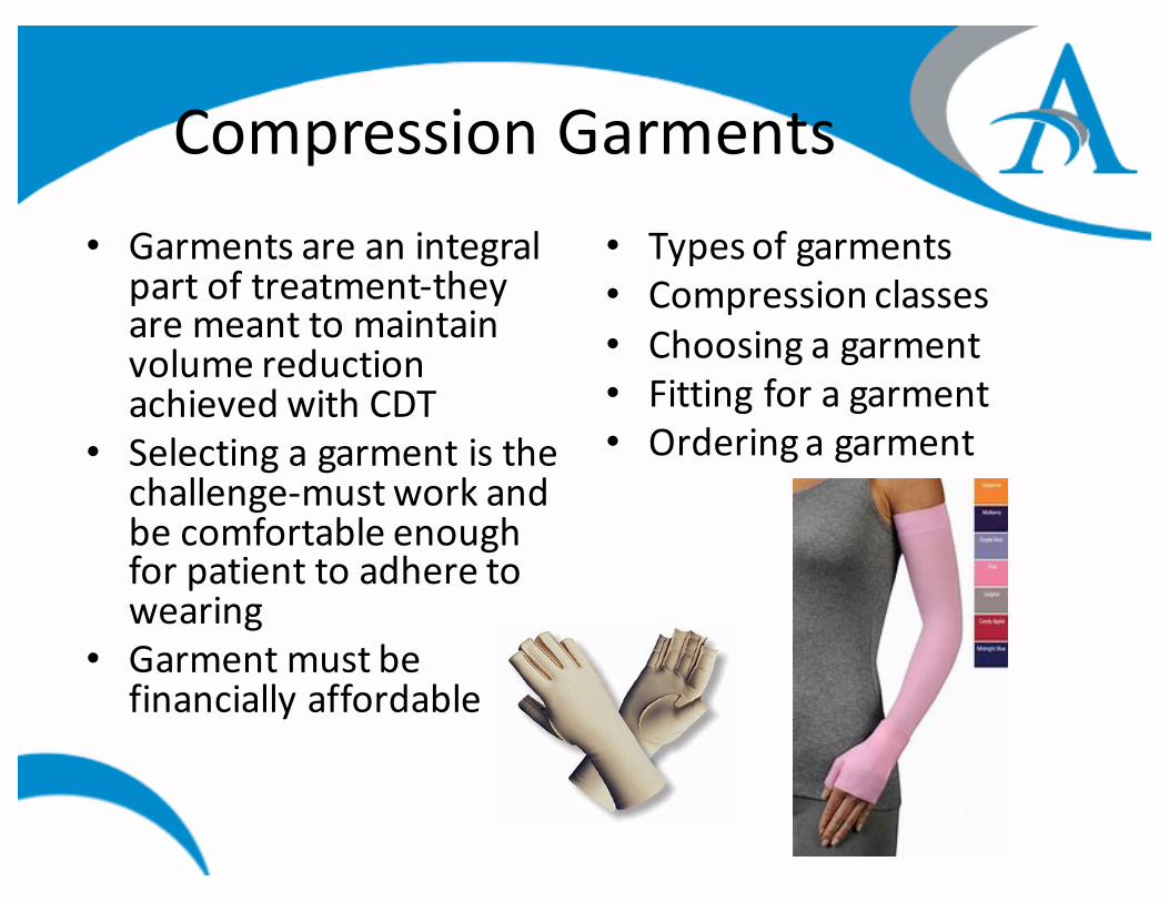
Compression Garments• Garments are an integral
part of treatment-‐they are meant to maintain volume reduction achieved with CDT
• Selecting a garment is the challenge-‐must work and be comfortable enough for patient to adhere to wearing
• Garment must be financially affordable
• Types of garments• Compression classes• Choosing a garment• Fitting for a garment• Ordering a garment

Types of GarmentsReady To Wear-‐ RTW• Circular knit• Usually come in several
sizes• Cheaper• Patient with mild to
moderate lymphedema• Relatively symmetrical
shape to limb• Usually lighter and
thinner material• Variety of colors available
Custom• Flat knit• Ideal for moderate
lymphedema• Need more containment• Perfect fit• Must be measured by a
fitter• Able to add variety of
customization• Expensive

Compression Classes
RTW
• Edema glove and preventative: 10-‐20 mmHg
• Class 1: 20-‐30 mmHg• Class 2: 30-‐40 mmHg• Class 3: 40-‐50 mmHg• Class 4: 50-‐60 mmHg
Custom
• Class 1: 18-‐21 mmHg • Class 2: 23-‐32 mmHg • Class 3: 34-‐46 mmHg

Choosing a Garment
• Balance of cost, ease of care, effectiveness, cosmesis
• Better to sacrifice amount of compression for patient adherence – go lighter to ensure patient will wear vs. higher compression class with poor adherence
• For UE’s usually want CCL 1 or 2• For LE’s usually want CCL 2 or 3

Fitting and Ordering Garments
• Measuring techniques• Insurance reimbursement• Local ordering options• Online ordering options

Elastic Taping
• Improves blood and lymphatic circulation• Increases tissue mobility to reduce scar formation
• Can assist in releasing adhered tissue• Reduced pain• Facilitates muscle activation

Taping Techniques

Acknowledgments• Foldi M, Foldi E. Foldi’s Textbook of Lymphology. 2nd ed. Munich: Elsevier: 2006.• Guenter Klose MLD/CDT Certified Instructor, CLT-‐LANA. Lymphedema Therapy
Certification Course. Presented by Klose Training, March 2010.• Courneya KS, Friedenreich CM (eds) (2011), Physical Activity and Cancer, Recent
Results in Cancer Research,. Springer-‐Verlag Berlin Heidelberg. Chapter 8 Physical Activity and Breast Cancer Survivorhip, Kathryn Schmitz.
• Fialka-‐Moser V, Crevenna R, Korpan M, Quittan M. Cancer Rehabilitation. Particularly with Aspects on Physical Impairments. J Rehabil Med 2003; 35: 153-‐162
• Kathryn Schmitz, PhD, MPH, FACSM. Beyond the PAL Trial: Making Resistance Exercise Training Standard of Care for Breast Cancer Rehabilitation. Presented at the Klose Lymphedema Conference, October 2011
• Jodi Winicour,PT, CMT, CLT-‐LANA. Breast Cancer Rehabilitation. Presented by Klose Training, September 2012
• Julie Silver, MD, STAR Program Certification Course. Oncology Rehab Partners, February 2014
• Rodrick JR, Poage E, Wanchai A. Management of lymphedema with complimentary, alternative, and other non complete decongestive therapies: a summary of the ALFP. Lymph Link. 2013; 26(4).
• Kase K, Wallis J, Kase T. Clinical Therapeutic Applications of the Kinesio Taping Method -‐ 3rd Edition. Kinesio Taping Association;2013.
• Netter F, Atlas of Human Anatomy. 2nd ed. Novartis: 1997.
Top Related