


Languages
Pages
Legal


Update of WHO and Molecular Classifications in Myelodysplastic syndromes
Mario Cazzola, MD
Department of Hematology Oncology, Fondazione IRCCS Policlinico San Matteo, and Department of Molecular Medicine, University of Pavia,
Pavia, Italy
MDS Foundation Symposium
San Francisco, CA, December 5th, 2014

Mario Cazzola Disclosures
• PI in sponsored clinical trials: no personal financial relationship with pharmaceutical companies
• Research grants from non-profit organizations or governmental agencies exclusively
• Associate Editor for Blood
RARSRARS

Pivotal role of morphology in diagnosis and prognostication of MDS
Cazzola et al. Blood. 2013 Dec 12;122(25):4021-34

WHO classification of MDS
• Refractory Cytopenia with Unilineage Dysplasia (RCUD) (mainly refractory anemia)
• Refractory Anemia with Ring Sideroblasts (RARS)
• Refractory Cytopenia with Multilineage Dysplasia (RCMD)
• Refractory Anemia with Excess Blasts (RAEB type I and II)
• Myelodysplastic Syndrome with Isolated del(5q)
Swerdlow et al (Editors). IARC 2008
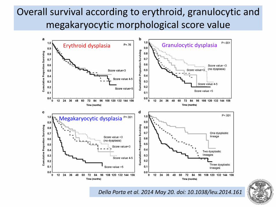
Overall survival according to erythroid, granulocytic and megakaryocytic morphological score value
Della Porta et al. 2014 May 20. doi: 10.1038/leu.2014.161
Erythroid dysplasia Granulocytic dysplasia
Megakaryocytic dysplasia

Prognostic relevance of dysplasia: number of lineages involved
Della Porta et al. 2014 May 20. doi: 10.1038/leu.2014.161
1 dysplastic lineage (mainly erythroid)
2 dysplastic lineages
3 dysplastic lineages

Outcome of MDS according to WHO classification
Cazzola. Haematologica. 2011 Mar;96(3):349-52.
943 MDS pts
Unilineage dysplasia
Excess blasts
Multilineage dysplasia

RPS14
miR-145(miR-146a)
Chr. 5
MDS with isolate del(5q): distinct nosologic entity caused by haploinsufficiency of genes mapping on the deleted region
Loss of a micro RNA and thrombocytosis Starczynowski et al. Nat Med. 2010 Jan;16(1):49-58.
Activation of p53 and apoptosis of immature red cellsBarlow et al. Nat Med. 2010 Jan;16(1):59-66Pellagatti et al. Blood. 2010 Apr 1;115(13):2721-3Dutt et al. Blood. 2011 Mar 3;117(9):2567-76
Haploinsufficiency of RPS14 phenocopies the disease in normal hematopoietic progenitor cellsEbert et al. Nature. 2008 Jan 17;451(7176):335-9
Coordinate loss of a microRNA and protein-coding gene cooperate in the pathogenesis of 5q- syndromeKumar et al. Blood. 2011 Oct 27;118(17):4666-73
Van den Berghe H, Cassiman JJ, David G, Fryns JP, Michaux JL, Sokal G. Distinct haematological disorder with deletion of long arm of no. 5 chromosome. Nature. 1974 Oct 4;251(5474):437-8.

Lenalidomide induces ubiquitination and degradation of CSNK1A1 in MDS with del(5q)
• Lenalidomide induces the ubiquitination and consequent degradation of CSNK1A1
• del(5q) cells have only one copy of CSNK1A1, so they are selectively depleted over wild-type cells
Fink et al. ASH 2014, abstract #4

MDS prognostic scoring systems
• WPSS– WHO classification (ring sideroblasts, multilineage dysplasia, excess
blasts)
– IPSS cytogenetics
– severity of anemia (transfusion requirement)
• IPSS-R– degree of cytopenia (Hb, ANC, PLT)
– excess blasts (≤2%, 3-4%, 5-10%, >10%)
– revised cytogenetics

A study of the International Working Group for Prognosis in Myelodysplasia (IWG-PM) on 5326 untreated MDS patients
Della Porta et al. 2014, unpublished results
Overall survival
WPSS
IPSS-R
Kendall’s tau 0.72 (P<.001)
Leukemia-free survival
Overall survival
Leukemia-free survival

Somatic gene mutations in patients with MDS
Haferlach et al. Leukemia. 2014 Feb;28(2):241-7
Papaemmanuil et al. Blood. 2013 Nov 21;122(22):3616-27
Genes mutated in ≥10% of MDS pts: SF3B1, TET2, SRSF2, ASXL1, DNMT3A, RUNX1
17/20 most frequently mutated genes are common to both studies (Papaemmanuil et al & Haferlach et al)
Most patients have somatic mutations of RNA splicing and/or DNA methylation

The blood cells of individuals with solid tumors contain mutations that may represent premalignant events that cause clonal hematopoietic expansion
Xie et al. Nat Med. 2014 Oct 19. doi: 10.1038/nm.3733
The Cancer Genome Atlas (TCGA)
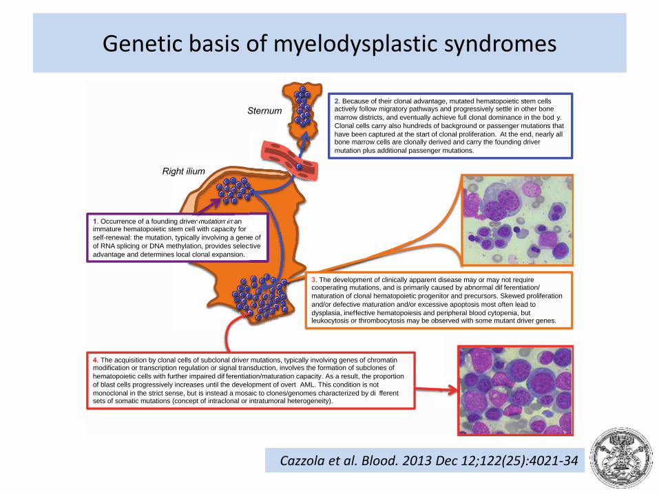
Genetic basis of myelodysplastic syndromes
Sternum
Right ilium
4. The acquisition by clonal cells of subclonal driver mutations, typically involving genes of chromatin modification or transcription regulation or signal transduction, involves the formation of subclones of
hematopoietic cells with further impaired dif ferentiation/maturation capacity. As a result, the proportion
of blast cells progressively increases until the development of overt AML. This condition is not
monoclonal in the strict sense, but is instead a mosaic to clones/genomes characterized by di fferent sets of somatic mutations (concept of intraclonal or intratumoral heterogeneity).
2. Because of their clonal advantage, mutated hematopoietic stem cells actively follow migratory pathways and progressively settle in other bone
marrow districts, and eventually achieve full clonal dominance in the bod y.
Clonal cells carry also hundreds of background or passenger mutations that
have been captured at the start of clonal proliferation. At the end, nearly all bone marrow cells are clonally derived and carry the founding driver
mutation plus additional passenger mutations.
1. Occurrence of a founding driver mutation in an immature hematopoietic stem cell with capacity for
self-renewal: the mutation, typically involving a gene of
of RNA splicing or DNA methylation, provides selective
advantage and determines local clonal expansion.
er mutation in
e of
ctive
3. The development of clinically apparent disease may or may not require cooperating mutations, and is primarily caused by abnormal dif ferentiation/
maturation of clonal hematopoietic progenitor and precursors. Skewed proliferation
and/or defective maturation and/or excessive apoptosis most often lead to
dysplasia, ineffective hematopoiesis and peripheral blood cytopenia, but leukocytosis or thrombocytosis may be observed with some mutant driver genes.
Cazzola et al. Blood. 2013 Dec 12;122(25):4021-34

Frequencies and distribution of spliceosome pathway gene mutations in myeloid neoplasms
Yoshida et al. Nature. 2011 Sep 11;478(7367):64-9

Splicing factors
Yoshida et al. Nature. 2011 Sep 11;478(7367):64-9
The SF3B1 protein is a core component of the U2 snRNP, which recognizes the 3′ splice site at intron–exon junctions

Precursor mRNA (pre-mRNA) splicing
Cazzola et al. Blood. 2013 Jan 10;121(2):260-9

Co-transcriptional RNA splicing and potential outcomes of mutations of genes encoding proteins of the spliceosome
Cazzola et al. Blood. 2013 Jan 10;121(2):260-9

Relationship between somatic SF3B1 mutations and ring sideroblasts
Quantitative enumerationof ring sideroblasts:325 MDS patients
101 (31%) patientswith mutation in SF3B1
91 patients >15% ring sideroblasts,7 patients 1-14%,
3 patients no ring sideroblasts
SF3B1 mutation: positive predictive value for ring sideroblasts 97.7%
Absence of ring sideroblasts: negative predictive valuefor SF3B1 mutation 97.8%
P=.002
Malcovati et al. Blood. 2011 Dec 8;118(24):6239-46

Cazzola et al. Blood. 2013 Jan 10;121(2):260-9
Relationship between the occurrence of a somatic SF3B1 mutation and the formation of ring sideroblasts in patients with RARS

Comprehensive analysis of aberrant RNA splicing in myelodysplastic syndromes
• RNA sequencing of CD34+ cells revealed 230 splicing events significantly enriched in SF3B1-mutated cases, of which 206 (90%) were caused by misrecognition of 3' splice sites.
• About 50% of these altered 3' splice sites resulted in frameshift, indicating that SF3B1 mutations cause deleterious effects in many genes simultaneously.
• Altered splice sites were found in genes involved in heme biosynthesis, cell cycle progression, and DNA repair
Shiozawa et al. ASH 2014, abstract #826

Novel disease paradigm
Shiozawa et al. ASH 2014, abstract #826
Stem cells Hematopoietic precursors
Occurrence of SF3B1 mutation in a multipotent hematopoietic stem cell
Misrecognition of 3' splice sites and frameshift in hundreds of genes
Gain of function at hematopoietic stem cell level
Loss of function at hematopoietic precursor level
Mutation detectable by DNA sequencing
Mutations detectable only by RNA seq
Ineffective erythropoiesis

The ability TGF-β superfamily ligand-trapping proteins to alleviate anemia with ineffective erythropoiesis in a mouse model of MDS
Suragani et al. Nat Med. 2014 Apr;20(4):408-14
Paulson RF. Nat Med. 2014 Apr;20(4):334-5

Clinical significance of SF3B1 mutation in MDS
Malcovati et al. Blood. 2011 Dec 8;118(24):6239-46

RNA splicing factors: SF3B1, SRSF2 and U2AF1
Yoshida et al. Nature. 2011 Sep 11;478(7367):64-9
SF3B1SRSF2
U2AF1

Clinical effect of spliceosome pathway gene mutations in myelodysplastic syndromes
Papaemmanuil et al. Blood. 2013 Nov 21;122(22):3616-27

Clinical effect of spliceosome pathway gene mutations in MDS with ring sideroblasts
Yoshida et al. Nature. 2011 Sep 11;478(7367):64-9
SF3B1SRSF2
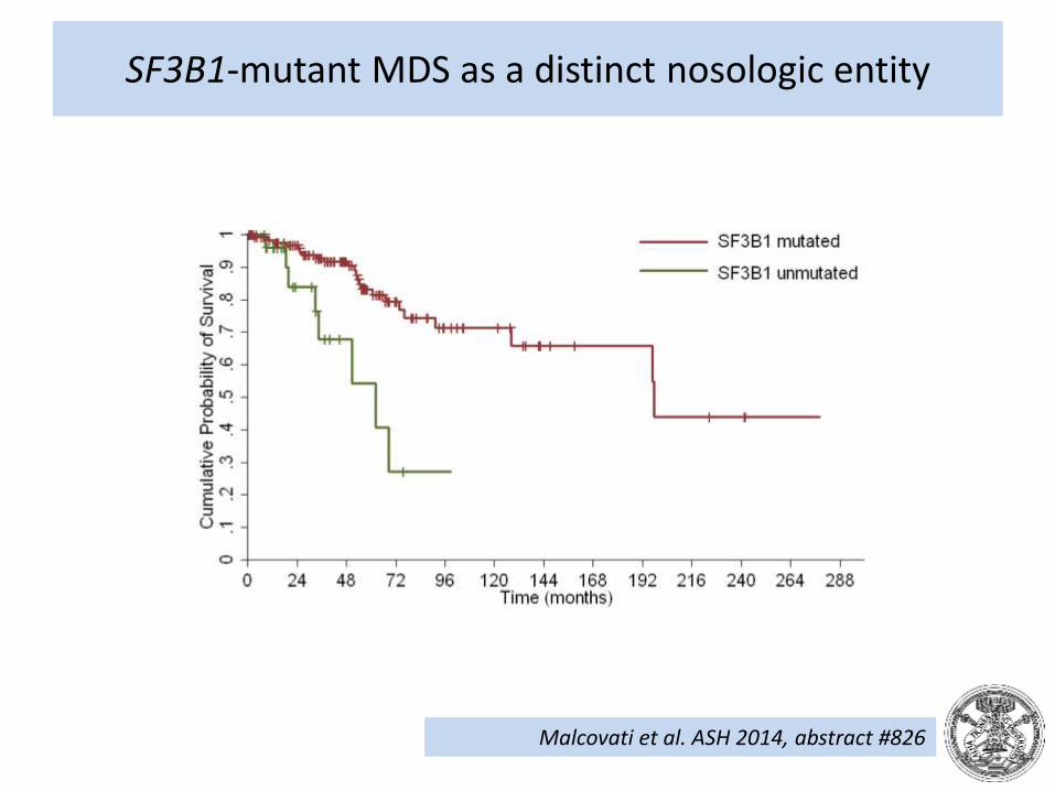
SF3B1-mutant MDS as a distinct nosologic entity
Malcovati et al. ASH 2014, abstract #826

Genetic predestination
Papaemmanuil et al. Blood. 2013 Nov 21;122(22):3616-27
Genetic “predestination”: early founding driver mutations shape the future trajectories of clonal evolution of a cancer through constraints on the repertoire of cooperating subclonal genetic lesions
SF3B1 mutSF3B1 mutJAK2 V617F
SRSF2 mut

Transition from RARS to RARS-T
Malcovati et al. Blood. 2009 Oct 22;114(17):3538-45

Genetic predestination
Papaemmanuil et al. Blood. 2013 Nov 21;122(22):3616-27
Genetic “predestination”: early founding driver mutations shape the future trajectories of clonal evolution of a cancer through constraints on the repertoire of cooperating subclonal genetic lesions
SF3B1 mutSF3B1 mutJAK2 V617F
SRSF2 mut
RARS->RARS-T
SRSF2 mutSTAG2 mut
RCMD-RS->RAEB

Unsupervised hierarchical clustering analysis of MDS patients
Cluster 1 (SF3B1 mut) Cluster 2 (NOS) Cluster 3 (MD) Cluster 4 (EB)
Malcovati et al. Blood. 2014 Aug 28;124(9):1513-21
SF3B1SRSF2U2AF1ZRSR2TET2DNMT3AIDH1/2………..
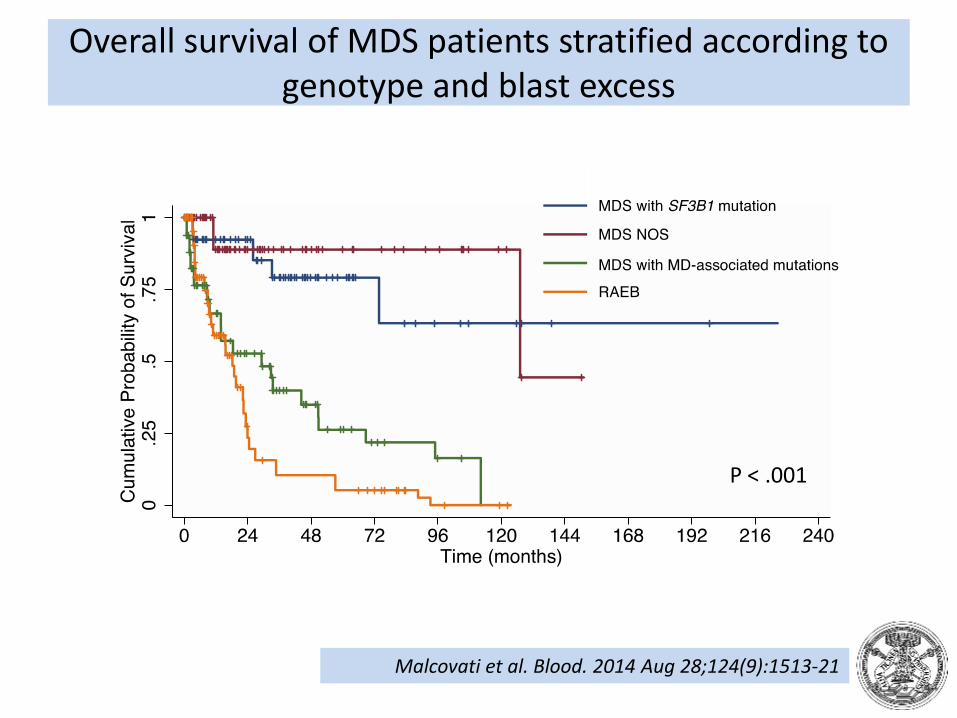
Overall survival of MDS patients stratified according to genotype and blast excess
P < .001
Malcovati et al. Blood. 2014 Aug 28;124(9):1513-21

Progression to AML in MDS patients stratified according to genotype and blast excess
P < .001
Malcovati et al. Blood. 2014 Aug 28;124(9):1513-21

Co-occurrence of TET2 and SRSF2 (or ZRSR2) mutations is highly specific for myelomonocytic phenotype
Malcovati et al. Blood. 2014 Aug 28;124(9):1513-21
CMML: Monocyte count ≥ 1.5 x 10⁹/L or TET2/SRSF2 co-mutation?

Somatic mutations of ASXL1, RUNX1 and SETBP1improve prognostic stratification of CMML
• TET2 (44%), SRSF2 (43%), ASXL1 (34%), KRAS (11%), NRAS(10%), CUX1 (10%), CBL (9%), RUNX1 (7%), SETBP1 (7%), JAK2(6%), SF3B1 (6%), and U2AF1 (5%)
• Lasso Cox regression model for genetic variable selection. The statistically significant variables were CPSS-specific cytogenetic risk groups (HR=2.49, P=.001), mutations in ASXL1(HR=2.77, P=.018), RUNX1 (HR=5.39, P=.009) and SETBP1(HR=3.96, P=.013).
• CPSS-Mol performed better than the original CPSS cytogenetic risk classification
Elena et al. ASH 2014, abstract #1915

Conclusions
• The identification of somatic mutations of RNA splicing machinery has provided a paradigm shift
• Already established genotype/phenotype relationships include
– SF3B1-mutant MDS
– TET2/SRSF2-comutant MDS/MPN (CMML)
• The time has come for us to develop a genotype-based (molecular) classification of MDS
Acknowledgments
Associazione Italiana per la Ricerca sul Cancro
MDS patients
Pavia:Luca MalcovatiMatteo G. Della PortaCristiana PascuttoIlaria AmbaglioAntonio BianchessiElisa BonoChiara ElenaAnna GallìErica TravaglinoMarta UbezioEmanuela BoveriRosangela Invernizzi
International collaborations:
• Eva Hellström-Lindberg & Karolinska investigators
• Jackie Boultwood & Oxford investigators
• Elli Papaemmanuil & Cambridge investigators
• Ulrich Germing & Duesseldorf Investigators
• Guillermo Sanz & Spanish Investigators
• Seishi Ogawa & Kyoto Investigators
Top Related