


Languages
Pages
Legal


UNIVERSITI PUTRA MALAYSIA
WOUND HEALING AND ANTIOXIDANT PROPERTIES OF EUCHEUMA COTTONII EXTRACT ON SPRAGUE DAWLEY RATS
SAMANEH GHASEMI FARD FSTM 2009 31

WOUND HEALING AND ANTIOXIDANT
PROPERTIES OF EUCHEUMA COTTONII
EXTRACT ON SPRAGUE DAWLEY RATS
SAMANEH GHASEMI FARD
MASTER OF SCIENCE
UNIVERSITI PUTRA MALAYSIA
2009
WOUND HEALING AND ANTIOXIDANT PROPERTIES OF
EUCHEUMA COTTONII EXTRACT ON SPRAGUE DAWLEY RATS
By
SAMANEH GHASEMI FARD
Thesis Submitted to the School of Graduate Studies, Universiti Putra Malaysia,
in Fulfilment of the Requirements for Degree of Master of Science
October 2009

DEDICATIONS
To the infinite source of wisdom and understanding…
“God does not play dice”
Albert Einstein
To my brother, Ashkan, who is not with us but he is alive forever in my heart
To my father who has supported me unconditionally in this journey
To my mother for having faith in me before I learned to have faith in myself
To my sister and brother, who I am alive because of them
I
Abstract of thesis presented to the Senate of Universiti Putra Malaysia in fulfilment
of the requirement for the degree of Master of Science
WOUND HEALING AND ANTIOXIDANT PROPERTIES OF EUCHEUMA
COTTONII EXTRACT ON SPRAGUE DAWLEY RATS
By
SAMANEH GHASEMI FARD
October 2009
Chair: Suhaila Mohamed, PhD
Faculty: Food Science and Technology
Wounds and their treatment are a big burden on the healthcare system, both in terms
of cost, time and energy of care required. The lost in productivity and decreased
quality of life is immeasurable. This study reports on the potential wound healing and
antioxidant properties of oral consumption of ethanolic and aqueous extracts of
Eucheuma cottonii. Two cm diameter excision of skin wound model was used, with
honey (100 mg/kg body weight) as positive control and untreated normal rats as
negative control groups. Both extracts significantly (P<0.05) increased the rate of
wound contraction, better than honey. The extracts decreased lipid peroxidation in the
plasma and increased erythrocyte antioxidant enzyme activities (superoxide
dismutase) and reduced glutathione compared to both the positive and negative
control groups. The ethanolic extract was more effective than the aqueous extract by
20%. Histopathological wound tissue observations showed both extracts significantly

II
reduced scars, enhanced epithelization, hair follicle growth and tissue granulation
compared to both control groups. The HPLC results revealed that E.cottonii
possessed several antioxidant compounds, which may be responsible for the wound
healing acceleration. This is the first report showing that oral consumption of tropical
seaweed extracts could enhance wound healing.

III
Abstrak tesis yang dikemukakan kepada Senat Universiti Putra Malaysia
sebagai memenuhi keperluan untuk Ijazah Master Sains
CIRI-CIRI PENYEMBUHAN LUKA DAN ANTIOKSIDA DARI EKSTRAK
EUCHEUMA COTTONII PADA TIKUS SPRAGUE DAWLEY
Oleh
SAMANEH GHASEMI FARD
October 2009
Pengerusi: Suhaila Mohamed, PhD
Fakulti: Sains dan Teknologi Makanan
Luka yang teruk dan rawatannya telah menjadi beban kepada sistem rawatan
kesihatan dari segi kos, masa dan tenaga. Kehilangan produktiviti dan pengurangan
kualiti di dalam hidup tidak dapat dinafikan lagi. Pengurangan ini melaporkan kesan
pengambilan ekstrak ethanolik dan akues Eucheuma cottonii terhadap potensinya
untuk mempercepatkan penyembuhan luka. Luka berdiameter 2 cm telah dilakukan
ke atas tikus sebagai model. Madu (100 mg/kg berat badan) telah digunakan sebagai
kawalan positif dan tikus yang normal dan tidak diberikan rawatan dijadikan sebgai
kawalan negatif. Kedua-dua ekstrak menunjukkan peningkatan yang bermakna
terhadap kadar pengecutan luka berbanding madu. Ekstrak ini telah mengurangkan
peroksidaan lipid di dalam plasma dan meningkatkan aktiviti enzim antioksidan
(superoxide dismutase) dan reduced glutathione di dalam darah berbanding kedua-
dua kumpulan kawalan positif dan negatif. Ekstrak etholik lebih efektif berbanding

IV
ekstrak akues sebanyak 20%. Pemerhatian luka secara histopatologi terhadap kedua-
dua esktrak menunjukkan pengurangan jelas terhadap parut, peningkatan epithelisasi
dan pertumbuhan folikel rambut dan granulasi tisu berbanding kedua-dua kumpulan
kawalan. Keputusan HPLC menunjukkan E.cottonii mempunyai beberapa bahan
antioksidan yang dapat mempercepatkan penyembuhan luka. Ini adalah laporan yang
pertama yang menunjukkan pengambilan rumpai laut secara oral dapat
mempercepatkan penyembuhan luka.

V
ACKNOWLEDGEMENTS
Although I cannot express in words the extent of my gratitude, I would like to thank
my advisor Prof. Dr. Suhaila Mohamed from whom I have learned a great deal, not
just scientifically but also personally. It goes without saying that this dissertation
would not have been possible without her guidance and keen advice. Her ability to
keep the big picture in sight has pulled me back on track on numerous occasions. I
truly appreciate the long discussions we have had where she helped infuse clarity into
my own thoughts and ideas. Most of all, I have to thank her for teaching me how to
communicate and present myself as a scientist and for showing me, by example, what
it takes to be a successful scientist/person.
I would also like to thank the members of my thesis committee, Dr. Kharidah
Muhammad and Dr. Goh Yong Meng
for taking the time and effort to come to my
thesis committee meetings and for offering very helpful advice and suggestions to
keep my research as focused as possible.
I would also like to thank Dr. Ajwad Awad Mohammed and Professor Karim Alwan
AL-Jashamy for technical support in histopathological evaluation.
In addition, my deep appreciation goes to all of my lab members and friends who
were the biggest support throughout my years as a graduate student, Rosalina Tan
Roslan Tan, Fatemeh Shamsabadi, Mahsa Motshakeri, Mehdi Javadi, Maslia Manja
B.Z and Anyanji Victor Uchenna.

VI
I would like to acknowledge Dr. Gururaj Bagal Kotkar in the Institute of Bioscience
(IBS) and Mr. Abd. Halim bin Abd. Rahman at the faculty of Food Science and
Technology for their valuable assistance in HPLC. I would like to thank Mr. Azman
Bin Abu Yamin whom I had bothered with many technical questions in the past few
years. My gratitude to Mr. Kufli Che Noor for his help in animal study.
I would like to thank my parents, Tahereh and Aliakbar for their consistent support
and love. They have shared all the bad and good times together with me in my life.
Finally, I gratefully acknowledge the support for this work provided by the Ministry
of Higher Education Fundamental Research Grant Scheme under Grant No: 91030.

VII
I certify that a Thesis Examination Committee has met on 15 October 2009 to
conduct the final examination of Samaneh Ghasemi Fard on her thesis entitled
Wound Healing and Antioxidant Properties of Eucheuma cottonii extract on
Sprague Dawley rats in accordance with the Universities and University Colleges
Act 1971 and the Constitution of the Universiti Putra Malaysia [P.U.(A) 106] 15
march 1998. The Committee recommends that the student be awarded the Master of
Science.
Members of the Examination Committee were as follows:
Faridah Abas, PhD
Lecturer
Faculty of Food Science and Technology
Universiti Putra Malaysia
(Chairperson)
Fauziah Othman, PhD
Professor
Faculty of Medicine and Health Science
Universiti Putra Malaysia
(Internal Examiner)
Azizah Abdul Hamid, PhD
Associate Professor
Faculty of Food Science and Technology
Universiti Putra Malaysia
(Internal Examiner)
Ayub Mohd Yatim, PhD
Associate Professor
Faculty Science and Technology
Universiti Kebangsaan Malaysia
(External Examiner)
BUJANG KIM HUAT, PhD
Professor and Deputy Dean
School of Graduate Studies
Universiti Putra Malaysia
Date: 24 December 2009

VIII
This thesis was submitted to the Senate of Universiti Putra Malaysia and has been
accepted as fulfilment of the requirement for the degree of Master of Science. The
members of the Supervisory Committee were as follows:
Suhaila Mohamed, PhD
Professor
Faculty of Food Science and Technology
Universiti Putra Malaysia
(Chairman)
Goh Yong Meng
Lecturer
Faculty of Veterinary Medicine
Universiti Putra Malaysia
(Member)
Sharifah Kharidah Bt Syed Muhammad
Associate professor
Faculty of Food Science and Technology
Universiti Putra Malaysia
(Member)
HASANAH MOHD/GHAZALI, PhD
Professor and Dean
School of Graduate Studies
Universiti Putra Malaysia
Date: 14 Jauary 2010

IX
DECLARATION
I declare that the thesis is my original work except for quotations and citation which
have been duly acknowledged. I also declare that it has not been previously, and is
not concurrently, submitted for any other degree at Universiti Putra Malaysia or at
any other institution.
SAMANEH GHASEMI FARD
Date: 18 June 2009

X
TABLE OF CONTENTS
Page
DEDICATIONS
ABSTRACT I
ABSTRAK III
ACKNOWLEDGEMENTS V
APPROVAL VII
DECLARATION IX
LIST OF TABLES XII
LIST OF FIGURES XIII
LIST OF ABBREVIATIONS XV
CHAPTER
1 INTRODUCTION 1
1.1 General introduction 1
1.2 Objectives 3
2 LITERATURE REVIEW 4
2.1 Seaweeds 4
2.1.1 Description 4
2.1.2 Compounds in seaweeds 6
2.1.3 Biological effects 8
2.2 Free radical in wound healing 9
2.3 Wound healing 10
2.3.1 Normal wound healing 10
2.3.2 Nutritional support for wound healing 13
2.3.3 Botanical medicines for wound healing 14
2.4 Hair follicle 15
2.4.1 Hair follicle structure 15
2.4.2 Morphological stages of hair cycle 17
2.4.3 Nutritional support for hair follicle growth 17
2.4.4 Botanical medicines for hair follicle 18
2.5 High pressure liquid chromatography (HPLC) 19
2.5.1 Flavonoids analysis using HPLC 19
3 MATERIALS AND METHODS 22
3.1 Chemicals and reagents 22
3.2 Sample preparation 22
3.3 Preparation of sample extract 23
3.4 Animal subjects 25
3.5 Wound creation and treatments 26
3.6 Sampling and wound healing evaluation 27
3.6.1 Wound contraction 28
3.6.2 Period of epithelization 29

XI
3.7 Sampling and biochemical assay 29
3.7.1 Plasma and red blood cells (RBC) preparation 29
3.7.2 Catalase (CAT) activity measurement 30
3.7.3 Superoxide dismutase (SOD) activity measurement 30
3.7.4 Reduced glutathione (GSH) assay 32
3.7.5 Malondialdehyde (MDA) level determination 32
3.8 Determination of flavonoids 33
3.8.1 Chemicals and reagents 33
3.8.2 Sample preparation and HPLC procedure 34
3.9 Statistical analysis 35
4 RESULTS AND DISCUSSION 36
4.1 Body weight 36
4.2 Wound healing 38
4.2.1 Wound contraction 38
4.2.2 Period of epithelization 42
4.2.3 Histopathology of wound 43
4.2.3.1 Re-epithelization 43
4.2.3.2 Granulation tissue development 52
4.3 Hair follicle growth and hair cycle 54
4.3.1 Percentage of hair growth 54
4.3.2 Counting hair follicles 59
4.3.3 Sebaceous glands 62
4.4 Analysis of antioxidants in blood 63
4.4.1 Catalase (CAT) activity 63
4.4.2 Superoxide dismutase (SOD) activity 66
4.4.3 Reduced glutathione (GSH) activity 69
4.5 Malondialdehyde (MDA) level 72
4.6 Antioxidants in ethanolic extract of seaweed 74
4.6.1 Flavonoids 74
5 CONCLUSION 82
REFERENCES 84
BIODATA OF STUDENT 99

XII
LIST OF TABLES
Table Page
3.1 Gradient program used for the separation of seaweed flavonoids 35
4.1 Variation in body weights of rats fed with E. cottonii extracts during the
experimental study (in grams) 37
4.2 The wound area (mm2) of different groups of rats fed with E. cottonii extracts
over a period of 15 days 40
4.3 The wound healing (%) of different groups of rats fed with E. cottonii extracts
over a period of 15 days 41
4.4 Period of epithelialization (day) of rats fed with E. cottonii extracts 42
4.5 Number of hair follicle, area size of hair bulb, and length of hair follicle
around the wound in rats treated with various extractions of E. cottonii at day
15 60
4.6 Catalase activity (k/mg protein) in erythrocytes rats fed with E. cottonii
extracts over a period of 15 days 65
4.7 Superoxide dismutase activity (U/mg protein) in erythrocytes rats fed with E.
cottonii extracts over a period of 15 days 68
4.8 Reduced glutathione (µg/g protein) in erythrocytes rats fed with E. cottonii
extracts over a period of 15 days 71
4.9 Malondialdehyde level (µM/g protein) in erythrocytes rats fed with E. cottonii
extracts over a period of 15 days 73
4.10 Compounds identified in Er.t by HPLC 76
XIII
LIST OF FIGURES
Figure Page
2.1 The sequence of events during normal wound healing 10
2.2 The blood components spill into the site of injury 12
2.3 Impact of nutrients on the different phases of wound healing 13
2.4 Hair follicle structure 15
3.1 Eucheuma cottonii 23
3.2 The sequence of extraction method 25
3.3 Photograph showing excision wound 27
3.4 Evaluation method of wound healing 28
4.1 Photomicrographs of epidermis in Er.t group 46
4.2 Photomicrographs of epidermis in E70 group 47
4.3 Photomicrographs of epidermis in Wr.t group 48
4.4 Photomicrographs of epidermis in W70 group 49
4.5 Photomicrographs of epidermis in HNY group 50
4.6 Photomicrographs of epidermis in WTR group 51
4.7 Granulation tissue of the wounded skin treated orally with Er.t (A), E70 (B),
Wr.t (C), W70 (D), HNY (E) and WTR (F) (H&E stain; ×400) 53
4.8 Photograph showing the hair growth in Er.t group at the 15 day post treatment
56
4.9 Photograph showing the hair growth in E70 group at the 15 day post treatment
56
4.10 Photograph showing the hair growth in Wr.t group at the 15 day post
treatment 57

XIV
4.11 Photograph showing the hair growth in W70 group at the 15 day post
treatment 57
4.12 Photograph showing the hair growth in HNY group at the 15 day post
treatment 58
4.13 Photograph showing the hair growth in WTR group at the 15 day post
treatment 58
4.14 Micrographs of hair follicle after 15 days oral treatment in different treatment
groups (H&E stain; ×40) 61
4.15 Light microscopy of sebaceous glands in ethanolic extract group (a) and
negative control group (b) at 15 day post treatment (H&E stain; ×100) 62
4.16 Chromatogram of five flavonoids standards 76
4.17 Flavonoids in ethanolic extract of E. cottonii 77
4.18 Co-elution of putative catechin (1), rutin (3) and quercetin (5) peaks in
ethanolic extract of E. cottonii with standard compounds added 77
4.19 Structure of the flavonol quercetin showing features important in defining the
classical antioxidant potential of flavonoids 79
4.20 Hypothesis of the links between the working mechanisms of flavonoids and
their effects on disease 80
XV
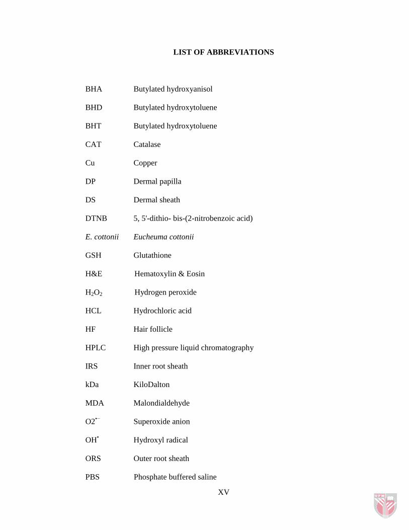
LIST OF ABBREVIATIONS
BHA Butylated hydroxyanisol
BHD Butylated hydroxytoluene
BHT Butylated hydroxytoluene
CAT Catalase
Cu Copper
DP Dermal papilla
DS Dermal sheath
DTNB 5, 5'-dithio- bis-(2-nitrobenzoic acid)
E. cottonii Eucheuma cottonii
GSH Glutathione
H&E Hematoxylin & Eosin
H2O2 Hydrogen peroxide
HCL Hydrochloric acid
HF Hair follicle
HPLC High pressure liquid chromatography
IRS Inner root sheath
kDa KiloDalton
MDA Malondialdehyde
O2•−
Superoxide anion
OH• Hydroxyl radical
ORS Outer root sheath
PBS Phosphate buffered saline
XVI
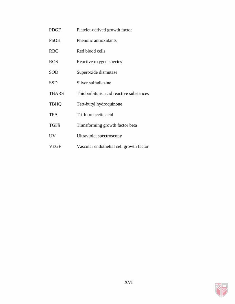
PDGF Platelet-derived growth factor
PhOH Phenolic antioxidants
RBC Red blood cells
ROS Reactive oxygen species
SOD Superoxide dismutase
SSD Silver sulfadiazine
TBARS Thiobarbituric acid reactive substances
TBHQ Tert-butyl hydroquinone
TFA Trifluoroacetic acid
TGFß Transforming growth factor beta
UV Ultraviolet spectroscopy
VEGF Vascular endothelial cell growth factor

1
CHAPTER ONE
INTRODUCTION
1.1 General introduction
Wounds are unavoidable events of life and might arise due to any agent that
induces injury or stress and wound has been a menace the world over. Healing is a
survival mechanism and represents an attempt to maintain normal anatomical
structure and function. Treatment is therefore aimed at minimizing the undesired
consequences. Wound healing management is a complicated and expensive
program. Research on drugs that improve wound healing is developing in modern
biomedical sciences. Several drugs obtained from plant sources are known to
improve healing of different wound types. Some of these drugs have screened
scientifically for evaluation of their wound healing activity in different
pharmacological models and patients, but the potential of many herbal agents used
traditionally, remains unexplored (Sandeep et al., 2009).
Hair has many useful biological functions, including protection from the elements
and dispersion of sweat gland products. It also has psychosocial importance in our
society and patients with hair loss (alopecia) often suffer tremendously (Paus and
Cotsarelis, 1999). The demand for drugs that alter hair growth and appearance has
led to multibillion dollar industries; also synthetic based product may cause human
health hazard with several side effects, therefore investigation on plant extracts in
order to find natural products are effective for these purposes (Rathi et al., 2008).

2
In order to find an effective natural product, that possesses both wound healing
and hair re-growth properties, Eucheuma cottonii was chosen as one of the edible
tropical seaweeds. It was obtained from Sabah area of Malaysia and was studied as
a novel source of variety compounds (Matanjun et al., 2008) that is necessary for
both properties, like polyphenols, vitamin C, -tocopherol, minerals and protein.
The seaweeds are used worldwide for many medicinal purposes.
There are reports in the literature of sulphated polysaccharides as antiviral
substances and fucoidans as anticoagulant, antithrombotic, anti-inflammatory and
antitumoral. Also there are a clear understanding of the mechanisms of action of
flavonoids, either as antioxidants or modulators of cell signalling, and the
influence of their metabolism on these properties are key to the evaluation of these
potent biomolecules as wound healing (Williams et al., 2004)
Flavonoids have certain health effects and their antioxidant, radical scavenging,
anti-mutagenic and anti-carcinogenic properties are well known (Middleton et al.,
2000). The therapeutic applications of flavonoids on inflammation have previously
been reported. Inflammation is important in many serious diseases. Therefore,
intake of flavonoids is very important in the management of wound repair
(Havsteen, 2002).

3
1.2 Objectives
With the above background, this study has the following objectives:
1. To evaluate the efficacy of seaweed extracts for wound healing and hair
follicle growth in normal rats.
2. To study blood antioxidant activities of seaweed extracts.
3. To identify the flavonoids responsible for wound healing and hair follicle
growth by HPLC.

4
CHAPTER TWO
LITERATURE REVIEW
2.1 Seaweeds
2.1.1 Description
Seaweeds (algae) are not true plants. They do not have flowers, any clearly
marked steam or leaves. They do not have true roots but has held fast, which does
not absorb food but simply attaches the plant firmly to a stone or rock. All
seaweeds at some stage in their life cycles are unicellular, as spore or zygotes, and
may be temporarily plank tonic (Guiry, 1998).
There are over 9,000 species of seaweeds which can be organized into three major
types: green (Chlorophyta), brown (Phaeophyta) and red (Rhodophyta). Species of
the genera Caulerpa, Durvillea, Laminaria, Monostroma, Nereocytstis,
Oedogonium, Porphyra, Rhodymenia, Sargassum, and Spirogyra are particularly
commonly used as food in different parts of the world. Red is the most species-
rich group (6,000) followed by brown (2,000) and green (1,200) (Guiry, 1999).
Seaweeds have been consumed in Asia since ancient times. Consumption of
brown (66.5%), red (33%) and green algae (5%) is high in Japan and China
compared to other Asian countries (Dawes, 1998). McHugh (2003) showed other
countries, such as the Republic of Korea, the United States of America, South
America, Ireland, Iceland, Canada and France have significantly increased

5
consumption, production and marketing of seaweeds. Approximately one million
tonnes of wet seaweeds were harvested in 35 countries as a source of food,
polysaccharides, fertilizer, fuel and cosmetics annually (Ruperez and Saura-
Calixto, 2002). More recently marine algae have been utilized in Japan as raw
materials in the manufacture of many seaweed food products such as jam, cheese,
wine, tea, soup and noodles (Nisizawa et al., 1987) and in the western countries,
mainly as a source of polysaccharides (agar, alginates, carrageenans) for the food
and pharmaceutical industries. Seaweeds as a food in Malaysia are not as common
as in countries like Japan and China. At present, seaweed is only consumed in
certain coastal areas especially along the east coast of peninsula Malaysia, where
it is occasionally eaten as a salad dish (Wong and Peter, 2000).
Seaweeds are a valuable food source as they contain protein, lipids, vitamins and
minerals (Norziah and Ching, 2000; Sa'nchez-Machado et al., 2004). Seaweeds are
not only a useful food source to humans, whole plants and seaweed mixes have
been used in animal nutrition (Chapman and Chapman, 1980; Robledo and Freile-
Pelegrin, 1997), poultry feed (Briand, 1991) and fish feed (McHugh, 2003). Some
countries like Hong Kong used seaweeds as animal feeds or fertilizers, especially
among the coastal villagers (Hodgkiss and Lee, 1983). However, very few of the
world‟s available seaweed species are used commercially. This may be because
they cannot be harvested or cultivated on a commercially viable scale, or because
their composition simply makes them unsuitable.
Top Related