


Languages
Pages
Legal

8/2/2019 Unit 5, Topic 7 NOTES
http://slidepdf.com/reader/full/unit-5-topic-7-notes 1/10
Biology Notes – Topic 7 Katherine Burke
Biology Unit 5
Joints and Movement
Muscles bring about movement at a joint, at least two are needed to move a boneto and fro because muscles can only pull. A pair of muscles working in this way
are described as antagonistic. A muscle that causes contraction is called an
extensor while the flexor muscle contracts in the reverse movement.
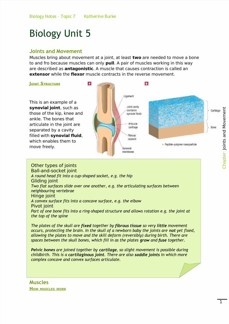
JOINT STRUCTURE
This is an example of a
synovial joint, such as
those of the kip, knee andankle. The bones that
articulate in the joint are
separated by a cavity
filled with synovial fluid,
which enables them to
move freely.
MusclesHOW MUSCLES WORK
C
h a p t e r : J o i n t s a n d M o v e m e
n t
1
Other types of jointsBall-and-socket joint A round head fit into a cup-shaped socket, e.g. the hip
Gliding jointTwo flat surfaces slide over one another, e.g. the articulating surfaces betweenneighbouring vertebrae
Hinge joint A convex surface fits into a concave surface, e.g. the elbow
Pivot jointPart of one bone fits into a ring-shaped structure and allows rotation e.g. the joint atthe top of the spine
The plates of the skull are fixed together by fibrous tissue so very little movementoccurs, protecting the brain. In the skull of a newborn baby the joints are not yet fixed,allowing the plates to move and the skill deform (reversibly) during birth. There arespaces between the skull bones, which fill in as the plates grow and fuse together.
Pelvic bones are joined together by cartilage, so slight movement is possible duringchildbirth. This is a cartilaginous joint. There are also saddle joints in which morecomplex concave and convex surfaces articulate.

8/2/2019 Unit 5, Topic 7 NOTES
http://slidepdf.com/reader/full/unit-5-topic-7-notes 2/10
Biology Notes – Topic 7 Katherine Burke
Muscle is made up of bundles of muscle fibres. Each fibre is a single muscle cell.
Each cell has several nuclei, referred to as multinucleate. This is because one
nucleus does not effectively control the metabolism of such a long cell. During
prenatal development, several cells fuse together to form the length of muscle
fibres. The muscle cells are stripped which is important for them to be able to
contract.
Tendon
Tendons at each end of the muscle connect the muscle to bone.
Bundle of Muscle Fibres
The muscle is made up of bundles of muscle fibres up to 2cm across. These are
bound together by connective tissue, which is continuous with the tendons.
Muscle Fibre
Each muscle fibre is a single muscle cell surrounded by a cell surface membrane.
Each muscle fibre may be several centimetres long, but is less than 0.1mm in
diameter. Inside the muscle fibre is the cytoplasm containing mitochondria and
other organelles.
Myofibrils
Within each muscle fibre there are numerous myofibrils; each is composed of
repeated contractile units called sarcomeres.
INSIDE MUSCLE FIBRES
C
h a p t e r : J o i n t s a n d M o v e m e
n t
2
8/2/2019 Unit 5, Topic 7 NOTES
http://slidepdf.com/reader/full/unit-5-topic-7-notes 3/10
3. The banding patterns created on an extendedmuscle myofibril
Biology Notes – Topic 7 Katherine Burke
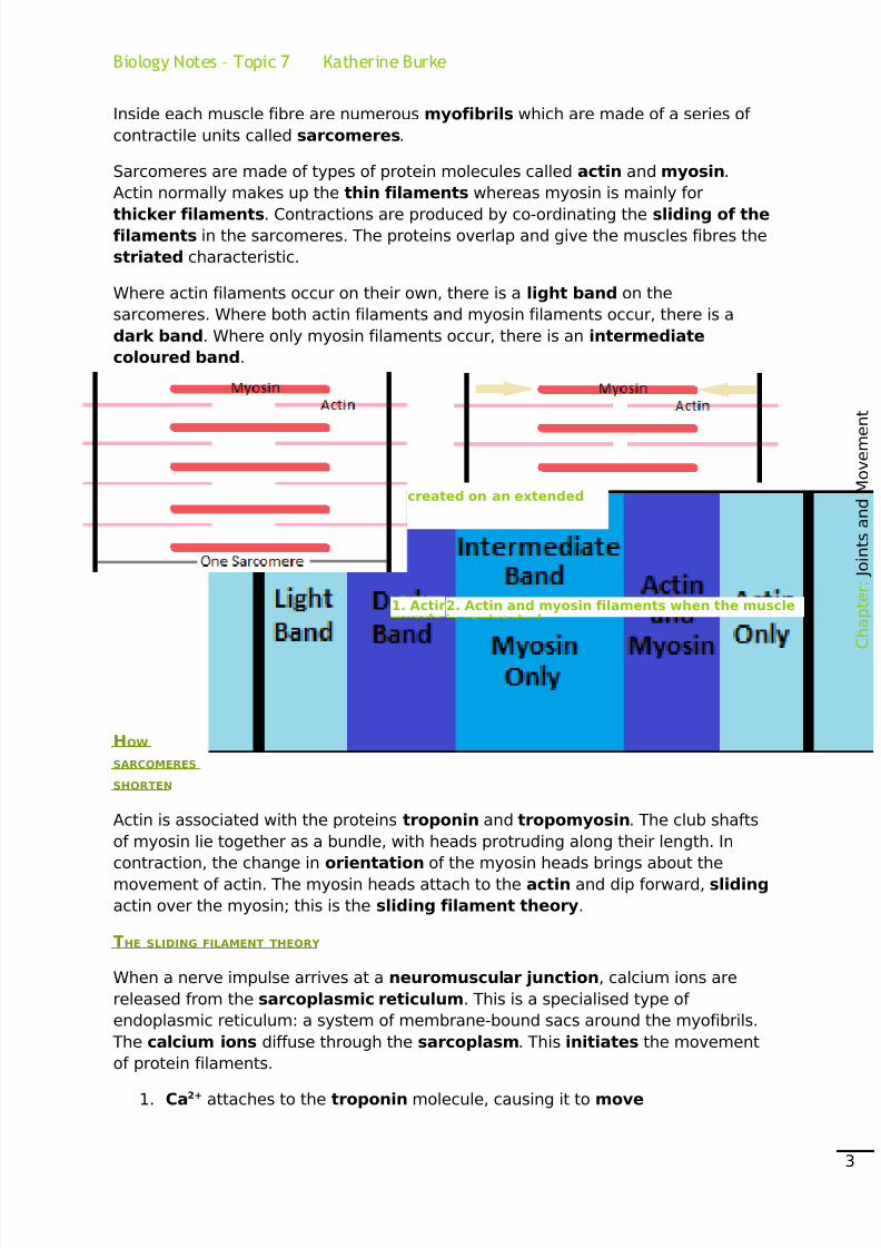
Inside each muscle fibre are numerous myofibrils which are made of a series of
contractile units called sarcomeres.
Sarcomeres are made of types of protein molecules called actin and myosin.
Actin normally makes up the thin filaments whereas myosin is mainly for
thicker filaments. Contractions are produced by co-ordinating the sliding of thefilaments in the sarcomeres. The proteins overlap and give the muscles fibres the
striated characteristic.
Where actin filaments occur on their own, there is a light band on the
sarcomeres. Where both actin filaments and myosin filaments occur, there is a
dark band. Where only myosin filaments occur, there is an intermediate
coloured band.
HOW
SARCOMERES
SHORTEN
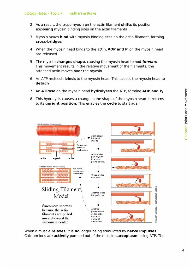
Actin is associated with the proteins troponin and tropomyosin. The club shafts
of myosin lie together as a bundle, with heads protruding along their length. In
contraction, the change in orientation of the myosin heads brings about themovement of actin. The myosin heads attach to the actin and dip forward, sliding
actin over the myosin; this is the sliding filament theory.
THE SLIDING FILAMENT THEORY
When a nerve impulse arrives at a neuromuscular junction, calcium ions are
released from the sarcoplasmic reticulum. This is a specialised type of
endoplasmic reticulum: a system of membrane-bound sacs around the myofibrils.
The calcium ions diffuse through the sarcoplasm. This initiates the movement
of protein filaments.
1. Ca2+ attaches to the troponin molecule, causing it to move
C
h a p t e r : J o i n t s a n d M o v e m e
n t
3
1. Actin and myosin filaments when the2. Actin and myosin filaments when the muscle
8/2/2019 Unit 5, Topic 7 NOTES
http://slidepdf.com/reader/full/unit-5-topic-7-notes 4/10
Biology Notes – Topic 7 Katherine Burke
2. As a result, the tropomyosin on the actin filament shifts its position,
exposing myosin binding sites on the actin filaments
3. Myosin heads bind with myosin binding sites on the actin filament, forming
cross-bridges
4. When the myosin head binds to the actin, ADP and Pi on the myosin head
are released
5. The myosin changes shape, causing the myosin head to nod forward.
This movement results in the relative movement of the filaments; the
attached actin moves over the myosin
6. An ATP molecule binds to the myosin head. This causes the myosin head to
detach
7. An ATPase on the myosin head hydrolyses the ATP, forming ADP and Pi
8. This hydrolysis causes a change in the shape of the myosin head. It returns
to its upright position. This enables the cycle to start again
When a muscle relaxes, it is no longer being stimulated by nerve impulses.
Calcium ions are actively pumped out of the muscle sarcoplasm, using ATP. The
C
h a p t e r : J o i n t s a n d M o v e m e
n t
4

8/2/2019 Unit 5, Topic 7 NOTES
http://slidepdf.com/reader/full/unit-5-topic-7-notes 5/10
Biology Notes – Topic 7 Katherine Burke
troponin and tropomyosin move back, once again blocking the myosin binding
sites on the actin. In the absence of ATP, the cross-bridges remain attached. This
is what happens in rigor mortis when the muscles that are contracted become
rigid.
Energy for Action The minimum energy requirement is called the basal metabolic rate (BMR),
measured in kJ g-1 h-1. It is used to measure the minimum energy requirement of
the body at rest to fuel basic metabolic processes. BMR is measured by recording
oxygen consumption under strict conditions of no food consumption for 12
hours before measurement; the body had to be totally at rest in a
thermostatically controlled room. BMR is roughly proportional to the body’s
surface area. It varies between individuals depending on their age and gender.
Percentage body fat seems to be important in accounting for these differences.
Physical activity increases the body’s total daily energy expenditure. Energy is
needed for muscle contraction to move the body but the energy can vary on how
the muscles are used. For example, an elite marathon runner uses energy at
almost half the rate of a sprinter.
Releasing energy
Food is the source of energy for all animal activity. The main energy sources are
carbohydrates and fats that have been absorbed or stored around the body.
Respiration is linked to ATP synthesis as the cells use the molecule ATP as an
energy carrier molecule.
ATP is created from ADP by the addition of Pi. In solution, phosphate ions are
hydrated and so the phosphate needs to be separated from these water
molecules to make ATP, requiring energy. ATP in water is higher in energy thanADP and phosphate ions in water, so ATP is water is a way of storing chemical
Other types of muscleMuscles found in the gut wall, blood vessels and the iris of the eye are known as smoothmuscle as their fibres do not appear to be striped. These are small cells with a single nucleus. They have a similar mechanism of contraction to skeletal muscle, using myosinand actin protein filaments. However, they are not arranged in the same way as they have gap junctions. These intercellular channels less than 2nm in diameter, and arebetween the smooth muscle cells to give cytoplasmic continuity between the cells. Thisallows chemical and electrical signals to pass between adjacent cells, and so allowssynchronised contraction. Contractions in smooth muscle fibres are slower and longer lasting and the fibres fatigue very slowly if at all.
The heart walls are made of specialised muscle fibres called cardiac muscle. These arestriped and interconnected to ensure that a co-ordinated wave of contraction occurs inthe heart. Cardiac muscle fibres do not fatigue. Neither smooth nor cardiac muscles areunder conscious control.
C
h a p t e r : J o i n t s a n d M o v e m e
n t
5
8/2/2019 Unit 5, Topic 7 NOTES
http://slidepdf.com/reader/full/unit-5-topic-7-notes 6/10
Biology Notes – Topic 7 Katherine Burke
potential energy. ATP keeps the phosphate separated from the water, but they
can be brought together in an energy-yielding reaction every time energy is
needed for reactions within the cell.
When one phosphate group is removed from ATP by hydrolysis, ADP forms. A
small amount of energy is required to break the bond holding the end phosphatein the ATP. Once removed, the phosphate group becomes hydrated. A lot of
energy is released as bonds form between water and phosphate. This energy can
be used to supply energy-requiring reactions in the cell. Some of the energy
transferred during hydration of phosphate from ATP will raise the temperature
of the cell; some is available to drive other metabolic reactions such as muscle
contraction, protein synthesis or active transport. The hydrolysis of ATP is coupled
to these other reactions:
Carbohydrate oxidation
If exercise is low intensity, enough oxygen is supplied to cells to enable ATP to be
regenerated through aerobic respiration of fuels. Fates and carbohydrates are
oxidised to carbon dioxide and water. A summary of the equation for aerobic
respiration:
Photosynthesis was described as a process that separates hydrogen from oxygen
by photolysis. The hydrogen from water is stored by combining it was carbon
dioxide to form carbohydrate. In aerobic respiration, the hydrogen stored in
glucose is brought together with oxygen to form water again. The bonds between
hydrogen and carbon atoms in glucose are not as strong as the bonds between
hydrogen and oxygen atoms in water. Therefore, the input of energy needed to
break the bonds in glucose and oxygen is not as great as the energy released
when the bonds in carbon dioxide and water are formed. Overall, there is a
release of energy and this can be used to generate ATP.
Glucose and oxygen are not brought together directly, as this would release large
amounts of energy too quickly and therefore damage the cell. Glucose is split
apart in a series of small steps with carbon dioxide as the waste product.Hydrogen from the glucose is eventually reunited with oxygen to release large
amounts of energy as water is formed.
C
h a p t e r : J o i n t s a n d M o v e m e
n t
6

8/2/2019 Unit 5, Topic 7 NOTES
http://slidepdf.com/reader/full/unit-5-topic-7-notes 7/10
Biology Notes – Topic 7 Katherine Burke
GLYCOLYSIS FIRST
The initial stage of carbohydrate
breakdown, known as glycolysis, occurs in
the cytoplasm of cells and the sarcoplasm
of muscle cells.
Stores of glycogen in muscle or liver cellsmust first be converted to glucose. It is a
good fuel but it is quite stable and
unreactive. Therefore, the first reactions of
glycolysis need an input of energy from ATP
to get started. Two phosphate groups are
added to the glucose from two ATP
molecules increasing the reactivity of
glucose. It can then be split into two 3-
carbon molecules.
Each intermediate 3C sugar is oxidised to produce pyruvate, a 3C compound.
Two hydrogen atoms are removed during the reaction and taken up by the
coenzyme NAD, which is a non-protein organic molecule. Glucose is at a higher
energy level than the pyruvate and so some energy becomes available for the
direct creation of ATP. Phosphate from the intermediate compounds is transferred
to ADP to create ATP, known as the substrate-level phosphorylation.
In summary, glycolysis reactions yield a net gain of two ATPs, two pair of
hydrogen atoms, and two molecules of 3C pyruvate.
THE FATE OF PYRUVATE IF OXYGEN IS AVAILABLE
If oxygen is available, the pyruvate created passes into the mitochondria where
it is completely oxidised to form carbon dioxide and water.
THE LINK REACTION
In the first step, pyruvate is decarboxylated, when carbon
dioxide is released as a waste product, and
dehydrogenated, where two hydrogens are removed and
taken up by the coenzyme NAD. The resulting 2C molecule
combines with coenzyme A to form acetyl coenzyme A
C
h a p t e r : J o i n t s a n d M o v e m e
n t
7
8/2/2019 Unit 5, Topic 7 NOTES
http://slidepdf.com/reader/full/unit-5-topic-7-notes 8/10
Biology Notes – Topic 7 Katherine Burke
(acetyl CoA). Two hydrogen atoms released are involved in ATP formation. The
coenzyme A carries the 2C acetyl groups to the Krebs cycle.
UNDERSTANDING THE CHEMISTRY OF RESPIRATION
The chemical reactions inside cells are controlled by enzymes. There are four
important types of reaction in the Krebs cycle:
• Phosphorylation reactions
• Decarboxylation reactions
• Redox reactions
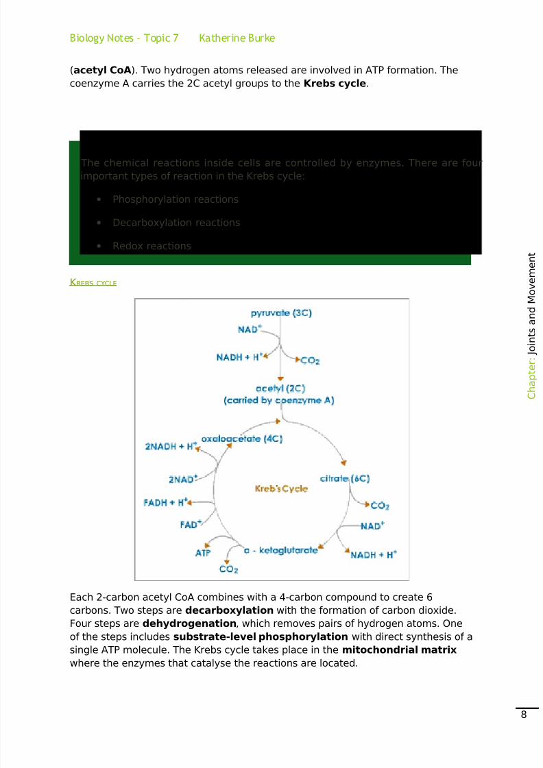
K REBS CYCLE
Each 2-carbon acetyl CoA combines with a 4-carbon compound to create 6
carbons. Two steps are decarboxylation with the formation of carbon dioxide.
Four steps are dehydrogenation, which removes pairs of hydrogen atoms. One
of the steps includes substrate-level phosphorylation with direct synthesis of a
single ATP molecule. The Krebs cycle takes place in the mitochondrial matrix
where the enzymes that catalyse the reactions are located.
C
h a p t e r : J o i n t s a n d M o v e m e
n t
8

8/2/2019 Unit 5, Topic 7 NOTES
http://slidepdf.com/reader/full/unit-5-topic-7-notes 9/10
Biology Notes – Topic 7 Katherine Burke
Each 2-carbon molecule entering the Krebs cycle results in the production of two
carbon dioxide molecules, one molecule of ATP by substrate-level
phosphorylation, and four pairs of hydrogen atoms, which are taken up by the
hydrogen acceptors, the coenzymes NAD and FAD. The hydrogen atoms are
subsequently involved in ATP production via the electron transport chain.
The Electron Transport Chain
For most hydrogen produced, the coenzyme NAD is the hydrogen acceptor but
those released in one-step of the Krebs cycle are accepted by the coenzyme FAD
rather than NAD.
When a coenzyme accepts hydrogen with its electron, the coenzyme is reduced,
becoming reduced NAD or reduced FAD. This reduced coenzyme ‘shuttles’ the
hydrogen atoms to the electron transport chain on the mitochondrial inner
membrane. Each hydrogen atom’s electron and proton then separate, with the
electron passing along a chain of electron carriers in the inner mitochondrial
membrane.
ATP S YNTHESIS BY CHEMIOSMOSIS
Energy is released as electrons pass along the electron transport chain. This
energy is used to move hydrogen ions from the matrix, across the inner
mitochondrial membrane, and into the intermembrane space. This creates a
steep electrochemical gradient across the inner membrane. There is a large
difference in the concentration of hydrogen ions across the membrane, and a
large electrical difference, making the intermembrane space more positive thanthe matrix.
C
h a p t e r : J o i n t s a n d M o v e m e
n t
9
8/2/2019 Unit 5, Topic 7 NOTES
http://slidepdf.com/reader/full/unit-5-topic-7-notes 10/10
Biology Notes – Topic 7 Katherine Burke
The hydrogen ions diffuse down this electrochemical gradient through hollow
protein channels in stalked particles on the membrane. As the hydrogen ions pass
through the channel, ATP synthesis is catalysed by ATPase located in each
stalked particle. The hydrogen ions cause a conformational change in the
enzyme’s active site, so the ADP can bind.
Within the matrix, the hydrogen ions and electrons recombine to form hydrogen
atoms. These combine with oxygen to form water. The oxygen, acting as the final
carrier in the electron transport chain, is thus reduced. This method of ATP
synthesis is known as oxidative phosphorylation.
HOW MUCH ATP IS PRODUCED?
The total number of ATP produced by one glucose molecule can vary according to
the efficiency of the cell. A simple explanation would give a maximum number of
38 ATP molecules per glucose molecule. This is based on the assumption that
that reduced NAD that is reoxidised results in the formation of three ATPmolecules and each reduced FAD results in production of two ATP molecules.
RATE OF RESPIRATION
The rate of aerobic respiration can conveniently be determined by measuring the
uptake of oxygen using a respirometer. The rate is determined by the any factor
affecting the rate of the enzyme-controlled reactions.
The concentration of ATP in the cell has a role in the control of respiration. ATPinhibits the enzyme in the first step of glycolysis, the phosphorylation of glucose.
The enzyme responsible for glucose phosphorylation can exist in two different
forms. As the ATP is broken down, the enzyme is converted back to the active form
and catalysis the phosphorylation of glucose. This is known as end point inhibition.
Google: W H Freedman Animations
C
h a p t e r : J o i n t s a n d M o v e m e
n t
10
Top Related