


Languages
Pages
Legal

Presented by:Sushant Kumar Swatantra
TRY-IN For Complete Denture
Patients

2DefinitionTry-in Verification / Aesthetic try-in :
“A preliminary insertion of a removable denture wax- up or a partial denture casting or a finished restoration to determine the fit, aesthetics, maxillomandibular relation.”
– GPT
Trial denture :
“A preliminary arrangement of denture teeth that has been prepared for placement into the patients mouth to evaluate aesthetics & maxillomandibular relationships”
- GPT
Importance: It is the last opportunity to evaluate many of the
previous steps already accomplished.
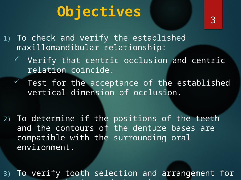
3Objectives
1) To check and verify the established maxillomandibular relationship:
Verify that centric occlusion and centric relation coincide.
Test for the acceptance of the established vertical dimension of occlusion.
2) To determine if the positions of the teeth and the contours of the denture bases are compatible with the surrounding oral environment.
3) To verify tooth selection and arrangement for proper esthetics and phonetics.

4Various aspects of try in: Extraoral examination of the trial dentures.
Intraoral examination of the trial dentures.

5Extraoral Examination Of The
Trial Dentures

The master cast
As the finished denture is processed on the master cast. So the master cast should be:
In good shape. Free from air bubbles or scratches. Free from wax debris which lead to improper
adaptation of the trial denture bases leading to false relationships.
If there are any undercuts present in the cast, these undercuts should be relieved to avoid scratching of the cast by the trial denture bases.

7The trial denture bases
Check the following:
The trial denture bases must be stable.
The borders of the trial denture base should be smooth, round, and have no sharp edges.
Also the border should be shaped to conform to the depth and width of the sulci.

8On the articulators
The mounted cast is checked for:
a) Maintaining of the vertical dimension of occlusion Top of the incisal pin is flush
with the upper member of the articulator.
The incisal pin is in contact with the incisal table.

9b) The mounting rings are firmly screwed in their position
c) Moving of the articulator smoothly from centric to eccentric positions without cuspal interlocking.
d) The trial denture bases lie properly on their casts and the teeth meet evenly in centric relation.

10
The teeth
It is the dentist responsibility to select the proper shade, and mould of the teeth and to determine that the teeth are set correctly.
Elimination of the excess wax is done to avoid the camouflages of the teeth relationships to overlook the occlusion.

11
Intraoral Examination Of The Trial Dentures

To reduce the risk of cross- contamination, the trial denture should be sprayed with suitable antiseptic solution and washed in running water, before inserted in patient mouth.
1) Checking the trial dentures separately: Trying- in the upper denture. Trying- in the lower denture.
2) Checking the upper and lower dentures together.

Maxillary trial dentureDenture base extension: The labial and buccal extension: marked overextension of the flanges, will stretch
the sulcus tissues and when denture is inserted, leads to elastic recoil resulting in dislodgment of the denture, immediate denture displacement after its seating.
Examination of the extension: Insertion of the upper trial denture in its place with
light pressure on the occlusal surface, move the cheek in functional movement. With the release of the pressure, the denture will fall down.
Need adjustment till little or no movement occurs.

14
Also under extension of the upper trial denture leads to poor physical retention.
Correction will usually entail making a new final
impression.
Provision of the frena {labial and buccal} should be done to ensure that they have adequate clearance.

15Posterior extension
The posterior border of the upper trial denture base should extended from the one hamular notch to the other along the vibrating line of the soft palate, and correctly placed on the master cast.
If the p.p.s is not done before, it should be done at this stage. Arbitary scraping of the cast and redapting the
record base

16Establishment of
Posterior Palatal seal
Advantages to placing the seal in Trial dentures :
1)Trial base will be more retentive, this can produce more accurate maxillomandibular records
2) Patients will be able to experience retentive qualities of the trial base, giving them psychologic security about retention
3)Denture wearer will be able to realize the posterior extent of denture.
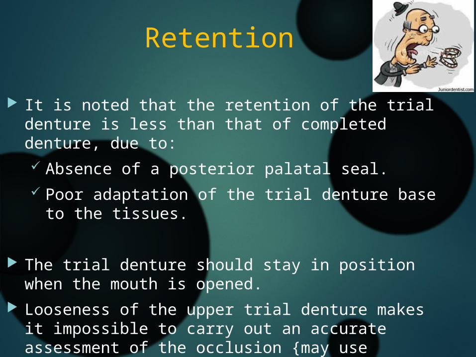
Retention
It is noted that the retention of the trial denture is less than that of completed denture, due to: Absence of a posterior palatal seal. Poor adaptation of the trial denture base to the
tissues.
The trial denture should stay in position when the mouth is opened.
Looseness of the upper trial denture makes it impossible to carry out an accurate assessment of the occlusion {may use denture fixative} especially, in patients with unfavorable anatomical factors.

18
How to test the retention of upper denture?
Seat the upper trial denture with a firm upward and backward pressure.
Allow the tissues to settle around the denture
Grip the labial and lingual surfaces of the upper denture teeth between the thumb and forefinger
Apply a firm downward vertical pull to dislodge the denture away from the tissues
If the retention is good, dislodgment of the trial denture may be difficult
19
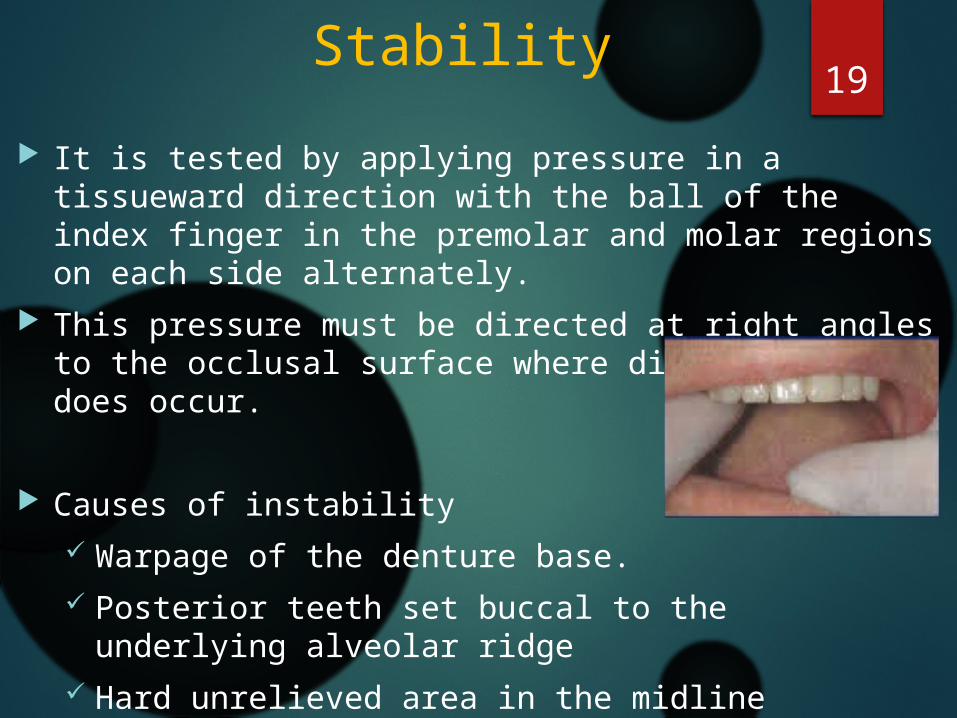
Stability It is tested by applying pressure in a tissueward
direction with the ball of the index finger in the premolar and molar regions on each side alternately.
This pressure must be directed at right angles to the occlusal surface where displacement does occur.
Causes of instability Warpage of the denture base. Posterior teeth set buccal to the underlying
alveolar ridge Hard unrelieved area in the midline
e.g. torus palatinus.

20Orientation of the
occlusal plane Properly oriented occlusal plane is
important to: Patient esthetics. Patient comfort Chewing function Balance of occlusion

21
Orientation of the anterior end of the occlusal plane is determined by esthetics.
The amount of the upper anterior teeth that will be seen during speech and facial expression depends on length and movement of the upper lip.
If the upper lip is relatively long, the natural teeth may not be visible when the lip is relaxed or even during speech.
The movement of the lips during function varies considerably among patient thus , the amount of the upper teeth that will be visible varies for each patient.

Lower denture trial
Denture base extension:
The lower trial denture extension should be tested with the patient mouth is opened no more than half opened position to allow the surrounding musculature is in an acceptable state of relaxation.
Labial and buccal extensions are checked as for the upper trial denture.

How to evaluate lower denture retention? Usually the lower denture retention is poor when
compared to the upper denture due to: 1. Small denture bearing area 2. The difficulty in obtaining an efficient border seal.
Testing of the lower trial denture retention:a) Ask the patient to open his mouth slightly and let his
tongue touch the cingula of the lower anterior teeth, support the chin of the patient with the left hand and pull the teeth straight upwards to check the retention of the anterior labial and lingual flanges.
b) Tilt the lower trial denture outward from the canine region to test the retention of the opposite retro molar pad.

24Lower occlusal plane
In most patients, the incisal edges of the natural lower canines and the cusp tips of the lower first premolars are located at the level of the lower lip at the corner of the mouth when the mouth is slightly open.
The posterior end of the occlusal plane should be at the level of the anterior two thirds of the retro molar pad.

25Tongue space
Natural teeth occupy a position in the mouth where the inward pressure of the lips and the cheeks is neutralized by an equal and opposite outward pressure of the tongue, and it is in this zone of neutral pressure that the artificial teeth must be set (neutral zone).

26
To check for the neutral zone in the patient’s mouth, let the patient open his mouth half-way and touch the lower anterior teeth with the tip of his tongue, while his tongue is relaxed. Feel the amount of pressure exerted by the tongue and cheek on the lower teeth, using a plastic filling instrument. Pressure should be roughly equal on the lingual and buccal sides of the teeth.

27
Lack of tongue space (cramped tongue)
If the tongue is more mobile than the cheeks will cause greater instability of the lower denture.
Cramped tongue may be due to: 1. Posterior teeth set lingually to the neutral zone. 2. Posterior teeth tilted lingually3. Posterior teeth too broad bucco-lingual.
Testing of the tongue space: Ask the patient to raise the tongue. If the tongue is
cramped, the denture will begin to rise immediately. As the tongue moves it tries to expand laterally and whenever the tongue moves the denture will move.

28
Checking the upper and lower dentures together
It is usually advisable to insert the lower trial denture first and then the upper because there is less chance of having the upper denture drop down.
The patient should be seated in an upright position.
The patient head is not supported by the headrest (the headrest may effect the physiologic rest position of the mandible so, it effect the amount of interocclusal distance).

Perfection & Verification of Jaw-relation records

30
Verifying the Vertical dimension Checking of labial frenum
Evaluation of Vertical dimension at rest & at occlusion
pre extraction records amount of inter occlusal distance to which pt. was
accustomed phonetics & esthetics facial dimension & facial expressions lip length in relation to teeth inter arch distance & parallelism of the ridges

31
Verifying Centric Relation
Intra Oral Observation of Intercuspation Pt. is guided into CR by a thumb placed on the
anteroinferior portion of the chin & index finger bilaterally on the buccal flanges of the lower denture.
Any Error in CR will be apparent when teeth slide over each other.

32
If Error is due to mounting :
anterior teeth if not placed to support lip, are corrected.
vertical overlap of anterior teeth are carefully noted .
posterior teeth are removed from lower occlusal rim
Impression plaster is mixed & placed on the rim.

Pt. is instructed to close the mouth slowly until the anterior teeth have same vertical overlap as they had before the posterior teeth were removed.
After the plaster is set, rims are removed & this corrected new record is mounted on a articulator.
33

34
Extra oral articulator methodProcess : - Impression material (eg. Aluwax) is placed over
mandibular posterior teeth - wax sealed – denture placed in mouth – just wax
portion is immersed in water bath of 130oF for 30 secs – denture placed back in pts mouth - mandible guided into CR so that upper teeth makes contact with the wax – denture removed & chilled in ice water & returned back to patients mouth for re-checking

– CR is confirmed – Trial dentures are then locked in articulator – opposing teeth should fit in the indentation in every way (anteriorly, posteriorly, laterally & vertically ) if the original CR was correct.
- If it does not fit, mandibular cast should be separated & remounted with last occlusal record.
35

36
Eccentric relation records
It is essential that the movements of articulator should simulate movements of the patient within the range of normal functional contacts of teeth.
For this, condylar elements of articulator must be adjusted so that they approximate condylar guiding factors within TMJ.

37
Contact during protrusion
At least three widely separated points or areas of occlusion must exist.

38
Contact during lateral movement
Working side Balancing side

Creating facial & functional harmonywith Anterior teeth
Appearance of entire lower half of face depends on dentures.
After vertical dimension of occlusion & CR has been verified. To obtain a harmonious effect with the patient face, modifications are made in arrangement of teeth
Incorrect positioning of anterior teeth or supporting base material alters normal appearance of vermilion border, the philtrum & mentolabial sulcus
39

40

1. Preliminary selection of artificial teeth
Evaluated for size, form & color
6 anterior teeth should be of sufficient overall width to extend approx. corner of mouth
Color should blend with the face
Any records used in initial selection should be consulted & changes should be made if it improves the appearance of patient

2. Horizontal orientation of anterior teethTeeth set directly over ridges causes insufficient lip
support characterized by:
Drooping of corners of mouth, Reduction in visible part of vermilion border, Deepening of nasolabial sulcus, Wrinkles over vermilion border
Excessive lip support causes stretched lips, tendency of lips to dislodge dentures during function, elimination of normal contours of lips, philtrum & sulci.
42
43
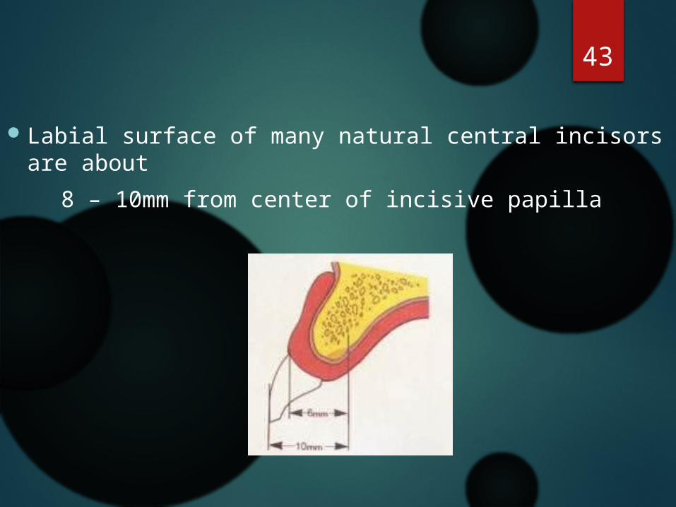
Labial surface of many natural central incisors are about 8 – 10mm from center of incisive papilla

44
3. Vertical orientation of anterior teeth
Index finger is placed on incisive papilla with relaxed upper lip & amount of finger covered gives indication of length of upper lip.
Lower lip is better guide for vertical orientation of anterior teeth. Incisal edges of lower canine & cusp tip of lower first premolar are even with corner of mouth when mouth is slightly open.

45
If lower teeth are above then,
1. plane of occlusion may be too high 2. vertical overlap of anterior teeth may be too
much high 3. vertical space between the jaws may be
excessive

46
4. Inclination of anterior teeth
Inclination of labial surface of residual ridges seen on edentulous cast gives guide to inclination of anterior teeth
Inclination of anterior teeth parallels the profile line of face

47
5. Harmony in general composition of anterior teeth
A. Harmony of dental arch form & form of residual ridge:
I. Square arch form Central incisors are more nearly in line with
canine 4 incisors should have little rotation to give
broader effect to teeth II. Tapering arch form Central incisors are greater distance forward
from canine rotating or lapping of teeth may be present III. Ovoid arch form : teeth seldom rotated show greater amount of labial surface

48
B. Harmony of long axis of central incisors & face
Imaginary perpendicular line from mid point of Inter pupillary line should mark middle of dental arch to be in harmony with face
Mid line also determined by observing position of incisive fossa
Mid line of mandibular central incisor is aligned with that of maxillary incisors

49C. Harmony of the teeth with smiling line of the
lower lip
Line formed by upper anterior teeth should follow curved line of lower lip during smiling
Vertical positioning of upper canines are responsible for shape of smile line

50
Canines should be arranged in such a way that their incisal edges should be slightly shorter than that of lateral incisors, if not it will create reverse smile line.
Reverse smile line is one of the frequent cause of artificial appearing dentures.

51
D. Harmony of opposing lines of Labial & Buccal
surfaces
Asymmetrical symmetry is essential for natural appearing teetheg. If maxillary right lateral incisor is set at 5 degrees to
perpendicular then opposing lateral should be of same angle but in opposite direction
Labial & buccal lines should be in harmony with lines of face.

E. Harmony of incisal wear & age
Incisal edges of denture teeth should be grinded to simulate the wear surface that would have developed by the time patient reached his current age.
This wear is placed on teeth where it would have occurred during function & also where it assists in the mechanics of balancing the occlusion

53
F. Harmony of spaces & individual tooth position
Use of spaces between teeth can be effective in emphasizing individual tooth position & natural appearing arrangement
Spaces designed should be self cleansing

54Conclusion
For the success of a complete denture the teeth should be arranged in harmony with the intraoral and circumoral structures and adjusted so that they occlude and articulate evenly.
After the preliminary arrangement of the artificial teeth on the occlusion rims, it is essential that the accuracy of the jaw relation records made with the occlusion rims be tested, perfected if incorrect, and then verified to be correct.
During Try-in all the procedures carried out in fabrication of denture are verified clinically.

55
References Prosthodontic Treatment for Edentulous patients, 12th edition
: Zarb – Bolender
Essentials of complete denture prosthodontics, 2nd edition : Sheldon Winkler
Text book of prosthodontics, Deepak Nallaswamy
http://www.slideshare.net/narendrabasutkar/try-in-complete-dentures
www.gr.dentistbd.com


57
Top Related