


Languages
Pages
Legal


Treatment Concepts for
EXTRACTIONSOCKETS
NEWNew clinical cases
Latest scientific literature
QR codes linked to clinical videos, 3D-animation...
?
1 – Treatment Concepts for Extraction Sockets
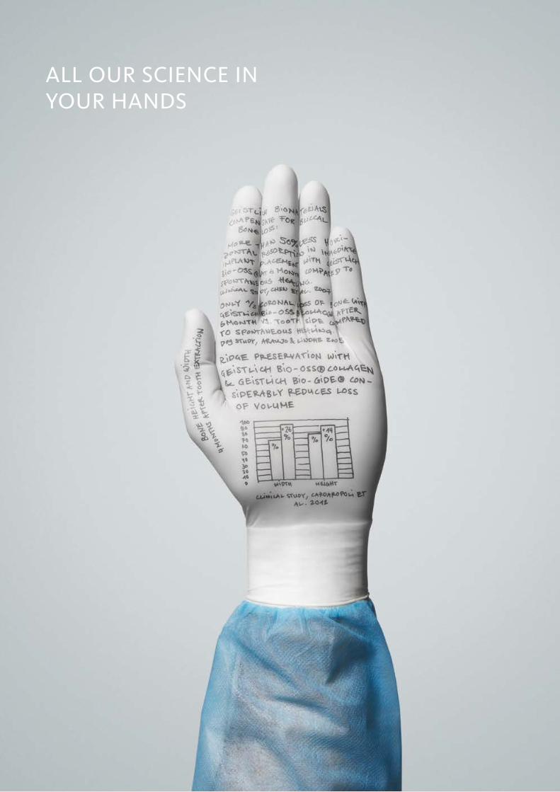
ALL OUR SCIENCE IN YOUR HANDS
2
TABLE OF CONTENTS
3 Clinical evidence
7 Immediate implant placement
Dr. Michael Back & Dr. Oliver Blume (Munich, Germany) Dr. Franck Bonnet (Le Cannet, France)
11 Early implant placement
Prof. Daniel Buser & Prof. Urs Belser (Berne, Switzerland) Dr. Luca Cordaro (Rome, Italy) Dr. Raffaele Cavalcanti (Bari, Italy)
17 Delayed/late implant placement
Dr. Hadi Antoun & Dr. Bouchra Sojod (Paris, France) Dr. Stefan Fickl (Würzburg, Germany)
21 Extraction socket treatment options
23 Delayed/late implant placement
Dr. Célia Coutinho Alves (Porto, Portugal) Prof. Ronald E. Jung (Zurich, Switzerland) Prof. Carlo Maiorana (Milan, Italy) Prof. Julio Cesar Joly, Prof. Robert Carvalho da Silva & Prof. Paulo Fernando M. de Carvalho (Sao Paulo, Brazil) Dr. Fernán López (Medellin, Colombia) Dr. Ham Byung-Do (Kainos Dental Clinic, Seoul, Korea)
35 No implant placement
Dr. Jeffrey Ganeles (Boca Raton, USA) Dr. Philipp Grohmann (Berikon, Switzerland)
39 Product Information
3 – Treatment Concepts for Extraction Sockets
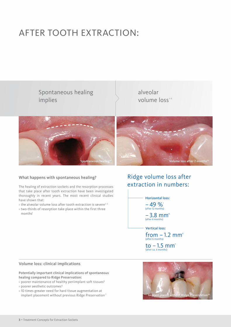
AFTER TOOTH EXTRACTION:
Spontaneous healing implies
alveolar volume loss1–5
What happens with spontaneous healing?
The healing of extraction sockets and the resorption processes that take place after tooth extraction have been investigated thoroughly in recent years. The most recent clinical studies have shown that: › the alveolar volume loss after tooth extraction is severe1–5
› two-thirds of resorption take place within the first three months1
Volume loss: clinical implications
Potentially important clinical implications of spontaneoushealing compared to Ridge Preservation: › poorer maintenance of healthy periimplant soft tissues6
› poorer aesthetic outcomes6
› 10 times greater need for hard tissue augmentation at implant placement without previous Ridge Preservation 7
Ridge volume loss after extraction in numbers:
from – 1.2 mm4 (after 6 months)
to – 1.5 mm7 (after ca. 6 months)
Horizontal loss:
Vertical loss:
– 49 %1 (after 12 months)
– 3.8 mm4 (after 6 months)
Spontaneous healing19 Volume loss after 2 months19
Implant placed without Ridge Preservation20
4
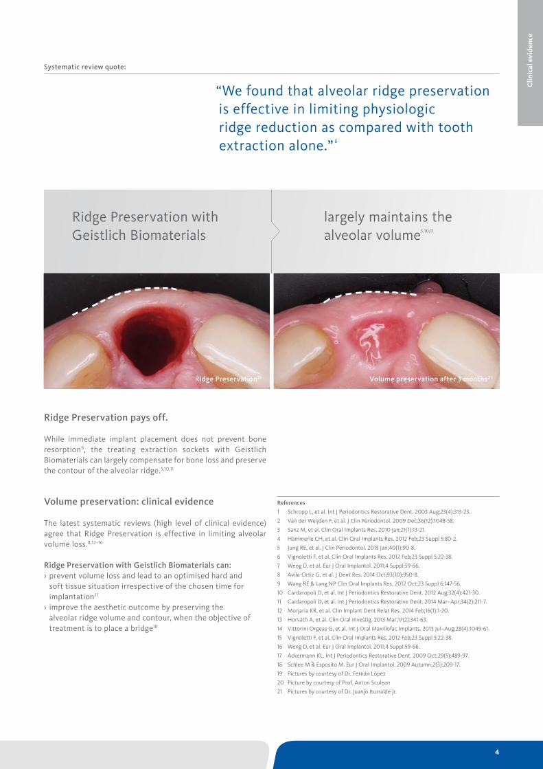
Ridge Preservation pays off.
While immediate implant placement does not prevent bone resorption9, the treating extraction sockets with Geistlich Biomaterials can largely compensate for bone loss and preserve the contour of the alveolar ridge.5,10,11
Volume preservation: clinical evidence
The latest systematic reviews (high level of clinical evidence) agree that Ridge Preservation is effective in limiting alveolar volume loss.8,12–16
Ridge Preservation with Geistlich Biomaterials can: › prevent volume loss and lead to an optimised hard and soft tissue situation irrespective of the chosen time for implantation17
› improve the aesthetic outcome by preserving the alveolar ridge volume and contour, when the objective of treatment is to place a bridge18
Ridge Preservation with Geistlich Biomaterials
largely maintains the alveolar volume5,10,11
“We found that alveolar ridge preservation is effective in limiting physiologic ridge reduction as compared with tooth extraction alone.” 8
References
1 Schropp L, et al. Int J Periodontics Restorative Dent. 2003 Aug;23(4):313-23.
2 Van der Weijden F, et al. J Clin Periodontol. 2009 Dec;36(12):1048-58.
3 Sanz M, et al. Clin Oral Implants Res. 2010 Jan;21(1):13-21.
4 Hämmerle CH, et al. Clin Oral Implants Res. 2012 Feb;23 Suppl 5:80-2.
5 Jung RE, et al. J Clin Periodontol. 2013 Jan;40(1):90-8.
6 Vignoletti F, et al. Clin Oral Implants Res. 2012 Feb;23 Suppl 5:22-38.
7 Weng D, et al. Eur J Oral Implantol. 2011;4 Suppl:59-66.
8 Avila-Ortiz G, et al. J Dent Res. 2014 Oct;93(10):950-8.
9 Wang RE & Lang NP Clin Oral Implants Res. 2012 Oct;23 Suppl 6:147-56.
10 Cardaropoli D, et al. Int J Periodontics Restorative Dent. 2012 Aug;32(4):421-30.
11 Cardaropoli D, et al. Int J Periodontics Restorative Dent. 2014 Mar–Apr;34(2):211-7.
12 Morjaria KR, et al. Clin Implant Dent Relat Res. 2014 Feb;16(1):1-20.
13 Horváth A, et al. Clin Oral Investig. 2013 Mar;17(2):341-63.
14 Vittorini Orgeas G, et al. Int J Oral Maxillofac Implants. 2013 Jul–Aug;28(4):1049-61.
15 Vignoletti F, et al. Clin Oral Implants Res. 2012 Feb;23 Suppl 5:22-38.
16 Weng D, et al. Eur J Oral Implantol. 2011;4 Suppl:59-66.
17 Ackermann KL. Int J Periodontics Restorative Dent. 2009 Oct;29(5):489-97.
18 Schlee M & Esposito M. Eur J Oral Implantol. 2009 Autumn;2(3):209-17.
19 Pictures by courtesy of Dr. Fernán López
20 Picture by courtesy of Prof. Anton Sculean
21 Pictures by courtesy of Dr. Juanjo Iturralde Jr.
Clin
ical
evi
denc
e
Systematic review quote:
Ridge Preservation21 Volume preservation after 3 months21

5 – Treatment Concepts for Extraction Sockets
RIDGE PRESERVATION WITH GEISTLICH BIOMATERIALS
Not all Bone Substitutes are the same – Take a closer look!In recent controlled clinical trials, Geistlich Bio-Oss® showed:
better ridge preservation than fast resorbing ß-TCP1
better ridge preservation than synthetic hydroxyapatite or gelatine sponge10
more mineralized tissue in sockets than allografts11
The use of a biofunctional material such as Geistlich Bio-Oss® is crucial to the long-term successful outcome of extraction socket treatment. After tooth extraction, the slowly resorbing bone matrix Geistlich Bio-Oss® / Geistlich Bio-Oss® Collagen
preserves the ridge volume over time and thus makes a major contribution towards the success of Ridge Preservation1–3
or ridge contouring at a later time point (e.g. for early implant placement after spontaneous healing)4,5
Clinical benefits of Ridge Preservation with Geistlich Bio-Oss®
Clinical studies indicate that Ridge Preservation using Geistlich Bio-Oss® allows for: › stable crest heights in sites with thin buccal bone walls6
› reduced horizontal bone loss in immediate implantation7
› increased mineralized tissue portion in the socket8
› preserved ridge volume under pontics9
Defective extraction socket
+
Geistlich Bio-Oss® Collagen
Geistlich Bio-Oss® Collagen
+=
=
Intact extraction socket*
Geistlich Mucograft® Seal
Geistlich Bio-Gide®
6
Geistlich Bio-Gide® – more new bone12
Due to its bilayer structure, the Geistlich Bio-Gide® membra-ne not only prevents ingrowth of soft tissue, but also acts as a guide for the appropriate early blood vessel13 development and new bone formation12.
Geistlich Bio-Gide®: › allows for uneventful wound healing in an open healing approach2,3
› provides for more new bone formation when combined with Geistlich Bio-Oss® vs. Geistlich Bio-Oss® without membrane12
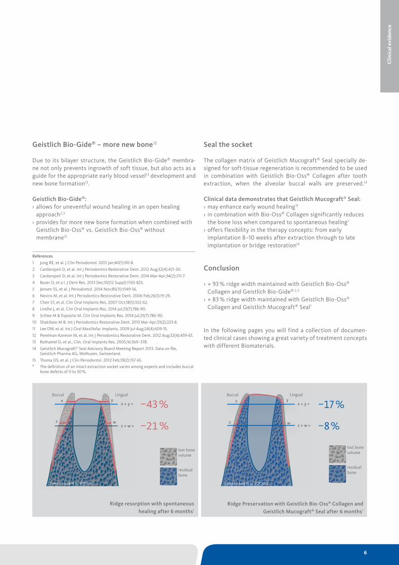
Ridge Preservation with Geistlich Bio-Oss® Collagen and Geistlich Mucograft® Seal after 6 months1
–43 %
–21 %
lost bone volume
residual bone
lost bone volume
residual bone
Ridge resorption with spontaneous healing after 6 months1
Seal the socket
The collagen matrix of Geistlich Mucograft® Seal specially de-signed for soft-tissue regeneration is recommended to be used in combination with Geistlich Bio-Oss® Collagen after tooth extraction, when the alveolar buccal walls are preserved.14
Clinical data demonstrates that Geistlich Mucograft® Seal: › may enhance early wound healing15
› in combination with Bio-Oss® Collagen significantly reduces the bone loss when compared to spontaneous healing1
› offers flexibility in the therapy concepts: from early implantation 8–10 weeks after extraction through to late implantation or bridge restoration14
Conclusion
› + 93 % ridge width maintained with Geistlich Bio-Oss® Collagen and Geistlich Bio-Gide® 2,3
› + 83 % ridge width maintained with Geistlich Bio-Oss® Collagen and Geistlich Mucograft® Seal1
In the following pages you will find a collection of documen-ted clinical cases showing a great variety of treatment concepts with different Biomaterials.
–17 %
–8 %
Buccal Lingualx
z
yx + y =
z + w =w
based on Jung et al., JCP 2013
Buccal Lingualx
z
yx + y =
z + w =w
based on Jung et al., JCP 2013
Clin
ical
evi
denc
e
References
1 Jung RE, et al. J Clin Periodontol. 2013 Jan;40(1):90-8.
2 Cardaropoli D, et al. Int J Periodontics Restorative Dent. 2012 Aug;32(4):421-30.
3 Cardaropoli D, et al. Int J Periodontics Restorative Dent. 2014 Mar-Apr;34(2):211-7.
4 Buser D, et a l. J Dent Res. 2013 Dec;92(12 Suppl):176S-82S.
5 Jensen SS, et al. J Periodontol. 2014 Nov;85(11):1549-56.
6 Nevins M, et al. Int J Periodontics Restorative Dent. 2006 Feb;26(1):19-29.
7 Chen ST, et al. Clin Oral Implants Res. 2007 Oct;18(5):552-62.
8 Lindhe J, et al. Clin Oral Implants Res. 2014 Jul;25(7):786-90.
9 Schlee M & Esposito M. Clin Oral Implants Res. 2014 Jul;25(7):786-90.
10 Shakibaie-M B. Int J Periodontics Restorative Dent. 2013 Mar-Apr;33(2):223-8.
11 Lee DW, et al. Int J Oral Maxillofac Implants. 2009 Jul-Aug;24(4):609-15.
12 Perelman-Karmon M, et al. Int J Periodontics Restorative Dent. 2012 Aug;32(4):459-65.
13 Rothamel D, et al., Clin. Oral Implants Res. 2005;16:369–378.
14 Geistlich Mucograft® Seal Advisory Board Meeting Report 2013. Data on file, Geistlich Pharma AG, Wolhusen, Switzerland.
15 Thoma DS, et al. J Clin Periodontol. 2012 Feb;39(2):157-65. The definition of an intact extraction socket varies among experts and includes buccal bone defects of 0 to 50 %.
*

7 – Treatment Concepts for Extraction Sockets
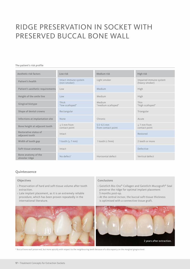
The patient’s risk profile
Quintessence
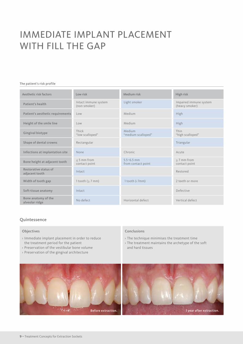
IMMEDIATE IMPLANT PLACEMENT WITH MINOR BONY DEFECT
Objectives
› Immediate implant placement maintaining a good aesthetic contour
› Minimally invasive procedure › The materials are unlimited and easy to use
Objectives
› Immediate implant placement maintaining a good aesthetic contour
› Minimally invasive procedure
› The materials are unlimited and easy to use
Before extraction.
Conclusions
› One-stage treatment of hard and soft tissues › The three combined Geistlich Biomaterials heal uneventfully and preserve the alveolar bone volume
› Long-term good aesthetic outcome in front teeth with short treatment time.
1 year after extraction.
Aesthetic risk factors Low risk Medium risk High risk
Patient’s health Intact immune system (non-smoker)
Light smoker Impaired immune system (heavy smoker)
Patient’s aesthetic requirements Low Medium High
Height of the smile line Low Medium High
Gingival biotype Thick“low scalloped”
Medium “medium scalloped”
Thin “high scalloped”
Shape of dental crowns Rectangular Triangular
Infections at implantation site None Chronic Acute
Bone height at adjacent tooth ≤ 5 mm from contact point
5.5–6.5 mmfrom contact point
≥ 7 mm from contact point
Restorative status of adjacent tooth
Intact Restored
Width of tooth gap 1 tooth (≥ 7 mm) 1 tooth (< 7mm) 2 teeth or more
Soft-tissue anatomy Intact Defective
Bone anatomy of the alveolar ridge
No defect Horizontal defect Vertical defect

8
Case documentation
1 Clinical situation before extraction of tooth 11.
2 Immediate implant placement after flap elevation.
3 Implant and Geistlich Bio-Oss® covered with Geistlich Bio-Gide®.
4 Geistlich Mucograft® sutured with single sutures on top of augmented area.
5 Clinical situation 2 days post-op.
6 Clinical situation 3 weeks after surgery.
7 Clinical situation 6 weeks after surgery (occlusal).
8 Clinical situation 6 weeks after surgery (buccal).
“Geistlich Biomaterials are innovative materials and products and have many years of experience.”
Material selection
1 2 3
4 5 6
7 8 9
Geistlich Bio-Oss® small granules (0.25–1 mm)
Geistlich Bio-Gide® (25 × 25 mm)
Geistlich Mucograft® (15 × 20 mm; punch 8 mm diameter)
9 Minimally invasive re-entry: roll flap preparation after 12 weeks.
10 The flap is rolled bucally to increase the thickness of the soft tissues in the buccal area and the abutment is connected.
11 a) X-ray after roll flap, 3 months after extraction. b) X-ray at 1-year follow-up.
12 Clinical situation 1 year after extraction.
10 1211 a 11 b
Dr. Michael Back & Dr. Oliver Blume (Munich, Germany)
Cas
e 1
| Im
med
iate
impl
ant p
lace
men
t
9 – Treatment Concepts for Extraction Sockets
The patient’s risk profile
Quintessence
IMMEDIATE IMPLANT PLACEMENT WITH FILL THE GAP
Aesthetic risk factors Low risk Medium risk High risk
Patient’s health Intact immune system (non-smoker)
Light smoker Impaired immune system (heavy smoker)
Patient’s aesthetic requirements Low Medium High
Height of the smile line Low Medium High
Gingival biotype Thick“low scalloped”
Medium “medium scalloped”
Thin “high scalloped”
Shape of dental crowns Rectangular Triangular
Infections at implantation site None Chronic Acute
Bone height at adjacent tooth ≤ 5 mm from contact point
5.5–6.5 mmfrom contact point
≥ 7 mm from contact point
Restorative status of adjacent tooth
Intact Restored
Width of tooth gap 1 tooth (≥ 7 mm) 1 tooth (< 7mm) 2 teeth or more
Soft-tissue anatomy Intact Defective
Bone anatomy of the alveolar ridge
No defect Horizontal defect Vertical defect
Objectives
› Immediate implant placement in order to reduce the treatment period for the patient
› Preservation of the vestibular bone volume › Preservation of the gingival architecture
Before extraction.
Conclusions
› The technique minimises the treatment time › The treatment maintains the archetype of the soft and hard tissues
1 year after extraction.

10
Case documentation
2 a
1 The patient presents with a fractured central incisor. The biotype is rather thin with scalloped marginal gingiva.
2 a) X-ray of the fractured tooth. b) Analysis of the bony situation through CBCT allows planning of Type 1 implant placement.
3 The gap from implant to the buccal bone is filled with Geistlich Bio-Oss®. A connective tissue graft is placed between the mucosa and the buccal bone.
4 The implant (NobelActive™) is positioned optimally, with a more palatal vestibular orientation. The provisional abutment is placed.
5 An ideal emergence profile is effected. The provisional crown allows maintenance of the papillae.
6 The provisional prosthesis is placed and left out of occlusion.
7 Clinical situation 8 days post-operative. The healing occurs uneventfully.
8 Situation 4 months after extraction, prior to finalising the prosthetic restoration.
Dr. Franck Bonnet (Le Cannet, France)
1 2 2 b 3
4 5 6
7 8 9
10 11 12
Geistlich Bio-Oss® small granules (0.25–1 mm)
9 The natural profile of the soft tissues has been preserved.
10 An individual impression post is created for precise transfer of the emergence profile to the lab.
11 The final crown is made directly over a zirconia abutment (Procera®).
12 Vestibular view of the final restoration 12 months after tooth extraction. Note the perfect alignment of the neck of the teeth and ideal position of the papillae in relation to the contact points.
Material selection
Cas
e 2
| Im
med
iate
impl
ant p
lace
men
t
Find the long-term follow-up here.

11 – Treatment Concepts for Extraction Sockets
The patient’s risk profile
Quintessence
EARLY IMPLANT PLACEMENT WITH GBR AFTER 8 WEEKS OF SPONTANEOUS HEALING
Aesthetic risk factors Low risk Medium risk High risk
Patient’s health Intact immune system (non-smoker)
Light smoker Impaired immune system (heavy smoker)
Patient’s aesthetic requirements Low Medium High
Height of the smile line Low Medium High
Gingival biotype Thick“low scalloped”
Medium “medium scalloped”
Thin “high scalloped”
Shape of dental crowns Rectangular Triangular
Infections at implantation site None Chronic Acute
Bone height at adjacent tooth ≤ 5 mm from contact point
5.5–6.5 mmfrom contact point
≥ 7 mm from contact point
Restorative status of adjacent tooth
Intact Restored
Width of tooth gap 1 tooth (≥ 7 mm) 1 tooth (< 7mm) 2 teeth or more
Soft-tissue anatomy Intact Defective
Bone anatomy of the alveolar ridge
No defect Horizontal defect Vertical defect
Objectives
› Pleasing aesthetic outcome › Long-term stable bone and soft-tissue situation in the aesthetic region
Before extraction.
Conclusions
› The low substitution rate of Geistlich Bio-Oss® helps to maintain the volume of the alveolar ridge over time, which is crucial for the long-term aesthetic outcome.
› Minimal marginal bone loss and low risk of mucosal recession.
7.5 years after implant therapy.
12
Case documentation
1 Clinical findings in the initial examination. The patient exhibits a high smile line and reports an accident several years ago, which affected tooth 11.
2 The extraction socket and the soft tissue are allowed to heal for 4–8 weeks after debridement of the inflammatory tissue.
3 Within 4–8 weeks of soft tissue healing, no reduction is visible in the crest width in the approximal region of the socket.
4 Special attention is payed to correct prosthetic positioning of the implant in all three dimensions with good primary stability.
5 The defect is covered with locally harvested autogenous bone chips to promote new bone formation as quickly as possible.
6 The bone volume is further optimised by local augmentation using Geistlich Bio-Oss® granules.
7 Geistlich Bio-Gide® is applied in two layers to act as a temporary barrier and as a stabiliser for the graft.
Prof. Daniel Buser & Prof. Urs Belser (Berne, Switzerland)
1 2 3
4 5 6
7 8 9
12
8 Following the release of the flap by means of mucoperiosteal incisions, a tension-free primary wound closure is achieved. Provi-sional implant prosthesis starts after 8 weeks.
9 The 7.5-year follow-up shows a stable aesthetic outcome.
10 X-rays a) at 1 year: implant optimally integrated in the bone; b) at 4 years: absolutely stable peri-implant bony conditions.
11 CBCT findings at 7.5 years a) section showing a completely intact facial wall; b) 3-dimensionally correctly placed implant.
12 The long-term aesthetic result is excellent.
Geistlich Bio-Oss® small granules (0.25–1 mm)
Geistlich Bio-Gide® (25 × 25 mm)
11 a10 a 11 b10 b
Material selection
Cas
e 3
| Ear
ly im
plan
t pla
cem
ent
Find the detailed surgical approach here.

13 – Treatment Concepts for Extraction Sockets
The patient’s risk profile
Quintessence
SPONTANEOUS HEALING FOR CANTILEVER IMPLANT BRIDGE
Aesthetic risk factors Low risk Medium risk High risk
Patient’s health Intact immune system (non-smoker)
Light smoker Impaired immune system (heavy smoker)
Patient’s aesthetic requirements Low Medium High
Height of the smile line Low Medium High
Gingival biotype Thick“low scalloped”
Medium “medium scalloped”
Thin “high scalloped”
Shape of dental crowns Rectangular Triangular
Infections at implantation site None Chronic Acute
Bone height at adjacent tooth ≤ 5 mm from contact point
5.5–6.5 mmfrom contact point
≥ 7 mm from contact point
Restorative status of adjacent tooth
Intact Restored
Width of tooth gap 1 tooth (≥ 7 mm) 1 tooth (< 7mm) 2 teeth or more
Soft-tissue anatomy Intact Defective
Bone anatomy of the alveolar ridge
No defect Horizontal defect Vertical defect
Objectives
› Prosthetic restoration of 2 side-by-side sockets in the anterior area
› Ridge Preservation for cantilever implant bridge
Before extraction.
Conclusions › Early implant placement is suitable for 2 side-by-side sockets
› The collapse of the tissues during the 6-week healing period can be compensated with a GBR contouring with Geistlich Bio-Oss® and Geistlich Bio-Gide®.
5.5 months after extraction.
14
Case documentation
1 Initial situation before extraction of 21 and 22.
2 Clinical close-up of the pre-operative site prior to extraction of the teeth.
3 a) Radiographic findings of the pre-operative site. Note the apical bone resorption at 22 and internal root resorption of tooth 21. b) Scheme of the 2 side-by-side sockets.
4 Teeth 21 and 22 are extracted and left heal spontaneously under a provisional restoration.
5 Buccal view after 6 weeks of spontaneous healing. Immediately before re-entry. Note the flattening of the ridge anticipating a horizontal defect.
6 Occlusal view 6 weeks post-extraction. The soft tissues are healed.
7 After flap elevation and implant placement, the resorption of the alveolar bone is compensated with Geistlich Bio-Oss®.
8 Geistlich Bio-Gide® is placed over the treated area to stabilise the graft and to obtain the desired contour augmentation.
Dr. Luca Cordaro (Rome, Italy)
1 2
4 5 6
7 8 9
10 12
9 Healing of the treated site 18 weeks post-extraction.
10 Occusal view after 18 weeks. Transmucosal healing took place with conditioning of the soft tissues with the provisional crown. The recession on tooth 23 has been covered with a coronally advanced flap and a connective tissue graft.
11 a) X-ray of the final prosthetic restoration. b) Schematic represen-tation of the cantilever implant bridge.
12 Final situation with the cantilever implant bridge in place 5.5 months after tooth extraction.
Geistlich Bio-Oss® small granules (0.25–1 mm)
Geistlich Bio-Gide® (25 × 25 mm)
23 a 3 b
11 a 11 b
“Early implantation with simultaneous contour augmentation is predictable in challenging cases in the aesthetic zone.”
Material selection
Cas
e 4
| Ear
ly im
plan
t pla
cem
ent

15 – Treatment Concepts for Extraction Sockets
The patient’s risk profile
Quintessence
EARLY IMPLANT PLACEMENT IN EXTRACTION SOCKET WITH PRESERVED BONE WALLS
Aesthetic risk factors Low risk Medium risk High risk
Patient’s health Intact immune system (non-smoker)
Light smoker Impaired immune system (heavy smoker)
Patient’s aesthetic requirements Low Medium High
Height of the smile line Low Medium High
Gingival biotype Thick“low scalloped”
Medium “medium scalloped”
Thin “high scalloped”
Shape of dental crowns Rectangular Triangular
Infections at implantation site None Chronic Acute
Bone height at adjacent tooth ≤ 5 mm from contact point
5.5–6.5 mmfrom contact point
≥ 7 mm from contact point
Restorative status of adjacent tooth
Intact Restored
Width of tooth gap 1 tooth (≥ 7 mm) 1 tooth (< 7mm) 2 teeth or more
Soft-tissue anatomy Intact Defective
Bone anatomy of the alveolar ridge
No defect Horizontal defect Vertical defect
Objectives
› Compensation of the bone resorption through Ridge Preservation
› Provide the patient with a final restoration in a relatively short time period of time
Before extraction.
Conclusions
› Almost complete maintenance of the ridge volume is achieved
› After 8–10 weeks, the soft tissue has a quality and maturity that is adequate for early implant placement.
7 months after extraction.

16
Case documentation
1 Initial situation before extraction of tooth 14.
2 No buccal bone defect is detected after tooth extraction.
3 Extraction socket with de-epithelialised wound margins.
4 Extraction socket filled with Geistlich Bio-Oss® Collagen.
5 The extraction socket is sealed with Geistlich Mucograft® Seal.
6 Geistlich Mucograft® Seal sutured with single interrupted sutures.
7 Pre-op clinical situation 10 weeks after extraction (prior to implant placement).
Dr. Raffaele Cavalcanti (Bari, Italy)
1 2 3
4 5 6
7 8 9
10 11 12
8 Preparation of a minimally invasive flap.
9 Implant placement with a minimally invasive roll flap technique to improve soft-tissue thickness at the buccal aspect.
10 Clinical situation of the soft tissues 4 months after implant placement.
11 Final restoration 7 months after tooth extraction (buccal).
12 Final restoration 7 months after tooth extraction (occlusal).
Geistlich Bio-Oss® Collagen (100 mg)
Geistlich Mucograft® Seal (8 mm diameter)
Material selection
Cas
e 5
| Ear
ly im
plan
t pla
cem
ent
Find a surgical video here.
17 – Treatment Concepts for Extraction Sockets
The patient’s risk profile
Quintessence
RIDGE PRESERVATION IN SOCKET WITH PRESERVED BUCCAL BONE WALL
Aesthetic risk factors Low risk Medium risk High risk
Patient’s health Intact immune system (non-smoker)
Light smoker Impaired immune system (heavy smoker)
Patient’s aesthetic requirements Low Medium High
Height of the smile line Low Medium High
Gingival biotype Thick“low scalloped”
Medium “medium scalloped”
Thin “high scalloped”
Shape of dental crowns Rectangular Triangular
Infections at implantation site None Chronic Acute
Bone height at adjacent tooth ≤ 5 mm from contact point
5.5–6.5 mmfrom contact point
≥ 7 mm from contact point
Restorative status of adjacent tooth
Intact Restored
Width of tooth gap 1 tooth (≥ 7 mm) 1 tooth (< 7mm) 2 teeth or more
Soft-tissue anatomy Intact Defective
Bone anatomy of the alveolar ridge
No defect* Horizontal defect Vertical defect
Objectives
› Preservation of hard and soft-tissue volume after tooth extraction.
› Late implant placement, as it is an extremely reliable procedure, which has been proven repeatedly in the international literature.
Before extraction.
Conclusions
› Geistlich Bio-Oss® Collagen and Geistlich Mucograft® Seal preserve the ridge for optimal implant placement 5 months post-op.
› At the central incisor, the buccal soft-tissue thickness is optimised with a connective tissue graft.
2 years after extraction.
* Buccal bone wall preserved, but more apically with respect to the neighbouring teeth because of a discrepancy on the marginal gingiva level.
18
Case documentation
12 b12 a
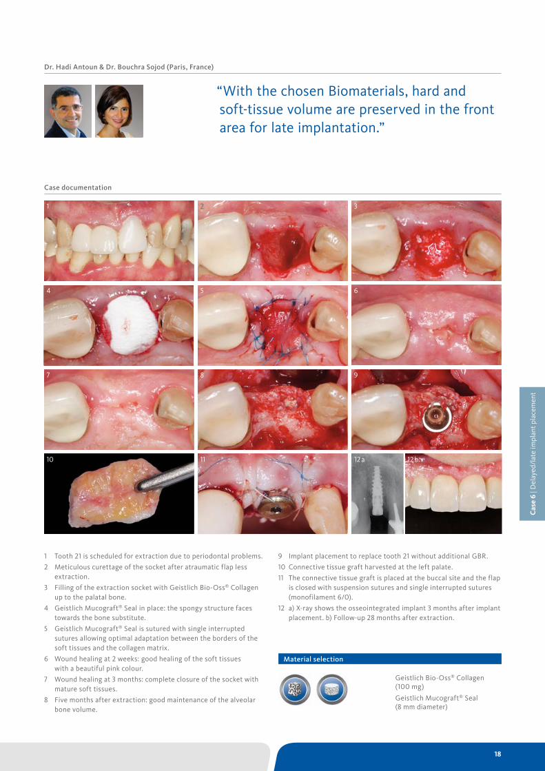
1 Tooth 21 is scheduled for extraction due to periodontal problems.
2 Meticulous curettage of the socket after atraumatic flap less extraction.
3 Filling of the extraction socket with Geistlich Bio-Oss® Collagen up to the palatal bone.
4 Geistlich Mucograft® Seal in place: the spongy structure faces towards the bone substitute.
5 Geistlich Mucograft® Seal is sutured with single interrupted sutures allowing optimal adaptation between the borders of the soft tissues and the collagen matrix.
6 Wound healing at 2 weeks: good healing of the soft tissues with a beautiful pink colour.
7 Wound healing at 3 months: complete closure of the socket with mature soft tissues.
8 Five months after extraction: good maintenance of the alveolar bone volume.
Cas
e 6
| Del
ayed
/lat
e im
plan
t pla
cem
ent
Dr. Hadi Antoun & Dr. Bouchra Sojod (Paris, France)
“With the chosen Biomaterials, hard and soft-tissue volume are preserved in the front area for late implantation.”
1 2 3
4 5 6
7 8 9
10 11
Geistlich Bio-Oss® Collagen (100 mg)
Geistlich Mucograft® Seal (8 mm diameter)
9 Implant placement to replace tooth 21 without additional GBR.
10 Connective tissue graft harvested at the left palate.
11 The connective tissue graft is placed at the buccal site and the flap is closed with suspension sutures and single interrupted sutures (monofilament 6/0).
12 a) X-ray shows the osseointegrated implant 3 months after implant placement. b) Follow-up 28 months after extraction.
Material selection

19 – Treatment Concepts for Extraction Sockets
The patient’s risk profile
Quintessence
RIDGE PRESERVATION IN EXTRACTION SOCKET WITH PRESERVED BUCCAL BONE
Aesthetic risk factors Low risk Medium risk High risk
Patient’s health Intact immune system (non-smoker)
Light smoker Impaired immune system (heavy smoker)
Patient’s aesthetic requirements Low Medium High
Height of the smile line Low Medium High
Gingival biotype Thick“low scalloped”
Medium “medium scalloped”
Thin “high scalloped”
Shape of dental crowns Rectangular Triangular
Infections at implantation site None Chronic Acute
Bone height at adjacent tooth ≤ 5 mm from contact point
5.5–6.5 mmfrom contact point
≥ 7 mm from contact point
Restorative status of adjacent tooth
Intact Restored
Width of tooth gap 1 tooth (≥ 7 mm) 1 tooth (< 7mm) 2 teeth or more
Soft-tissue anatomy Intact Defective
Bone anatomy of the alveolar ridge
No defect Horizontal defect* Vertical defect
Objectives
› Delayed implant placement 4 months after extraction › Minimally invasive treatment of the socket
Before extraction.
Conclusions
› Good/mature/solid bone obtained 4 months after treatment
› Fast and scar-free soft-tissue regeneration › Optimal clinical and aesthetic result for the patient
2 years after extraction.
* Intact extraction socket, with a minor bony defect up to 50 % of the buccal bone wall

20
Case documentation
1 Situation on the day of tooth extraction.
2 Pre-op situation (buccal).
3 The sulcus is de-epithelialised using a diamond bur.
4 The extraction socket is filled with Geistlich Bio-Oss® Collagen.
5 Geistlich Mucograft® Seal in place sutured with single and double interrupted sutures.
6 Healing of soft tissues 3 days after tooth extraction.
Dr. Stefan Fickl (Würzburg, Germany)
“Soft and hard tissues are well preserved without any scarring on the buccal or occlusal aspect.”
1 2 3
4 5 6
7 8 9
10 11 12
7 Healing of the soft tissues at the time of suture removal 10 days after surgery.
8 Tissue healing 9 weeks after tooth extraction.
9 Situation after 4 months at the time of implant placement.
10 The flap elevation reveals ideal bony situation for implant placement.
11 Implant seated.
12 Final restoration 11 months after tooth extraction.
Geistlich Bio-Oss® Collagen (100 mg)
Geistlich Mucograft® Seal (8 mm diameter)
Material selection
Cas
e 7
| Del
ayed
/lat
e im
plan
t pla
cem
ent
21 – Treatment Concepts for Extraction Sockets
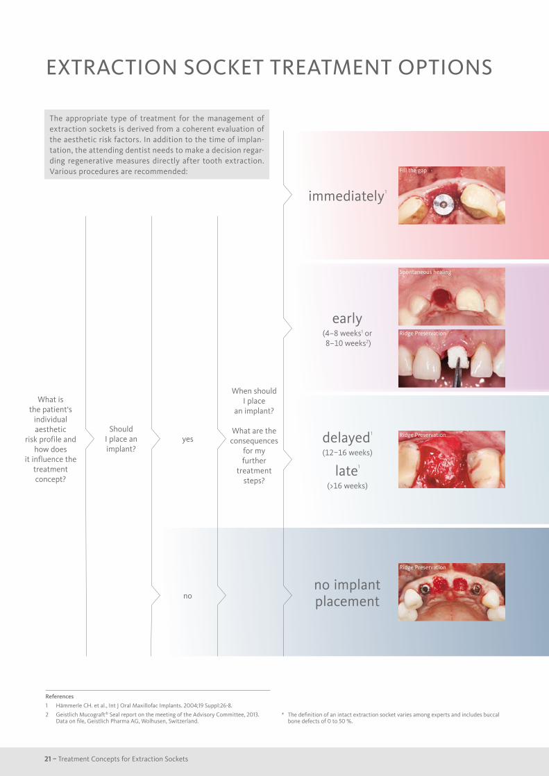
EXTRACTION SOCKET TREATMENT OPTIONS
delayed1
(12–16 weeks)
late1 (>16 weeks)
Ridge Preservation
no implant placement
imme diately1
Fill the gap
early(4–8 weeks1 or 8–10 weeks2)
Spontaneous healing
Ridge Preservation
What is the patient‘s
individual aesthetic
risk profile and how does
it influence the treatment concept?
Should I place an implant?
yes
no
References
1 Hämmerle CH. et al., Int J Oral Maxillofac Implants. 2004;19 Suppl:26-8.
2 Geistlich Mucograft® Seal report on the meeting of the Advisory Committee, 2013. Data on file, Geistlich Pharma AG, Wolhusen, Switzerland.
The appropriate type of treatment for the management of extraction sockets is derived from a coherent evaluation of the aesthetic risk factors. In addition to the time of implan-tation, the attending dentist needs to make a decision regar-ding regenerative measures directly after tooth extraction. Various procedures are recommended:
* The definition of an intact extraction socket varies among experts and includes buccal bone defects of 0 to 50 %.
Ridge Preservation
When should I place
an implant?
What are the consequences
for my further
treatment steps?
22
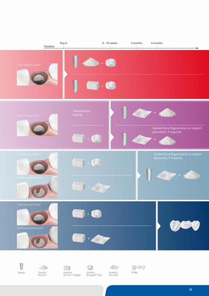
Day 0 8 – 10 weeks 3 months 6 monthsTimeline
Guided Bone Regeneration at implant placement, if required
Guided Bone Regeneration at implant placement, if required
Spontaneous healingIntact extraction socket*
Intact extraction socket*
+
+
+
+
Intact extraction socket*
Defective extraction socket
Intact extraction socket*
Defective extraction socket
+
+
+
+
+
Geistlich Bio-Oss®
Bridge Geistlich Bio-Oss® Collagen
Geistlich Mucograft® Seal
Geistlich Bio-Gide®
Implant
+
23 – Treatment Concepts for Extraction Sockets
The patient’s risk profile
Quintessence
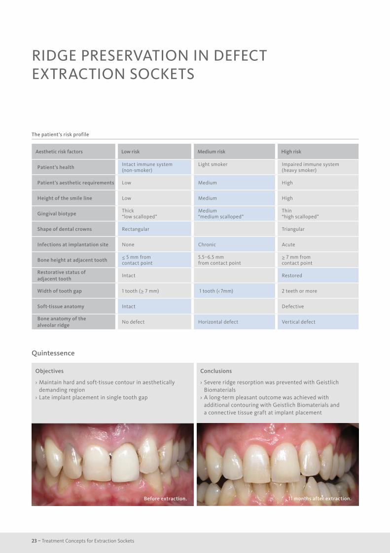
RIDGE PRESERVATION IN DEFECT EXTRACTION SOCKETS
Aesthetic risk factors Low risk Medium risk High risk
Patient’s health Intact immune system (non-smoker)
Light smoker Impaired immune system (heavy smoker)
Patient’s aesthetic requirements Low Medium High
Height of the smile line Low Medium High
Gingival biotype Thick“low scalloped”
Medium “medium scalloped”
Thin “high scalloped”
Shape of dental crowns Rectangular Triangular
Infections at implantation site None Chronic Acute
Bone height at adjacent tooth ≤ 5 mm from contact point
5.5–6.5 mmfrom contact point
≥ 7 mm from contact point
Restorative status of adjacent tooth
Intact Restored
Width of tooth gap 1 tooth (≥ 7 mm) 1 tooth (< 7mm) 2 teeth or more
Soft-tissue anatomy Intact Defective
Bone anatomy of the alveolar ridge
No defect Horizontal defect Vertical defect
Objectives
› Maintain hard and soft-tissue contour in aesthetically demanding region
› Late implant placement in single tooth gap
Before extraction.
Conclusions
› Severe ridge resorption was prevented with Geistlich Biomaterials
› A long-term pleasant outcome was achieved with additional contouring with Geistlich Biomaterials and a connective tissue graft at implant placement
11 months after extraction.

24
Case documentation
1 Initial situation before removal of tooth 21.
2 Inspection of the extraction socket with the periodontal probe shows a buccal bony defect.
3 Geistlich Bio-Gide® is placed buccally on the inner alveolar wall, slightly protruding the crestal bone. Geistlich Bio-Oss® Collagen fills the socket up to the crestal bone level.
4 Geistlich Bio-Oss® (small granules) are packed on top of Geistlich Bio-Oss® Collagen up to soft-tissue level.
5 The collagen membrane is folded over the filled socket, adapted under the palatinal sulcus, fixed with vertical mattress sutures and heals by secondary intention.
6 Uneventful healing situation 3 days post-extraction.
7 Clinical situation 1 week after tooth extraction.
8 Situation after site-conditioning of the soft tissues 4 months post-extraction.
Dr. Célia Coutinho Alves (Porto, Portugal)
“Whenever possible we prefer to preserve rather than to rebuild the bone later, specially in the front teeth.”
1 2 3
4 5 6
7 8 9
10 11 12
9 Flap elevation and implant placement reveal a fenestration 4 months after tooth extraction.
10 The ridge is contoured with a GBR (Geistlich Bio-Oss® and Geistlich Bio-Gide®) and a connective tissue graft on the buccal-crestal area.
11 The flap is closed over the graft.
12 Loading of the implant with the final restoration 7 months after implant placement (11 months after extraction).
Geistlich Bio-Oss® small granules (0.25 –1 mm)
Geistlich Bio-Oss® Collagen (100 mg)
Geistlich Bio-Gide® (25 × 25 mm)
Material selection
Cas
e 8
| Del
ayed
/lat
e im
plan
t pla
cem
ent

25 – Treatment Concepts for Extraction Sockets
The patient’s risk profile
Quintessence
RIDGE PRESERVATION IN THE ANTERIOR REGION FOR LATE IMPLANTATION
1 Jung RE, et al. J Clin Periodontol. 2013 Jan;40(1):90–8
* Intact extraction socket, with a minor bony defect up to 50 % of the buccal bone wall
Aesthetic risk factors Low risk Medium risk High risk
Patient’s health Intact immune system (non-smoker)
Light smoker Impaired immune system (heavy smoker)
Patient’s aesthetic requirements Low Medium High
Height of the smile line Low Medium High
Gingival biotype Thick“low scalloped”
Medium “medium scalloped”
Thin “high scalloped”
Shape of dental crowns Rectangular Triangular
Infections at implantation site None Chronic Acute
Bone height at adjacent tooth ≤ 5 mm from contact point
5.5–6.5 mmfrom contact point
≥ 7 mm from contact point
Restorative status of adjacent tooth
Intact Restored
Width of tooth gap 1 tooth (≥ 7 mm) 1 tooth (< 7mm) 2 teeth or more
Soft-tissue anatomy Intact Defective
Bone anatomy of the alveolar ridge
No defect Horizontal defect* Vertical defect
Objectives
› Preservation of hard and soft-tissue volume after extraction in the anterior region for late implant placement.
› Prevention of extensive guided bone regeneration procedures at implant placement.
Right after extraction.
Conclusions
› Volume of hard and soft tissue can be preserved better with Geistlich Bio-Oss® Collagen and Geistlich Mucograft® Seal than with spontaneous healing.1
› A minimally invasive GBR is peformed to contour the ridge at implant placement.
10 months after extraction.
26
Case documentation
1 Extraction of tooth 21 due to a trauma with concomitant external resorptions. Care was taken in preserving the alveolar bone.
2 Occlusal view of the socket after tooth extraction. No flaps are raised around the affected area. A slight buccal bone defect was observed.
3 The socket is gently curetted for removal of granulation tissue. Subsequently, the wound margins were de-epithelialised with a diamond in a counter-piece with water cooling.
4 Filling of the extraction socket with Geistlich Bio-Oss® Collagen to the level of the palatal bone.
5 Geistlich Mucograft® is applied dry and adapts perfectly to the wound margins.
6 Suturing of the Geistlich Mucograft® with 6-0 single interrupted sutures.
7 The tissues are left to heal beneath the provisional, taking care not to apply pressure to the biomaterials.
Prof. Ronald E. Jung (Zurich, Switzerland)
1 2 3
4 5 6
7 8 9
10 11 12
8 Situation 7.5 months after extraction revealing nice soft-tissue situation with a slight dip at the buccal aspect.
9 Flap elevation shows the healed bony situation 7.5 months after Ridge Preservation.
10 Implant placement in fully mature bone. A small GBR for contouring is performed.
11 Excellent emergence profile after 10 months.
12 Situation with the final restoration 10 months after tooth extraction.
Geistlich Bio-Oss® Collagen (100 mg)
Geistlich Mucograft®
(15 × 20 mm punch 8 mm diameter)
Material selection
Cas
e 9
| Del
ayed
/lat
e im
plan
t pla
cem
ent
Find the abstract of the publica-tion1 here.
27 – Treatment Concepts for Extraction Sockets
The patient’s risk profile
Quintessence
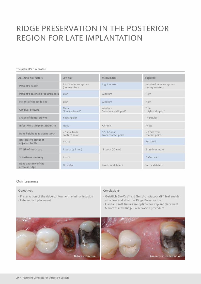
RIDGE PRESERVATION IN THE POSTERIOR REGION FOR LATE IMPLANTATION
Aesthetic risk factors Low risk Medium risk High risk
Patient’s health Intact immune system (non-smoker)
Light smoker Impaired immune system (heavy smoker)
Patient’s aesthetic requirements Low Medium High
Height of the smile line Low Medium High
Gingival biotype Thick“low scalloped”
Medium “medium scalloped”
Thin “high scalloped”
Shape of dental crowns Rectangular Triangular
Infections at implantation site None Chronic Acute
Bone height at adjacent tooth ≤ 5 mm from contact point
5.5–6.5 mmfrom contact point
≥ 7 mm from contact point
Restorative status of adjacent tooth
Intact Restored
Width of tooth gap 1 tooth (≥ 7 mm) 1 tooth (< 7 mm) 2 teeth or more
Soft-tissue anatomy Intact Defective
Bone anatomy of the alveolar ridge
No defect Horizontal defect Vertical defect
Objectives
› Preservation of the ridge contour with minimal invasion › Late implant placement
Before extraction.
Conclusions
› Geistlich Bio-Oss® and Geistlich Mucograft® Seal enable a flapless and effective Ridge Preservation
› Hard and soft tissues are optimal for implant placement 6 months after Ridge Preservation procedure
6 months after extraction.
28
Case documentation
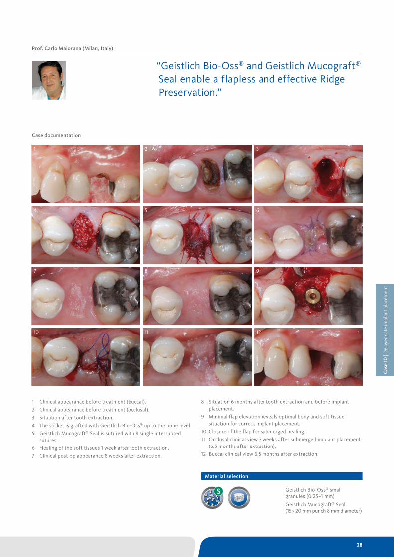
1 Clinical appearance before treatment (buccal).
2 Clinical appearance before treatment (occlusal).
3 Situation after tooth extraction.
4 The socket is grafted with Geistlich Bio-Oss® up to the bone level.
5 Geistlich Mucograft® Seal is sutured with 8 single interrupted sutures.
6 Healing of the soft tissues 1 week after tooth extraction.
7 Clinical post-op appearance 8 weeks after extraction.
Prof. Carlo Maiorana (Milan, Italy)
“Geistlich Bio-Oss® and Geistlich Mucograft® Seal enable a flapless and effective Ridge Preservation.”
1 2 3
4 5 6
7 8 9
10 11 12
8 Situation 6 months after tooth extraction and before implant placement.
9 Minimal flap elevation reveals optimal bony and soft-tissue situation for correct implant placement.
10 Closure of the flap for submerged healing.
11 Occlusal clinical view 3 weeks after submerged implant placement (6.5 months after extraction).
12 Buccal clinical view 6.5 months after extraction.
Geistlich Bio-Oss® small granules (0.25–1 mm)
Geistlich Mucograft® Seal (15 × 20 mm punch 8 mm diameter)
Material selection
Cas
e 10
| D
elay
ed/l
ate
impl
ant p
lace
men
t

29 – Treatment Concepts for Extraction Sockets
The patient’s risk profile
Quintessence
RIDGE PRESERVATION IN DEFECT EXTRACTION SOCKET
Objectives
› Replace a falling hopeless central incisor with high aesthetic demands.
Before extraction.
Conclusions
› Ridge Preservation techniques are effective in minimising volume loss.
6 months after extraction.
Aesthetic risk factors Low risk Medium risk High risk
Patient’s health Intact immune system (non-smoker)
Light smoker Impaired immune system (heavy smoker)
Patient’s aesthetic requirements Low Medium High
Height of the smile line Low Medium High
Gingival biotype Thick“low scalloped”
Medium “medium scalloped”
Thin “high scalloped”
Shape of dental crowns Rectangular Triangular
Infections at implantation site None Chronic Acute
Bone height at adjacent tooth ≤ 5 mm from contact point
5.5–6.5 mmfrom contact point
≥ 7 mm from contact point
Restorative status of adjacent tooth
Intact Restored
Width of tooth gap 1 tooth (≥ 7 mm) 1 tooth (< 7mm) 2 teeth or more
Soft-tissue anatomy Intact Defective
Bone anatomy of the alveolar ridge
No defect Horizontal defect Vertical defect

30
Case documentation
1 Initial Smile.
2 Initial front view.
3 Alveolar inspection after the extraction. Note the presence of deep buccal defect.
4 Positioning of Geistlich Bio-Gide® by buccal and palatal.
5 Socket filling with Geistlich Bio-Oss®.
6 Geistlich Bio-Oss® accommodation .
7 Repositioning of Geistlich Bio-Gide® for socket sealing.
8 Provisional prosthesis installed.
Prof. Julio Cesar Joly, Prof. Robert Carvalho da Silva & Prof. Paulo Fernando M. de Carvalho (São Paulo, Brazil)
“Geistlich Biomaterials grant us safe choices, effective treatments and predictable out-comes.”
1 2 3
4 5 6
7 8 9
10 11 12
* Prosthesis rehabilitation by Prof. Dr. Oswaldo Scopin de Andrade and Luis Alves
Geistlich Bio-Oss® small granules (0.25–1 mm)
Geistlich Bio-Gide® (13 × 25 mm)
9 Healing aspect after 6 months. Note the volume preservation and tissue contour. Connective tissue graft and late implant place-ment after 6 months.
10 Final rehabilitation with ceramic crown*. Note the natural contour and emergence profile.
11 Final clinical appearance showing harmony in gingival margin positioning.
12 Harmonic smile after rehabilitation.
Material selection
Cas
e 11
| D
elay
ed/l
ate
impl
ant p
lace
men
t
31 – Treatment Concepts for Extraction Sockets
The patient’s risk profile
Quintessence
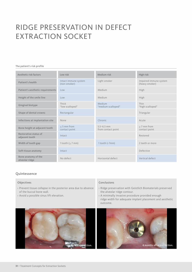
RIDGE PRESERVATION IN DEFECT EXTRACTION SOCKET
Aesthetic risk factors Low risk Medium risk High risk
Patient’s health Intact immune system (non-smoker)
Light smoker Impaired immune system (heavy smoker)
Patient’s aesthetic requirements Low Medium High
Height of the smile line Low Medium High
Gingival biotype Thick“low scalloped”
Medium “medium scalloped”
Thin “high scalloped”
Shape of dental crowns Rectangular Triangular
Infections at implantation site None Chronic Acute
Bone height at adjacent tooth ≤ 5 mm from contact point
5.5–6.5 mmfrom contact point
≥ 7 mm from contact point
Restorative status of adjacent tooth
Intact Restored
Width of tooth gap 1 tooth (≥ 7 mm) 1 tooth (< 7mm) 2 teeth or more
Soft-tissue anatomy Intact Defective
Bone anatomy of the alveolar ridge
No defect Horizontal defect Vertical defect
Objectives
› Prevent tissue collapse in the posterior area due to absence of the buccal bone wall.
› Avoid a possible sinus lift elevation.
Before extraction.
Conclusions
› Ridge preservation with Geistlich Biomaterials preserved the alveolar ridge contour.
› A minimally invasive procedure provided enough ridge width for adequate implant placement and aesthetic outcome.
6 months after extraction.
32
Case documentation
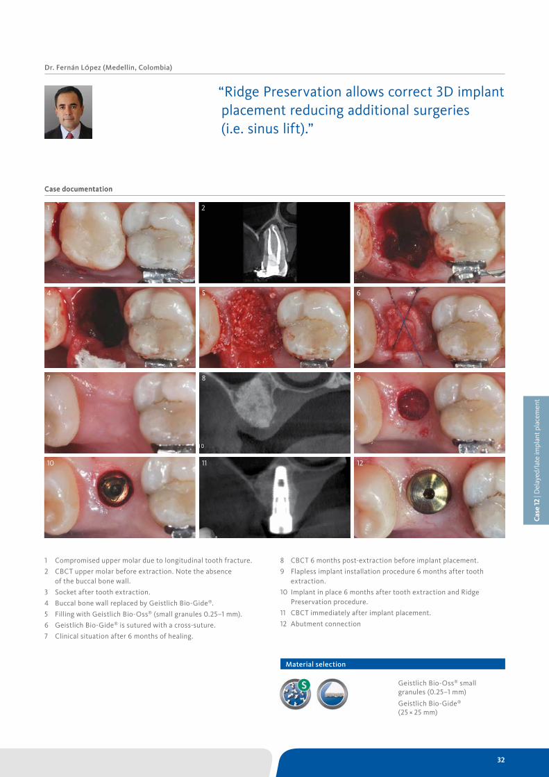
1 Compromised upper molar due to longitudinal tooth fracture.
2 CBCT upper molar before extraction. Note the absence of the buccal bone wall.
3 Socket after tooth extraction.
4 Buccal bone wall replaced by Geistlich Bio-Gide®.
5 Filling with Geistlich Bio-Oss® (small granules 0.25–1 mm).
6 Geistlich Bio-Gide® is sutured with a cross-suture.
7 Clinical situation after 6 months of healing.
Case documentation
Dr. Fernán LÓpez (Medellin, Colombia)
“Ridge Preservation allows correct 3D implant placement reducing additional surgeries (i.e. sinus lift).”
1 2 3
4 5 6
7 8 9
10 11 12
8 CBCT 6 months post-extraction before implant placement.
9 Flapless implant installation procedure 6 months after tooth extraction.
10 Implant in place 6 months after tooth extraction and Ridge Preservation procedure.
11 CBCT immediately after implant placement.
12 Abutment connection
Geistlich Bio-Oss® small granules (0.25–1 mm)
Geistlich Bio-Gide® (25 × 25 mm)
2
Material selection
Cas
e 12
| D
elay
ed/l
ate
impl
ant p
lace
men
t

33 – Treatment Concepts for Extraction Sockets
The patient’s risk profile
Quintessence
RIDGE PRESERVATION FOR DELAYED IMPLANT PLACEMENT
Aesthetic risk factors Low risk Medium risk High risk
Patient’s health Intact immune system (non-smoker)
Light smoker Impaired immune system (heavy smoker)
Patient’s aesthetic requirements Low Medium High
Height of the smile line Low Medium High
Gingival biotype Thick“low scalloped”
Medium “medium scalloped”
Thin “high scalloped”
Shape of dental crowns Rectangular Triangular
Infections at implantation site None Chronic Acute
Bone height at adjacent tooth ≤ 5 mm from contact point
5.5–6.5 mmfrom contact point
≥ 7 mm from contact point
Restorative status of adjacent tooth
Intact Restored
Width of tooth gap 1 tooth (≥ 7 mm) 1 tooth (< 7mm) 2 teeth or more
Soft-tissue anatomy Intact Defective
Bone anatomy of the alveolar ridge
No defect Horizontal defect Vertical defect
Objectives
› Reconstruct alveolar bone with severe vertical loss from chronic periodontitis at the lower left second molar
› Investigate the clinical and histological result by using Geistlich Combi-Kit Collagen after tooth extraction.
Before extraction.
Conclusions
› The defect was completely filled with newly-formed hard tissue after 6 months
› Histomorphometric analysis revealed 45% of the hard tissue area including bone substitute material and 28% of the soft tissue area.
9 months after extraction.

34
Case documentation
1 Radiological status prior to extraction. Initial Smile.
2 Starting situation.
3 Status following atraumatic extraction of tooth 17.
4 A flap is raised.
5 Filling of the extraction socket up to the level of the crestal bone level using Geistlich Bio-Oss® Collagen.
6 Insertion of the Geistlich Bio-Gide® membrane over the defect .
Dr. Ham Byung-Do (Kainos Dental Clinic, Seoul, Korea)
1 2 3
4 5 6
7 8 9
10 11 12
7 Closure of the extraction socket with a mattress suture. Open healing.
8 Situation 6 months post-op.
9 Newly formed hard tissue. Geistlich Bio-Oss® Collagen is not obvious.
10 One stage protocol with healing abutment.
11 Provisional prosthesis.
12 Radiological view after implantation.
Geistlich Combi-Kit Collagen:
Geistlich Bio-Oss® Collagen (100 mg)
Geistlich Bio-Gide® (16 × 22 mm)
“After 6 months the defect was completely filled with newly-formed hard tissue.”
Material selection
Cas
e 13
| D
elay
ed/l
ate
impl
ant p
lace
men
t
35 – Treatment Concepts for Extraction Sockets
The patient’s risk profile
Quintessence
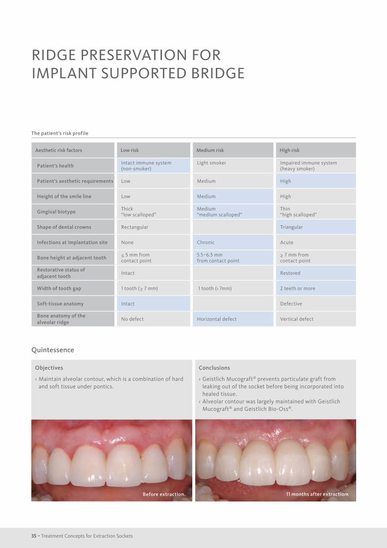
RIDGE PRESERVATION FOR IMPLANT SUPPORTED BRIDGE
Aesthetic risk factors Low risk Medium risk High risk
Patient’s health Intact immune system (non-smoker)
Light smoker Impaired immune system (heavy smoker)
Patient’s aesthetic requirements Low Medium High
Height of the smile line Low Medium High
Gingival biotype Thick“low scalloped”
Medium “medium scalloped”
Thin “high scalloped”
Shape of dental crowns Rectangular Triangular
Infections at implantation site None Chronic Acute
Bone height at adjacent tooth ≤ 5 mm from contact point
5.5–6.5 mmfrom contact point
≥ 7 mm from contact point
Restorative status of adjacent tooth
Intact Restored
Width of tooth gap 1 tooth (≥ 7 mm) 1 tooth (< 7mm) 2 teeth or more
Soft-tissue anatomy Intact Defective
Bone anatomy of the alveolar ridge
No defect Horizontal defect Vertical defect
Objectives
› Maintain alveolar contour, which is a combination of hard and soft tissue under pontics.
Before extraction.
Conclusions
› Geistlich Mucograft® prevents particulate graft from leaking out of the socket before being incorporated into healed tissue.
› Alveolar contour was largely maintained with Geistlich Mucograft® and Geistlich Bio-Oss®.
11 months after extraction.

36
Case documentation
Cas
e 14
| N
o im
plan
t pla
cem
ent
1 Radiographic findings prior to implant placement in teeth 12 and 22.
2 Clinical initial situation prior to implant placement in teeth 12 and 22.
3 Maxillary central incisors scheduled for extraction due to recurrent endodontic infections 2 months after implant placement in lateral incisors.
4 Extraction sockets grafted with Geistlich Bio-Oss®. The bone sub-stitute fills the socket up to slightly above the bone crest.
5 Geistlich Mucograft® is placed over the occlusal surfaces as a socket seal.
6 Provisional restoration.
7 Provisional restoration contoured to maintain Geistlich Mucograft® in place, taking care not to compress the grafted site.
8 Vascularisation and integration of Geistlich Mucograft® after two weeks.
9 Clinical situation 1 month post-op.
Dr. Jeffrey Ganeles (Boca Raton, USA)
“This treatment is ideal for extraction sockets to preserve aesthetic contours when there are limited bony defects.”
10 Occlusal view at 9 months with the final restoration (11 months after teeth extraction).
11 Buccal view at 9 months with the final restoration (11 months after teeth extraction).
12 Radiograph showing integration of the graft material in the sockets. Final restoration in place.
1 2 3
4 5 6
7 8 9
10 11 12
Geistlich Bio-Oss® small granules (0.25–1 mm)
Geistlich Mucograft® (15 × 20 mm punch 8 mm diameter)
Material selection

37 – Treatment Concepts for Extraction Sockets
The patient’s risk profile
Quintessence
RIDGE PRESERVATION IN MULTIPLE EXTRACTION SOCKETS
Aesthetic risk factors Low risk Medium risk High risk
Patient’s health Intact immune system (non-smoker)
Light smoker Impaired immune system (heavy smoker)
Patient’s aesthetic requirements Low Medium High
Height of the smile line Low Medium High
Gingival biotype Thick“low scalloped”
Medium “medium scalloped”
Thin “high scalloped”
Shape of dental crowns Rectangular Triangular
Infections at implantation site None Chronic Acute
Bone height at adjacent tooth ≤ 5 mm from contact point
5.5–6.5 mmfrom contact point
≥ 7 mm from contact point
Restorative status of adjacent tooth
Intact Restored
Width of tooth gap 1 tooth (≥ 7 mm) 1 tooth (< 7mm) 2 teeth or more
Soft-tissue anatomy Intact Defective
Bone anatomy of the alveolar ridge
No defect Horizontal defect Vertical defect
Objectives
› Ridge profile maintenance under full arch bridge. › Flapless procedure.
Before extraction.
Conclusions
› Good and quick soft-tissue healing during the early healing phase.
› Bone volume has been largely preserved with a minimally invasive approach.
12 months after extraction.
38
Case documentation
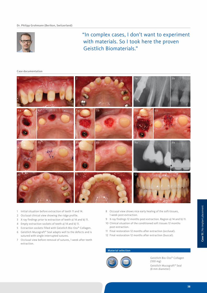
Dr. Philipp Grohmann (Berikon, Switzerland)
“In complex cases, I don’t want to experiment with materials. So I took here the proven Geistlich Biomaterials.”
1 Initial situation before extraction of teeth 11 and 14.
2 Occlusal clinical view showing the ridge profile.
3 X-ray findings prior to extraction of teeth a) 14 and b) 11.
4 Empty extraction sockets of teeth a) 14 and b) 11.
5 Extraction sockets filled with Geistlich Bio-Oss® Collagen.
6 Geistlich Mucograft® Seal adapts well to the defects and is sutured with single interrupted sutures.
7 Occlusal view before removal of sutures, 1 week after teeth extraction.
8 Occusal view shows nice early healing of the soft-tissues, 1 week post-extraction.
9 X-ray findings 12 months post-extraction. Region a) 14 and b) 11.
10 Clinical situation of the conditioned soft tissues 12 months post-extraction.
11 Final restoration 12 months after extraction (occlusal).
12 Final restoration 12 months after extraction (buccal).
10 11 12
Geistlich Bio-Oss® Collagen (100 mg)
Geistlich Mucograft® Seal (8 mm diameter)
1 2 3 a 3 b
7 a 7 b 9 a 9 b8 a 8 b
Material selection
Cas
e 15
| N
o im
plan
t pla
cem
ent
6 a 6 b5 a 5 b4 a 4 b
39 – Treatment Concepts for Extraction Sockets
TECHNICAL GUIDELINES
Reference
1 Adapted from Geistlich Mucograft® Seal Advisory Board Meeting Report 2013. Data on file, Geistlich Pharma AG, Wolhusen, Switzerland.
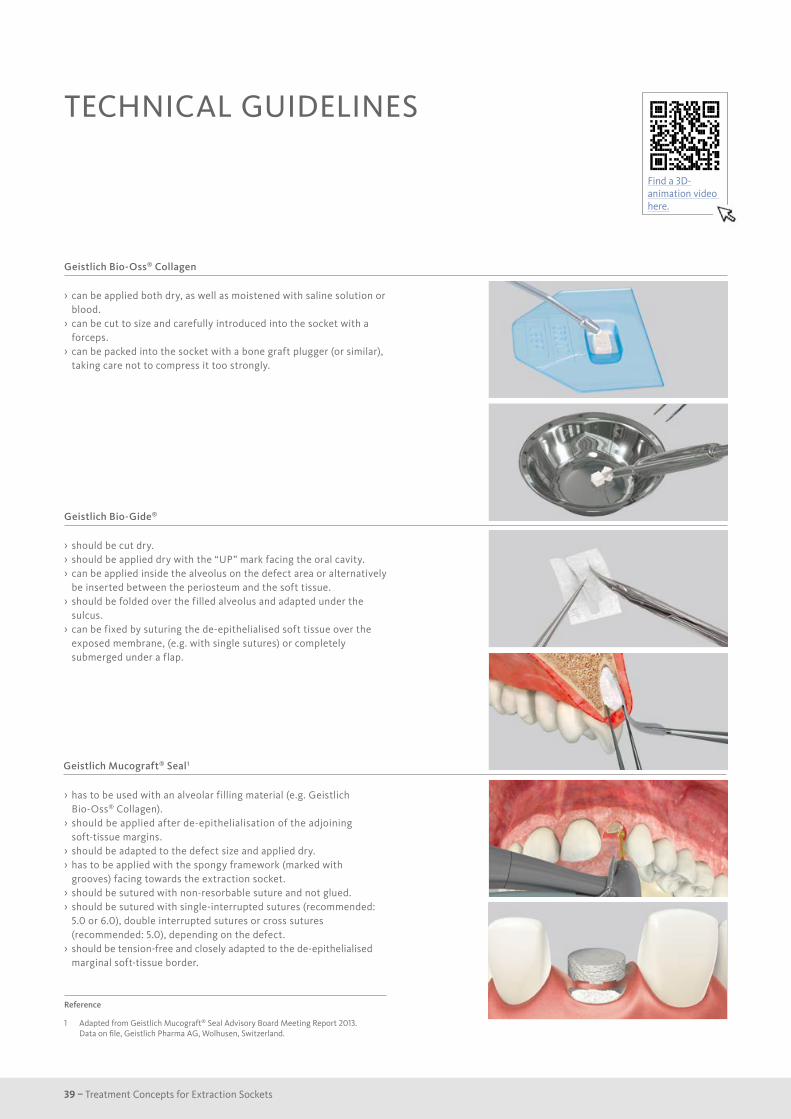
› can be applied both dry, as well as moistened with saline solution or blood.
› can be cut to size and carefully introduced into the socket with a forceps.
› can be packed into the socket with a bone graft plugger (or similar), taking care not to compress it too strongly.
› should be cut dry. › should be applied dry with the “UP” mark facing the oral cavity. › can be applied inside the alveolus on the defect area or alternatively be inserted between the periosteum and the soft tissue.
› should be folded over the filled alveolus and adapted under the sulcus.
› can be fixed by suturing the de-epithelialised soft tissue over the exposed membrane, (e.g. with single sutures) or completely sub merged under a flap.
› has to be used with an alveolar filling material (e.g. Geistlich Bio-Oss® Collagen).
› should be applied after de-epithelialisation of the adjoining soft-tissue margins.
› should be adapted to the defect size and applied dry. › has to be applied with the spongy framework (marked with grooves) facing towards the extraction socket.
› should be sutured with non-resorbable suture and not glued. › should be sutured with single-interrupted sutures (recommended: 5.0 or 6.0), double interrupted sutures or cross sutures (recommended: 5.0), depending on the defect.
› should be tension-free and closely adapted to the de-epithelialised marginal soft-tissue border.
Geistlich Bio-Oss® Collagen
Geistlich Bio-Gide®
Geistlich Mucograft® Seal1
Find a 3D- animation video here.
40
PRODUCT RANGE
Geistlich Bio-Oss® (small granules) + 10% collagen (porcine) Sizes: 100 mg (0.2–0.3 cm3), 250 mg (0.4–0.5 cm3), 500 mg (0.9–1.1 cm3)
Geistlich Bio-Oss® Collagen is indicated for use in periodontal defects and extraction sockets. Through the addition of collagen, Geistlich Bio-Oss® Collagen can be tailored to the morphology of the defect and is particularly easy to apply.
Resorbable bilayer membrane Sizes: 25 × 25 mm, 30 × 40 mm
Geistlich Bio-Gide® consists of porcine collagen and has a bilayer structure – a rough side that faces the bone and a smooth side that faces the soft tissue. Geistlich Bio-Gide® is easy to handle: it can be positioned easily, adheres well to the defect, and is resistant to tension and tearing.
Geistlich Bio-Oss® Collagen 100 mg + Geistlich Bio-Gide® 16 × 22 mm
When used in combination, the system has optimised properties for Ridge Preservation and minor bone augmentations according to the GBR principle.
Collagen matrix Size: 8 mm diameter
Geistlich Mucograft® Seal consists of a compact structure that gives stability while allowing open healing, and a spongy structure that supports blood clot stabilisation and ingrowth of soft-tissue cells.
Small granules (0.25–1 mm) | Quantities: 0.25 g ≈ 0.5 cc, 0.5 g ≈ 1.0 cc Large granules (1–2 mm) | Quantity: 0.5 g ≈ 1.5 cc
Geistlich Bio- Oss® granules are available in an applicator. It allows the bone substitute material to be applied faster and more precisely to the surgical site. Geistlich Bio-Oss Pen® is available containing both the small granules and the large granules.
Geistlich Bio-Oss Pen®
Geistlich Bio-Oss® Collagen
Geistlich Bio-Gide®
Geistlich Combi-Kit Collagen
Geistlich Mucograft® Seal
Geistlich Bio-Oss®
Small granules (0.25–1 mm) | Quantities: 0.25 g, 0.5 g, 2.0 g (1 g ≈ 2.05 cm3)Large granules (1–2 mm) | Quantities: 0.5 g, 2.0 g (1 g ≈ 3.13 cm3)
The small Geistlich Bio-Oss® granules are recommended for smaller 1–2 socket defects and for contouring auto genous block grafts. The large Geistlich Bio-Oss® granules enable improved regeneration over large distances and provide enough space for the ingrowing bone.
Prod
uct i
nfor
mat
ion

41 – Treatment Concepts for Extraction Sockets
Outstanding Quality
Quality and safety are high priorities at Geistlich Pharma. At Geistlich Pharma every-thing is done under one roof: from the selection and control of the raw material to production and storage until dispatch, and all steps are taken seamlessly and meet the company’s high standards of quality and safety.
Unique Biofunctionality
The excellent results of Ridge Preservation with Geistlich Biomaterials are largely due to their unsurpassed biofunctionality: Geistlich Bio-Oss® with its porous structure1 serves as guide rail for the in-growing blood vessels2 and integrates into newly formed bone3, while the unique bilayer Geistlich Bio-Gide® shields the young bone from the surrounding connec-tive tissue cells and supports wound healing4 and early vascularisation5. The 3-dimensional matrix of Geistlich Mucograft® Seal facilitates soft-tissue cells ingrowth6 and may enhance early wound healing7. Clinically relevant: › Geistlich Biomaterials are perfectly suited to combined use for treatment of extraction sockets.
› Geistlich Bio-Oss® Collagen combined with Geistlich Bio-Gide® preserves up to 93 % of the ridge width8,9 and they promote more new bone formation vs. no membrane10
› Geistlich Bio-Oss® Collagen combined with Geistlich Mucograft® Seal increases preserved bone volume when compared to spontaneous healing11
Your Worldwide No. 1 Reference
Geistlich Biomaterials is constantly working to offer you solutions for easy, predictable and successful management and regeneration of extraction sockets. The company’s own re-search departments along with global experts develop the product portfolio, and try new techniques and applications for existing pro-ducts.In more than 15 worldwide Round Table Meetings*, expert clinicians and Geistlich Biomaterials cooperate on the aim of pro-moting discussion and evolving a consensus on the treatment concepts for extraction so-ckets. These Round Table Meetings also help to define what is the current published clinical evidence and where research still needs to be done.
Unique Biofunctionality
OutstandingQuality
Your Worldwide No. 1 Reference
References
1 Weibrich G et al., Mund Kiefer Gesichtschirurg 4, 2000; 148–152.
2 Degidi M et al., Oral Dis. 2006 Sep; 12(5): 469–475.
3 Artzi Z, et al. J Periodontol. 2001 Feb;72(2):152-9.
4 Becker J et al., Clin. Oral Implants Res. 2009; 20(7): 742–93.
5 Rothamel D et al., Clin. Oral Implants Res. 2005;16:369–378.
6 Ghanaati S, et al. Biomed Mater. 2011 Feb;6(1):015010.
7 Thoma DS, et al. J Clin Periodontol. 2012 Feb;39(2):157-65.
8 Cardaropoli D, et al. Int J Periodontics Restorative Dent. 2012 Aug;32(4):421-30.
9 Cardaropoli D, et al. Int J Periodontics Restorative Dent. 2014 Mar-Apr;34(2):211-7.
10 Perelman-Karmon et al. Int J Periodontics Restorative Dent. 2012 Aug;32(4):459-65.
11 Jung RE, et al. J Clin Periodontol. 2013 Jan;40(1):90-8.
* Data on file (Wolhusen, Switzerland): Austria, Baltics, Belgium, Brazil, France, Germany, Greece,Holland, Korea, Nordics, Poland, Russia, Spain (2009), Spain/Portugal (2014, 2015), Switzerland (2009, 2011, 2013), UK.
41 – Treatment Concepts for Extraction Sockets
GET THE NO. 1 INTO YOUR PRACTICE
42

6012
79/1
501/
en
Manufacturer© Geistlich Pharma AGBusiness Unit BiomaterialsBahnhofstrasse 40CH-6110 WolhusenPhone +41 41 492 56 30Fax +41 41 492 56 39www.geistlich-biomaterials.com
Subsidiary Great Britain, IrelandGeistlich BiomaterialsGeistlich Sons Limited1st Floor, Thorley HouseBailey LaneManchester AirportGB-Manchester M90 4ABPhone +44 161 490 2038Fax +44 161 498 6988www.geistlich.co.uk
More details about our distribution partners:www.geistlich-biomaterials.com
Top Related