


Languages
Pages
Legal


Thyroid FNAKey Cytology and Histology for “Clinicians”
Zubair W. Baloch, MD, PhDProfessor of Pathology & Laboratory Medicine
UPENN Medical Center Perelman School of Medicine

Faculty/Presenter Disclosure
• Faculty: [Zubair Baloch, MD, PhD]
• Relationships with commercial interests: None

Objectives of Thyroid FNA
• Recognize specific diagnostic entities
• Provide meaningful, management oriented diagnosis
• Potential utilization of ancillary techniques

Thyroid FNA Bethesda Classification Scheme
The Bethesda System for Reporting Thyroid Cytopathology:
Implied Risk of Malignancy and Recommended Clinical Management
Diagnostic Category Risk of Malignancy
(%)
Usual Management
Non-diagnostic or Unsatisfactory Repeat FNA with ultrasound
guidance
Benign 0-3% Clinical follow-up
Atypia of Undetermined Significance or
Follicular Lesion of Undetermined
Significance (AUS/FLUS)
~ 5-15% Repeat FNA
Follicular Neoplasm or Suspicious for a
Follicular Neoplasm (Specify if Hurthle
type or Oncocytic)
15-30% Surgical lobectomy
Suspicious for Malignancy 60-75% Near-total thyroidectomy or
surgical lobectomy
Malignant 97-99% Near-total thyroidectomy
Easy-Breezy Thyroid Pathology
Concordant Ultrasound Features, FNA cytomorphology & Histologic
Follow-up
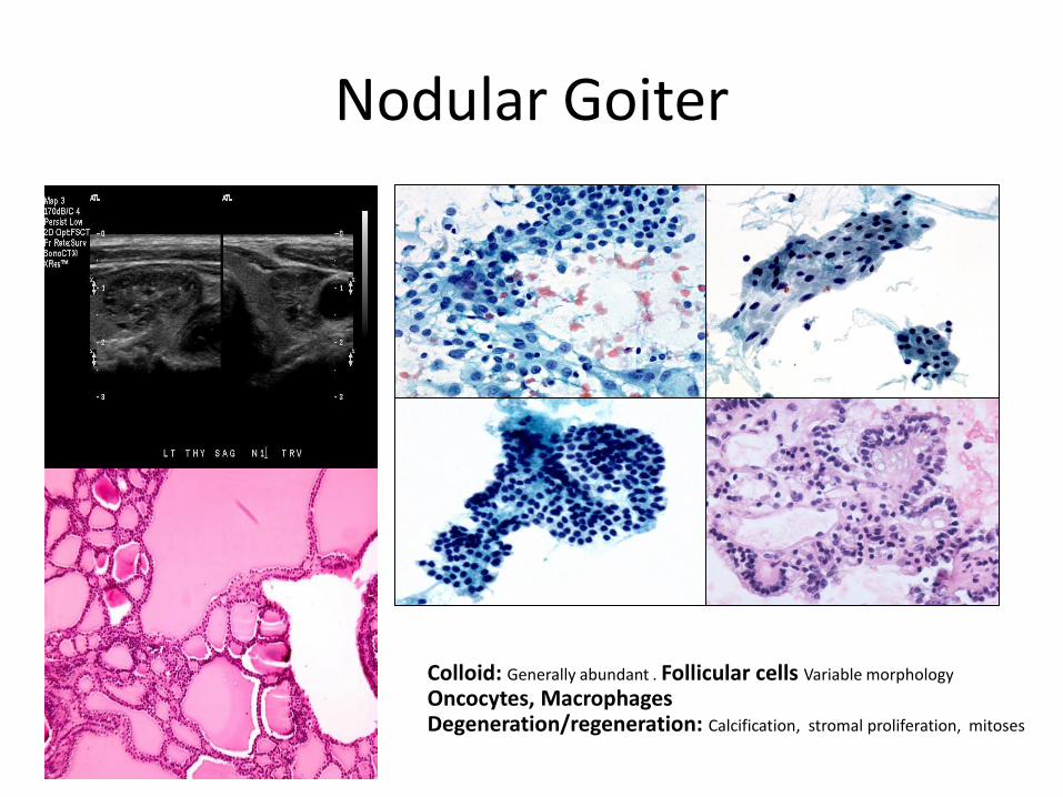
Nodular Goiter
Colloid: Generally abundant . Follicular cells Variable morphology
Oncocytes, MacrophagesDegeneration/regeneration: Calcification, stromal proliferation, mitoses

Chronic Lymphocytic Thyroiditis
OncocytesLymphocytes: In the background & infiltrating the cell groups

Papillary Thyroid Carcinoma
Nuclear features – Major Diagnostic FeaturesElongation, chromatin clearing, Nuclear membrane irregularities Intranuclear groovesInclusionsSmall peripheral nucleoli
Not so easy - Head Scratching Everyday Thyroid Cytopathology
Intdeterminate Lesions
Or
Indeterminate Pathologist?

Thyroid FNA Bethesda Classification Scheme
The Bethesda System for Reporting Thyroid Cytopathology:
Implied Risk of Malignancy and Recommended Clinical Management
Diagnostic Category Risk of Malignancy
(%)
Usual Management
Non-diagnostic or Unsatisfactory Repeat FNA with ultrasound
guidance
Benign 0-3% Clinical follow-up
Atypia of Undetermined Significance or
Follicular Lesion of Undetermined
Significance (AUS/FLUS)
~ 5-15% Repeat FNA
Follicular Neoplasm or Suspicious for a
Follicular Neoplasm (Specify if Hurthle
type or Oncocytic)
15-30% Surgical lobectomy
Suspicious for Malignancy 60-75% Near-total thyroidectomy or
surgical lobectomy
Malignant 97-99% Near-total thyroidectomy

Diagnosis Follicular Neoplasm
80% Benign on Surgical Excision
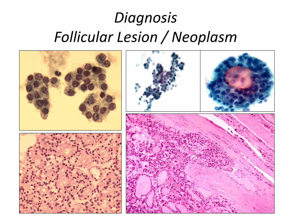
DiagnosisFollicular Lesion / Neoplasm
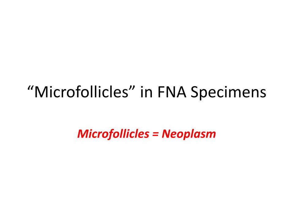
“Microfollicles” in FNA Specimens
Microfollicles = Neoplasm

Is It That Easy
Don’t Think So

Microfollicles
• Inter-observer Agreement on Microfollicles– Renshaw AA et al. (Arch Pathol Lab Med 2006)
– 12 cytopathologists were shown 45 small groups of follicular cells• 20 Microfollicles
• 7 Macrofollicles
• 18 Indeterminate
– <15 cells arranged in circle that is at least two-thirds complete, should be classified as microfollicles.
Microfollicles
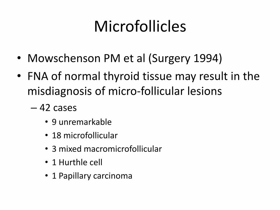
• Mowschenson PM et al (Surgery 1994)
• FNA of normal thyroid tissue may result in the misdiagnosis of micro-follicular lesions
– 42 cases
• 9 unremarkable
• 18 microfollicular
• 3 mixed macromicrofollicular
• 1 Hurthle cell
• 1 Papillary carcinoma

The Atypical Category
The Dreaded AUS/FLUS

Lets Talk About FLUS/AUSResponsible Factors
• History– TFT’s, H/O prior FNA
• Ultrasound features– Cystic vs. solid
• Operator – sampling
• Adequacy
• Cytology Preparation
• Interpretation
• Surgical follow-up – ? Gold standard

The so Called Gold Standard

Case-1

Case 1Thyroid Experts Diagnoses
The Cytopathologists Gold Standard
Diagnoses: Hyperplastic nodule – BenignorFollicular Adenoma – Benignor Follicular Variant of Papillary Thyroid Carcinoma - Malignant
LiVolsi Rosai Asa Lloyd

Case 1 - Sampling
Benign
Malignant

Case 2 Lesional Morphology
Benign
Atypical Architecture

Case 3History of Prior FNA-
Making Sense of Atypia
Markedly Atypical Cells

Case 4: Inadequate History 52-year-old woman. Ultrasound – Left thyroid lobe occupied by a predominantly
ill-defined hypoechoic structure – suspicious for anaplastic carcinoma
Original Diagnosis Suspicious for Anaplastic
CarcinomaMore History
Transient symptomatic hyperthyroidism (TSH –
0.03) followed by hypothyroidism.
Current medication: Synthroid
Second opinion DxSuspect sub-acute
thyroiditisSurgical excision of
left lobe

Dealing with AUS/FLUSThe Dreaded Call From the Clinician
Too ManyFLUS Cases
What is going on?
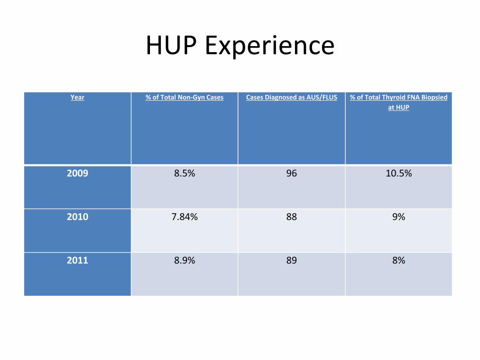
HUP Experience
Year % of Total Non-Gyn Cases Cases Diagnosed as AUS/FLUS % of Total Thyroid FNA Biopsied
at HUP
2009 8.5% 96 10.5%
2010 7.84% 88 9%
2011 8.9% 89 8%

HUP ExperienceRates and Interpretations

How to Relay The AUS/FLUS Diagnosis
Explain, Explain & Explain

HUP Experience
• AUS/FLUS cases are further sub-classified into Following subcategories (SC):
• SC1 - favor benign, however, a follicular neoplasm (FN) could not be excluded due to increased cellularity
• SC2 - specimens with focal nuclear overlapping and crowding• SC3 - scant specimens with focal nuclear overlapping and crowding• SC4 - specimens with focal nuclear overlapping and crowding in a
background of lymphocytic thyroiditis• SC5 - few cells with features suspicious for papillary thyroid cancer
(PTC)• SC6 - specimens in which a FN cannot be excluded (with
miscellaneous morphologic descriptors).

0.00% 5.00% 10.00% 15.00% 20.00% 25.00% 30.00% 35.00% 40.00%
SC1
SC2
SC3
SC4
SC5
SC6
Malignancy Rate by AUS-FLUS Subtype
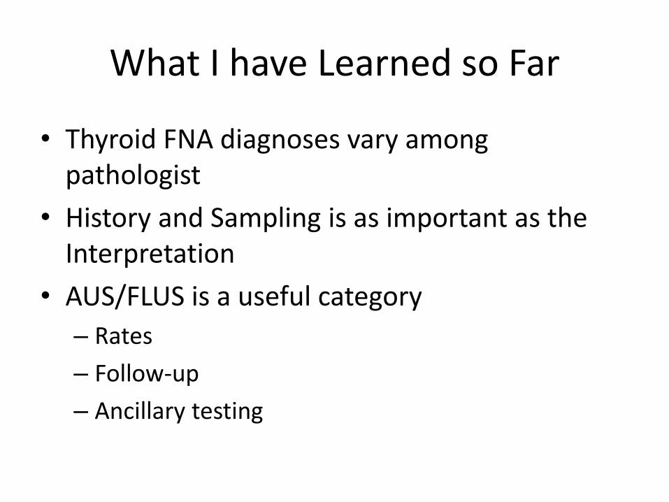
What I have Learned so Far
• Thyroid FNA diagnoses vary among pathologist
• History and Sampling is as important as the Interpretation
• AUS/FLUS is a useful category
– Rates
– Follow-up
– Ancillary testing

Modern ApproachA Gentle Mix of Old and New
Nothing is 100%
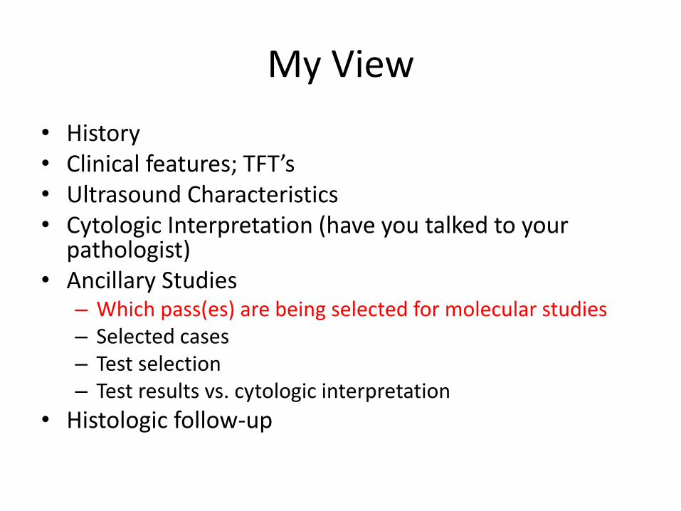
My View
• History• Clinical features; TFT’s• Ultrasound Characteristics• Cytologic Interpretation (have you talked to your
pathologist)• Ancillary Studies
– Which pass(es) are being selected for molecular studies– Selected cases– Test selection– Test results vs. cytologic interpretation
• Histologic follow-up
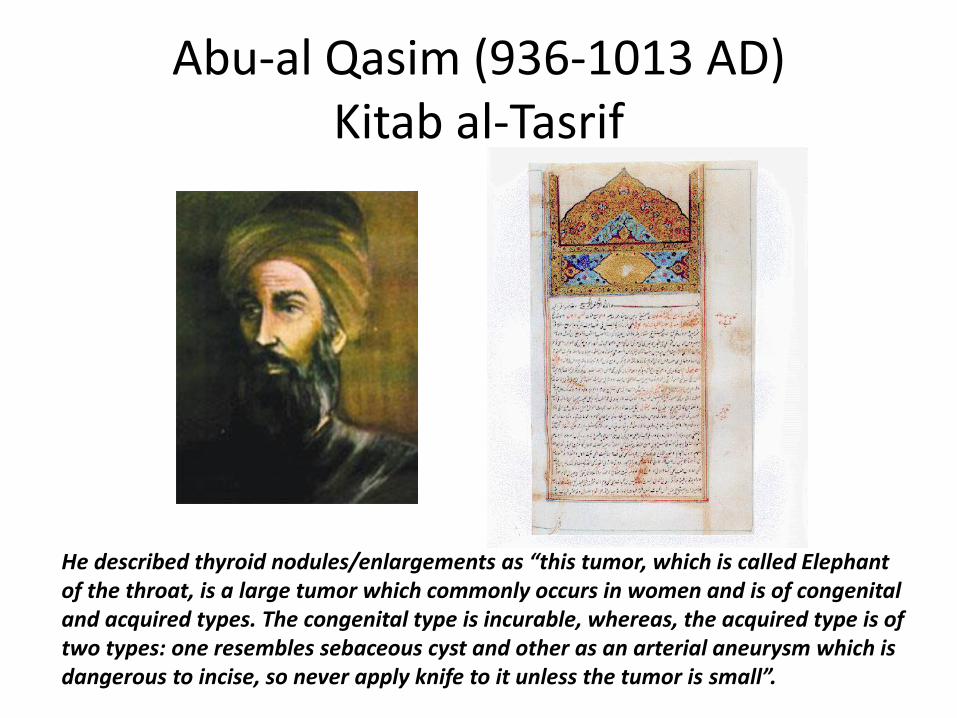
Abu-al Qasim (936-1013 AD)Kitab al-Tasrif
He described thyroid nodules/enlargements as “this tumor, which is called Elephant of the throat, is a large tumor which commonly occurs in women and is of congenital and acquired types. The congenital type is incurable, whereas, the acquired type is of two types: one resembles sebaceous cyst and other as an arterial aneurysm which is dangerous to incise, so never apply knife to it unless the tumor is small”.
Top Related